
Definition
• Bacterial pneumonias can be classified by their pattern of anatomical involvement:
• Lobar pneumonia: consolidation due to intraalveolar neutrophils affecting a large and continuous area of the lobe(s) of lung(s)
• Bronchopneumonia (lobular, bronchial or bronchogenic pneumonia): acute bronchiolitis with consolidation of peribronchial alveoli affecting one or more pulmonary lobules
Pathogenesis
• Causative organisms usually invade the host via airways
• In congenital pneumonia the infection is usually secondary to in utero aspiration of infected amniotic fluid
• Bronchopneumonia often leads to lobar pneumonia as the infection worsens
Clinical features
• Presenting features may include a cough, fever, shortness of breath, and chest pain
• Clinical presentation is altered by treatment: patients have very few signs after 48 to 72 hours of antibiotics
• Streptococcus pneumonia accounts for 90% to 95% of cases of lobar pneumonia (Klebsiella pneumoniae, staphylococci, streptococci, Hemophilus influenzae, or gram-negative bacteria occasionally can be found to be causative)
• Bronchopneumonia is a common hospital-acquired pneumonia with causative organisms such as Staphylococcus aureus, Klebsiella pneumoniae, Escherichia coli, and Pseudomonas (less likely than lobar pneumonia to be linked with streptococcus)
• Viral and fungal infections may also be seen clinically as either bronchopneumonia or lobar pneumonia
• Clinically, it is more important to identify the causative agent, extent of disease, and antibiotic susceptibility rather than distinguish the exact anatomical subtype of pneumonia
Pathology
• Lobar pneumonia:
• Congestion (first 24 hours): grossly, the lung is heavy and hyperemic. Histologically characterized by vascular engorgement, intraalveolar fluid accumulation, mild inflammatory infiltrate, and often numerous bacteria
• Red hepatization: grossly, vascular congestion persists; solidification is present and is due to exudate accumulation in airspaces. The lung is likened to the appearance of the liver, thus the term hepatization. Histologically, there is extravasation of red cells into alveolar spaces, along with increased numbers of neutrophils and fibrin
• Gray hepatization: grossly, the lung is paler and the cut surface drier with consolidation. Histologically, red cells disintegrate, with persistence of the neutrophils and fibrin. The alveoli still appear consolidated
• Resolution: the exudate is digested by enzymatic activity and cleared by macrophages or by the coughing mechanism
• Bronchopneumonia: histologically, similar stages of evolution are thought to occur (as lobar pneumonia) but small areas of involvement and temporal differences among foci make demonstration of this evolution difficult
• Grossly: multiple often bilateral 2- to 4-cm dry, gray-brown foci of consolidation may be present in the basal lobes of the lung. Lesions are often centered around a bronchiole
• Histologically: extensive congestion, parabronchiolar suppurative inflammation with acute bronchiolitis, and leukocytic alveolitis are present. Inflammatory foci are separated by normal, aerated parenchyma
• In the immunocompromised patient, there may be little or no inflammatory reaction to the organism(s)
Complications
• Although the majority of the bacterial pneumonias resolve, complications include
• Abscess formation
• Empyema formation
• Organization and fibrosis
• Bacteremia and sepsis

Fig 1 Lobar pneumonia and bronchopneumonia. Chest X-ray film showing right upper lobe pneumonia.
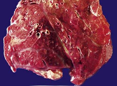
Fig 2 Lobar pneumonia and bronchopneumonia. Gross photograph of cut surface of lung showing patchy pale raised areas in this autopsy of a patient with bronchopneumonia.

Fig 3 Lobar pneumonia and bronchopneumonia. This high-power view of bronchopneumonia shows numerous neutrophils filling the alveolar spaces. Adjacent less involved lung is evident. In lobar pneumonia, the inflammatory infiltrate would be similar, except it would involve the majority of the lobe.
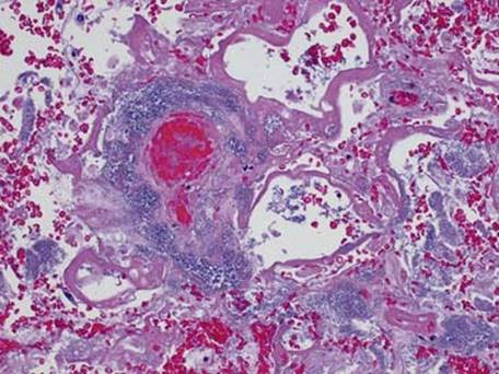
Fig 4 Lobar pneumonia and bronchopneumonia. In this severely immunocompromised patient the bacterial colonies are seen rimming blood vessels with no inflammatory response. This perivascular rimming by bacterial colonies is characteristic of Pseudomonas infection. Diffuse alveolar damage is also present.
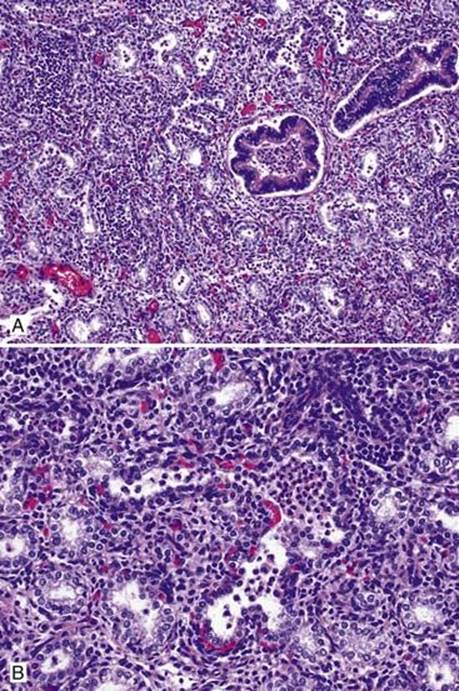
Fig 5 Lobar pneumonia and bronchopneumonia. Low- (A) and high- (B) power views of congenital pneumonia in an infant of 26 weeks’ gestation. Note that the lung is immature, and peripheral airspaces are lined by cuboidal epithelium.