
Definition
• Infection caused by weakly pathogenic bacteria; rare cause of bronchopneumonia and lung abscess
Clinical features
Epidemiology
• Actinomyces spp. are normal commensal flora of the oral cavity
• Infection occurs from aspiration of organisms, usually in patients with poor dental hygiene, impaired consciousness, or abnormal swallowing from neurological conditions
Presentation
• Gradual onset of a productive cough, fever, and pulmonary infiltrates on chest x-ray film
• Important to consider in cases of unresolving bronchopneumonia
Prognosis and treatment
• Frequently cured with high-dose penicillin and tetracycline
• Surgical excision may be required for a lung abscess or pulmonary fistula
Pathology
Histology
• Usually single or multiple lung abscesses, which may form interlobar fistulas
• Extensive acute inflammation and necrosis surrounded by granulation tissue with chronic inflammatory cells and giant cells but no granulomas
• Characteristic amphophilic crystalline sulfur granules found within areas of acute inflammation
• Sulfur granules may have peripheral eosinophilic deposits (Splendore-Hoeppli phenomenon), which is not specific to actinomycosis
• Long branching, beaded, filamentous rods, 0.2-0.5 microns thick, seen within sulfur granules
Immunopathology/special stains
• Gomori methenamine silver (GMS) positive
• Gram positive
• Acid-fast negative
Main differential diagnoses
• Nocardiosis (weakly acid-fast, no sulfur granules)
• Botryomycosis (cocci and short rods of mixed bacteria forming pseudogranules)
• Fungal infection (thicker, not filamentous)
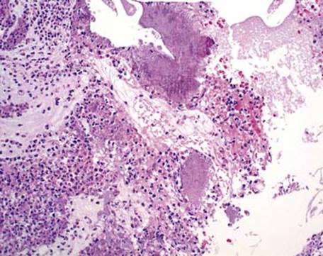
Fig 1 Actinomycosis. Actinomyces typically forms sulfur granules in a background of neutrophils.
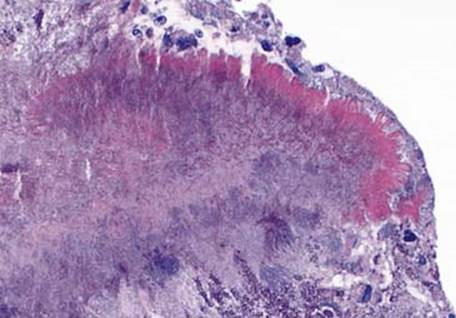
Fig 2 Actinomycosis. The bright eosinophilic rim around this sulfur granule is characteristic of the Splendore-Hoeppli phenomenon, which is not specific to Actinomyces.
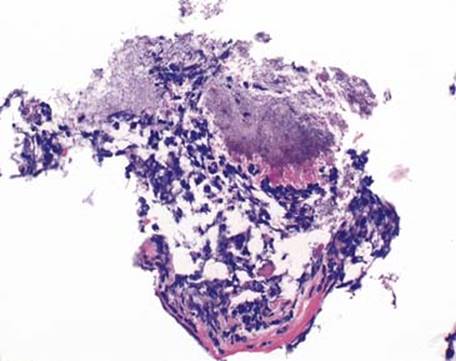
Fig 3 Actinomycosis. Filamentous rods of Actinomyces can be seen radiating toward the periphery of this sulfur granule from a transbronchial needle aspirate.
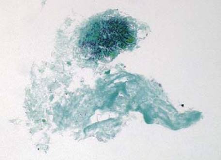
Fig 4 Actinomycosis. The filamentous rods of Actinomyces are GMS positive.

Fig 5 Actinomycosis. Tissue Gram stain (Brown and Hopps) highlights the gram-positive filamentous rods of Actinomyces.
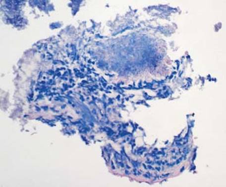
Fig 6 Actinomycosis. Actinomyces are acid-fast negative, appearing as blue filamentous rods in this acid-fast bacillus stain.