
Definition
• A pulmonary infection caused by adenovirus, a linear, double-stranded DNA virus
Clinical features
Epidemiology
• Adenovirus: worldwide distribution, infections occur throughout the year, causing 5% to 10% of all febrile illnesses in infants and young children; most individuals have serological evidence of prior adenoviral infection by the age of 10
• Transmission: via aerosol droplets, the fecal–oral route, and by contact with contaminated fomites; epidemics occur in military recruits and dense civilian populations; neonates may acquire adenoviral infection from exposure to cervical secretions at birth if mother is infected
• More than 50 human adenovirus serotypes have been identified; serotypes 3, 7, 21 (subgroup B), 1, 2, 5 (subgroup C), and 4 (subgroup E) are associated with upper respiratory disease and pneumonia, and serotypes 3, 2, and 1 are the most common
• Adenoviruses are responsible for approximately 10% of pneumonias in children
Presentation
• Signs and symptoms are usually similar to other pneumonias
• More severe in infants than older children and may be associated with lethargy, diarrhea, and vomiting; extrapulmonary complications occasionally occur, including meningoencephalitis, hepatitis, myocarditis, nephritis, neutropenia, and disseminated intravascular coagulation
• In immunosuppressed patients, adenovirus pneumonia is severe and frequently fatal, particularly in transplantation patients and those with severe immunodeficiency
• Chest radiography is classically described as bilateral hyperaeration, bronchial wall thickening, and diffuse bilateral bronchopneumonia
Prognosis and treatment
• Treatment consists of management of the clinical manifestations as effective medical therapy is not available
• Incidence of pulmonary sequelae is high in children. Up to 50% of affected children develop chronic lung disease, including bronchiectasis, bronchiolitis obliterans, and unilateral hyperlucent lung
Pathology
Gross
• Involved lungs show various degrees of consolidation, with patchy areas of hemorrhage and necrosis
Histology
• Two major patterns of lung injury:
• Diffuse alveolar damage with or without necrotizing bronchitis/bronchiolitis. The muscularis of involved airways is usually preserved, but the epithelium is frequently destroyed
• Pneumonitis (interstitial pneumonia) with karyorrhectic necrosis
• The two patterns may coexist in some cases
• The cytopathic effects are helpful for diagnosis. Two types of nuclear inclusions can be caused by adenovirus, which are usually present in viable tissue at the edges of the necrotic zones
• Smudge cell type inclusion is most distinct, which is basophilic or amphophilic, obscures the nuclear membrane, and gives a “smudgy” appearance to the nucleus
• The other type of inclusion is dense eosinophilic and surrounded with a clear halo, similar to that seen in herpesvirus infection (Cowdry type A)
• The nuclear inclusions appear as a lattice-like crystalline array of virions by electron microscopy
• Reactive and reparative type atypia may be present in the background
Immunopathology/special stains
• Immunohistochemical testing for adenovirus is helpful in confirming the diagnosis
• Adenoviral antigen can also be detected via polymerase chain reaction (PCR) and direct fluorescence assay
Main differential diagnoses
• Other potential causes of necrotizing bronchitis/bronchiolitis and/or diffuse alveolar damage
• Other Cowdry type A viral inclusions: herpesvirus (multinucleation, intranuclear inclusions, no smudge cells), cytomegalovirus (enlarged cell with nuclear and cytoplasmic inclusions), and measles (multinucleation, nuclear and cytoplasmic inclusions). Immunohistochemical analysis and PCR are helpful. Culture not only confirms diagnosis but also types the virus
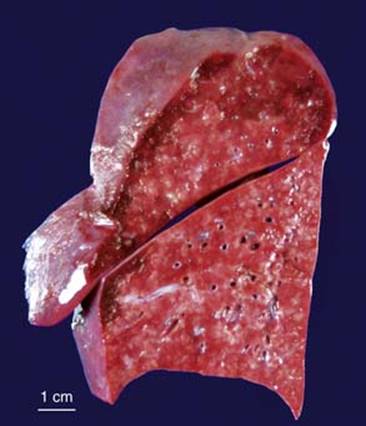
Fig 1 Adenovirus pneumonia. Cut surface of a lung involved by adenovirus pneumonia showing consolidation with numerous foci of necrosis.
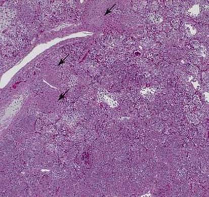
Fig 2 Adenovirus pneumonia. Low-power photomicrograph of lung showing alveoli filled with inflammatory debris with focal necrosis (arrows).
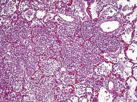
Fig 3 Adenovirus pneumonia. Intermediate-power photomicrograph showing intraalveolar inflammatory debris.
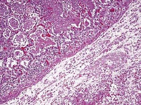
Fig 4 Adenovirus pneumonia. Large airway filled with mucus. The bronchial epithelium is destroyed, but the muscle layer is preserved. Adjacent lung has intraalveolar fibrin and inflammatory cells.
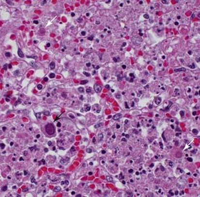
Fig 5 Adenovirus pneumonia. Adenoviral inclusions. Both smudgy cell type (basophilic, smudgy nucleus; black arrow) and Cowdry type A (eosinophilic with clear halo; white arrow) are present in this field.
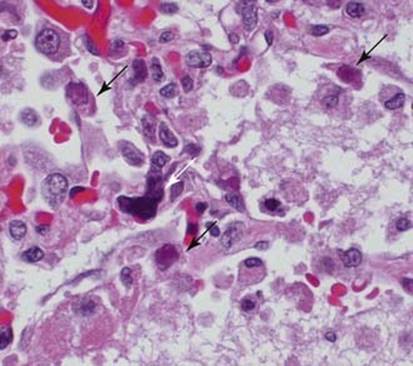
Fig 6 Adenovirus pneumonia. Smudgy cells (basophilic/amphophilic, smudgy nucleus; black arrows). Note one megakaryocyte entrapped in a pulmonary capillary (white arrow), which should not be mistaken as a viral inclusion.

Fig 7 Adenovirus pneumonia. Electron microscopy demonstrates an adenoviral nuclear inclusion as a lattice-like crystalline array of virions.