
Definition
• Respiratory illness due to infection with RSV, an RNA paramyxovirus
Pathogenesis
• The virus enters the body through the eye or nose, rarely through the mouth, and spreads along the epithelium of the respiratory tract by cell to cell transfer
Clinical features
Epidemiology
• Most common cause of lower respiratory tract infection in children worldwide
• Infects up to 50% of children in the first year of life and virtually all children by the age of 3
• Repeated infections may occur during the first three years of life, but after 3 years of age they are generally milder
• Socioeconomic factors play a role in the severity of the illness: in a nonurban middle class area, 1 in 1000 infants infected in the first year of life require hospitalization, whereas in industrialized poorer areas, the rate may be as high as 1 in 50
• Recently recognized as an etiologic agent in community-acquired pneumonia in adults
• Transmitted via person to person through large-particle aerosols or hand contamination of secretions with self-inoculation
• Incubation period is usually 3 to 6 days but ranges from 2 to 8 days
• Viral shedding starts 1 to 2 days before symptoms and lasts as long as 2 weeks after symptom onset
• Outbreaks occur in the late autumn and winter
Presentation
• Upper respiratory tract infection or tracheobronchitis: this is the most common form in both adults and children and presents with coryza, coughing, pharyngitis, and a low-grade fever
• Lower respiratory tract infection (bronchiolitis and bronchopneumonia): findings include rhinorrhea, tachypnea, wheezing, intercostal and suprasternal retractions, a hyperresonant chest, crackles, or inspiratory rales
• Bronchopneumonia: seen especially in the first few months of life. Localized or diffuse crackles may be present. Bronchiolitis and pneumonia often represent a continuum and may be difficult to differentiate clinically
• Complicated, severe, or fatal RSV infection: infants with underlying cyanotic congenital heart disease, those with underlying pulmonary disease, and immunocompromised infants are at risk
• Nonobstructive apnea occurs in 20% of infants, especially in the setting of prematurity, and may be the initial symptom of the infection
• Secondary bacterial infection is unusual
Prognosis and treatment
• Mild cases: no therapy or symptomatic therapy
• RSV pneumonia: correct the hypoxemia; aerosolized ribavirin, (a synthetic nucleoside that interferes with the expression of mRNA), administered 12 or more hours per day for 3 to 5 days, may help by reducing the viral load and correct the hypoxemia
• Complications may include prolonged pulmonary function alterations that may lead to chronic lung disease in later life
• Mortality rate: 0.5% to 1.0% for hospitalized previously healthy infants; 15% to 40% in immunocompromised patients
Pathology
Histology
• Bronchiolar and peribronchiolar chronic inflammation, which may be associated with ulceration and necrosis of the epithelium
• Accumulation of mucus, inflammatory cells, and necrotic epithelium within the airways is a hallmark of bronchiolitis
• Infiltration of interstitial tissue with mononuclear cells is seen in pneumonia
• Rarely presents with features of giant cell viral pneumonia associated with diffuse alveolar damage
• Multinucleation (due to fusion of the viral envelope with host cell membranes caused by surface F glycoprotein) and eosinophilic cytoplasmic/paranuclear inclusions are characteristic but often difficult to find
• Virions are pleomorphic with surface spikes on electron microscopy
Immunopathology/special stains
• Direct and indirect immunofluorescence antibody staining of epithelial cells from the respiratory tract: both polyclonal (80% to 90% sensitivity) and monoclonal (95% to 100% sensitivity) immunofluorescence antibodies are easy to perform, but the interpretation is subjective
• RSV antigen detection in nasopharyngeal specimens with enzyme-linked immunosorbent assay (ELISA): several kits are available. Advantages include objective interpretation, speed, and ability to screen large number of specimens, but these tests have poorer sensitivity, can give equivocal results, and require confirmation with time-consuming blocking ELISA procedure
• Cell culture: RSV has a high liability and should be transported to the laboratory promptly; nasopharyngeal aspirates, nasal washes, or tracheal secretions are the best specimen for isolation; human heteroploid cells (HEP-2 and HeLa) provide the best tissue culture; characteristic syncytia formation appears in 4 to 5 days
• Serology: based on detection of rising of antibody on acute and convalescent sera; very lengthy method; serological response in infants may be poor
• Multiplex reverse transcriptase respiratory virus PCR: most sensitive method. This helps to distinguish multiple pathogens simultaneously and is becoming more popular in clinical laboratories
Main differential diagnoses
• Herpes simplex/varicella zoster: rare multinucleation is seen; can cause diffuse alveolar damage and necrosis; ultrastructurally, the particle size is 150 to 200 nm and shows a round core with a double membrane
• Measles pneumonia: multinucleation is seen; causes interstitial pneumonia; ultrastructurally, there is a 120 to 150 nm, spherical inclusion with 15 to 20 tubular filaments
• Parainfluenza virus: occasional multinucleation; can cause diffuse alveolar damage and interstitial pneumonia; ultrastructurally, the particle size is 150 to 250 nm; it is more pleomorphic than influenza
• Human metapneumovirus (RNA virus associated with respiratory illness in the first year of life)
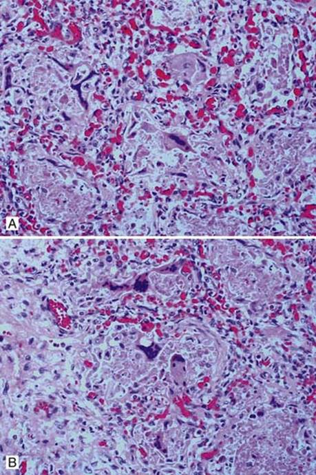
Fig 1 Respiratory syncytial virus pneumonia. A and B, Case of RSV infection showing alveolar fibrin, interstitial thickening, and syncytial multinucleated giant cells.
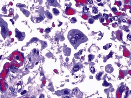
Fig 2 Respiratory syncytial virus pneumonia. High-power view of eosinophilic cytoplasmic inclusions in mononucleated and multinucleated cells.

Fig 3 Respiratory syncytial virus pneumonia. Necrosis of the bronchial epithelial cells is a common finding, as seen here.

Fig 4 Respiratory syncytial virus pneumonia. Electron microscopic image demonstrates viral particles.