
Definition
• Pulmonary infection due to the measles virus (rubeola), a single-stranded RNA paramyxovirus in the Morbillivirus genus
Pathogenesis
• The virus first gains access to the body via the upper respiratory tract or the conjunctiva and spreads to the immediate lymph nodes. A primary viremia ensues with subsequent infection of the reticuloendothelial system
• A secondary viremia follows in 5 to 7 days after the initial infection, whereby the virus spreads through blood monocytes to other organs such as the spleen, thymus, lung, liver, kidney, conjunctivae, and skin
• The Koplik’s spots and the rash are thought to result from a delayed hypersensitivity reaction
Clinical features
Epidemiology
• Highly contagious worldwide infection
• Recent outbreaks have primarily affected nonvaccinated preschool-age children and previously vaccinated school-age children
• A high number of cases are seen in developing countries among children younger than 12 months because of inadequate vaccination; causes significant public health problems
• Disease is more severe in malnourished children, specifically in those with vitamin A deficiency
• Complications include diarrhea, dehydration, stomatitis, inability to feed, and bacterial infections
• Pneumonia, an uncommon complication, occurs in up to 6% of reported cases
• Measles is a leading cause of blindness in African children
Presentation
• Prodrome with fever, rhinorrhea, coughing, and conjunctivitis
• Koplik’s spots (small, irregular spots with central bluish white speck), seen in the buccal mucosa in 50% to 90% of patients
• Maculopapular rash starts on the face, spreads to the trunk and the extremities, and lasts 6 days
• Acute measles panencephalitis: CNS involvement is common, even in uncomplicated measles, with transient electroencephalographic abnormalities detected in 50% of patients
• Pulmonary complications: secondary pneumonia (bacterial), giant cell pneumonia (Hecht’s pneumonia; seen as an opportunistic infection in immunocompromised patients, especially in deficient cell-mediated immunity), and atypical measles pneumonia (associated with inactivated vaccine; not seen nowadays)
• Immunocompromised patients: primary viral pneumonia is uncommon, even in immunocompromised individuals
• Immunocompetent patients: measles pneumonia may be complicated by secondary bacterial pneumonia, which is associated with the vast majority of measles-related deaths
Prognosis and treatment
• In general, immunocompetent patients have a good prognosis
• In developed countries, the death rate is less than 1 in 1000 children, but in endemic areas in subSaharan Africa, the measles case fatality rate ranges from 5% to 10%
• In immunocompromised patients the fatality rate may reach as high as 25%
• Immunocompromised patients with severe infections may benefit from immunoglobulin if given within the first week of exposure
• Measles vaccine may be helpful if given within 72 hours of exposure
• Antibiotics are recommended for secondary bacterial infections
Pathology
Histology
• Characteristic morphological feature is the presence of multinucleated giant cells (a few to up to 60 nuclei) with intranuclear and intracytoplasmic inclusions
• Inclusions occur within alveolar lining cells, endothelial cells, and macrophages
• Peribronchiolar inflammation
• Necrotizing bronchiolitis associated with squamous metaplasia and epithelial regenerative changes
• May also cause giant cell interstitial pneumonia
• Diffuse alveolar damage with hyaline membranes and intraalveolar proteinaceous exudate seen in severe cases
Immunopathology/special stains
• Antigen detection: use swab to obtain cells from nasopharyngeal specimens or urine (2-5 days after the appearance of the rash)
• Serology: enzyme-linked immunosorbent assay detects IgG and IgM antibodies
• Virus isolation: should only be attempted in complicated cases and in immunocompromised patients with pneumonia (nasopharyngeal aspirate/washing, throat swab, lung tissue, cerebrospinal fluid, or urine samples)
Main differential diagnoses
• Respiratory syncytial virus: cytoplasmic but not nuclear inclusions
• Influenza: no inclusions seen
• Cytomegalovirus: large cell with both nuclear and cytoplasmic inclusions
• Herpes: nuclear but not cytoplasmic inclusions
• Hard metal pneumoconiosis, associated with tungsten carbide, cobalt, and titanium (giant cell interstitial pneumonia), often presents as a chronic disease in adults: these metals can be demonstrated in lung tissues by energy dispersive X-ray spectroscopy
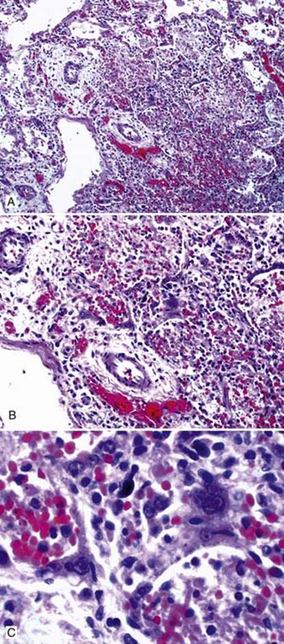
Fig 1 Measles pneumonia. A to C, Low- to high-power views showing multinucleated giant cells with characteristic red intranuclear and intracytoplasmic inclusions.