
See also Mediastinal Bronchogenic Cyst.
Definition
• Cystic lesion arising from anomalous budding of the tracheobronchial anlage of the primitive foregut during development
Clinical features
Epidemiology
• Rare congenital malformation, primarily diagnosed in children
Presentation
• Mostly mediastinal; however, they can be found at any point along the tracheobronchial tree, occasionally found in the lung parenchyma or within the cervical, intrapleural, or suprasternal cutaneous regions, rarely, below the diaphragm or pericardium
• Incidentally detected on chest imaging
• Rarely, patients present with obstruction of tracheobronchial tree, a ruptured cyst, or infection
Prognosis and treatment
• Surgery is curative
Pathology
Gross
• Unilocular cyst containing clear fluid or, rarely, hemorrhagic secretions
• Communication with the tracheobronchial tree is rare
Histology
• Thin-walled cyst lined by ciliated pseudostratified columnar epithelium
• Wall can contain smooth muscle, cartilage, seromucinous bronchial glands, and occasionally linear calcifications
• Squamous metaplasia and/or chronic inflammation may be present
• Bronchogenic cysts lack alveolar tissue
• Cysts do not communicate with the tracheobronchial tree
Immunopathology/special stains
• Not contributory
Main differential diagnoses
• Pulmonary sequestration
• Congenital pulmonary airway malformation
• Mediastinal cysts: esophageal cyst, enteric cyst, thymic cyst, cystic teratoma, pericardial cyst
• Abscess
• Cystic bronchiectasis
• Postinfarction cyst
• Interstitial emphysema
• Pleuropulmonary blastoma

Fig 1 Bronchogenic cyst. Chest imaging showing a bronchogenic cyst in the mediastinum.
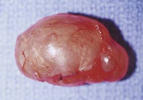
Fig 2 Bronchogenic cyst. Gross image of the same bronchogenic cyst as in Fig 1.

Fig 3 Bronchogenic cyst. Thin-walled cyst lined by ciliated pseudostratified columnar epithelium with adjacent cartilage and seromucinous bronchial glands.