
Definition
• An infection or allergic reaction caused by Aspergillus species. Aspergillus:
• Is the most common fungus in the environment: >160 species identified
• Has 16 species that cause human disease; A. fumigatus is the most common pathogen among them
• Causes pulmonary aspergillosis; usually A. fumigatus, A. niger, or A. flavus is indicated
• Releases fungal spores that are transmitted to human lung by inhalation
Clinical features (pulmonary aspergillosis)
• Allergic bronchopulmonary aspergillosis (ABPA):
• Develops in patients with asthma or cystic fibrosis
• Due to hypersensitivity reaction to Aspergillus antigen
• Causes wheezing, mucous plugs, fever, chest pain, and eosinophilia
• May occur in conjunction with allergic fungal sinusitis
• Treatment: corticosteroids to suppress the allergic reaction; antifungal treatment is also therapeutic
• Complications: bronchiectasis and pulmonary fibrosis
• Aspergilloma (fungus ball):
• Develops in preexisting cavitary lung lesions (most commonly from tuberculosis)
• Most common symptom is hemoptysis; may be asymptomatic
• Good prognosis after surgical resection
• Chronic necrotizing aspergillosis (CNA, or semiinvasive aspergillosis)
• Rare; develops in chronic lung disease (e.g., chronic obstructive pulmonary disease) or mildly immunocompromised patients (alcoholics, long-term corticosteroid users)
• Subacute clinical course; common symptoms: fever, coughing, night sweats, and weight loss
• Often unrecognized for prolonged periods
• Usually single lesion involving upper lobes or superior segments of lower lobes
• No vascular invasion or disseminated spreading
• Treatment: intravenous (IV) amphotericin or itraconazole
• Variable mortality rate (10% to 40%); mortality due to low suspicion and failure to diagnose early
• Invasive bronchopulmonary aspergillosis (IBPA)
• Develops in immunocompromised patients; it is the most serious type
• Patients present with hemoptysis and pleuritic chest pain; hemoptysis may be massive and even life-threatening
• Multiple nodules with necrosis, infarction and vascular invasion, and dissemination to other organs
• High mortality rate (30% to 95%), even after IV amphotericin or voriconazole treatment
Pathology
Histology
• Aspergillus hyphae is the usual form in human body
• Characteristic Aspergillus hyphae: thick-walled, parallel, septated, and acute angle (dichotomous) branching
• Rarely, conidial heads can also be seen where organism is exposed to air
• Oxalate crystals are associated with Aspergillus infection; they can be seen within necrotic debris and are polarizable
• ABPA:
• Mucous plug of bronchi with abundant eosinophils and Charcot-Leyden crystals
• Eosinophilic pneumonia
• Bronchocentric granuloma
• Aspergilloma: fungus ball with radiating eosinophilic hyphae at periphery
• CNA:
• Chronic granulomatous inflammation with Aspergillus hyphae
• No vascular invasion
• IBPA:
• Hemorrhagic infarct with necrotizing pneumonia
• Fungal hyphae are found invading blood vessel walls
• Fungal emboli may completely occlude vessels, causing “target lesion”
Immunopathology/special stains
• GMS and PAS highlight characteristic fungal hyphae
Main differential diagnoses
• Zygomycosis: irregular fungal hyphae; nonseptated, with right angle branching
• Candidiasis: pseudohyphae with budding yeasts
• Fusariosis:
• An emerging mycosis in immunocompromised patients caused by Fusarium spp.
• The second most common opportunists after Aspergillus in immunocompromised patients with hematological malignancies
• Clinically and histopathologically resembles Aspergillus
• Distinguishable only by culture
• Other fungal infections: Penicillin spp., Pseudallescheria boydii
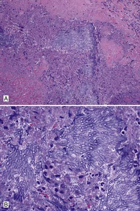
Fig 1 Aspergillosis. Necrotizing pneumonia with aspergillus hyphae: Low (A) and high (B) powers.
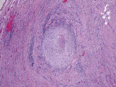
Fig 2 Aspergillosis. Granuloma with central necrosis; low power.
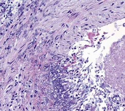
Fig 3 Aspergillosis. Aspergillus hyphae invading through the arterial wall.
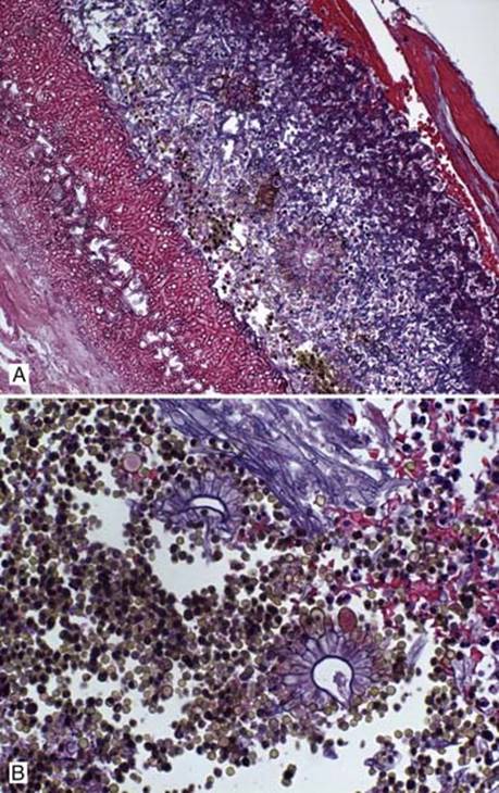
Fig 4 Aspergillosis. Fungal hyphae with fruiting head (conidia) and spores of A. niger (A and B). Note black spores, which give it the name “niger.”
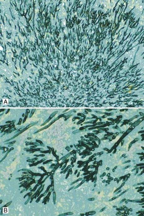
Fig 5 Aspergillosis. GMS stain of aspergillus hyphae. Low (A) and high (B) powers; note septated hyphae with 45-degree branching.
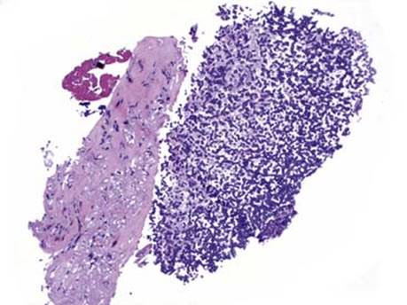
Fig 6 Aspergillosis. Fungal hyphae cut in cross-section can be easily identified on this H&E-stained section of a transbronchial biopsy. Culture will be needed to speciate the fungus.