
Definition
• Infection by the dimorphic fungus, Coccidioides immitis; also called Valley Fever
Clinical features
Epidemiology
• Endemic to southwestern United States and parts of Central and South America
• Pulmonary infection occurs after inhaling fungal spores that become airborne after disturbance of contaminated soil
• Up to half of people living in endemic areas will have evidence of exposure
• Most infections are asymptomatic and self-limited
• Disseminated disease occurs in immunocompromised and pregnant patients
Presentation
• Primary pulmonary coccidioidomycosis is usually asymptomatic but may be seen as mild respiratory symptoms and mild nonspecific infiltrates on chest x-ray films, as well as peripheral blood eosinophilia
• Patients with persistent pulmonary coccidioidomycosis typically present with symptoms related to pneumonia and chest x-ray findings of patchy consolidation or miliary nodularity
• A residual circumscribed fibrotic nodule, or coccidioidoma, may persist after acute or persistent disease
• Apical consolidation or nodules on chest x-ray films, bronchopleural fistula, and or empyema may be seen in chronic progressive coccidioidal pneumonia
• Patients with disseminated disease often have skin or central nervous system involvement
Prognosis and treatment
• Primary pulmonary coccidioidomycosis spontaneously resolves within a few weeks in the majority of cases
• Persistent disease, chronic progressive disease, miliary disease, and disseminated disease are treated with antifungal agents and may take several months to resolve
Pathology
Histology
• Necrotizing or nonnecrotizing granulomas with prominent eosinophils
• Large (30-100 micron diameter) spherules at various stages of maturation
• Mature spherules contain numerous 2- to 5-micron diameter endospores and have a thick refractile wall
• Immature spherules contain few to no endospores
• Endospores have a single nucleus and punctate cytoplasmic inclusions and may be found outside of ruptured spherules
• In aerated tissues, such as bronchopleural fistulas or cavitary lesions, mycelial forms with hyphae may be seen
Immunopathology/special stains
• Spherules and endospores are GMS and PAS positive
Main differential diagnoses
• Immature (empty) spherules: Blastomyces dermatitidis: look for mature spherules of coccidioides and broad-based budding of Blastomyces
• Free endospores: Histoplasma capsulatum or Cryptococcus: look for spherules of coccidioides, budding of Histoplasma and Cryptococcus, and capsule of Cryptococcus
• Mature spherules: Rhinosporidium or myospherulosis
• Mycelial form: Candida spp.
• Other: eosinophilic pneumonia
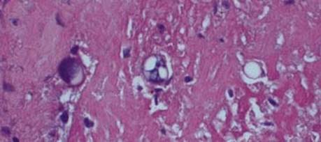
Fig 1 Coccidioidomycosis. Immature spherules of Coccidioides immitis with few to no endospores.
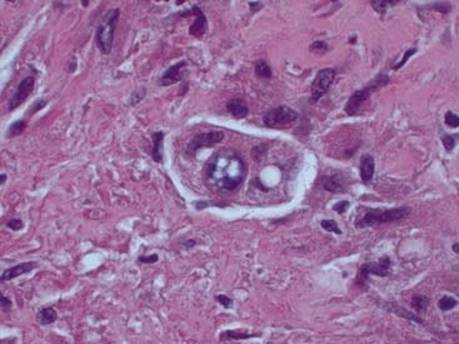
Fig 2 Coccidioidomycosis. Spherules of Coccidioides immitis engulfed by a macrophage.

Fig 3 Coccidioidomycosis. Empty spherules of Coccidioides immitis can be mistaken for Blastomyces dermatitidis (GMS stain).
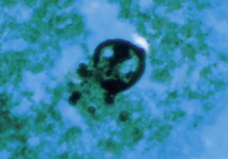
Fig 4 Coccidioidomycosis. This spherule has ruptured, releasing its endospores (GMS stain).
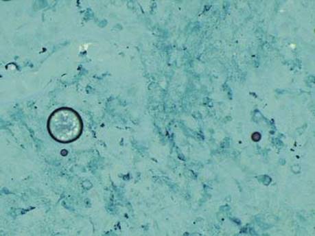
Fig 5 Coccidioidomycosis. Both an empty spherule (left) and endospores can be identified in this GMS-stained section from a necrotic chest mass.
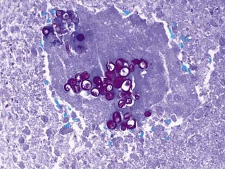
Fig 6 Coccidioidomycosis. Endospores have a single nucleus, punctate cytoplasmic inclusions, and are PAS positive (PAS stain).
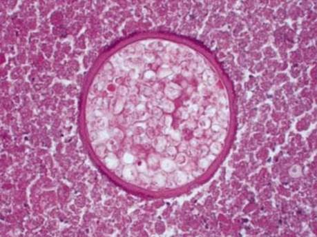
Fig 7 Coccidioidomycosis. This large mature spherule has a thick refractile wall and contains numerous small endospores. Note the necrotic background.
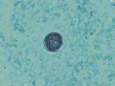
Fig 8 Coccidioidomycosis. GMS stains both the wall of the spherule and the endospores inside.
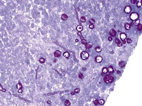
Fig 9 Coccidioidomycosis. In this cavitary lesion, variably sized spherules as well as mycelial hyphae of C. immitis are present.