
Definition
• An infection caused by fungi from Cryptococcus genus
Pathogenesis
• Infection of humans and animals by inhalation of the spores
• Starts with lung infection; may spread to brain, causing meningoencephalitis
• Patient’s immune status is critical to the pathogenicity of the organism
Clinical features
Epidemiology
• Although there are more than 50 Cryptococcus species, majority of cryptococcosis cases are caused by C. neoformans and C. gattii
• C. neoformans:
• Found worldwide in bird feces, especially pigeon feces
• Only infect immunocompromised patients
• C. gattii:
• Associated with plants found in tropical and subtropical climates
• Recently, outbreaks found in Canada and U.S. Northwest (Washington and Oregon)
• Can also infect immunocompetent patients
Presentation
• Cryptococcus may infect lung, brain, or both
• Majority of immunocompetent patients are asymptomatic; some have respiratory symptoms including fever, cough, chest pain, hemoptysis, and malaise
• Immunocompromised patients are usually symptomatic and often with symptoms of brain involvement like headache, nausea, vomiting, visual change, seizures, or even coma
• Small percentage of patients may also have skin changes like rash, pustules, or ulcers
Prognosis and treatment
• Treatment:
• Long-term antifungal therapy with multiple medications
– Immunocompetent patients: 1 to 2 years
– Immunocompromised patients: 1 to 2 years of treatment followed by lifetime suppressive therapy
• Surgery to remove a fungal mass (cryptococcoma), if necessary
• Good prognosis in immunocompetent patients with early diagnosis and treatment
• Poor prognosis (approximately 30% mortality rate) in immunocompromised patients or immunocompetent patients who were diagnosed and treated late
Pathology
Histology
• Granulomatous reaction with fibrosis occurs in patients with normal or near-normal immunity; fewer organisms are usually found within giant cells and histiocytes
• In immunocompromised patients, there is minimal inflammatory reaction with numerous organisms present:
• Histiocytic pneumonia: histiocytic reaction with numerous, densely packed fungal yeasts, resembling lipoid pneumonia
• Mucoid pneumonia: numerous yeasts filling alveolar spaces without an inflammatory reaction
• Intravascular/intracapillary invasion: usually seen in most severe immunodeficient hosts
• Characteristic yeast forms: round to oval yeasts (2-10 μm) with a thick capsule and narrow budding; they are pale gray to light blue on H&E sections
• Capsule-deficient forms may occur, which causes diagnostic difficulties
Immunopathology/special stains
• Mucin stains highlight the mucinous capsule, which is diagnostic for Cryptococcus
• GMS stain identifies all forms of the organism, including the capsule-deficient form
• Electron microscopy and immunofluoresence studies have been used to identify attenuated capsules in capsule-deficient cases
Main differential diagnoses
• Capsule-deficient forms need to be distinguished from other fungal organisms:
• Histoplasma capsulatum
• Blastomyces
• Pneumocystis jiroveci
• Sporothrix schenckii

Fig 1 Cryptococcosis. Cryptococcosis with sheets of organisms mimicking exogenous lipoid pneumonia. Low (A) and high (B) powers.
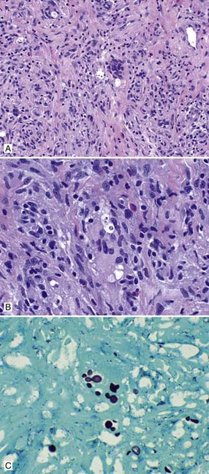
Fig 2 Cryptococcosis. Cryptococcosis with granulomatous reaction. A, Low power; B, high power; and C, GMS showing yeasts within giant cells.

Fig 3 Cryptococcosis. Mucoid pneumonia pattern. A, Low; B, medium; and C, high powers. Note the pale stained yeasts with clear surrounding spaces.
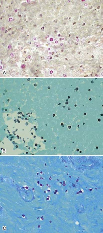
Fig 4 Cryptococcosis. A, Mucicarmine stains the capsule red; B, GMS stain shows variable-sized yeasts, some with narrow-based budding; and C, PAS stains the organism.