The accompanying illustrations are a guide for examining sensory and selected motor function of certain peripheral nerves: radial (Figure A-1), median (Figure A-2), ulnar (Figure A-3), fibular (peroneal) (Figure A-4), and femoral (Figure A-5). The sensory distribution of the lateral femoral cutaneous and obturator nerves is also shown (Figures A-6 and A-7). It is not intended to illustrate the findings of a lesion at any particular level of the nerves depicted. Sensory deficits may be less extensive than the full sensory field of a nerve because the fields of two nerves overlap, because a distal nerve lesion affects only part of the field, or because different sensory modalities are differentially involved.
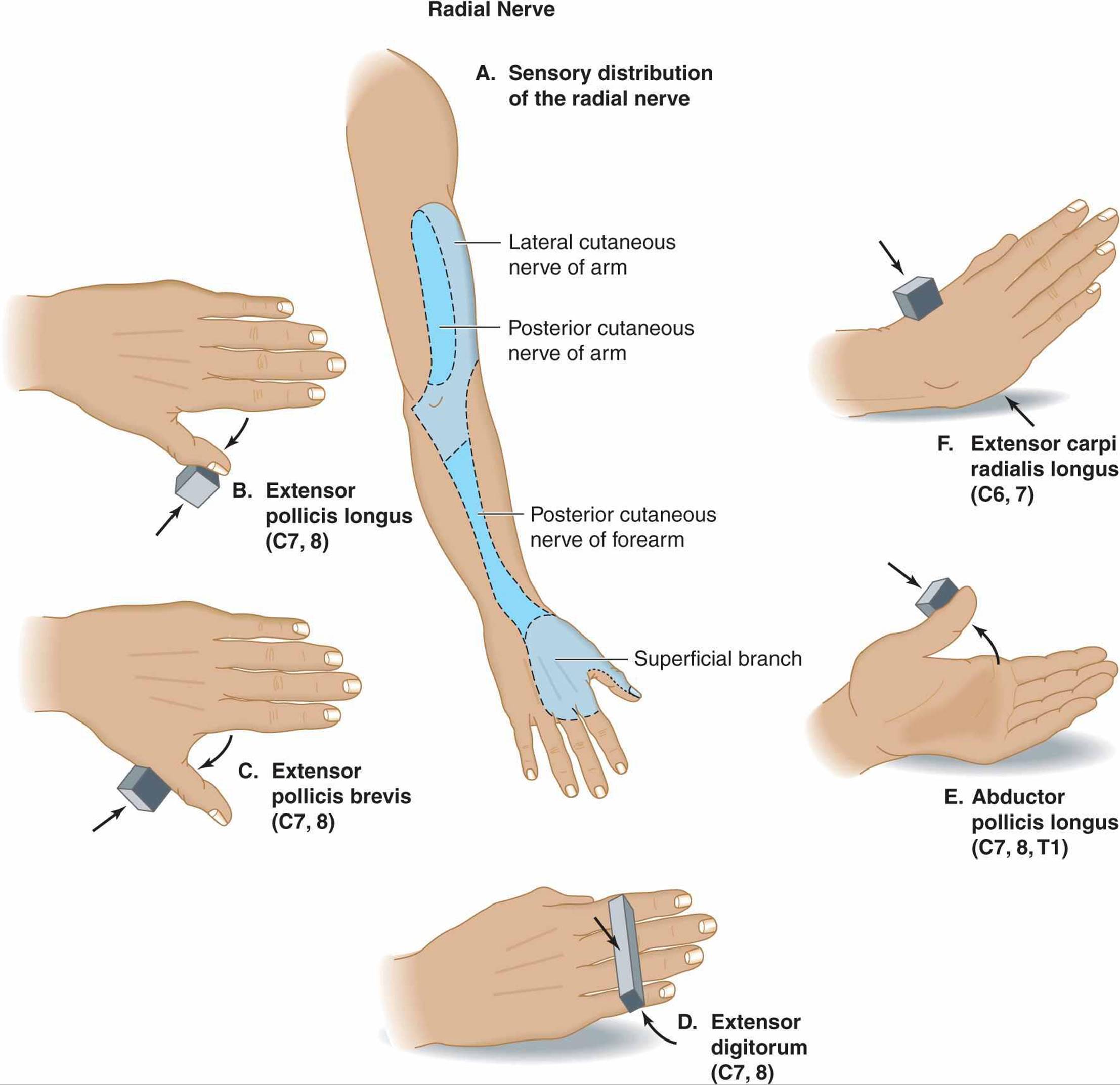
Figure A-1. Testing the radial nerve. A: Sensory distribution. The radial nerve supplies the dorsolateral surface of the upper arm, forearm, wrist, and hand; the dorsal surface of the thumb; the dorsal surface of the index and middle fingers above the distal interphalangeal joints; and the lateral half of the dorsal surface of the ring finger above the distal interp halangeal joint. B: Extensor pollicis longus. The thumb is extended at the interphalangeal joint against resistance. C: Extensor pollicis brevis. The thumb is extended at the metacarpophalangeal joint against resistance. D: Extensor digitorum. The fingers are extended at the metacarpophalangeal joints against resistance. E: Abductor pollicis longus. The thumb is abducted (elevated in a plane at 90 degrees to the palm) at the carpometacarpal joint against resistance. F:Extensor carpi radialis longus. The wrist is extended toward the radial (thumb) side against resistance.
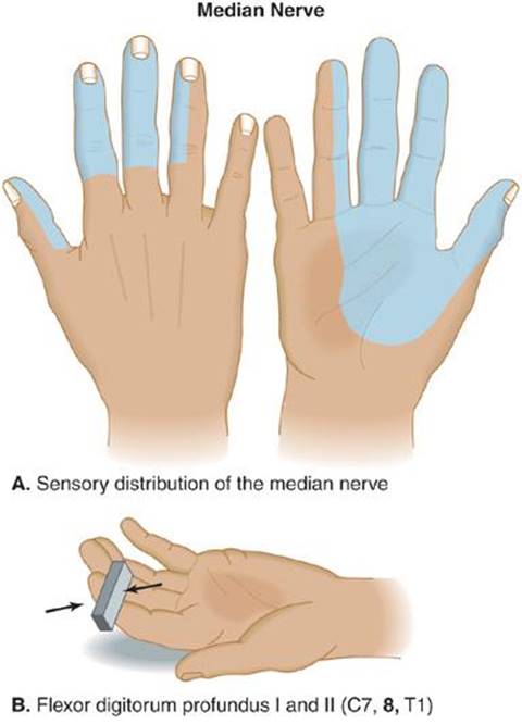
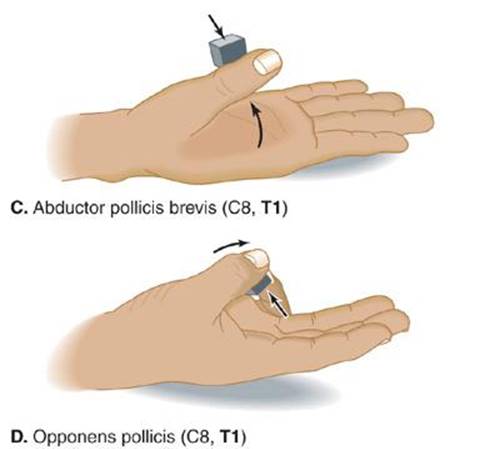
Figure A-2. Testing the median nerve. A: Sensory distribution. The median nerve supplies the dorsal surface of the index and middle fingers, the lateral half of the dorsal surface of the ring finger, the lateral two-thirds of the palm, the palmar surface of the thumb, index finger, middle fingers, and the lateral half of the palmar surface of the ring finger. B: Flexor digitorum profundus I and II. The index and middle fingers are flexed at the distal inter-phalangeal joints against resistance. C: Abductor pollicis brevis. The thumb is abducted (elevated at 90 degrees to the plane of the palm) at the metacarpophalangeal joints against resistance. D: Opponens pollicis. The thumb is crossed over the palm to touch the little finger against resistance.
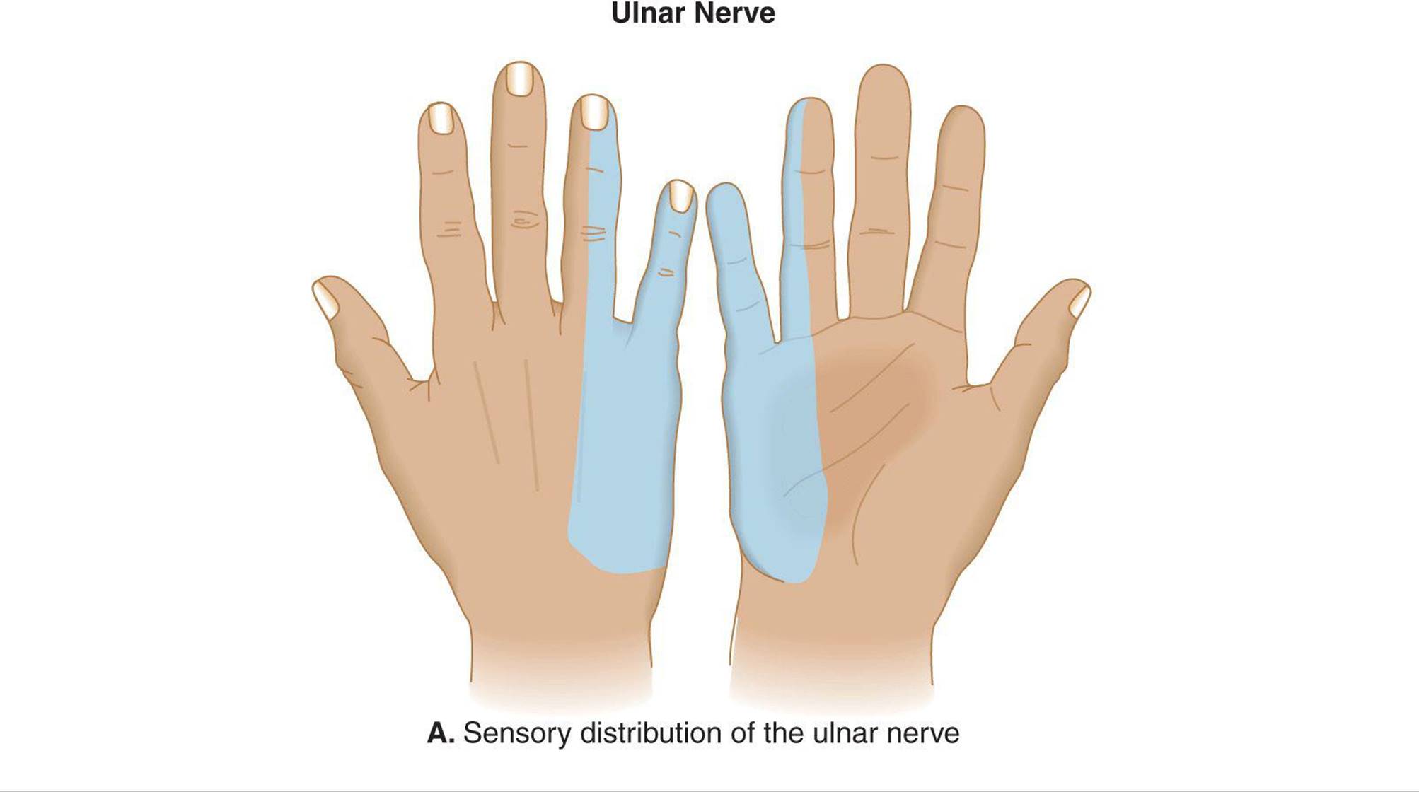
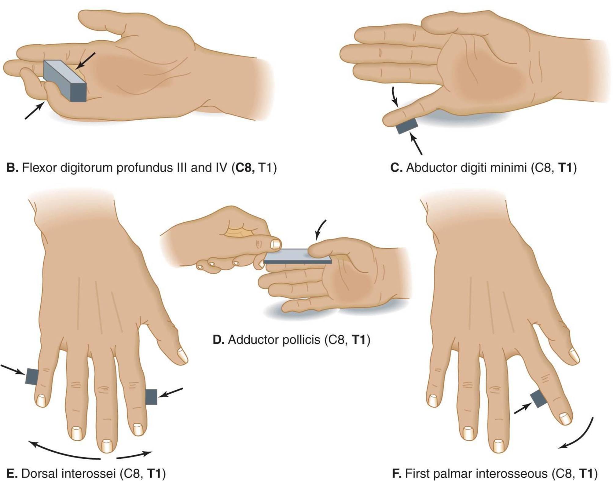
Figure A-3. Testing the ulnar nerve. A: Sensory distribution. The ulnar nerve supplies the dorsal and palmar surfaces of the medial one-third of the hand, the dorsal and palmar surfaces of the little finger, and the dorsal and palmar surfaces of the medial half of the ring finger. B: Flexor digitorum profundus III and IV. The index and middle fingers are flexed at the distal interphalangeal joints against resistance. C:Abductor digiti minimi. The little finger is abducted against resistance. D: Adductor pollicis. A piece of paper is grasped between the thumb and the palm with the thumbnail at 90 degrees to the plane of the palm while the examiner tries to pull the paper away. E: Dorsal interossei. The fingers are abducted against resistance. F: First palmar interosseous. The abducted index finger is adducted against resistance.
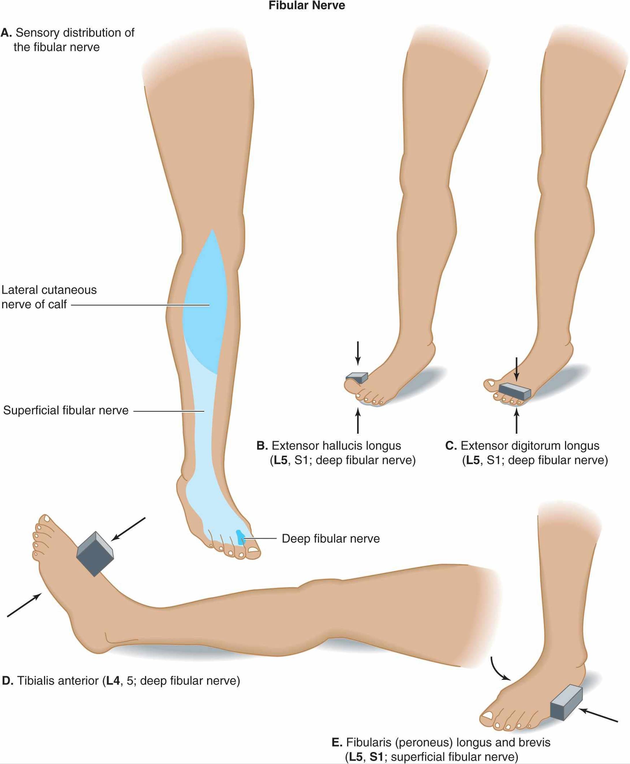
Figure A-4. Testing the fibular (peroneal) nerve. A: Sensory distribution. The common fibular (peroneal) nerve has three main sensory branches. The lateral cutaneous nerve of the calf supplies the lateral surface of the calf, the superficial fibular (peroneal) nerve supplies the lateral surface of the lower leg and the dorsum of the foot, and the deep fibular (peroneal) nerve supplies a roughly triangular patch of skin on the dorsum of the foot between the first and second toes. B: Extensor hallucis longus. The large toe is extended (dorsiflexed) against resistance. C: Extensor digitorum longus. The second, third, fourth, and fifth toes are extended against resistance. D: Tibialis anterior. The foot is dorsiflexed at the ankle against resistance. E: Fibularis (peroneus) longus and brevis. The foot is everted (rotated laterally) at the ankle against resistance.
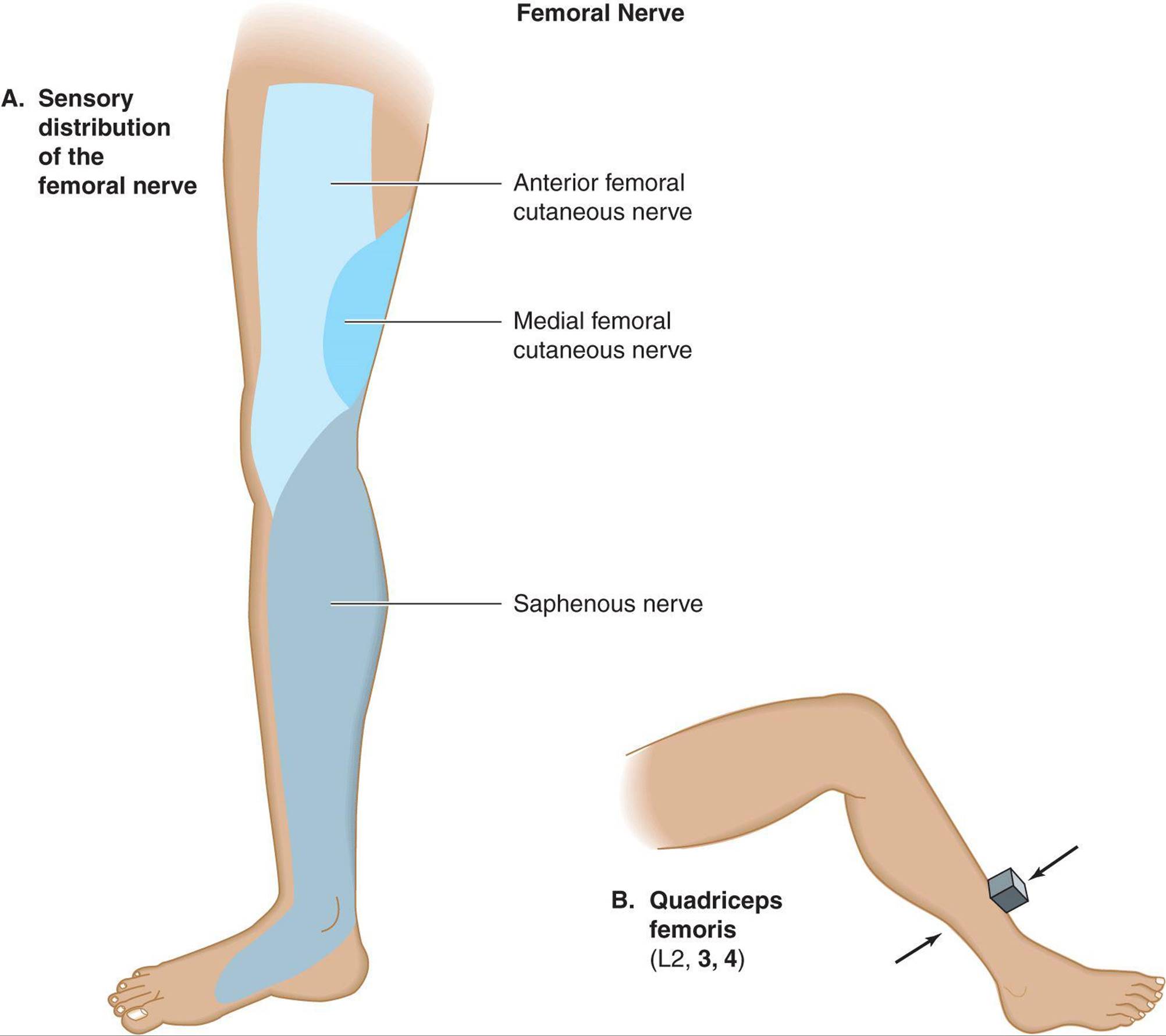
Figure A-5. Testing the femoral nerve. A: Sensory distribution. The femoral nerve has three main sensory branches. The anterior femoral cutaneous nerve supplies the anterior surface of the thigh, the medial femoral cutaneous nerve supplies the anteromedial surface of the thigh, and the saphenous nerve supplies the medial surface of the lower leg, ankle, and foot. B: Quadriceps femoris. The leg is extended at the knee against resistance.
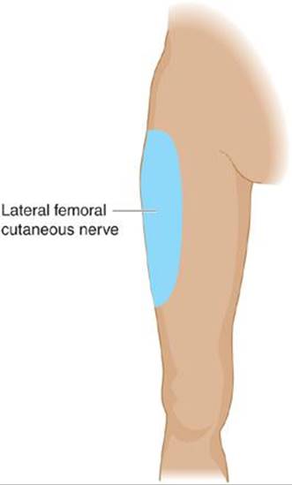
Figure A-6. Sensory distribution of the lateral femoral cutaneous nerve.
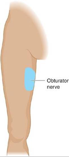
Figure A-7. Sensory distribution of the obturator nerve.