
Approach to Diagnosis
Emergency Management
History & Examination
History
General Physical Examination
Neurologic Examination
Pathophysiologic Assessment
Causes of Coma
Supratentorial Structural Lesions
Subdural Hematoma
Epidural Hematoma
Cerebral Contusion
Intracerebral Hemorrhage
Brain Abscess
Stroke (Cerebral Infarction)
Brain Tumor
Subtentorial Structural Lesions
Basilar Artery Thrombosis or Embolic Occlusion
Pontine Hemorrhage
Cerebellar Hemorrhage or Infarction
Posterior Fossa Subdural and Epidural Hematomas
Diffuse Encephalopathies
Meningitis & Encephalitis
Subarachnoid Hemorrhage
Hypoglycemia
Global Cerebral Ischemia
Drug Intoxication
Hepatic Encephalopathy
Hyperosmolar States
Hyponatremia
Hypothermia
Hyperthermia
Seizure or Prolonged Postictal State
Other Diffuse Encephalopathies
Differential Diagnosis
Psychogenic Unresponsiveness
Persistent Vegetative State
Locked-In Syndrome
Brain Death
Cessation of Brain Function
Irreversibility of Brain Dysfunction
Persistence of Brain Dysfunction
Additional Confirmatory Tests
References
Coma is a sleep-like state in which the patient makes no purposeful response to the environment and from which he or she cannot be aroused. The eyes are closed and do not open spontaneously. The patient does not speak, and there is no purposeful movement of the face or limbs. Verbal stimulation produces no response. Painful stimulation may produce no response or nonpurposeful reflex movements mediated through spinal cord or brainstem pathways.
Coma results from a disturbance in the function of either the brainstem reticular activating system above the mid pons or of both cerebral hemispheres (Figure 3-1), as these are the brain regions that maintain consciousness.

Figure 3-1. Anatomic basis of coma. Consciousness is maintained by the normal functioning of the brainstem reticular activating system above the mid pons and its bilateral projections to the thalamus and cerebral hemispheres. Coma results from lesions that affect either the reticular activating system or both hemispheres.
APPROACH TO DIAGNOSIS
The approach to diagnosis of the comatose patient consists first of emergency measures to stabilize the patient and treat presumptively certain life-threatening disorders, followed by efforts to establish an etiologic diagnosis.
EMERGENCY MANAGEMENT
As summarized in Table 3-1, emergency management of the comatose patient includes the following steps:
1. Ensure patency of the airway and adequacy of ventilation and circulation. This is accomplished by rapid visual inspection and by measuring the vital signs. If the airway is obstructed, the obstruction should be cleared and the patient intubated. If there is evidence of trauma that may have affected the cervical spine, however, the neck should not be moved until stability has been established by x-rays of the cervical spine. If spinal instability is present and intubation is required, tracheostomy should be performed. Adequacy of ventilation can be established by the absence of cyanosis, a respiratory rate greater than 8/min, the presence of breath sounds on auscultation of the chest, and the results of arterial blood gas and pH studies (see later). If any of these suggest inadequate ventilation, the patient should be ventilated mechanically. Measurement of the pulse and blood pressure provides a rapid assessment of the status of the circulation. Circulatory embarrassment should be treated with intravenous fluid replacement, pressors, and antiarrhythmic drugs, as indicated.

Table 3-1. Emergency management of the comatose patient.
2. Insert an intravenous catheter and withdraw blood for laboratory studies. These studies should include measurement of serum glucose and electrolytes, hepatic and renal function tests, prothrombin time, partial thromboplastin time, and a complete blood count. Extra tubes of blood should also be obtained for additional studies that may be useful in certain cases, such as drug screens, and for tests that become necessary as diagnostic evaluation proceeds.
3. Begin an intravenous infusion and administer dextrose, thiamine, and naloxone. Every comatose patient should be given 25 g of dextrose intravenously, typically as 50 mL of a 50% dextrose solution, to treat possible hypoglycemic coma. Because administration of dextrose alone may precipitate or worsen Wernicke encephalopathy (see Chapter 4) in thiamine-deficient patients, all comatose patients should also receive 100 mg of thiamine by the intravenous route. To treat possible opiate overdose, the opiate antagonist naloxone, 0.4 to 1.2 mg intravenously, should also be administered routinely to comatose patients. The benzodiazepine antagonist flumazenil, 1 to 10 mg intravenously, may be useful when benzodiazepine overdose contributes to coma. However, it should not be used in patients with a history of seizures, chronic benzodiazepine abuse, or suspected coingestion of tri- or tetracyclic antidepressants. The latter should be suspected if the electrocardiogram (ECG) shows sinus tachycardia at a rate of >130/min, QTc interval >0.5 seconds, and QRS duration >0.1 seconds.
4. Withdraw arterial blood for blood gas and pH determinations. In addition to assisting in the assessment of ventilatory status, these studies can provide clues to metabolic causes of coma (Table 3-2).

Table 3-2. Metabolic coma: differential diagnosis by acid–base abnormalities.
5. Institute treatment for seizures, if present. Persistent or recurrent seizures in a comatose patient should be considered to represent status epilepticus and treated accordingly, as described in Chapter 12 (see particularly Table 12-6).
After these measures have been taken, the history (if available) is obtained, and general physical and neurologic examinations are performed.
HISTORY & EXAMINATION
HISTORY
The most crucial aspect of the history is the time over which coma develops. In the absence of precise details about the mode of onset, information about when the patient was last seen in an apparently normal state may assist in establishing the time course of the disease process.
1. A sudden onset of coma suggests a vascular origin, especially a brainstem stroke or subarachnoid hemorrhage.
2. Rapid progression from hemispheric signs, such as hemiparesis, hemisensory deficit, or aphasia, to coma within minutes to hours is characteristic of intracerebral hemorrhage.
3. A more protracted course leading to coma (days to a week or more) is seen with tumor, abscess, or chronic subdural hematoma.
4. Coma preceded by a confusional state or agitated delirium, without lateralizing signs or symptoms, is probably due to a metabolic derangement or infection (meningitis or encephalitis).
GENERAL PHYSICAL EXAMINATION
Signs of Trauma
1. Inspection of the head may reveal signs of basilar skull fracture, including the following:
A. Raccoon eyes—Periorbital ecchymoses (see Figure 1-4).
B. Battle sign—Swelling and discoloration overlying the mastoid bone behind the ear (see Figure 1-4).
C. Hemotympanum—Blood behind the tympanic membrane.
D. Cerebrospinal fluid (CSF) rhinorrhea or otorrhea—Leakage of CSF from the nose or ear. CSF rhinorrhea must be distinguished from other causes of rhinorrhea, such as allergic rhinitis. Glucose concentration does not reliably distinguish CSF from nasal mucus, but beta-2 transferrin is unique to CSF, and its presence documents a CSF source of rhinorrhea.
2. Palpation of the head may demonstrate a depressed skull fracture or swelling of soft tissues at the site of trauma.
Blood Pressure
Elevated blood pressure in a comatose patient may reflect long-standing hypertension, which predisposes to intracerebral hemorrhage or stroke. In the rare condition of hypertensive encephalopathy, the blood pressure is above 250/150 mm Hg in chronically hypertensive patients; it may be lower in children or after acute elevation of blood pressure in previously normotensive patients (eg, in acute renal failure). Elevated blood pressure may also be a consequence of the process causing the coma, as in intracerebral or subarachnoid hemorrhage or, rarely, brainstem stroke.
Temperature
Hypothermia occurs in coma caused by ethanol or sedative drug intoxication, hypoglycemia, Wernicke encephalopathy, hepatic encephalopathy, and myxedema. Coma with hyperthermia is seen in heat stroke, status epilepticus, malignant hyperthermia related to inhalational anesthetics, anticholinergic drug intoxication, pontine hemorrhage, and certain hypothalamic lesions.
Signs of Meningeal Irritation
Signs of meningeal irritation (eg, nuchal rigidity or the Brudzinski sign [see Figure 1-5]) can be invaluable in the prompt diagnosis of meningitis or subarachnoid hemorrhage, but these signs are lost in deep coma, so their absence does not exclude these conditions.
Optic Fundi
Examination of the optic fundi may reveal papilledema or retinal hemorrhages compatible with chronic or acute hypertension, or an elevation in intracranial pressure (Figure 1-11). Subhyaloid (superficial retinal) hemorrhages in an adult strongly suggest subarachnoid hemorrhage (Figure 6-3).
NEUROLOGIC EXAMINATION
The neurologic examination is the key to etiologic diagnosis in the comatose patient. Pupillary size and reactivity, reflex eye movements (oculocephalic and oculovestibular reflexes), and the motor response to pain should be evaluated in detail (Figure 3-2).
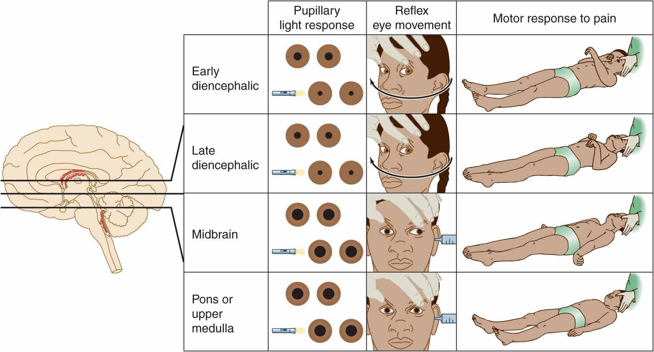
Figure 3-2. Neurologic signs in coma with downward transtentorial herniation. In the early diencephalic phase, the pupils are small (approximately 2 mm in diameter) and reactive, reflex eye movements are intact, and the motor response to pain is purposeful or semipurposeful (localizing) and often asymmetric. The late diencephalic phase is associated with similar findings, except that painful stimulation results in decorticate (flexor) posturing, which may also be asymmetric. With midbrain involvement, the pupils are fixed and midsized (approximately 5 mm in diameter), reflex adduction of the eyes is impaired, and pain elicits decerebrate (extensor) posturing. Progression to involve the pons or medulla also produces fixed, midsized pupils, but these are accompanied by loss of reflex abduction as well as adduction of the eyes and by no motor response or only leg flexion upon painful stimulation. Note that although a lesion restricted to the pons produces pinpoint pupils as a result of the destruction of descending sympathetic (pupillodilator) pathways, downward herniation to the pontine level is associated with midsized pupils. This happens because herniation also interrupts parasympathetic (pupilloconstrictor) fibers in the oculomotor (III) nerve.
Pupils
1. Normal pupils—Normal pupils are typically 3 to 4 mm in diameter (but larger in children and smaller in the elderly) and equal in size bilaterally; they constrict briskly and symmetrically in response to light. Normally reactive pupils in a comatose patient are characteristic of a metabolic cause.
2. Thalamic pupils—Slightly smaller (~2 mm) reactive pupils are present in the early stages of thalamic compression from mass lesions, perhaps because of interruption of the descending sympathetic pathways.
3. Fixed, dilated pupils—Pupils greater than 7 mm in diameter and fixed (unreactive to light) usually result from compression of the oculomotor (III) cranial nerve (and associated sympathetic, pupillodilator nerve fibers) anywhere along its course, from the midbrain to the orbit, but may also be seen in anticholinergic or sympathomimetic drug intoxication. The most common cause of a fixed, dilated pupil in a comatose patient is transtentorial herniation of the medial temporal lobe from a supratentorial mass.
4. Fixed, midsized pupils—Pupils fixed at approximately 5 mm in diameter are the result of brainstem damage at the midbrain level, which interrupts both sympathetic, pupillodilator and parasympathetic, pupilloconstrictor nerve fibers.
5. Pinpoint pupils—Pinpoint pupils (1-1.5 mm in diameter) in a comatose patient usually indicate opioid overdose or, less commonly, a focal structural lesion in the pons. These causes can be distinguished by the administration of naloxone (see earlier) or by the associated defects in horizontal eye movements that usually accompany pontine lesions. Pinpoint pupils may appear unreactive to light except when viewed with a magnifying glass. Pinpoint pupils can also be caused by organo-phosphate poisoning, miotic eye drops, or neurosyphilis (Argyll Robertson pupils).
6. Asymmetric pupils—Asymmetry of pupillary size (anisocoria) with a difference of 1 mm or less in diameter is a normal finding that occurs in 20% of the population. In such physiologic anisocoria, pupils constrict to a similar extent in response to light, and extraocular movements are unimpaired. In contrast, a pupil that constricts less rapidly or to a lesser extent than its contralateral fellow usually implies a structural lesion affecting the midbrain, oculomotor nerve, or eye.
Eye Movements
1. Pathways tested—The neuronal pathways examined by testing eye movements begin at the pontomedullary junction (vestibular [VIII] nerve and nucleus), synapse in the caudal pons (horizontal gaze center and abducens [VI] nerve nucleus), ascend through the central core of the brainstem reticular activating system (medial longitudinal fasciculus), and arrive at the contralateral mid-brain (oculomotor [III] nucleus and nerve; Figure 3-3).

Figure 3-3. Brainstem pathways mediating reflex conjugate horizontal eye movements. In a comatose patient with intact brainstem function, irrigation of the tympanic membrane with ice water inhibits the vestibuloocular pathways shown, resulting in tonic deviation of both eyes toward the irrigated side; head rotation causes eye deviation away from the direction of rotation.
2. Methods of testing—In the comatose patient, eye movements are tested by stimulating the vestibular system (semicircular canals of the middle ear) by passive head rotation (the oculocephalic reflex, or doll’s-head maneuver) or using the stronger stimulus of ice-water irrigation against the tympanic membrane (oculovestibular reflex, or cold-water caloric testing) (Figure 3-3).
The doll’s-head (oculocephalic) maneuver is performed by rotating the head horizontally to elicit horizontal eye movements and vertically to elicit vertical movements. The eyes should move in the direction opposite to that of head rotation. This may be an inadequate stimulus for inducing eye movements, however, and the reflex may be overridden in conscious patients.
Cold-water caloric (oculovestibular) stimulation is a more potent stimulus and is performed by irrigating the tympanic membrane with ice water. Otoscopic examination should always be undertaken before this maneuver is attempted, because it is contraindicated if the tympanic membrane is perforated. In conscious patients, unilateral cold water irrigation produces nystagmus with the fast phase directed away from the irrigated side. In comatose patients with intact brainstem function, unilateral ice water irrigation results in tonic deviation of the eyes toward the irrigated side. Bilateral irrigation with ice water causes tonic downward deviation, whereas bilateral stimulation with warm (44°C) water induces tonic upward deviation. An absent or impaired response to caloric stimulation with large volumes (eg, 50 mL) of ice water is indicative of peripheral vestibular disease, a structural lesion involving the posterior fossa (cerebellum or brainstem), or intoxication with sedative drugs.
3. Normal movements—A comatose patient with intact brainstem function has full conjugate horizontal eye movements, which occur either spontaneously (as “roving eye movements”) or during the doll’s-head maneuver, as well as tonic conjugate deviation of both eyes toward the side of the ice-water irrigation during cold-water caloric testing. Full horizontal eye movements in a comatose patient exclude a structural lesion in the brainstem as the cause of coma and suggest either a nonstructural (eg, metabolic) cause or, less commonly, bilateral hemispheric lesions.
4. Abnormal movements
a. With lesions affecting the oculomotor (III) nerve or nucleus, such as hemispheric mass lesions causing downward transtentorial herniation (Figure 3-2), cold-water caloric testing fails to produce adduction of the ipsilateral eye, whereas the contralateral eye abducts normally.
b. Complete unresponsiveness to cold-water caloric testing in a comatose patient implies either a structural lesion of the brainstem affecting the pons or a metabolic disorder that affects the brainstem preferentially, such as sedative drug intoxication.
c. Downward deviation of one or both eyes in response to unilateral cold-water caloric testing also suggests sedative drug intoxication.
Motor Response to Pain
The motor response to pain is assessed by applying strong pressure on the supraorbital ridge, sternum, or nail beds. The response to such stimuli can indicate whether the condition causing coma affects the brain symmetrically (as is typical of metabolic and other diffuse disorders) or asymmetrically (as in unilateral structural lesions). The motor response to pain may also help to localize the anatomic level of cerebral dysfunction or provide a guide to the depth of coma.
1. With cerebral dysfunction of only moderate severity, patients may localize an offending stimulus by reaching toward the site of stimulation. Although such “semipurposeful” localizing responses can be difficult to distinguish from the reflex responses described later, movements that involve limb abduction almost never represent reflexes.
2. A decorticate response to pain (flexion of the arm at the elbow, adduction at the shoulder, and extension of the leg and ankle) is classically associated with lesions that involve the thalamus directly or large hemispheric masses that compress the thalamus from above.
3. A decerebrate response (extension at the elbow, internal rotation at the shoulder and forearm, and leg extension) tends to occur when brain dysfunction has descended to the level of the midbrain. Thus decerebrate posturing generally implies more severe brain dysfunction than decorticate posturing, although neither response localizes the site of dysfunction precisely.
4. Bilateral symmetric posturing may be seen in both structural and metabolic disorders.
5. Unilateral or asymmetric posturing suggests structural disease in the contralateral cerebral hemisphere or brainstem.
6. In patients with pontine and medullary lesions, there is usually no response to pain, but occasionally some flexion at the knee (a spinal reflex) is noted.
Glasgow Coma Scale
The pupillary, eye movement, and motor responses described earlier are sometimes translated to a numerical scale so that changes in the examination (and thus the numerical score) may be more easily noticed over time and compared between different examiners (Table 3-3).

Table 3-3. Glasgow coma scale.
PATHOPHYSIOLOGIC ASSESSMENT
The most important step in evaluating a comatose patient is to decide whether the cause is a structural brain lesion (for which emergency neurosurgical intervention may be required) or a diffuse disorder caused by a metabolic disturbance, meningitis, or seizures (for which immediate medical treatment may be needed).
Supratentorial Structural Lesions
When coma is the result of a supratentorial mass lesion, the history and physical findings early in the course usually point to dysfunction of one cerebral hemisphere. Symptoms and signs include contralateral hemiparesis, contralateral hemisensory loss, aphasia (with dominant, usually left, hemisphere lesions), and agnosia (indifference to or denial of the deficit, with injury to the nondominant hemisphere).
As the mass expands (commonly from associated edema), the patient becomes increasingly lethargic due to compression of the contralateral hemisphere or thalamus. Stupor progresses to coma, but findings on examination often remain asymmetric. With rostral–caudal (downward) progression of brain injury, the thalamus, midbrain, pons, and medulla become sequentially involved, and the neurologic examination reveals dysfunction at successively lower anatomic levels (Figure 3-2). This segmental pattern of involvement strongly supports the diagnosis of a supratentorial mass with downward transtentorial herniation (Figure 3-4) and dictates the need for neurosurgical intervention. At the fully developed midbrain level (midsized, unreactive pupils), chances of survival without severe neurologic impairment decrease rapidly, especially in adults. Once the pontine level of dysfunction is reached (unreactive pupils, and absent horizontal eye movements), a fatal outcome is inevitable.
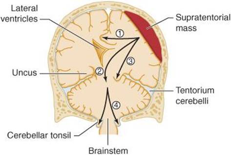
Figure 3-4. Anatomic basis of herniation syndromes. An expanding supratentorial mass lesion may cause brain tissue to be displaced into an adjacent intracranial compartment, resulting in (1) cingulate herniation under the falx, (2) downward transtentorial (central) herniation, (3) uncal herniation over the edge of the tentorium, or (4) cerebellar tonsillar herniation into the foramen magnum. Coma and ultimately death result when (2), (3), or (4) produces brainstem compression.
Supratentorial mass lesions may cause herniation of the medial portion of the temporal lobe (the uncus) over the edge of the cerebellar tentorium (Figure 3-4). This exerts direct pressure on the upper brainstem and produces signs of oculomotor (III) nerve and midbrain compression, such as ipsilateral pupillary dilatation and impaired adduction of the eye (uncal syndrome), which may precede loss of consciousness. Neurosurgical decompression must occur early in the course of oculomotor (III) nerve involvement if functional recovery is to occur.
Subtentorial Structural Lesions
Coma of sudden onset with focal signs of brainstem dysfunction strongly suggests a subtentorial structural lesion. Abnormal pupillary function and eye movement are the findings most suggestive of a subtentorial structural lesion, especially if these abnormalities are asymmetric. Midbrain lesions cause loss of pupillary function: the pupils are midsized (approximately 5 mm in diameter) and ureactive to light. Pontine hemorrhage, pontine infarction, or compression of the pons by adjacent cerebellar hemorrhage or infarction produces pinpoint pupils. Brainstem lesions may also be associated with conjugate gaze deviation away from the side of the lesion (and toward a hemiparesis) (Figure 7-17), or disconjugate eye movements such as internuclear ophthalmoplegia (selective impairment of eye adduction). Motor responses are generally not helpful in separating subtentorial from supratentorial lesions. Ventilatory patterns associated with subtentorial lesions are abnormal but variable and may be ataxic or gasping (Figure 3-5). Because the fully developed syndrome of transtentorial herniation from a supratentorial mass is characterized by extensive brainstem dysfunction, its differentiation from a primary subtentorial process may be impossible except by history.

Figure 3-5. Ventilatory patterns in coma. Cheyne-Stokes respiration and central hyperventilation are seen with metabolic disturbances and with structural lesions at a variety of sites in the brain. They are therefore not useful for anatomic localization of disorders producing coma. Ataxic and gasping ventilatory patterns are most commonly seen with pontomedullary lesions.
Diffuse Encephalopathies
Diffuse encephalopathies that result in coma (sometimes termed metabolic coma) include not only metabolic disorders such as hypoglycemia and drug intoxication, but other processes that affect the brain diffusely, such as meningitis, subarachnoid hemorrhage, and seizures.
The clinical presentation of diffuse encephalopathy is distinct from that of a mass lesion. There are usually no focal signs, such as hemiparesis, hemisensory loss, or aphasia, and—except in some cases of subarachnoid hemorrhage—consciousness is lost only gradually, typically after a period of progressive somnolence or agitated delirium.
A symmetric neurologic examination is the rule, although hypoglycemia, hyperosmolar nonketotic hyperglycemia, and hepatic encephalopathy may sometimes be accompanied by focal signs, such as hemiparesis, which may alternate from side to side. Asterixis, myoclonus, and tremor preceding coma are important clues that suggest metabolic disease. Symmetric decorticate or decerebrate posturing can be seen with hepatic, uremic, anoxic, hypoglycemic, or sedative drug–induced coma.
Reactive pupils in the presence of otherwise impaired brainstem function is the hallmark of metabolic encephalopathy. Although coma with intact pupillary reaction can also be seen early in transtentorial herniation (Figure 3-2), this is associated with asymmetric neurologic findings, such as hemiparesis. A few metabolic causes of coma can also impair pupillary light reflexes, including massive barbiturate overdose with apnea and hypotension, acute anoxia, marked hypothermia, anticholinergic poisoning (large pupils), and opioid overdose (pinpoint pupils), but even in these settings, completely unreactive pupils are uncommon.
Ventilatory patterns in metabolic coma vary widely, but measuring arterial blood gases and pH may help to establish an etiologic diagnosis. Arterial blood gas abnormalities in coma are outlined in Table 3-2.
Summary
The relationship between neurologic signs and the pathophysiology of coma is summarized in Table 3-4. Examining pupil size and reactivity and testing reflex eye movements and the motor response to pain help determine whether brain function is disrupted at a discrete anatomic level (structural lesion) or in a diffuse manner (metabolic coma).

Table 3-4. Pathophysiologic assessment of the Comatose patient.
Supratentorial structural lesions compromise the brain in an orderly way, producing dysfunction at progressively lower anatomic levels. In patients with metabolic coma, such localization is not possible, and scattered, anatomically inconsistent findings may be seen. An impressive example of the anatomically discordant findings characteristic of metabolic coma is the retention of pupillary reactivity in the face of otherwise depressed brainstem functions—including paralysis of eye movements, respiratory depression, flaccid muscle tone, and unresponsiveness to painful stimuli—after sedative drug overdose. The same degree of low brainstem dysfunction produced by a supratentorial mass lesion would first compromise the more rostrally situated midbrain structures that mediate pupillary reactivity before affecting the lower brainstem centers.
CAUSES OF COMA
SUPRATENTORIAL STRUCTURAL LESIONS
SUBDURAL HEMATOMA
Subdural hematoma is a collection of blood in the subdural space between the dura mater and the arachnoid. Because subdural hematoma is resectable, it must always be considered early in any comatose patient with a suspected supratentorial mass lesion. Subdural hematoma is more common in older patients, because cerebral atrophy stretches bridging cortical veins and renders them more susceptible to laceration from shearing injury or apparently spontaneous rupture.
Trauma is the most common cause, and in the acute stage after head injury, focal neurologic deficits are often conspicuous. The severity of injury needed to produce a subdural hematoma becomes less with advancing age; in perhaps 25% of cases a history of trauma is not present.
The most common clinical findings are headache and altered consciousness, but symptoms and signs may be absent, nonspecific, or nonlocalizing, especially with chronic subdural hematomas that appear months or years after injury (Table 3-5). The classic history of waxing and waning signs and symptoms is too infrequent to be relied on for diagnosis. Hemiparesis, when present, is contralateral to the lesion in approximately 70% of cases. Pupillary dilation, when present, is ipsilateral in approximately 90% of cases. The frequency of bilateral hematomas may make localization difficult, as may coexisting cerebral contusion.
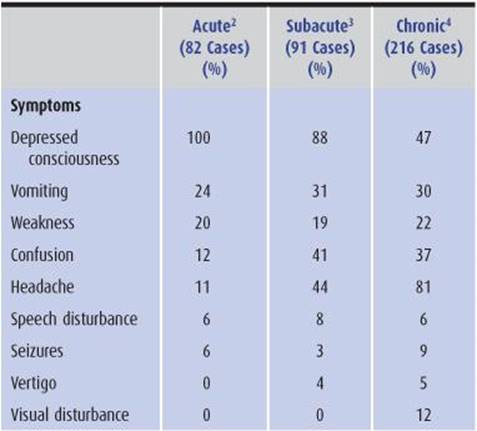

Table 3-5. Clinical features of subdural hematoma.1
Diagnosis is by computed tomography (CT) scan or magnetic resonance imaging (MRI) (Figure 3-6).
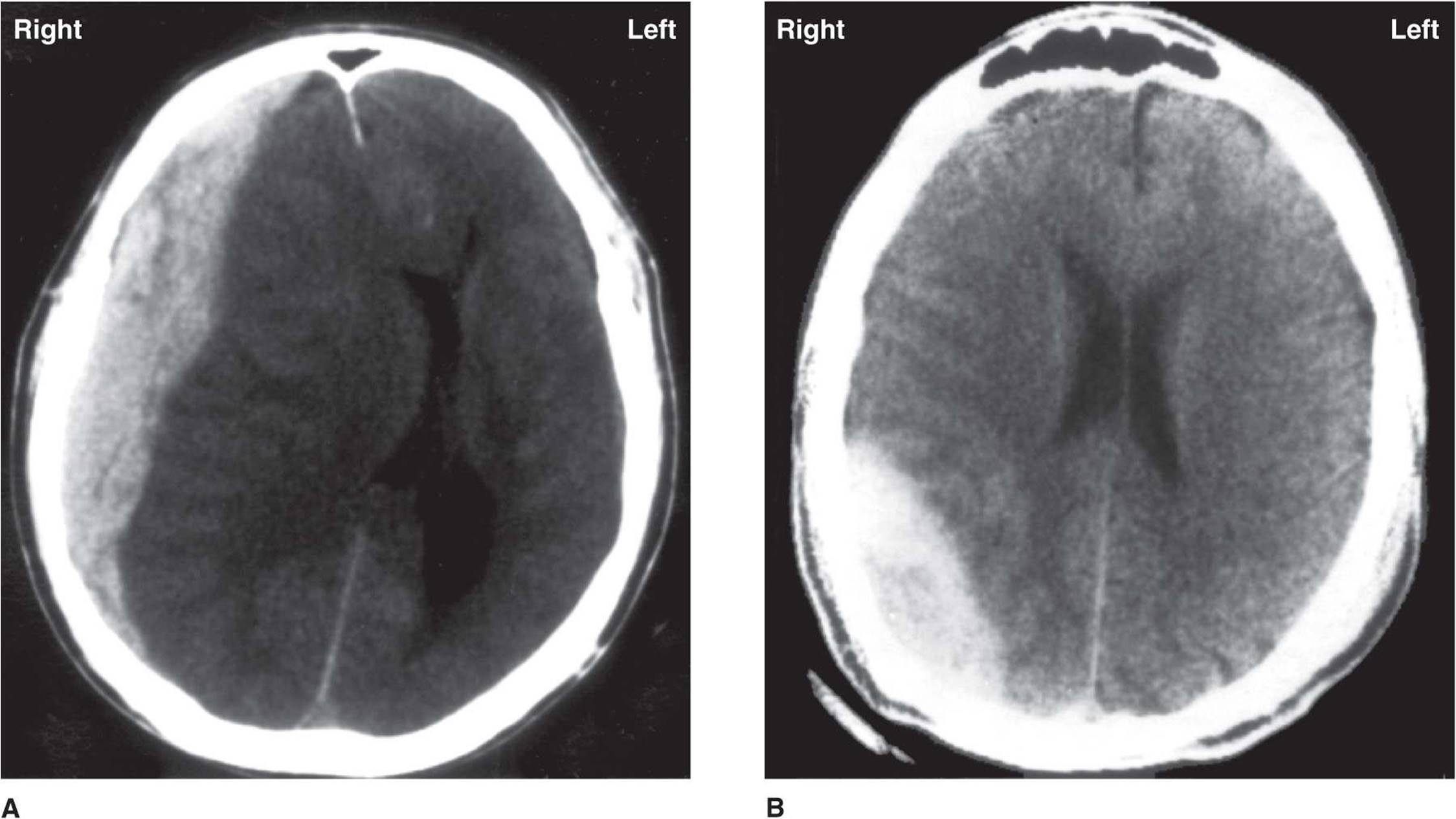
Figure 3-6. A: Subdural hematoma. Unenhanced CT scan showing a large, high-density crescentic mass over the right cerebral hemisphere, with shift of the lateral ventricles across the midline. B: Epidural hematoma. Unenhanced CT scan showing a large, high-density lens-shaped mass in the right parietooccipital region. Fracture of the occipital bone was seen on bone windows.
Treatment of subdural hematoma causing coma is by surgical evacuation.
EPIDURAL HEMATOMA
Epidural hematoma typically results from head trauma associated with a lateral skull fracture and tearing of the middle meningeal artery and vein. Patients may or may not lose consciousness initially. There is often a lucid interval of several hours before the onset of coma, during which headache, vomiting, obtundation, seizures, and focal neurologic signs may occur. The diagnosis is made by CT scan or MRI (Figure 3-6), which classically shows a radiodense biconvex lens-shaped mass compressing the cerebral hemisphere. Prompt surgical evacuation of the hematoma is essential to prevent a fatal outcome.
CEREBRAL CONTUSION
Cerebral contusion is bruising of the brain caused by head trauma. It is associated with initial unconsciousness (concussion) from which the patient recovers. Edema surrounding the contusion may cause the level of consciousness to fluctuate, and seizures and focal neurologic signs may develop. Patients must be carefully monitored for neurologic deterioration related to progressive edema and herniation.
Lumbar puncture is unnecessary and potentially dangerous. CT scan or MRI is the diagnostic procedure of choice. In contrast to subdural and epidural hematomas, cerebral contusions are rarely operated upon.
INTRACEREBRAL HEMORRHAGE
Etiology
The most common cause of nontraumatic intracerebral hemorrhage is chronic hypertension. This and other causes are discussed in more detail in Chapter 13.
Clinical Findings
Intracerebral hemorrhage usually occurs while the patient is awake. Hemorrhage is not preceded by transient prodromal symptoms, such as the transient ischemic attacks (TIAs) often associated with cerebral infarction (see Chapter 13).
Headache occurs in many cases and can be moderate to severe. If present, headache may be localized to the site of hemorrhage or generalized. Nausea and vomiting are common. Altered consciousness may progress steadily to stupor or coma over minutes to hours.
On examination, patients are nearly always hypertensive (blood pressure 170/90 mm Hg or higher), even in the late stages of transtentorial herniation. The funduscopic examination usually shows vascular changes associated with chronic hypertension. Nuchal rigidity is common. Gaze deviation—toward the side of a putaminal or lobar hemorrhage or downward and medially in thalamic hemorrhage—may occur. Hemiparesis is frequent because of the proximity of common hemorrhage sites, such as the basal ganglia and thalamus, to the internal capsule, which conveys descending motor fibers from the cerebral cortex.
Seizures occur in approximately 10% of cases and are often focal. Neurologic deficits do not fluctuate spontaneously.
Investigative Studies
CT brain scan without contrast or MRI shows intraparenchymal blood and confirms the diagnosis (Figure 13-18).
Treatment
1. Blood pressure—Systemic blood pressure should be reduced to ≤140 mm Hg to limit hematoma expansion, but excessive blood pressure reduction should be avoided, as it may compromise blood flow in brain tissue adjacent to the hemorrhage.
2. Cerebral edema—The mass effect of intracerebral hemorrhage is typically compounded by progressive cerebral edema, which becomes evident at approximately 24 hours and maximal within 5 to 6 days. Cerebral edema may be treated with mannitol or intravenous hypertonic saline (Table 3-6), but this is usually only useful as a temporizing measure prior to surgery, when indicated, and alone rarely alters the eventual outcome.

Table 3-6. Drug therapy for Cerebral Edema.
3. Surgical treatment—Evacuation of the clot may be appropriate in cases (approximately 10%) in which hemorrhage is located superficially in the cerebral hemisphere and produces a mass effect. However, most hemorrhages are deep within the brain and less accessible to surgery.
Prognosis
Early mortality from intracerebral hemorrhage is high, with approximately 25% of patients dying within 72 hours. However, those who survive may be left with surprisingly mild deficits as the clot resolves over a period of weeks to months.
BRAIN ABSCESS
Brain abscess is an uncommon disorder, accounting for only 2% of intracranial masses.
Etiology
The common conditions predisposing to brain abscess, in approximate order of frequency, are blood-borne metastasis from distant systemic (especially pulmonary) infection, direct extension from parameningeal sites (otitis, cranial osteomyelitis, sinusitis), an unknown source, infection associated with recent or remote head trauma or craniotomy, and infection associated with cyanotic congenital heart disease.
The most common pathogenic agents are aerobic, anaerobic, and microaerophilic streptococci, and gram-negative anaerobes such as bacteroides, Fusobacterium, and Prevotella. Staphylococcus aureus, Proteus, and other gram-negative bacilli are less common. Actinomyces, Nocardia, and Candida are also found. Multiple organisms are present in the majority of abscesses.
Clinical Findings
The course is that of an expanding mass lesion, usually presenting with headache and focal neurologic deficits in a conscious patient. Coma may develop over days but rarely over hours. Common presenting signs and symptoms are shown in Table 3-7. It is important to note that common correlates of infection may be absent: Temperature is normal in 40% of patients, and the peripheral white blood cell count is below 10,000/μL in 20%.

Table 3-7. Brain abscesses: presenting features in 123 cases.
Investigative Studies
The diagnosis is strongly supported by finding a mass lesion with a contrast-enhanced rim on CT scan or MRI or an avascular mass on angiography. Examination of the CSF reveals opening pressure >200 mm water in 75% of patients, pleocytosis of 25 to 500 or more white cells/μL (depending on the proximity of the abscess to the ventricular surface and its degree of encapsulation), and elevated protein (45-500 mg/dL) in approximately 60% of patients. CSF cultures are usually negative. However, marked clinical deterioration may follow lumbar puncture in patients with brain abscess; therefore, lumbar puncture should not be performed if brain abscess is suspected.
Treatment
Treatment of pyogenic brain abscess can be with antibiotics alone or combined with surgical drainage. Surgical therapy should be strongly considered when there is a significant mass effect or the abscess is near the ventricular surface, because catastrophic rupture into the ventricular system may occur.
Medical treatment alone is indicated for surgically inaccessible, multiple, or early abscesses. If the causal organism is unknown, broad-spectrum antibiotic coverage is indicated. The first-line recommendation in North America is ceftriaxone 2 g every 12 hours plus metronidazole, although penicillin G 3 to 4 million units intravenously every 4 hours and metronidazole 7.5 mg/kg intravenously or orally every 6 hours is still used in some countries. If staphylococcal infection is suspected, oxacillin or nafcillin 3 g intravenously every 6 hours should be added. Because methicillin-resistant staphylococcal infection is now so common, many centers begin treatment with vancomycin 1 g every 12 hours adjusted to serum levels. Nafcillin plus cefotaxime or ceftriaxone is recommended to cover staphylococci and aerobic gram-negative bacilli in trauma patients. Glucocorticoids (Table 3-6) may reduce edema surrounding the abscess. The response to medical treatment should be assessed by clinical examination and serial CT scans or MRIs. When medically treated patients do not improve, needle aspiration of the abscess is indicated to identify the organisms present.
STROKE (CEREBRAL INFARCTION)
Embolic or thrombotic occlusion of one carotid artery does not cause coma directly, because bilateral hemispheric lesions are required for consciousness to be lost. However, cerebral edema after massive hemispheric infarction can compress the contralateral hemisphere or cause transtentorial herniation, either of which can produce coma. Edema becomes maximal within 48 to 72 hours after infarction, and may cause progression of the original neurologic deficit and ultimately stupor and coma. Cerebral hemorrhage is excluded by CT scan or MRI.
The use of corticosteroids and dehydrating agents to treat cerebral edema associated with stroke has produced no clear benefit. Stroke is discussed in more detail in Chapter 13.
BRAIN TUMOR
Clinical Findings
Primary or metastatic brain tumors (see Chapter 6) rarely present with coma, although they can do so when hemorrhage into the tumor or tumor-induced seizures occur. More often, coma occurs late in the clinical course of brain tumor, and there is a history of headache, focal neurologic deficits, and altered consciousness. Papilledema is a presenting sign in 25% of cases.
Investigative Studies
If brain tumor is suspected, a CT scan or MRI should be obtained. It may or may not be possible to determine the nature of the tumor by its radiographic appearance alone; biopsy may be required. Chest x-ray is useful, because lung carcinoma is the most common source of intracranial metastasis and because other tumors that metastasize to the brain commonly involve the lungs first.
Treatment
In contrast to their lack of therapeutic effect in cerebral infarction, corticosteroids (Table 3-6) are often remarkably effective in reducing tumor-associated vasogenic brain edema from leaking capillaries and improving related neurologic deficits. Specific approaches to the treatment of tumors include excision, radiotherapy, and chemotherapy, depending on the site and nature of the lesion.
SUBTENTORIAL STRUCTURAL LESIONS
BASILAR ARTERY THROMBOSIS OR EMBOLIC OCCLUSION
Clinical Findings
These relatively common vascular syndromes (discussed in more detail in Chapter 13) produce coma by impairing blood flow to the brainstem reticular activating system. Patients are typically middle-aged to elderly and often have a history of hypertension, atherosclerotic vascular disease, or TIAs. Thrombosis usually affects the middle portion, and embolic occlusion the top, of the basilar artery. Virtually all patients present with some alteration of consciousness, and 50% are comatose at presentation. Focal neurologic signs are present from the outset.
Pupillary abnormalities vary with the site of the lesion and include midsized fixed pupils with midbrain involvement and pinpoint pupils with pontine lesions. Vertically skewed deviation of the eyes is common, and horizontal eye movements may be absent or asymmetric during doll’s-head or cold-water caloric testing. Conjugate eye deviation, if present, is directed away from the side of the lesion and toward the hemiparesis (Figure 7-17). Vertical eye movements may be impaired or intact. Symmetric or asymmetric long-tract signs, such as hemiparesis, hyperreflexia, and Babinski responses, may be present. There is no blood in the CSF.
Treatment & Prognosis
Conventional treatment involves anticoagulation for progressive subtotal basilar artery thrombosis, despite the absence of clear evidence of efficacy. Anticoagulation reduces the frequency of recurrent cardiac emboli if these are the underlying problem. Intravascular clot retrieval has been used in specialized centers. The prognosis depends directly on the degree of brainstem injury.
PONTINE HEMORRHAGE
Pontine hemorrhage occurs almost exclusively in hypertensive patients, but only approximately 6% of hypertensive intracerebral hemorrhages are at this site. The sudden, “apoplectic” onset of coma is the hallmark of this syndrome. Physical examination reveals many of the findings noted in basilar artery infarction, but transient ischemic episodes do not occur. Features especially suggestive of pontine involvement include pinpoint pupils, loss of horizontal eye movements, and ocular bobbing (spontaneous, brisk, periodic, mainly conjugate, downward movements of the eyes, with slower return to the primary position). Hyperthermia, with temperature elevations to ≥39.5°C (103°F), occurs in most patients who survive for more than a few hours. The diagnosis is made by CT scan or MRI. CSF is grossly bloody and under increased pressure, but lumbar puncture is not indicated. There is no effective treatment. Pontine hemorrhage is considered in greater detail in Chapter 13.
CEREBELLAR HEMORRHAGE OR INFARCTION
The clinical presentation of cerebellar hemorrhage or infarction ranges from sudden onset of coma with rapid evolution to death to a syndrome in which headache, dizziness, vomiting, and inability to stand progress to coma over hours or even several days. Acute deterioration may occur without warning; this emphasizes the need for careful observation and early treatment of all patients. CT scan or MRI is helpful in confirming the diagnosis.
Surgical decompression may produce dramatic reduction of symptoms, and with proper surgical treatment, lethargic or even stuporous patients may survive with minimal or no residual deficits and intact intellect. If the patient is deeply comatose, however, the likelihood of useful survival is small. Additional discussion of these disorders can be found in Chapter 13.
POSTERIOR FOSSA SUBDURAL AND EPIDURAL HEMATOMAS
These very uncommon lesions have similar clinical pictures and are important to recognize because they are treatable. Occipital trauma typically precedes the onset of brainstem involvement by hours to many weeks. Physical findings result from extra-axial (extrinsic) compression of the brainstem and include ataxia, nystagmus, vertigo, vomiting, and progressive obtundation. Nuchal rigidity may be present, as may papilledema in more chronic cases. CT scans of the skull often reveal a fracture line crossing the transverse and sigmoid sinuses. The source of the hematoma is the traumatic tearing of these vessels. Examination of the CSF is not helpful. Treatment is by surgical decompression.
DIFFUSE ENCEPHALOPATHIES
MENINGITIS & ENCEPHALITIS
Meningitis and encephalitis may be manifested by an acute confusional state or coma and are characteristically associated with fever and headache. In meningitis, signs of meningeal irritation are also typically present and should be sought meticulously so that lumbar puncture, diagnosis, and treatment can be undertaken promptly. These signs include resistance of the neck to full forward flexion, knee flexion during passive neck flexion, and flexion of the neck or contralateral knee during passive elevation of the extended straight leg (Figure 1-5). Meningeal signs may be absent in encephalitis without meningeal involvement and in meningitis occurring at the extremes of age, or in patients who are deeply comatose or immunosuppressed. Findings on neurologic examination are usually symmetric, but focal features may be seen in certain infections, such as herpes simplex encephalitis or bacterial meningitis complicated by vasculitis. CSF findings and treatment are considered in Chapter 4. If signs of meningeal irritation are present, CSF examination should not be delayed in order to obtain a CT scan.
SUBARACHNOID HEMORRHAGE
In subarachnoid hemorrhage, discussed in detail in Chapter 6, symptoms begin suddenly and almost always include headache, which is typically, but not invariably, severe. Consciousness is frequently lost, either transiently or permanently, at onset. Decerebrate posturing or, rarely, seizures may occur at this time. Other than oculomotor (III) or abducens (VI) nerve palsies, prominent focal neurologic signs are uncommon, although bilateral extensor plantar responses occur frequently. Subarachnoid blood causes meningeal irritation and meningeal signs. Examination of the optic fundi may show acute hemorrhages from suddenly increased intracranial pressure or the more classic superficial subhyaloid hemorrhages (Figure 6-3). The CSF is bloody and the CT brain scan shows blood in the subarachnoid space (Figure 6-5).
HYPOGLYCEMIA
Etiology
Hypoglycemic encephalopathy and coma usually result from insulin overdose. Other causes include alcoholism, severe liver disease, oral hypoglycemic agents, insulin-secreting neoplasms (insulinoma), and large retroperito-neal tumors.
Clinical Findings
As the blood glucose level falls, signs of sympathetic nervous system hyperactivity (tachycardia, sweating, and anxiety) appear and may warn patients of hypoglycemia. These prodromal symptoms may be absent, however, in patients with diabetic autonomic neuropathy. Neurologic findings in hypoglycemia include seizures, focal neurologic signs that may alternate sides, delirium, stupor, and coma. Progressive hypothermia is common.
Investigative Studies
There is no precise correlation between blood glucose levels and symptoms; thus a level of 30 mg/dL can be associated with coma in one patient, delirium in a second, and hemiparesis with preserved consciousness in a third. Coma, stupor, and confusion have been reported with blood glucose concentrations of 2 to 28, 8 to 59, and 9 to 60 mg/dL, respectively.
Treatment
Permanent brain damage from hypoglycemia can be avoided if glucose is rapidly administered intravenously, orally, or by nasogastric tube. Because hypoglycemia is so easily treated and because a delay in treatment can have tragic consequences, every patient presenting with altered consciousness (acute confusional state, coma, or psychosis) should have blood drawn for subsequent glucose determination and immediately receive 50 mL of 50% dextrose intravenously. This allows blood to be analyzed without delaying therapy.
Prognosis
The duration of hypoglycemia that will result in permanent damage to the brain is variable. Hypoglycemic coma may be tolerated for 60 to 90 minutes, but once the stage of flaccidity with hyporeflexia has been reached, glucose must be administered within 15 minutes if recovery is to be expected. If the brain has not been irreparably damaged, full recovery should occur within seconds after intravenous administration of glucose and within 10 to 30 minutes after nasogastric administration. Rapid and complete recovery is the rule, but gradual improvement to full normality may take hours to several days. Any lingering signs or symptoms suggest irreversible brain damage from hypoglycemia or an additional neuropathologic process.
GLOBAL CEREBRAL ISCHEMIA
Global cerebral ischemia produces encephalopathy and coma, which occur most often after cardiac arrest. The pupils dilate rapidly, and there may be tonic, often opistho-tonic, posturing with a few seizure-like tonic–clonic movements. Fecal incontinence is common.
If cerebral perfusion is promptly reestablished, recovery can occur and begins at the brainstem level with the return of reflex eye movements and pupillary function. Reflex motor activity (extensor or flexor posturing) then gives way to purposive movements, and consciousness is regained.
Prognosis is related to the rapidity with which brain function returns (Table 3-8). Patients without pupillary reactivity within 1 day—or those who fail to regain consciousness within 4 days—have a poor prognosis.


Table 3-8. Prognostic signs in Coma from global Cerebral Ischemia: comparison of findings in two studies.1
Persistent impairment of brainstem function (unreactive pupils) in adults after the return of cardiac function essentially precludes meaningful recovery. Incomplete recovery may occur, leading to the return of brainstem function and wakefulness (ie, eye opening with sleep–wake cycles) without higher-level intellectual functions. The condition of such patients—awake but not aware—has been termed persistent vegetative state (see later). Although such an outcome is possible after other major brain insults such as trauma, bihemispheric stroke, or subarachnoid hemorrhage, global ischemia is the most common cause.
Therapeutic hypothermia is being used in some centers to treat patients in coma after resuscitation from cardiac arrest, but the clinical predictors of outcome at day 3 remain valid in these patients.
DRUG INTOXICATION
Sedative Drugs
Sedative drug overdose is the most common cause of coma in many series; barbiturates and benzodiazepines are the prototypical drugs.
Coma is preceded by a period of intoxication marked by prominent nystagmus in all directions of gaze, dysarthria, and ataxia. Shortly after consciousness is lost, the neurologic examination may briefly suggest a structural lesion affecting motor pathways, with hyperreflexia, ankle clonus, extensor plantar responses, and (rarely) decerebrate or decorticate posturing. However, the characteristic feature of sedative-hypnotic overdose is the absence of eye movements on doll’s-head or cold-water caloric testing, with preserved pupillary reactivity. Rarely, concentrations of barbiturates or other sedative drugs sufficient to produce severe hypotension and respiratory depression requiring pressors and ventilatory support can also compromise pupillary reactivity, resulting in pupils 2 to 3 mm in diameter that are nonreactive to light. Bullous skin eruptions and hypothermia are also characteristic of barbiturate-induced coma.
The electroencephalogram (EEG) may be flat—and in overdose with long-acting barbiturates may remain iso-electric for at least 24 hours—yet full recovery will occur with support of cardiopulmonary function.
Treatment should be supportive, centered on maintaining adequate ventilation and circulation. Barbiturates are dialyzable, but with shorter-acting barbiturates, morbidity and mortality rates are lower in more conservatively managed patients. The benzodiazepine-receptor antagonist flumazenil (0.2-0.3 mg intravenously, repeated once, then 0.1-mg intravenous dosing to maximum of 1 mg) can be used to reverse sedative drug intoxication in some cases, but can precipitate status epilepticus.
Ethanol
Ethanol overdose produces a syndrome similar to that seen with sedative drug overdose, although nystagmus during wakefulness, early impairment of lateral eye movements, and progression to coma are not as common. Peripheral vasodilation is prominent, as are tachycardia, hypotension, and hypothermia. Stupor is typically associated with blood ethanol levels of 250 to 300 mg/dL and coma with levels of 300 to 400 mg/dL, but alcoholic patients who have developed tolerance to the drug may remain awake and even apparently sober with considerably higher levels.
Opioids
Opioid overdose is characterized by pupillary constriction, which can also be produced by miotic eye drops, pontine hemorrhage, Argyll Robertson pupils, and organophosphate poisoning. The diagnosis of opioid intoxication is confirmed by rapid pupillary dilation and awakening after intravenous administration of 0.4 to 1.2 mg of the opioid antagonist naloxone. The duration of action of naloxone is typically 1 to 4 hours. Repeated doses may therefore be necessary, especially after intoxication with long-acting opioids such as methadone.
HEPATIC ENCEPHALOPATHY
Clinical Findings
Hepatic encephalopathy (also discussed in Chapter 4) leading to coma can occur in patients with severe liver disease, especially those with portacaval shunting. Jaundice need not be present. Coma may be precipitated by an acute insult, especially gastrointestinal hemorrhage. The production of ammonia by colonic bacteria may contribute to pathogenesis. Neuronal depression may result from an increase in inhibitory γ-aminobutyric acid–mediated neurotransmission, perhaps from elevated levels of endogenous benzodiazepine-receptor agonists in the brain. As in other metabolic encephalopathies, the patient presents with somnolence or delirium. Asterixis may be especially prominent. Muscle tone is often increased, hyperreflexia is common, and alternating hemiparesis and decorticate or decerebrate posturing have been described. Generalized and focal seizures occur but are infrequent.
Investigative Studies
A helpful diagnostic clue is the nearly invariable presence of hyperventilation with resultant respiratory alkalosis; however, serum bicarbonate levels are rarely depressed below 16 mEq/L. The CSF is usually normal but may appear yellow (xanthochromic) in patients with serum bilirubin levels greater than 4 to 6 mg/dL. The diagnosis is confirmed by an elevated CSF glutamine concentration. Coma is usually associated with concentrations above 50 mg/dL but may occur with values as low as 35 mg/dL. Hepatic encephalopathy is treated by controlling gastrointestinal bleeding or systemic infection, decreasing protein intake to less than 20 g/d, and decreasing intracolonic pH with lactulose (30 mg orally 2-3 times per day or titrated to produce 2-4 bowel movements daily). Abdominal cramping may occur during the first 48 hours of lactulose treatment. Production of ammonia by colonic bacteria may be reduced with neomycin 6 g/d orally in three or four divided doses.
HYPEROSMOLAR STATES
Coma with focal seizures is a common presentation of the hyperosmolar state, which is most often associated with nonketotic hyperglycemia. Hyperosmolar nonketotic hyperglycemia is discussed in Chapter 4.
HYPONATREMIA
Hyponatremia can cause neurologic symptoms if serum sodium levels fall below 120 mEq/L, especially when the serum sodium level falls rapidly. Delirium and seizures are common presenting features. Hyponatremia is considered in detail in Chapter 4.
HYPOTHERMIA
All patients with temperatures below 26°C (79°F) are comatose, whereas mild hypothermia (>32.2°C [90°F]) does not cause coma. Causes of coma associated with hypothermia include hypoglycemia, sedative drug intoxication, Wernicke encephalopathy, and hypothyroidism (myxedema). Exposure can also produce hypothermia, such as may occur when a structural brain lesion causes acute coma out of doors or in another unheated area; therefore, such a lesion should not be excluded from consideration in the differential diagnosis of coma with hypothermia.
On physical examination, the patient is obviously cold to the touch but may not be shivering (which ceases at temperatures <32.5°C [90.5°F]). Neurologic examination shows the patient to be unresponsive to pain, with diffusely increased muscle tone. Pupillary reactions may be sluggish or even absent.
The ECG may show prolonged PR, QRS, and QT intervals; bradycardia; and characteristic J point elevation (Osborn waves). Serum creatine phosphokinase may be elevated in the absence of myocardial infarction, and high levels of serum amylase are common. Arterial blood gas values and pH must be corrected for temperature, otherwise, falsely high PO2 and PCO2 and falsely low pH values will be reported.
Treatment is aimed at the underlying disease responsible for hypothermia and at restoration of normal body temperature. The optimal method and speed of rewarming are controversial, but passive rewarming with blankets in a warm room is an effective and simple treatment. Ventricular fibrillation may occur during rewarming. Because warming produces vasodilation and can lead to hypotension, intravenous fluids may be required.
Most patients who recover from hypothermia do so without neurologic sequelae. Except in myxedema, there is no direct correlation between recorded temperature and survival. Death, when it occurs, is caused by the underlying disease process responsible for hypothermia or by ventricular fibrillation, to which the human myocardium becomes especially susceptible at temperatures <30°C (86°F); myocardial sensitivity is maximal below 21 to 24°C (70-75°F).
HYPERTHERMIA
At body temperatures >42 to 43°C (107.6-109.4°F), the brain’s metabolic activity cannot meet the increased energy demands, and coma ensues. The most common cause of hyperthermia is exposure to elevated environmental temperatures (heat stroke). Additional causes include status epilepticus, idiosyncratic reactions to halogenated inhalational anesthetics (malignant hyperthermia) or antipsychotic drugs (neuroleptic malignant syndrome), anticholinergic drugs, hypothalamic damage, and delirium tremens. Patients surviving pontine hemorrhage for more than a few hours have centrally mediated temperature elevations ranging from 38.5 to 42.8°C (101.3-109°F).
The neurologic examination in hyperthermia reveals reactive pupils and a diffuse increase in muscle tone, as well as coma.
Treatment is immediate reduction of body temperature to 39°C (102.2°F) by sponging the patient with ice water and alcohol and using an electric fan or cooling blanket. Care must be taken to prevent overhydration, because cooling results in vasoconstriction that may produce pulmonary edema in volume-expanded patients.
SEIZURE OR PROLONGED POSTICTAL STATE
Status epilepticus should always be considered in the differential diagnosis of coma. Motor activity may be restricted to repetitive movements of part of a single limb or one side of the face. Although these signs of seizure activity can be subtle, they must not escape notice: Status epilepticus requires urgent treatment (see Chapter 12).
Coma may also be due to a prolonged postictal state, which is also discussed in Chapter 12.
OTHER DIFFUSE ENCEPHALOPATHIES
Rare causes of coma include multifocal disorders that present as metabolic coma: disseminated intravascular coagulopathy, sepsis, pancreatitis, vasculitis, thrombotic thrombocytopenic purpura, fat emboli, hypertensive encephalopathy, and diffuse micrometastases.
DIFFERENTIAL DIAGNOSIS
Coma can be confused with a variety of psychiatric and neurologic disorders.
PSYCHOGENIC UNRESPONSIVENESS
Psychogenic unresponsiveness is a diagnosis of exclusion that should be made only on the basis of compelling evidence. It may be a manifestation of schizophrenia (catatonic type), somatoform disorders (conversion disorder or somatization disorder), or malingering.
The general physical examination reveals no abnormalities; neurologic examination generally reveals symmetrically decreased muscle tone, normal reflexes, and a normal (flexor) response to plantar stimulation. The pupils are 2 to 3 mm in diameter or occasionally larger and respond briskly to light. Lateral eye movements on doll’s-head testing may or may not be present because visual fixation can suppress this reflex. The slow conjugate roving eye movements of metabolic coma cannot be imitated, however, and, if present, are incompatible with a diagnosis of psychogenic unresponsiveness. Likewise, the slow, often asymmetric and incomplete eye closure commonly seen after the eyes of a comatose patient are passively opened cannot be voluntarily reproduced. The patient with psychogenic unresponsiveness usually exhibits some voluntary muscle tone in the eyelids during passive eye opening. A helpful diagnostic test is irrigation of the tympanic membrane with cold water. Brisk nystagmus is the characteristic response in conscious patients, whereas no nystagmus occurs in coma. The EEG in psychogenic unresponsiveness is that of a normal awake person.
PERSISTENT VEGETATIVE STATE
Some patients who are comatose because of cerebral hypoxia, global cerebral ischemia, head trauma, or bilateral hemispheric strokes (Figure 3-7) regain wakefulness but not awareness. If this persists for at least 1 month, it is termed persistent vegetative state. Such patients exhibit spontaneous eye opening and sleep–wake cycles, which distinguish them from patients in coma, and have intact brainstem and autonomic function. However, they neither comprehend nor produce language, and they make no purposeful motor responses. This condition may persist for years. Recovery of consciousness from nontraumatic causes is rare after 3 months, and recovery from traumatic causes is rare after 12 months. A subset of these patients may have minimal but definite evidence of environmental awareness, which has been referred to as a minimally conscious state. Late recovery of responsiveness with severe residual disability has been reported.
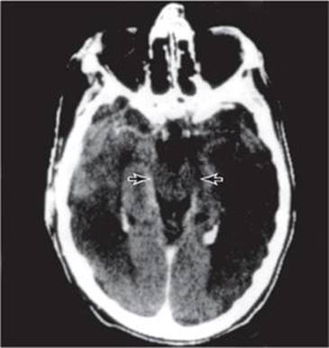
Figure 3-7. CT brain scan (contrast-enhanced) of a patient with bilateral middle cerebral artery infarcts who is in a persistent vegetative state. The reticular activating system in the intact midbrain (arrows) allows wakefulness, but the bihemispheric lesions preclude awareness.
LOCKED-IN SYNDROME
Because the portion of the reticular formation responsible for consciousness lies above the level of the midpons, functional transection of the brainstem below this level—by pontine infarction (Figure 3-8), hemorrhage, central pontine myelinolysis, tumor, or encephalitis—can interrupt descending neural pathways to produce an akinetic and mute state, with preserved consciousness. Such patients appear comatose but are awake and alert although mute and quadriplegic. Decerebrate posturing or flexor spasms may be seen. The diagnosis is made by noting that voluntary eye opening, vertical eye movements, ocular convergence, or some combination of these volitional midbrain-mediated movements is preserved. During the examination of any apparently comatose patient, the patient should be told to “open your eyes,” “look up,” “look down,” and “look at the tip of your nose” to elicit such movements. The EEG is normal. Outcome is variable and related to the underlying cause and the extent of the brainstem lesion. Mortality, usually from pneumonia, is approximately 70% when the cause is a vascular disturbance and approximately 40% in nonvascular cases. Survivors may recover partially or completely over a period of weeks to months.

Figure 3-8. CT brain scan (contrast-enhanced) of a man with basilar artery occlusion who exhibits the “locked-in” syndrome. The pontine infarction (arrows) is below the level of the reticular activating system, allowing wakefulness, but the bilateral descending motor tracts have been transected.
BRAIN DEATH
Current standards for the determination of brain death, developed by the President’s Commission for the Study of Ethical Problems in Medicine and Biomedical and Behavioral Research (1981), are summarized here. Irreversible cessation of all brain function is required for a diagnosis of brain death. The diagnosis of brain death in children younger than 5 years must be made with caution.
CESSATION OF BRAIN FUNCTION
Unresponsiveness
The patient must be unresponsive to sensory input, including pain and speech.
Absent Brainstem Reflexes
Pupillary, corneal, and oropharyngeal responses are absent, and attempts to elicit eye movements with doll’s-head and cold-water caloric testing are unsuccessful. Respiratory responses are also absent, with no ventilatory effort after the patient’s PCO2 is permitted to rise to 60 mm Hg for maximal ventilatory stimulation, while oxygenation is maintained by giving 100% oxygen by a cannula inserted into the endotracheal tube (apnea test).
IRREVERSIBILITY OF BRAIN DYSFUNCTION
The cause of coma must be known, it must be adequate to explain the clinical picture, and it must be irreversible. Sedative drug intoxication, hypothermia (<32.2°C [90°F]), neuromuscular blockade, and shock must be ruled out, because these conditions can produce a clinical picture that resembles brain death, but in which neurologic recovery may still be possible.
PERSISTENCE OF BRAIN DYSFUNCTION
The criteria for brain death described in the preceding section must persist for an appropriate length of time, as follows:
1. Six hours with a confirmatory isoelectric (flat) EEG, performed according to the technical standards of the American Electroencephalographic Society.
2. Twelve hours without a confirmatory isoelectric EEG.
3. Twenty-four hours for anoxic brain injury without a confirmatory isoelectric EEG.
ADDITIONAL CONFIRMATORY TESTS
Demonstrating the absence of cerebral blood flow confirms brain death without a waiting period. Cerebral angiography provides the most unequivocal assessment, although Doppler techniques and technetium imaging are used in some centers.
REFERENCES
General
Fisher CM. The neurological examination of the comatose patient. Acta Neurol Scand. 1969;45(suppl 36):1-56.
Posner JB, Saper CB, Schiff N, Plum F. Plum and Posner’s Diagnosis of Stupor and Coma (Contemporary Neurology Series, 71). 4th ed. Oxford, UK: Oxford University Press; 2007.
Weiss N, Galanaud D, Carpentier A, Naccache L, Puybasset L. Clinical review: prognostic value of magnetic resonance imaging in acute brain injury and coma. Crit Care. 2007;11:230.
Young GB, Wijdicks EF, Eds. Disorders of Consciousness. Handbook of Clinical Neurology, Vol 90 (Series Eds: Aminoff, Boller, and Swaab). New York, NY: Elsevier; 2008.
Emergency Management
Ngo AS, Anthony CR, Samuel M, Wong E, Ponampalam R. Should a benzodiazepine antagonist be used in unconscious patients presenting to the emergency department? Resuscitation. 2007;74:27-37.
History and Examination
Mantur M, Łukaszewicz-Zajac M, Mroczko B, et al. Cerebrospinal fluid leakage: reliable diagnostic methods. Clin Chim Acta. 2011;412:837-840.
Structural Lesions
Baird TA, Muir KW, Bone I. Basilar artery occlusion. Neurocrit Care. 2004;1:319-329.
Caplan LR. Cerebellar infarcts: key features. Rev Neurol Dis. 2005;2:51-60.
Honda H, Warren DK. Central nervous system infections: meningitis and brain abscess. Infect Dis Clin North Am. 2009;23: 609-623.
Parvizi J, Damasio AR. Neuroanatomical correlates of brainstem coma. Brain. 2003;126:1524-1536.
Qureshi AI, Mendelow AD, Hanley DF. Intracerebral haemorrhage, Lancet. 2009;373:1632-1644.
Rabinstein AA, Tisch SH, McClelland RL, Wijdicks EF. Cause is the main predictor of outcome in patients with pontine hemorrhage. Cerebrovasc Dis. 2004;17:66-71.
Schneck MJ, Maheswaran M, Leurgans S. Predictors of outcomes after nontraumatic subdural hematoma. J Stroke Cerebrovasc Dis. 2004;13:192-195.
Weil AG, Rahme R, Moumdjian R, Bouthillier A, Bojanowski MW. Quality of life following hemicraniectomy for malignant MCA territory infarction. Can J Neurol Sci. 2011;38:434-438.
Diffuse Encephalopathies
Auer RN. Hypoglycemic brain damage. Metab Brain Dis. 2004;19:169-175.
Bouchama A, Knochel JP. Heat stroke. N Engl J Med. 2002;346: 1978-1988.
Buylaert WA. Coma induced by intoxication. Acta Neurol Belg. 2000;100:221-224.
Fischbeck KH, Simon RP. Neurological manifestations of accidental hypothermia. Ann Neurol. 1981;10:384-387.
Fugate JE, Wijdicks EF, Mandrekar J, et al. Predictors of neurologic outcome in hypothermia after cardiac arrest. Ann Neurol. 2010;68:907-914.
Hart SP, Frier BM. Causes, management and morbidity of acute hypoglycaemia in adults requiring hospital admission. Q J Med. 1998;91:505-510.
Holzer M. Targeted temperature management for comatose survivors of cardiac arrest. N Engl J Med. 2010;363:1256-1264.
Stevens RD, Pronovost PJ. The spectrum of encephalopathy in critical illness. Semin Neurol. 2006;26:440-451.
Wijdicks EF, Hijdra A, Young GB, Bassetti CL, Wiebe S; Quality Standards Subcommittee of the American Academy of Neurology. Practice parameter: prediction of outcome in comatose survivors after cardiopulmonary resuscitation (an evidence-based review): report of the Quality Standards Subcommittee of the American Academy of Neurology. Neurology. 2006;67:203-210.
Persistent Vegetative State
Bernat JL. Chronic disorders of consciousness. Lancet. 2006;367(9517):1181-1192. Erratum in: Lancet. 2006; 367(9528):2060.
Estraneo A, Moretta P, Loreto V, Lanzillo B, Santoro L, Trojano L. Late recovery after traumatic, anoxic, or hemorrhagic long-lasting vegetative state. Neurology. 2010;75:239-245.
Luauté J, Maucort-Boulch D, Tell L, et al. Long-term outcomes of chronic minimally conscious and vegetative states. Neurology. 2010;75:246-252.
Monti MM, Vanhaudenhuyse A, Coleman MR, et al. Willful modulation of brain activity in disorders of consciousness. N Engl J Med. 2010;362:579-589.
Locked-In Syndrome
Leon-Carrion J, van Eeckhout P, Domínguez-Morales Mdel R, Pérez-Santamaría FJ. The locked-in syndrome: a syndrome looking for a therapy. Brain Inj. 2002;16:571-582.
Smith E, Delargy M. Locked-in syndrome. BMJ. 2005;330: 406-409.
Brain Death
Drazkowski J. Determining brain death: back to the basics. Semin Neurol. 2007;27:393-399.
Saposnik G, Basile VS, Young GB. Movements in brain death: a systematic review. Can J Neurol Sci. 2009;36:154-160.
Wijdicks EF. The case against confirmatory tests for determining brain death in adults. Neurology. 2010;75:77-83.
Wijdicks EF, Varelas PN, Gronseth GS, Greer DM; American Academy of Neurology. Evidence-based guideline update: determining brain death in adults: report of the Quality Standards Subcommittee of the American Academy of Neurology. Neurology. 2010;74:1911-1918.