
Introduction
Fittingly, a book on localization in clinical neurology should begin with a chapter explaining what the term localization means. Localization derives from the Latin term locus or site. Localization is the diagnostic exercise of determining from the signs (most often) or symptoms of the patient what site of the nervous system has been affected by a disease process. Important injury to the nervous system results in abnormal function, be it behavioral, motor, or sensory. Characteristics of the dysfunction often pave the way for a topographic (from the Greek term topos or place) diagnosis. Localization and topographic diagnosis refer to the same thing: the determination of where in the nervous system the damage has occurred.
Even in the age of sophisticated neurophysiology, neuroimaging, and molecular biology, the clinical diagnosis should precede the use of these other techniques if their full diagnostic potential is to be realized. Clinical localization has particular relevance to the adequate use of ancillary procedures. For instance, false-positive findings from “gunshot approach” neuroimaging can only be avoided by careful localization. As an example, congenital brain cysts, strikingly visible on imaging procedures, are often wrongly blamed for a variety of neurologic disorders, while the actual disease remains overlooked and untreated. The thoughtful use of ancillary procedures in neurology, guided by clinical localization, minimizes discomfort for patients and the waste of resources.
A Brief History of Localization: Aphasia as an Example
The history of localization is the history of early neurology, concerned with topographic diagnosis that would eventually lead to therapy. In few areas of neurology was the development of localization as interesting and so much at the center of famous controversies as it was in the case of aphasia. In fact, the oldest known document on neurologic localization concerns aphasia. It was recorded in an Egyptian papyrus from the Age of the Pyramids (about 3000–2500 BC), where an Egyptian surgeon described the behavior of an aphasic individual:
If thou examinest a man having a wound in his temple, penetrating to the bone, (and) perforating his temporal bone; … if thou ask of him concerning his malady and he speak not to thee; while copious tears fall from both his eyes, so that he thrusts his hand often to his face so that he may wipe both his eyes with the back of his hand … Edwin Smith surgical papyrus, Case 20, 2800 BC [12].
From the time of Hippocrates, in ancient Greece, it was documented that injury to the left part of the brain resulted in weakness of the right side of the body. However, paired organs in the body were thought to have identical functions. In the mid-19th century, Paul Broca (1824–1880) revolutionized the then current understanding of the functional organization of paired organs by describing lateralization of language to the left hemisphere [5,13]. He called aphemia the disorder that we now call Broca’s aphasia. In his 1865 paper, he wrote:
Now, this function of the intellectual order, which controls the dynamic element as well as the mechanical element of articulation, seems to be the nearly constant privilege of the left hemisphere convolutions, since lesions that result in aphemia are almost always localized in that hemisphere … That is tantamount to saying that we are left-brained with regard to language. Just as we control movements in writing, drawing, embroidering, etc, with the left hemisphere, so we speak with the left hemisphere.
Broca defined the inferior frontal gyrus as the area that, when injured, would lead to aphemia [13]. He also noted the variation in the expression of diverse lesions in the inferior frontal gyrus, characteristic of the plasticity found in cortical organization:
During the course of our study of brains of patients with aphemia, many times before, we had determined that the lesion of the third left frontal convolution was not always in direct relation to the intensity and the impairment of language. For example, we had observed that speech was completely wiped out as a result of a lesion with the size of 8 to 10 mm, whereas, in other cases, lesions that were tenfold more extensive had only partly impaired the capacity for articulate speech.
In the few years after Broca’s remarkable statements, knowledge about the localization of the language centers in the brain grew rapidly. Already in 1874, Carl Wernicke (1848–1905) wrote:
The whole area of convolution encircling the Sylvian fissure, in association with the cortex of the insula, serves as a speech center. The first frontal gyrus, being motor, is the center for representation of movement, and the first temporal gyrus, being sensory, is the center for word images … The first temporal gyrus should be considered as the central end of the auditory nerve, and the first frontal gyrus (including Broca’s area) as the central end of the nerves to the speech muscles … Aphasia can result from any interruption of this path …
Knowledge of the cortical organization for language had been derived from careful clinicopathologic correlation [67]. After describing a 73-year-old woman with the sudden onset of confused speech, Wernicke goes on to describe the pathologic findings:
The branch of the artery of the left Sylvian fissure, running down into the inferior sulcus of Burdach, was occluded by a thrombus tightly adherent to the wall. The entire first temporal gyrus, including its junction with the second temporal gyrus and the origin of the latter from Bischof’s inferior parietal lobule were converted into a yellowish-white brei [67,68].
Wernicke’s diagram of the language areas is illustrated in Figure 1.1.
Current techniques, such as functional brain mapping, promise to clarify further the localization of mechanisms underlying neurologic dysfunction. For instance, conduction aphasia, initially described by Wernicke in 1874, has traditionally been associated with damage of the arcuate fasciculus, purportedly connecting Wernicke’s with Broca’s area. Recent neurophysiological and neuroimaging findings, obtained with the use of diffusion tensor imaging and other functional magnetic resonance imaging (MRI) techniques, are challenging this notion [6].
Clinical Diagnosis and Lesion Localization
Clinical diagnosis in neurology requires several steps:
1. Recognition of impaired function
2. Identification of what site of the nervous system has been affected, that is, localization
3. Definition of the most likely etiology, often resulting in a differential diagnostic list
4. Use of ancillary procedures to determine which of the different possible etiologies is present in the given patient

FIG. 1.1. Wernicke’s diagram of the language areas. In the original, the label on the superior temporal gyrus was simply a, but from the context, it should have been a1. Wernicke’s explanation of this figure is as follows:
Let F be the frontal, O the occipital, and T the temporal end of a schematically drawn brain. C is the central fissure; around the Sylvian fissure (S) extends the first primitive convolution. Within this convolution, ai is the central end of the acoustic nerve, a its site of entry into the medulla oblongata; bdesignates the representation of movements governing sound production, and is connected with the preceding through the association fibers ai b running in the cortex of the insula. From b the efferent pathways of the sound-producing motor nerves run to the oblongata and exit there …
(From: Wernicke C, Der aphasische symptomencomplex; eine psychologische studie auf anatomischer basis, Breslau: Max Cohn & Weigert, 1874 [67].)
Each of these steps is important. The first one, recognition of impaired function, depends on a good history and neurologic examination. Only by storing the range of normal neurologic functions in their mind can physicians recognize an abnormal neurologic function. Inexperience or carelessness in examining a patient often results in overlooking a neurologic deficit and therefore missing a diagnosis. For instance, mild chorea may appear to the inexperienced as normal fidgetiness. The slow eye movements of a pontocerebellar disorder may pass completely unrecognized by someone who looks only for a full excursion of the eyes.
Abnormal neurologic findings come in the form of abnormal behavior, impaired posture or gait, difficulty with movements of the face or extremities, and, finally, sensory disturbances, including pain. Pain exemplifies well several of the difficulties physicians face when confronting possible neurologic dysfunction.
1. First, is the dysfunction real? Is the pain really there or is the patient trying to deceive? We have witnessed the plight of a paraplegic patient who had been repeatedly asked by health care personnel to stop pretending not to be able to move his legs. They had misinterpreted the triple flexion response witnessed when they pulled the sheets off the patient’s legs as evidence of volitional movement. Movement disorders, such as the dystonias, were frequently considered psychogenic in the past and have gradually emerged from this realm into a phase of general recognition of their “organicity.” Unless accompanied by clear psychiatric manifestations, neurologic symptoms or signs should be taken at face value.
2. Second, to what extent is the dysfunction pathologic, that is, indicative of injury serious enough to warrant a formal diagnostic workup? Many aches and pains do not reflect serious disease. Sending everyone with a “little pain” to a physician would hopelessly clog up the health care system. Interestingly, the child learns from falls and other minor injuries what to expect as “normal pain,” and when a person seeks medical attention for any symptom, the likelihood is that the problem is serious enough to warrant at least a thoughtful physical examination.
3. Third, is the dysfunction neurologic in origin? Is the pain due to injury of the affected body part or neurologic dysfunction? Is the dysfunction a manifestation of a disease of the nervous system rather than of the organ mediating the function? Is the patient unable to walk because of arthritis or because the motor system is affected? All these questions find an answer when the physician recognizes patterns that belie neurologic impairment, for instance, in the case of pain, a characteristic radicular nature and distribution. In other cases, the neurologic examination may reveal other manifestations of unquestionable neurologic dysfunction. A patient with pain in the hand may also have atrophy of the muscles in the thenar eminence and a Tinel’s sign—pain on percussion of the median nerve at the wrist. Knowledge of localization tells us that the pain derives from injury of the median nerve at the point where the pain increases on percussion. What is needed to localize the lesion, in this case as in any other, is a good working knowledge of neuroanatomy.
Neuroanatomy is a key to localization. In this book, a synopsis of the anatomy of each structure of the nervous system precedes the discussion on localization of lesions of that structure. Neuroanatomy has two broad aspects: the morphology of the structure and its “functional representation.” Functional representation refers to the function mediated by a given structure of the nervous system. Damage to the structure alters the function mediated by this structure. For example, an injury to the oculomotor nerve results in mydriasis in the eye supplied by this nerve.
Neuroanatomy provides the road map for localization. Localizing is identifying the site of injury on the neuroanatomic map. As with any other map, we need either an address, with street name and number, or the intersection between two well-defined streets or roads. Injury expresses itself through neurologic dysfunction, be it behavioral, motor, or sensory. If we know what kind of dysfunction can result from injury of the different parts of the nervous system, we will be able to identify the source of the injury. Some types of dysfunction directly give us the address we are looking for. A combination of resting tremor, bradykinesia, and rigidity tells us that the substantia nigra of the patient has been injured. At other times, we use the approach of looking for the intersection between two streets. From some signs we deduce that a particular pathway must be affected. From others, we infer that a second pathway is affected as well. The injury must be in the place where these pathways meet. For instance, by the presence of left-sided hemiparesis we infer that the corticospinal tract has been affected. But the corticospinal tract can be affected at the level of the spinal cord, brainstem, or cerebral hemispheres. To precisely identify the location of damage we need to use other clues. If, in addition to the left-sided hemiparesis, we find a right third nerve palsy, we are well on our way to localizing the lesion. This well-known syndrome, named after Weber, typifies a general principle of localization: the lesion is where the two affected pathways cross. If the patient only had a third nerve palsy, the lesion could be anywhere between the fascicle of the nerve (in the brainstem) and the superior orbital foramen (in the orbit). The addition of a contralateral hemiparesis precisely defines that the lesion affects the crus cerebri on the same side of the third nerve palsy. This is where the corticospinal tract and the fibers of the third nerve meet. Neuroanatomy provides the roadmap for a correct assessment.
Localization tends to be more precise when the lesion affects the lower levels of the nervous system. When we localize lesions of the nervous system, it is helpful to think about the major syndromes that result from lesions at different functional and anatomic levels, from the muscle to the cortex. At the simplest level, injury to a muscle impairs the movement mediated by that muscle. One level higher, we find that injury to a peripheral nerve causes weakness of the muscles innervated by that nerve and sensory loss in its cutaneous distribution. Lesions in the spinal cord below the low cervical level cause weakness of one or both legs and sensory loss that often has a horizontal level in the trunk. Lesions in the cervical cord or brainstem typically cause weakness or sensory loss on one or both sides of the body, often more severe on one side, and findings characteristic of the level affected. For instance, lesions of the cervical cord may cause radicular pain or weakness affecting the arms or hands. Lesions of the lower pons give rise to gaze palsies or peripheral facial weakness. The localization of lesions in the cranial nerves (CNs) is fairly straightforward because they may affect a peripheral nerve or a neuroanatomic structure that is relatively simple, such as the visual pathways. As we ascend the neuraxis, the localization of lesions becomes less precise. Lesions in the cerebellum may cause ataxia. Lesions in the thalamus often, but not always, cause sensory loss and postural disorders, or memory loss. Lesions in the hemispheric white matter may give rise to weakness or visual field defects. Finally, lesions in the cortex manifest themselves by an array of motor, sensory, or behavioral findings that vary according to the area that has been injured.
Similarly, lesions of the lower levels tend to cause findings that change little over time, whereas lesions of the higher levels may be very “inconsistent” in the course of an examination. An ulnar nerve lesion may be responsible for atrophy of the first dorsal interosseous muscle. The atrophy diagnosed by the examiner will be consistent. By contrast, a patient with a Broca’s aphasia may have a great deal of difficulty repeating some words, but not others of apparently similar difficulty. The examiner may be puzzled and not know what to document: can the patient repeat or can she not? In this case, what should be noted is not whether the patient can do something, but whether she does it consistently in a normal way. Any difficulty repeating a sentence on the part of a native speaker of a language should be considered as abnormal. Higher neurologic function should be sampled enough to avoid missing a deficit that the more complex neural networks of higher levels can easily mask.
For the anatomic localization of lesions, the neurologic examination is much more important than the history. It must be noted that when we speak here about “examination,” we include the sensory findings reported by the patient during the examination. A complaint of pain or of numbness is usually as “objective” as a wrist drop. By tracking back the pathways that mediate the functions that we find are impaired in the neurologic examination, we can generally localize the site of the lesion, even without a history. The history, that is, the temporal evolution of the deficits witnessed in the neurologic examination, is important in defining the precise etiology. For instance, a left-sided hemiparesis is detected in the neurologic examination. If it occurred in a matter of minutes, cerebrovascular disease or epilepsy is most likely. If it evolved over a few days, we should think about an infection or demyelinating disease. If it developed insidiously, in a matter of months, a tumor or a degenerative process is more likely. In all of these cases, the localization is derived from the findings of the examination: we detect a left-sided hemiparesis. If we also find a right third nerve palsy and determine that it has appeared at the same time as the hemiparesis, we will emphasize the need for a careful look at the midbrain when we obtain an MRI. In this sense, the history is also important for localization: we may witness in the examination the end result of multiple lesions that affected the nervous system over time. In the previous example, if the third nerve palsy occurred when the patient was 10 years old and the hemiparesis appeared when he was in his sixties, the lesion responsible for the hemiparesis would probably not be in the midbrain.
Finally, there is the issue of discrete lesions versus system lesions. Much of the work on localization has been done on the basis of discrete lesions, such as an infarct affecting all the structures in the right side of the pons. Some types of pathologies tend to cause this type of lesion. Cerebrovascular disease is the most common, but demyelinating lesions, infections, trauma, and tumors also often behave like discrete, single, or multiple lesions. Other neurologic disorders affect arrays of neurons, often responsible for a functional system. Parkinson’s disease is an example. Here, the localization to the substantia nigra is simple. The localization of other degenerative disorders, such as the spinocerebellar degeneration of abetalipoproteinemia or vitamin E deficiency, is more complicated [56]. Here, the clinical syndrome seems to point to the spinal cord, but the real damage is inflicted to the large neurons in the sensory nuclei of the medulla, dorsal root ganglia, and Betz cells. The puzzle is resolved when one realizes that the destruction of the corticospinal tract logically follows metabolic injury to the neurons that give rise to it. The larger neurons, with the longest axons reaching the lumbar segments, are affected first. The neuron may not die, but, incapable of keeping an active metabolism, it begins to retract its axon (dying-back phenomenon). Likewise, the lesion in the dorsal columns of the spinal cord (and peripheral nerve) simply reflects the damage inflicted to the larger sensory neurons by the lack of vitamin E. Therefore, a precise knowledge of the functional significance of the different structures of the neuraxis facilitates the localization of degenerative or system lesions as much as it helps with discrete lesions.
Having reviewed some general principles of localization in the nervous system, we will now review in more detail the principles of localization in the motor and sensory systems. Finally, we will review the localization of gait disorders.
Localization of Lesions of the Motor System
Anatomy of the Motor System
The motor neurons of the ventral horn of the spinal cord and the motor nuclei in the brainstem, whose axons synapse directly on striated muscles, are the “final common pathway” for muscle control. These large alpha (a) motor neurons supply the extrafusal fibers of the skeletal muscles providing the only axons to skeletal muscle. Scattered among the a motor neurons are many small gamma (g) motor neurons, which supply the intrafusal fibers of the muscle spindles. These muscle spindles are the receptors for the muscle stretch reflexes. The motor neuron, together with its axon, and all the muscle fibers it supplies, is called the motor unit. The junction between the terminal branches of the axon and the muscle fiber is called the neuromuscular junction [23,49]. There is a somatotopic organization of the cell columns of anterior horn cells in the ventral gray horn of the spinal cord. Neurons that supply the axial muscles, including the neck muscles, are located in ventromedially placed columns; neurons that supply proximal muscles are situated in the midregion; and neurons supplying the musculature of the distal aspect of the limbs are located in laterally placed columns [23,49].
Motor neuronal cell groups receive input from the contralateral motor cortex (MC) through the descending corticospinal and corticobulbar tracts (Figs. 1.2A and B). The corticospinal tract contains on each side approximately 1 million fibers of various sizes, but only 3% of all the fibers originate from the giant pyramidal cells of Betz found in layer V in the primary MC. All corticospinal fibers are excitatory and appear to use glutamate as their neurotransmitter. The neurons from which the corticospinal and corticobulbar tracts arise are known as upper motor neurons [2,4,23,31,66]. The corticospinal pathway, which controls voluntary, discrete, highly skilled movements of the distal portion of the limbs, arises from somatotopically organized areas of the primary MC, lateral premotor cortex (PMC), and supplementary motor area (SMA). These fibers arise from both precentral (60%) and postcentral (40%) cortical areas. The corticospinal neurons are found primarily in Brodmann’s area 4 (40%), which occupies the posterior portion of the precentral gyrus (primary MC). The lateral PMC, on the lateral aspect of the frontal lobe, and the SMA, on its medial part, are located in Brodmann’s area 6 (20%). Corticospinal axons also arise from neurons in the primary sensory cortex in the postcentral gyrus (Brodmann’s areas 3, 1, and 2), particularly from area 3a, anterior paracentral gyri; superior parietal lobule (Brodmann’s areas 5 and 7); and portions of the cingulate gyrus on the medial surface of the hemisphere. Fibers of the corticospinal system descend in the corona radiata, the posterior limb of the internal capsule, the middle three-fifths of the cerebral peduncle, the basis pontis (where the tract is broken into many bundles by the transverse pontocerebellar fibers), and the medullary pyramids.
In the caudal end of the medulla, nearly 75% to 90% of the corticospinal fibers in the pyramid cross the ventral midline (pyramidal decussation or Mistichelli crossing) before gathering on the opposite side of the spinal cord as the lateral corticospinal tract. In the posterior limb of the internal capsule, the corticospinal tract is organized somatotopically, with hand fibers lateral and slightly anterior to foot fibers [32]. The corticospinal fibers also follow a somatotopic organization in the pons. Fibers controlling the proximal muscles are placed more dorsal than those controlling the more distal muscle groups. Because of the ventral location of the pyramidal tract in the pons, a pure motor hemiparesis of brainstem origin is usually observed with pontine lesions. Unilateral motor deficits may predominantly involve the upper or lower limb, but a difference in the pontine lesion location among these patterns of weakness distribution is not observed [43]. There is also a somatotopic organization of the corticospinal fibers within the medullary pyramids, with fibers of the lower extremities placed more laterally and decussating more rostrally than those of the upper extremities [24]. The remaining fibers that do not decussate in the medulla descend in the ipsilateral ventral funiculus as the ventral or anterior corticospinal tract (Türck’s bundle). Most of these fibers ultimately decussate at lower spinal cord levels as they further descend in the anterior column of the spinal cord. Therefore, only approximately 2% of the descending corticospinal fibers remain truly ipsilateral, forming the bundle of Barnes [2]. These ipsilateral descending projections control the axial musculature of the trunk and proximal limbs.


FIG. 1.2. A simplified diagram of the motor system. A: Corticospinal tract. B: Corticobulbar tract.
The corticobulbar fibers, originating in the lower third of the cortical motor fields, especially the MC and SMA, descend in the genu of the internal capsule, the medial part of the cerebral peduncle, and the basis pontis, where they are intermixed with corticospinal fibers. The corticobulbar pathway has bilateral input to the nuclei of the trigeminal and hypoglossal CNs, as well as the facial nucleus supplying the upper facial muscles. Traditional localization concepts postulate that ventral brainstem lesions rostral to the lower pons result in contralateral central facial paresis, whereas lesions of the lower dorsolateral pons result in ipsilateral facial paresis of the peripheral type. However, an aberrant fiber bundle branching off the main pyramidal tract at the midbrain and upper pons, along the tegmentum in a paralemniscal position, has been described. Therefore, whereas the muscles of the lower face receive predominantly crossed corticobulbar input, the muscles of the upper face are represented in the ipsilateral, as well as the contralateral, hemisphere, with transcranial magnetic stimulation (TMS) studies showing that the amount of uncrossed pyramidal projections are no different from the muscles of the upper than those of the lower face [21,61]. TMS studies in patients with and without central facial paresis due to brainstem lesions have also shown that a supranuclear facial paresis may be contralateral to a lesion of the cerebral peduncle, pontine base, aberrant bundle or ventral medulla, or ipsilateral to a lateral medullary lesion [64].
The ventral part of the facial nucleus, innervating the lower two-thirds of the face, has a predominantly crossed supranuclear control. This schema of supranuclear facial muscle control holds true for voluntary facial movements. Emotional involuntary movements and voluntary facial movements may be clinically dissociated, and therefore, a separate supranuclear pathway for the control of involuntary movements probably exists. A prevailing view is that the SMA and/or cingulate motor areas are critical for emotional facial innervation [35]. Fibers mediating emotional facial movements do not descend in the internal capsule in their course to the facial motor nuclei. The right cerebral hemisphere is also involved in supranuclear emotional facial movement control and is “dominant” for the expression of facial emotion [10]. Furthermore, some of the facial corticobulbar fibers seem to descend ipsilaterally before making a loop as low as the medulla and decussating and ascending to the contralateral facial nucleus (located dorsolaterally in the caudal pons) that innervates the perioral facial musculature [17,62]. This anatomic understanding explains the emotional facial paresis of pontine origin resulting from the involvement of the dorsal lateral pontine area [33].
Within the MC, corticospinal neurons are somatotopically organized in patterns that reflect their functional importance (motor homunculus). The size of the cortical representation in the motor homunculus varies with the functional importance of the part represented; therefore, the lips, jaw, thumb, and index finger have a large representation, whereas the forehead, trunk, and proximal portions of the limbs have a small one. As an example, isolated hand weakness of cortical origin may present with loss of thumb and finger movements and impaired hand flexion and extension or with partial involvement of a few digits (pseudoradicular pattern). This cortical motor hand area has been localized in the middle to lower portion of the anterior wall of the central sulcus (Brodmann’s area 4), adjacent to the primary sensory cortex of the hand (Brodmann’s areas 3a and 3b) [60]. Neurons in the medial aspect of the MC and the anterior paracentral gyrus influence motor neurons innervating the muscles of the foot, leg, and thigh. Neurons in the medial two-thirds of the precentral gyrus influence motor neurons innervating the upper extremity and trunk. Neurons in the ventrolateral part of the precentral gyrus contribute to the corticobulbar tract and project to motor nuclei of the trigeminal (CN V), facial (CN VII), glossopharyngeal (CN IX), vagus (CN X), accessory (CN XI), and hypoglossal (CN XII) nerves to influence the cranio–facial–oral musculature [4,31]. As an example, each hypoglossal nucleus receives impulses from both sides of the cerebral cortex, except for the genioglossus muscle that has probably crossed unilateral innervation. Therefore, a lingual paresis may occur with lesions at different anatomic levels including the medulla, hypoglossal foramen, cervical (neck) region, anterior operculum, and posterior limb of the internal capsule [26].
Sensory cortical pathways (e.g., thalamocortical connections), corticofugal projections to reticulospinal and vestibulospinal tracts, direct corticospinal projections to the spinal cord, and projections to the basal ganglia and cerebellum have an active role in the planning and execution of movements. The cerebellum and basal ganglia are critically important for motor function [2,4,66]. The cerebellum has a major role in the coordination of movements and control of equilibrium and muscle tone. The cerebellum controls the ipsilateral limbs through connections with the spinal cord, brainstem, and contralateral MC through the thalamus. A corticofugal pathway of major clinical importance is the corticopontine pathway, which arises primarily from the precentral and postcentral gyri, with substantial contributions from the PMC, SMA, and posterior parietal cortices, and few from the prefrontal and temporal cortices. These fibers descend in the anterior limb of the internal capsule and the medial fifth of the cerebral peduncle before reaching the basis pontis, where they project to pontine nuclei. Second-order neurons from pontine nuclei cross to the contralateral basis pontis and give rise to the pontocerebellar pathway.
The basal ganglia play a major role in the control of posture and movement and participate in motor planning through reciprocal connections with ipsilateral MC. The corticostriate pathway includes direct and indirect projections from the cerebral cortex to the striatum. Corticostriate projections arise mainly from motor–sensory cortex (Brodmann’s areas 4 and 3, 1, and 2), PMC (Brodmann’s area 6), and frontal eye fields (Brodmann’s area 8). Direct corticostriate projections reach the striatum through the internal and external capsules and the subcallosal fasciculus. The indirect pathways include the cortico–thalamo–striate pathway, collaterals of the corticoolivary pathway, and collaterals of the corticopontine pathway. All parts of the cerebral cortex give rise to efferent fibers to the caudate and putamen. Cortical association areas project mainly to the caudate nucleus, whereas sensorimotor areas project preferentially to the putamen. These corticostriate projections mainly terminate ipsilaterally in a topographic pattern (e.g., the frontal cortex projects fibers to the ventral head of the caudate and rostral putamen). The cortex also sends fibers to the substantia nigra, subthalamic nucleus, and claustrum.
Another corticofugal tract of major clinical importance is the corticothalamic pathway. This pathway arises from cortical areas receiving thalamic projections and, therefore, serves as a feedback mechanism from the cortex to the thalamic nuclei. Except for the reticular nucleus of the thalamus, examples of such reciprocal connections include the anterior nucleus and the posterior cingulate cortex, the ventral lateral nucleus and the MC, the ventral anterior nucleus and the SMA, the ventral posterior nucleus and the primary sensory cortex, the lateral geniculate body and the primary visual cortex, the medial geniculate body and the primary auditory cortex, and the dorsomedial nucleus and the prefrontal cortex. Corticothalamic fibers descend in various parts of the internal capsule and enter the thalamus in a bundle known as the thalamic radiation.
Additional corticofugal tracts include the corticoreticular pathway, which arises from one cerebral hemisphere, descends in the genu of the internal capsule, and projects to both sides of the brainstem reticular formation, and the highly integrated corticohypothalamic tract, which arises from the prefrontal cortex, cingulate gyrus, amygdala, olfactory cortex, hippocampus, and septal area. Corticofugal areas from the frontal eye fields (Brodmann’s area 8) and the middle frontal gyrus (Brodmann’s area 46) project to the superior colliculus and centers in the brainstem reticular formation that influence the motor nuclei of the oculomotor (CN III), trochlear (CN IV), and abducens (CN VI) nerves [7].
The internal capsule, a compact lamina of white matter, contains afferent and efferent nerve fibers passing to and from the brainstem to the cerebral hemispheres, that is, continuous rostrally with the corona radiata and caudally with the cerebral peduncles. Located medially between the caudate nucleus and the thalamus, and laterally in the lenticular nucleus (globus pallidus and putamen), in a horizontal (Flechsig) section, the internal capsule is somewhat curved with its convexity inward. The prominence of the curve (genu) projects between the caudate nucleus and the thalamus. The portion in front of the genu is called the anterior limb, which measures approximately 2 cm in length and separates the lenticular nucleus from the caudate nucleus (lenticulocaudate segment of the internal capsule). The portion behind the genu is the posterior limb, which measures 3 to 4 cm in length and separates the lenticular nucleus from the thalamus (lenticulothalamic segment). The internal capsule extends further to include sublenticular and retrolenticular segments.
The anterior limb of the internal capsule contains frontopontine fibers, and thalamocortical and corticothalamic fibers (reciprocally connecting the frontal lobe to the thalamus), as well as caudatoputaminal fibers. Corticobulbar fibers, and perhaps motor corticopontine fibers, occupy the genu of the internal capsule. This fiber arrangement explains the facial and lingual hemiparesis with mild limb involvement observed in the capsular genu syndrome [9]. In the caudal half of the posterior limb of the internal capsule, the corticospinal bundle is somatotopically organized in such a way that the fibers to the upper extremity are located more anteriorly (i.e., shoulder, elbow, wrist, and fingers), followed by fibers to the trunk and then by the fibers to the lower extremity (i.e., hip, knee, ankle, toes), bladder, and rectum. As the corticospinal tract descends through the internal capsule, its fibers intermix with other fiber systems including corticorubral, corticoreticular, and corticopontine fibers. Corticorubral, corticothalamic, and thalamocortical fibers (carrying sensory tracts from the thalamus to the parietal lobes) are also located dorsal to the corticospinal fibers, in the posterior limb of the internal capsule. Finally, the sublenticular segment of the internal capsule contains the auditory and visual radiations, while the retrolenticular segment contains the visual radiations of Gratiolet’s radiating fibers and corticotectal, corticonigral, and corticotegmental fibers. The anterior limb of the internal capsule receives its vascular supply from the artery of Heubner, a branch of the anterior cerebral artery; the genu and the middle and inferior part of the posterior limb receive their blood supply from the anterior choroidal artery; while the superior aspect of the anterior and posterior limb of the internal capsule receives their blood supply from the lenticulostriates, branches of the middle cerebral artery.
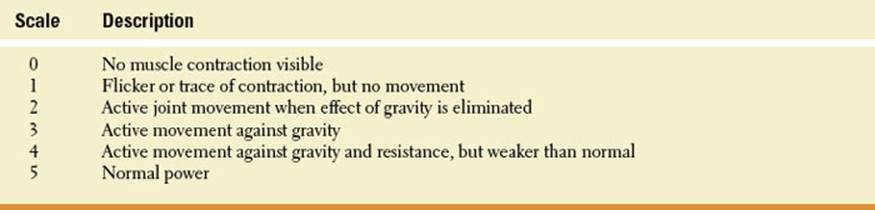
TABLE 1.1 Medical Research Council’s Scale for Assessment of Muscle Power
Motor Signs and Symptoms and Their Localization
Patients with motor deficits may present with plegia or paresis. Plegia denotes complete paralysis; paresis denotes a lesser degree of weakness. However, in daily clinical parlance, the word paralysis is often used for both complete and partial loss of motor function. Muscle strength testing is graded according to the Medical Research Council’s scale for muscle power (Table 1.1), which has a good interobserver reliability. Normal grading means that the muscle is capable of holding the test position against strong pressure. Grade 4 is often subdivided into 4−, 4, and 4+ to indicate movement against slight, moderate, and strong resistance, respectively. Common patterns of weakness include monoplegia (single limb weakness), hemiplegia (loss of motor function down one side of the body), paraplegia (bilateral loss of lower limb motor function), quadriplegia or tetraplegia (loss of motor function in all four extremities), brachial diplegia (loss of motor function of both upper extremities), or facial diplegia (loss of motor function of both halves of the face). Other patterns seen in children include double hemiplegia, characterized by severe spasticity in all four extremities, which is more severe in the arms than in the legs, and cerebral diplegia, where the spastic paralysis usually affects all four extremities and involves the legs more than the arms.
When examining patients afflicted with any of these patterns of weakness, one should have three fundamental questions in mind: (a) where is the lesion? (b) Is the lesion focal, multifocal, or diffuse? and (c) What is the likely underlying cause? The first and second questions are answered by performing a focused neurologic examination; the answer to the last question requires detailed history and investigations.
Lesions in the descending motor system can be located in the cerebral cortex, internal capsule, brainstem (cerebral peduncles, pons, medulla oblongata), or spinal cord. Cortical lesions leading to spasticity involve the primary motor and premotor cortical areas. Although the upper motor neuron type of paralysis is often referred to as pyramidal syndrome, lesions accounting for this clinical picture involve more than the pyramidal tract, and therefore, the usage of this term is to be discouraged. Lesions of the lower motor neurons can be located in the cells of the ventral gray column of the spinal cord or brainstem or in the axons of these neurons.
The upper motor neuron syndrome may follow head or spinal cord injury, perinatal brain injuries, stroke, demyelinating diseases such as multiple sclerosis, or motor neuron diseases such as amyotrophic lateral sclerosis or primary lateral sclerosis. The clinical presentation of the upper motor neuron syndrome following cortical lesions is somewhat different from that of spinal cord lesions. Likewise, there may be subtle differences between incomplete and complete spinal cord lesions [57]. In general, spasticity is less severe with cerebral lesions than with spinal cord lesions. Damage to the upper motor neuronsresults in muscles that are initially weak and flaccid but eventually become spastic and exhibit hypertonia and hyperactivity of the stretch reflexes (hyperreflexia). Muscle stretch reflexes consist of a monosynaptic arc with large-diameter afferent (sensory) nerve fiber input from muscle spindle fibers and large-diameter efferent (motor) nerve fiber output from a motor neuron fibers. Clonus, characterized by a series of rhythmic contraction and relaxation of a group of muscles, is best seen at the ankle. Spasticity, a motor component of the upper motor neuron syndrome, is best characterized by a velocity-dependent increase in tonic stretch reflexes [39]. Spasticity predominates in antigravity muscles (flexors of the upper extremities and extensors of the lower extremities). Evaluation of muscle tone shows variable degree of resistance to passive movements with changes in speed and direction of passive motion and a clasp-knife character; in other words, greater resistance is felt with faster stretches. Weakness of the muscles of the upper extremity is most marked in the deltoid, triceps, wrist extensors, and finger extensors; this predilection for involvement of the extensors and supinators explains the pronation and flexion tendencies of the upper limb. In cases of spastic hemiparesis, the affected arm is adducted at the shoulder, and flexed at the wrist and fingers. Weakness of the muscles of the lower extremity is most marked in hip flexors, knee flexors, foot dorsiflexors, and foot evertors.
Different anatomic substrates may underlie hyperreflexia and spasticity; likewise, spasticity must be clearly separated from flexor spasms (see subsequent text). As an example, corticospinal lesions in the cerebral peduncle do not result in spasticity, and lesions confined to the medullary pyramid may cause weakness and hyperreflexia without spasticity [58]. The upper motor neuron syndrome is associated with the presence of pathologic reflexes and signs, such as the extensor plantar reflex or Babinski’s sign, a disinhibited flexion withdrawal reflex, characterized by dorsiflexion (extension) of the big toe often accompanied by a spreading movement of the other toes (“signe de l’eventail”). However, such response is to be considered normal until the age of 1 year. Furthermore, severe flexor or less-common extensor muscle spasms may also occur in response to a variety of nociceptive or nonnociceptive sensory stimuli, or may develop spontaneously. Flexor spasms, resembling the flexor withdrawal reflex, often consist of flexion of the hip, knee, and ankle, whereas extensor spasms often involve the extensors of the hip and knee with plantar flexion and ankle inversion. Unlike cerebral lesions, spinal cord lesions are often associated with marked flexor spasms, except for incomplete or high spinal cord lesions that usually have a dominant extensor tone. Severe flexor spasms may also be accompanied by bladder and, occasionally, fecal incontinence. Other manifestations (negative features) seen with the upper motor neuron syndrome include muscle weakness, muscle slowness, impaired dexterity, and fatigability. In addition, patients with severe spasticity may exhibit muscle deformities, contractures, and associated reactions including synkinesias [11].
Finally, the superficial reflexes (e.g., abdominal reflexes, cremasteric reflex, etc) are absent on the affected side. With lesions above the pyramidal decussation, the previously discussed signs are detected on the opposite side of the body; with lesions occurring below the pyramidal decussation, these signs are observed ipsilaterally.
When the lower motor neurons or their axons are damaged, the innervated muscles will show some combination of the following signs: weakness or paralysis of the involved muscles, flaccidity, hypotonia, diminished or absent muscle stretch reflexes (hyporeflexia or areflexia), and eventually atrophy. In the spinal muscular atrophies, weakness and amyotrophy predominate in the proximal segments of the limbs, but distal, fascioscapulohumeral, scapulohumeral, and segmental forms are well known [19]. Some patterns of discrete muscle atrophy have localizing value, as is the case of early neuropathic compromise, with involvement of the first dorsal interosseus of the hand and the extensor digitorum brevis in the feet. Fasciculations, which are visible twitches of small groups of muscle fibers, may be present. No pathologic reflexes are elicited.
The topographic diagnosis of a hemiplegia requires a structured approach to patient evaluation on the basis of localization and a basic understanding of applied neuroanatomy. When a patient presents with hemiplegia or hemiparesis, it is important to determine whether the lower half of the face is involved with relative sparing of upper facial function. Then, one must determine whether the hemiparesis is proportionate or disproportionate (e.g., similar degree of muscle weakness of the upper and lower limbs). A careful search for neighboring signs or symptoms, such as ipsilateral hemisensory deficit, aphasia, homonymous hemianopia, anosognosia, side-gait, or history of partial motor or somatosensory seizures could greatly assist with localization and in distinguishing organic from psychogenic hemiplegia.
Facial weakness can be of the upper or the lower motor neuron type. Muscles of the upper facial portion, which have bilateral cortical innervation, are not affected in supranuclear lesions, or at least not to the same extent as to the lower facial musculature. Thus, if there is facial weakness of the upper motor neuron type (involvement of the lower half of the face with relative sparing of muscles of the upper part of the face such as the frontalis and orbicularis oculi) on the same side of the hemiplegia, the lesion is generally localizable above the upper pons; likely sites are the MC, corona radiata, or internal capsule. However, a lesion on the cerebral peduncles and upper pons can also cause a hemiplegia or hemiparesis with an associated upper motor neuron type of facial paresis. If the hemiparesis is disproportionate, that is, the face and arm are characteristically more severely affected than the leg (e.g., faciobrachial predominance), the lesion is often corticosubcortical and laterally placed on the contralateral hemisphere. If the leg is more severely affected than the arm and face (e.g., crural predominance of the hemiparesis), the lesion most likely involves the contralateral paracentral region. In cases of internal capsule lesions, the hemiplegia is often proportionate, with equal involvement of the face and upper and lower limbs. Internal capsular lesions usually cause a pure motor hemiplegia; other locations of lesions causing a pure motor hemiplegia include the basis pontis, the cerebral peduncle, and the medullary pyramid. Capsular lesions may rarely cause a faciobrachial or crural predominant type of hemiplegia. Infarctions in the territory of the anterior choroidal artery result in hemiparesis because of the involvement of the pyramidal tract in the posterior limb of the internal capsule, hemisensory loss due to involvement of the superior thalamic radiations situated in the thalamogeniculate segment of the posterior limb of the internal capsule, and hemianopia secondary to the involvement of the optic tract, the lateral geniculate body, the optic radiations, or combination of these (see Chapter 21). In cases of alternating hemiplegia, there are “crossed” signs, with CN involvement ipsilateral to the lesion and hemiparesis or hemiplegia contralateral to the lesion. This type of crossed syndrome points to a brainstem lesion (see Chapter 15). For example, a lesion at the level of the cerebral peduncle may damage the pyramidal fibers and the fascicle of CN III, causing an ipsilateral oculomotor paresis with pupillary involvement and a contralateral hemiparesis including the lower portion of the face (Weber’s syndrome). Likewise, the presence of purposeful movements of the hand associated with rest, postural and a vigorous kinetic tremor (rubral tremor), would localize the lesion near the red nucleus in the midbrain.
In psychogenic hemiplegia, the lower half of the face ipsilateral to the hemiplegia is not involved. The protruded tongue, if it deviates at all, deviates toward the normal side [36]. The abdominal, plantar, and muscle stretch reflexes are always normal. The hand is not preferentially affected as in organic hemiplegia. The side-gait (patient is asked to move sideways along a straight line) is as a rule equally impaired in both directions alike [8,50].
If the patient presents with paraparesis or paraplegia, the lesion can be located in the cerebrum (e.g., parasagittal meningioma) or cervical or thoracic spinal cord, or may be peripheral (e.g., Guillain–Barré syndrome and bilateral lumbar plexopathies). In patients presenting with quadriparesis or quadriplegia, the lowest level of central nervous system pathology is in the high cervical cord (quadriparesis can also be due to diffuse peripheral problems). Examination of the muscle stretch reflexes can be used to find the lowest point at which the spinal cord pathology can be located. In a spinal cord lesion, the muscle stretch reflexes are lost at the level of the lesion and increased below this level. As an example, compression of the lower cervical spinal cord causes lower motor neuron signs at the corresponding segmental level and upper motor neuron signs below the lesion (e.g., spastic paraplegia). With C5 spinal cord segment lesions, the biceps reflex (segments C5–C6) and the brachioradialis reflex (segments C5–C6) are absent or diminished, whereas the triceps reflex (segments C7–C8) and the finger flexor reflex (segments C8–T1) are exaggerated (see Chapter 5). Occasionally, percussion of a tendon accounts for unexpected results. Inverted or paradoxical reflexes resulting from combined spinal cord and root (e.g., radiculomyelopathy) pathology show contractions opposite of what may be expected. As an example, with a C5–C6 lesion, when the biceps tendon is tapped, there is no biceps jerk, but the triceps contract (inverted biceps reflex).
Single limb weakness may be due to an upper motor neuron lesion (e.g., anterior cerebral artery territory infarction and paracentral lobule mass lesion) or an extramedullary spinal cord lesion (e.g., Brown-Séquard syndrome, where there is ipsilateral lower motor neuron paralysis in the segment of the lesion, ipsilateral spastic paralysis below the level of the lesion due to interruption of the descending corticospinal tract, ipsilateral loss of proprioceptive function below the level of the lesion due to interruption of ascending fibers in the posterior column, and contralateral loss of pain and temperature due to interruption of the crossed spinothalamic tract). However, when patients present with an isolated monoplegia and no involvement, even minor, of the other limb or the face, a lower motor neuron type of syndrome, attributable to a root, plexus, or nerve lesion, must always be considered.
A wide range of conditions can affect the motor unit. Lesions of the lower motor neuron may involve the motor neurons, roots, plexus, peripheral nerves, neuromuscular junction, and muscle and are discussed in detail in subsequent chapters. Muscle weakness, atrophy, fasciculations, and exaggerated muscle stretch reflexes suggest motor neuron disease (e.g., amyotrophic lateral sclerosis). Diseases of the peripheral nervous system may affect motor, sensory, or autonomic neurons. Absent reflexes are indicative of dysfunction of large-diameter sensory fibers. However, the patient’s age must be taken into consideration because muscle stretch reflexes diminish with advanced age. As an example, an absent Achilles reflex after age of 80 may be a normal finding [15]. Generalized distal weakness is likely to be due to a peripheral neuropathy, although proximal weakness occurs in some cases and can imitate myopathy. Severe unilateral pain made worse with movement of the arm, minor sensory loss, weakness more proximal than distal, and atrophy of muscles innervated by the upper trunk of the brachial plexus suggest a diagnosis of Parsonage–Turner syndrome or neuralgic amyotrophy. Generalized proximal weakness is likely to be due to a myopathy or neuromuscular junction disorder. Fluctuating weakness with a predilection for the extraocular muscles and proximal limb muscles, worse with exercise and better with rest, is the hallmark of myasthenia gravis. Symmetric upper and lower girdle muscle involvement associated with muscle pain and dysphagia is often seen in patients with idiopathic inflammatory myopathies. Asymmetric distal (e.g., foot extensors and finger flexors) and proximal (e.g., quadriceps) weakness may be a clue to the diagnosis of inclusion body myositis. Delayed relaxation of skeletal muscle following voluntary contraction is present in myotonic disorders. Episodic attacks of flaccid limb muscle weakness, with sparing of ocular and respiratory muscles, are characteristic of periodic paralysis. Pseudohypertrophy of the calves is seen in most boys with Duchenne’s muscular dystrophy. The Gowers’ maneuver, resulting from weakness in the proximal hip muscles, may be observed, with affected patients using their hands to rise from the ground. Other early features include hyperlordosis of the lumbar spine and a waddling wide-based gait and toe walking.
The Localization of Sensory Abnormalities
Anatomy of the Sensory System
The peripheral sensory unit consists of the sensory receptor (each with a characteristic modality and receptive field), its contiguous axon, the cell body in the dorsal root ganglion, the dorsal root, and the axonal terminus in the dorsal horn or dorsal column nuclei (depending on the specific sensory system) [14]. Cutaneous sensory afferent fibers are histologically divided into C-type (small unmyelinated), A-d(small, thinly myelinated), and A-a/b (myelinated).
The somatosensory pathways are illustrated in Figure 1.3. Small lateral group fibers (conveying pain, temperature, and soft touch) enter the spinal cord and dichotomize into collaterals, which ascend and descend one or two levels before synapsing in the dorsal horn. The secondary sensory neurons decussate in the anterior commissure of the spinal cord and then ascend in the contralateral anterolateral funiculi as the spinothalamic tracts. Within the spinothalamic tract, the fibers mediating sensation of pain and temperature appear to occupy the dorsolateral part of the anterolateral funiculus and those conveying the sensation of touch are found in the ventromedial part. The fibers in the spinothalamic tract are somatotopically arranged. At cervical levels, fibers from sacral segments are found most superficially followed by fibers originating at successively more rostral levels. Intraparenchymal lesions of the cord may therefore cause a loss of sensation of pain, temperature, and soft touch below the level of cord damage but with sparing of sacral sensation (i.e., “sacral sparing”). The somatotopic arrangement is maintained during the further course of the spinothalamic tract in the medulla, pons, and midbrain, with the tract ending in the thalamus, predominantly in the ventral–posterior–lateral (VPL) nucleus, the posterior complex, and parts of the intralaminar nucleus.
Large medial group sensory fibers (conveying proprioception, vibratory sensation, deep pressure, and soft touch) enter the white matter closely medial to the dorsal horn and ascend in the posterior column of the spinal cord ipsilateral to their corresponding nerve root and ganglion cells. These fibers give off few collaterals and terminate in the nucleus gracilis and cuneatus in the caudal medulla oblongata. During their ascending course, nerve fibers in the dorsal columns are steadily pushed more medially because fibers entering at succeeding rostral levels intrude between the ascending fibers and the dorsal horn. Therefore, fibers occupying the most medial part of the medial funiculus gracilis in the upper cervical region will belong to the sacral dorsal roots, and then follow the fibers from the lumbar dorsal roots (i.e., the fibers from the lower extremity are found more medially in the dorsal columns). Fibers belonging to the upper extremity are found more laterally in the funiculus cuneatus, close to the dorsal horn, with fibers from the upper cervical roots found more laterally than those from lower cervical roots. Of the thoracic fibers, approximately the lower six occupy the lateral part of the funiculus gracilis; the upper six occupy the medial part of the funiculus cuneatus. The ascending fibers of the dorsal columns are therefore somatotopically organized [14].
The axons of the cells of the nuclei gracilis and cuneatus form the medial lemniscus, which crosses the midline in the medulla. The segmental somatotopic organization present in the dorsal columns and their nuclei are maintained in the medial lemniscus [40]. In the medulla, the fibers of the medial lemniscus, after crossing, occupy a triangular area dorsal to the pyramidal tract. Here, fibers from the gracile nucleus are situated ventrolaterally and those from the cuneate nucleus dorsomedially. This same arrangement is maintained in the pons. Further along the tract, a certain rotation takes place, so that fibers that were originally ventrolateral occupy the lateral position, whereas the originally dorsomedial fibers from the cuneate nucleus are found medially. In this order, the fibers enter the VPL nucleus of the thalamus.
The pathways for joint position sense and vibration sense are probably more complicated than the scheme provided in the preceding text suggests (Fig. 1.4) [28]. Gilman reviewed the anatomic organization of joint sense and vibration sensation and noted that proprioception consists of the sense of position and movement of the limbs and body in the absence of vision [28]. Proprioception includes two components: the sense of stationary position of the limbs (limb position sense) and the sense of limb movement (kinesthesia). Each of these components can be tested individually. The primary afferent fibers innervating muscle spindles provide the principal receptors for both of these aspects of proprioception. Afferent fibers mediating proprioception enter the dorsal horn of the spinal cord and many of these afferents synapse with second-order neurons in deeper layers of the dorsal horn. Second-order neurons then ascend through the ipsilateral dorsolateral funiculus to synapse in the lateral cervical nucleus (LCN) located in the two upper cervical cord segments, immediately ventral to the dorsal horn. Postsynaptic neurons then project across the midline of the cord and ascend to enter the medulla and join the medial lemniscus. Some proprioceptive afferents project directly into the dorsal columns and ascend the cord, terminating in the dorsal column nuclei. The dorsal columns (cuneate and gracile fascicles), however, mediate only the discrimination of frequency and duration of repetitive tactile stimuli. Most fibers conveying proprioception from the trunk and upper limbs that enter the cuneate fasciculus run their full length up to the medulla in this structure. In contrast, most fibers conveying proprioception from the lower limbs depart from the gracile fasciculus in the upper lumbar cord and terminate on the neurons of Clarke’s column; these neurons project to nucleus Z in the medulla, and neurons from this nucleus project to the medial lemniscus with fibers from the cuneate nucleus. The fibers remaining in the gracile fascicle principally contain these conveying tactile sensation. Afferents from the dorsal columns synapse in the dorsal column nuclei of the medulla. Axons from the gracile and cuneate nucleus form the medial lemniscus, which crosses the midline and receives fibers from the LCN and nucleus Z. The medial lemniscus ascends in the brainstem to terminate in the VPL nucleus of the thalamus.
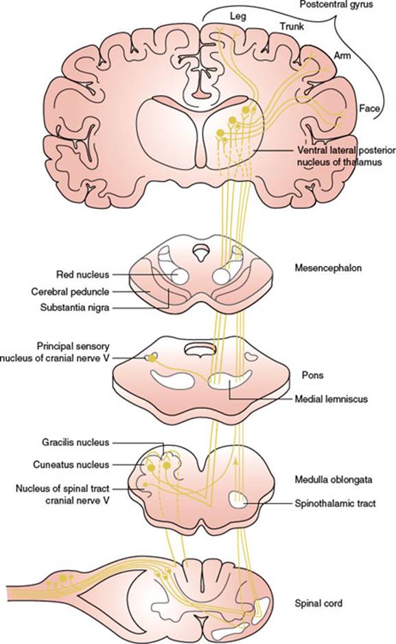
FIG. 1.3. A simplified diagram of the somatosensory pathways. (Adapted from Brodal A. The somatic afferent pathways. In: Neurological anatomy. In relation to clinical medicine, 3rd ed. New York: Oxford University Press, 1981, with permission.)
Vibration sense is mediated by different receptors from proprioception [28]. These receptors include Merkel disk receptors and Meissner’s corpuscles. Fibers mediating vibration enter the cord and bifurcate, with one branch terminating on neurons in the deeper layers of the dorsal horn and others entering the dorsal columns. Second-order neurons from the dorsal horn ascend through the ipsilateral dorsolateral funiculus, terminating on neurons in the LCN, which in turn projects fibers across the cord midline to ascend and join the medial lemniscus in the medulla. Other dorsal root collaterals enter the dorsal columns and ascend ipsilaterally, terminating in the dorsal column nuclei. Further projections from these pathways are the same as those conveying proprioception, although fibers for vibration and proprioception terminate in separate distributions within the thalamus and cerebral cortex.
From the thalamus, the sensory impulses are conveyed mainly to the somatosensory areas of the cerebral cortex (e.g., the postcentral gyrus). Within the somatosensory cortex, there is a somatotopic organization. For example, in the postcentral gyrus, the calf and foot are represented on the medial surface of the hemisphere, followed by the thigh, abdomen, thorax, shoulder, arm, forearm, hand, digits, and face. Therefore, a parasagittal lesion may cause sensory changes confined to the lower limb.
Sensory Signs and Symptoms and Their Localization
Sensory symptoms may be positive or negative. Positive symptoms include paresthesias, which are spontaneous sensations occurring without stimulation. Hyperesthesia refers to exaggerated sensation, dysesthesia to altered sensation, allodynia to a painful response to nonnoxious stimulation, and hyperpathia to exaggerated sensation to a painful stimulus. Hypesthesia is decrease in sensation, whereas anesthesia is complete loss of sensation; both may occasionally be associated with pain (anesthesia dolorosa). Proprioceptive impairment may cause ataxia and pseudoathetosis.
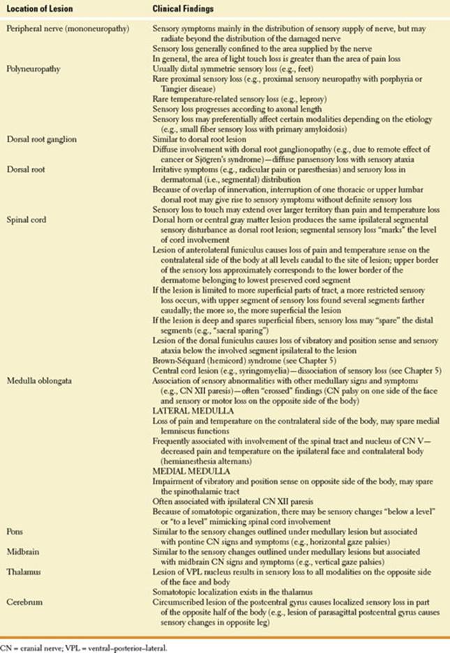
The localization of lesions affecting the somatosensory pathways is outlined in Table 1.2.
Localization of Postural and Gait Disorders
Both the sensory and motor systems play a crucial role in the maintenance of a stable stance, or posture, and in the mediation of gait. It is however worth summarizing separately the localization of disorders of posture and gait because they are frequent and require a slightly different approach. Posture and gait are complex functions that require input from the nervous system but can also be altered by the disorders of nonneurologic structures, including the muscles and joints. Often clinical bias tends to favor a nonneurologic diagnosis when the problem is actually in the neural control of gait or posture. Although initiated and modified volitionally, both functions run largely in the background. For instance, when concentrating on getting something from the refrigerator, a person pays no attention to the complex movements of the legs and paravertebral muscles while walking. Likewise, the person is not aware of the movements the same muscles make when shifting in bed, an activity mediated by similar neural structures.
Neurologic disorders of gait and posture can be localized using two main approaches:
1. Characterization of the gait disorder the patient has. In other words, we study how the patient walks or stands, or moves in bed, and from the pattern of movement or posture, we try to identify the lesioned structures. Some types of gait, such as the hemiparetic gait, are highly stereotypic and define the cause as damage to a specific structure (e.g., the corticospinal tract in the case of hemiparesis). Other types of gait, such as the cautious gait or central disequilibrium, may have many different etiologies and the lesion causing it is more difficult to localize.
2. Identification of accompanying neurologic signs. Many lesions causing neurologic gait disorders also cause other neurologic findings that may be helpful in localizing the lesion. In the case of the hemiparetic gait, we may find a Babinski’s sign pointing to a lesion in the corticospinal tract. Many structures of the nervous system participate in the control of gait, as indicated in the subsequent text. The signs or symptoms caused by lesions of these structures are described in the rest of the book.
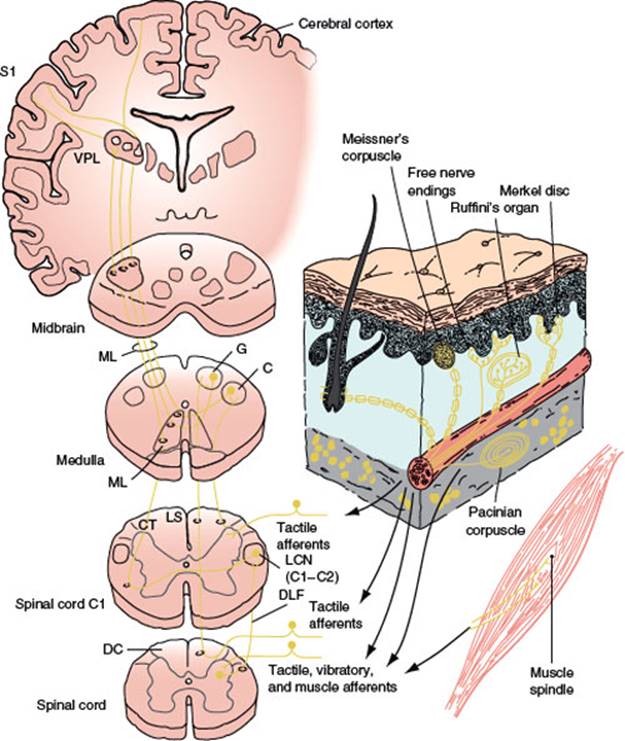
FIG. 1.4. Diagram of the peripheral receptors and central pathways mediating joint position sense, vibration sense, and tactile sensation. The lower diagram on the right illustrates the receptors principally responsible for position sense, which are muscle spindle primary and secondary afferents. The upper diagram on the right illustrates the location and morphology of mechanoreceptors in glabrous (hairless) and hairy skin of the human hand. The receptors are located both in the superficial skin at the junction of dermis and epidermis and in the deeper dermis and subcutaneous tissue. Glabrous skin contains Meissner’s corpuscles, located in dermal papillae; Merkel disc receptors, located between dermal papillae; and free nerve endings. Hairy skin contains hair receptors, Merkel receptors, and free nerve endings. Subcutaneous receptors located in both glabrous and hairy skin include pacinian corpuscles and Ruffini’s endings. Merkel disc receptors, Meissner’s corpuscles, and pacinian corpuscles are capable of mediating vibration sense, but pacinian corpuscles are responsible for detecting vibration as tested clinically. Multiple receptors mediate tactile sensation, including Meissner’s corpuscles, Merkel discs, Ruffini’s endings, pacinian corpuscles, and hair follicle receptors. The diagram on the left illustrates the central pathways mediating joint position sense, vibration sense, and tactile sensation. Afferent fibers innervating pacinian corpuscles, muscle spindles, and tactile receptors make synaptic connections with dorsal horn neurons that project rostrally through the dorsolateral funiculus (DLF) and terminate in the lateral cervical nucleus (LCN) at spinal cord segments C1 and C2. Fibers from the LCN project across the midline and ascend into the medulla, where they join the medial lemniscus. Some afferent fibers innervating tactile receptors bifurcate in the dorsal horn, with one branch entering the dorsal columns (DCs) and the other making a synaptic connection on dorsal horn neurons with axons that cross the midline and project through the lateral spinothalamic tract (not shown in the diagram) or the DLF. Fibers in the DC are laminated, with those from the sacral region (S) most medial, and lumbar (L), thoracic (T), and cervical (C) sequentially more lateral. DC fibers from sacral and lumbar segments terminate in the gracile (G) nucleus and fibers from thoracic and cervical segments terminate in the cuneate (C) nucleus of the medulla. Fibers projecting from the G and C nuclei pass across the midline and enter the medial lemniscus, which ascends to the ventral–posterior–lateral (VPL) nucleus of the thalamus. Thalamocortical fibers from VPL project to the primary somatosensory cortex (S1) of the postcentral gyrus. ML = medial lemniscus, LS = lateral sulcus, CT. (From: Gilman S. Joint position sense and vibration sense: anatomical organization and assessment. J Neurol Neurosurg Psychiatry 2002;73(5):473–477, with permission.)
Neural Structures Controlling Posture and Gait
At the simplest level of analysis, the act of standing and walking requires sensory information reaching specific brain centers and a motor output from these centers [51,53]. Sensory information includes proprioception, vision, and vestibular input. Some brain centers important for posture are the vestibular nuclei, the medullary and pontine reticular formation, the pedunculopontine and cuneiform nuclei (at the junction between the pons and midbrain), and the substantia nigra (in the midbrain). The cerebellum, basal ganglia, and thalamus play a major role in the central control of gait. In humans, the medial frontal cortex, particularly the SMA and the paracentral lobule, also contribute to the control of gait. On the motor side, the corticospinal, vestibulospinal, and reticulospinal tracts, among others, convey output from higher centers to the spinal cord. In turn, the anterior horn cells, through their axons, stimulate muscles that turn this output into specific movements.
EXAMINATION OF GAIT AND BALANCE
If a patient can stand from a low chair without using his or her arms, walk normally, maneuver turns well, walk on his or her heels and in tandem, and is steady with eyes closed and feet together and denies any imbalance or tendency to fall, gait and balance are probably normal. When examining a child, ask him or her to run for a brief stretch, while distracting from the action of running by asking to come and get something. If any abnormality is suspected from these screening maneuvers, the neural systems involved in gait should be tested further. Sensory systems can be tested by exploring the performance of the patient when one or two varieties of sensory input are removed and the postural reflexes depend on the remaining sensory information. For instance, the Romberg test explores the patient’s ability to maintain a steady upright posture with vision removed and the base of support reduced by keeping the feet together. Proprioceptive or vestibular loss will result in difficulty maintaining balance. To test the intactness of the corticospinal tract, spinal cord, peripheral nerves, and muscles, the patient is asked to wiggle the toes, draw a circle on the floor with each foot, and to extend the big toe against resistance. Proximal muscle strength in the legs can be tested by asking the patient to rise from a low chair without using his or her arms to prop himself or herself up. Despite the patient’s ability to complete all these tasks quite well, there may still be difficulty in walking and a propensity to fall. This apparent discrepancy highlights the importance of neural systems critical for posture, which are distinct from the system mediating volitional leg and foot movements [51].
TABLE 1.2 The Localization of Lesions Affecting the Somatosensory Pathways
For the description of gait disorders and their localization, we will follow a classification reminiscent of the one by Marsden and Thompson [42]. They considered gait disorders in terms of the hierarchy of lower, middle, and higher sensorimotor levels.
SENSORY AND LOWER MOTOR GAIT DISORDERS
These disorders occur with myopathies or lesions of the peripheral nervous system or their nuclei of origin, particularly in younger patients. When a sensory system is affected in isolation, the disorder is seldom long lasting. Blind people, those with bilateral destruction of the semicircular canals, and those with prosthetic limbs can walk. The intact central mechanisms use the information arriving from the other sensory systems to eventually compensate for the single-modality sensory loss. The problem can be more devastating when multiple sensory systems are affected.
Steppage Gait. Severe deafferentation or a bilateral foot drop may result in an excessive flexion of the hips and knees with every step. With sensory loss, the heel tends to strike the ground heavily. The greater foot clearance is used to prevent the patient from tripping on the toes or on the floor irregularities that are poorly felt. The most common cause of this problem is severe thick-fiber neuropathy of the kind encountered with the Guillain–Barré syndrome and other demyelinating neuropathies, including hereditary disorders such as Charcot–Marie–Tooth disease.
Vestibular Ataxia. Acute vestibular lesions cause instability and a tendency for the patient to veer or even fall to the side of the lesion. The base of support is widened and performance is markedly degraded by the Romberg’s maneuver or when the patient is asked to walk with eyes closed.
Visual Ataxia. Acute distortion of visual perception can lead to ataxia, with a broad base of support and tentative steps. In the past, this type of gait difficulty was common after cataract surgery, with removal of the affected lens leaving the patient with a severe refractive defect. Lens replacement has reduced the incidence of this problem.
Waddling Gait. The waddling gait is seen with severe proximal muscle weakness. Weakness of the hip muscles, particularly the gluteus medius, results in an excessive drop of the hip and trunk tilting to the side opposite the foot placement. The hips oscillate up and down with every step, making the patient seem to waddle. With muscle weakness, there is accentuation of the lumbar lordosis.
SIMPLER GAIT DISORDERS OF CENTRAL ORIGIN
Simpler gait disorders of central origin follow lesions located more centrally than the ones causing sensory and lower motor gait disorders. Disorders of pyramidal, cerebellar, or nigral motor systems cause distortion of appropriate postural and locomotor synergies [42]. In general, the correct postural and locomotor responses are selected, but their execution is faulty.
SPASTIC GAIT
Corticospinal tract lesions give rise to a spastic gait, unilateral or hemiparetic when the lesion is unilateral and paraparetic when the lesion is bilateral. The base of support is narrow, so much so that with bilateral lesions the legs tend to cross in front of each other in a pattern that has been called “scissors gait.” The leg is externally rotated at the hip. The knee is extended and stiff, so the patient walks as if on a stilt. The foot is plantar flexed and inverted; for this reason, the patient tends to scrape the floor with the outer edge of the foot; the patient’s turns are slow. With each step the affected leg is rotated away from the body, then toward it (circumduction). There is also difficulty picking up the toes on the hemiparetic side, when instructed to walk on the heels and decrease cadence of gait. The lesion can be anywhere along the corticospinal tract. When the lesion is unilateral, the abnormality is easy to diagnose. Bilateral lesions, particularly when they cause a slowly progressive syndrome, are more difficult to diagnose early in the course of the disease. The cervical myelopathy of cervical spondylosis, a relatively common syndrome, belongs to this category. Cervical spondylosis tends to cause demyelinating lesions in the posterior columns and corticospinal tracts of the cervical spinal cord. The most common place of involvement is at the C5–C6 interspace. Severe lesions in this location result in paraparesis and clumsiness of the hand with atrophy in the small muscles of the hand. Milder lesions may only give rise to unsteadiness while walking or standing, often accompanied by a positive Romberg’s sign [41]. The brachioradialis reflex may be depressed, and instead, a brisk finger flexor response is elicited when percussing the brachioradialis tendon (inverted radial reflex). Careful testing of vibratory sense may reveal a sensory level in the cervical region. Sometimes the patient perceives the stimulus better in the thumb than in the small finger. Early diagnosis is important because the myelopathy of cervical spondylosis is often progressive if untreated [55].
CEREBELLAR ATAXIC GAIT
Lesions of the anterior lobe of the cerebellum can also be accompanied by a discrete impairment in gait, and those affecting the flocculonodular lobe affect equilibrium [27]. Cerebellar lesions may affect gait by causing disequilibrium and by altering limb and trunk kinematics and interlimb coordination [18]. The cerebellum does not appear to actually generate postural and gait synergies because these automatic responses, albeit very dysmetric, are present in dogs with total cerebellectomy [54]. Disturbances of gait and balance are primarily caused by lesions of the vestibulocerebellum and spinocerebellum or their connections. Lesions of the cerebellar hemispheres cause irregular timing, force, and cadence of leg movements, leading to inaccurate and variable stepping [30]. Lesions of the vestibulocerebellum, or flocculonodular lobe, can produce balance and gait disturbances that resemble those caused by vestibular lesions [18]. Tremor of the head and trunk, truncal imbalance, and swaying and falling in all directions are characteristic of vestibulocerebellar lesions. Vestibular nystagmus may be present. Although most often patients with cerebellar lesions tend to fall to the side of the lesion, some patients with lesions in the tonsillar area develop increased tone (and increased reflexes) in the ipsilateral side and fall to the contralateral side.
The clinical syndrome caused by lesions of the spinocerebellum is best characterized by alcoholic cerebellar degeneration, which primarily affects the anterior lobe of the cerebellum but also involves the olivary complex and the vestibular nuclei [65]. Patients with alcoholic cerebellar degeneration have a widened base, instability of the trunk, slow and halting gait with irregular steps and superimposed lurching. The gait abnormalities are accentuated at the initiation of gait, on turning, and with changes in gait speed. These patients may have severe gait ataxia without nystagmus, dysarthria, or arm dysmetria. Even the heel-to-shin test may give little inkling of the severity of the gait disturbance. The anterior lobe of the cerebellum is exquisitely sensitive to many metabolic injuries, not just alcohol. For instance, in severe hypoxia, the anterior lobe can be severely damaged, whereas the rest of the cerebellum may be spared.
PARKINSONIAN GAIT
The patient with Parkinson’s disease walks with a rigid trunk, reduced arm swing, slow and short steps, and a tendency for the knees to be flexed. The gait of patients with classical Parkinson’s disease differs from the gait of patients with the atypical Parkinsonian syndromes, such as progressive supranuclear palsy. Festination, a tendency for the patient to begin running after taking a few steps, may be present with classical Parkinson’s disease, but seldom with atypical Parkinson’s syndrome. The base of support is generally normal in early Parkinson’s disease but is often widened in atypical Parkinson’s disease, which is also often accompanied by impaired balance. Whereas a stoop is characteristic of classical Parkinson’s disease, patients with progressive supranuclear palsy walk quite erect. Early reduction of arm swing is more characteristic of classical Parkinson’s disease. This disease follows destruction of neurons of the substantia nigra. The Parkinsonian syndromes are caused by more widespread lesions, some of which involve the lenticular nucleus.
CHOREIC, HEMIBALLISTIC, AND DYSTONIC GAITS
In choreic, hemiballistic, and dystonic gaits, the abnormal choreic, hemiballistic, or dystonic movements are superimposed to the normal gait. Whereas chorea or hemiballismus usually interferes little with the ability to walk, dystonia can cause severe gait difficulties. Intorsion of the foot is a relatively common dystonic movement in patients on dopaminergic agents. Chorea is most frequent, with lesions of the anterior putamen resulting in an excessive suppression of the inhibitory activity of the globus pallidus medialis over the lateral thalamus. Hemiballismus, most pronounced in the leg while the patient is sitting or lying down, abates partially in the lower extremity when the patient begins to walk. It is due to a lesion of the subthalamic nucleus. Dystonia can be found with lenticular nucleus lesions [7].
COMPLEX GAIT DISORDERS OF CENTRAL ORIGIN
Complex gait disorders of central origin are less well characterized than the ones previously described. Nonetheless, they are probably more common, particularly in the elderly population. In some cases, they are caused by lesions of brainstem nuclei. Some others are due to damage of the control loop that begins in the paracentral cortex and PMC and projects to the putamen. Through direct and indirect pathways, modified by input from the substantia nigra and subthalamic nucleus, the putamen projects to the medial globus pallidus, which inhibits the activity of thalamic neurons in the ventrolateral and ventral anterior nuclei. These thalamic nuclei send facilitatory projections to the frontal cortex. This loop probably plays an important role in mediating overlearned, unconscious motor activity that runs in the background, such as gait and postural reflexes. Patients with lesions in this loop can markedly improve their gait by paying attention to it. They have a faulty “automatic pilot” for postural reflexes. Finally, other gait disorders result from direct dysfunction of the cortex in the posterior portion of the medial frontal region.
The Cautious Gait. The cautious gait is characterized by a normal or mildly widened base, a shortened stride, slowness of walking, and turning en bloc [42,59]. Anyone who has to walk on an icy street may have adopted a similar gait pattern to minimize the risk of falling. With this gait strategy, the center of gravity remains within the limits of the base of support. This gait disorder is seen mainly in older people. It may represent a milder or compensatory phase of any of the disorders causing poor balance and is not localizing.
Brainstem Disequilibrium. To a lesser or greater degree, patients with brainstem disequilibrium have poor equilibrium. Some may feel unsteady, although there is little evidence in the neurologic examination. Others are so unsteady that they cannot stand or even sit up unassisted.
It is well known that damage of the vestibular nuclei can result in marked impairment in equilibrium, with a tendency to fall to the side of an acute injury. Milder vestibular dysfunction may be an important cause of gait disturbances in older people without overt vestibular disease [20]. Fife and Baloh found vestibular dysfunction in 26 patients older than 75 years who complained of disequilibrium and in whom no cause was evident after clinical evaluation. Although none had Romberg’s sign, the patients tend to sway more and do poorer on semiquantitative gait and balance testing than the controls did [20]. Their base of support was slightly widened, their turns unsteady, and they had a tendency to stagger when pushed and veer when walking.
In patients with atherosclerosis, isolated pontine hyperintense lesions on MRI correlated with disequilibrium [37]. The lesions were located in the basis pontis, possibly involving the corticopontine or corticospinal fibers, the pontocerebellar fibers, and the pontine nuclei. The rest of the brain appeared normal on MRI. Pyramidal signs were equally distributed among patients and controls [37].
The laterodorsal region of the midbrain contains the mesencephalic locomotor region, which plays an important role in locomotion in animals [25]. Stimulation of this region in the cat induces rapid walking, followed by running. This area contains the cuneiform nucleus and the cholinergic pedunculopontine nucleus. In humans, loss of neurons in the pedunculopontine nucleus has been found in patients with progressive supranuclear palsy and Parkinson’s disease but not in patients with Alzheimer’s disease, perhaps implying a role of this nucleus in ambulation [69]. Discrete vascular damage in this region can give rise to severe disequilibrium and a loss of rhythmic, alternating feet movement that characterize normal walking [44]. It is conceivable that other brainstem nuclei, still poorly identified, may also play an important role in postural mechanisms.
DISEQUILIBRIUM WITH AUTOMATIC PILOT DISORDER
The disorders described next are characterized not only by disequilibrium but also by a striking difference between the patients’ performance when they walk spontaneously and a better performance when they think about walking, for instance, by stepping over an obstacle or trying to take long strides. All of these lesions affect the corticobasal ganglionic-thalamo-cortical loop, described at the beginning of this section. The basal ganglia are part of an important loop that controls proximal movements participating in postural synergies.
1. Basal ganglia lesions. Early disequilibrium characterizes progressive supranuclear palsy and multiple system atrophy and helps differentiate them from early Parkinson’s disease. Acute lesions of the basal ganglia can also produce a syndrome of unsteadiness without the loss of isometric power, in which a patient without an apparent weakness cannot stand normally [38].
2. Thalamic lesions. Whereas chronic lesions of the basal ganglia are better known to cause axial motor impairment than acute ones, the opposite is true for thalamic lesions. A syndrome of impaired axial postural movements has been described with acute infarction or hemorrhage in the ventrolateral nucleus of the thalamus or suprathalamic white matter [46]. Although alert, with normal or near-normal strength on isometric muscle testing and a variable degree of sensory loss, these patients could not stand, and some with acute lesions could not sit up unassisted for several days after the acute insult. They fell backwards or toward the side contralateral to the lesion. These patients appeared to have a deficit of overlearned motor activity of an axial and postural nature. The syndrome has been called thalamic astasiaand grouped by some among the central disequilibrium syndromes [34].
3. Hemispheric paracentral periventricular white matter lesions. The output of the thalamus that is critical for gait is directed to the areas of the cortex involved in lower extremity movements. This area of the cortex is the medial frontal region, specifically, the paracentral lobule and the SMA. The fibers reaching this area from the thalamus course through the periventricular white matter. Therefore, it is possible or even likely, that lesions in this area may result in impaired gait. Ischemic disease of the white matter is common in the elderly population. Beginning with a report in 1989, many studies have confirmed that white matter abnormalities on computed tomography scan and MRI correlate with impaired gait and balance in older people [3,16,47]. The kind of gait impairment seen in these patients corresponds to what has been termed the cautious gait [59]. Because the patients have poor balance, the steps are shorter, possibly to lessen the single-foot stance portion of the gait cycle. Like patients with thalamic lesions, these individuals may seem to walk rather normally so long as they pay attention to their gait. However, when they engage the automatic pilot, and the motor control system begins to be relied on for involuntary movements, they tend to fall. Sudden buckling of the knees may precipitate them to the floor.
Disequilibrium may also be prominent in patients with hydrocephalus and with lesions in the medial aspect of the frontal lobe. However, these patients tend to have the gait disorder described in the subsequent text as “magnetic gait.”
Central disequilibrium is probably the most common cause of the so-called drop attacks, sudden falls without warning or loss of consciousness in older individuals. Drop attacks were originally attributed to the disease of the vertebrobasilar system, but this etiology of drop attacks in the elderly is probably not as common as subcortical hemispheric disease [45].
Freezing of Gait. With preserved balance, patients with isolated gait ignition failure or freezing of gait cannot start walking because of hesitation and may freeze in the course of locomotion, particularly on a turn [34,42]. Once the patient begins to walk, steps are short and shuffling, but they become larger and the foot clearance increases as the patient continues to walk. The base of support is normal. Postural responses are preserved. Eye closure does not induce abnormal swaying. Maneuvers that bring about a “cortical strategy,” such as trying to kick an imaginary ball, step over a cane, or count the steps, help the patient initiate and maintain gait. Minus the disequilibrium, this disorder mimics the “automatic pilot disorder” described in the previous section. The anatomic localization of this disorder is still undefined, in part because the phenomenology is not uniform: freezing in the course of walking normally along a straight line may not be the same as freezing initiating gait or making a turn. The second features are characteristic of mild magnetic gait [52], described in the following paragraph. Freezing of gait is present in a minority of patients with Parkinson’s disease and may antedate by years a diagnosis of progressive supranuclear palsy (PSP). As the upper brainstem is markedly affected in PSP, damage of this region could be most often responsible for freezing of gait.
Magnetic Gait. Magnetic gait is a disorder that corresponds to what has been described as frontal gait disorder, marche à petit pas or arteriosclerotic parkinsonism [34]. Meyer and Barron called it apraxia of gait because despite the severe gait disorder the patients can move their legs at will [48]. Although able to stand, these patients have such an inability to lift their feet and walk that their feet may seem to be glued to the floor. Some patients have great difficulty initiating walking and, when pushed forward, the heels are lifted but the toes seem to grab the floor. There may be a dissociation between gait and distal volitional movements, in that the patients may be quite able to draw figures with their feet or do the heel–shin maneuver normally. Given the preservation of even complex motor patterns for the lower extremities, it is perhaps better to not use the term apraxia for this type of gait. Milder forms of the same disorder resemble the Parkinsonian gait, with short, shuffling steps and truncal rigidity. Arm swing during walking may be preserved and, if so, helps differentiate this disorder from Parkinson’s disease [63]. The turns are very slow and broken down into many steps. Turning may bring up the tendency for the feet (or for one foot more than the other when the problem is asymmetrical) to become glued to the floor. Freezing may become evident as the steps halt and the patient remains motionless or develops tremor-like movements of the lower legs. Falls are common, particularly in patients who have disequilibrium. This disorder may be caused by bilateral lesions of the medial frontal cortex, severe hydrocephalus, or bilateral ischemic lesions of the white matter. Gait impairment is part of the classical triad for the diagnosis of normal-pressure hydrocephalus [1]. Some authors have described this entity as a rather prevalent cause of gait disorders in the aging population [22]. However, other studies, looking at the outcome of shunting for large ventricles in older individuals, have concluded that this is a relatively rare entity [29].
Disequilibrium and Disorganized Gait. Disequilibrium and disorganized gait has also been described as frontal disequilibrium [34]. There is disequilibrium and a disorganization of gait patterns, such that the patients do not move the legs appropriately for locomotion. They may cross the legs or move them in directions that are inappropriate to keep balance or even to sit up from a sitting position. This disorder has been described with a variety of frontal lobe lesions [34] and also with lesions in the mesencephalic locomotor center [44].
References
1. Adams R, Fisher C, Hakim S, et al. Symptomatic occult hydrocephalus with “normal” cerebrospinal fluid pressure. N Engl J Med 1965;273:117–126.
2. Afifi AK, Bergman RA. Functional neuroanatomy. Text and atlas. Major sensory and motor pathways. New York, Saint Louis, San Francisco: McGraw Hill, 1998:587–600, Chapter 31.
3. Baloh RW, Yue Q, Socotch TM, et al. White matter lesions and disequilibrium in older people. I. Case-control comparison. Arch Neurol 1995;52:970–974.
4. Benarroch EE, Westmoreland BF, Daube JR, et al. Medical neurosciences. An approach to anatomy, pathology, and physiology by systems and levels. The motor system, 4th ed. Philadelphia, PA: Lippincott Williams & Wilkins, 1999:193–247, Chapter 8.
5. Berker EA, Berker AH, Smith A. Translation of Broca’s 1865 report. Localization of speech in the third left frontal convolution. Arch Neurol 1986;43: 1065–1072.
6. Bernal B, Ardila A. The role of the arcuate fasciculus in conduction aphasia. Brain 2009;132:2309–2316.
7. Bhatia KP, Marsden CD. The behavioural and motor consequences of focal lesions of the basal ganglia in man. Brain 1994;117:859–876.
8. Biller J, Gruener G, Brazis PW. DeMyer’s neurologic examination, a programmed text. New York: McGraw Hill, 2011.
9. Bojowskslausks J, Repli F. Capsular genu syndrome. Neurology 1990;40:1499–1502.
10. Borod JC, Koff E, Lorch MP, et al. Emotional and non-emotional facial behaviour in patients with unilateral brain damage. J Neurol Neurosurg Psychiatry 1988; 521: 826–832.
11. Boubonnais D, Boissy P, Gravel D, et al. Quantitation of upper limb synkinesis in hemiparetic subjects. Rehabilitation R and D Progress Report, No 32. Department of Veterans Affairs, 1994:118–119.
12. Breasted J. The Edwin Smith surgical papyrus. Chicago, IL: Chicago University Press, 1930.
13. Broca P. Sur le siège de la faculté du langage articulé. Bull Soc Anthropol 1865;6:337–393.
14. Brodal A. The somatic afferent pathways. In: Neurological anatomy. In relation to clinical medicine, 3rd ed. New York, NY: Oxford University Press, 1981: 46–147.
15. Bromberg MB. An approach to the evaluation of peripheral nerve diseases. In: Bromberg MR, Smith AG, eds. Handbook of peripheral neuropathy. Boca Raton, London, Singapore: Taylor and Frances, 2005: 1–13, Chapter 1.
16. Camicioli R, Moore MM, Sexton G, et al. Age-related brain changes associated with motor function in healthy older people. J Am Geriatr Soc 1999;47:330–334.
17. Cavazos JE, Bulsara K, Cavess J, et al. Pure motor sensory hemiplegia including the face induced by an infarct of the medullary pyramid. Clin Neurol Neurosurg 1996;98:21–23.
18. Diener H, Nutt J. Vestibular and cerebellar disorders of equilibrium and gait. In: Masdeu J, Sudarsky L, Wolfson L, eds. Gait disorders of aging. Falls and therapeutic strategies. Philadelphia, PA: Lippincott-Raven, 1997: 261–272.
19. Dubowitz V. Muscle disorders in childhood, 2nd ed. Philadelphia, PA: WB Saunders, 1995.
20. Fife TD, Baloh RW. Disequilibrium of unknown cause in older people. Ann Neurol 1993;34:694–702.
21. Fischer U, Hess CW, Rosler KM. Uncrossed cortico-muscular projections in humans are abandoned to facial muscles of the upper and lower face, but may differ between sexes. J Neurol 2005;252:21–26.
22. Fisher C. Hydrocephalus as a cause of disturbances of gait in the elderly. Neurology 1982;32:1358–1363.
23. Fitzgerald MJT. Neuroanatomy. Basic and clinical. In: Spinal cord: descending pathways, 3rd ed. London, Philadelphia, Toronto, Sydney, Tokyo: WB Saunders, 1996:117–130, Chapter 13.
24. Fung HC, Chen ST, Tang LM, et al. Triparesis: MRI documentation of bipyramidal medullary infarction. Neurology 2002;58:1130–1131.
25. Garcia-Rill E. The pedunculopontine nucleus. Prog Neurobiol 1991;36:363–389.
26. Gerace C, Fele RM, Pingi A. Capsular lingual paresis. Neurology 2005;76:595.
27. Gilman S. Cerebellum and motor dysfunction. In: Asbury AK, McKhann GM, McDonald WI, eds. Diseases of the nervous system. Clinical neurobiology, 2nd ed. Philadelphia, PA: WB Saunders, 1992:319–341.
28. Gilman S. Joint position sense and vibration sense: anatomical organization and assessment. J Neurol Neurosurg Psychiatry 2002;73:473–477.
29. Graff-Radford N, Godersky J. A clinical approach to symptomatic hydrocephalus in the elderly. In: Masdeu J, Sudarsky L, Wolfson L, eds. Gait disorders of aging. Falls and therapeutic strategies. Philadelphia, PA: Lippincott-Raven, 1997:245–259.
30. Hallett M, Stanhope S, Thomas S. Pathophysiology of posture and gait in cerebellar ataxia. In: Shimamura M, Grillner S, Edgerton V, eds. Neurobiological basis of human locomotion. Tokyo: Japan Scientific Societies Press, 1991:275–283.
31. Heimer L. The human brain and spinal cord. Functional neuroanatomy and dissection guide. Spinal cord and the descending supraspinal pathways, 2nd ed. New York, Berlin, Heidelberg, London, Paris, Tokyo, Hong Kong, Barcelona, Budapest: Springer-Verlag, 1995: 316–334, Chapter 15.
32. Holodny AI, Gor DM, Watts R, et al. Diffusion-tensor MR tractography of somatotopic organization of corticospinal tracts in the internal capsule; initial anatomic results in contradistinction to prior reports. Radiology2005;234:649–653.
33. Hopf HC, Fitzek C, Marx J, et al. Emotional facial paresis of pontine origin. Neurology 2000;54:1217.
34. Jankovic J, Nutt JG, Sudarsky L. Classification, diagnosis, and etiology of gait disorders. Adv Neurol. 2001;87: 119–133.
35. Jox R, Bruning R, Hamann G, et al. Volitional facial palsy after a vascular lesion of the supplemental motor area. Neurology 2004;63:756–757.
36. Keane JR. Wrong-way deviation of the tongue with hysterical hemiparesis. Neurology 1986;36:1406–1407.
37. Kwa VI, Zaal LH, Verbeeten B Jr, et al. Disequilibrium in patients with atherosclerosis: relevance of pontine ischemic rarefaction. Amsterdam Vascular Medicine Group. Neurology 1998;51:570–573.
38. Labadie EL, Awerbuch GI, Hamilton RH, et al. Falling and postural deficits due to acute unilateral basal ganglia lesions. Arch Neurol 1989;46:492–496.
39. Lance JW. Symposium synopsis. In: Feldman RG, Young RR, Koella WP, eds. Spasticity disorder of motor control. Chicago, IL: Year Book Medical Publishers, 1980:485–494.
40. Lee S-H, Kim D-E, Song E-C, et al. Sensory dermatomal representation in the medial lemniscus. Arch Neurol 2001;58:649–651.
41. Lee TT, Manzano GR, Green BA. Modified open-door cervical expansive laminoplasty for spondylotic myelopathy: operative technique, outcome, and predictors for gait improvement. J Neurosurg1997;86:64–68.
42. Marsden C, Thompson P. Toward a nosology of gait disorders: descriptive classification. In: Masdeu J, Sudarsky L, Wolfson L, eds. Gait disorders of aging. Falls and therapeutic strategies. Philadelphia, PA: Lippincott-Raven, 1997:135–146.
43. Marx JJ, Iannetti GD, Thomke F, et al. Somatotopic organization in the corticospinal tract in the human brainstem. Ann Neurol 2005;5:824–831.
44. Masdeu J, Alampur U, Cavaliere R, et al. Astasia and gait failure with damage of the pontomesencephalic locomotor region. Ann Neurol 1994;35:619–621.
45. Masdeu JC. Cerebrovascular disorders. In: Masdeu JC, Sudarsky L, Wolfson L, eds. Gait disorders of aging. Philadelphia, PA: Lippincott-Raven, 1997:221–243.
46. Masdeu JC, Gorelick PB. Thalamic astasia: inability to stand after unilateral thalamic lesions. Ann Neurol 1988;23:596–603.
47. Masdeu JC, Wolfson L, Lantos G, et al. Brain white-matter changes in the elderly prone to falling. Arch Neurol 1989;46:1292–1296.
48. Meyer JS, Barron DW. Apraxia of gait: a clinico-physiological study. Brain 1960;82:261.
49. Mihailoff GA, Haines DE. Motor system II: corticofugal systems and the control of movement. In: Haines DE, ed. Fundamental neuroscience. New York, Edinburgh, London, Madrid, Melbourne, San Francisco, Tokyo: Churchill Livingstone, 1997:347–362, Chapter 24.
50. Monrad-Krohn GH. The clinical examination of the nervous system. 8th ed. New York: Paul B. Hoeber Inc. 1997, pp 207.
51. Mori S. Neurophysiology of locomotion: recent advances in the study of locomotion. In: Masdeu J, Sudarsky L, Wolfson L, eds. Gait disorders of aging. Falls and therapeutic strategies. Philadelphia, PA: Lippincott-Raven, 1997:55–78.
52. Nadeau SE. Gait apraxia: further clues to localization. Eur Neurol 2007;58:142–145.
53. Nashner L. Physiology of balance, with special reference to the healthy elderly. In: Masdeu J, Sudarsky L, Wolfson L, eds. Gait disorders of aging. Falls and therapeutic strategies. Philadelphia, PA: Lippincott-Raven, 1997:37–53.
54. Rademaker GGJ. The physiology of standing: postural reactions and equilibrium, with special references to the behavior of decerebellate animals. Minneapolis: University of Minnesota Press, 1980.
55. Sadasivan KK, Reddy RP, Albright JA. The natural history of cervical spondylotic myelopathy. Yale J Biol Med 1993;66:235–242.
56. Saito K, Matsumoto S, Yokoyama T, et al. Pathology of chronic vitamin E deficiency in fatal familial intrahepatic cholestasis (Byler disease). Virchows Arch [Pathol Anat] 1982;396:319–330.
57. Sheehan G. Neurophysiology of spasticity. In: Barnes MP, Johnson GR, eds. Upper motor neurone syndrome and spasticity. Clinical management and neurophysiology. Cambridge: University Press, 2001:12–78, Chapter 2.
58. Sherman SJ, Koshland GF, Laguna JF. Hyperreflexia without spasticity after unilateral infarct of the medullary pyramid. J Neurol Sci 2000;175:145–155.
59. Sudarsky L, Tideiksaar R. The cautious gait, fear of falling, and psychogenic gait disorders. In: Masdeu J, Sudarsky L, Wolfson L, eds. Gait disorders of aging. Falls and therapeutic strategies. Philadelphia, PA: Lippincott-Raven, 1997:283–295.
60. Takahashi N, Kawamura M, Araki S. Isolated hand palsy due to cortical infarction: localization of the motor hand area. Neurology 2002;58:1412–1414.
61. Terao S, Miura N, Takeda A, et al. Course and distribution of facial tract bulbar tract fibers in the lower brainstem. J Neurol Neurosurg Psychiatry 2000;69: 262–265.
62. Terao S, Takatsu S, Izumi M, et al. Central facial weakness due to medial medullary infarction: the course of facial corticobulbar fibers. J Neurol Neurosurg Psychiatry 1997;63:391–393.
63. Thompson P, Marsden C. Clinical neurological assessment of balance and gait disorders. In: Bronstein A, Brandt T, Woollacott M, eds. Clinical disorders of balance, posture and gait. London: Arnold, 1996: 79–84.
64. Urban PP, Witch S, Vucorevic G, et al. The course of cortico-facial projections in the human brainstem. Brain 2001;124:1866–1876.
65. Victor M, Ferrendelli JA. The nutritional and metabolic diseases of the cerebellum: clinical and pathological aspects. In: Fields W, Willis W, eds. The cerebellum in health and disease. St. Louis, MO: Green, 1970.
66. Waxman SG. Control of movement. In: Correlative neuroanatomy, 24th ed. New York, St. Louis, San Francisco: Lange Medical Books/McGraw Hill, 2000: 189–201, Chapter 13.
67. Wernicke C. Der aphasische symptomencomplex; eine psychologische studie auf anatomischer basis. Breslau: Max Cohn & Weigert, 1874.
68. Wilkins RH, Brody IA. Wernicke’s sensory aphasia. Arch Neurol 1970;22:279–282.
69. Zweig R, Whitehouse P, Casanova M, et al. Pedunculopontine cholinergic neurons in progressive supranuclear palsy. Ann Neurol 1987;22:18–25.