
Anatomy of the Olfactory Pathways
Although the olfactory system is not of major importance in neurologic diagnosis, certain clinical information, useful in neuroanatomic localization, may be attained by investigating the sense of smell. This investigation requires a basic knowledge of the anatomy of the olfactory pathways [3,6,9,30,37], especially their relationship with the surrounding neural structures (e.g., frontal lobes) (Figs. 6.1 and 6.2).
The olfactory receptors, the sensory cells of the olfactory epithelium, are located on the superior– posterior nasal septum and the lateral wall of the nasal cavity. These ciliated cells give off central processes that form small bundles (approximately 20 in number). These bundles, the filaments of the olfactory nerve, penetrate the cribriform plate of the ethmoid bone and enter the olfactory bulb. Here, the olfactory afferent fibers synapse with the dendrites of the second-order neurons called the mitral and tufted cells. At the points of synapse, conglomerates of fibers called the olfactory glomeruli are formed. The axons of the mitral and tufted cells leave the olfactory bulb and course posteriorly, as the olfactory tract, in the olfactory sulcus on the orbital surfaces of the frontal lobe. The olfactory tract divides into a median and a lateral olfactory stria on either side of the anterior perforated substance (the triangular area formed by the two striae is called the olfactory trigone). Some of these strial fibers decussate in the anterior commissure and join the fibers from the opposite olfactory pathways, terminating in the contralateral cerebral hemisphere. Other strial fibers, especially those of the lateral stria, supply the ipsilateral piriform lobe of the cerebral (temporal) cortex (the primary olfactory cortex) and terminate in the amygdaloid nucleus, septal nuclei, and hypothalamus.
Although relatively quantitative methods [6,30,37] are available to test olfaction (e.g., tests of the minimal perceptible odor or measurements of olfactory fatigue), the sense of smell is usually tested by asking the patient to sniff various nonirritating substances (each nostril is tested separately) and then attempt to identify the odor (perception of the smell is of more value than identification of the specific substance). Irritating substances (e.g., ammonia) are to be avoided because they stimulate the trigeminal nerve fibers in the nasal mucosa as well as the olfactory fibers.
Localization of Lesions Affecting the Olfactory Nerve
Lesions Causing Anosmia
Anosmia (loss of smell) or hyposmia (diminished olfactory functioning) may or may not be apparent to the patient. He or she may have some difficulty in tasting various flavors because the identification of tasted substances depends in part on the olfactory system.
Decreased smell function occurs in the “normal” elderly [12]. Generally, age-related decline in olfaction is more severe for men than for women. Approximately 2% of the population younger than 65 years has a chronic impairment of smelling. Between 65 and 80 years, this rises dramatically with about half of the population experiencing significant decrements in the ability to smell. For the population older than 80 years, this figure rises to nearly 75% [12]. The basis for age-related changes in smell function is multiple and includes ossification and closure of the foramina of the cribriform plate, development of early neurodegenerative disease pathology, and cumulative damage to the olfactory receptors from repeated viral and other insults throughout life [8].
Local nasal disease (e.g., allergic rhinitis, nasal obstruction, polyposis) must first be sought as the cause of anosmia, especially if the olfactory difficulty is bilateral. The most common cause of transient and bilateral anosmia is the common cold. Exposure to several airborne toxins, including herbicides, pesticides, solvents, and heavy metals can alter the ability to smell, especially when exposure has been chronic. Among the heavy metals, the best documented cases are for cadmium, chromium, nickel, and manganese [8].
After local nasal disease has been ruled out, anosmia, especially unilateral anosmia, should raise the suspicion of a lesion affecting the olfactory nerve filaments, bulb, tract, or stria. Because the cortical representation for smell in the piriform cortex is bilateral, a unilateral lesion distal to the decussation of the olfactory fibers causes no olfactory impairment.

FIG. 6.1. The olfactory nerve (lateral view).
Head injury [13,36,41] is probably the most common cause of disruption of the olfactory fibers prior to their decussation. Frontal impact produces less dysfunction than back or side impacts [13]. The olfactory nerve proper (olfactory filaments) may be torn by fractures involving the cribriform plate of the ethmoid bone, but closed head injury without fracture may also disrupt the olfactory pathways unilaterally or bilaterally. Closed head injury can produce impairment of olfactory recognition despite relatively preserved olfactory detection [25]. Olfactory naming and recognition may be impaired by traumatic forces affecting the orbitofrontal and temporal lobes, and the degree of olfactory disturbances is directly related to the severity of the injury [25]. Disturbances of complex olfactory function (e.g., discrimination) despite the relatively preserved detection of odors have been reported with alcoholic Korsakoff syndrome [23] and following thalamic or prefrontal cortical lesions [34]. Significant olfactory dysfunction has also been described with Alzheimer disease [27,28,46], Lewy body disease [32], Huntington chorea (HC) [28], corticobasal ganglionic degeneration [33], Creutzfeldt–Jakob disease [42,52], frontotemporal dementia [33], multiple sclerosis [10,20], Parkinson disease (PD) [11,21,27,43], Refsum disease [17], spinocerebellar ataxias (including Friedreich ataxia) [4], Wilson disease [31], narcolepsy [39], pure autonomic failure [38], and in adults with Down syndrome [45]. Impaired olfaction can predate clinical PD in men by at least 4 years and may be a useful screening tool to detect those at high risk for development of PD in later life [48]. Patients with PD have decreased performance on odor discrimination tests in addition to deficits of odor detection and identification [43]. REM (rapid eye movement) sleep disorder and olfactory dysfunction are common and very early features of alpha-synucleinopathies, in particular, PD [40]. Olfactory loss in patients with multiple sclerosis has been associated with plaque formation in the central olfactory (i.e., inferior frontal and temporal) brain regions [10].

FIG. 6.2. The olfactory nerve (inferior view).
Hawkes [19] noted that there has been an increase of interest in olfactory dysfunction because it was realized that anosmia was a common feature of idiopathic PD and Alzheimer- dementia (AD). In his review of PD, parkinsonian syndromes, essential tremor, AD, motor neuron disease, and HC, the following observations are made [19]:
1. Olfactory dysfunction is frequent and often severe in PD and AD.
2. Normal smell identification in PD is rare and should prompt the review of diagnosis unless the patient is a female with a tremor-dominant disease.
3. Anosmia in suspected progressive supranuclear palsy and corticobasal degeneration is atypical and should likewise provoke diagnostic review.
4. Hyposmia is an early feature of PD and AD and may precede motor and cognitive signs, respectively.
5. Subjects with anosmia and one apoE4 allele have an approximate fivefold increased risk of later AD.
6. Impaired sense of smell is seen in some patients at 50% risk of parkinsonism.
7. Smell testing in HC and motor neuron disease, where abnormality may be found, is not likely to be of clinical value.
8. Biopsy of olfactory nasal neurons shows nonspecific changes in PD and AD and, at present, will not aid diagnosis.
Incidental Lewy bodies (ILB), the presence of Lewy bodies in the brains of deceased individuals without a history of PD or dementia during life, are thought to represent a presymptomatic stage of PD. Olfactory dysfunction is associated with ILB [35]. If ILB represent a presymptomatic stage of PD, olfactory testing may be a useful screening tool to identify those at high risk for developing PD.
Congenital anosmia or hyposmia may occur owing to cleft palate in men, absent or hypoplastic olfactory bulbs or tracts, familial dysautonomia, and Turner syndrome. A familial syndrome of permanent anosmia with hypogonadotropic hypogonadism (Kallmann syndrome) has also been described [50,51]; patients with this syndrome may also have cerebellar ataxia and mirror movements of the hands [18].
Olfactory discrimination and detection may be abnormal after unilateral frontal or temporal lobectomy [53]. After temporal lobectomy, deficits in olfactory discrimination are confined to the nostril ipsilateral to the lesion. After frontal lobectomy, discrimination is also impaired; however, in patients with right frontal lesions including the orbital cortex, the impairment is found in both nostrils. Therefore, the orbitofrontal cortex is important in olfactory discrimination, and the nostril difference found in healthy subjects, together with the birhinal impairment in patients with right orbitofrontal damage, suggests a relative advantage of the right orbital region in olfactory processing [53]. Anosmia may also complicate rhinoplasty, ethmoidectomy, laryngectomy, submucous resection of the nasal septum, radiotherapy [30], and surgery for anterior communicating artery aneurysms owing to the olfactory nerve dysfunction [15]. Olfactory damage is much more common after an anterior interhemispheric surgical approach rather than after a basal interhemispheric approach.
The olfactory bulb and tract are frequently affected by tumors of the olfactory groove (especially meningiomas) [49], which may cause the Foster Kennedy syndrome (see the discussion on Foster Kennedy syndrome in the next section). Tumors of the sphenoid or frontal bone (e.g., osteomas), pituitary tumors with suprasellar extension, nasopharyngeal carcinoma [44], and saccular aneurysms of the anterior portion of the circle of Willis (e.g., a giant anterior communicating artery aneurysm) may also compress the olfactory bulb or tract [26]. Any diffuse meningeal process (e.g., meningitis) may involve the olfactory pathways. The anatomic relationship of the frontal lobe to the olfactory bulb and tract is especially important. Mass lesions of the frontal lobe (e.g., glioma or abscess) often exert pressure on the olfactory system and may lead to anosmia even before clear-cut frontal lobe signs and symptoms are noted. Therefore, in any patient with personality changes or subtle signs of frontal lobe involvement, olfaction should be carefully tested.
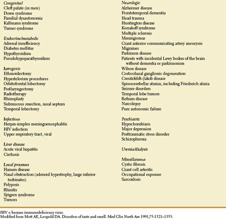
TABLE 6.1 Conditions Associated with Disturbance of Olfaction
Esthesioneuroblastomas (olfactory neuroblastomas) are tumors that arise in the upper nasal cavity, often superior and lateral near the ethmoid sinus [29]. These tumors may present with anosmia as well as persistent nasal obstruction and epistaxis [29]. They may occasionally involve the orbit and cause periorbital swelling, proptosis, diplopia, and visual loss [2,29].
The Foster Kennedy Syndrome
The Foster Kennedy syndrome is occasionally noted with olfactory groove or sphenoid ridge masses (especially meningiomas) or space-occupying lesions of the frontal lobe. This syndrome consists of the following three signs:
1. Ipsilateral anosmia due to direct pressure on the olfactory bulb or tract.
2. Ipsilateral optic atrophy due to direct injury of the ipsilateral optic nerve.
3. Contralateral papilledema due to raised intracranial pressure secondary to the mass lesion.
Many cases of Foster Kennedy syndrome may actually be due to direct bilateral optic nerve compression; even increased intracranial pressure without optic nerve compression may cause the syndrome [47].
A pseudo–Foster Kennedy syndrome may rarely be noted when increased intracranial pressure of any cause occurs in a patient who has previous unilateral optic atrophy. Because the atrophic disc cannot become swollen, only the previously normal fundus demonstrates papilledema. Olfactory nerve involvement varies depending on the etiology of the increased intracranial pressure, but increased intracranial pressure per se may impair olfaction without any evidence of local olfactory pathway damage. A pseudo–Foster Kennedy syndrome is most often due to sequential anterior ischemic optic neuropathy (arteritic or nonarteritic) or optic neuritis in which optic disc edema on one side is associated with optic disc atrophy on the other side.
Lesions Causing Parosmia and Cacosmia
Parosmia or dysosmia (perversion of smell) and cacosmia (experiencing unpleasant odors) [7] are rare phenomena that are usually seen after a head injury or with a psychiatric disease (e.g., depression). Various scents are interpreted as “abnormal” and, often, unpleasant. Occasionally, these unpleasant odors may persist or occur spontaneously as an olfactory hallucination [14,24]. It is not clear whether these phenomena are of cortical origin (due to primary olfactory cortex injury), and therefore possibly ictal in nature, or are due to direct irritation of the olfactory pathways. Unilateral paroxysmal olfactory hallucinations (paroxysmal unilateral dysosmia) have been cured by resection of the homolateral olfactory bulb suggesting that, in at least some patients, olfactory hallucinations may be due to structural nerve damage [24]. Olfactory hallucinations (especially foul odors) may occur with partial seizures with complex symptomatology or with migraine [1,16]. Olfactory epileptic auras are not necessarily unpleasant [1]. In patients with olfactory epileptic auras, tumors are the most common etiology of the seizures, and mesial temporal sclerosis is relatively rare; the amygdala is the most likely symptomatogenic zone in these patients [1]. Hyperosmia (increased sensitivity to smell) may occur with migraine or hyperemesis gravidarum; phantosmia refers to the perception of an odor when none is present [5].
Hyposmia and parosmia have been associated with hypogeusia (diminished taste acuity) and dysgeusia (distorted taste perception) and have been correlated with zinc and vitamin A deficiencies [22]. Other conditions associated with altered olfaction include adrenal insufficiency, diabetes mellitus, hypothyroidism, pseudohypoparathyroidism, cystic fibrosis, and sarcoidosis [30].
Conditions associated with disturbances of olfaction are outlined in Table 6.1.
References
1. Acharya V, Acharya J, Lüders H. Olfactory epileptic auras. Neurology 1998;51:56–61.
2. Berman EL, Chu A, Wirtschafter JD, et al. Esthesioneuroblastoma presenting as sudden unilateral blindness. Histopathologic confirmation of optic nerve demyelination. J Clin Neuroophthalmol1992;12:31–36.
3. Brodal A. Neurological anatomy in relation to clinical medicine, 3rd ed. New York: Oxford University Press, 1981.
4. Connelly T, Farmer JM, Lynch DR, et al. Olfactory dysfunction in degenerative ataxias. J Neurol Neurosurg Psychiatry 2003;74:1435–1437.
5. Cowart BJ, Young IM, Feldman RS, et al. Clinical disorders of taste and smell. In: Beauchamp GK, Bartoshuk L, eds. Tasting and smelling. New York, NY: Academic Press, 1997,175–198.
6. Davidson TM, Jalowayski A, Murphy C, et al. Evaluation and treatment of smell dysfunction. West J Med 1987;146:434–438.
7. Dejong RN. The neurologic examination—incorporating the fundamentals of neuroanatomy and neurophysiology, 4th ed. Hagerstown, MD: Harper & Row, 1979: 83–88.
8. Doty RL. The olfactory system and its disorders. Semin Neurol 2009;29:74–81. In: Campbell WW, ed. Disorders of the cranial nerves. New York, NY: Thieme.
9. Doty RL, Kimmelman GP, Lesser RP. Smell and taste and their disorders. In: Asbury AK, McKhann GM, McDonald WI, eds. Diseases of the nervous system. Clinical neurobiology, 2nd ed. Philadelphia, PA: WB Saunders, 1992:390–403.
10. Doty RL, Li C, Mannon LJ, et al. Olfactory dysfunction in multiple sclerosis: relation to longitudinal changes in plaque numbers in central olfactory structures. Neurology 1999;53:880–882.
11. Doty RL, Riklan M, Deems DA, et al. The olfactory and cognitive deficits of Parkinson’s disease: evidence for independence. Ann Neurol 1989;25:166–171.
12. Doty RL, Shaman P, Dann M. Development of the University of Pennsylvania Smell Identification Test: a standardized microencapsulated test of olfactory function. Physiol Behav 1984;32:489–502.
13. Doty RL, Yousem DM, Pham LT, et al. Olfactory dysfunction in patients with head trauma. Arch Neurol 1997;54:1131–1140.
14. Estrem SA, Renner G. Disorders of smell and taste. Otolaryngol Clin North Am 1987;20:133–147.
15. Fujiwara H, Yasui N, Nathal-Vera E, et al. Anosmia after anterior communicating artery aneurysm surgery: comparison between the anterior interhemispheric and basal interhemispheric approaches. Neurosurgery 1996; 38:325–328.
16. Fuller GN, Guiloff RJ. Migrainous olfactory hallucinations. J Neurol Neurosurg Psychiatry 1987;50:1688–1690.
17. Gibberd FB, Feher MD, Sidey MC, et al. Smell testing: an additional tool for identification of adult Refsum’s disease. J Neurol Neurosurg Psychiatry 2004;75: 1334–1336.
18. Hardelin JP, Levilliers J, del Castillo I, et al. X chromosome-linked Kallmann syndrome: stop mutations validate the candidate gene. Proc Natl Acad Sci 1992;89:8190.
19. Hawkes C. Olfaction in neurodegenerative disorder. Mov Disord 2003;18:364–372.
20. Hawkes CH, Shephard BC, Kobal G. Assessment of olfaction in multiple sclerosis: evidence of dysfunction by olfactory evoked response and identification tests. J Neurol Neurosurg Psychiatry1997;63:145–151.
21. Henderson JM, Lu Y, Wang S, et al. Olfactory deficits and sleep disturbances in Parkinson’s disease: a case-control survey. J Neurol Neurosurg Psychiatry 2003;74:956–958.
22. Henkin RI, Schechter PJ, Hoye R, et al. Idiopathic hypogeusia with dysgeusia, hyposmia, and dysosmia. JAMA 1971;217:434–440.
23. Jones BP, Butters N, Moskowitz HR, et al. Olfactory and gustatory capacities of alcoholic Korsakoff patients. Neuropsychologia 1988;16:323–337.
24. Kaufman MD, Lassiter KRL, Shenoy BV. Paroxysmal unilateral dysosmia: a cured patient. Ann Neurol 1988;24:450–451.
25. Levin HS, High WM, Elsenberg HM. Impairment of olfactory recognition after closed head injury. Brain 1985;108:579–591.
26. Manconi M, Paolino E, Casetta I, et al. Anosmia in a giant anterior communicating artery aneurysm. Arch Neurol 2001;58:1474–1475.
27. Mesholam RI, Moberg PJ, Mahr RN, et al. Olfaction in neurodegenerative disease: a meta-analysis of olfactory functioning in Alzheimer’s and Parkinson’s diseases. Arch Neurol 1998;55:84–90.
28. Moberg PL, Pearlson GD, Speedie LJ, et al. Olfactory recognition—differential impairments in early and late Huntington’s and Alzheimer’s disease. J Clin Exp Neurol 1987;9:650–664.
29. Morita A, Ebersold MJ, Olsen KD, et al. Esthesioneuroblastoma: prognosis and management. Neurosurgery 1993;32:706–714.
30. Mott AE, Leopold DA. Disorders of taste and smell. Med Clin North Am 1991;75:1321–1353.
31. Mueller A, Reuner U, Landis B, et al. Extrapyramidal symptoms in Wilson’s disease are associated with olfactory dysfunction. Mov Disord 2006;21:1311–1316.
32. Olichney JM, Murphy C, Hofstetter CR, et al. Anosmia is very common in the Lewy body variant of Alzheimer’s disease. J Neurol Neurosurg Psychiatry 2005;76:1342–1347.
33. Pardini M, Huey ED, Cavanagh AL, et al. Olfactory function in corticobasal syndrome and frontotemporal dementia. Arch Neurol 2009;66:92–96.
34. Potter H, Butters N. An assessment of olfactory deficits in patients with damage to prefrontal cortex. Neuropsychologia 1980;18:621–628.
35. Ross GW, Abbott RD, Petrovitch H, et al. Association of olfactory dysfunction with incidental Lewy bodies. Mov Disord 2007;21:2062–2067.
36. Schechter PJ, Henkin RI. Abnormalities of taste and smell after head trauma. J Neurol Neurosurg Psychiatry 1974;37:802–810.
37. Schiffman SS. Taste and smell in disease. N Engl J Med 1983;308:1275–1279 and 1337–1343.
38. Silveira-Moriyama L, Mathias C, Mason L, et al. Hyposmia in pure autonomic failure. Neurology 2009; 72:1677–1681.
39. Stiasny-Kolster K, Clever SC, Möller JC, et al. Olfactory dysfunction in patients with narcolepsy with and without REM sleep behaviour disorder. Brain 2007;130:442–449.
40. Stiasny-Kolster K, Doerr Y, Möller JC, et al. Combination of ‘idiopathic’ REM sleep behaviour disorder and olfactory dysfunction as possible indicator for alpha-synucleinopathy demonstrated by dopamine transporter FP-CIT-SPECT. Brain 2005;128:126– 137.
41. Sumner D. Post-traumatic anosmia. Brain 1964;87: 107–202.
42. Tabaton M, Monaco S, Cordone F, et al. Prion deposition in olfactory biopsy of sporadic Creutzfeldt-Jakob disease. Ann Neurol 2004;55:294–296.
43. Tissingh G, Berendse HW, Bergmans P, et al. Loss of olfaction in de novo and treated Parkinson’s disease: possible implication for early diagnosis. Mov Disord 2001;16:41–46.
44. Turgman J, Braham J, Modan B, et al. Neurological complications in patients with malignant tumors of the nasopharynx. Eur Neurol 1978;17:149–154.
45. Warner MD, Peabody CA, Berger PA. Olfactory deficits and Down’s syndrome. Biol Psychiatry 1988;23: 833–839.
46. Warner MD, Peabody CA, Flattery JJ, et al. Olfactory deficits and Alzheimer’s disease. Biol Psychiatry 1986;21:116–118.
47. Watnick RL, Trobe JA. Bilateral optic nerve compression as a mechanism for the Foster Kennedy syndrome. Ophthalmology 1989;96:1793–1798.
48. Webster Ross G, Petrovitch H, Abbot RD, et al. Association of olfactory dysfunction with risk for future Parkinson’s disease. Ann Neurol 2008;63:167–173.
49. Welge-Luessen A, Temmel A, Quint C, et al. Olfactory function in patients with olfactory groove meningioma. J Neurol Neurosurg Psychiatry 2001;70:218–221.
50. White BJ, Rogel AD, Brown KS, et al. The syndrome of anosmia with hypogonadotropic hypogonadism: a genetic study of 18 new families and a review. Am J Med Genet 1983;15:417–435.
51. Yousem DM, Geckle RJ, Bilker W, et al. MR evaluation of patients with congenital hyposmia or anosmia. Am J Radiology 1996;166:439–443.
52. Zanusso G, Ferrari S, Cardone F, et al. Detection of pathologic prion protein in the olfactory epithelium in sporadic Creutzfeldt-Jakob disease. N Eng J Med 2003;348: 711–719.
53. Zatorre RJ, Jones-Gotman M. Human olfactory discrimination after unilateral frontal or temporal lobectomy. Brain 1991;114:71–84.