
Anatomy of the Visual System
The Retina
The retina extends anteroposteriorly from the ora serrata to the optic disc, which corresponds to the attachment of the optic nerve, slightly nasal to the posterior pole of the eyeball. Approximately at the posterior pole of the globe is the macula, a circular area of the retina that appears yellow when viewed with the ophthalmoscope. Each retina can be divided into four quadrants by a vertical and a horizontal meridian intersecting at the macula (Fig. 7.1). The horizontal meridian separates the retina into superior and inferior portions. The vertical meridian separates the nasal (medial) retina from the temporal (lateral) retina.
The first neuronal elements in the visual system are located deep in the retina, separated from the choroid by the retinal pigment epithelium. These elements, the rods and cones, contain pigments that, reacting to visible light, produce electrical activity. This activity is conveyed to the more superficially located ganglion cells by short bipolar cells and by horizontally disposed amacrine cells (Fig. 7.2). The ganglion cells send their axons predominantly to the lateral geniculate body or to the superior colliculus.
The photoreceptors, rods and cones, are oriented toward the pupillary opening rather than toward the center of the globe. The pigment of the rods is a glycoprotein called rhodopsin, which reacts to light within the visible wavelength, from 400 to 800 nm. Approximately 100 million rods are unevenly distributed throughout the retina. They become more tightly packed in the fundus of the globe but are absent from the optic disc (blind spot) and from the macula.
There are three different types of cones that react maximally to red, green, or blue light. The retina contains approximately 7 million cones, 100,000 of which are concentrated in the macular region. In the center of the macula, there is a small region (the foveola, measuring 0.35 mm across) that is devoid of vessels and neural elements other than the tightly packed cones. Visual discrimination is greatest here, where light can reach the photoreceptors avoiding the layers present in the rest of the retina.
An estimated 1.2 million ganglion cells populate the inner aspect of the retina. Their receptive fields become smaller in the region of the posterior pole of the globe, where ganglion cells are much more numerous than in the periphery and the cones have one-to-one connections with their own ganglion cells. By contrast, in the periphery, receptive fields overlap extensively. This anatomic arrangement may explain the relative sparing of the peripheral vision with lesions that affect the ganglion cells preferentially.
Morphologically different classes of retinal ganglion cells (M cells and P cells) project to different divisions of the lateral geniculate nucleus, which, in turn, project to the visual cortex in a segregated distribution. M cells make up approximately 10% of the retinal ganglion cells and are engaged with “where” the target of regard is in space. They are concerned with depth perception or stereopsis, are color ignorant, and have high contrast sensitivity, low spatial resolution, and fast temporal resolution. Retinal M cells project to magnocellular neurons in layers 1 and 2 of the lateral geniculate nucleus, which in turn project to 4C a neurons in cortical area 17. The 4C a neurons project to 4 bcortical area 17 neurons, which in turn project to cortical area MT (see Chapter 8). P cells only slightly outnumber M cells in the peripheral retina, whereas the macula is composed predominantly of P cells, which are concerned with “what is being seen” (they have color opponency, low contrast sensitivity, and high spatial resolution). P cells make up approximately 90% of retinal ganglion cells and project to parvocellular neurons in layers 3, 4, 5, and 6 of the lateral geniculate nucleus, which project to 4C b neurons in cortical area 17. The 4C b neurons project to layers 2 and 3 of cortical area 17 which, in turn, project to cortical area 18, which then sends fibers to areas V3 and V4 [48].
Certain pathologic processes may preferentially affect M cells or P cells. In Alzheimer’s disease, for example, there is a predominant loss of M cells in the retina, resulting in difficulty with determining motion and depth and inaccurate fast eye movements (saccades) with preserved acuity and color vision [168]. In optic neuritis (ON), more P than M ganglion cells are lost, which may explain contrast sensitivity abnormalities, central scotomata, and color vision impairment [198].
The axons of the ganglion cells constitute the innermost layer of the retina, which is separated from the vitreous by a thin basement membrane. The position of the axons in the nerve fiber layer depends on their origin in the retina. As the axons converge toward the optic disc, the ganglion cells closer to the disc send their axons through the whole thickness of the nerve fiber layer. Therefore, the more peripherally generated axons are deeper in this layer, whereas the ones originating centripetally rest nearer to the vitreous (Fig. 7.2.)

FIG. 7.1. Retinal nerve fiber layer and arteries. Note the temporal raphe formed by the fibers from the superior and inferior retina.
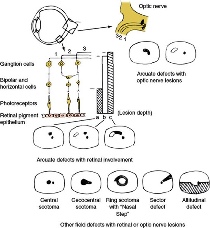
FIG. 7.2. Diagrammatic representation of the retinal layers, disposition of fibers in the nerve fiber layer and optic nerve, and visual field defects caused by retinal or optic nerve lesions. The vertical bars (a, b, c) represent partial (a) to complete (c) retinal lesions; the corresponding field defects are depicted underneath. Retinal lesions affecting the nerve fiber layer have an arcuate shape with the base located peripherally and, in temporal retinal lesions, in the horizontal meridian. Compare with Figure 7.1.
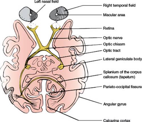
FIG. 7.3. Schematic horizontal section at the level of the lateral geniculate bodies, depicting the optic pathways. The right hemifield has been shaded, and fibers from the corresponding retina have been traced.
Nerve fibers nasal to the optic disc and those originating in the nasal side of the macula (papillomacular bundle) take a straight course as they converge into the optic disc (Fig. 7.1). The remaining fibers arch around the papillomacular bundle, adopting a disposition that has a bearing on the visual field defects that are secondary to retinal and optic nerve lesions. Fibers from the superior half of the temporal aspect of the macula arch superiorly and then down toward the disc. Fibers from the inferior half of the temporal aspect of the macula arch inferiorly and then ascend to reach the disc. Fibers from the temporal retina, particularly those closer to the horizontal meridian, follow a similar course. Therefore, between the nerve fibers from the superior temporal retina and those from the inferior temporal retina a raphe is formed, located in the horizontal meridian (Fig. 7.1).
The axons of the ganglion cells on the temporal side of a vertical line drawn through the fovea project to the ipsilateral lateral geniculate body, whereas the ones from the nasal side cross at the optic chiasm (Fig. 7.3). However, this separation is not sharp. The neurons subserving the macular region and a vertical strip of approximately 1 degree, centered in the fovea, project to either lateral geniculate body.
The Optic Nerves and Optic Chiasm
Each optic nerve is approximately 50 mm long and has four portions from the globe to the chiasm (Fig. 7.4).
1. Intraocular Portion. In this portion, also called the optic nerve head (1 mm long), the axons become myelinated (central type of myelin). The funduscopic appearance of the optic nerve depends on the angle of the nerve head to the eye. When the angle between the nerve and the sclera is <90 degrees, a rim or crescent of choroid or sclera may be seen on the flat temporal side of the disc, whereas the nasal edge appears elevated.
2. Intraorbital Portion. This (section 25 mm long) is shaped like an elongated S to allow mobility within the orbit. Here the optic nerve is surrounded by fat contained in the cone formed by the ocular muscles. The apex of this cone (which is open to the optic foramen and the superior orbital fissure) is directed posteriorly and slightly displaced nasosuperiorly in the orbit (Fig. 7.4). In addition to the ophthalmic artery, the ciliary ganglion and nerves, and the nerves to the extraocular muscles are in close relation to the optic nerve here.

FIG. 7.4. Superolateral view of the contents of the sella and cranial nerves in the cavernous sinus.
3. Intracanalicular Portion. This portion (approximately 9 mm long) is the part of the nerve that travels the optic canal. Each optic canal is oriented posterosuperomedially, at an angle that approximates 45 degrees to the sagittal and horizontal planes. The ophthalmic artery and some filaments of the sympathetic carotid plexus accompany the optic nerve within the optic canal.
4. Intracranial Portion. This part (approximately 4–16 mm long, depending on the position of the chiasm) stretches between the proximal opening of the optic canal and the chiasm (Fig. 7.4). Each optic nerve lies above the respective carotid artery as this vessel exits from the cavernous sinus and gives off the ophthalmic artery. Inferomedially, the optic nerve lies over the bony roof of the sphenoid sinus, which can be quite thin, and over the contents of the sella turcica when the chiasm is posteriorly placed. Superior to each optic nerve is the horizontal portion of the anterior cerebral artery, which is overlaid by the gyrus rectus of the frontal lobe, the olfactory tract, and the anterior perforated substance (Fig. 7.5). The anterior communicating artery is superior to the optic nerves or to the optic chiasm.
Proximal to the angled optic canal, the optic nerves maintain a 45-degree angle to the horizontal plane, and the chiasm is similarly tilted over the sella turcica, with the suprasellar cistern lying between them. The relation between the chiasm and the sella varies between individuals. In brachycephalic heads the chiasm tends to be more anterior and dorsal than in dolichocephalic heads. Autopsy studies have shown that in approximately 5% of individuals the chiasm overlies the anterior margin of the sella (prefixed chiasm), in 12% it lies over the diaphragma sellae, in 79% it is above the dorsum sellae, and in 4% it projects behind the dorsum sellae (postfixed chiasm). The chiasm is located below the suprachiasmatic recess of the third ventricle in close proximity to the hypothalamus. Above the chiasm are the lamina terminalis and the anterior commissure. Immediately posterior to it, the pituitary stalk runs an anteroinferior course. The optic tracts originate from the posterolateral corners of the chiasm (Fig. 7.5).
Nerve fibers in the optic nerve follow a topical arrangement similar to that found in the retina (Fig. 7.6). Superior retinal fibers run superiorly in the optic nerve, inferior fibers are below, and those from the temporal and nasal retina run in the corresponding parts of the optic nerve. In the proximal portion of the nerve, near the globe, the macular fibers occupy a wedge-shaped sector just temporal to the central vessels (Fig. 7.6). More distally, they shift toward the core of the nerve.

FIG. 7.5. Inferior aspect of the brain showing some relationships of the optic nerves, chiasm, and optic tracts.
At the chiasm, more than half of the fibers (those originating in ganglion cells of the nasal retina) cross to reach the contralateral optic tract (Fig. 7.3). The ratio of crossed to uncrossed fibers is approximately 53:47. Fibers from the inferior part of the nasal retina are ventral in the chiasm and loop into the proximal portion of the contralateral optic nerve (Wilbrand’s knee) before reaching the lateral aspect of the optic tract (Fig. 7.7). Those from the superior nasal retina remain dorsal in the chiasm and become medial in the optic tract.
The anatomic existence of Wilbrand’s knee has come into question. Wilbrand was restricted to examining human subjects who had undergone enucleation. In the enucleated eye, the nerve fibers atrophied and became distinct from the nerve fibers of the normal eye as seen on myelin staining. Horton, utilizing axon labeling techniques in nonenucleated monkeys, was unable to demonstrate crossing fibers looping into the contralateral optic nerve (Wilbrand’s knee) [72]. In one monkey that had undergone enucleation 4 years previously, however, nerve fiber topography similar to that described by Wilbrand was found. Horton hypothesized that Wilbrand’s knee may be an artifact of enucleation caused by atrophy of the optic nerve and not a normal anatomic finding. However, the concept of Wilbrand’s knee is still clinically useful (see subsequent text).

FIG. 7.6. Disposition of the ganglion cell axons in a cross-section of the optic nerve. A: Distal portion, near the globe. B: Proximal portion, where the macular fibers have shifted to the core of the nerve.

FIG. 7.7. Crossing of nasal fibers in the optic chiasm. Fibers from the inferior retina make a forward loop into the opposite optic nerve (Wilbrand’s knee). The existence of Wilbrand’s knee has recently been questioned (see text). (Figure modified from Hoyt WF, Luis O. Visual fiber anatomy in the infrageniculate pathway of the primate. Uncrossed and crossed retinal quadrant fiber projections studied with Nauta silver stain. Arch Ophthalmol 1962;68:94–138.)
Uncrossed fibers, originating from the temporal retina, maintain their dorsal or ventral position in the chiasm. The macular fibers, which constitute a large proportion of the total number of chiasmal fibers, are also crossed and uncrossed. However, the separation between temporal and nasal ganglion cells is not sharp. Crossed and uncrossed fibers originate in both nasal and temporal sides of the macula. In the optic tract, the macular fibers occupy a dorsal position.
The Optic Tracts and Lateral Geniculate Bodies
The optic tracts extend from the dorsolateral corners of the chiasm to the lateral geniculate bodies. From the chiasm the tracts run posterolaterally, limiting the hypothalamus to a triangular space; they then sweep around the cerebral peduncles, and, as soon as they cross them, reach the lateral geniculate bodies in the posterior part of the ventral aspect of the thalami (Fig. 7.3). Several large vessels are located below the optic tracts. The posterior communicating artery crosses their distal portion in the suprasellar cistern. In the perimesencephalic cistern, the posterior cerebral artery and the basilar vein of Rosenthal are apposed to the tracts. Inferolaterally, the uncus of the temporal lobe covers the proximal portion of each tract (Fig. 7.5).
The lateral geniculate body, a thalamic nucleus, provides a relay station for all the axons of the retinal ganglion cells subserving vision. Neurons from the lateral geniculate body project, by way of the optic radiations, to the pericalcarine cortex of the occipital lobe, which is the primary cortical area for vision (Fig. 7.3). The lateral geniculate body is in the roof of the perimesencephalic cistern (cisterna ambiens), just medial to the hippocampal gyrus of the temporal lobe. Anteriorly, the lateral geniculate body receives the optic tract and sends out the ventral optic radiations, which lie in a close association with the posterior limb of the internal capsule. Dorsolaterally, the lateral geniculate body is covered by the optic radiations. Dorsomedial to the lateral geniculate body, the auditory radiations, originating from the medial geniculate body, pass on their way to the transverse temporal gyrus of Heschl, where the primary auditory cortex is located. Superomedial to the lateral geniculate body is the pulvinar of the thalamus.
Shaped on midsection like Napoleon’s hat, with its concave aspect (hilus) facing inferoposteromedially, the lateral geniculate body has a deep brown color with stripes (striae) of white matter that are visible to the naked eye [76]. The geniculate neurons, as numerous as the fibers in the optic tract, are disposed in six laminae, numbered from I to VI, beginning from the hilus of the nucleus. Layers I, IV, and VI serve the contralateral eye, whereas II, III, and V are connected with the ipsilateral eye. These six laminae are clearly distinguished in the center of the lateral geniculate body, where the macular region of the retina is represented, but only one or two are present in the peripheral part of the nucleus, which receives axons from ganglion cells in the peripheral retina.
The postchiasmal shift in the position of the fibers (the superior retinal fibers become superomedial and the inferior fibers become inferolateral; Fig. 7.7) persists in the synaptic areas of the lateral geniculate body. This shift is straightened out in the optic radiations, where again the superior fibers correspond to the superior retina and those below to the inferior retina. A similar representation is found in the calcarine cortex.
The Optic Radiations
The optic radiations sweep posteriorly around the lateral aspect of the posterior portion of the lateral ventricles (Fig. 7.8), forming the external sagittal stratum, which is separated from the ventricle by the internal sagittal stratum, made up of occipitomesencephalic fibers. Three bundles can be distinguished in the radiations: (a) the upper bundle, originating in the medial part of the lateral geniculate body and corresponding to the superior retina, which courses through the deep parietal white matter and ends in the superior lip of the calcarine fissure; (b) the central bundle, originating from the medial part of the nucleus and serving the macular region, which travels through the posterotemporal and occipital white matter and ends in the posterior part of the calcarine fissure, on both lips; and (c) the lower bundle, originating from the lateral part of the nucleus and corresponding to the lower retina, which sweeps first anteriorly and then posteriorly around the temporal horn of the lateral ventricle (Fig. 7.8), terminating in the lower lip of the calcarine fissure. As they sweep lateral to the ventricle (Meyer’s loop), the lower radiations reach a point located approximately 5 cm behind the tip of the temporal lobe.
In the anterior part of the radiations, fibers corresponding to adjacent retinal units are spatially separated and the macular fibers, instead of being interposed between fibers from the superior and inferior peripheral retina, run medial to them. Also, fibers from either eye seem to have a similar anterior extent in Meyer’s loop, with those from the contralateral eye lying medial to the ones coming from the ipsilateral eye.
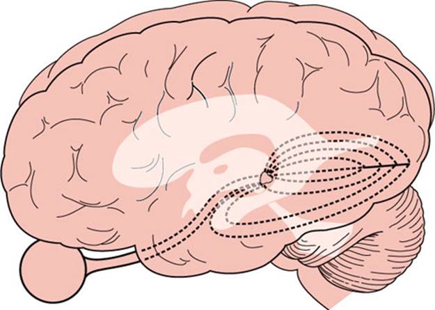
FIG. 7.8. Lateral view of the brain showing the arrangement of the optic radiations in the parietal and temporal lobes, lateral to the ventricular system.

FIG. 7.9. Schematic diagram showing arrangement of V1, V2, and V3 along the medial and posterior occipital surface. Most of V1 is buried within the calcarine fissure. (From Horton JC, Hoyt WF. The representation of the visual field in human striate cortex. A revision of the classic Holmes’ map. Arch Ophthalmol 1991;109:816. Copyright 1991, American Medical Association. Reprinted with permission.)
The Visual Cortex and Visual Association Areas
Cortical area 17 of Brodmann, located along the superior and inferior lips of the calcarine fissure in the medial aspect of the occipital lobe, receives the axons from the neurons of the lateral geniculate body and represents the first link in the cortical processing of visual information (primary visual cortex, see Chapter 20). The primary visual cortex actually extends farther than the posterior extent of the calcarine fissure, spreading for approximately 1 cm around the posterolateral aspect of the occipital pole. On cross-section of the cortex, a white matter stria (stria Gennari) can be seen with the naked eye. This characteristic feature has won the term striate cortex for area 17. The line of Gennari corresponds to a thick band of white matter in layer IV of the cortex, which is devoid at this point of pyramidal cells but is very rich in granular cells.
Each occipital lobe receives projections from the nasal half of the opposite eye and from the temporal half of the ipsilateral retina. More simply, it receives projections from the two halves of the retinas on the same side as the occipital lobe (Fig. 7.3). This unilateral representation includes the macular region. The superior and inferior retinal projections extend to the superior and inferior lips of the calcarine fissure, respectively. Finally, the macular retina is represented in the posterior pole of the calcarine cortex, whereas the more peripheral retina is more anteriorly represented [180]. The foveal representation is located at the occipital pole, where the striate cortex usually extends approximately 1 cm onto the lateral convexity of the occipital lobe. The extreme periphery of the visual field is represented anteriorly at the junction of the calcarine and parieto-occipital fissures. The central 10 to 15 degrees of vision fill most of the total surface area of the occipital cortex (as much as 50%–60%) (Figs. 7.9 and 7.10) [75,125].
Vascular Supply of the Visual Pathways
The vascular supply of the retina is derived from the ophthalmic artery, which branches from the carotid artery shortly after this vessel exits from the cavernous sinus. At the optic canal, the ophthalmic artery lies below and lateral to the nerve. At a point 5 to 15 mm from the globe, it gives off the central retinal artery, which pierces the optic nerve and courses forward in its core, to divide into a superior and an inferior branch at the optic disc (Fig. 7.1). Second-order nasal and temporal branches supply the nerve fiber layer and the inner layers of the retina (including ganglion cells). From the anatomic arrangement of these vessels, it follows that the complete occlusion of the central retinal artery results in global retinal ischemia, except when the macular area is supplied by cilioretinal arteries, and occlusion of one of its branches causes superior or inferior retinal ischemia. The consequence of such a lesion is an inferior or superior altitudinal field defect (Fig. 7.2). Infarction in the territory of the central retinal artery may be caused by emboli, thrombi, hypercoagulable states, migraine, and arteritis (e.g., giant cell arteritis).

FIG. 7.10. A: Artificially flattened map showing retinotopic organization of V1 (stippled area), V2 (small triangles), and V3 (hatched) in the left occipital lobe. B: Right visual field coordinates corresponding to map in (A). More than half of the visual cortex is devoted to processing the central 10 degrees of vision. (From Horton JC, Hoyt WF. The representation of the visual field in human striate cortex. A revision of the classic Holmes’ map. Arch Ophthalmol 1991;109:816. Copyright.)
In addition to the central retinal artery, the ophthalmic artery gives off dural branches (anterior falcine and recurrent meningeal arteries), orbital branches, and several posterior ciliary arteries. The posterior ciliary arteries form a rich anastomotic circle on the posterior aspect of the globe near the optic nerve and supply some sectors of the optic disc, the outer layers of the retina (including photoreceptors), and the choroid. In about half of the population, the region of the macula and papillomacular bundle receives its vascular supply from one or more cilioretinal arteries, which are branches of the posterior ciliary arteries (Fig. 7.1). This explains the sparing of central vision that occurs in some individuals despite central retinal artery occlusion (CRAO) with global retinal ischemia. Unlike ischemia in the territory of the central retinal artery, ischemia in the territory of the posterior ciliary arteries is seldom related to emboli but is usually caused by either atherosclerotic disease or hypotension (nonarteritic anterior ischemic optic neuropathy [AION]) or a vasculitis (arteritic ischemic optic neuropathy [ION] due to temporal or giant cell arteritis). However, field defects resulting from lesions in the territory of the posterior ciliary arteries are also altitudinal, and the differentiation of lesions in either territory rests on the ophthalmoscopic findings. Edema of the retina is obvious in the acute stages of retinal infarction owing to CRAO, but the retina may appear normal or show axonal swellings (cotton wool spots) in a segmental distribution in the presence of ischemia in the territory of the posterior ciliary arteries.
The distal part of the optic nerve (near the globe) is supplied by small branches of the ophthalmic artery and, as it approaches the chiasm, by thin vessels from the carotid and anterior cerebral arteries. Similarly, thin vessels originating in the region of the anterior communicating artery supply the dorsal aspect of the chiasm, whereas the inferior aspect receives arterioles from the carotid, posterior communicating, and posterior cerebral arteries. The latter two vessels also supply the optic tract, which in addition is fed by the anterior choroidal artery, a branch of the internal carotid. The lateral geniculate body receives a dual supply, from the anterior choroidal artery laterally and from the lateral posterior choroidal artery medially. The upper (parietal) portion of the optic radiations is supplied by branches of the middle cerebral artery, whereas the lower part receives branches from the posterior cerebral artery. The posterior cerebral artery, particularly its calcarine branch running in the calcarine fissure, supplies the primary visual cortex. Anastomotic branches from the middle cerebral artery (generally the angular or posterior temporal arteries) also play an important role in the vascular supply of the occipital pole, in which the macular region is represented.
Localization of Lesions in the Optic Pathways
The long course of the visual pathways along the base of the brain and their relative simplicity render them a very useful tool in lesion localization. Various techniques of neuro-ophthalmologic testing are reviewed by Glaser [48]. Detailed quantitative testing allows the following:
1. The detection of subtle deficits that may escape detection by bedside maneuvers
2. Better definition of abnormalities, such as the exact shape of a field defect, which may be important in lesion localization
3. The quantification of the extent and intensity of a deficit, which are very useful data when judging the evolution of a disease process
However, detailed testing requires equipment that is unavailable at the bedside and a degree of active cooperation that is often lacking in patients with brain disorders.
Lesions in the visual system may cause impaired visual perception or objective deficits. Impaired visual perception may include (a) poor discrimination of fine details of high contrast (visual acuity), which results in difficulty with tasks such as reading a printed page; (b) impaired color recognition; (c) impaired discrimination of objects that have little contrast with the background (contrast discrimination); and (d) visual field defects, the pattern of which is often the most helpful clue to lesion localization. Objectively, retinal changes caused by retinal or more proximal lesions can be seen with the ophthalmoscope, and an impaired pupillary response to light may betray a lesion in the afferent arc of this reflex.
Changes in Visual Perception
VISUAL ACUITY
Visual acuity, the capacity for visual discrimination of fine details of high contrast, such as small black letters on a white page, reflects the function of the macular region. A subnormal value of acuity indicates a fault in the visual system (e.g., optical faults, retinal lesions, or visual pathway lesions), faulty foveation (i.e., an eye motility defect), or poor cooperation, singly or in combination [42]. It remains unimpaired by unilateral lesions dorsal to the optic chiasm [42]. In practice, visual acuity is most often impaired by changes in the shape of the globe and in the refractory characteristics of the transparent media of the eye. Patients with these refractory defects regain a much better acuity when looking through a pinhole (pinhole test) because this maneuver restricts vision to the central beam of light, which is undisturbed by abnormal ocular distances or transparent media. At the bedside, visual acuity can be tested by asking the patient to read a “near card” with the Snellen optotypes printed on it. The card should be well-illuminated and held 14 inches in front of the patient’s eyes.
Once refractory defects have been excluded, it can be accepted that changes in visual acuity are secondary to lesions in the macular region or its projection. The macula is the only part of the retina that has high visual acuity. Virtually, all compressive and most noncompressive lesions of the optic nerve cause a drop in visual acuity, often even before a field defect can be detected. Medial chiasmal lesions behave in a similar manner. Lateral chiasmatic lesions tend to impair visual acuity in the ipsilateral eye only. From these findings and from the sparing of visual acuity that occurs with retrochiasmatic lesions, Frisen postulated that acuity will remain normal if either the crossing or the noncrossing set of nerve fibers from the fovea remains intact [42]. Both sets of fibers are often affected with medial chiasmatic lesions. Unilateral lesions of the optic tract, lateral geniculate body, visual radiations, or striate cortex do not impair visual acuity. When the retrochiasmal pathways are affected bilaterally, visual acuity fails to the same degree in both eyes.
CONTRAST SENSITIVITY
Contrast sensitivity testing may detect more subtle impairments in the function of the macula, optic nerve, and chiasm than visual acuity testing [18,103]. For instance, visual acuity may become normal after an acute ON, yet the patient may complain of “dimness” or “fuzzy vision” in that eye. This patient’s ability to perceive a series of bars that have very little contrast from the background will probably be abnormal. Impaired contrast sensitivity probably has localizing significance that is similar to that of impaired visual acuity, but it has been studied less thoroughly.
PERCEPTION OF COLOR
Color perception is often degraded in areas of the visual fields that correspond to a partial field defect. For instance, a scotoma for blue or for red may be demonstrated when vision for white targets is still good. In confrontation testing of the visual fields, one of the most useful techniques is to ask the patient which one of two identically bright red objects is more red, because a desaturation for red is often caused by lesions of the visual pathways. A color sample that appears red to the healthy eye appears more yellowish to the defective eye and passes from orange to yellow to colorless as disease severity increases. Impairment of color vision may also be detected by asking the patient to read numbers composed of an assembly of dots of different colors embedded in a background of differently colored dots (Ishihara or Hardy–Rand–Rittler pseudoisochromatic color plates). Color-blind patients cannot perform this task, which mainly reflects macular function. Because optic nerve and chiasmatic lesions often affect the macular fibers, monocular reading of the Ishihara or similar plates may be defective on the side of the lesion. However, Ishihara plates generally have poor sensitivity for acquired dyschromatopsia. It should also be noted that the interpretation of pseudoisochromatic color plates requires that the patient is able to “put the dots together” to make a visual whole. Therefore, patients with simultanagnosia (see Chapter 20) due to bilateral occipitoparietal damage (e.g., in the “posterior” form of Alzheimer’s disease) may have difficulty in identifying the images on the plates despite adequate visual acuity and the ability to name all the colors in the plates correctly [17].
Color vision loss usually parallels visual acuity loss (e.g., in ION), but in ON color vision loss may be much worse. In ON, chromatic sensitivity is more severely impaired than luminance sensitivity [132]. Another exception to the general rule of color vision failure paralleling visual acuity impairment is that color vision does not depend equally on perfect foveation and a well-focused retinal image, so that patients with nystagmus and anisometropia usually have normal color vision unless acuity is severely impaired. Acuity may also be normal with acquired achromatopsia due to cerebral cortical lesions [32,157].
Congenital color vision defects are much more common in men than women, with deficits mainly of red and green hues. Acquired color vision defects cluster primarily in the blue–purple or blue–green hues [131]. Therefore, bluish–purple objects may have superior sensitivity over red targets in assessing acquired dyschromatopsia. In general, patients with primary optic nerve disease frequently show a tendency for hue discrimination difficulties between reds and greens, whereas patients with primary retinochoroidal disorders more frequently show evidence of hue discrimination difficulties between blues and yellows (Köllner’s rule) [63]. However, Köllner’s rule has numerous exceptions (e.g., primary open angle glaucoma is an optic nerve disease characterized by blue–yellow deficits).
Another way of revealing impaired color processing involves the flight-of-colors phenomenon, which consists of a succession of color impressions that normally follows shining a bright light into the eye. This phenomenon is absent or reduced in duration with acquired dyschromatopsia [39].
Impairment of color perception also occurs with lesions in the posterior visual pathways. A visual field defect for red may betray the presence of a lesion when the fields for white stimuli are full. Patients with bilateral lesions of the inferomedial occipital region often have color blindness with normal visual acuity [32,157].
VISUAL FIELDS
The shape and distribution of visual field loss closely reflects the site of the lesion (Fig. 7.11). Therefore, careful plotting of the visual fields is most helpful in the localization of lesions of the visual pathways when examining a cooperative patient [42]. In patients with a markedly reduced attention span or other disturbances in alertness or mentation, the gross extent of the visual fields can be estimated from the patient’s response to moving objects in different quadrants. A moving object strongly induces the patient to look at it. However, small field defects are missed with confrontation techniques [184]. In cooperative patients, visual field testing with the tangent (Bjerrum) screen or static or kinetic perimetry provides a detailed map of the visual fields. Testing of the central 20 to 30 degrees of vision is most important because very few disease processes affect the peripheral fields alone; exceptions to this are the tapetoretinal degenerations and retinal detachment (both diagnosed by ophthalmoscopic exam) and anterior visual cortex lesions [42].
Adequate visual field testing requires patient cooperation and a skilled examiner. Shadowing facial contours (e.g., the eyebrows and nose), ptosis, disorders of eye motility, blinks, pupillary size, eyelashes, and spectacle rims must all be taken into account when interpreting the visual fields [42]. Ametropia, presbyopia, or both may affect the fields. For example, uncorrected astigmatism may cause an upper temporal depression suggesting a chiasmatic lesion; however, unlike a true chiasmatic defect, this defect does not respect the vertical meridian and spares central fixation [42]. Spherical ametropia may cause generalized field depression and occasionally an upper temporal depression, which may run under the blind spot (baring of the blind spot) due to local ametropia or local deviation from the normal retinal curvature [42].
By convention, in representing the visual fields, the field for the left eye is represented to the left of the field for the right eye (Fig. 7.11). Therefore, the nasal retina of the left eye “sees” the temporal field of the left eye. This terminology explains why a chiasmatic lesion that destroys the nasal fibers from both retinas causes a bitemporal hemianopia. Similarly, a macular lesion yields a central defect, whereas a lesion in the nasosuperior retina of the right eye results in a field defect in the temporoinferior portion of the visual field corresponding to the right eye.

FIG. 7.11. Visual field defects with chiasmatic and retrochiasmatic lesions. Visual fields from both eyes are usually abnormal. There is greater similarity between the field defects in each eye (congruity) with more posteriorly located lesions.
Any localized area of poor vision surrounded by areas of normal vision is termed a scotoma. The blind spot, the projection of the optic nerve in the visual field, is a physiologic scotoma that cannot be perceived because it lacks representation in the brain. Angioscotomata, the shadow images of the superficial retinal vessels on the underlying retina, is another type of physiologic scotoma that may be noted under certain circumstances. Absolute defects involving the outer limits of the visual field are called contractions, whereas depressions are smoothly tapering but not absolute deficits in the field [42].
Types of Visual Field Defects
A central defect occupies the position of the macula (Fig. 7.2). A cecocentral defect affects the area of the macula and of the papillomacular bundle. Nerve fiber bundle defects are field abnormalities in which at least part of the border coincides with the course of the retinal nerve fiber layer [42]. Peripheral nerve fiber bundle defects in the nasal field tend to have an arcuate shape when they are secondary to retinal or optic nerve disease, as they often are. The field defect takes this arcuate shape because of the disposition of the fiber layer in the retina and in the optic nerve (Fig. 7.2). Arcuate field defects may occur with glaucoma, AION, drusen of the disc, and congenital optic pits. Small deep retinal lesions result in a discrete defect localized to the point of the lesion, because the fiber layer remains unaltered. Larger lesions affect the superficial fiber layer and therefore give rise to a fan-shaped arcuate defect, with its tip pointing to the lesion and its base fanning peripherally and toward the nasal horizontal meridian. Nerve fiber bundle defects occur most commonly with lesions in the optic nerve head, where the tip of the defect reaches to the blind spot (Fig. 7.2), but may also occur with branch retinal artery or vein occlusion and with juxtapapillary inflammation. Defects in the temporal field lateral to the blind spot have the appearance of a sector rather than an arcuate shape. Visual defects in the temporal field do not “respect” the horizontal meridian because neither the blood supply nor the fibers of the nasal retina are arranged along a horizontal raphe (as opposed to temporal retinal fibers). The straight course of the retinal fibers of the nasal retina toward the nerve head explains this sector configuration (Fig. 7.1).
Enlargement of the blind spot is often noted with any process causing disc swelling (e.g., increased intracranial pressure); however, any peripapillary retinal disorder (e.g., the peripapillary conus or crescent seen with aging, myopia, or glaucoma and congenital optic nerve pit) may also enlarge the blind spot. These abnormalities are usually observed on ophthalmoscopic examination.
Occasionally a field defect has the appearance of a ring, with preserved vision central and peripheral to the scotoma. Usually the center coincides with the fovea. Annular or ring scotomas may occur with retinopathies or optic neuropathies. Retinitis pigmentosa often results in a large midperipheral ring scotoma. Cancer-associated retinopathy (CAR) syndrome may also cause a ring scotoma, often associated with funduscopic evidence of arteriolar narrowing and optic atrophy [29]. Ring scotomas of small diameter may occur with macular lesions, especially associated with a “bull’s-eye” appearance on ophthalmoscopy (e.g., chloroquine retinopathy [64]) or with the retinal disorder fundus flavimaculatus [156]. Annular or ring scotomas may also occur with retinitis, choroiditis, retinal migraine, and myopia. Paracentral and arcuate scotomas are characteristic of glaucoma; fusion of superior and inferior arcuate defects gives rise to ring scotomata. These ring-shaped defects have a characteristic horizontal or nasal step (Fig. 7.2), which distinguishes them from lesions located more distally in the visual pathways. Physiologic ring scotomas may be caused by corrective lenses, the prismatic effects of strongly curved correcting lenses, and the shallow ring scotoma surrounding the blind spot [42].
When only central vision is intact, the visual field is said to be narrowed, and the patient has funnel vision, not to be confused with tunnel vision, a field defect characteristic of hysteria or malingering. This latter field defect can easily be mapped onto a tangent screen by plotting the fields with the patient seated 1 and 2 m from the screen (the target size is doubled at 2 m) or can be detected with confrontation methods. Logically, with an organic field defect, the field projected at 2 m is larger than the field plotted at 1 m (funnel vision). Identical fields are obtained when the constriction of the field is not due to a lesion of the visual system. Constricted visual fields with retained acuity may be due to glaucoma, retinitis pigmentosa, CAR, hyaline bodies of the disc, postpapilledema optic atrophy, bilateral occipital infarcts with macular sparing, and feigned visual loss.
Hemianopia is a field defect that encompasses roughly half of the field, with a fairly sharp cutoff at the vertical or horizontal meridian (Fig. 7.11) [178]. Vertical hemianopia can be nasal or temporal. Horizontal or “altitudinal” hemianopia can be superior or inferior. When only one-fourth of the field is affected, the resulting deficit is called quadrantanopia.
Bilateral field defects are said to be homonymous when they are similarly located in both visual fields. They are congruous when there is a point-to-point correspondence of the defect in either field; otherwise they are called incongruous (Fig. 7.11).
Unilateral visual inattention refers to the phenomenon found in some patients with parieto-occipital lesions. No field defect is found on unilateral testing, but when stimuli are placed on both right and left hemifields, the patient appears not to see the object on the field opposite to the lesion. Unilateral visual inattention is often seen with incomplete homonymous defects and in the process of recovery of a dense field defect, particularly at the margins of the defect.
Dissociation of the perception of kinetic and static stimuli (Riddoch’s phenomenon) occasionally occurs with occipital lesions, and less often with lesions anywhere in the optic pathways (e.g., optic tract and optic chiasm lesions). In this case, the patient can still appreciate moving objects within a dense field defect for static stimuli [12]. In some cases, nonstriated projections might mediate visual function in the absence of striate cortex (e.g., through superior colliculus-pulvinar-prestriate cortex paths).
Localization of Visual Field Defects
Most important for lesion localization, is to note whether the field defect is monocular, in which case the lesion usually affects the retina or the optic nerve, or binocular, in which case the lesion is localized to or beyond the optic chiasm (Figs. 7.2 and 7.11). Obviously, multiple lesions in the visual pathways, which occur frequently with multiple sclerosis (MS) and other conditions, may result in bilateral loss even when the anterior optic pathways are involved. The pattern of the visual field loss can seldom differentiate retinal from optic nerve disease. However, retinal involvement generally accompanies obvious ophthalmoscopic abnormalities. Also, most optic neuropathies involve visual acuity; spared acuity should raise the suspicion of preretinal, retinal, or retrochiasmal disease [42].
Monocular visual field defects are almost always due to disease of the choroid, retinal pigment epithelium, retina, optic disc, or optic nerve. Lesions affecting the retina, nerve fiber layer, or optic nerve produce visual field defects in the ipsilateral eye, which correspond in position, shape, extent, and intensity to the lesion. Almost all retinal lesions resulting in visual field loss are visible ophthalmoscopically. Careful attention should be directed to the retina and retinal nerve fiber layer corresponding to the visual field defect. Patients with macular disease may also complain of metamorphopsia, micropsia, and positive photopsias (e.g., flashing lights), which are unusual in patients with optic neuropathies.
In assessing optic nerve-related visual field defects, several anatomic points are worth remembering:
1. Fibers from peripheral ganglion cells occupy a more peripheral position of the optic disc, whereas fibers from ganglion cells located closer to the disc occupy a more central position.
2. Peripheral fibers course peripherally through the entire extent of optic nerve.
3. The papillomacular bundle occupies a large sector-shaped region of the temporal disc. This bundle of fibers moves centrally in the more distal (posterior) portions of the orbital optic nerve.
4. All retinal fibers retain their relative positions throughout visual pathways except in the optic tract and at the lateral geniculate nucleus where there is a rotation of 90 degrees that becomes “straightened out” in the optic radiations.
Central visual field defects (unilateral or bilateral) are the result of damage to the papillomacular bundle or optic nerve. Any visual field defect produced by a retinal lesion may be produced by a lesion of the optic nerve [128] and virtually any etiology may be responsible (e.g., glaucomatous, degenerative, ischemic, traumatic, inflammatory, infiltrative, compressive, or vascular optic neuropathy). For example, unilateral central scotomas are often seen with ON, compressive optic neuropathies, or early macular disease. Bilateral central or cecocentral scotomas usually indicate hereditary (e.g., Leber’s hereditary optic neuropathy) or toxic-nutritional optic neuropathies, but may also be seen with bilateral macular lesions, bilateral compressive lesions affecting the optic nerves, or even bilateral lesions affecting the occipital poles. The clinical features and etiologies of bilateral superior and inferior altitudinal defects and bilateral central or cecocentral scotomas are noted in Table 7.1.
TABLE 7.1 Clinical Features and Etiologies of Bilateral Superior or Inferior Altitudinal Defects and Bilateral Central or Cecocentral Scotomas

Although monocular visual field defects are usually due to retinal or optic nerve disease, in the early stages of a chiasmatic lesion, the loss may be restricted to the temporal portion of the field corresponding to the ipsilateral eye [70]. This monocular (often scotomatous) temporal hemianopia (junctional scotoma of Traquair, Fig. 7.12) is attributed to the involvement of the ipsilateral optic nerve close enough to the chiasm to impair conduction selectively in ipsilateral crossing fibers but too anterior to affect nasal retinal fibers crossing from the fellow eye (i.e., nasal compression of the distal intracranial optic nerve ipsilateral to the defect) [70]. Also, lesions located in the most anterior extent of the calcarine cortex cause a crescent-shaped defect restricted to the temporal field of the contralateral eye from 60 to 90 degrees (monocular temporal crescent or “half-moon syndrome”) [26,109]. This is the only retrochiasmatic lesion that may cause a strictly unilateral visual field defect (Fig. 7.11). Similarly, an occipital lesion that spares the foremost part of the calcarine cortex results in a homonymous hemianopia that spares the unpaired temporal crescent (Fig. 7.11).
Monocular altitudinal defects (Fig. 7.2), which are often accompanied by macular sparing, are characteristic of disease in the distribution of the central retinal artery. Central vision may be spared because the blood supply for the macula often derives from the cilioretinal arteries (Fig. 7.1). AION (infarction involving the anterior portion of the optic nerve), due to ischemia involving the posterior ciliary arteries, is another common cause of an altitudinal (usually inferior) defect (see subsequent text). Other causes of a monocular altitudinal defect include choroiditis, choroidal coloboma, retinal detachment, glaucoma, optic nerve hypoplasia, chronic atrophic papilledema, drusen, ON, optic nerve trauma, and masses affecting the optic nerve or chiasm. Bilateral altitudinal defects may result from bilateral lesions, often ischemic, of the retinas or optic nerves, but bilateral occipital lesions, especially trauma or infarction, may also be responsible for this type of defect (Table 7.1) [62,94,106,138]. Rarely, a large prechiasmal lesion compresses both nerves inferiorly to cause bilateral superior altitudinal defects. The compression of nerves from below may also elevate them against the dural shelves extending out from the intracranial end of the optic canals and cause bilateral inferior altitudinal defects. Bilateral lesions of medial aspect of the lateral geniculate body may cause bilateral inferior altitudinal defects. It is important to emphasize that the nerve fiber layer of the retina respects the horizontal meridian only in the nasal field, not in the temporal field; therefore, incomplete altitudinal field defects are more common with retinal lesions. Because of the anastomotic blood supply of the occipital pole, only altitudinal defects due to occipital infarcts spare macular vision [62,106]. Diagnosis of retinal branch artery occlusion or AION is aided by the presence of a unilateral altitudinal defect along with ipsilateral funduscopic changes and, in most bilateral cases, by sequential temporal development; bilateral occipital infarcts are characterized by the sudden, simultaneous onset of altitudinal visual field defects with an absence of retinal or optic nerve abnormality or abnormalities of the pupillary response [106].
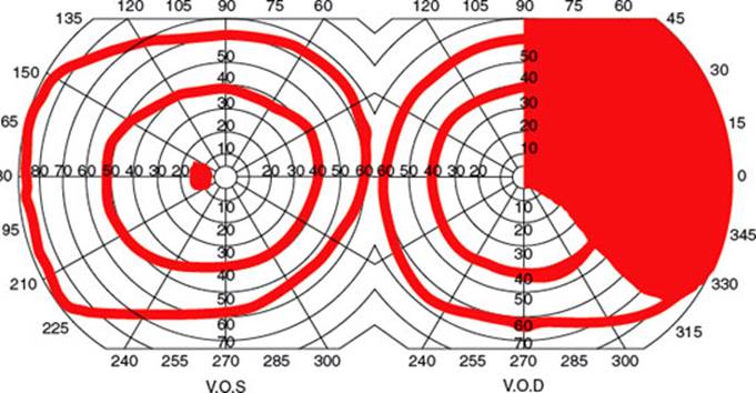
FIG. 7.12. Junctional scotoma of Traquair.
Bilateral ring defects may be the consequence of retinal disease, but bilateral occipital involvement can cause a similar field defect. In the case of occipital lesions, however, a vertical step can be regularly identified between the two halves of the ring (Fig. 7.11).
Bitemporal field defects [137] are most often due to a compressive mass lesion affecting the optic chiasm, such as pituitary tumors. Rarely, processes that cause rapidly developing hydrocephalus in children may result in bitemporal defects, perhaps through dilation of the optic recess of the third ventricle. True pure complete bitemporal hemianopias are rare because it is difficult for any pathogenetic mechanism, except trauma, to affect crossing fibers only. Bitemporal hemianopsia may be peripheral, paracentral, or central. The visual field defect may “split” or “spare” the macular central field. Certain anatomical relationships are important in evaluating chiasmal visual field defects:
1. The ratio of crossed to uncrossed fibers in the chiasm is 53:47.
2. Uncrossed fibers, both dorsal and ventral, maintain their relative position at the lateral aspects of the chiasm and pass directly into the ipsilateral optic tract.
3. Dorsal extramacular crossing fibers from each eye decussate posteriorly in the chiasm and then directly enter the dorsomedial aspect of contralateral optic tract.
4. Macular fibers that cross do so in the central and posterior portions of chiasm.
5. Some inferonasal retina fibers, primarily peripheral fibers, loop in Wilbrand’s loop (although the anatomic existence of this structure has been questioned).
Early chiasmal compression with pituitary tumors affecting crossing fibers in isolation usually results in relative rather than absolute defects limited to the central parts of the upper temporal quadrants. With increasing tumor growth, the contact area with the chiasm increases in size so that noncrossing fibers are always affected and acuity is impaired. Therefore, complete bilateral hemianopia almost never occurs in isolation and is usually combined with binasal depression and subnormal acuity [42]. Although most patients with midchiasmal compression demonstrate bitemporal superior visual depression, occasional patients may demonstrate bitemporal scotomas or, rarely, bilateral arcuate defects [48].
Clinically, three chiasmatic syndromes may be recognized (Fig. 7.13) [128].
1. The anterior chiasm or junctional syndrome (different from the junctional syndrome of Traquair, above), in which a unilateral optic defect is associated with a superior temporal defect in the other eye.
2. Body of the chiasm syndrome, in which patients demonstrate bitemporal visual field defects. These visual field defects may be peripheral, central, or a combination of both, with or without “splitting of the macula,” and may be quadrantic or hemianopic. Visual acuity is usually normal, and the optic discs are normal or pale.
3. The posterior chiasm syndrome, in which visual field testing reveals bitemporal scotomas (the peripheral visual fields are intact). Visual acuity and the optic discs are normal.
Lesions affecting the optic chiasm are listed in Tables 7.2 and 7.3. Superior bitemporal field defects may also occur with tilted discs, an optic disc anomaly in which the discs have an elliptical shape. This field anomaly differs from that due to a chiasmatic lesion in that with tilted discs, the defect crosses the median into the nasal field (i.e., does not “respect” the vertical meridian) [47,60]. Pseudochiasmal visual field defects (i.e., bitemporal defects that do not respect the vertical midline) may also be due to ametropia, astigmatism, colobomas, bilateral nasal retinal disease (e.g., schisis), glaucoma, and bilateral optic neuropathies. Rarely a bitemporal hemianopia due to retinal disease can respect the vertical meridian [170].
A central defect in one field with a superior temporal defect in the opposite field points to the involvement of the anterior angle of the chiasm, with damage of the ipsilateral optic nerve and of the loop made by the fibers from the inferonasal retina of the other eye (Wilbrand’s knee) (Figs. 7.7 and 7.13A). Because of its localizing implications, this type of visual field defect has been termed junctional scotoma(different from the junctional syndrome of Traquair, as explained in preceding text) [188]. Such junctional scotomas stress the importance of meticulous testing of the visual fields in the “normal” eye in patients with apparently unilateral visual impairment. As noted in the preceding text, the existence of Wilbrand’s knee has come into question. Nevertheless, whether Wilbrand’s knee exists anatomically, the localizing value of junctional visual field loss to the junction of the optic nerve and chiasm remains undiminished because chiasmal compression alone or ON affecting the junction of the posterior optic nerve and chiasm may result in the contralateral superotemporal visual field defect (junctional scotoma) [71,87].
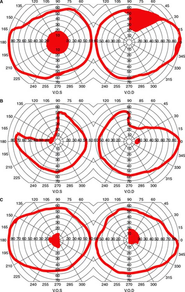
FIG. 7.13. Visual field defects with chiasm lesions: A: Anterior chiasm or junctional syndrome.B: Body of chiasm syndrome. C: Posterior chiasm syndrome.
TABLE 7.2 Compressive Chiasmal Syndromes
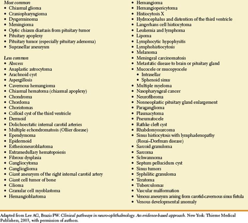
Trobe and Glaser noted that junctional visual field loss was due to a mass lesion in 98 of 100 cases [185]. The differential diagnosis of a junctional syndrome includes pituitary tumors, suprasellar meningiomas, supraclinoid aneurysms, craniopharyngiomas, and gliomas [188]. Chiasmal neuritis, pachymeningitis, and trauma are rare etiologies of the junctional syndrome [159,197]. Junctional visual field abnormalities may also occur on a functional (nonorganic) basis.
Binasal hemianopias and quadrantanopias may occur, are usually asymmetric, and often do not respect the vertical meridian. Binasal defects are usually due to bilateral intraocular disease of the retina or optic nerve (e.g., chronic papilledema, ION, glaucoma, optic nerve drusen, or retinal disease such as sector retinitis pigmentosa or retinoschisis) [171]. Rarely, bilateral compression of the lateral chiasm may result in a binasal defect [144]. Bilateral nasal defect may occur with hydrocephalus with third ventricle enlargement causing lateral displacement of optic nerves against the supraclinoid portion of the internal carotid arteries. Binasal defects have also been described in patients with primary empty sella syndrome and with other suprasellar lesions [25,120]. An unusual binasal visual field impairment has been noted with spontaneous intracranial hypotension from a dural cerebrospinal fluid leak [73]. Some of these patients have a binasal defect with peripheral depressions that are most severe in the upper nasal quadrants but also involving the lower nasal and upper temporal quadrants.
Homonymous hemianopias appear with lesions in the retrochiasmatic pathways [79]. Homonymous hemianopia may, therefore, be caused by lesions affecting the optic tract, lateral geniculate body, optic radiations, or occipital lobe. Rarely, an occipital lesion may cause a monocular field defect (see preceding text). Homonymous hemianopias affecting the tract and lateral geniculate body tend to be incongruous, but the more posteriorly the lesion is located in the optic pathways, the greater the congruity of the defect in either field. In general, tumors produce sloping field defects, whereas vascular lesions produce sharp field defects. Complete homonymous hemianopias are nonlocalizing and may be seen with any lesion of the retrochiasmal pathway, including lesions of the optic tract, lateral geniculate body, optic radiations, and striate cortex.
TABLE 7.3 Other Causes of Chiasmal Syndrome
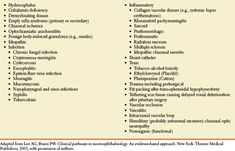
In the optic tract, macular fibers lie dorsolaterally, peripheral fibers from the upper retina are situated dorsomedially, and peripheral fibers from the lower retinas run ventrolaterally. Complete unilateral optic tract lesions cause a complete macular splitting homonymous hemianopia, usually without impaired visual acuity, unless the lesion extends to involve the optic chiasm or nerve [173]. Partial optic tract lesions are more common than complete lesions and result in an incongruous field defect that may be scotomatous [8,9,173]. The only other postchiasmal location for a lesion causing a scotomatous hemianopic visual field defect is the occipital lobe.
Optic tract lesions are often associated with a relative afferent pupillary defect (RAPD) (see subsequent text) in the eye with temporal field loss (contralateral to the side of the lesion) [8,20,145]. The RAPD that occurs in this setting reflects the difference in light sensitivity between the intact temporal and nasal hemifields. Its magnitude does not correlate with the difference in the number of crossed and uncrossed axons, but its sidedness contralateral to the side of the optic tract lesion is consistent with the greater percentage of decussating pupillomotor input [88]. Therefore, an afferent pupillary defect in the contralateral eye in a patient with normal visual acuity bilaterally and a complete homonymous hemianopia are usually indicative of optic tract involvement [128]. Another abnormality of the pupil that may occur with optic tract lesions is due to concurrent third nerve involvement by the pathologic process causing the tract damage. In these cases, the pupil ipsilateral to the lesion may be large and poorly reactive. Finally, many patients with chronic optic tract lesions develop bilateral optic atrophy with a characteristic “wedge,” “band,” or “bow tie” pallor in the contralateral eye (identical to that seen in some patients with bitemporal visual field loss from chiasmal lesions), and a more generalized pallor in the ipsilateral optic nerve associated with loss of nerve fiber layer in the superior and inferior arcuate regions corresponding to the bulk of temporal fibers subserving the nasal visual fields (hemianopic optic atrophy) [128,173]. Hemianopic optic atrophy indicates the involvement of the postchiasmal, preoptic radiations (i.e., optic tract or lateral geniculate body damage) but has also been rarely described in congenital retrogeniculate lesions [4,78,128]. Etiologies of optic tract lesions include space-occupying lesions (e.g., glioma, meningioma, craniopharyngioma, metastasis, pituitary adenoma, ectopic pinealoma, abscess, sella arachnoid cyst), aneurysms, arteriovenous malformations, dolichoectatic basilar artery, demyelinating disease, neurosyphilis, and trauma, including neurosurgical procedures (e.g., temporal lobectomy, insertion of intraventricular shunt, pallidotomy for parkinsonism) [11,28,55,57,82,111,117,128,164,173,178,191]. A congenital optic tract syndrome has also been described [134].
In the lateral geniculate body, axons from ganglion cells superior to fovea are located medially, axons originating from ganglion cells inferior to fovea are located laterally, and macular fibers terminate in a large central area. As axons leave the lateral geniculate body they rotate back to their original positions so that within the optic radiations and the striate cortex, fibers that have synapsed with axons from superior retinas are located in superior radiations and above the calcarine fissure in the striate cortex, whereas fibers that have synapsed with axons from the inferior retinas are located in the inferior optic radiations and below the calcarine fissure. Upper field fibers originate in the medial aspect of lateral geniculate nucleus and travel through the parietal lobes, while lower field fibers originate from the lateral aspect of the lateral geniculate body and make a loop in the temporal lobe (Meyer’s loop or the Meyer-Archambault loop). Lateral geniculate body lesions may also cause a complete macular splitting homonymous hemianopia [58,77,128]. Partial lesions result in an incongruous homonymous field defect. Hemianopic optic atrophy may develop and no RAPD is usually evident.
Although lesions of the optic tract or lateral geniculate body often cause incongruous field defects, two relatively specific patterns of congruous homonymous field defects with abruptly sloping borders, associated with sectorial optic atrophy, have been attributed to focal lesions of the lateral geniculate body caused by infarction in the territory of specific arteries. Occlusion of the anterior choroidal artery may cause a homonymous defect in the upper and lower quadrants with the sparing of a horizontal sector (quadruple sectoranopia) (Fig. 7.14A), which is essentially diagnostic of a lateral geniculate body lesion in the anterior choroidal artery distribution [41,68,119]. This defect occurs because the lateral geniculate body is organized in projection columns oriented vertically that represent sectors of the field parallel to the horizontal meridians, and the anterior choroidal artery supplies to the hilum and anterolateral part of the nucleus. Bilateral lateral geniculate lesions may therefore cause bilateral hourglass-shaped visual field defects or bilateral blindness [34,127]. Quadruple sectoranopia has also been described with posterior cerebral artery infarction [96]. As noted in the preceding text, the lateral geniculate body has a dual blood supply; therefore, interruption of the posterior lateral choroidal artery, which perfuses the central portion of the lateral geniculate, causes a horizontal homonymous sector defect (wedge-shaped) (Fig. 7.14B) [14,41,119,135,176,190,195]. A similar sector defect may occur with lesions affecting the optic radiations [23] or, rarely, with lesions affecting the occipital cortex in the region of the calcarine fissure [56], lesions of the temporo-occipital junction, parietotemporal lesions, or lesions in the distribution of the superficial sylvian artery territory [54]. Several patients have been described with bilateral lateral geniculate lesions with bilateral sector defects with the preservation of the visual fields in an hourglass distribution [53,133].
Patients with lesions of the lateral geniculate body may have no other signs or symptoms of neurologic involvement or may have associated findings related to thalamic or corticospinal tract involvement. Etiologies for lateral geniculate damage include infarction, arteriovenous malformation, trauma, tumor, inflammatory disorders, demyelinating disease, and toxic exposure (e.g., methanol) [14,34,53,55,68,101,119,127,135,176].
Superior homonymous quadrantic defects (“pie-in-the-sky” field defects; Fig. 7.11) may result from a lesion in the temporal (Meyer’s) loop of the optic radiations or in the inferior bank of the calcarine fissure. To cause a quadrantic defect the lesion has to be quite extensive; small lesions result in scotomata. In a study of 30 patients with superior quadrantanopias, lesions were occipital in 83%, temporal in 13%, and parietal in 3% [83]. In temporal lobe lesions, the superior quadrantic defect is usually, but not always, incongruous and the inferior margins of the defects may have sloping borders and may cross beyond the horizontal midline [128]. Also, the ipsilateral nasal field defect is often denser and comes closer to fixation than the defect in the contralateral eye. Macular vision may or may not be involved with the quadrantic defect [84,121,128]. Etiologies for temporal lobe dysfunction include space-occupying lesions (e.g., tumors, abscesses, hemorrhage), arteriovenous malformations, infarction, infections, congenital malformations, demyelinating disease, and trauma (e.g., temporal lobectomy) [80,84,121,178,187,203]. In one study, 36.6% of patients undergoing selective amygdalohippocampectomy for hippocampal sclerosis developed visual field defects owing to the interruption of the anterior bundle of the optic radiation fibers while opening the temporal horn through the inferior limiting sulcus of the insula [203].
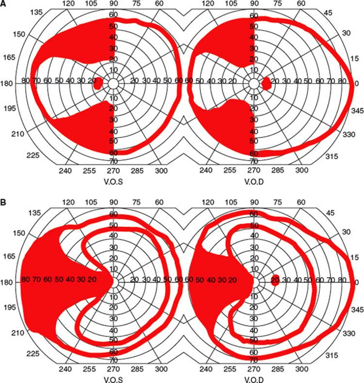
FIG. 7.14. Visual field defects seen with vascular lesions of the lateral geniculate body. A: Anterior choroidal artery lesion. B: Lateral choroidal artery lesion.
Hughes et al. studied the visual field defects in 32 patients after temporal lobe resection [80]. Visual field defects were present in 31 of the 32 patients but none of the patients was aware of the deficits. Points nearest fixation were relatively spared and defects were greatest in the sector closest to the vertical meridian in the eye ipsilateral to the resection. Ipsilateral and contralateral field defects differed in topography and in depth. This study, therefore, demonstrated that certain fibers from the ipsilateral eye travel more anteriorly and laterally in Meyer’s loop and supports the hypothesis that visual field defects due to anterior retrogeniculate lesions are incongruous because of anatomic differences in the afferent pathway [80].
The involvement of the optic radiations in the depth of the parietal lobe gives rise to an inferior quadrantic defect (“pie-on-the-floor” defect) (Fig. 7.11). Such defects are usually more congruous than those produced by lesions of the temporal lobe, and because the entire optic radiation passes through the parietal lobe, large lesions may produce complete homonymous hemianopia with macular splitting [128]. Patients may often be unaware of their visual field defects [152,193].
In a study of 41 patients with inferior quadrantanopias, 76% were due to occipital lesions, 22% to parietal lesions, and 2% to temporal lesions [83]. In patients with occipital lesions, the field defects often occurred in isolation, while other localizing signs of parietal involvement were evident in 89% of patients with parietal lesions. Therefore, although visual field defects may occur in relative isolation with parietal lobe lesions, lesions in this location more often betray themselves by other signs of neurologic dysfunction. Parietal lobe lesions may be associated with contralateral somatosensory impairment, including impaired object recognition, impaired position sense, impaired touch and pain sensation, and tactile extinction. Dominant parietal lesions may cause apraxia, finger agnosia, acalculia, right–left disorientation, alexia, and aphasic disturbances, whereas nondominant lesions may be associated with anosognosia (denial of neurologic impairment), autotopagnosia (failure to recognize hemiplegic limbs as belonging to self), spatial disorientation, hemispatial neglect, constructional apraxia (abnormal drawing and copying), and dressing apraxia [113].
Homonymous quadrantic visual field defects may occur with unilateral occipital lesions [74]. Often, these field defects have a sharp horizontal edge that would be difficult to develop with tumors or missile injuries because it is unlikely that they would injure only one bank of the calcarine fissure and leave the fellow calcarine bank untouched. Therefore, Horton and Hoyt [74] suggest that a lesion of the extrastriate cortex (areas V2 and V3) would more likely explain the sharp horizontal edge of the defect because areas V2 and V3 are divided along the horizontal meridian into separate halves flanking the striate (V1) cortex and, consequently, the upper and lower quadrants in the extrastriate cortex are physically isolated on opposite sides of the striate cortex. Although a lesion in this location (e.g., tumor) may have irregular margins, if it crosses the representation of the horizontal meridian in the extrastriate cortex, it produces a quadrantic visual field defect with a sharp horizontal border because of the split layout of the upper and lower quadrants of V2 and V3 [74]. A congruous inferior quadrantanopia with borders aligned on both the vertical and horizontal meridians has, however, also been described with a lesion of the superior fibers of the optic radiations near the contralateral trigone, where the fascicles of visual axons become compact as they approach the calcarine cortex [14]. Therefore, a homonymous quadrantanopia respecting the horizontal meridian is not a “pathognomonic” sign of extrastriate cortical disease but may occur with striate lesions [126]. A congruous inferior quadrantanopia with borders aligned on both the vertical and horizontal meridians has also been described with a lesion of the superior fibers of the optic radiations near the contralateral trigone where the fascicles of visual axons become compact as they approach the calcarine cortex [15].
Medial occipital lesions [75,158] cause highly congruous homonymous hemianopias (Fig. 7.11). When both the upper and the lower calcarine cortices are affected, a complete homonymous hemianopia, usually with macular sparing, develops. Sparing of the central 5 degrees of vision (macular sparing) is common with occipital lesions, probably due to a combination of a large macular representation and dual blood supply [128]. The central 10 to 15 degrees of vision fill most of the total surface area of the occipital cortex (as much as 50%–60%) [52,75,126,202]. Occipital infarcts in the distribution of the posterior cerebral artery are a common cause of such field defects and are most commonly due to emboli to the basilar apex (e.g., from a cardiac source, or vertebrobasilar atherosclerotic occlusive disease) [158].
Patients with purely occipital lesions are partially or fully aware of the hemianopia, whereas patients with larger or more anterior lesions, affecting parietal regions or associative pathways to the primary or secondary visual association cortex, may be unaware of their deficit [99]. Celesia et al., however, studied prospectively 32 consecutive patients with homonymous field defects due to ischemic infarcts and found hemianopic anosognosia, defined as the unawareness of visual loss in the homonymous hemifield (or hemiquadrant), in 20 patients (62%) [24]. Hemianopic anosognosia occurred predominantly in right-sided lesions (16/26 patients or 62%), but was also present in four of six patients (or 67%) with left-sided lesions. Hemianopic anosognosia was associated with somatic anosognosia in nine patients and hemineglect in 17 patients. Eight patients had pure homonymous hemianopia without cognitive, motor, or somatosensory deficits, four of these patients had awareness of the visual defect, and three patients had hemianopic anosognosia. Patients in these two groups had similar anatomic lesions. Patients with phosphenes, photopsias, or visual hallucinations were usually aware of their visual field loss. The authors suggest that hemianopic anosognosia is most often related to failure of the discovery of the deficits, occasionally with severe visual hemineglect, sometimes to generalized cognitive impairment, or to a combination of these factors. The authors further conclude that (a) there is no specific cortical area for conscious visual perception; (b) visual awareness is processed by a distributed network including multiple visual cortices, parietal and frontal lobes, the pulvinar, and the lateral geniculate bodies (lesions localized at various nodes or centers in the network may produce similar phenomena); and (c) both hemispheres are involved in visual processing and conscious awareness [24].
Lesions of the striate cortex may be classified into anterior, intermediate, and posterior locations [10,75,109,124,126,128]. Anterior lesions lie adjacent to the parieto-occipital fissure and affect the monocular temporal crescent of the contralateral visual field (temporal crescent or half-moon syndrome) [109]. This area constitutes <10% of the total surface area of the striate cortex. Conversely, the temporal crescent may be spared with lesions that destroy the entire calcarine cortex except for the anterior tip [109,114]. Posterior lesions are located in the posterior 50% to 60% of the striate cortex, including the occipital pole and operculum, and affect macular vision (i.e., the central 10 degrees in the contralateral hemifield). Intermediate lesions lie between the anterior and posterior confines and affect from 10 to 60 degrees in the contralateral hemifield (Figs. 7.9 and 7.10) [75,125].
Gray et al. report two patients with unique homonymous hemianopias from occipital lesions [51]. One patient had vertical meridian sparing and the other displayed horizontal meridian sparing. Magnetic resonance imaging (MRI) correlation with the defects confirmed that the vertical hemianopic meridian is represented along the border of the calcarine lip and the horizontal meridian lies at the base of the calcarine banks deep within the calcarine fissure. Galetta and Grossman reported two patients further demonstrating that the horizontal meridian is represented at the calcarine fissure base in the primary visual cortex [44].
The most common cause of unilateral occipital disease is infarction in the distribution of the posterior cerebral artery [6,7,10,43,46,124,158,172]. Other etiologies include venous infarction, hemorrhage, arteriovenous malformation and fistulas, tumor, abscess, and trauma [5,104,117,129,130,189].
The clinical-anatomic correlations of homonymous hemianopia (HH) was studied in 904 cases [205]. HH were found in 852 patients. A total of 340 HH (37.6%) were complete and 564 HH (62.4%) were incomplete. Homonymous quadrantanopia (264 HH, 29%) was the most common type of incomplete HH, followed by homonymous scotomatous defects (116 HH, 13.5%), partial HH (114 HH, 13%), and HH with macular sparing (66 HH, 7%). A total of 407 HH (45.0%) were isolated. Causes of HH included stroke (629 HH, 69.6%), trauma (123, 13.6%), tumor (102, 11.3%), brain surgery (22, 2.4%), demyelination (13, 1.4%), other rare causes (13, 1.4%), and unknown etiology (2, 0.2%). The lesions were most commonly located in the occipital lobes (45%) and the optic radiations (32.2%) [205]. The same authors noted that in patients with HH due to stroke, 84.4% were from infarction and 15.6% from primary intraparenchymal hemorrhage [206]. Spontaneous improvement of homonymous hemianopia is seen in at least 50% of patients first seen within 1 month of injury [204]. In most cases, the improvement occurs within the first 3 months from injury. Spontaneous improvement after 6 months postinjury should be interpreted with caution as it is most likely related to improvement of the underlying disease or to improvement in the patient’s ability to perform visual field testing reliably [204].
Kedar et al. evaluated the value of congruency in the localization of brain lesions in patients with homonymous hemianopia (HH) [93]. Five hundred and thirty patients with 548 incomplete HH were included (373 congruent HH and 175 incongruent HH). Stroke caused 75% of congruent HH and 55.8% of incongruent HH; trauma and tumors caused 20.5% of congruent HH and 34.5% of incongruent HH. The lesion locations in congruent HH versus incongruent HH included occipital lobe in 47.9% versus 21.3%, occipital lobe and optic radiations in 8.3% versus 5.6%, optic radiations in 32.4% versus 50.6%, optic tract in 7.2% versus 16.3%, and other locations in 4.2% vs 6.3%. Although there was a trend toward more congruent HH for lesions of the posterior visual pathways, 50% of optic tract lesions and 59% of optic radiation lesions produced congruent HH. The authors concluded that although lesions involving the occipital lobe characteristically produce congruent HH, at least 50% of lesions in other locations also produce congruent HH, especially if these lesions are stroke related. They suggested that the rule of congruency should be used cautiously and may not apply to optic tract lesions [93].
Bilateral occipital lobe lesions may occur from a single or from consecutive events and may cause bilateral homonymous scotomas, usually with some macular sparing (“ring” scotomas), that respect the vertical midline [69,128]. In some cases there may be “keyhole” fields with bilateral complete homonymous hemianopias except for macular sparing. Careful testing in these cases reveals that the macular sparing respects the vertical midline. Bilateral lesions affecting the superior or inferior calcarine cortices may produce bilateral altitudinal defects that may mimic the visual field abnormalities seen with bilateral optic nerve or retinal disease [62,106,138,147].
Bilateral homonymous hemianopia (double hemianopia) may occur from a single or from consecutive events and may result in cortical blindness. Cortical blindness is most often due to simultaneous or successive posterior cerebral artery occlusion. There are many etiologies of cerebral and cortical blindness, including hypoxia, infarction, hemorrhage, eclampsia, preeclampsia, hypertensive encephalopathy, tentorial herniation from cerebral mass, tumor, arteriovenous malformation, infection (e.g., progressive multifocal leukoencephalopathy, Creutzfeldt– Jacob disease, subacute sclerosing panencephalitis, human immunodeficiency virus encephalitis, syphilis, encephalitis, abscess), inflammation (e.g., sarcoidosis), demyelinating disease, trauma, migraine, metabolic disorders (e.g., adrenoleukodystrophy, hypoglycemia, porphyria, mitochondrial encephalopathies), toxins (e.g., lead, mercury, ethanol, carbon monoxide), scorpion sting, alcoholic ketoacidosis, medications (e.g., cyclosporine, tacrolimus, interleukin-2), reversible posterior leukoencephalopathy syndrome (RPLES) (e.g., due to thrombotic thrombocytopenic purpura, bilateral carotid artery dissection, hypertension, preeclampsia, drugs), pre-eclampsia, radiation encephalopathy, Alzheimer’s disease, postictal after seizures, and complications of cerebral angiography [3,92]. These patients may be left with a small central field around the point of fixation (macular sparing or keyhole vision) or may have complete blindness. Occasionally, patients with cortical blindness deny their visual defect (Anton’s syndrome).
OTHER CHANGES IN VISUAL PERCEPTION
Patients with lesions in the anterior optic pathways usually complain of difficulty in reading and the dimming of vision. Altitudinal field defects are often described as a curtain coming down or the sensation of looking over the horizon. Vertical hemianopic defects are often detected when the patient finds himself colliding with objects in the blind field or is unable to see half of the page or the keyboard. Other, less-common subjective complaints also have some localizing value.
Metamorphopsia (objects appearing misshapen), micropsia (objects appearing reduced in size), and macropsia (objects appearing enlarged) may be due to retinal disease, which causes displacement of the receptor cells. Micropsia is probably related to excessive separation of the photoreceptors by edematous fluid (macular edema), whereas retinal macropsia is caused by the retinal photoreceptors being closer together than normal (e.g., from macular scarring). Micropsia may rarely occur with lesions of the optic chiasm. Irregular distortion (irregular metamorphopsia) results when the photoreceptors are no longer evenly spaced (e.g., scarring of the retina or retinal traction). Distorted perception of the shape and size of objects can also occur with occipital or temporal lobe disease. In this case the misperception is often transient because it is linked to the prodroma of migraine or to focal seizures. Hemimicropsia is a rare disorder of visual perception characterized by an apparent reduction of the size of objects when presented in one hemifield [30]. Hemimicropsia may result from contralateral focal lesions affecting the unimodal visual association cortex in areas 18 and 19 and the underlying white matter [30].
ON may cause movement phosphenes (a sensation of flashes of light when moving the eyes in the dark) [33]. This phenomenon may originate within the optic nerve and represent the visual equivalent of Lhermitte’s sign [33] (see Chapter 5) with the increased mechanosensitivity of the area of demyelination resulting in “spontaneous” impulse generation. Persistent photophobia may occasionally be a symptom of compressive lesions of the chiasm [91].
Albinism, cone degeneration, achromatopsia, and corneal, lenticular, and vitreous opacities may cause a painless intolerance of the eyes to bright light, called dazzle [59]. Central dazzle may occur with lesions of the optic nerves, chiasm, thalamus, occipitotemporal region, or brainstem [31,35,169]. Central dazzle has even been described with trigeminal sensory neuropathy secondary to a lesion in the trigeminal nucleus [59].
Patients with chiasmatic lesions and bitemporal hemianopia may lose central vision when their eyes converge, because convergence makes the bitemporal defects overlap. This deficit stands in the way of activities such as threading a needle or drawing. Chiasmatic lesions may also cause image displacement in the absence of damage to the ocular motor nerves. Small motor imbalances, which are easily compensated by binocular fixation when the fields are full, are manifest in the presence of a bitemporal defect by horizontal or vertical deviation of the images from either eye (hemifield slide phenomenon).
Visual hallucinations may be seen with optic nerve and retinal disease. Retinal-associated hallucinations occur in the form of flashes of light referred to as retinal phosphenes (lights without structure) or photopsias (lights with geometric shapes). They are due to stimulation of retinal photoreceptors by diseased states (e.g., inflammation), traction (e.g., retinal detachment), or mechanical events (e.g., trauma). Entopic ocular phenomena are not hallucinations but are visual sightings of ocular structures. Posterior vitreous detachment is a common condition that results in brief bursts of light flashes, especially with eye movements in the dark, and floaters in the vision. Optic disc edema from any cause may also cause photopsias or phosphenes by irritation of the surrounding retina by edema. Simple or unformed hallucinations may also occur in patients with ON without significant optic disc edema.
Bilateral posterior brain lesions affecting the lateral temporo-occipital cortex and underlying white matter, especially the upper part of the occipital gyri and adjacent portion of the middle temporal gyri, may cause an unusual and severe disturbance of movement vision [207]. This disorder is characterized by a difficulty in perceiving motion stimuli in general, whereby all moving objects induce very unpleasant and disturbing experiences, especially when moving at higher velocities. The selectivity of the movement vision deficit and the irreversibility of the disorder support the idea that movement vision is a separate function that is subserved by a visual pathway specialized for the processing of visual motion [207].
Objective Findings with Lesions of the Optic Pathways
In addition to neurologic abnormalities, such as ocular motor paresis or hemiparesis, that are due to the involvement of neighboring structures, lesions in the optic pathways may betray their presence by changes in the appearance of the optic nerve or retina or by impairment of the afferent arc of the light reflex.
OPHTHALMOSCOPIC APPEARANCE OF THE RETINA AND OPTIC NERVE
Lesions of the retina and optic nerve may produce identical visual field defects and loss of visual acuity. However, retinal lesions are often apparent on ophthalmoscopic examination. Chronically increased intraocular pressure in glaucoma results in cupping of the optic disc. This finding is evident by the time visual acuity decreases or arcuate scotomas appear.
Many optic nerve lesions cause an initial swelling of the optic nerve head, appreciable with the ophthalmoscope, followed in time by optic atrophy. In general, the appearance of the optic nerve (e.g., normal, swollen, or pale) is not specific and cannot differentiate among various possible etiologies for optic neuropathy. Trobe et al. reviewed 163 color fundus photographs of several entities resulting in optic atrophy, including glaucoma, CRAO, ION, ON, hereditary optic neuropathy (Leber’s and non-Leber’s types), compressive optic neuropathy, and traumatic optic neuropathy [186]. These photographs were reviewed by five ophthalmologists as “unknowns.” Glaucoma, CRAO, and ION were correctly identified as the etiology by at least one of the five observers with an accuracy above 80%, but the remaining etiologies were correctly identified in <50% of cases! Helpful features in differentiating the entities included the following:
1. The presence of retinal arteriolar attenuation and sheathing in ischemic lesions (e.g., CRAO or ION)
2. Temporal pallor in entities selectively involving central vision and central visual field with sparing of peripheral visual field (e.g., ON and toxic optic neuropathies)
3. Superior or inferior (sector) optic disc pallor in ION
Although optic disc cupping was often identified in glaucoma, it was also seen in 20% of cases not associated with glaucoma. Optic disc cupping in glaucoma cases, however, was more profound than in nonglaucomatous cases and greater neuroretinal rim pallor occurred in the nonglaucomatous cases. In patients with glaucoma, there is often absence of at least part of the neuroretinal rim and the color of the remaining rim is normal. With nonglaucomatous optic neuropathy, rarely is any area of the rim completely absent and the remaining rim is often pale. Interestingly, only 11% of these cases with a known history of papillitis or ION had sufficient clues to identify previous disc swelling [186]. Another study suggested that optic disc appearance may help differentiate AION from ON although there are overlapping features [192]. Altitudinal disc swelling was more than three times more common in AION than ON, although most discs were diffusely swollen. Most patients with AION had hemorrhages, while most ON cases did not. Almost all discs with ON had normal color or were hyperemic; only 35% of discs with AION had pallid swelling. Pallid swelling was so rare in ON, however, in that of the discs with pallor, 93% had AION. Arterial attenuation was also much more typical of AION. AION was the clinical diagnosis in 82% of cases with altitudinal edema, in 81% of the cases with disc hemorrhage, in 93% of the cases with pallid edema, and in 90% of the cases with arterial attenuation. A pale nerve with hemorrhage, regardless of the type of edema, always represented AION (100%). A normal color nerve without hemorrhage reflected ON in 91% of the cases, increased from only 76% if hemorrhage was not considered. A hyperemic nerve with hemorrhage represented AION in 82% of cases, but if altitudinal edema was also present, AION incidence increased to 93%.
Papilledema (optic disc swelling secondary to increased intracranial pressure from any cause) is manifest by disc hyperemia (dilatation of capillaries in the disc surface), disc swelling, loss of venous pulsations, and blurring of the disc margins, followed later by associated retinal hemorrhages and exudates [128]. The presence of venous pulsations synchronous with the arterial pulse is a reliable indicator of intracranial pressure below 180 to 190 mm of water; absent venous pulsations may be found in normal individuals and in patients with increased intracranial pressure [116]. Syndromes causing increased intracranial pressure are outlined in Table 7.4 [104].
TABLE 7.4 Syndromes Causing Increased Intracranial Pressure
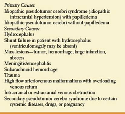
The clinical features and clinical stages of papilledema are outlined in Tables 7.5 and 7.6.
Patients with a history of a ventriculoperitoneal shunt for hydrocephalus may develop papilledema and visual loss or signs of a dorsal midbrain syndrome (see Chapter 8) due to shunt failure. Usually computed tomography or MRI reveals recurrence of the hydrocephalus. However, in some individuals shunt malfunction may occur without apparent ventriculomegaly, perhaps due to “stiff ventricles” [89,110,141]. Shunt revision is, therefore, indicated when there are signs or symptoms of increased intracranial pressure, even if ventriculomegaly is absent, to prevent deterioration of visual function and potentially irreversible visual loss.
TABLE 7.5 The Clinical Features of Papilledema
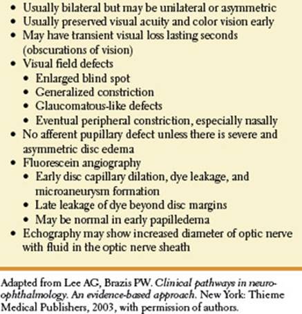
TABLE 7.6 The Stages of Papilledema
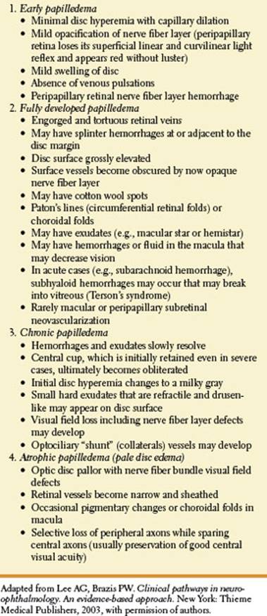
True disc swelling must be distinguished from pseudopapilledema and anomalously elevated discs caused by optic nerve head drusen [105,118,166]. Pseudopapilledema, with or without optic disc drusen, is not an uncommon condition. Drusen of the disc may be obvious, tiny, or buried. Ophthalmoscopic criteria that distinguish pseudopapilledema form true papilledema include the following [48]:
1. An absent central cup with small disc diameter
2. Vessels arise from the central apex of the disc
3. Anomalous branching of vessels, increased number of disc vessels, venous pulsations present
4. Disc may be transilluminated, with glow of drusen when present
5. Disc margins irregular with derangement of peripapillary retinal pigment epithelium
6. Absence of superficial capillary telangiectasia
7. No hemorrhages (rare exceptions)
8. No exudates or cotton wool spots
Other disc anomalies that may be mistaken for papilledema include “crowded” or hyperopic discs and tilted discs. In these cases, the peripapillary nerve fiber layer and the retinal vessels that traverse it remain normal, venous pulsations are usually present, there is no vascular engorgement or hemorrhages, there are no cotton wool spots, and the discs do not leak dye on fluorescein angiography. Myelinated nerve fibers may occasionally resemble disc swelling but are characterized by a white feathery appearance. Hyaloid traction on the optic disc and epipapillary glial tissue may also occasionally be mistaken for disc swelling.
When a lesion affects both optic nerves successively, optic atrophy may be seen in the eye involved earlier while the other still has disc edema (Foster-Kennedy syndrome). True Foster-Kennedy syndromes are associated with depression or loss of the sense of smell. Foster-Kennedy syndrome due to a basofrontal tumor (e.g., olfactory groove meningioma) may result from direct compression of the optic nerve on the side of the lesion, whereas contralateral papilledema is due to increased intracranial pressure. For example, intracranial meningiomatosis caused a Foster-Kennedy syndrome by unilateral optic nerve compression and blockage of the superior sagittal sinus resulting in increased intracranial pressure [1]. Bilateral direct optic nerve compression is a more common cause of Foster-Kennedy syndrome; however, the syndrome may even occur with longstanding increased intracranial pressure without direct optic nerve compression [194]. Pituitary adenoma may rarely cause the Foster-Kennedy syndrome [102]. Although classically described with frontal lobe tumors on the side of the optic atrophy, a pseudo-Foster-Kennedy syndrome occurs more often with bilateral and sequential ON, AION, arachnoiditis, syphilis, and occult trauma [174]. Lesions in the chiasm, optic tract, or lateral geniculate body may also induce optic atrophy.
Striking optociliary shunt vessels may appear in the region of the disc or at the disc margins in cases of chronic increased pressure in the optic canal or cranial cavity. They represent anastomotic channels between the central retinal vein and the peripapillary choroidal venous system, which are enlarged in an effort to bypass the compressed venous channels of the optic nerve. They are most commonly seen with central retinal vein occlusion or optic nerve sheath meningiomas but also occur with optic nerve glioma, neonatal hydrocephalus, pseudotumor cerebri, drusen of the optic disc, glaucomatous optic atrophy, high myopia, chronic atrophic papillitis, arachnoid cyst of the optic nerve, neurofibromatosis, optic nerve coloboma, and osteosclerosis [122].
Because the nerve fiber layer of the retina is composed of axons of ganglion cells on their way to the lateral geniculate body, any lesion between this nucleus and the eye may cause changes in the ophthalmoscopic appearance of the fiber layer. These changes, better appreciated with red-free light, may have one of four basic patterns: slit or rake defects, sector atrophy, diffuse atrophy, or density changes in the nerve fibers themselves [142]. Because the course of the axons through the anterior optic pathways is known, the retinal distribution of these changes may suggest the location of the process. For instance, chiasmatic lesions affect the fibers nasal to the optic disc and those nasal to the macula (papillomacular bundle; Figs. 7.1 and 7.2). As a result, sector atrophy occurs at both sides of the disc, and the remaining fibers, somewhat thinned out, adopt the shape of a vertically disposed bow tie, with the knot at the optic disc. This pattern is present in both eyes when the lesion is in the chiasm but may appear in only one eye, the one contralateral to the lesion, when the lesion is in the optic tract or geniculate body [173].
PUPILLARY LIGHT REFLEX
The fibers that constitute the afferent arc of the pupillary light reflex leave the visual sensory pathway just before the lateral geniculate body, without synapsing in it, to reach the dorsal midbrain. Retinal lesions must be quite large to impair the light reflex, but changes in pupillary responses, particularly the so-called RAPD or Marcus Gunn pupil, are very helpful in detecting asymmetric optic nerve or chiasmatic lesions. This pupillary sign is characterized by a normal bilateral pupillary response when the sound eye is illumined, but pupillary dilatation occurs when the flashlight is quickly switched to the diseased side [181]. Anisocoria larger than 2 mm in diameter may induce a small clinically RAPD in the eye with the smaller pupil [108].
Optic tract disease may cause a modest RAPD when the light is directed into the eye contralateral to the lesion [173]. It was thought that the RAPD resulted from the fact there are more crossed than uncrossed fibers (ratio 53:47 of crossed to uncrossed fibers) running in the optic tract. However, in patients with a unilateral optic tract lesion, the pupillary responses from full-field stimulation to each eye are the same as comparing the functioning temporal field with the functioning nasal field [88]. The percentage of decussating fibers is reflected in the ratio of the maximal pupil contraction amplitudes resulting from stimulus input between the two eyes. The RAPD that occurs in this setting reflects the difference in light sensitivity between the intact temporal and nasal hemifields. Its magnitude does not correlate with the difference in the number of crossed and uncrossed axons, but its sidedness contralateral to the side of the optic tract lesion is consistent with the greater percentage of decussating pupillomotor input [88]. RAPD without visual dysfunction may occur with lesions that selectively interrupt the pupillary afferents to the pretectal nucleus (e.g., lesions of the brachium of the superior colliculus) or damage the pretectal nucleus itself [45,90,97,153]. RAPD without visual dysfunction has been described with cerebral infarction, tumors of the pineal region, thalamic tumors, thalamic and midbrain glioma, and brainstem arteriovenous malformations [27,37,40,85,97]. A lesion affecting the brachium of the superior colliculus and the adjacent trochlear nucleus or fascicle (e.g., mesencephalic astrocytoma) may cause a contralateral RAPD without visual impairment and a contralateral superior oblique paresis [36]. Therefore, an RAPD may occur with disease of the macula, retina, optic nerve, optic chiasm, optic tract, brachium of the superior colliculus, or pretectal nucleus [38] (Table 7.7). Macular disease must be substantial and usually easily visualized on funduscopic examination to produce an RAPD. Lateral geniculate bodylesions and those in the geniculocalcarine segment of the optic sensory pathways leave the pupillary light reflex unimpaired. In functional visual loss, there is no RAPD.
Cataracts, even when very dense and pigmented, produce little or no RAPD and, in fact, often increase the pupillomotor effectiveness of light [107,181]. Therefore, an RAPD may occur contralateral to an eye with a unilateral dense cataract, with the RAPD disappearing after cataract extraction [107]. If an RAPD is found in an eye with a cataract, a visual pathway defect should be suspected. An RAPD may occur in an eye with better visual acuity when visual loss is due to abnormality of the ocular media (e.g., corneal opacity, hyphema, anterior segment membrane, cataract, or vitreous opacity), amblyopia, refractive error, age-related macular degeneration, or cystoid macular edema [19]. An RAPD is not proportional to visual acuity loss but may be proportional to visual field loss [86,182].
TABLE 7.7 Etiologies of a Relative Afferent Pupillary Defect

Optic Neuropathy
The diagnosis of optic neuropathy is usually made on clinical grounds alone. The clinical features of optic neuropathies are summarized in Table 7.8 [111].
Two of the most common causes of acute optic neuropathy are AION and ON. Although there is considerable overlap in their clinical presentation, age can be used as an initial differentiating feature in many cases [161]. In younger patients (<40 years old) with acute unilateral optic disc edema and evidence of optic neuropathy, ON is more likely than AION. Conversely, in the older patient with acute optic disc edema and visual loss, AION is more common.
OPTIC NEURITIS
ON is an inflammatory or autoimmune disease process affecting the optic nerve causing relatively acute impaired vision, progressing over hours or days [48]. Visual function is lowest by 1 week, and the disease process is predominantly unilateral. ON is more common in women (77%) and usually affects patients who are 20 to 50 years of age (mean age, 32 years) [148]. Pain, often induced or exacerbated by eye movement, accompanies visual loss in >90% of patients [148]. The optic disc is normal in approximately two-thirds of patients (retrobulbar ON) and swollen in one-third [148]. Color vision is often affected more than visual acuity [132]. Visual function is especially decreased in the central 20 degrees of the visual field, with various abnormalities noted on perimetry [95]. In most patients, vision improves in the second or third week and is often normal by the fourth or fifth week. However, some patients do not improve to a functional level or at all. The clinical features of typical ON are outlined in Table 7.9.
TABLE 7.8 The Clinical Features of Optic Neuropathy

ON is the presenting feature in 25% of patients with MS and occurs at some stage of the disease in 73%. In a study of 60 New England whites with isolated ON, the risk of clinical MS developing in 15 years was 69% for women and 33% for men [160]. In another study, life-table analysis showed that 39% of patients with isolated ON progress to clinically definite MS by 10 years of follow-up; 49% by 20 years; 54% by 30 years; and 60% by 40 years [162]. This latter study did not note any difference in the risk of developing MS between men and women. In the Optic Neuritis Treatment Trial, a prospective study of 388 patients who did not have probable or definite MS at study entry, the 5-year cumulative probability of clinically definite MS was 30% [149]. Brain MRIs performed at study entry were a strong predictor of the development of MS, with the 5-year risk of clinically definite MS ranging from 16% in 202 patients with no MRI lesions to 51% in 89 patients with three or more MRI lesions. The Optic Neuritis Study Group further studied 388 patients who experienced acute ON and followed up prospectively for the development of MS [150]. The 10-year risk of MS was 38%. Patients (160) who had one or more typical lesions on the baseline MRI of the brain had a 56% risk; those with no lesions (191) had a 22% risk.
TABLE 7.9 Features of Typical Optic Neuritis

Uhthoff’s symptom (worsening of vision under bright light or with exercise) may occur with ON; its presence may be a prognostic indicator for the early development of MS [175]. Occasionally, a chiasmatic syndrome may occur with ON (chiasmal neuritis) [139].
NEUROMYELITIS OPTICA (NMO)
Neuromyelitis optica (NMO), which includes Devic’s disease, is a distinct clinical and pathologic entity separate from MS. This entity most commonly presents clinically with acute or subacute loss of vision in one or both eyes caused by optic neuropathy preceded or followed by a transverse or ascending myelopathy. The clinical features of NMO are outlined in Table 7.10.
In NMO, the primary pathologic change is loss of aquaporin 4 water channel protein on astrocytes, occasionally with secondary demyelination [163]. NMO is phenotypically similar to optico-spinal MS (OSMS) described in Asian MS populations; particularly in patients with longitudinally extensive transverse myelitis (LETM) [136].
In 2004, Lennon et al. reported a characteristic immunofluorescence autoantibody staining pattern of CNS tissues with serum from patients with NMO; IgG deposition occurred around microvessels of the pia, subpia, and Virchow–Robin spaces and colocalized with laminin [112]. This autoantibody was named NMO-IgG and was subsequently shown to bind to the predominant CNS water channel aquaporin-4 (AQP4). NMO-IgG positivity (and by inference anti-AQP4 antibody positivity) has now been included as one of the three supportive criteria in the recently revised diagnostic criteria for NMO [200]. Inclusion of NMO-IgG positivity as part of the diagnostic criteria for NMO has resulted in phenotypic spread. For example, involvement of the brain, previously considered an exclusion criterion for the diagnosis of NMO, is now acceptable. Hypothalamic and periventricular brain lesions in the appropriate clinical context appear to be specifically associated with NMO-IgG/anti-AQP4 seropositivity; this particular distribution of lesions corresponds with the distribution of AQP4 expression in the brain and preliminary experiments suggest anti-AQP4 autoantibodies may be pathogenic.
TABLE 7.10 Clinical Features of Neuromyelitis Optica (NMO)


Other clinical phenotypes associated with NMO-IgG or anti-AQP4 positivity, but not fulfilling diagnostic criteria for NMO, include bilateral or recurrent ON, chronic relapsing inflammatory optic neuropathy (CRION), relapsing longitudinally extensive transverse myelitis (LETM), and acute brainstem and hypothalamic syndromes associated with abnormal imaging. It has also emerged that NMO-IgG/anti-AQP4 seropositivity may be associated with other systemic autoimmune diseases, most notably Sjögren syndrome, systemic lupus erythematosus, and myasthenia gravis.
Papais-Alvarenga described the clinical characteristics, course, and prognosis of ON in recurrent neuromyelitis optica [154]. ON was the initial feature in 53.3% of patients, most with unilateral disease. Recurrent ON before myelitis occurred in 18.3%. The visual impairment was severe (less than or equal to 20/200) at nadir of the visual index event in 78.3%, with a high remission rate. In the median disease duration of 8 years (range, 0.5–30 years), 380 relapses (118 ON, 223 myelitis, 39 ON and myelitis) occurred. At the last follow-up, 53.3% of patients had bilateral visual impairment and 63.3% were blind in at least 1 eye. A high mortality rate (23.3%) was due to cervical myelitis. Mortality rates were significantly higher among Afro Brazilian patients (58.3%) [154].
NMO is a relapsing disease. In the Mayo Clinic series of 96 cases of NMO, followed for a median of 7 years, the course was relapsing in 87% and monophasic in 13% of cases [201]. Secondary progression was only observed in 2 of the 96 patients. Unlike the situation in MS, most of the disability that accrues in patients with NMO is the result of a failure to recover from the initial attack or subsequent relapses: preventing relapses is therefore an important aim [201].
Matiello et al. report that after a median follow-up of 8.4 years, 6 of 12 patients (50%) presenting with recurrent ON, who tested positive for NMO-IgG, went on to have an episode of myelitis and fulfill diagnostic criteria for NMO, versus only 1 of 15 seronegative patients (7%); the seropositive patients had a poorer visual outcome versus the seronegative patients. NMO-IgG seropositivity at the initial presentation of LETM predicts relapse of myelitis or development of ON [123]. Twenty-three patients presenting with a first attack of LETM spanning three or more vertebral segments on MRI were followed up for 1 year: none of the 14 who were NMO-IgG seronegative developed recurrent myelitis or ON, vs 5 of 9 NMO-IgG seropositive patients [196].
ANTERIOR ISCHEMIC OPTIC NEUROPATHY
AION is the most frequent cause of optic disc swelling in adults older than 50 and usually affects individuals in the sixth to eighth decades [13,81]. It is due to the occlusion of the posterior ciliary artery, which may be due to atherosclerosis (nonarteritic AION) or temporal (giant cell) arteritis (arteritic AION). AION may also occur with collagen vascular diseases, the antiphospholipid antibody syndrome, diabetes, migraine, and with acute blood loss, surgery, or after cataract extraction. [111]. With AION there is an acute or subacute, usually painless loss of vision with a sudden visual field defect, usually an altitudinal (especially inferior) defect [13,81]. A small cup to disc ratio (“disc at risk”) in the fellow eye is a predisposing factor to nonarteritic AION [21]. The optic disc is swollen and often pale, usually with small peripapillary flame-shaped hemorrhages. In contrast to ON, visual loss is usually permanent with subsequent optic atrophy, although 32.6% of patients improve somewhat over a 6-month period [81]. The clinical features of typical nonarteritic AION are outlined in Table 7.11. Arteritic AION differs from nonarteritic AION by affecting older patients, by its association (in 10% of patients) with premonitory transient visual symptoms (amaurosis fugax), by its causing more profound visual loss, by its association with other symptoms (headache, jaw claudication, and polymyalgia rheumatica), by its association with an elevated erythrocyte sedimentation rate or C-reactive protein, and by evidence that steroid treatment prevents visual loss in the fellow eye [13,65–67,111]. ION presenting without disc swelling (posterior ION) is uncommon but its occurrence should always raise a concern for giant cell arteritis as the etiology.
TABLE 7.11 Typical Clinical Features of Nonarteritic Anterior Ischemic Optic Neuropathy
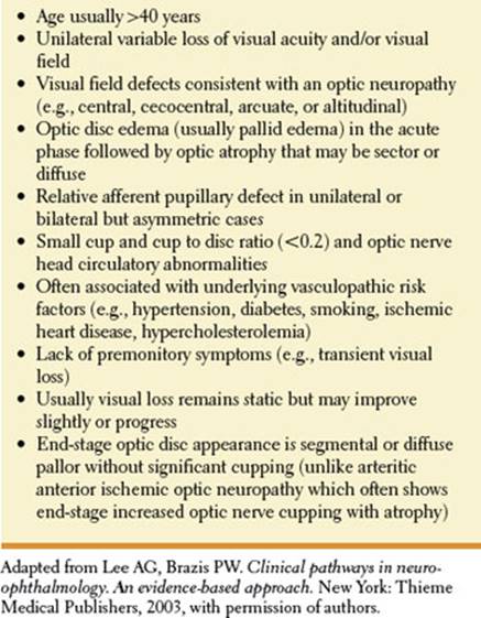
The subacute or sudden onset of visual loss in ON and ION contrast with the progressive visual disturbance noted with a compressive lesion of the optic nerve [115]. Therefore, patients with visual loss from optic neuropathy must be followed closely for deterioration in visual function to rule out potentially correctable optic nerve compressive lesions.
MASS LESIONS OF THE ORBIT
Tumors of the orbit usually cause progressive unilateral visual failure that may be variably associated with the following signs and symptoms [128]:
1. Optic disc swelling that is followed by atrophy.
2. Optociliary shunt vessels. The triad of optociliary shunt veins, disc pallor, and visual loss (the Hoyt-Spencer sign) is characteristic of chronic optic nerve compressive lesions, especially sphenoorbital optic nerve sheath meningiomas.
3. Limitation of ocular movements is seen along with diplopia.
4. Proptosis (protrusion of the eyeball). Eyeball protrusion may also be seen with disease of the cavernous sinus and may be rarely due to intracranial disease (e.g., a tumor of the middle cranial fossa may cause pressure on the veins of the cavernous sinus leading to secondary intra-orbital venous congestion and “false localizing” proptosis [2]). Intermittent proptosis may occur with venous angioma within the orbit and develops when the patient strains, cries, bends the head forward, hyperextends the neck, coughs, or blows the nose against a closed nostril and when the jugular vein is compressed. During these episodes, the eye may become tense and painful, the pupil may enlarge, and occasional bradycardia or syncope may develop (oculocardiac syndrome). Pulsation of the globe may occur with congenital sphenoid dysplasia, with orbital cranial encephalocele with neurofibromatosis, from orbital arteriovenous malformations or venous varices, due to tricuspid regurgitation, with arterial pulsation of the orbital vein, due to arteriovenous fistula, or from transmission of pulsations of intracranial pressure through surgical or traumatic defects in the orbital wall [48].
Exophthalmos is most often caused by orbital Graves’ disease. Causes of pseudoexophthalmos include an enlarged globe (e.g., due to myopia, buphthalmos, or congenital cystic eye), eyelid or palpebral fissure asymmetry (e.g., due to lid retraction, ptosis, seventh nerve palsy, or postsurgical effect), extraocular muscle abnormality (weakness or paralysis), shallow or asymmetric bony orbits, or contralateral enophthalmos (e.g., metastatic breast cancer, orbital floor fracture, or congenital bone defect) [143].
Rather than causing proptosis, scirrhous carcinoma of the breast or carcinoma of the lung, gastrointestinal tract, or prostate metastatic to the orbit may cause progressive fibrotic change and enophthalmos[49,50,183]. This enophthalmos may be caused by posterior traction and tethering on the eyeball or by the tumor mass destroying the orbital wall resulting in “biologic orbital decompression.” Other causes of enophthalmos include senile orbital fat atrophy, traumatic orbital floor fracture, traumatic orbital fat atrophy, facial hemiatrophy (Parry-Romberg disease), facial osteomyelitis, and orbital fat necrosis [48,61]. Pulsating enophthalmos in patients with type 1 neurofibromatosis may occur due to sphenoid wing dysplasia [167]. Spontaneous enophthalmos and ptosis of the globe (hypoglobus), unassociated with orbital trauma, may be associated with ipsilateral chronic maxillary sinusitis or hypoplasia (the “silent sinus syndrome”) [179,199]. The apparent dissolution and resorption of the orbital floor causes the loss of inferior support and orbital expansion. Enophthalmos and hypoglobus unassociated with prior trauma, surgery, or other symptoms have been called the silent sinus syndrome, which is ipsilateral maxillary sinus hypoplasia and orbital floor resorption [179].
TABLE 7.12 Signs and Symptoms in Visual Pathway Lesions

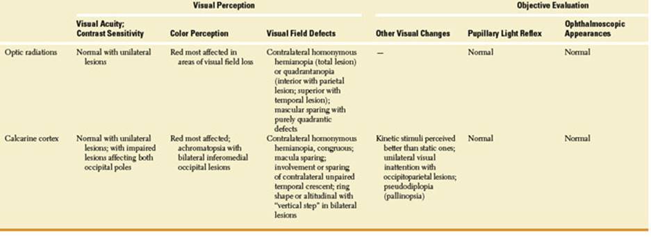
5. Swelling of the eyelids and chemosis are seen.
6. Gaze-evoked amaurosis. This refers to loss of vision whenever the eye is placed in an eccentric position of gaze and has been noted most often with cavernous hemangiomas and optic nerve sheath meningiomas [16,151]. Gaze-evoked amaurosis has also been described with orbital osteoma, glioma, medial rectus granular cell myoblastoma, varix, pseudotumor cerebri, orbital trauma, and metastatic orbital tumor [100]. This phenomenon is thought to be due to decreased blood flow to the retina or optic nerve with eye movement (e.g., the mass compresses the central retinal artery) [98]. Although most often due to intrinsic orbital disease, gaze-evoked monocular obscurations in lateral and upward gaze have also been described with pseudotumor cerebri [146,155] and gaze-evoked transient visual loss on upward gaze has been noted with an intracranial internal carotid artery aneurysm [177].
7. Facial pain and paresthesias. Several branches of the trigeminal nerve may be affected by orbital disease, especially those of the ophthalmic division, which has a large number of branches passing through the orbit [165]. The extent of cutaneous sensory loss is indicative of the position of the orbital disease, with the lacrimal, supraorbital, or supratrochlear nerves being affected by disease along the orbital roof and the zygomatic and infraorbital nerves being affected by diseases along the orbital floor. Disease at the orbital apex or the superior orbital fissure may cause hypesthesia affecting several or even all of the periorbital dermatomes. In contrast with cutaneous sensory loss, however, corneal hypesthesia appears unrelated to the position of disease within the orbit [165]. If the tumor erodes through the floor of the orbit, it may damage the maxillary division of cranial nerve V (the trigeminal nerve), resulting in ipsilateral maxillary pain, anesthesia, or both, over the distribution of the maxillary branch of the trigeminal nerve. Orbital pain is common with orbital lesions, especially with orbital malignancy or inflammatory disease.
Goldberg et al.[49,50] described the manifestation of orbital metastatic tumors into five syndromes: (a) infiltrative—characterized by prominent restriction of motility, a firm orbit, ptosis, and often enophthalmos; (b) mass—characterized by proptosis, displacement of the globe, and often a palpable orbital mass; (c) inflammatory—characterized by pain, chemosis, erythema, and periorbital swelling; (d) functional—characterized by cranial nerve findings (e.g., problems with ocular motility) disproportionate with the degree of orbital involvement; and (e) silent—orbital metastatic lesions detected by computerized tomography or MRI but asymptomatic. Infiltrative and mass lesions were by far the most common manifestations. Direct metastases to the orbital muscles may occur, especially with carcinoma of the breast and malignant melanoma [22].
This chapter has dealt with the signs and symptoms that are most helpful in localizing a lesion in the optic pathways. These signs are summarized in Table 7.12, which also lists the most likely findings with lesions in each portion of the visual system.
References
1. Acebes X, Arruga J, Acebes JJ, et al. Intracranial meningiomatosis causing Foster Kennedy syndrome by unilateral optic nerve compression and blockage of the superior sagittal sinus. J Neuro-Ophthalmol 2009;29:140–142.
2. Acers TE. Pseudo-orbital apex syndrome. Am J Ophthalmol 1979;88:623–625.
3. Aldrich MS, Alessi AG, Beck RW, et al. Cortical blindness: etiology, diagnosis, and prognosis. Ann Neurol 1987;21:149–158.
4. Bajandas FJ, McBeath JB, Smith JL. Congenital homonymous hemianopia. Am J Ophthalmol 1976; 82:498–500.
5. Bartolomei J, Wecht DA, Chaloupka J, et al. Occipital lobe vascular malformations: prevalence of visual field deficits and prognosis after therapeutic intervention. Neurosurgery 1998;43:415–423.
6. Beal MF, Chapman PH. Cortical blindness and homonymous hemianopia in the postpartum period. JAMA 1980;244:2085–2087.
7. Belden JR, Caplan LR, Pessin MS, et al. Mechanism and clinical features of posterior border-zone infarcts. Neurology 1999;53:1312–1318.
8. Bell RA, Thompson HS. Relative afferent pupillary defect in optic tract hemianopsias. Am J Ophthalmol 1978;85:538–540.
9. Bender MB, Bodis-Wollner I. Visual dysfunctions in optic tract lesions. Ann Neurol 1978;3:187–193.
10. Benton S, Levy I, Swash M. Vision in the temporal crescent in occipital infarction. Brain 1980;103: 83–97.
11. Biousse V, Newman NJ, Carroll C, et al. Visual fields in patients with posterior Gpi pallidotomy. Neurology 1998;50:258–265.
12. Blythe IM, Kennard C, Ruddock KH. Residual vision in patients with retrogeniculate lesions of the visual pathways. Brain 1987;110:887–905.
13. Boghen DR, Glaser JS. Ischaemic optic neuropathy. Brain 1975;98:689–708.
14. Borruat F-X, Maeder P. Sectoranopia after head trauma: evidence of lateral geniculate body lesion on MRI. Neurology 1995;45:590–592.
15. Borruat F-X, Siatkowski RM, Schatz NJ, et al. Congruous quadrantanopia and optic radiation lesion. Neurology 1993;43:1430–1432.
16. Bradbury PG, Levy IS, McDonald WI. Transient uniocular visual loss on deviation of the eye in association with intraorbital tumours. J Neurol Neurosurg Psychiatry 1987;50:615–619.
17. Brazis PW, Graff-Radford NR, Newman NJ, et al. Ishihara color plates as a test for simultanagnosia. Am J Ophthalmol 1998;126:850–851.
18. Bulens C, Meerwaldt JD, van der Wildt GJ, et al. Spatial contrast sensitivity in unilateral cerebral ischemic lesions involving the posterior visual pathway. Brain 1989;112:507–528.
19. Bullock JD. Relative afferent papillary defect in the “better” eye. J Clin Neuroophthalmol 1990;10:45–51.
20. Burde RM. The pupil. Int Ophthalmol Clin 1967;7: 839–855.
21. Burde RM. Optic disk risk factors for nonarteritic anterior ischemic optic neuropathy (Review). Am J Ophthalmol 1993;116:759–764.
22. Capone A, Slamovitz TL. Discrete metastasis of solid tumors to extraocular muscles. Arch Ophthalmol 1990;108:237–243.
23. Carter JE, O’Conner P, Shacklett D, et al. Lesions of the optic radiations mimicking lateral geniculate nucleus visual field defects. J Neurol Neurosurg Psychiatry 1985;48:982–988.
24. Celesia GG, Brigell MG, Vaphiades MS. Hemianopic anosognosia. Neurology 1997;49:88–97.
25. Charteris DG, Cullen JF. Binasal field defects in primary empty sella syndrome. J Clin Neuroophthalmol 1996;16:110–114.
26. Chavis PS, Al-Hazmi A, Clunie D, et al. Temporal crescent syndrome with magnetic resonance correlation. J Clin Neuroophthalmol 1997;17:151–155.
27. Chen CJ, Scheufele M, Sheth M, et al. Isolated relative afferent pupillary defect secondary to contralateral midbrain compression. Arch Neurology 2004;61: 1451–1453.
28. Chun BB, Lee AG, Coughlin WF, et al. Unusual presentation of sellar arachnoid cyst. J Clin Neuroophthalmol 1998;18:246–249.
29. Chung SM, Selhorst JB. Cancer-associated retinopathy. Ophthalmol Clin North Am 1992;5:587.
30. Cohen L, Gray F, Meyrignac C, et al. Selective deficit of visual size perception: two cases of hemimicropsia. J Neurol Neurosurg Psychiatry 1994;57:73–78.
31. Cummings GL, Gittinger JW Jr. Central dazzle: a thalamic syndrome? Arch Neurol 1981;38:372–374.
32. Damasio A, Yamada T, Damasio H, et al. Central achromatopsia: behavioral, anatomic, and physiologic aspects. Neurology 1980;30:1064–1071.
33. Davis FA, Bergen D, Schayf C. Movement phosphenes in optic neuritis: a new clinical sign. Neurology 1977;26:1100–1104.
34. Donahue SP, Kardon RH, Thompson HS. Hourglass-shaped visual fields as a sign of bilateral lateral geniculate myelinolysis. Am J Ophthalmol 1995;119: 378–380.
35. Du Pasquier RA, Genoud D, Safran AB, et al. Monocular central dazzle after thalamic infarcts. J Clin Neuroophthalmol 2000;20:97–99.
36. Elliot D, Cunningham ET Jr, Miller NR. Fourth nerve paresis and ipsilateral relative afferent pupillary defect without visual sensory disturbance. A sign of contralateral dorsal midbrain disease. J Clin Neuroophthalmol1991;11:169–172.
37. Ellis CJK. Afferent pupillary defect in pineal region tumour. J Neurol Neurosurg Psychiatry 1984;47: 739–741.
38. Ellis CJK. The afferent pupillary pathway. In: Kennard C, Ross FC, eds. Physiological aspects of clinical neuro-ophthalmology. Chicago, Ill: Year Book, 1988:393–408.
39. Feldman M, Todman L, Bender MB. “Flight of colours” in lesions of the visual system. J Neurol Neurosurg Psychiatry 1974;37:1265–1272.
40. Forman S, Behrens MM, Odel J, et al. Relative afferent pupillary defect with normal visual function. Arch Ophthalmol 1990;108:1074–1075.
41. Frisen L. Quadruple sectoranopia and sectorial optic atrophy: a syndrome of the distal anterior choroidal artery. J Neurol Neurosurg Psychiatry 1979;42: 590–594.
42. Frisen L. Clinical tests of vision. New York, NY: Raven Press, 1990.
43. Fujino T, Kigazawa K, Yamada R. Homonymous hemianopia: a retrospective study of 140 cases. Neuroophthalmology 1986;6:17.
44. Galetta SL, Grossman RI. The representation of the horizontal meridian in the primary visual cortex. J Clin Neuroophthalmol 2000;20:89–91.
45. Girkin CA, Perry JD, Miller NR. A relative afferent pupillary defect without any visual sensory deficit. Arch Ophthalmol 1998;116:1544–1545.
46. Gittinger JW. Occipital infarction following chiropractic cervical manipulation. J Clin Neuroophthalmol 1986;6:11–13.
47. Giuffre G. The spectrum of the field defects in the tilted disc syndrome. Clinical study and review. Neuroophthalmology 1986;6:239.
48. Glaser JS. Neuro-ophthalmology, 3rd ed. Philadelphia, PA: Lippincott Williams &Wilkins, 1999.
49. Goldberg RA, Rootman J. Clinical characteristics of metastatic orbital tumors. Ophthalmology 1990;47: 620–624.
50. Goldberg RA, Rootman J, Kline RA. Tumors metastatic to the orbit: a changing picture. Surv Ophthalmol 1990;35:1–24.
51. Gray LG, Galetta SL, Schatz NJ. Vertical and horizontal meridian sparing in occipital lobe homonymous hemianopias. Neurology 1998;50:1170–1173.
52. Gray LG, Galetta SL, Siegal T, et al. The central visual field in homonymous hemianopia. Evidence for unilateral foveal representation. Arch Neurol 1997;54:312–317.
53. Greenfield DS, Siatkowski RM, Schatz NJ, et al. Bilateral lateral geniculitis associated with severe diarrhea. Am J Ophthalmol 1996;122:280–281.
54. Grochowicki M, Vighetto A. Homonymous horizontal sectoranopia: report of four cases. Br J Ophthalmol 1991;75:624–628.
55. Groomm M, Kay MD, Vicinanza-Adami C, et al. Optic tract syndrome secondary to metastatic breast cancer. Am J Ophthalmol 1997;125:115–118.
56. Grossman M, Galetta SL, Nichols CW, et al. Horizontal homonymous sectoral field defect after ischemic infarction of the occipital cortex. Am J Ophthalmol 1990;109:234–236.
57. Guirgis MF, Lam BL, Falcone SF. Optic tract compression from dolichoectatic basilar artery. Am J Ophthalmol 2001;132:282–286.
58. Gunderson CH, Hoyt WF. Geniculate hemianopia: incongruous visual defects in two patients with partial lesions of the lateral geniculate nucleus. J Neurol Neurosurg Psychiatry 1971;34:1–6.
59. Gutrecht JA, Lessell IM, Zamani AA. Central dazzle in trigeminal sensory neuropathy. Neurology 1990; 40:722–723.
60. Hackelbusch R, Nover A, Scherer U. Bitemporal visual field defects in the tilted disk syndrome. Neuroophthalmology 1986;6:125.
61. Hakin KN, Yokoyama C, Wright JE. Hemifacial atrophy: an unusual cause of enophthalmos. Br J Ophthalmol 1990;74:496–497.
62. Hansen HV. Bilateral inferior altitudinal hemianopia. Neuroophthalmology 1993;13:81.
63. Hart WM. Color vision testing in clinical neuro-ophthalmology. In: Tusa RJ, Newman SA, eds. Neuro-ophthalmologic disorders-diagnostic work-up and management. New York, NY: Marcel Dekker, 1995: 77–98.
64. Hart WM Jr, Burde RM, Johnston GP, et al. Static perimetry in chloroquine retinopathy. Arch Ophthalmol 1984;102:377–380.
65. Hayreh SS, Podhajsky PA, Raman R, et al. Giant cell arteritis: validity and reliability of various diagnostic criteria. Am J Ophthalmol 1997;123:285–296.
66. Hayreh SS, Podhajsky PA, Zimmerman B. Occult giant cell arteritis: ocular manifestations. Am J Ophthalmol 1998;125:521–526.
67. Hayreh SS, Podhajsky PA, Zimmerman B. Ocular manifestations of giant cell arteritis. Am J Ophthalmol 1998;125:509–520.
68. Helgason C, Caplan LR, Goodwin J, et al. Anterior choroidal artery territory infarction—report of cases and review. Arch Neurol 1980;43:681–686.
69. Helpern JL, Sedler RR. Traumatic bilateral homonymous hemianopic scotomas. Ann Ophthalmol 1980; 12:1022–1026.
70. Hershenfeld SA, Sharpe JA. Monocular temporal hemianopia. Br J Ophthalmol 1993;77:424.
71. Hickman SJ, Kupersmith MJ, Straga J, et al. Upper temporal visual field depressions in the fellow eye in posterior acute optic neuritis: “knee” or no “knee,” Wilbrand’s concept remains clinically significant. Neuro-Ophthalmology 2002;28:69–76.
72. Horton JC. Wilbrand’s knee of the optic chiasm is an artifact of long-term monocular enucleation. Presented at the North American Neuro-Ophthalmology Society Meeting, Snowbird, Utah, February 10–11, 1996.
73. Horton JC, Fishman RA. Neurovisual findings in the syndrome of spontaneous intracranial hypotension from dural cerebrospinal fluid leak. Ophthalmology 1994;101:244–251.
74. Horton JC, Hoyt WF. Quadrantic visual field defects. A hallmark of lesions in extrastriate (V2/V3) cortex. Brain 1991;114:1703–1718.
75. Horton JC, Hoyt WF. The representation of the visual field in human striate cortex. A revision of the classic Holmes map. Arch Ophthalmol 1991;109: 816–824.
76. Horton JC, Landau K, Maeder P, et al. Magnetic resonance imaging of the human lateral geniculate body. Arch Neurol 1990;47:1201–1206.
77. Hoyt WF. Geniculate hemianopia: incongruous visual defects from partial involvement of the lateral geniculate nucleus. Proc Aust Assoc Neurol 1975; 12:7–16.
78. Hoyt WF, Rios-Montenegro EN, Behrens MM, et al. Homonymous hemioptic hypoplasia: funduscopic features in standard and red-free illumination in three patients with congenital hemiplegia. Br J Ophthalmol1972;56:537–545.
79. Huber A. Homonymous hemianopia. Neuroophthalmology 1993;12:351–366.
80. Hughes TS, Abou-Khalil B, Lavin PJM, et al. Visual field defects after temporal lobe resection. A prospective quantitative analysis. Neurology 1999;53: 167–172.
81. Ischemic Optic Neuropathy Decompression Trial Research Group (IONDT). Optic nerve decompression for nonarteritic Anterior Ischemic Optic Neuropathy (AION) is not effective and may be harmful. JAMA1995;273:625–632.
82. Iwamoto K, Aoyagi J, Kiyozuka T, et al. Neurosyphilis with unilateral optic tract lesion causing homonymous hemianopia. Neurologist 2009;15:345–346.
83. Jacobson DM. The localizing value of a quadrantanopsia. Arch Neurol 1997;54:401–404.
84. Jensen I, Seedorf HH. Temporal lobe epilepsy and neuro-ophthalmology: ophthalmological findings in 74 temporal lobe resected patients. Acta Ophthalmol 1976;54:827–841.
85. Johnson RE, Bell RA. Relative afferent pupillary defect in a lesion of the pretectal afferent pupillary pathway. Can J Ophthalmol 1987;22:282–284.
86. Johnson LN, Hill RA, Bartholomew MJ. Correlation of afferent pupillary defect with visual field loss on automated perimetry. Ophthalmology 1988;95:1649–1653.
87. Karanjia N, Jacobson DM. Compression of the prechiasmatic optic nerve produces a junctional scotoma. Am J Ophthalmol 1999;128:256–258.
88. Kardon R, Kawasaki A, and Miller NR. Origin of the relative afferent pupillary defect in optic tract lesions. Ophthalmology 2006;113:1345–1353.
89. Katz DM, Trobe JD, Muraszko KM, et al. Shunt failure without ventriculomegaly proclaimed by ophthalmic findings. J Neurosurg 1994;81:721–725.
90. Kawasaki A, Miller NR, Kardon R. (2010) Pupillographic investigations of the relative afferent papillary defect associated with midbrain lesion. Ophthalmology 2010:117:175–179.
91. Kawasaki A, Purvin VA. Photophobia as the presenting visual symptom of chiasmal compression. J Clin Neuroophthalmol 2002;22:3–8.
92. Keane JR. Blindness following tentorial herniation. Ann Neurol 1980;8:186–190.
93. Kedar S, Zhang X, Lynn MJ, et al. Congruency in homonymous hemianopia. Am J Ophthalmol 2007; 143:772–780.
94. Keklikoglu HD, Yoldas TK, Coruh Y. A case of bilateral superior altitudinal hemianopia with cerebral infarction. The Neurologist 2010:16:132–135.
95. Keltner JL, Jonmson CA, spur JO, et al. Baseline visual field profile of optic neuritis. The experience of the optic neuritis treatment trial. Arch Ophthalmol 1993;111:231–234.
96. Kim J-Y, Roh J-K. Quadruple sectoranopia caused by posterior cerebral artery infarction. Neuroophthalmology 1999;22:233–237.
97. King JT, Galetta SL, Flamm ES. Relative afferent pupillary defect with normal vision in a glial brainstem tumor. Neurology 1991;41:945–946.
98. Knapp ME, Flaharty PM, Sergott RC, et al. Gaze-induced amaurosis from central retinal artery compression. Ophthalmology 1992;99:238–240.
99. Koehler PJ, Endtz LJ, TeVelde J, et al. Aware or non-aware. On the significance of awareness for the localization of the lesion responsible for homonymous hemianopia. J Neurol Sci 1986;75:255–262.
100. Kohmoto H, Oohira A. Gaze-evoked scotoma in metastatic orbital tumor. Neuroophthalmology 1993;13: 223–226.
101. Kosmorsky G, Lancione RR Jr. When fighting makes you see black holes instead of stars. J Clin Neuroophthalmol 1998;18:255–257.
102. Kuchle M, Sever H. Foster Kennedy syndrome caused by an extensive pituitary adenoma. Neuroophthalmology 1992;12:77.
103. Kupersmith MJ, Siegel IM, Carr RE. Reduced contrast sensitivity in compressive lesions of the anterior visual pathway. Neurology 1981;31:550–554.
104. Kupersmith MJ, Vargas ME, Yashar A, et al. Occipital arteriovenous malformations; visual disturbances and presentation. Neurology 1996;46:953–957.
105. Kurz-Levin MM, Landau K. A comparison of imaging techniques for diagnosing drusen of the optic nerve head. Arch Ophthalmol 1999;117:1045–1049.
106. Lakhanpal A, Selhorst JB. Bilateral altitudinal visual fields. Ann Ophthalmol 1990;22:112–117.
107. Lam BL, Thompson HS. A unilateral cataract produces a relative afferent pupillary defect in the contralateral eye. Ophthalmology 1990;97:334–337.
108. Lam BL, Thompson HS. An anisocoria produces a small relative afferent pupillary defect in the eye with the smaller pupil. J Clin Neuroophthalmol 1999;19: 153–159.
109. Landau K, Wichmann W, Valavanis A. The missing temporal crescent. Am J Ophthalmol 1995;119: 345–349.
110. Lee AG. Visual loss as the manifesting symptom of ventriculoperitoneal shunt malfunction. Am J Ophthalmol 1996;122:127–129.
111. Lee AG, Brazis PW. Clinical pathways in neuro-ophthalmology. An evidence-based approach. New York, NY: Thieme Medical Publishers, 1998.
112. Lennon VA, Wingerchuk DM, Kryzer TJ, et al. A serum autoantibody marker of neuromyelitis optica: distinction from multiple sclerosis. Lancet 2004;364: 2106–2112.
113. Lepore FE. Visual deficits in alexia without agraphia. Neuroophthalmology 1998;19:1–6.
114. Lepore FE. The preserved temporal crescent: the clinical implications of an “endangered” finding. Neurology 2001;57:1918–1921.
115. Lessell S. Optic neuropathies. N Engl J Med 1978; 299: 533–536.
116. Levin BE. The clinical significance of spontaneous pulsations of the retinal vein. Arch Neurol 1978;35: 37–40.
117. Liu GT, Galetta SL. Homonymous hemifield loss in childhood. Neurology 1997;49:1748–1749.
118. Lorentzen SE. Drusen of the optic disc. A clinical and genetic study. Acta Ophthalmol 1966;90:1–181.
119. Luco C, Hoppe A, Schweitzer M, et al. Visual field defects in vascular lesions of the lateral geniculate body. J Neurol Neurosurg Psychiatry 1992;55:12–15.
120. Manor RS, Ouaknine GE, Matz S, et al. Nasal visual field loss with intracranial lesions of the optic pathways. Am J Ophthalmol 1980;90:1–10.
121. Marino R Jr, Rasmussen T. Visual field changes after temporal lobectomy in man. Neurology 1968;18: 825–835.
122. Masuyama Y, Kodama Y, Matsura Y, et al. Clinical studies on the occurrence and the pathogenesis of optociliary veins. J Clin Neuroophthalmol 1990; 10:1–8.
123. Matiello M, Lennon VA, Jacob A, et al. NMO-IgG predicts the outcome of recurrent optic neuritis. Neurology 2008;70:2197–2200.
124. McAuley DL, Russell RW. Correlation of CAT scan and visual field defects in vascular lesions of the posterior visual pathways. J Neurol Neurosurg Psychiatry 1979;42:298–311.
125. McFadzean R, Brosnahan D, Hadley D, et al. Representation of the visual field in the occipital striate cortex. Br J Ophthalmol 1994;78:185–190.
126. McFadzean RM, Hadley DM. Homonymous quadrantanopia respecting the horizontal meridian. A feature of striate and extrastriate cortical disease. Neurology 1997;49:1741–1746.
127. Merren MD. Bilateral lateral geniculate body necrosis as a cause of amblyopia. Neurology 1972;22:263–268.
128. Miller NR, Newman NJ. Topical diagnosis of lesions in the visual sensory pathway. In: Miller DR, Newman NJ, eds. Walsh and Hoyt’s. Clinical neuro- ophthalmology, 5th ed. Baltimore, MD: Lippincott Williams & Wilkins, 1998:237–386.
129. Molia L, Winterkorn JMS, Schneider SJ. Hemianopic visual field defects in children with intracranial shunts: report of two cases. Neurosurgery 1996;39: 599–603.
130. Monteiro MLR, Hoyt WF, Imes RK. Puerperal cerebral blindness: transient bilateral occipital involvement from presumed cerebral venous thrombosis. Arch Neurol 1984;41:1300–1301.
131. Mullen KT, Plant GT. Colour and luminance vision in human optic neuritis. Brain 1986;109:1–13.
132. Mullen KT, Plant GT. Anomalies in the appearance of colours and of hue discrimination in optic neuritis. Clin Vis Sci 1987;1:303.
133. Mudumbai RC and Bhandari A. Bilateral isolated lateral geniculate body lesions in a patient with pancreatitis and microangiopathy. J Neuro-Ophthalmol 2007;27:169–175.
134. Murphy MA, Grosof DH, Hart WM Jr. Congenital optic tract syndrome: magnetic resonance imaging and scanning laser ophthalmoscopy findings. J Neuro-Ophthalmol 1997;17:226–230.
135. Neau J-P, Bogousslavsky J. The syndrome of posterior choroidal artery territory infarction. Ann Neurol 1996;39:779–788.
136. Nakashima I, Fukazawa T, Ota K, et al. Two subtypes of optic-spinal form of multiple sclerosis in Japan: clinical and laboratory features. J Neurol 2007; 254:488–492.
137. Neetens A. Traumatic (bi)temporal hemianopia. Neuroophthalmology 1993;12:375–383.
138. Newman RP, Kinkel WR, Jacobs L. Altitudinal hemianopia caused by occipital infarcts: clinical and computerized topographic correlations. Arch Neurol 1984;41:413–418.
139. Newman NJ, Lessell S, Winterkorn MS. Optic chiasmal neuritis. Neurology 1991;41:1203–1210.
140. Newman SA, Miller NR. Optic tract syndrome: neuro-ophthalmologic considerations. Arch Ophthalmol 1983;101:1241–1250.
141. Newman NJ, Sedwick LA, Boghen DR. Bilateral visual loss and disc edema in a 15-year-old girl. Surv Ophthalmol 1994;38:365–370.
142. Newman NJ, Tornambe PE, Corbett JJ. Ophthalmoscopy of the retinal nerve fiber layer. Use in detection of neurologic disease. Arch Neurol 1982;39: 226–233.
143. Nowinski TS, Flanagan JC. Evaluation of exophthalmous and thyroid ophthalmopathy. In: Gonzalez CF, Becker MN, Flanagan JC, eds. Diagnostic imaging in ophthalmology. New York, NY: Springer-Verlag New York, 1986:189–199.
144. O’Connell JEA. The anatomy of the optic chiasma and heteronymous hemianopia. Mayo Clin Proc 1976;51:563.
145. O’Connor PS, Kasdon D, Tredici TJ, et al. The Marcus Gunn pupil in experimental optic tract lesions. Ophthalmology 1982;89:160–164.
146. O’Duffy D, James B, Elston J. Idiopathic intracranial hypertension presenting with gaze-evoked amaurosis. Acta Ophthalmol Scand 1998;76:119–120.
147. Ogawa K, Ishikawa H, Tamura M, et al. Bilateral superior altitudinal hemianopia due to bilateral occipital lobe infarction. Neuro-Ophthalmology 2009;33: 264–267.
148. Optic Neuritis Study Group. The clinical profile of optic neuritis. Experience of the optic neuritis treatment trial. Arch Ophthalmol 1991;109:1673–1678.
149. Optic Neuritis Study Group. The 5-year risk of MS after optic neuritis. Experience of the optic neuritis treatment trial. Neurology 1997;49:1404–1413.
150. Optic Neuritis Study Group. High- and low-risk profiles for the development of multiple sclerosis within 10 years after optic neuritis experience of the optic neuritis treatment trial. Arch Ophthalmol2003;121: 944–949.
151. Orcutt JC, Tucker WM, Mills RP, et al. Gaze-evoked amaurosis. Ophthalmology 1987;94:213–218.
152. Pahwa JM. Homonymous hemianopias from lesions of parietotemporal lobes. Mediscope 1963;5:543–547.
153. Papageorgiou E, Wermund T, and Wilhelm H. Pupil perimetry demonstrates hemifield pupillary hypokinesia in a patient with a pretectal lesion causing a relative afferent pupillary defect but no visual fields loss. J Neuro-Ophthalmol 2009:29:33–36.
154. Papais-Alvarenga RM, Carellos SC, Alvarenga MP, et al. Cinical course of optic neuritis in patients with relapsing neuromyelitis optica. Arch Ophthalmol 2008;126:12–16.
155. Pascual J, Combarros O, Berciano J. Gaze-evoked amaurosis in pseudotumor cerebri. Neurology 1988; 38:1654–1655.
156. Passmore JA, Robertson DM. Ring scotomata in fundus flavimaculatus. Am J Ophthalmol 1975;80:907–912.
157. Pearlman AL, Birch J, Meadows JC. Cerebral color blindness: an acquired defect in hue discrimination. Ann Neurol 1979;5:253–261.
158. Pessin MS, Lathi ES, Cohen MB, et al. Clinical features and mechanism of occipital infarction. Ann Neurol 1987;21:290–299.
159. Reynolds WD, Smith JL, McCrary JA. Chiasmal optic neuritis. J Clin Neuroophthalmol 1982;2: 93–110.
160. Rizzo JF, Lessell S. Risk of developing multiple sclerosis after uncomplicated optic neuritis. A long-term prospective study. Neurology 1988;38:185–190.
161. Rizzo JF III, Lessell S. Optic neuritis and ischemic optic neuropathy: overlapping clinical problems. Arch OphthaImol 1991;109:1668–1672.
162. Rodriguez M, Siva A, Cross SA, et al. Optic neuritis: a population-based study in Olmsted County, Minnesota. Neurology 1995;45:244–250.
163. Roemer SF, Parisi JE, Lennon VA, et al. Pattern-specific loss of aquaporin-4 immunoreactivity distinguishes neuromyelitis optica from multiple sclerosis. Brain 2007;130:1194–1205.
164. Rogers GM, Jhaveri M, and Rucker JC. Traumatic optic tract injury. Neuro-Ophthalmology 2008;32: 312–314.
165. Rose GE, Wright JE. Trigeminal sensory loss in orbital disease. Br J Ophthalmol 1994;78:427–429.
166. Rosenberg MA, Savino PJ, Glaser JS. A clinical analysis of pseudopapilledema. I. Population, laterality, acuity, refractile error, ophthalmoscopic characteristics, and coincident disease. Arch Ophthalmol 1979;97:65–70.
167. Rufa A, Zicari E, Cerase A, et al. Pulsating enophthalmos in an adult patient with type 1 neurofibromatosis. Neurology 2006;67:2169.
168. Sadun AA, Bassi CJ. Optic nerve damage in Alzheimer’s disease. Ophthalmology 1990;97:9–17.
169. Safran AB, Kline LB, Glaser JS. Positive visual phenomena in optic nerve and chiasm disease: photopsias and photophobia. In: Glaser JS, ed. Neuro-ophthalmology, Vol. 10. St. Louis: Mosby, 1980:225–231.
170. Salgado CM, Gislin G, Miller NR. Bitemporal hemianopia caused by retinal disease. Arch Opthalmology 2009;127:1690–1693.
171. Salinas-Garcia RF, Smith JL. Binasal hemianopia. Surg Neural 1978;10:187–194.
172. Sato M, Tanaka S, Kohama A, et al. Occipital lobe infarction caused by tentorial herniation. Neurosurgery 1986;18:300–305.
173. Savino PJ, Paris M, Schatzz NJ. Optic tract syndrome: a review of 21 patients. Arch Ophthalmol 1978;96:656–663.
174. Schatz NJ, Smith JL. Non-tumor causes of the Foster-Kennedy syndrome. J Neurosurg 1967;27:37–44.
175. Scholl GB, Song HS, Wray SH. Uhthoff’s symptom in optic neuritis: relationship to magnetic resonance imaging and development of multiple sclerosis. Ann Neurol 1991;30:180–184.
176. Shacklett DE, O’Connor PS, Dorwart AH, et al. Congruous and incongruous sectoral visual field defects with lesions of the lateral geniculate nucleus. Am J Ophthalmol 1984;98:283–290.
177. Sivak-Callcott J, Carpenter J, Rosen C, et al. Gaze-evoked amaurosis associated with an intracranial aneurysm. Arch Ophthalmol 2004;122:1404–1406.
178. Slavin ML. Acute homonymous field loss: really a diagnostic dilemma. Surv Ophthalmol 1990;34: 399–407.
179. Soparkar CN, Patrinely JR, Cuaycong MJ, et al. The silent sinus syndrome. A cause of spontaneous enophthalmos. Ophthalmology 1994;101:772–778.
180. Spector RH, Glaser JS, David NJ, et al. Occipital lobe infarctions: perimetry and computed tomography. Neurology 1981;31:1098–1106.
181. Thompson HS, Corbett JJ. Swinging flashlight test. Neurology 1989;38:154.
182. Thompson HS, Montague P, Cox TA, et al. The relationship between visual acuity, pupillary defect, and visual field loss. Am J Ophthalmol 1982;93: 681–688.
183. Thompson PD, Wise RJS, Kendall BE. Enophthalmous and metastatic carcinoma of the breast. J Neurol Neurosurg Psychiatry 1985;48:1305–1306.
184. Trobe JD, Acosta PC, Krischer JP, et al. Confrontation visual field techniques in the detection of anterior visual pathway lesions. Ann Neurol 1981;10:28–34.
185. Trobe JD, Glaser JS. The visual fields manual: a practical guide to testing and interpretation. Gainesville, FL: Triad, 1983:176.
186. Trobe JD, Glaser JS, Cassady JC. Optic atrophy: differential diagnosis by fundus observation. Arch Ophthalmol 1980;98:1040–1045.
187. Trobe JD, Lorber ML, Schlezinger NS. Isolated homonymous hemianopia. Arch Ophthalmol 1973;89: 377–381.
188. Trobe JD, Tao AJ, Schuster JJ. Pre-chiasmal tumor: diagnostic and prognostic features. Neurosurgery 1984;15:391–399.
189. Troost BT, Newton TH. Occipital lobe arteriovenous malformations. Clinical and radiologic features in 26 cases with comments on differentiation from migraine. Arch Ophthalmol 1975;93:250–256.
190. Tsuda H, Ishikawa H, Koshinaga M, et al. Case report: Homonymous hemianopia due to cerebral infarction of the lateral geniculate. Neuro-Ophthalmology 2005;29:43–47.
191. Vargas ME, Kupersmith MJ, Setton A, et al. Endovascular treatment of giant aneurysm which cause visual loss. Ophthalmology 1994;101: 1091– 1098.
192. Warner JEA, Lessell S III, Rizzo JF, et al. Does optic disc appearance distinguish ischemic optic neuropathy from optic neuritis? Arch Ophthalmol 1997;115: 1408–1410.
193. Warrington BK Jr. The completion of visual forms across hemianopic field defects. J Neurol Neurosurg Psychiatry 1962;25:208–217.
194. Watnick RL, Trobe JA. Bilateral optic nerve compression as a mechanism for the Foster-Kennedy syndrome. Ophthalmology 1989;96:1793–1798.
195. Wein F, Miller NR, Vaphiades MS. An unusual homonymous visual field defect. Surv Ophthalmol 2000;44:324–328.
196. Weinshenker BG, Wingerchuk DM, Vukusic S, et al. Neuromyelitis optica IgG predicts relapse after longitudinally extensive transverse myelitis. Ann Neurol 2006;59:566–569.
197. Weinstein GW, Powell SR, Thrush WP. Chiasmal neuropathy secondary to rheumatoid pachymeningitis. Am J Ophthalmol 1987;104:439–440.
198. Well M. Loss of P retinal ganglion cell function in resolved optic neuritis. Neurology 1990;40:649.
199. Wilkins RB, Kulwin DR. Spontaneous enophthalmos associated with chronic maxillary sinusitis. Am J Ophthalmol 1981;88:981–985.
200. Wingerchuk DM, Lennon VA, Pittock SJ, Lucchinetti CF, Weinshenker BG. Revised diagnostic criteria for neuromyelitis optica. Neurology 2006;66: 1485–1489.
201. Wingerchuk DM, Pittock SJ, Lucchinetti CF, Lennon VA, Weinshenker BG. A secondary progressive clinical course is uncommon in neuromyelitis optica. Neurology 2007;68:603–605.
202. Wong AMF, Sharpe JA. Representation of the visual field in the human occipital cortex. A magnetic resonance imaging and perimetric correlation. Neurology 1999;117:208–217.
203. Yeni SN, Tanriover N, Uyanik O, et al. (2008). Visual field defets in selective amygdalohippocampectomy for hippocampal sclerosis: The fate of Meyer’s loop during the transsylvian approach to the temporal horn. Neurosurgery 63:507–515.
204. Zhang X, Kedar S, Lynn MJ, et al. Natural history of homonymous hemianopia. Neurology 2006;66: 901–905.
205. Zhang X, Kedar S, Lynn MJ, et al. Homonymous hemianopias: Clinical-anatomic correlations in 904 cases. Neurology 2006;66:906–910.
206. Zhang X, Kedar S, Lynn MJ, et al. Homonymous hemianopia in stroke. J Neuro-Ophthalmology 2006;26: 180–183.
207. Zihl J, von Cramon D, Mai N, et al. Disturbance of movement vision after bilateral posterior brain damage. Further evidence and follow-up observations. Brain 1991;114:2235–2252.