
Visual loss as a primary complaint varies depending on whether one or both eyes are affected. Is the visual loss abrupt in onset, gradual or is it suddenly discovered long after onset? Is the visual loss complete or partial? Finally, does the visual loss include visual hallucinations or illusions? The causes of visual loss rapidly narrow down to a very small number of possibilities based on the temporal sequence of the patient’s symptoms, age, and sex, and the presumed anatomic location of the lesion (Table 10.1).
Patient’s visual complaints usually first seek an ophthalmologist or optometrist. Referral to a neurologist from the ophthalmologist usually includes a CT or MRI already in hand. If the problem appears to be a tumor, the eye specialist will most often refer the patient directly to a neurosurgeon.
The visual pathway provides a number of diagnostic constellations of easily examined elements that can be carried out at the bedside. The pupils, the retina, and the optic disc can be objectively examined. Subjective visual tests include color, visual acuity, and visual fields, and these help to direct localization to the retina optic nerve, chiasm, optic tract, lateral geniculate, geniculocalcarine tract, and visual cortex. Damage to visual association cortices, especially parietal and inferior temporal, will produce higher function disturbances such as central achromatopsia, alexia without agraphia, prosopagnosia, and Anton’s syndromes.
I. BEDSIDE OR OFFICE CLINICAL EXAMINATION OF THE VISUAL SYSTEM
A. Visual acuity (high contrast/low contrast). Although it is best to examine visual acuity using a distance Snellen chart, the neurologist almost always examines visual acuity using a handheld Rosenbaum card or a Jaeger print card. If a near card is used, be sure that the patient has their glasses on. In the office you can keep a pair of drugstore reading glasses of plus 2 or plus 3 for those patients over 40 years old who have forgotten their reading glasses. Push the patient to give you the very best acuity possible. Do not take no for an answer. Do not rush them. Start by telling them the first letter or number of the line and allow them to move the card back and forth. Do not begin with the largest letter; rather, ask the patient to read the 20/30 and the 20/25 lines and finally the 20/20 lines. If that fails, gradually work your way down the card. If the patient cannot read any letters on the Snellen card, the next approach is counting fingers. Three fingers held up are the equivalent of the big E on the distance acuity chart. A patient who can count fingers at 20 feet has 20/200 acuity. Thus counting fingers at 5 feet would be a rough equivalent of 20/800. If they cannot count fingers, try hand movements. If the patient cannot detect hand movement, try light perception by turning a hand light off and on. If the patient sees light reliably, see if the light can be localized in space, above, below, or side to side.
Visual acuity should be normal with a retrochiasmal pure HH or with a pure bitemporal hemianopia. Acquired visual acuity deficits not due to refractive error (nearsightedness or farsightedness) imply that macular or central vision is defective. Loss of Snellen acuity is commonly accompanied by other central visual loss such as defective color vision, Amsler’s grid defects, pallor of the optic disc, evidence of macular disease and, if unilateral, a RAPD. Bilateral, but grossly asymmetrical, retinal or optic nerve visual loss will also show an RAPD in the eye with the greatest field loss.
B. Confrontation visual fields should be performed at a distance of 1 m from the patient. Have the patient cover one eye with the palm of the hand and direct him to look at your nose. Divide the visual field in front of the patient into an imaginary plane of superior and inferior nasal and temporal quadrants. The center of the visual field is your nose. Present your fingers (one, two, or five) rapidly in the periphery of each quadrant. Redirect the patient’s attention to your nose and not to look at your fingers. Slowness or failure to respond accurately in one quadrant or hemifield may be the earliest sign of a homonymous field loss. After rapid finger counting, present your hands, palms forward, first in the two upper quadrants, then in the lower quadrants. Ask the patient to compare the palms for brightness and clarity. Finally, place the index finger of one hand on your nose and the index finger of the other hand on the peripheral nasal, temporal, superior, and inferior fields. Have the patient look at your nose and ask which finger is brightest and clearest. This tests for a central scotoma. You can also place one hand above and one below the horizontal meridian and have the patient look at your nose. This allows the patient to compare hands for brightness and clarity and helps identify altitudinal visual field defects as are seen with retinal and optic disc diseases, especially anterior ischemic optic neuropathy (AION) or BRAO.
TABLE 10.1 Sources of TMVL
Intraocular
Recurrent hyphema
Glaucomaa
Papilledemaa
Disc drusena
Congenital cavitary disc anomaliesa
AION
Arteritic (due to giant cell arteritis)
Non-arteritic rarely causes TMVL
Choroidal insufficiency (ocular ischemia syndrome)
Intraorbital (intermittent vascular compression)
Hemangioma
Osteoma
Meningioma
Intracranial
AVMs
Brain tumors
Embolic to retina (central or branch retinal artery occlusion)
Intracranial aneurysm
Cardiac
Valvular debris
Infective endocarditis
Rheumatic valvular disease
Bicuspid aortic valve
Mitral valve prolapse
Clot
Congenital cardiac malformations
Atrial fibrillation
Ventricular subendocardial ischemia
Akinetic segment
Ventricular aneurysm
Right-to-left shunt
PFO Patent Foramen Ovale
ASD Atrial Septal Defect
ASA Atrial Septal Aneurysm
Atrial myxoma
Aortic atherosclerosis
Carotid disease
Dissection
Atherosclerosis
Fibromuscular dysplasia
Fat embolism
Pancreatitis
Long bone and flat bone fractures
Hematologic
Polycythemia
Sickle cell disease
Thrombocytosis
Hypotensiona
Demyelinating disease
Uhthoff’s phenomenon
Vasospasm
Hypertensive crises especially high in paraplegic or quadriplegic patients
Migraine
aThese causes of TMVL usually last seconds and are known as transient visual obscurations or TVO. They are frequently binocular as well as monocular.
C. Color vision testing. A book of Ishihara or Hardy–Rand–Rittler pseudoisochromatic color plates can be used to test color vision one eye at a time. Although these tests were designed to identify red-green color blindness, they can be used as a rough indicator of color acuity, which is another test of central macular function. Test one eye at a time when looking for evidence of unilateral optic nerve damage. About 1% or 2% of men are red-green color blind or defective as are 0.5% of women. There are other, more complicated, color tests but neurologically they provide no more information.
D. Pupil tests. The single most important and useful objective bedside test of anterior visual pathway function is the pupillary light response. When the ganglion cells or their axons in one eye, optic disc or optic nerve, are damaged, the pupil reaction to light will be less vigorous in the affected eye as compared with the unaffected or normal eye. If both optic nerves or retinas are damaged, a relative afferent pupil defect will be seen in the eye with the largest amount of visual field loss. There is no such entity as a bilateral relative afferent pupil defect. The RAPD is a defect in the reaction to light of one eye relative to the other. One should swing the light from eye to eye allowing it to rest for 1-second intervals and there will be a brisker reaction (a more complete response) to light in the normal unaffected eye and a less brisk reaction or dilation of the pupil in the affected eye. This is the RAPD. If one eye is blind, the direct reaction to light will be absent—this is an amaurotic pupil. For details of the RAPD, refer to Chapter 12.
E. Ophthalmoscopy. Ophthalmoscopy is an objective part of the neuro-ophthalmologic examination. Recognition of changes in the optic disc is a key to the diagnosis of diseases affecting the anterior visual pathways. Details of this part of the examination are not considered in this chapter. When in doubt, dilate the pupils (with two drops of 2.5% epinephrine or 1% mydracil eye drops) for a good look. Observe and record the size of the ratio of cup to disc, appearance of the vessels, whether there are hemorrhages, exudates, or pigment changes or swelling of the disc.
F. Visual evoked potentials (VEPs). This test tends to be overused by neurologists in the evaluation of the visual pathways, but it can be especially valuable in the patient with a past history suggestive of optic neuritis (ON) where visual acuity is normal, there is no color vision loss and only slight optic disc pallor. Such patients frequently have a prolonged P100 latency. The VEP can be useful in patients suspected of functional visual loss. Patients, however, can confound the VEP by focusing in the distance, past the VEP screen, and thereby alter p100. A normal VEP latency in this setting is useful, but an abnormal latency may be a red herring.
G. Electroretinography (ERG) is a test of retinal receptor function most commonly used to detect conditions like retinitis pigmentosa and paraneoplastic retinopathies. With certain bilateral retinopathies this may be a definitive test.
H. Formal visual field testing consists of three types of tests.
Tangent screen exam is rarely used today. It is carried out on a flat black felt screen located 1 m away from the patient. It can be especially helpful in that it allows the examiner to back the patient up to 2 or 3 m and can be used to look for functional “tunnel” vision or to magnify small central visual defects.
Kinetic perimetry is carried out on a Goldmann perimeter using variable size and brightness lights and uses a supra-threshold kinetic technique. It is done in an ophthalmologist’s office. This form of perimetry is technician-dependent and has largely been supplanted by static perimetry.
Static perimetry done on Humphrey® or Octopus perimetry® tests threshold static targets presented at every 2° and tests the inner 24° to 30° of the visual field. This test is also done in an ophthalmologist’s office and is the usual formal perimetry done today.
II. ACUTE TRANSIENT MONOCULAR VISUAL LOSS
A. Clinical features. Acute transient monocular visual loss (TMVL), also called amaurosis fugax, is relatively common and has a host of different causes (Table 10.1). Before concluding that the patient has had a monocular event, explore the possibility that the “monocular” event was really a transient binocular HH. This is particularly true if the visual loss is followed by a headache. Patients with homonymous visual defects rarely complain that half of objects are gone or that they cannot see well to one side. The patient may insist that they closed one eye or the other and the visual loss was only unilateral, but this is just seeing what they think they should be seeing. Monocular visual loss is occasionally reported as being sudden in onset when in reality it is suddenly discovered when the unaffected normal eye was covered and the patient suddenly appreciates that they could not see well out of the affected eye. It is understandable why the patient would assume visual loss was sudden. The mystery is why some people do not detect severe visual loss in one eye until it is brought to their attention. A most common cause of true transient monocular visual loss is artery-to-artery or cardiac-to-artery embolism, but there are many other causes of transient or permanent acute visual loss (Fig. 10.1).
B. Approach to transient monocular visual loss.
1. Be sure that the spells occur in one eye and are not homonymous.
2. Look for ophthalmoscopic evidence of asymmetric optic disc cupping (glaucoma), optic disc anomaly, optic disc swelling, or residues of retinal embolism (hemorrhages, exudates, embolic plugs of cholesterol, platelets, fibrin, or calcium).
3. Look for a relative afferent pupil defect, visual field loss in both eyes, loss of visual acuity, and finally whether color vision is equal and normal in both eyes.
4. Look for proptosis (sign of intraorbital disease) causing intermittent amaurosis due to vascular compression particularly occurring with eye movement.
5. Auscultate the heart and carotid arteries for murmurs and bruits.
6. Laboratory studies.
a. CBC, including platelet count.
b. Sedimentation rate (ESR)—In all patients over age 50.
c. C-reactive protein (CRP)—In all patients over age 50.
In patients of age 40 or less, strongly consider evaluation for hypercoagulable states:xs
a. Protein C.
b. Protein S.
c. Factor V Leiden.
d. Antithrombin.
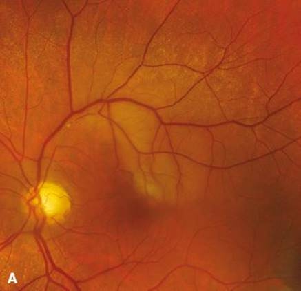

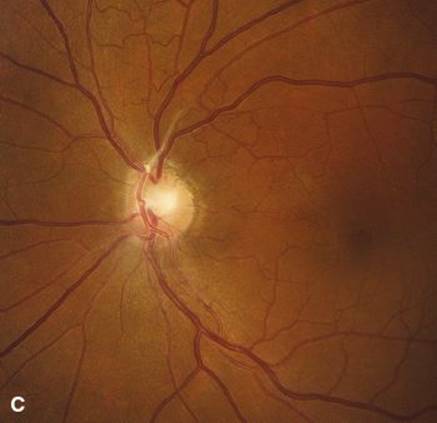
FIGURE 10.1 A: Superior branch retinal artery occlusion due to platelet-thrombin embolus. B: Magnified view of 1A showing arteriolar narrowing. C: Platelet cholesterol embolus occluding superior branch retinal artery.
e. Prothrombin gene mutation 20210A.
f. PT (INR) and aPTT.
g. Lupus anticoagulant (LA).
h. Anticardiolipin antibodies (IgG, IgM, and IgA).
i. Fibrinogen.
7. Transesophageal echocardiogram, looking especially at the aortic arch, the interatrial septum and left atrial appendage for evidence of patent foramen ovale (PFO), atrial septal defect (ASD) and/or atrial septal aneurysm (ASA), or transthoracic echocardiogram with agitated saline bubble study.
8. Perform carotid Doppler ultrasound and transcranial Doppler.
9. Consider an MRA, Computed tomography angiography (CTA), or four vessel catheter angiography.
Carotid artery emboli are not the only cause of TMVL, and there may be more than one convincing potential cause of TMVL in a single patient. It is always valuable to refer the patient with TMVL for examination by an ophthalmologist who can measure intraocular pressure, perform a dilated indirect ophthalmoscopic examination, perform formal visual fields, and obtain fundus photos. Most patients with TMVL seen by neurologists have already been seen by an ophthalmologist or optometrist if the primary complaint is visual. Be sure that the ophthalmic examination recommended above is performed.
III. SUBACUTE MONOCULAR VISUAL LOSS
Subacute monocular visual loss may occur in 2 age groups: 15 to 45 years of age where it is usually painful especially with eye movement and in those over the age of 50 years where it is commonly stepwise and usually painless.
A. Clinical syndromes.
1. Optic neuritis. This condition occurs in younger patients and presents with painful (>90%) monocular visual loss. Pain may precede the onset of visual loss by a few days and is worse with eye movement. Visual loss is characterized as a “skim, scum, blur, fog, or haze” or may be described as if there were a cloud in front of the eye. Vision may also be characterized as dim, dark, or bright. Colors are dim, washed out or gone entirely, and low-contrast images will be lost. One-third of patients with ON will have a swollen optic disc (optic papillitis). Occasionally, patients complain of photopsias (spots and sparkles) occurring with loud noise. Visual acuity may range from 20/20 to no light perception, but 20/50 to 20/200 is the rule. The visual field loss is typically monocular and is central and/or altitudinal. Prognosis for return of visual acuity to 20/40 or better is the rule and occurs within weeks. If visual acuity remains severely depressed, obtain a neuromyelitis optica (NMO) antibody.
2. AION. The older patient with monocular painless, acute, occasionally stepwise visual loss, most commonly appreciated on awakening in the morning, is suffering from hypotensive ischemia of the optic disc known as AION.This condition has two forms.
a. Non-arteritic AION (NAION). The non-arteritic form is related to hypotensive ischemia predisposed to by an anatomically small cupless optic disc through which all 800,000 to 1,200,000 axons of the optic nerve pass, on their way to the chiasm and beyond. This small, tightly packed “disc at risk” is a setup for an ischemic cascade that may occur either abruptly or in a stepwise fashion over a few days (Fig. 10.2). The NAION is usually painless and characteristically produces inferior altitudinal, inferior nasal quadrantic, and/or central scotomatous visual field defects. Twenty-five percent of patients with NAION in one eye will have the second eye affected within 2 years.
b. Arteritic AION (AAION). The obvious major difference between NAION and the arteritic form of AION is age. AAION is caused by giant cell arteritis (GCA), a disease occurring in the elderly. GCA should be considered when symptoms include a new kind of headache, usually continuous, with soreness of the scalp, and occasional brief amaurotic attacks due to recurrent choroidal ischemia. Symptoms of polymyalgia rheumatica (PMR) include tenderness of the scalp, jaw claudication, aching pains in the shoulders and hips, anorexia, weight loss, fever, and night sweats. The occult form of AAION in which there are no PMR symptoms occurs 20% of the time. AAION causes sudden severe, devastating permanent visual loss. It is a diagnostic and therapeutic emergency. Visual acuity is usually 20/200 or less and the visual field shows an altitudinal defect with a large central scotoma or complete blindness. There is an RAPD. With total blindness in one eye there is an amaurotic pupil. In addition to an ESR, CRP, and CBC, a temporal artery biopsy should be performed. As with NAION, the disc is swollen, but it is usually very pallid with a few small splinter hemorrhages. (Fig. 10.3) Prognosis for visual return is poor. Risk of second eye involvement, if giant cell arteritis (GCA) is left untreated, is very high. Treatment is immediate high dose corticosteroids, given as soon as the diagnosis is suspected.
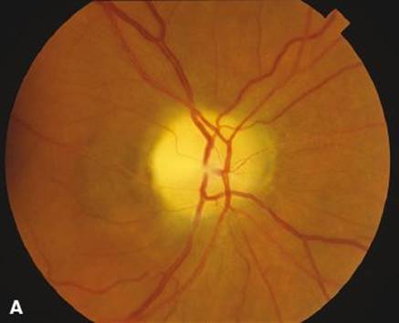
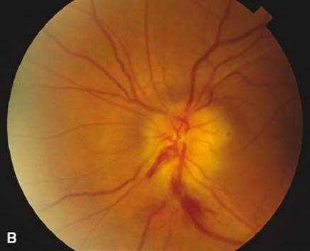
FIGURE 10.2 Old and new NAION in the same patient. A: The right disc affected 1 year ago shows ischemic altitudinal pallor. B: The left disc is acutely swollen with splinter hemorrhages which occurred the day before the photo was taken.
c. Leber optic neuropathy. Leber optic neuropathy is an inherited disorder of mitochondrial DNA. It presents as subacute, painless, monocular visual loss over days characteristically seen in men in the teens to twenties (8:1 men to women). The second eye becomes similarly affected weeks to months after the first eye (Fig. 10.4).
The disc appears slightly swollen with tortuous small telangiectatic vessels on the disc surface. After a few weeks, the nerve fiber layer becomes atrophic, particularly in a temporal wedge of the papillomacular bundles. This nerve fiber loss is most prominent between the 7 and 11 o’clock hours in the right eye and the 1 and 5 o’clock hours in the left eye. Later, as more nerve fibers are lost, both discs become diffusely pale. Visual acuity loss ranges from 20/80 to 20/800, and there are dense central scotomas that may break out into the periphery. Confirmation of the diagnosis is made with the identification of mitochondrial DNA for appropriate mutations.
B. Approach to subacute monocular visual loss.
1. Historically determine the pace of visual loss. Was it actually suddenly discovered, or was there gradual and progressive visual dysfunction in the eye, or was the visual loss abrupt and remained poor?
2. Look for evidence of earlier optic disc swelling or pallor and for evidence of embolic material in the arterioles. Look also for pigment changes characteristic of infections, inflammation, or “bone spicule” clumps seen in retinitis pigmentosa. Dilate the pupils.
3. Look for a relative afferent pupil defect, do confrontation visual fields, document visual acuity, and record color vision one eye at a time. Obtain fundus photographs and formal visual fields.
4. Decide whether visual loss appears to have been due to embolic vascular disease in which case you will proceed as if there was an embolic source.
5. If the visual loss appears to be due to ON (pain on eye movement and subacute progressive visual loss over a few days), proceed to MRI and lumbar puncture to look for evidence of demyelination.
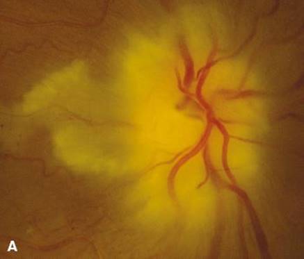
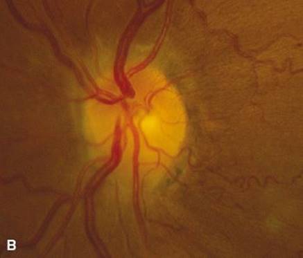
FIGURE 10.3 Pallid complete disc swelling that includes cilio-retinal arteriolar occlusion in A and normal in B.

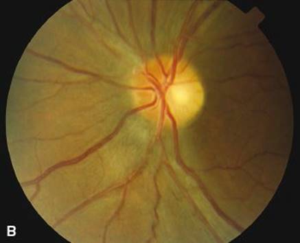
FIGURE 10.4 Leber optic neuropathy. Right eye is acutely affected and the left eye was previously affected with large segment of temporal nerve fiber layer dropout and temporal pallor of the disc.
6. If it appears that the visual loss was actually gradual in onset or has been present longer than recognized and there is evidence of optic disc pallor, do an MRI to look for a compressive lesion from the globe to orbital apex to optic chiasm.
IV. THE SYNDROME OF CHRONIC PROGRESSIVE MONOCULAR VISUAL LOSS
The syndrome of chronic progressive monocular visual loss is a characteristic of optic nerve compression. Visual acuity may well be normal early, but the patient will notice that something is not quite right. They complain of a blur or a smudge and may repeatedly clean their glasses or have their refraction checked and rechecked resulting in one of the “handful of glasses” syndromes (Table 10.2). Compressive visual loss is usually painless. If the optic nerve is compressed by a mass within the orbit, there may be proptosis, limitation of ocular motility, eye movement-induced transient, conjunctival congestion, and chemosis. If optic nerve compression occurs within the optic canal or intracranially, proptosis will occur late or may not occur at all.
Severe loss of visual acuity may occur rapidly, and visual field testing reveals a central scotoma that may break out into the periphery. Color vision will be defective in the affected eye, and there is an RAPD in the affected eye. Optic disc swelling, or disc pallor, or a combination of pallid swelling is common. Retino-choroidal collateral vessels may develop on the disc. These vessels are evidence of chronic disc swelling and retarded venous drainage. They may also be seen in end-stage glaucoma but most commonly they are the result of retrobulbar strangulation of the optic nerve by meningioma, glioma, sarcoidosis, or disc swelling of any cause (Fig. 10.5).
TABLE 10.2 “Handful of Glasses” Syndromes
Early optic nerve compression
Bitemporal hemianopia with hemifield slide
Alexia without agraphia
Bilateral small occipital tip infarcts
Vertical eye movement-induced myopia with convergence-retraction nystagmus—the dorsal midbrain syndrome
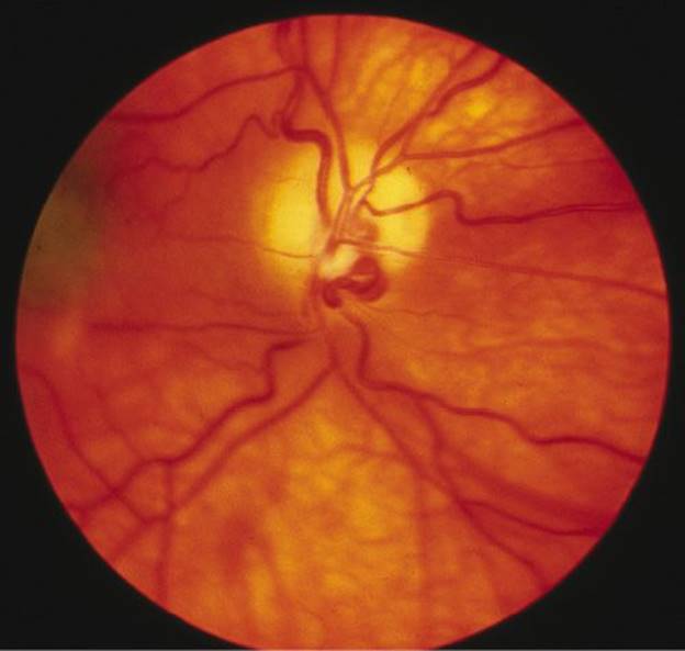
FIGURE 10.5 Swollen pallid disc with retino-choroidal collateral veins caused by an optic nerve sheath meningioma.
The VEP in the early stages of optic nerve compression when visual acuity is normal or near normal will show a prolonged P100. After visual acuity has dropped, VEP latency becomes strikingly prolonged and the VEP amplitude flattens. Consultation with an ophthalmologist is appropriate and should be done to rule out other potentially treatable ocular causes of visual loss. Ask for formal visual fields, photos of the optic discs, and intraocular pressure measurement.
V. BINOCULAR VISUAL LOSS THAT IS ABRUPT IN ONSET
Binocular visual loss that is abrupt in onset (Table 10.3) is occasionally due to bilateral optic disc disease such as ischemic optic neuropathy or ON, especially Devic disease or NMO. When this scenario occurs, the patient typically announces that the vision in both eyes has acutely or subacutely been lost. On examination, most commonly, one optic disc is pale and atrophic, evidence of earlier damage, and the other disc is swollen by new damage. This“Foster Kennedy syndrome” is attributed in most textbooks to a frontal tumor causing ipsilateral compressive visual loss and optic atrophy combined with papilledema due to increased intracranial pressure in the contralateral eye. The most common cause of this ophthalmoscopic combination of atrophy in one eye and disc swelling in the other is a “pseudo-Foster Kennedy syndrome,” caused by bilateral, sequential AION in a patient who did not notice visual loss in the first eye to be affected (the eye with disc pallor). (Fig. 10.2). The subsequent loss of vision with disc swelling in the previously unaffected eye suddenly plunges the patient into unexpected bilateral visual loss.
TABLE 10.3 Causes of Bilateral Visual Loss—Anterior Visual Pathways
Ocular causes
Anomalous discs
Papilledema—of any cause
Disc drusen
Pseudo-Foster Kennedy syndrome (bilateral AION)
Toxic—nutritional (tobacco–alcohol amblyopia), B12 deficiency
Medications—ethambutol, chloramphenicol, Plaquenil, thioridazine
Leber optic neuropathy
Neuromyelitis Optica NMO (Devic disease)
Sarcoidosis
Intracranial causes
Chiasmal
Tumor (craniopharyngioma, Rathke’s cleft cyst, pituitary tumor, meningioma and other rarer tumors)
Pituitary apoplexy
Aneurysm (ophthalmic or anterior cerebral)
Sphenoid mucocele
Trauma (chiasmal tear)
Demyelination
Toxic/metabolic
Vascular (dolichoectatic anterior cerebral artery)
Combined chiasmal and optic nerve disease due to any of the above will give combinations of bitemporal and central visual loss
Optic tract
Tumor—same as chiasm
Demyelination
Trauma
Stroke
Transient visual obscurations (TVO) occur in patients who have papilledema, drusen of the optic disc, and other disc-related conditions (see Table 10.1). These TVOs are seconds-long transient bouts of unilateral or binocular visual dimming or blindness frequently precipitated by Valsalva maneuvers or postural change. Examination of the fundus photos and B-mode scans should identify the congenitally anomalous swollen discs.
Binocular visual loss, which is abrupt in onset, occurs rarely following arteriographic procedures. Posterior fossa angiography and coronary angiography will also, occasionally be attended by what appears to be a toxic reaction to iodinated contrast associated with vasospasm. Permanent visual loss owing to such toxic contrast responses is rare.
VI. BINOCULAR VISUAL LOSS DUE TO CHIASMAL DAMAGE
Binocular visual loss due to chiasmal damage is seen in the pure form where there is no concomittant damage to optic nerves or optic tracts.
A. Clinical features.
1. Bitemporal visual loss. If the lesion causing bitemporal visual loss arises from below (typically a pituitary adenoma), visual loss is dense in the superior bitemporal visual field. If the lesion originates from above (typically an aneurysm of the anterior cerebral artery or craniopharyngioma), the visual field defect will be in the inferior bitemporal visual fields. Complete, macula-splitting, bitemporal defects are usually due to tumor or traumatic chiasmal tears.
The patient with bitemporal visual loss rarely complains of loss of peripheral vision, but their symptoms are caused by instability of the two preserved nasal visual fields that abut the midline. The visual complaints consist of intermittent and brief doubling of objects, loss of objects, and strange visual effects that occur as a result of vertical sliding of the one hemifield relative to the other. This “slip” in the retinas causes the right half of images to slip vertically and/or horizontally in relation to the left half. This odd group of symptoms is collectively known as the “hemifield slide phenomena” and is another cause of the “handful of glasses” syndrome (Table 10.2). As patients with visual symptoms see eye doctors first and are rarely able to articulate the nature of the visual dysfunction, they are commonly provided with a new refraction, particularly if confrontation or formal visual fields are not performed.
2. Junctional syndrome. If the chiasmal compression occurs where the optic nerve enters the chiasm, symptomatic optic nerve compression occurs in that eye (see IV) and asymptomatic superior temporal quadrantic visual loss is found in the contralateral eye. This is caused by damage to fibers from the inferior nasal part of the contralateral eye and the optic nerve at its junction with the chiasm. This visual field combination of central scotoma in one eye and a superior temporal defect in the other eye is known as a junctional syndrome.
3. Pituitary apoplexy. Abrupt onset of unilateral or bilateral visual loss usually combined with ocular motility disturbances due to paralysis of cranial nerves III, IV, and VI, associated with headache, agitation, fever, stiff neck, and occasionally blood in the CSF, occurs with acute hemorrhage into a pituitary tumor or the pituitary gland. This may occur spontaneously or result from embolic infarction following carotid endarterectomy or cardiac surgery. The diagnosis is confirmed with an MRI or CT scan. Patient agitation frequently makes these studies less than optimal for interpretation. Pituitary apoplexy only occasionally causes pure visual loss; there is usually some ocular motility disturbance. The diagnosis of pituitary apoplexy should rank high on the list of causes of sudden onset of bilateral visual loss.
4. Combination of central and bitemporal visual loss. Both optic nerves may be gradually compressed with large lesions that also compress the chiasm. This may cause unilateral and bilateral central visual loss and a roughly bitemporal visual field defect. Unless the bitemporal field loss is complete, it is rare for the area of field loss to be equal in both eyes. Thus the eye with the greatest visual field loss will have an RAPD.
B. Clinical approach to binocular visual loss that is subacute or chronic.
1. Historically look for symptoms consistent with hemifield slide. Ask which eye seems to be affected most severely.
2. Do confrontation visual fields to look for bitemporal and/or central visual field loss. Do visual acuity and color vision testing to look for evidence of optic nerve dysfunction. Perform ophthalmoscopy to look for evidence of optic disc swelling, pallor, or retinal hemorrhages.
3. A CT or MRI should be done to look at the suprasellar space, the pituitary fossa, and sphenoid sinus and cavernous sinuses.
4. Fundus findings in chiasmal compression depend on the nature of the lesion and if there is increased intracranial pressure. Papilledema is rarely seen with pituitary adenomas unless the tumor causes hydrocephalus. Optic atrophy with nerve fiber loss may preclude the development of disc swelling. For reasons that are not clear, disc swelling is fairly common with craniopharyngiomas as is severe optic atrophy.
VII. BILATERAL VISUAL LOSS DUE TO HOMONYMOUS HEMIANOPSIA
Bilateral visual loss due to HH can be due to lesions in the optic tract, the lateral geniculate nucleus, geniculocalcarine tract, or occipital cortex. Total bilateral right and left homonymous visual loss is very rare.
A. Localization of lesions. If a hemianopia is complete and splits fixation and there are no other symptoms or signs, the lesion is either in the optic tract, geniculocalcarine tract, or the occipital cortex. Lesions occurring between the lateral geniculate, the geniculocalcarine tract, and visual cortex are rarely complete without creating other neurologic problems such as hemiparesis, hemisensory loss, aphasia, or parietal neglect. Most HH are incomplete, and their location, density, and congruity (super-imposability of one eye’s visual field on the other) broadly help to tell where the lesion is located. For practical purposes, CT and MRI provide images of lesions and their location is now rarely a mystery. Homonymous visual field defects owing to tumor, stroke, and arteriovenous malformations (AVM) are easily accounted for with these techniques. Occipital lobe damage will produce pure visual loss if damage is confined to the calcarine cortex. Total loss of calcarine cortex on one side will give a complete HH. Commonly, the macular region of calcarine cortex is “spared” to some extent because a large part of the visual cortex subserves the inner 20° of the visual field. Conversely, small cortical infarcts that occur in the inner 20° of the visual field may cause very small but visually troubling visual field defects that may not be identified by standard perimetry. These visual field defects are best identified using a “magnifying technique” with a tangent screen exam done at 2 or 3 m. These patients complain bitterly of difficult-to-describe loss of vision which cannot be corrected with glasses (Table 10.2) and they go from one eye doctor to another without having the occipital lobe damage discovered.
B. Migraine auras. The most common cause of episodic homonymous visual loss is the visual aura of migraine. These homonymous auras are frequently mistaken by the patient for visual loss in one eye. The characteristic features of this visual event are movement, brightness, and “buildup” of the visual loss usually beginning in the center or off to one side of the center of the visual field. It then moves over minutes toward the periphery of the visual field. The typical auras consist of zigzag lines that are silvery or red, yellow, blue, bright white, and black; they pulsate, turn, swirl, or glimmer. Although most patients characterize this as a “c-shaped” or “horseshoe” shape to one side, occasional patients describe a visual image that is central in both eyes “as if a flashbulb just went off” or an arc of shimmering flashing zigzags in both the right and left fields “like a rainbow.” The next most common description is a “heat wave” sensation or the image is of water running down a window. These visual events usually last from 5 to 60 minutes and are followed by a headache. The headache is usually unilateral but may be generalized and need not be particularly severe or long lasting. Some patients have only the visual aura and no headache, so-called acephalgic migraine. This is a common event in older migraineurs.
C. Visual seizures occipital lobe epilepsy. Metastatic brain tumors, meningiomas, gliomas, and AVMs may have primary visual seizures with no secondary generalization. These seizures produce homonymous sparkles, flashes, and colors, but, as contrasted with migraine, there is no characteristic “buildup” and progression of the visual event from center to periphery. A CT and MRI will identify these conditions, and EEG will show epileptiform activity.
D. Degenerative diseases. The common causes of HH are benign and leave no trace (migraine) or produce lesions in the brain that can be identified with CT or MRI (stroke or tumor). Creutzfeldt–Jakob disease (CJD) may present with HH (the so-called Heidenhain variety) and rarely patients with Alzheimer disease (AD) may develop HH. The CJD has characteristic MRI and EEG changes, but AD will show no disease specific imaging findings. Progressive multifocal leukoencephalopathy, common in immune-compromised patients, often presents with dense HH and demyelinating changes on MRI, typically sparing the U-fibers.
VIII. SYNDROMES OF VISUAL DISTURBANCE DUE TO HIGHER COGNITIVE DYSFUNCTION
These syndromes can be caused by tumors primary or metastatic, however, most are caused by embolic stroke and are particularly common after cardiac surgical procedures. They frequently go unrecognized and present to the physician with unusual visual complaints.
A. Alexia without agraphia. This syndrome is caused by damage to connections between both visual cortices and the angular gyrus. This is due to either (1) a lesion in the splenial outflow from the right occipital lobe and the connections of the left occipital lobe to the angular gyrus where there is no hemianopia or (2) a combination of a left occipital infarct with a right HH and a callosal splenium lesion. These patients may present to the neurologist with a history of going to many eye doctors for new glasses because they are unable to read (Table 10.2). The diagnosis is particularly problematic when there is no HH.
B. Balint’s syndrome. Bilateral damage to the watershed region between the middle and posterior cerebral artery circulation high in the parietal lobes will cause Balint syndrome, which consists of visual disorientation, spasm of fixation (apraxia of gaze), optic ataxia (defect in visually guided hand movements), and simultanagnosia (loss of panoramic vision). Although these patients do not deny their visual troubles, they usually suffer in silence because they are frequently unable to adequately verbalize what is wrong. They are not agitated and are not aphasic or demented. Frequently they have alexia without agraphia as well.
C. Bilateral inferior temporal lobe syndrome. Damage to the inferior temporal lobe bilaterally in the region of area V4 of the fusiform gyrus will cause one or both of the following syndromes: prosopagnosia (the inability to recognize faces) and central achromatopsia (central color vision loss).
1. Prosopagnosia. Patients with prosopagnosia present with either bilateral sudden visual disturbance or a previously damaged inferior temporal lobe (which may or may not have been recognized) and then a second lesion in the other inferior temporal lobe. Occasionally, these patients will have only a unilateral lesion, usually of the right inferior temporal lobe. Prosopagnosic patients complain of being unable to recognize individual faces, but they also have trouble picking out their own car from other cars, their dog from other dogs, and so on. In short, while they can identify classes of objects, they have trouble singling out a specific individual within a general class or group without other clues.
These patients become expert at recognizing features of a person’s voice or the way a person walks to garner clues in identifying people. They commonly complain that colors are washed out and that whites (linens, snow) look dirty or brownish. They may have homonymous superior quadrantic visual field defects or may have no visual field loss at all.
2. Central achromatopsia. Patients with central achromatopsia involving the entire visual field may also have prosopagnosia or they may have only a hemifield defect in color vision known as hemiachromatopsia. Color loss may be profound or it may consist simply of desaturation and “dirtying” of color complained of by the patients with prosopagnosia. The lesions are inferior temporal in location.
D. Anton syndrome. Extensive bilateral damage to both the occipital and parietal lobes produces visual loss and the denial of blindness known as Anton syndrome. These patients confabulate elaborately in response to questions about their visual environment.
E. Approach to patients with homonymous visual field defects.
1. Obtain best corrected visual acuity (even alexic patients can usually read numbers or individual letters).
2. Confrontation visual fields will tell you the completeness and the rough location of the visual field defect.
3. Fundus examination will confirm that there is no significant disease of the retina or disc responsible for the visual loss.
4. A CT scan or preferably an MRI should confirm localization of the lesion(s).
5. Referral should be made to an ophthalmologist for visual fields. Patients who are hemiplegic, aphasic, or have right parietal lesions will not perform fields well, if at all.
6. Neuropsychologic evaluation of patients with higher cortical defects will be helpful.
IX. FUNCTIONAL VISUAL LOSS
The patient with functional visual loss either claims total or partial loss in one eye or both eyes. Whatever the motivation or underlying issue, it is possible to uncover functional monocular visual loss simply by looking for an RAPD. In the absence of an RAPD, severe, or even moderate, significant monocular visual loss does not exist. Patients with functional binocular blindness can be uncovered in one of two ways. Use of an optokinetic target (tape or drum) is usually sufficient; however, some patients are able to “look through” these targets. A foolproof method that can be used to uncover a single “blind” eye or bilateral “blindness” is a mirror held in front of the patient’s face and then tilted up and down and side to side. This maneuver produces an irresistible sensation of environmental movement and the patient’s eyes will involuntarily move to orient the patient in space. Eye movement proves that the patient can see at least partially if the claim has been that of total blindness.
The most difficult problem is that of the patient who presents with moderate functional visual loss that is equal in both eyes (e.g., 20/50 OU). An RAPD is of no help because the visual loss is bilateral and the acuity is too good to make the mirror or optokinetic tangent useful. Remember that it is possible for patients to voluntarily alter the VEP latency and wave form, thus an abnormal VEP may not be helpful. However, a normal VEP is reassuring. Tubular visual fields done at 1 and 3 m are also commonly seen in functional visual loss. Before pronouncing a patient’s visual loss functional, be sure to either use a pinhole to “refract” the patient or send them for ophthalmological refraction. In the setting of presumed functional visual loss referral to an ophthalmologist is prudent to be certain that there is no underlying serious ocular pathology. Patients who complain loudly that the testing is uncomfortable, painful, or an otherwise onerous chore are likely trying to get you to stop examining them for fear of being “found out.”
A. Approach to a patient with functional visual loss.
1. Document every visual test performed and the patient’s response. This documentation should include their ' text-align:justify;line-height:normal'>2. The greater the resistance to examination and complaints about the tests, the more likely one is dealing with a deliberate malingerer, whereas the more naive functional patient will gladly go along with the exam regardless of the obvious contradictions in performance behavior.
3. Confronting the patient is fruitless and counterproductive. Gentle suggestion that their vision is “better than the patient thinks it is” and that the patient’s vision is likely to improve will reassure all but the hard-core malingerer. Remind them gently but confidently that you have seen this problem before and it gets better by itself. Malingerers commonly will accuse the examiner of not believing that their symptoms are real or that they are simply “all in their mind.”
4. Radiographic and/or electrophysiologic studies (ERG and VEP) should be done and interpreted for the patient to be sure that no nagging doubts remain. The patient should be told that there is no disease of the central or peripheral nervous system that has been identified. Be sure to do a Schirmer test to identify dry eyes.
5. Do not prescribe eye drops or any medication or refer the patient to a psychiatrist. Prescriptions for medication, glasses, and drops give a double message, that is, “nothing is wrong but take this medication” and psychiatric referrals for functional problems do not produce any more useful results than simple reassurance and encouragement. If they are found to have dry eyes, normal saline drops are reasonable. Explain why you are giving drops.
B. General rules for referral of patients who complain of any kind of visual loss.
1. If the patient has seen a number of eye care specialists (optometrists and ophthalmologists) and continues to complain of visual loss, consider the conditions listed in Table 10.2.
2. Do imaging studies with the visual pathways in mind. If the visual loss is monocular, look at the orbit and intracranial optic nerve. If there is monocular central visual field loss, always test the superior temporal field of the other eye. If the visual loss is bitemporal, look at chiasm and perichiasmal structures.
3. Enlist an ophthalmologist for a computed visual field examination if the patient has not yet had one. Ask for visual fields to be done and for an interpretation of the findings. If the optic disc looks abnormal or the fundus is abnormal, ask for photographs to be taken. As physicians, we always get chest X-rays for lung disease, and we should obtain fundus photos where there is suspected optic nerve or retinal disease. Remember that an ophthalmologist will be able to identify refractive, corneal, lenticular problems as well as vitreous and retinal causes of visual loss.

Recommended Readings
Barton JJ, Corbett JJ. Neuro-ophthalmologic vascular emergencies in the elderly. Clin Geriatr Med. 1991;7:525–548.
Bernstein EF, ed. Clinics in Geriatric Medicine. W.B. Saunders Co. Philadelphia. Amaurosis Fugax New York. Springer-Verlag; 1988.
Bruno A, Barton JJ, Corbett JJ, et al. Transient monocular visual loss patterns and underlying vascular abnormalities: a prospective study of 100 consecutive patients. Stroke. 1990;21:34–39.
Cooper SA, Murray KL, Heath CA, Will RG, Knight RS. Isolated visual symptoms at onset in sporadic Crutzfeldt-Jakob disease: the clinical phenotype of the “Heidenhain variant.” Br J Ophthalmol. 2005;89:1341–1342.
Hupp SL. Syndromes of the optic chiasm In: Tusa RJ, Newman SA, eds. Neuro-Ophthalmological Disorders: Diagnostic Workup and Management. New York, NY: Marcel Dekker; 1995:65–75.
Hupp SL, Kline LB, Corbett JJ. Visual disturbances of migraine. Surv Ophthalmol. 1989;33:221–236.
Lee AG, Martin CO. Neuro-ophthalmic findings in the visual variant of Alzheimer’s disease. Ophthalmology. 2004;111:376–380.
Liu GT, Volpe NJ, Galetta SL (Eds). Disorders of higher cortical visual function. In: Neuro-ophthalmology Diagnosis and Management. 2nd ed. Saunders Elsevier; 2010:339–362.
Rizzo M, Nawrat M. Human visual cortex and its disorders. Curr Opin Ophthalmol. 1993;4:30–37.
Thompson HS. Functional visual loss. Am J Ophthalmol. 1985;100:209–213.
Thompson HS, Corbett JJ. Swinging flashlight test. Neurology. 1989;39:154–156.
Trobe JD, Acosta PC, Krischer JP, Trick GL. Confrontation visual field techniques in the detection of anterior visual pathway lesions. Ann Neurol. 1981;10:28–34.
Xhang X, Kedar S, Lynn MJ, et al. Homonymous hemianopias. Clinical anatomic correlations in 904 cases. Neurology. 2006;66:906–910.