
The autonomic nervous system (ANS) maintains internal homeostasis and regulates protective responses by continuously monitoring and responding to internal and external stimuli. This is achieved through autonomic reflex pathways and extensive vasomotor, visceromotor, and sensory innervation. The baroreflex, an example of an autonomic reflex, regulates blood pressure (BP), heart rate (HR), and extracellular fluid volume (Fig. 31.1).
ANS dysfunction may be focal or generalized depending on the site of a lesion in an autonomic reflex pathway. Central forms of ANS dysfunction are because of lesions involving neurons of the CNS, brainstem, spinal cord, and preganglionic neurons, whereas with peripheral forms, dysfunction is because of lesions involving peripheral ganglia and postganglionic neurons, or to lesions involving afferent autonomic reflex limbs (Fig. 31.1). Selected autonomic symptoms and findings are summarized in Table 31.1. Numerous diseases result in autonomic dysfunction (Table 31.2).
I. EVALUATION OF THE PATIENT WITH ANS SYMPTOMS
A. History. All patients presenting with autonomic dysfunction should undergo a comprehensive medical and neurologic history and physical examination. Important elements of the autonomic history include the following:
1. Chief autonomic complaints, with severity of symptoms, their distribution and frequency, progression, the presence of aggravating and alleviating factors, and a measure of the degree of disability are determined.
2. Review of ANS systems including cardiovascular, sexual, urinary, gastrointestinal, vasomotor, thermoregulatory and sudomotor, secretomotor, pupillomotor, and sleep functions.
3. Medication review (antihypertensive, psychotropic, antiandrogenic, laxative medications, and alcohol and recreational drugs can produce ANS dysfunction).
4. Psychosocial evaluation to determine the impact of ANS dysfunction on quality of life.
5. Family history (inherited autonomic disorders).
B. Physical examination. The physical and neurologic examination indicates the site and extent of the lesion responsible for ANS dysfunction and defines associated illness. A comprehensive examination includes supine and upright BP and HR, and examination of the skin and mucosa to assess sweating patterns and to determine if trophic lesions are present. A vascular system examination is important. In patients with genitourinary and anorectal dysfunction complaints (see the following list), the physical examination also includes the following:
1. Abdominal examination to determine if aortic dilatation is present (1% of patients with erectile dysfunction [ED] have abdominal aortic aneurysm) or masses are present.
2. Stretching and palpating the penis for an indication of the integrity of erectile tissue and if Peyronie’s plaques (lumps within the penis) are present.
3. Testicular volume and consistency.
4. Cremasteric reflex (testicle retraction on stroking the thigh), anal wink reflex (anal sphincter contraction on stroking perianal skin), and bulbocavernosus reflex (anal sphincter contraction on squeezing the glans penis or clitoris).
5. Rectal examination to determine if prostatic hypertrophy, fecal impaction, and prolapse are present.
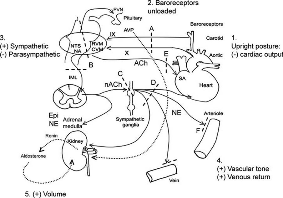
FIGURE 31.1 The baroreflex. 1. Standing produces “pooling” of 600–1,000 mL of blood to the lower body (mainly limb and splanchnic capacitance circulation), reducing venous return and cardiac output (by approximately 30%). 2. This is sensed by specialized stretch receptors (arterial and cardiopulmonary baroreceptors) that in turn activate (unload) baroreflexes. Inputs from carotid and aortic baroreceptors travel with the glossopharyngeal (IX) and vagus (X) nerves to converge on cardiovascular centers in the brainstem and medulla (mainly nucleus of the tractus solitarius) and their projections. 3. The physiologic baroreceptor reflex response to the volume shifts produced by upright posture is a compensatory increase in sympathetic tone with a decrease in parasympathetic outflow. 4. Sympathetic nerve terminals release NE that produces increased vasoconstriction of skeletal and mesenteric muscle vessels, HR, and cardiac contractility. Additional increases in venous return occur through a “pumping” effect of contracting limb and abdominal muscles engaged by the effort of standing (not shown). 5. A longer term response promoting extracellular fluid volume expansion includes baroreflex-mediated release of renin, angiotensin, and aldosterone, leading to increased renal Na++ absorption, and release of vasopressin, with an increase in free-water absorption.
A: Afferent baroreflex limb dysfunction is present when lesions involve baroreceptors and IX and X cranial nerves (neck surgery, radiotherapy, trauma, neuropathies, and autonomic disorders such as baroreflex failure, Holmes-Adie syndrome, and HSAN III). B: Central lesions involve the ventrolateral medulla (MSA), descending sympathetic pathways (medullary lesions, spinal cord lesions above T5), and IML (MSA, Lewy’s body disorders).
C: Sympathetic ganglia lesions involve sympathetic ganglia (autoimmune ganglionopathies associated with nAChR antibodies or paraneoplastic, Lewy’s body disorders).
D, E: Postganglionic sympathetic and efferent parasympathetic lesions are present with small fiber neuropathies (diabetes, amyloidosis, Sjögren’s syndrome, HSAN III).
F: Efferent sympathetic neuroeffector junction dysfunction occurs with dopamine β-hydroxylase deficiency and α-1 adrenoceptor blocking drugs.
nAChR, nicotinic acetylcholine receptor antibodies; AVP, arginine vasopressin; CVM, caudal ventrolateral medulla; Epi, epinephrine; HSAN III, hereditary sensory autonomic neuropathy type III; IML, intermediolateral cell columns of the spinal cord; NA, nucleus ambiguus; NE: morepimephrime NTS, nucleus tractus solitarius; RVM, rostral ventrolateral medulla; SA, sinus node.
TABLE 31.1 Clinical Features of ANS Dysfunction

C. Autonomic testing. ANS testing is considered an extension of the physical examination. The data from ANS testing is of most value when the selection of tests is guided by the clinical findings. The goal of testing is to confirm the presence of autonomic dysfunction, determine the extent of autonomic involvement, and to localize the site of a lesion in the ANS reflex arc, and distinguish primary from secondary autonomic disorders. Most ANS tests assess the integrity of a reflex arc by recording stimulus-evoked effector organ responses. The more popular tests rely on measuring those effector organ responses that are easily recorded (i.e., changes in HR, BP, pupillary size, etc.). Common bedside tests are shown in Table 31.3. More sophisticated tests include beat-to-beat arterial pressure recordings during different challenges, and the measurement of sweat output (the quantitative sudomotor axon reflex test [QSART], measuring efferent sudomotor function). Usually a battery of several tests is required to reach a diagnosis of autonomic dysfunction (abnormal tests do not always imply disease).
1. Screening tests. Selection of testing is directed by the clinical presentation.
a. Complete blood count and differential.
b. Urinalysis and renal function studies.
c. Hemoglobin A1C, fasting and postprandial glucose, and glucose tolerance test.
d. TSH levels.
e. HIV testing.
f. Immunoelectrophoresis (serum and urine).
g. Sweat gland nerve fiber density and epidermal nerve fiber density (skin biopsy).
TABLE 31.2 Selected Autonomic Disorders
|
Autonomic Disorders |
|
|
Central autonomic disorders |
MSA |
|
PAF (peripheral involvement predominant), Parkinson’s disease, DLB |
|
|
Disorders of different causes (cerebrovascular, epileptic, tumoral, demyelinating, traumatic, infectious, and degenerative) and autonomic presentations involving primarily: frontal lobes, limbic system, hypothalamus, brainstem, cerebellum, and spinal cord. |
|
|
Autonomic neuropathies |
a. Acute and subacute autonomic neuropathies: subacute autoimmune autonomic neuropathy (panautonomic neuropathy and pandysautonomia), subacute paraneoplastic autonomic neuropathy, Landry–Guillain–Barré’s syndrome, botulism, porphyrias, drug induced and toxic autonomic neuropathies. |
|
b. Chronic peripheral autonomic neuropathies: distal small fiber neuropathies. |
|
|
Sympathetic and parasympathetic neuropathies: DAN, amyloidosis, autoimmune autonomic neuropathy (paraneoplastic and idiopathic), sensory neuronopathy with autonomic failure, hereditary neuropathies. |
|
|
Catecholamine disorders |
Baroreflex failure, tumors that secrete catecholamines (pheochromocytoma, neuroblastoma, chemodectoma, and familial paraganglioma syndrome), disorders |
|
Orthostatic intolerance disorders |
POTS |
|
Mitral valve prolapse dysautonomia |
|
|
Idiopathic hypovolemia |
|
|
Paroxysmal syncope |
Neurally mediated hypotension and bradycardia (vasovagal), situational syncope |
|
Miscellaneous |
Hyperhidrosis (generalized, focal), anhidrosis (CNS, peripheral nerve, and dermatologic) |
|
Horner syndrome, Holmes–Adie’s syndrome, Ross’ syndrome, and crocodile tears |
|
|
Hirschsprung’s disease |
|
|
Brugada’s syndrome |
|
|
Abbreviation: DAN, diasetic autonomic meuropathy; DLB, dementia with Lewy bodies; PAF, pure autonomic Failure; POTS, postural tachycardia syndrome. |
|
h. Aminolevulinic acid, porphobilinogen, and porphyrins (24 hour urine collection), erythrocyte porphobilinogen deaminase activity.
i. Genetic testing (inherited neuropathies).
j. Amyloid staining in fat aspirate, rectal or gingival biopsy.
k. [Norepinephrine] plasma (supine and standing)
D. Antibody testing. Should always be guided by the clinical presentation.
1. Antinuclear antibodies, rheumatoid factor, Anti-Ro/SS-A, and Anti-La/SS-B.
2. Other antibodies: Neuronal nicotinic acetylcholine receptor, P/Q-type calcium channel, and acetylcholine receptor.
a. Paraneoplastic antibodies: Anti-Hu (ANNA-1); Purkinje-cell cytoplasmic antibodies type 2 (PCA-2); and collapsin response-mediator protein 5 (CRMP-5), voltage gated calcium channels (VGCC).
TABLE 31.3 Bedside Autonomic Tests

E. Electrophysiologic studies. Nerve conduction studies and EMG help define large fiber peripheral neuropathies. Sphincter and pelvic floor EMG is a specialized technique (useful when performed by experienced examiners) in detecting denervation potentials in selected muscles in lesions of the anterior horn cells in the spinal cord.
F. Imaging. MRI of the brain and spine is essential in CNS and spinal cord lesions. Pelvic imaging may be indicated in those in whom structural lesions are suspected.
Clinical Presentation of Autonomic Dysfunction
NEUROGENIC ORTHOSTATIC (POSTURAL) HYPOTENSION
Orthostatic hypotension (OH) is a frequent and disabling manifestation of autonomic disorders. It may be the initial sign (i.e., “tip of the iceberg”) heralding the onset of primary and secondary autonomic disorders.
A. Diagnosis. BP measured with a sphygmomanometer and pulse rate recorded while supine for a few minutes of quiet rest (i.e., once BP values have stabilized), and after standing up for 3 minutes is sufficient to determine if OH is present and may help with its differential diagnosis (Table 31.4). OH is defined as a sustained decrease in systolic pressure of at least 20 mm Hg (30 mm Hg in patients with supine hypertension) and of diastolic pressure of at least 10 mm Hg within 3 minutes of standing or head up tilt to at least 60 degrees on a tilt table.
B. Pathophysiology. The autonomic responses to gravitational volume shifts are complex, and rely on intact baroreflexes (Fig. 31.1). In neurogenic OH, a patient’s ability to normally increase vascular tone in upright postures is impaired as a result of a failure to appropriately release norepinephrine (NE), the sympathetic postganglionic neurotransmitter innervating blood vessels. This may be because of an impaired afferent baroreflex pathway or to impaired efferent sympathetic outflow at central or peripheral sites. In OH, the lesions are below the medullary circulation centers.
TABLE 31.4 BP and HR Responses to Active Standing

TABLE 31.5 Non-Pharmacologic Treatment of OH
|
Objective |
Intervention |
|
Understand mechanisms of OH and recognize factors that trigger/worsen OH. |
Avoid: abrupt standing, prolonged motionless standing, Valsalva-like maneuvers, hyperventilation, excessive exercise, hot environments, alcohol, large meals, high carbohydrate meals, and drugs with hypotensive effects. |
|
Increase venous return and cardiac output acutely to counter symptomatic hypotension |
Counter-maneuvers with “muscle pumping” effect: leg and arm crossing, muscle tensing, handgrip, squatting, sitting, lying down, and raising limbs. Mechanical compression of capacitance vessels of abdomen and lower limbs: muscle toning exercises, abdominal and thigh binders. |
|
Expand plasma volume |
Adequate water intake (2–2.5 L fluid/d), adequate salt intake (>8 g/d) with additional 4–6 g/d if symptomatic from OH and 24 hr urinary [Na++] <170 mmol/L. (Goal: light urine color, low urine specific gravity). |
|
Exercise (as tolerated). |
|
|
Enhance vasoconstriction |
Activate osmopressor reflexes (vasoconstriction in response to acute hypo-osmolarity): ~500 mL water by mouth over ~5 min. |
|
Tilt training by standing 30 min leaning with low back against a wall and feet 15 cm away from the wall, once or twice a day (as tolerated). |
|
|
Plasma and red cell volume expansion |
Stimulate renin and vasopressin release: head up at night in reverse |
C. Causes. Medications (in particular antihypertensive and diuretic agents), autonomic neuropathies, spinal cord lesions above T4 or T5, brainstem and medulla lesions, and multiple system atrophy (MSA), Parkinson’s disease, dementia with Lewy’s bodies (DLB), and pure autonomic failure (PAF) should be considered in the differential diagnosis of OH (Fig. 31.1). Extracellular volume depletion (dehydration), medications, deconditioning, and ageing are frequent exacerbating factors.
D. Treatment. Treatment of neurogenic OH is directed to improve symptoms and functional capacity and quality of life (rather than BP “numbers”). The use of diaries (recording symptoms and BP and HR while supine and after standing for <2 minutes, before and 1 hour after meals) facilitates management. Standing times (maximum time a patient is able to stand) can be used in those with severe OH who do not tolerate standing for enough time to allow BP measurements.
1. Non-pharmacologic treatment is recommended for all patients with neurogenic OH regardless of the cause (Table 31.5). Countermeasures to raise BP when symptoms of OH are present are keys to successful treatment. Two cups of coffee before meals (breakfast and lunch) may help abate postprandial hypotension.
2. Pharmacologic treatment. Fludrocortisone acetate is considered the first-line drug in the treatment of OH. Drugs with vasoconstrictor effects (Table 31.6) should be administered during the patient’s active hours to minimize supine hypertension.
SEXUAL DYSFUNCTION
Male and female sexual dysfunction is highly prevalent. Men and women have similar genital and bladder reflexes and lumbosacral innervation. Genital engorgement is a neurovascular event controlled by spinal autonomic centers, enhanced by genital stimulation and by supraspinal sexual centers. Up to half of men between 40 and 70 suffer from some degree of erectile dysfunction (ED).
TABLE 31.6 Pharmacologic Treatment of OH

A. Diagnosis. ED is the persistent inability to achieve and maintain an erection sufficient enough to permit satisfactory sexual performance. ED may result as a side effect of medications, or because of neurologic, endothelial, endocrine, metabolic, vascular, and psychogenic causes (depression and anxiety are common causes of sexual dysfunction).
B. Pathophysiology. In men, erection occurs with dilatation of the cavernous helical artery and compression of the cavernous vein against the tunica albuginea. Helical artery dilatation results from activation of cholinergic and nitrergic nerves that release nitric oxide (NO) from the vascular endothelium.
There are three types of erection, each depending on different stimuli:
1. Psychogenic erection by audiovisual stimulation (affected by lesions above the sacral cord).
2. Reflexive erection by somatosensory stimulation (affected by lesions of the sacral cord and intermediolateral cell columns-as in MSA).
3. Nocturnal penile tumescence and morning erection (affected by disturbed rapid eye movement [REM]-sleep).
C. Laboratory testing.
1. Screening blood tests: morning total testosterone levels (if low, total and free testosterone, luteinizing and follicle-stimulating hormone, and prolactin levels should be obtained), fasting serum glucose level or HgA1C, and cholesterol and lipid panel.
2. Further testing for men who do not respond to treatment with phosphodiesterase type 5 (PDE5) inhibitors, or when specific neurologic causes are considered, or when the cause of ED is not apparent, include glucose tolerance, liver function, prostatic specific antigen, blood urea nitrogen, creatinine, and thyroid function tests, psychiatric and urologic consultations, and various specialized tests including pharmaco-penile duplex ultrasonography (Doppler and ultrasound of the penis are combined with intracorporeal papaverine) and measurement of penile tumescence or sleep-related erections. When vascular causes are suspected, the appropriate vascular studies are obtained.
Particular attention should be paid to the history of symptoms of leg claudication and psychological symptoms. As ED is a strong predictor of future cardiovascular disease in younger men, a medical evaluation is recommended.
D. Treatment of sexual dysfunction.
1. Management of sexual dysfunction: Underlying endocrine, metabolic, vascular, and psychogenic causes are treated, and offending medications are adjusted or discontinued. Exercise, weight loss (goal: BMI <30 kg per m2), and smoking and alcohol cessation are recommended for all patients with sexual dysfunction. Pelvic floor muscle strengthening exercises may be helpful. Psychosexual counseling is important in many cases.
2. Pharmacologic treatment of ED. PDE5 inhibitors have revolutionized treatment of ED by their simplicity of use. They inhibit the destruction of cyclic guanosine monophosphate by PDE5, potentiating smooth muscle relaxation effects of NO on penile blood flow. PDE5 inhibitors do not increase libido and require sexual stimulation to be effective. As NO-mediated smooth muscle relaxation is androgen-dependent, supplementing testosterone in those who are deficient (i.e., morning testosterone <300 ng per dl) may offer benefit.
Sildenafil (50–100 mg, 1 hour prior to intercourse) and vardenafil (10–20 mg, 1 hour prior to intercourse) are shorter-acting agents (duration of effect up to 4 hours), whereas tadalafil (10–20 mg, 1–12 hours prior to intercourse) has a longer half-life (duration of effect up to 36 hours). Interaction between PDE5 inhibitors and NO donors (i.e., nitrates) may be precipitate serious hypotension, and the ongoing use of nitrates is an absolute contraindication to the use of PDE5 inhibitors. For patients whose cardiac risk from sexual activity is high, tadalafil should be avoided because of its long half-life.
Local therapies are indicated in patients in whom PDE5 inhibitors are not effective or contraindicated. Vacuum constriction devices and constriction rings applied to the base of the penis produce unnatural erections but are effective treatment alternatives. Alprostadil, a synthetic analog of prostaglandin E1 (PGE1), increases cAMP, producing penile smooth muscle relaxation and penile erection. The medicated urethral system for erection using alprostadil pellets involves inserting the medication through a small catheter into the urethra. Self-administered injectable medications (papaverine, alprostadil, phentolamine, and PGE1) into the corpus cavernosa to cause an erection are effective. In neurologically normal men, a normal erection in response to the intracavernosal injection of vasoactive agents (i.e., papaverine) indicates that vascular mechanisms involved in erection are intact and may support a diagnosis of psychogenic impotence (although approximately 30% of men with normal erections may lack a response).
URINARY BLADDER DYSFUNCTION
A wide range of neurologic diseases are associated with urinary bladder dysfunction.
A. Physiology.
1. Bladder contraction requires stimulation of parasympathetic cholinergic muscarinic receptors, and relaxation relies on stimulation of ß-adrenoceptors. Contraction of the urethra relies on stimulation of α-1A/D adrenoceptors, and relaxation relies on stimulation of nicotinic acetylcholine receptors. Micturition depends on a brainstem and a spinal cord autonomic reflex. It includes periaqueductal gray and the pontine micturition center (PMC), regulated by the hypothalamus and prefrontal cortex. Micturition is initiated by the hypothalamus and prefrontal cortex. The PMC facilitates the sacral bladder preganglionic nucleus and inhibits Onuf’s nucleus.
2. Urinary storage relies on a sacral cord autonomic reflex arc. It is tonically facilitated by the pontine storage center, hypothalamus, cerebellum, basal ganglia, and frontal cortex.
B. Clinical presentations.
1. Urinary urgency/frequency and urgency incontinence. Bladder (detrusor) overactivity is the most important cause. With lesions above S2–S4, the voiding reflex is intact but overactive because of decreased inhibition from the brain (upper motor neuron bladder). There is inability to sense bladder filling and inhibit bladder emptying, mimicking an exaggerated micturition reflex. Patients with urinary incontinence with urgency usually have upper motor neuron signs at neurologic examination. Causes include dementias, Parkinson’s disease, hydrocephalus, bilateral frontal lobe lesion, spinal cord disease, syphilis, and tethered cord. Frequency of micturition is often present in these patients, and bladder capacity is usually reduced. In the elderly, there may be impaired cortical ability to inhibit the voiding reflex with urge incontinence (overactive bladder). The nonneurologic cause of urgency usually is cystitis secondary to infection or inflammation with another cause.
2. Underactive detrusor (atonic bladder and bladder weakness) is the major cause of voiding difficulty in autonomic disorders. Typically, it is a “lower motor neuron bladder” as a result of lesions affecting S2–S4 neurons innervating the bladder muscles (although upper neuron lesions can also cause detrusor weakness). There is lack of awareness of bladder filling and inability to initiate voiding producing urinary retention with overflow incontinence. Atonic bladder incontinence occurs in cases of spinal shock, myelitis, conus medullaris and cauda equina lesions, and neuropathy of various types. It also occurs in the course of progressive neurologic diseases, such as MSA, Friedreich’s ataxia, tabes, diabetes, alcoholic neuropathy, plexopathy, and after pelvic radiation.
3. Detrusor hyperactivity with impaired contractile function (DHIC) reflects lesions in both the storage-facilitating areas (basal ganglia and pontine storage center) and the voiding-facilitating areas (PMC and sacral preganglionic intermediolateral cell column neurons). DHIC is a combination of detrusor overactivity in the filling phase and underactive detrusor in the voiding phase, accompanied by urgency incontinence and difficulty voiding with incomplete emptying (typical findings in MSA).
4. Detrusor-sphincter dyssynergia occurs with incomplete relaxation of the urethral sphincter during bladder contraction, and reflects interruption of brainstem–sacral cord micturition reflex pathways. There is often increased residual urine volume with low flow and an intermittent pattern of voiding.
5. Sphincter weakness produces urinary stress incontinence or continuous incontinence, and results from lesions involving Onuf’s neurons as in MSA.
C. Evaluation of urinary dysfunction. Consultation with a urologist is desirable in the evaluation and management of bladder dysfunction.
1. Bladder neck obstruction must be excluded, especially in the motor paralytic type of bladder dysfunction.
2. Urodynamic investigations include cystometry to provide information about the pressure–volume relation on filling (bladder compliance), bladder capacity, volume at first sensation and at urge to void, voiding pressure, and the presence of uninhibited detrusor contractions (Table 31.7).
3. Sphincter and pelvic floor EMG may detect denervation potentials in selected muscles in lesions of the anterior horn cells in the spinal cord.
D. Treatment.
1. Incontinence with urgency.
a. Bladder training. Timed bladder emptying, intermittent catheterization, and biofeedback techniques are used.
b. Pharmacotherapy. Anticholinergic and antimuscarinic agents reduce bladder contraction and alter bladder sensation and capacity, resulting in reduced frequency, incontinence, and increased voided volumes. Duloxetine decreases incontinence episode frequency in women with stress urge incontinence, and imipramine reduces urge and stress incontinence. Botulinum toxin A injections into the detrusor muscle also have been used, especially in cases of neurogenic detrusor overactivity of predominantly spinal cord origin. Table 31.8 provides the dosages of some of the drugs used in the medical management.
TABLE 31.7 Urodynamic Findings in Various Types of Neurogenic Bladder Dysfunction

TABLE 31.8 Drugs Used to Manage Bladder Dysfunction

2. Underactive detrusor (atonic bladder with overflow incontinence). The goal of therapy is to improve bladder tonus and to reduce bladder capacity. The following methods are used:
a. Credé’s maneuver or Valsalva’s maneuver can empty the bladder.
b. Intermittent self-catheterization is the mainstay of long-term treatment.
c. Pharmacotherapy usually is not an effective treatment modality. Drugs such as bethanecol in a dosage range of 25 to 100 mg four times a day can be used, but often there are unacceptable side effects.
FECAL INCONTINENCE
A. Clinical features. In upper motor neuron lesions rostral to the sacral cord, there is fecal retention, loss of voluntary control, increased anal sphincter tone, and inability to relax or contract the sphincter on command. Lesions of the sacral cord, conus medullaris, or cauda equina result in a weak and areflexic anal sphincter with a patulous anus. There may also be associated sensory loss. The extent of the sensory deficit and its recovery is important in determining bowel control.
B. Laboratory evaluation. The laboratory studies used in the investigation of fecal incontinence are limited.
1. Proctoscopy and other endoscopic studies, as indicated, demonstrate structural abnormalities.
2. Anorectal manometry assesses internal and external anal sphincter function and measurement of rectal pressure.
3. MRI and CT imaging studies: MRI of the spine (essential in spinal cord lesions) and pelvic CT and/or MRI for some patients with malformations and other structural abnormalities.
4. Endoanal ultrasonography visualizes anal canal musculature and the presence or absence of sphincter defects.
5. Barium enema radiographic examination is helpful in demonstrating obstruction and some structural abnormalities.
C. Neurophysiologic studies. Anal sphincter and puborectalis muscle EMG and pudendal nerve terminal motor latency may provide discriminative evidence of the type of neurologic disorder.
D. Management of fecal incontinence.
1. Dietary management. The goal is to increase the volume of the colonic contents and maintain them at near-normal consistency.
a. Diet high in fiber content.
b. Docusate sodium to prevent stool hardening.
c. Psyllium types of dietary fiber to decrease stool viscosity and increase volume.
d. Some patients may benefit from calcium polycarbophil, an insoluble, synthetic hydrophilic polymer.
2. Techniques for achieving orderly defecation.
a. Valsalva’s maneuver and abdominal pressure work for some patients who have preservation of some rectal sensation and feel the urge to defecate.
b. Glycerine suppositories and digital stimulation of the rectum with a gloved finger work for some patients. These methods are most effective with the patient in the sitting position.
c. Neural stimulators. Anterior sacral root stimulators (neuromodulation) may be useful in some patients.
d. Surgical intervention. Formation of a replacement sphincter and pelvic floor reconstruction may be considered in suitable cases.
3. Biofeedback. EMG feedback training has been effective for some patients with fecal incontinence.
AUTONOMIC DISORDERS
A. Primary autonomic failure. These are the classic forms of ANS failure for which there is no cure.
1. Pure autonomic failure (PAF), is a degenerative disorder of the ANS of unknown cause, presenting in middle to late life, more often affecting men. The name, PAF, reflects the clinical features with largely isolated impairment of efferent sympathetic and parasympathetic autonomic neurons (with relative sparing of the adrenal medulla). There is cell loss in the intermediolateral column of the spinal cord and loss of catecholamine uptake and catecholamine fluorescence in sympathetic postganglionic neurons. As in idiopathic Parkinson’s disease and DLB, in PAF there is α-synuclein accumulation in central and peripheral nervous systems, as well as Lewy’s bodies. Although the pathology is similar (and PAF may progress clinically to Parkinson’s disease with OH and DLB with OH), abnormalities in spinal cord and peripheral nerves are more prominent in PAF, perhaps explaining clinical differences between these disorders.
2. MSA is a progressive, adult-onset neurodegenerative disorder of unknown cause, affecting the autonomic and somatic nervous systems, causing autonomic cardiovascular, urinary, and anorectal dysfunction, parkinsonism, and ataxia in any combination. Onset is typically in the sixth decade of life, and men are affected twice as frequently. In many patients, chronic OH precedes other neurologic involvement, making differentiation of MSA from PAF difficult. Pathology demonstrates glial cytoplasmic inclusions containing α-synuclein and neuronal degeneration at multiple sites within the brain and spinal cord, but no Lewy’s bodies. There is no specific diagnostic test, but early urinary and anorectal dysfunction with an abnormal sphincter EMG (because of loss of neurons of the sacral nucleus of Onuf) is typical.
MSA has been classified as either MSA-P (parkinsonism) or MSA-C (cerebellar), depending on the presence of predominant parkinsonism or cerebellar ataxia. As some clinical features of MSA also occur with other disorders, such as Parkinson’s disease and PAF, the clinical diagnosis may be difficult.
a. Clinical features. Patients with MSA have OH, erectile and urinary dysfunction, hypohidrosis, early instability, rapid progression, abnormal postures, bulbar and respiratory dysfunction, and emotional incontinence and Parkinsonism and cerebellar features (Table 31.9).
b. Laboratory evaluation.
(1) Autonomic testing. See Table 31.3.
Polysomnography may help with the differential diagnosis as REM sleep behavior disorder supports the diagnosis of an α-synucleinopathy (MSA, PD, and DLB). Sleep apnea is more common in MSA than PD and PSP.
(2) EMG may show signs of denervation in limb muscles, suggesting involvement of the anterior horn cells in MSA. Abnormal spontaneous activity or marked motor unit potentials changes in sphincter muscles on an EMG study can distinguish MSA from Parkinson’s disease in the first 5 years after the onset of symptoms and signs, and from PAF, as well as from cerebellar ataxias, if other causes for sphincter denervation have been ruled out. EMG does not distinguish MSA from progressive supranuclear palsy. A normal EMG is unlikely in MSA.
(3) MRI. Linear hyperintense putaminal border rim, putaminal atrophy, and putaminal hypointensity relative to the globus pallidus signal are specific to MSA, but sensitivity is low. Cerebellar atrophy may be present in some patients even without clinical cerebellar signs.
(4) Radionuclide gastric emptying study may help evaluate gastroparesis.
TABLE 31.9 Differential Diagnosis of MSA

c. Management. Only symptomatic treatment is available. One-third of patients have temporary response to levodopa; some may respond to amantadine. Side effects, especially accentuation of hypotension, must be kept in mind.
d. Dopamine ß-hydroxylase deficiency is a rare inherited disorder characterized by an inability to metabolize dopamine to NE, with sympathetic and adrenomedullary failure, but normal parasympathetic and sympathetic cholinergic function. There is minimal or absent plasma NE and epinephrine with marked elevation of plasma dopamine. Symptomatic treatment involves administration of L-DOPS, a synthetic NE precursor.
AUTONOMIC NEUROPATHIES
A. Diabetic autonomic neuropathy (DAN) is a common complication of diabetes. As a length-dependent neuropathy, DAN affects the vagus nerve early on with abnormal cardiovascular autonomic function, manifested as reduced HR variation, the earliest indicator of cardiac autonomic neuropathy (CAN). Clinical symptoms of autonomic neuropathy generally do not occur until long after the onset of diabetes. The 5-year mortality rate is five times higher for those with CAN than for individuals without cardiovascular autonomic involvement. Impaired glucose regulation (IGT, non-diabetic hyperglycemia, prediabetes) with small fiber neuropathy is accompanied by mild autonomic neuropathy (sudomotor fibers tend to be affected earlier with IGT).
B. Amyloid autonomic neuropathy: common in both primary and familial amyloidosis, but uncommon in secondary amyloidosis.
C. Acute and subacute autonomic neuropathies: acute inflammatory demyelinating polyradiculoneuropathy, subacute autonomic neuropathy (pandysautonomia, sympathetic and cholinergic dysautonomias).
D. Immune-mediated and paraneoplastic neuropathies: Paraneoplastic autonomic neuropathy, autoimmune autonomic ganglionopathy, Lambert Eaton myasthenic syndrome. May be initial presentation of cancer.
E. Hereditary autonomic neuropathies: Familial amyloidotic, hereditary sensory and autonomic (Familial dysautonomia), hereditary motor and sensory, Friedreich’s ataxia, porphyria, Fabry’s disease, Navajo sensory-autonomic neuropathy with arthropathy.
F. Autonomic neuropathy because of infectious diseases: Chagas disease, HIV, leprosy, botulism.
G. Autonomic neuropathies because of toxins: alcohol, drugs (amiodarone, cis-platinum, cyclosporine A, vincristine, perhexiline, Taxol), heavy metals, toxins (acrylamide, hexacarbons, Taxol).
H. Autonomic neuropathies because of deficiency states: vitamin B12.
AUTONOMIC CRISIS
A. Autonomic crises. Acute autonomic dysfunction occurs in many conditions and a hypersympathetic state is most often encountered. Examples of such neurologic conditions are as follows.
1. Cerebral lesions: ischemic stroke, intracerebral hemorrhage (ICH), subarachnoid hemorrhage, intracranial mass lesion, Cushing response.
2. Spinal cord lesions.
3. Peripheral nerve disease: Landy–Guillain–Barré’s syndrome.
4. Systemic diseases: tetanus, episode of acute intermittent porphyria.
5. Drug-related conditions: neuroleptic malignant syndrome, sympathomimetic drug overdose, tricyclic antidepressant overdose.
B. Autonomic dysreflexia is a sympathetic storm that occurs in cases of spinal cord transection. The spinal cord lesion usually is above the midthoracic level. The episodes are paroxysmal and start several months after the acute spinal cord injury as recovery occurs. The episodes are characterized by sudden onset of severe hypertension, headache, sweating and flushing, piloerection, and sometimes chills. A precipitating cause can most often be identified and is a noxious stimulus. Urinary bladder distention and fecal impaction are common causes. Elimination of the precipitating cause often results in resolution of the episode and prevention is the best therapy. Hypertension is treated with antihypertensive agents with rapid onset and short duration.

Recommended Readings
Appell RA. Pharmacotherapy for overactive bladder. Drugs. 2006;66:1361–1370.
Benarroch E. The arterial baroreflex. Functional organization and involvement in neurological disease. Neurology. 2008;71:1733–1738.
Benarroch E. Neural control of the bladder. Recent advances and neurologic implications. Neurology. 2010;75; 1839–1846.
Bradley WG, Daroff RB, Fenichel GM, et al., eds. Neurology in Clinical Practice. 4th ed. Boston, MA: Butterworth-Heineman; 2004.
Cirino G, Fusco F, Imbimbo C, Mirone V. Pharmacology of erectile dysfunction in man. Pharmacol Ther. 2006;111:400–423.
Ehrenpreis ED, Chang D, Eichenwald E. Pharmacotherapy for fecal incontinence: a review. Dis Colon Rectum. 2006;50:641–649.
Fowler CJ. Electrophysiologic evaluation of sexual dysfunction. In: Low PA, ed. Clinical Autonomic Disorders, Evaluation and Management. 2nd ed. Philadelphia, PA: Lippincott-Raven Publishers; 1997.
Fowler CJ. Integrated control of lower urinary tract—clinical perspective. Br J Pharmacol. 2006;147:S14–S24.
Freeman R. Autonomic peripheral neuropathy. Neurol Clin. 2007; 25:277–301.
Garg BP. Disorders of micturition and defecation. In: Swaiman KF, Ashwal S, Ferriero DM, eds. Pediatric Neurology: Principles and Practice. 4th ed. St. Louis, MO: Mosby; 2006.
Husmann DA. Use of sympathetic alpha antagonists in the management of pediatric urologic disorders. Curr Opin Urol. 2006;16:277–282.
Ku JH. The management of neurogenic bladder and quality of life in spinal cord injury. BJU Int. 2006;98:739–745.
Lagi A, Spini S. Clinostatic hypertension and orthostatic hypotension. Clin Cardiol. 2010;33:E10–E15.
Maslekar S, Gardiner A, Maklin C, Duthie GS. Investigation and treatment of fecal incontinence. Postgrad Med J. 2006;82:363–371.
Oribe, E: Testing autonomic function. In: Appenzeller O, ed. The Autonomic Nervous System. Part 1—Normal Functions. Amsterdam and New York, Elsevier Science; 1999. Vinken PJ, Bruyn GW, eds. Handbook of Clinical Neurology.
Podnar S. Neurophysiology of the neurogenic lower urinary tract disorders. Clin Neurophysiol. 2007;118:1423–1437.
Rees PM, Fowler CJ, Maas CP. Sexual function in men and women with neurological disorders. Lancet. 2007;369: 512–525.
Robertson D. The pathology and diagnosis of orthostatic hypotension. Clin Auton Res. 2008;18(suppl 1):2–7.
Wein AJ, Kavoussi LR, Novick AC, et al., eds. Campbell-Walsh urology. 9th ed. Philadelphia, PA: WB Saunders; 2007.
Woderich R, Fowler CJ. Management of lower urinary tract symptoms in men with progressive neurological disease. Curr Opin Urol. 2006;16:30–36.