
Neurologic complications of cancer can be metastatic, treatment related, or remote (paraneoplastic). They can cause neurologic disability or death, even while systemic disease is under control. Timely recognition and management of some complications can have a beneficial effect on quality and length of life. In most instances, management of nervous system complications is palliative. Thus, quality-of-life judgments may be more important than longevity in making therapeutic decisions.
I. METASTASIS TO THE BRAIN PARENCHYMA
A. About 20% of cancer patients develop metastases to the CNS, but only one half are symptomatic. Metastases are the most common CNS tumors (approximately 170,000 per year).
1. Seventy-five percent of brain metastasis are from lung (50%), breast (15%), and melanoma (10%); gastrointestinal, gynecologic, urologic, and cancers of unknown primary cause another 10%.
2. Twenty-five percent of metastatic lesions are single; about 20% of patients have two metastatic lesions.
3. Patients with gastrointestinal, gynecologic, or urologic tumors tend to have single brain lesions; approximately 50% are to the posterior fossa. Only 10% of other tumors metastasize to the posterior fossa.
4. Patients with lung cancer, melanoma, and tumors of unidentified origin usually have multiple metastatic lesions.
5. Most metastases localize to the frontal and parietal lobes (because of their relative mass and higher blood flow) and a few (1%) to the brainstem. Metastases lodge at the arterial border zones of the major cerebral vessels because of decreased vascular caliber and flow; hence, their preference for the gray–white junction.
6. The tumors most likely to bleed are melanoma, renal, and thyroid—the most vascular.
B. In up to 10%, an intracranial mass in patients with cancer is not a metastasis. The differential diagnosis includes:
1. Primary CNS tumor.
2. Brain abscess.
3. Demyelinating plaque.
4. Arteriovenous malformation.
C. Management.
1. Increased intracranial pressure (ICP) from a metastasis is usually managed with a bolus of dexamethasone 10 mg intravenously (IV) followed by 16 mg IV or orally in divided doses for maintenance.
2. For patients with minimal neurologic deficit no bolus is needed
3. If needed, dexamethasone is increased by 8 or more mg IV or orally four times a day.
4. Clinical improvement should be apparent within 24 to 48 hours of treatment, continue for several days, and then plateau.
5. Dexamethasone is tapered as tolerated after the patient’s condition is stable and more definitive therapy has started. The goal is to maintain optimal neurologic function on the lowest steroid dose due to the large number of side effects.
6. A proton pump inhibitor or H2 blocker may be started as prophylaxis against gastric bleeding, ulceration, or perforation.
D. Patients in extremis from increased ICP may need an osmotic diuretic, e.g., mannitol 20% solution 1 g per kg IV.
1. Smaller doses of mannitol (0.25 to 0.5 g per kg) can be repeated, but ICP must be monitored.
2. Only short-term hyperosmolar therapy is useful because:
a. when serum sodium increases to >160 mEq per L, treatment is no longer useful,
b. dehydration can lead to cardiovascular collapse, and
c. a rebound increase in ICP occurs despite continued treatment, especially upon rehydration.
3. Patients given osmotic diuretics need bladder catheterization.
E. Hyperventilation and osmotic diuretics are used only if a definitive end point is first established, because beneficial effects are temporary. The target of hyperventilation is a pCO2 of 30 to 35 mm Hg (see Chapter 58).
F. Seizures.
1. Between 15% and 30% of patients with brain metastases have seizures.
2. Status epilepticus is managed in the standard manner (see Chapters 38 and 39).
3. Prophylactic antiepileptic drugs (AEDs) can be considered for patients with melanoma (50% of whom have seizures); otherwise there is no role for them. Posterior fossa metastases are not epileptogenic.
4. When phenytoin, dexamethasone, and whole-brain irradiation are used concurrently, the risk of erythema multiforme and erythema multiforme bullosa (Stevens–Johnson syndrome) increases.
5. Given the frequency of drug–drug interactions with older AEDs, newer agents should be considered first.
G. Surgical management.
1. The ideal candidate has minimal disability, a single, circumscribed, accessible lesion, and controlled systemic disease.
2. A shunt is indicated for obstructive hydrocephalus.
3. Current areas of controversy include:
a. Reoperation. A second resection may be considered for a lesion that recurs in the original tumor bed with minimal further parenchymal invasion.
b. Excision of multiple metastatic masses. If one or two of several metastatic lesions are symptomatic or life-threatening, palliative resection should be considered.
H. Radiation therapy (RT).
1. RT is the primary therapy for brain metastasis.
a. Standard dosage is 30 Gy to whole brain over 10 days; alternative dose-fractionation schedules are used in some cases.
b. Patients with radio-resistant tumors (melanoma and renal cell) may benefit from a radiosurgical boost to the tumor bed (section 3).
c. If dexamethasone is administered, acute complications of RT are few (mild headache, fatigue, hair loss, and asthenia).
d. CNS tolerance to RT is inversely proportional to the volume irradiated and dose used. Toxic effects include:
(1) Acute encephalopathy: headache, nausea, and changes in mental status from increased ICP. Occurs within a few days and is common if steroids are not used during RT. Steroids in high doses help.
(2) Early delayed encephalopathy: probably from demyelination; starts 14 to 120 days after RT with headache and drowsiness or brainstem dysfunction—ataxia, diplopia, and dysarthria. Spontaneous recovery in a few weeks is usual but steroids can help.
(3) Delayed radiation encephalopathy: occurs months to years after RT. It manifests as diffuse cerebral atrophy, focal deficits, increased ICP, or as a normal pressure hydrocephalus (NPH) like syndrome (see Chapter 8). Pathology reveals necrosis—either from direct RT damage or vascular changes such as microangiopathy or accelerated atherosclerosis. A VP shunt is sometimes helpful.
(4) Delayed radiation necrosis: usually occurs >1 year after RT; it can look and behave like a tumor.
(5) Myelopathy: occurs within the first year of RT from demyelination and is usually transient.
(6) Delayed severe myelopathy: occurs >1 year after RT from necrosis or atrophy, resembles cord compression with para- or quadriplegia but no pain. MRI is often normal. No specific treatment exists but steroids may help temporarily.
(7) Plexopathy: RT-induced plexopathy can occur early but is usually delayed and must be differentiated from direct involvement of the plexus. Clues to RT damage include doses >60 Gy, painless weakness, lack of lymphedema or induration of the supraclavicular fossa, and presence of myokymic discharges on electromyography (EMG).
2. Prophylactic cranial irradiation is standard of care for limited stage small-cell lung cancer (SCLC); although it decreases the incidence of subsequent brain metastasis, it does not significantly affect patient survival.
3. Stereotactic radiosurgery (SR) or Gamma Knife delivers a single large or several smaller RT fractions to a well-defined, limited intracranial target with a sharp peripheral dose fall-off and minimal exposure to normal surrounding brain.
a. A 10 to 24 Gy fraction can be administered before or after conventional RT to tumor diameters of about 3 cm.
b. To remain within brain tolerance parameters, doses vary inversely with tumor size and the number of isocenters.
c. Local tumor control rates >80% can be achieved especially with radioresistant tumors, with complete response rates of approximately 40%.
d. Tumors <2 cm may respond better than larger ones and the risk of radiation necrosis is also less.
e. For patients with a single lesion, SR after whole-brain RT is beneficial. SR without whole-brain RT is controversial as local and distant brain control decreases but survival is unchanged.
4. A patient with radiation necrosis has acute or subacute neurologic deterioration and signs and symptoms of a mass lesion.
a. Neither CT nor MRI can help differentiate a necrotic mass from recurrent tumor. However, MR spectroscopy, MR perfusion, or PET imaging may.
b. Necrosis can be managed conservatively with steroids; recent data suggest using bevacizumab.
c. Resection is indicated if neurologic deterioration continues, escalating steroid doses become necessary, or intolerable steroid toxicity develops.
I. Chemotherapy.
1. As most tumors are drug-resistant, chemotherapy is not routinely indicated for management of CNS metastasis.
a. CNS metastases most frequently occur in patients with advanced, unsuccessfully treated cancers. Breast cancer patients who are Her-2 positive often have limited or no systemic disease but have CNS disease because trastuzumab cannot penetrate the CNS.
b. The blood–brain barrier may hinder penetration of most agents; hence, the CNS relapses.
2. Chemotherapy may be considered in
a. Patients with a chemosensitive tumor, good performance status, and inactive systemic disease with a metastasis that recurs after RT with or without surgery.
b. CNS metastasis from SCLC, breast, lymphoma, and germ cell tumors may respond to chemotherapy at rates comparable with those of the systemic tumor.
J. Cerebellar metastasis.
1. Common symptoms include: gait or limb ataxia, nystagmus, papilledema, headache, dizziness, vomiting, and double vision.
2. Initial treatments are the same as for supratentorial metastases: dexamethasone and RT.
3. Acute complications of RT are more common with cerebellar than with supratentorial metastases. Therefore, dexamethasone is started 48 hours prior to RT.
4. The risk of brain herniation after lumbar puncture (LP) is greater in patients with posterior fossa masses.
5. The indications for resection are the same as for supratentorial metastases, but any sign of clinical instability or deterioration, an expanding mass, hydrocephalus, or lack of response to dexamethasone should prompt consideration of immediate neurosurgical intervention except in brain stem lesions.
6. After resection of a cerebellar metastasis, patients have a higher incidence of developing leptomeningeal dissemination due to the proximity of the tumor to CSF spaces.
II. PITUITARY APOPLEXY
A. Pituitary apoplexy is an emergency. Acute panhypopituitarism occurs when a metastasis in the sella turcica or the pituitary gland necroses or hemorrhages—causing headache, ophthalmoplegia, bitemporal hemianopsia, blindness, encephalopathy, coma, hypotension and signs of meningeal irritation.
B. Treatment is with high IV doses of corticosteroids; surgical decompression may be needed.
C. Complete endocrine work up is indicated as hormone replacement will be needed.
III. METASTASIS TO THE SKULL BASE
The hallmark of metastasis to the skull base is involvement of the cranial nerves (CN). Most tumors arise from the breast, lung, or prostate. Five major syndromes have been recognized.
A. Orbital syndrome characterized by dull, continuous, progressive pain over the affected eye with proptosis, external ophthalmoplegia, and blurred vision. There is decreased sensation over the distribution of ophthalmic division of the trigeminal nerve (CN V).
B. Parasellar syndrome (cavernous sinus metastasis) manifests as unilateral frontal headache and ophthalmoplegia. Patients may have decreased sensation over the distribution of the ophthalmic division of CN. V. If cavernous sinus thrombosis occurs, there may be chemosis, edema of the eyelids and forehead, proptosis, and papilledema with retinal hemorrhages. In both the parasellar and orbital syndromes, steroids are warranted before RT to prevent acute vision loss from radiation-induced edema.
C. Middle fossa syndrome (Gasserian ganglion) is characterized by pain, numbness, or paresthesias over the distribution of the second (maxillary) or third (mandibular) divisions of CN V. The initial presentation may be a “numb chin” or a “numb lip.” Pterygoid and masseter weakness and abducens palsy occur late. About 65% of these lesions are from breast cancer and 15% from lymphomas. Approximately half the patients have mandibular metastasis, 15% have skull base lesions, and 20% carcinomatous meningitis.
D. Jugular foramen syndrome manifests as hoarseness and dysphagia (CN X) with or without pain (CN IX or X). Examination may show asymmetric palatal elevation (CN IX), weakness of the ipsilateral sternocleidomastoid and trapezius muscles (CN XI), and Horner’s syndrome (oculo-sympathetic). Weakness and atrophy of the tongue may be found if the tumor extends to the adjacent hypoglossal canal (CN XII).
E. Occipital condyle syndrome manifests as stiff neck and severe occipital pain that increases with neck flexion. Dysarthria and dysphagia from unilateral involvement of CN XII are seen in approximately 50%.
IV. DURAL METASTASIS
Dural metastases can cause headache or underlying venous sinus thrombosis and may invade the parenchyma. Malignant subdural effusions can also occur. Breast and prostate tumors are most commonly implicated.
V. SPINAL EPIDURAL METASTASIS
A. Metastatic epidural spinal cord compression (ESCC) occurs in approximately 5% to 10% of patients with cancer. ESCC is an emergency.
B. Prognosis. The most important determinant is neurologic function at presentation.
1. Ninety percent of ambulatory patients remain so after treatment, and have a 75% probability of surviving 1 year. Fewer than 10% of nonambulatory patients survive 1 year.
2. Only 50% of paraparetic and 13% of paraplegic patients with “radiosensitive” tumors become ambulatory after treatment.
3. Once neurologic dysfunction begins, paraplegia and loss of sphincter control occur within hours and are usually irreversible.
C. ESCC must be suspected on clinical grounds and must prompt timely confirmation and treatment.
1. About 60% of epidural metastates are from prostate, lung, breast, and kidney cancer; about 15% from multiple myeloma.
2. Approximately 50% of adults with an acute transverse myelopathy have metastatic ESCC. In 50% it is the initial manifestation of cancer, and in one half of those, the primary is lung cancer.
D. Presentation.
1. About 95% of patients with epidural tumor have progressive axial pain with or without a radicular or referred component.
2. Some weakness and sensory disturbance are present in 80% of patients.
3. Almost 60% of patients have sphincter dysfunction, a poor prognostic sign that implies bilateral cord or root damage.
E. Site of involvement.
1. The vertebral column, and never the intervertebral disks, is involved in about 85% of epidural tumors from solid cancers.
a. The vertebral body is involved in 45%.
b. The posterior arch and pedicle are involved in 40%.
c. The entire vertebra is involved in 15%.
2. About 50% to 70% of lesions involve the thoracic spine.
3. About 20% to 30% involve the lumbosacral spine.
4. About 10% to 20% involve the cervical spine.
5. At least one-third of patients with breast and prostate cancer have metastatic lesions at multiple levels.
F. There are three potential mechanisms of metastatic ESCC.
1. Most common is hematogenous spread to the vertebra.
2. The valveless veins in Batson’s plexus allow tumor seeding when the intraabdominal pressure increases.
3. Direct invasion of a paravertebral mass through the intervertebral foramen occurs in 75% of ESCC from lymphoma.
G. Diagnosis. Non-contrast-enhanced followed by contrast-enhanced MRI should be performed in all cases of suspected ESCC to establish the diagnosis and the extent of tumor invasion (Fig. 53.1). Because disease is often multifocal and discontinuous, the entire spine should be imaged. CT scan with or without myelography may be needed if an MRI is contraindicated.
H. Initial management.
1. If spinal cord compression is suspected, or upon confirmation, a 10-mg IV bolus of dexamethasone is administered and followed by 4 mg IV every 6 hours. If pain is severe, or if there is paresis or sphincter involvement, a 100-mg IV bolus of dexamethasone is administered and followed by 24 mg IV every 6 hours until more definitive treatment is started.
2. Maintenance dosage of dexamethasone (4 mg IV or orally every 6 hours) is then tapered as tolerated.
3. Steroids can promote clinical improvement and decrease pain but rarely produce a dramatic reversal of established neurologic disability. Prognosis is better when they do.
4. An indwelling bladder catheter should be inserted to check PVR and remain in.
5. Deep venous thrombosis prophylaxis and a stool-softening regimen should be started.
I. Although the intent of therapy remains palliative, several treatment options are available.
1. Surgery is indicated for establishing a diagnosis, in cases of spinal instability and/or presence of bone fragments within the spinal canal or continued neurologic decline during RT.
2. Decompressive laminectomy works only temporarily. It fails because
a. Metastatic tumor is generally located in the vertebral body (anterior to the spinal cord) and not in the neural arch.
b. Laminectomy may contribute to spinal instability.
3. Anterior vertebral body resection
a. This must be coupled with surgical stabilization of the spine.
b. Operative morbidity (10%) is from non-healing, breakdown, or infection of the wound and failure to stabilize the spine due to bone metastases.
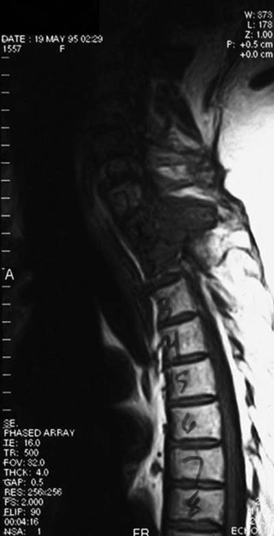
FIGURE 53.1 Sagittal MRI of the spine in a patient with metastatic ESCC. (Courtesy of José Biller, MD.)
4. RT alone is the procedure of choice for radiosensitive tumors.
a. A total dose of 20 to 40 Gy divided over 10 to 20 fractions is the usual treatment—30 Gy is the most common dose.
b. The port should encompass two vertebral bodies above and below the epidural defect and any discontinuous lesions.
c. RT is indicated promptly after the diagnosis is made and should follow administration of dexamethasone.
d. If surgery is performed first, RT should follow after the wound heals.
e. The tumors that most commonly produce ESCC—lung, breast, prostate, and lymphoma—are likely to respond to RT.
f. If neurologic deterioration continues, surgical intervention should be considered.
g. The complications of RT are:
(1) Myelosuppression.
(2) Radiation myelopathy or syrinx (6 to 18 months after therapy).
(3) Risk of a subacute syndrome characterized by Lhermitte’s sign several weeks after radiation.
5. Laminectomy plus radiation. For select patients, surgery followed by RT is better than RT alone for preserving function and increasing survival.
6. Epidural tumors do not respond rapidly enough to chemotherapy to warrant its use in most acute situations.
7. Recurrent ESCC.
a. Local metastatic lesions develop within two vertebral bodies of a previous lesion and within 3 months of the original diagnosis. They represent a failure of tumor control at the margin of the radiation port.
b. Distant metastatic lesions develop three or more vertebral bodies from a previous lesion and 15 months or longer after the original diagnosis.
c. Patients who previously responded to RT may be considered for repeat RT; they may benefit but not survive to experience the necrotizing consequences of exceeding spinal cord radiation tolerance.
d. Stereotactic body radiotherapy is being used more frequently for recurrent tumors and sometimes initially due to the focal nature of the treatment, thereby minimizing toxicity to the neural structures.
VI. LEPTOMENINGEAL METASTASIS
A. Leptomeningeal metastasis (LM) occurs when tumor cells invade the arachnoid and pia mater, focally or multifocally.
1. They develop in as many as 70% of patients with leukemia, and in 5% to 8% of patients with non-Hodgkin’s lymphoma. CNS lymphomas often involve the leptomeninges. Among solid tumors, breast is the most common, followed by lung, gastrointestinal tract, and melanoma.
B. Pathology. Typical pathologic features are:
1. Sheetlike layers and clumps of tumor cells (“lumpy-bumpy disease”).
2. Infiltration of cranial or spinal nerve roots.
3. Parenchymal invasion through the Virchow-Robin spaces.
C. Presentation.
1. Clinical features reflect the level of CNS involvement, alone or in some combination. Multifocal neurologic symptoms in a cancer patient is LM until proven otherwise.
2. The initial feature may be hydrocephalus.
3. New, worsening, unremitting headaches or somatic pain that present without apparent cause.
4. Extraocular, or any other cranial nerve palsies (see Chapter 12).
5. Involvement of lumbosacral nerve roots may cause a cauda equina syndrome.
6. Major differential diagnoses include bacterial, fungal, tubercular, or granulomatous meningitides.
D. Diagnosis.
1. LP should be performed in all cases where MRI is not diagnostic and there is no risk of herniation. Results are abnormal in >95% of cases.
2. LP may show lymphocytic pleocytosis, low to very low glucose, elevated protein, or malignant cells. The CSF pressure may be elevated.
3. Cytology is positive in only 50% on the first LP, so two to three may be needed. Cytology should be performed on a cytospin sample (not on stained smears) processed immediately after withdrawal to prevent cell lysis.
4. Markers such as carcinoembryonic antigen, CA27.29, PSA, ß2 microglobulin, and lactate dehydrogenase can be used for follow-up evaluation of the CSF, once initial cytologic diagnosis has been made.
5. Gadolinium-enhanced MR images show enhancement on the surface of nerve roots, spinal cord, CN, cerebellar folia, or over the cerebral convexities; fluid-attenuated inversion recovery (FLAIR) sequence may also provide evidence of LM. MRI may be abnormal even without positive cytology or spinal symptoms. Treatment can be initiated on the basis of MRI findings.
E. Initial treatment.
1. The systemic disease status and likelihood of successful palliation should be considered in the decision to treat.
2. Dexamethasone rarely provides symptomatic improvement and should be tapered after more definitive treatment is initiated.
F. Craniospinal or local RT.
1. Usual doses are 30 Gy to the entire neuraxis or 24 to 30 Gy to the most symptomatic areas.
2. Most patients are unable to tolerate RT to the entire neuraxis; it rarely controls LM and it produces myelosuppression. Craniospinal RT is reserved mainly for meningeal leukemia.
3. Local RT is indicated at the level of a radiographic or functional spinal block, especially if Intrathecal (IT) treatment is considered.
G. IT chemotherapy.
1. This modality is useful because tumor diffusely infiltrates the leptomeninges.
2. After systemic administration, most drugs reach CSF concentrations between 1% and 25% of the plasma concentration. Hence, the dose intensity of treatment in the CSF is decreased, predisposing to CNS failure. Some drugs like methotrexate (MTX) and cytarabine (cytosine arabinoside) have good CNS penetration and may provide adequate drug distribution to the CSF if administered IV in high doses.
3. Because the volume of distribution in the CSF is small, high concentrations can be achieved with small IT drug doses.
4. Drug clearance half-lives tend to be longer in the CSF than in plasma, maximizing exposure.
5. IT chemotherapy may be used alone or with neuraxis RT.
6. The first few doses can be administered by means of LP, but if continued, this can lead to an epidural hematoma, CSF leaks, and virtual subdural or epidural compartments through which drug can be lost.
7. Other constraints to delivery through LP relate to the dynamics of CSF flow, which is craniocaudal, tends to bypass the ventricles, is affected by patient position, may be severely disturbed in the presence of meningeal tumor with or without increased ICP and cannot be done with low platelets.
8. A ventricular catheter with a subcutaneous (Ommaya) reservoir should be implanted for more optimal drug delivery.
a. Infections are a serious but rare complication of Ommaya reservoirs; proper sterile technique should be followed with each use.
b. Fever, headache, lethargy, and CSF extravasation around the reservoir are signs of an Ommaya reservoir infection.
c. A chemical meningitis can sometimes occur.
9. About 50% of patients with LM from solid tumors respond to treatment (mostly breast cancer); about 75% with lymphoma or leukemia do. Response in solid tumors is less as available IT agents are more effective for hematologic malignancies.
10. MTX is the most commonly used drug for IT management of solid tumors.
11. Cytarabine for IT use is available encapsulated into multivesicular lipid-based particles. Severe or life-threatening arachnoditis from this treatment occurs in 19% to 30% of patients but its incidence and severity can be reduced by co-administration with dexamethasone. The terminal half-life of cytarabine in CSF can reach up to 82 hours and systemic exposure is minimal.
VII. METASTASIS TO THE SPINAL CORD PARENCHYMA
A. Treatment is palliative.
1. Dexamethasone is the initial treatment and should be followed by RT to the affected area.
2. Because approximately 75% of cases are secondary to lung and breast cancer and lymphoma, some clinical improvement can be expected, especially if the patient is treated before a myelopathy develops.
3. Surgical decompression accomplishes little because the disease is intrinsic to the spinal cord, and resection of the lesion itself is generally not feasible.
VIII. METASTASIS TO THE PERIPHERAL NERVOUS SYSTEM
A. Presentation.
1. Pain, numbness, paresthesiae, or weakness.
2. Must be differentiated from RT plexopathy, which is painless.
B. Treatment.
1. Aggressive pain relief is imperative.
2. Nonnarcotic drugs fail early, and adequate doses of narcotics should be prescribed.
3. If narcotics fail, several anesthetic or neurosurgical procedures are available.
4. RT or chemotherapy is used as appropriate.
IX. NEUROLOGIC COMPLICATIONS OF CHEMOTHERAPY
A. Most cytotoxic drugs can cause neurotoxicity as can some of the biologic agents. Reasons for recognizing iatrogenic toxicity are:
1. Drug toxicity can obscure or mimic the presentation of metastasis, paraneoplastic syndromes, or primary neurologic disease.
2. Neurotoxicity can contribute to morbidity, disability, and death.
3. Neurotoxicity can result from metabolic derangement of therapy or cancer-induced end-organ failure.
4. The offending agent can be identified during treatment with a multiple-drug regimen.
B. Common complications attributable to chemotherapy.
1. Peripheral neuropathy. Any chemotherapy can cause neuropathy (see Chapter 46), but most commonly implicated are vinca alkaloids, platinum-based agents, taxanes, etoposide, and suramin.
a. Vinca alkaloids. Length-dependent sensorimotor neuropathy from interference with axonal microtubule assembly is the dose-limiting side effect; it produces mild neuropathy in almost all patients at conventional doses. Autonomic dysfunction (gastroparesis, urinary retention, constipation, and ileus) can manifest early. Sometimes, cranial nerve involvement such as facial sensory loss, facial weakness, or recurrent laryngeal nerve palsy occurs. Reduction in dose or early withdrawal leads to complete recovery.
b. Cisplatin. Neuropathy is the dose-limiting side effect; it can develop even months after the drug is stopped. Large-fiber sensory neuropathy and ototoxicity are common, especially with cumulative doses >400 mg per m2. Symptoms include hearing loss, paresthesia, proprioceptive loss, ataxia and a Lhermitte’s sign (sudden, transient electric-like sensation spreading down the body when flexing the head). Autonomic symptoms (see above) are frequent. Most patients improve spontaneously if the total dose is <500 mg per m2.
c. Oxaliplatin causes acute and chronic peripheral neuropathy. Acute neurotoxicity can occur during or up to 2 days post-infusion and causes neuropathic symptoms (paresthesias, hypesthesias, and dysesthesiae), beginning in the hands or feet, perioral or in the throat. Acute side effects occur at a dose of about 130 mg per m2. Patients may develop a sense of dyspnea or dysphagia, may describe unusual sensations in the tongue, jaw spasms, eye pain, and muscle spasms or cramps that are sometimes described as stiffness in hands or feet or inability to release their grip. Cold temperature exacerbates the symptoms so patients should avoid cold drinks, wear gloves when handling refrigerated items, and avoid inhaling cold air. Symptoms usually last only a few days. Oxaliplatin causes prolonged opening of sodium-gated channels on peripheral nerves and leads to hyperexcitability from sequestration of calcium by the oxaliplatin–oxalate metabolite. Lowering the dose or increasing the infusion time may lessen the occurrence of these symptoms. Administering calcium gluconate and magnesium chloride decreases the occurrence of pharyngolaryngeal dysesthesia. More prolonged administration of oxaliplatin (total doses 540 to 850 mg per m2) causes neuropathic symptoms, changes in proprioception that do not resolve between cycles and Lhermitte’s sign. The chronic neuropathy is cumulative, with grade 3 toxicity occurring in 10% after 9 cycles, and in approximately 50% at 12 to 14 cycles (doses of 85 mg per m2 every 14 days). Urinary retention has been reported but is not common. Symptoms usually last months and resolve or improve to level 2 or 1 toxicity within 12 months. Rare complications include optic neuritis and visual field deficits. Gabapentin can reduce the acute neuropathic toxicity and prevent the chronic form as well.
d. Paclitaxel and Docetaxel. Sensory neuropathy occurs with doses >200 mg per m2. Painful paresthesia, multimodality sensory loss, ataxia, and mild distal weakness are common. Loss of the distal muscle stretch reflexes is almost universal. These agents can also cause a myopathy.
e. Etoposide is reported to cause axonal neuropathy in up to 10% of cases.
f. Suramin causes two distinct patterns of neurotoxicity that may be dose limiting. It causes a length-dependent axonal polyneuropathy or a subacute demyelinating polyradiculoneuropathy resembling Guillain–Barré’s syndrome (GBS). Inhibition of nerve growth factor may be the mechanism for neurotoxicity and occurs after peak plasma concentration increases to 0.350 mg per mL. Improvement after plasmapheresis has been reported.
g. Bortezomib is a proteosome inhibitor used in the treatment of multiple myeloma. It causes an axonal peripheral neuropathy often worsened in patients with dermatomyositis.
h. Ifosphamide is known to cause encephalopathy and busulfan causes seizures so patients are often placed on AEDs pre-treatment.
2. CNS toxicity.
a. The prototype drug is MTX. Route of administration, dosage, and simultaneous use with other neurotoxic therapy (particularly cranial RT) may cause additive or synergistic toxicity.
b. IT MTX may produce aseptic meningitis within a few hours after administration, and transient or permanent myelopathy.
c. Leukoencephalopathy is a delayed effect of either IT or high-dose systemic administration and can occur in up to 45% if combined with cranial RT or other neurotoxic drugs.
d. High-dose MTX can produce an acute, self-limited neurologic syndrome characterized by encephalopathy or, sometimes, a stroke-like syndrome of unknown causation.
3. Cerebellar toxicity.
a. The Purkinje cells are very sensitive to chemotherapy.
b. Cytarabine.
(1) Cerebellar toxicity occurs in 8% to 50% of patients given high-dose systemic cytarabine (doses 0.48 g per m2), especially if older than 60 and with renal dysfunction. Symptoms occur within 24 to 48 hours and manifest with nystagmus and mild ataxia followed by a florid encephalopathic and ataxic syndrome. See VI.G.11. for IT toxicity.
(2) Patients may improve within 1 week and recover completely within 2 weeks.
(3) The pathogenesis of this syndrome relates to the minimal amounts of cytidine deaminase in the CNS.
c. 5-Fluorouracil (5-FU).
(1) Causes abrupt onset of cerebellar dysfunction in as many as 8% of patients. Symptoms develop a few days to months after initiation of treatment and resolve after drug discontinuation. Cerebellar dysfunction is likely with schedules of one or more weekly boluses of >15 mg per kg and is linked to a deficiency of dihydropyrimidine dehydrogenase.
(2) Can cause an encephalopathy with hyperammonemia and lactic acidosis.
(3) Treatment of metastatic colorectal cancer with 5-FU and levamisole can cause a subacute multifocal leukoencephalopathy. MRI often shows supratentorial and infratentorial multifocal enhancing white-matter lesions.
(4) Other side effects include ophthalmoplegia, optic neuropathy, encephalopathy, focal dystonias, and parkinsonism.
(5) Capecitabine is a 5-FU prodrug that can have CNS toxicity, usually more acutely than 5-FU, but which resolves once the drug is stopped; it can cause diffusion restriction on MRI.
4. Bevacizumab is a monoclonal antibody against VEGF.
a. In 0.1% it has been associated with posterior reversible encephalopathy syndrome (PRES), which may present with headaches, seizures, lethargy, confusion, blindness, or other visual disturbances. Hypertension may or may not precede the symptoms. MRI shows characteristic findings of PRES.
b. Arterial thromboembolic events such as stroke, transient ischemic attack, and myocardial infarction may also occur.
c. Bleeding can occur—when intracranial or intratumoral, it can be fatal.
X. OTHER COMPLICATIONS
A. Neuropathic pain. Common neuropathic pain syndromes are:
1. Brachial plexus pain. Tumor invasion of the brachial plexus from breast carcinoma leads to pain most often referable to a C8-T1 distribution. Pain may precede neurologic findings by months. Management of the underlying tumor relieves pain in approximately 50% of cases. If pain returns, an exhaustive search for tumor recurrence must be undertaken.
2. Lumbar plexus pain. The lumbar plexus is commonly involved in extension of pelvic and colon tumors or metastasis from distant tumors (see Chapters 23 and 25).
3. Base of skull metastasis. (See III.) Pain in the face may be shock-like, and referable to the orbit or brow area. Swallow syncope refers to the occurrence of lightning-like pain upon swallowing followed by syncope. It is often caused by tumors involving the glossopharyngeal nerve (see Chapter 7).
4. Carcinomatous meningitis. Tumor seeding along nerve root sleeves may produce polyradicular pain symptoms.
5. Postsurgical pain. Five percent to twenty percent of patients undergoing mastectomy have characteristic postmastectomy pain characterized by a burning, tight feeling in the upper inner arm and across the chest. This may lead to a “frozen shoulder.” Thoracotomy is also very painful in the immediately postoperative period because the intercostal nerves are subjected to direct trauma during the procedure. Radical neck dissection can lead to poorly defined burning, stabbing pain from injury to the cervical nerves.
6. Radiation injury to peripheral nerves and plexus, although usually painless, can have painful sequelae with onset usually delayed for years after exposure. Pain may be the initial presentation of cervical plexus injury, whereas lumbar plexus injury often manifests as weakness. These must be differentiated from tumor recurrence.
7. Chemotherapy-related pain. Neuropathy caused by chemotherapeutic agents can be painful.
8. Treatment. Opioids are considered the mainstay of treatment. Other agents commonly used include tricyclic antidepressants, including tertiary and secondary amines. AEDs such as carbamazepine, gabapentin, and pregabalin are also used. Corticosteroids have been used in a variety of settings mainly as an adjunct to opioids. Local anesthetics such as capsaicin- and lidocaine-based creams and patches can also be used (see Chapter 50).
B. Encephalopathy.
1. Seizures.
a. Encephalopathic conditions can cause seizures, and seizures can cause encephalopathy.
b. A postictal state can mimic encephalopathy; in debilitated, elderly patients it can last 1 week or more.
2. If treatable causes are excluded, management is supportive.
C. CNS infection (see Chapters 43 and 44).
1. The most frequent organisms are Listeria monocytogenes, Cryptococcus neoformans, and Aspergillus fumigatus.
2. Next in frequency are gram-negative rods, Candida albicans, and varicella zoster virus.
3. Latent mycobacterial infections may become reactivated.
4. Presentation.
a. CNS infections often manifest as fever, changes in mental status, and seizures (see Chapter 43).
b. Headache and stiff neck can be subtle if patients are unable to mount an adequate inflammatory response.
c. In leukopenic patients, the CSF may not be purulent, and the infecting organism may not be readily detectable.
d. Gram staining or a polymerase chain reaction of cytospin sediment may reveal the pathogen before cultures.
5. LP should be approached with caution because a patient with increased ICP may herniate, or a patient with underlying thrombocytopenia may develop an epidural or subdural hematoma.
6. Treatment consists of antibiotics and support.
7. Progressive multifocal leukoencephalopathy (PML) (see Chapters 43 and 44).
a. PML is a CNS infection by an opportunistic papovavirus, the JC virus.
b. Rare cases go into long-term remission but most patients die of this disorder.
c. PML manifests as changes in mental status, speech and vision deficits, and weakness.
d. Diagnosis is from biopsy of a focal (nonenhancing) white-matter brain lesion if CSF for JC virus is negative.
e. Treatment with adenosine arabinoside (Ara-A) or cytarabine is of unproven benefit.
f. May be seen in patients treated with rituximab.
D. Cerebrovascular (CV) complications.
1. At autopsy, approximately 15% of cancer patients are found to have CV disease.
2. Atherosclerosis remains the leading cause of infarction, but only approximately 15% of infarcts are symptomatic.
3. Patients with cancer may also develop CV disease as a complication of the neoplastic process or its management.
4. Ischemic infarcts, rather than hemorrhages, predominate among patients with carcinoma. Nonbacterial thrombotic endocarditis (NBTE) and disseminated intravascular coagulation are frequent causes of symptomatic cerebral infarction in this population.
5. The most frequent causes of intraparenchymal hemorrhage are coagulopathies and hemorrhage into metastatic lesions from melanoma and germ cell tumors.
6. Mucinous cancers may produce infarction from occlusion of any cerebral artery.
7. A large proportion of patients with symptomatic CV disease and leukemia may experience hemorrhagic infarctions.
8. There is no specific therapy for these complications.
9. Chemotherapy, especially with cisplatin, can cause both acute and late vasculo-occlusive complications.
a. Acute vascular occlusion may be related to endothelial injury.
b. Late vascular occlusive abnormalities may be related to vasospasm from underlying hypomagnesemia.
10. Late effects of RT include a noninflammatory arteriopathy, causing large- and small-vessel occlusion, a mineralizing microangiopathy, or accelerated atherosclerosis.
11. Dural venous sinus thrombosis, particularly of the superior sagittal sinus (SSS), is underdiagnosed.
a. It is frequently asymptomatic and eventually recanalizes.
b. It is most frequent in patients with leukemia receiving chemotherapy (especially L-asparaginase), and in patients with underlying coagulopathies or widespread cancer especially with a large amount of skull metastases.
c. It manifests as headache, seizures, papilledema, focal motor signs, and encephalopathy.
d. Diagnosis is made by means of contrast-enhanced CT, MRI, MR angiography or venography.
e. Treatment is supportive, with short-term anticoagulation, if not clinically contraindicated.
12. Neoplastic angioendotheliosis or intravascular lymphomatosis, a rare complication of lymphoma, is intravascular occlusion of small blood vessels by malignant mononuclear cells.
a. When in the CNS, patients have multifocal deficits and encephalopathy with short-term or subacute progression to death.
b. This disorder is difficult to differentiate from PML, CNS vasculitis, and multiple emboli.
c. It can respond to treatment.
13. Patients with Waldenström’s macroglobulinemia can develop neurologic symptoms from peripheral neuropathy or CNS symptoms from hypervisocity or brain infiltration (Bing-Neel’s syndrome).
E. Syncope in patients with head and neck cancer.
1. This manifests in advanced or recurrent disease as syncope accompanied by paroxysmal head and face pain.
2. An abnormally strong carotid sinus reflex mediates the syncopal attacks.
3. A relation between the syncope and sudden death exists and should prompt a search for recurrent carcinoma.
4. Pain is managed with an AED such as gabapentin, pregabalin, or carbamazepine.
5. Syncope is managed with anticholinergic drugs such as propantheline at 15 to 30 mg orally four times a day; ephedrine at 25 mg orally four times a day may be added if there is a vasodepressor component and florinef may be used.
XI. PARANEOPLASTIC SYNDROMES
Paraneoplastic syndromes, or remote effects of cancer, are thought to originate from production of an antibody to onconeural antigens shared between the tumor and the CNS. Paraneoplastic neurologic syndromes may involve any part of the CNS or PNS. They must be differentiated from treatment-related toxicity and from metastasis. The antibodies found in these disorders are not thought to be directly pathogenic and should be considered as markers for these disorders. T-cell cytotoxic mechanisms might play an important role in nervous system injury. Any paraneoplastic syndrome may precede diagnosis of the underlying malignancy by up to several years. The best characterized paraneoplastic syndromes are
A. Paraneoplastic limbic encephalitis may develop in isolation or in association with disorders of brainstem, cerebellum, or peripheral nerves. The most common associated tumor is SCLC, followed by germ cell tumor, testicular tumors, breast, thymoma, and Hodgkin’s lymphoma.
1. Clinical features.
a. Most characteristic is a subacute amnestic syndrome with relative preservation of other cognitive functions. Short-term anterograde and variable retrograde amnesia are present. Memory deficit may be preceded by weeks of depression, personality changes, and emotional lability. Partial complex seizures are common.
b. Most patients eventually manifest a more generalized multifocal encephalomyelitis with involvement of brainstem, cerebellum, dorsal root ganglia, spinal cord, and autonomic nervous system.
c. The course is variable and unpredictable. Most patients appear to stabilize at a level of severe disability; a few patients become obtunded and comatose.
2. Diagnosis.
a. Anti-Hu antibodies are present in 50% of patients with SCLC and symptoms suggestive of limbic encephalitis. Prognosis is better among patients without anti-Hu antibodies.
b. Anti-Ta, anti-Ma1, and anti-Ma2 antibodies have been identified in the CSF and serum of patients with this syndrome.
c. “Atypical antibodies” including anti-CV2 associated with thymoma and antibodies that stain hippocampal neurons in patients with colon cancer have also been reported.
d. T2-weighted or FLAIR MRI may show abnormalities in the mesial temporal lobes (Fig. 53.2). CSF may demonstrate inflammatory changes, including increased protein content, moderate pleocytosis, presence of oligoclonal bands, and an increased level of immunoglobulin G (IgG).
B. Anti-NMDA receptor (NMDAR) encephalitis is a severe syndrome with psychiatric symptoms (such as agitation, paranoia, hallucinations, and irritability) and seizures associated with antibodies to the NR1 subunit of the NMDAR in women with ovarian teratomas. Outcome is favorable following removal of the teratoma.
C. Paraneoplastic cerebellar degeneration (PCD) may be profoundly disabling. PCD is more common among women and is often associated with adenocarcinoma of the ovary, uterus or adnexa, carcinoma of the breast, SCLC, and less commonly with Hodgkin’s lymphoma. In the setting of gynecologic malignancies, PCD usually occurs as an isolated syndrome. In the setting of SCLC, PCD often occurs as a part of more diffuse CNS involvement.
1. Clinical features.
a. The most common initial symptom is loss of coordination. Vertigo, nausea, and vomiting are common. Progression is usually subacute. Most patients display trunk and gait ataxia and dysarthric speech. Head titubation may be present.
b. Examination usually demonstrates ocular dysmotility, including nystagmus, limb dysmetria, and occasionally, opsoclonus. Patients are often left with a severe neurologic deficit without much improvement.
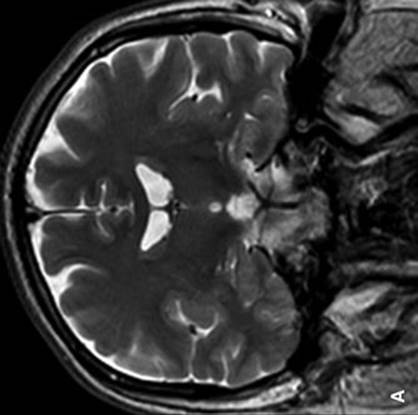
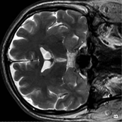


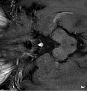
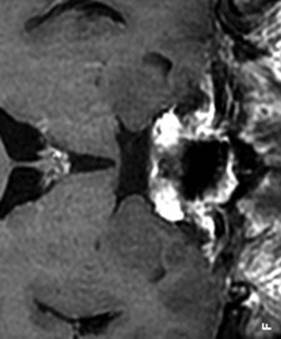

FIGURE 53.2 MRI of the brain in a patient with paraneoplastic limbic encephalitis associated with lung carcinoma demonstrates hyperintense signal in the medial temporal lobes. Note on T2 images increased signal and thickness in the mesial temporal lobe structures, and on post-contrast T1 images the presence of edema and the lack of significant enhancement. (Courtesy of José Biller, MD.)
2. Diagnosis.
a. Three major and several minor patterns of antineuronal antibody patterns have been associated with PCD. Only one of these, the anti-Yo antibody (type 1 antiparietal cell antibody) is strictly associated with PCD and is present exclusively in women with gynecologic malignancies or breast carcinoma. The other two, the anti-Hu (type 2, antineuronal nuclear antibody type 1) and the anti-Ri (type 2b, antineuronal nuclear antibody type 2) are associated with more complex disorders that may initially be diagnosed as PCD.
b. The anti-Hu pattern is associated with SCLC, and the anti-Ri with breast carcinoma. Other antibodies include anti-Tr autoantibodies described in patients with Hodgkin’s disease, and the anti-CV2 in patients with lung cancer. Most patients with PCD in association with Hodgkin’s disease, non-SCLC, or gastrointestinal carcinoma do not have any demonstrable antineuronal antibodies.
3. Management. Treatment is unsatisfactory, with best results among patients with no underlying gynecologic malignant tumors. Plasmapheresis or administration of IV immunoglobulin (IVIg) is beneficial in fewer than 10% of patients.
D. Opsoclonus–myoclonus (OM) is characterized by random, chaotic ocular movements. It is reported to occur with a variety of tumors and occurs in approximately 2% to 3% of children with neuroblastoma. Paraneoplastic OM usually appears as a part of a constellation of symptoms that include some combination of ataxia, myoclonus, and alteration in mental status.
1. Clinical features. Eye movements are conjugate, high amplitude, and in all directions of gaze. They are almost continuous and persist during eye opening. Unlike other paraneoplastic disorders, OM may go into spontaneous remission and be relieved with immunosuppressive therapy or tumor treatment. No clinical features differentiate paraneoplastic from non-paraneoplastic OM.
2. Diagnosis. Remains clinical, but patients with opsoclonus and ataxia associated with breast cancer may have anti-Ri antibodies. Patients with SCLC and opsoclonus usually have no detectable antineuronal antibodies. MRI of the brain is usually normal. Children with opsoclonus need a complete evaluation for neuroblastoma, including nuclear imaging with metaiodobenzylguanidine.
3. Management. Corticotropin or corticosteroids produce rapid and dramatic neurologic improvement in at least two-thirds of children independently of tumor status. In a number of adult patients with or without antineuronal antibodies, improvement has followed treatment with corticosteroids.
E. Myelopathy can occur with multiple-system neurologic involvement, as in paraneoplastic encephalomyelitis, in which case cord involvement is patchy. Necrotizing myelopathy, a distinct clinical entity, also occurs independently. Necrotizing myelopathy can occur in association with a variety of carcinomas and lymphoid tumors.
1. Clinical features of necrotizing myelopathy.
a. Onset is usually subacute with bilateral symptoms involving motor, sensory, and sphincter dysfunction and little or no pain. Examination shows findings consistent with transverse spinal cord dysfunction. Most patients have rapid deterioration with progressively ascending paralysis. Death may result from respiratory failure.
b. Differential diagnosis in a cancer patient includes intramedullary spinal metastasis, and spinal cord injury caused by RT or by IT chemotherapy.
2. Diagnosis. Spinal MRI may be normal or show intramedullary involvement. Although no antineuronal antibodies have been associated specifically with necrotizing myelopathy, in the absence of other causes it is felt to be paraneoplastic.
3. Management. There is anecdotal evidence of improvement with corticosteroids.
F. Motor neuron disease. Lower motor neuron signs are a predominant manifestation in about 25% of cases of multifocal paraneoplastic encephalomyelitis; most patients have SCLC. However, it is unclear how often pure motor neuron disease is paraneoplastic. As yet, there is no convincing evidence that nonhematogenous neoplasms occur in patients with amyotrophic lateral sclerosis any more frequently than would be expected in an age-matched control population. The prevalence of plasma cell dyscrasias among patients with motor neuron disease is higher than among controls; most have a monoclonal gammopathy of undetermined significance. Lymphoma has also been described in these patients.
1. Clinical features. Patchy weakness, fasciculations, and atrophy are usual manifestations. Patients may also have alteration in mental status, cerebellar ataxia, and brainstem findings.
2. Diagnosis. Anti-Hu antibodies may or may not be present.
3. Management. Improvement may follow plasma exchange and administration of corticosteroids and alkylating agents such as melphalan.
G. Stiff person syndrome. Muscle stiffness or rigidity may occur in the setting of several paraneoplastic disorders and reflect either CNS or PNS dysfunction. A syndrome resembling stiff person syndrome has been associated with several neoplasms; breast and SCLC most commonly, but also Hodgkin’s lymphoma, and carcinoma of the colon.
1. Clinical features. Patients have increasing aching and rigidity of axial and proximal limb muscles with sparing of the face. Severe painful spasms may occur spontaneously, or are precipitated by movements or sensory stimuli. Some patients display a fixed posture or opisthotonus in later stages of the disease.
2. Diagnosis. EMG shows continuous firing of motor units during the spasms. Most patients have serum and CSF antibodies against amphiphysin, a synaptic vesicle-associated protein but they are not specific for this syndrome and have also been found among patients with limbic encephalitis, PCD, or sensory neuronopathy. In contrast, antibodies to glutamic acid decarboxylase are found in stiff man syndrome of non-neoplastic origin.
3. Management. Some patients improve after tumor treatment. Benzodiazepines, baclofen, tizanidine, or prednisone might provide symptomatic relief.
H. Retinal degeneration (carcinoma-associated retinopathy) is a heterogenous disorder with varying tumor associations and different pathophysiologic mechanisms. More than 90% of patients have SCLC. Melanoma also is frequently associated.
1. Clinical features. Patients complain of bilateral visual blurring or dimming. Night blindness is a common complaint, especially among patients with melanoma. Positive visual symptoms such as sparkles and shimmering lights may be present. Examination may show an afferent pupillary defect and mild to moderate arteriolar narrowing. Otherwise, the examination is often unremarkable.
2. Diagnosis. The most common encountered antibody is the polyclonal IgG anti-CAR antibody. These antibodies react with recoverin, a calcium-binding protein. Some patients may also have antibodies against unidentified retinal proteins. However, many patients with cancer-associated retinopathy have no detectable antineuronal antibody.
3. Management. Most patients have mild to moderate improvement with oral prednisone. Management of the tumor appears to have no significant effect on this condition.
I. Neuronopathy.
1. Subacute sensory neuronopathy. More than 90% of patients have SCLC. Other neoplasms include carcinoma of the breast, prostate, colon, and lymphoma.
a. Clinical features. Development is subacute, and symptoms may begin in the face, trunk, or abdomen, and may be unilateral. Earliest symptoms are patchy numbness and paresthesia that may spread. Examination usually demonstrates involvement of all sensory modalities with loss of muscle stretch reflexes, flexor plantar responses, and preserved muscle strength if subacute sensory neuronopathy is the isolated or predominant paraneoplastic syndrome. Pseudoathetoid movements of the limbs may be present. Most patients stabilize at a severe level of disability.
b. Diagnosis.
(1) Most patients have anti-Hu antibodies in serum and CSF, usually in association with SCLC.
(2) Nerve conduction studies show reduced or absent sensory nerve action potentials and normal or borderline motor nerve conduction velocities. Some patients have features of both axonal and demyelinating neuropathy (see Chapter 33).
(3) CSF examination usually shows pleocytosis with mononuclear predominance, increased protein level, intrathecal IgG synthesis, and presence of oligoclonal bands.
(4) In patients with known cancer, neuronopathy is often due to the toxic effects of chemotherapy, metastasis, and nutritional deficits. Common drugs implicated are cisplatin, taxanes, and vinca alkaloids.
c. Management. Corticosteroids, plasma exchange, and administration of IVIg may be effective.
2. Demyelinating neuropathy associated with neoplasms. There are reports of acute, predominantly motor polyradiculoneuropathy occurring in the setting of a number of neoplasms, especially lymphoma. CSF and electrodiagnostic findings resemble those of GBS. Several cases of sensorimotor neuropathy fulfilling criteria for chronic inflammatory demyelinating polyneuropathy have been documented among patients with lymphoma.
3. Lambert–Eaton myasthenic syndrome (LEMS). In approximately 50% of cases, the LEMS is associated with a carcinoma, most commonly SCLC. An antibody for the P/Q type voltage-gated calcium channel is found in most patients with LEMS. The LEMS is discussed in further detail in Chapter 48.
J. Some patients have onconeural antibodies in the absence of a malignancy and may remain as such but need surveillance to be sure no malignancy develops; PET scans are the modality of choice.

Recommended Readings
Andrews DW, Scott CB, Sperduto PW, et al. Whole brain radiation therapy with or without stereotactic radiosurgery boost for patients with one to three brain metastases: phase III results of the RTOG 9508 randomised trial. Lancet. 2004;363:1665–1672.
Aoyama H, Shirato H, Tago M, et al. Stereotactic radiosurgery plus whole-brain radiation therapy vs stereotactic radiosurgery alone for treatment of brain metastases: a randomized controlled trial. JAMA. 2006;295:2483–2491.
Cestari DM, Weine DM, Panageas KS, et al. Stroke in patients with cancer, incidence and etiology. Neurology. 2004;62:2025–2030.
Deangeles LM, Posner JB, eds. Neurologic Complications of Cancer. 2nd ed. New York, NY: Oxford University Press; 2009.
Greenberg HS, Deck MDF, Vikram B, et al. Metastasis to the base of the skull: clinical findings in 43 patients. Neurology. 1981;1:530–537.
Langer CJ, Mehta MP. Current management of brain metastases, with a focus on systemic options. J Clin Oncol. 2005;23:6207–6219.
Loblaw DA, Perry J, Chambers A, et al. Systematic review of the diagnosis and management of malignant extradural spinal cord compression: the cancer care Ontario practice guidelines initiative’s neuro-oncology disease site group. J Clin Oncol. 2005;23:2028–2037.
Patchell R, Tibbs P, Regine W, et al. Direct decompressive surgical resection in the treatment of spinal cord compression caused by metastatic cancer: a randomised trial. Lancet. 2004;366:643–648.
Pruss H, Dalmau J, Harnms L, et al. Retrospective analysis of NMDA antibodies in encephalitis of unknown origin. Neurology. 2010;75:1735–1739.
Rogers LR. Cerebrovascular complications in cancer patients. Neurol Clin. 2003;21:167–192.
Rosenfeld MR, Dalmau J. Update on paraneoplastic and autoimmune disorders of the central nervous system. Semin Neurol. 2010;30:320–331.
Sirven JI, Wingerchuk DM, Drazkowski JF, et al. Seizure prophylaxis in patients with brain tumors: a meta-analysis. Mayo Clin Proc. 2004;79:1489–1494.
Titulaer MJ, Soffieti R, Dalmayu J, et al. Screening for tumors in paraneoplastic syndromes: report of an EFNS task force. Eur J Neurol. 2011;18:19-e3.
Vecht CJ, van Breemen M. Optimizing therapy of seizures in patients with brain tumors. Neurology. 2006;67:S10–S13.
Vogelbaum MA, Suh JH. Resectable brain metastases. J Clin Oncol. 2006;24:1289–1294.