
In the United States, there are 4,000,000 live births per year. It is thus common to see neurologic conditions occur in association with pregnancy. Furthermore, the physiologic changes in pregnancy can mimic neurologic diseases and can affect the severity of neurologic signs and symptoms. Not only can neurologic conditions be affected by pregnancy, but also treatment frequently must be altered to accommodate a developing fetus. Finally, pregnancy-specific conditions can present with neurologic symptoms and signs.
I. NORMAL PHYSIOLOGIC CHANGES IN PREGNANCY
A. Cardiovascular.
1. Increase of 30% to 50% in cardiac output and blood volume with singleton pregnancy (70% with twins).
2. Midpregnancy decrease in blood pressure.
B. Pulmonary.
1. Increase of 20% to 30% in minute volume.
2. Increase in respiratory rate and partially compensated respiratory alkalosis.
C. Renal.
1. Increase of 30% to 50% in renal blood flow.
2. Decreased serum level of blood urea nitrogen and creatinine (due to increased renal clearance).
D. Gastrointestinal.
1. Decreased motility.
2. Elevated alkaline phosphatase level (placental). No pregnancy-associated changes in any other liver function tests.
3. Increased cytochrome P-450 activity.
E. Hematologic.
1. Decreased hematocrit (20% to 30% increase in RBC volume but 30% to 50% increase in blood volume).
2. Increased WBC count; decreased platelet count.
F. Coagulation factors.
1. Increased levels of plasminogen, fibrinogen, and factors VII, VIII, IX, and X.
2. No change in factor V, antithrombin, or platelet adhesion.
3. Thrombophilias (e.g., antiphospholipid antibodies, protein C and S deficiency, and factor V Leiden) are more likely to produce thromboembolic complications.
G. Connective tissue. Thickening and fragmentation of reticular fibers with mild hyperplasia of smooth muscle cells.
H. Hormonal changes. Progressive increase in estrogens and progesterone until delivery.
I. Serum osmolality. Decreases from early in gestation, with resultant increase in extracellular fluid volume.
J. Neurologic. Increase in pituitary size; slight decrease in brain volume that returns to baseline postpartum.
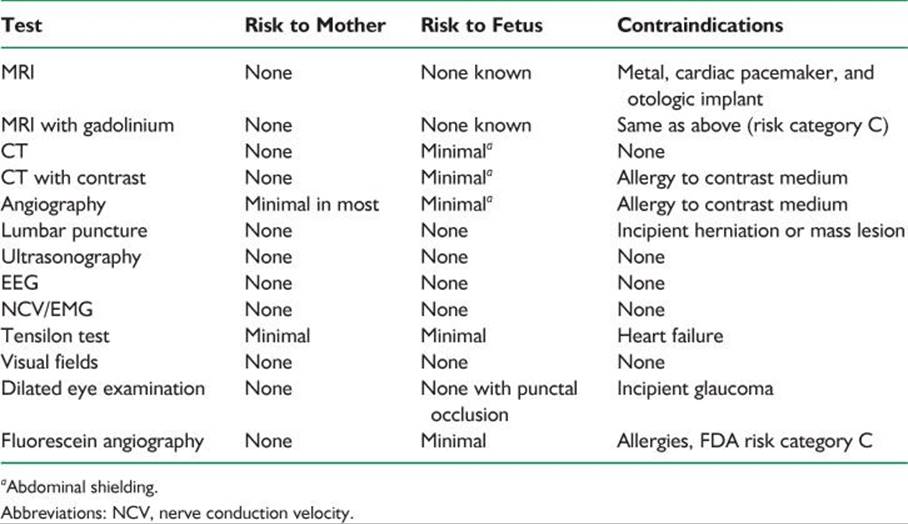
K. Evaluating neurologic conditions in pregnancy (Table 57.1).
L. U.S. Food and Drug Administration (FDA) risk factor classification of drugs in pregnancy.
1. Class A. Controlled studies show no risk to fetus in the first trimester; fetal harm is remote.
2. Class B. No controlled studies have been completed, but there are no known risks.
3. Class C. Studies on animals may show effects on fetuses, but no results of controlled studies are available. The drug can be used if the risk is justified.
TABLE 57.1 Evaluating Neurologic Conditions in Pregnancy
4. Class D. There are risks, but the drug may be used if serious disease or life-threatening conditions exist.
5. Class X. Human and animal studies show risk. The risk of use outweighs any benefit.
II. SEIZURE DISORDERS IN PREGNANCY
A. Frequency. One percent of the population.
1. In an unselected population, frequency is 7 to 8 per 1,000 deliveries.
2. Antiepileptic drugs (AEDs) lower the efficacy of some oral contraceptives in some individuals making pregnancy more likely.
B. Heredity.
1. About 2% to 5% if parent has idiopathic epilepsy. Relatively higher if the parent is the mother; relatively lower if the parent is the father.
2. No significant transmission if disease is acquired.
C. Course of disease in pregnancy.
1. The best figures for disease activity during pregnancy include the following:
a. Improved, 22%.
b. Exacerbated, 24% (most likely to occur in the first trimester).
c. No change, 54%.
2. Postulated mechanisms for changes in frequency during pregnancy include the following:
a. Physiologic.
(1) Hormonal (estrogens decrease and progestins increase seizure threshold).
(2) Metabolic (increased cytochrome P-450 activity).
b. Sleep deprivation.
c. Noncompliance (e.g., fear of birth defects from taking medications).
d. Pharmacokinetic changes in drug levels caused by: impaired absorption, increased volume of distribution, decreased albumin concentration, reduced plasma protein binding, and increased drug clearance.
e. Folate supplementation can reduce anticonvulsant levels.
f. Stress, anxiety.
g. Alcohol or other drug use.
3. Seizure frequency during pregnancy does not correlate with maternal age, seizure type, drug regimen, and seizure frequency in previous pregnancies.
4. Fetal risks with generalized convulsive seizure include the following:
a. Physical injury from maternal abdominal trauma.
b. Hypoxic–ischemic injury due to maternal hypoxia.
D. Therapeutic options.
1. Pharmacologic.
a. Be certain of the diagnosis.
b. Be familiar with and use the few drugs that are the most effective for the various types of seizures.
2. Surgery in general should be addressed before or after pregnancy.
3. General.
a. Maintain good daily habits (regularly scheduled meals, adequate sleep, and minimize stress).
b. Avoid alcohol and sedatives.
c. Avoid hazardous situations.
d. Avoid ketogenic diet.
E. Drug dosages, plasma levels, and clinical management.
1. AED levels decline during pregnancy in almost all women. This does not necessarily equate with a need to increase dosage, unless seizures are not controlled.
a. Free (non-protein-bound) drug level equates best with clinical status (seizure control and side effects) and should be obtained in pregnancies complicated by persistent or recurrent seizures or side effects.
b. Total drug level (usual laboratory result) sufficient if the patient has good clinical control.
c. With the exception of valproic acid, the average decline in free levels is less than that for total levels.
2. Frequency of measurement of levels.
a. Ideally, preconceptional total and free levels should be obtained and optimized.
b. Obtain non-protein-bound (free) levels every trimester (3 months), and again 4 weeks before term when: seizure types do not interfere with activities of daily living, the epilepsy is well-controlled.
c. Obtain monthly free levels when: uncontrolled seizures interfere with activities of daily living during the year before conception, previously controlled seizures recur during pregnancy, seizures are controlled but total drug levels decrease >50% on routine screens, troublesome or disabling side effects develop, lack of compliance is suspected or confirmed.
d. Always check levels postpartum and adjust dosage because levels often increase as the physiologic effects of pregnancy resolve within 10 to 15 days after delivery.
3. Changing drug dosage.
a. Reasons not to change dosage.
(1) Total drug levels are declining in a woman with well-controlled seizures, unless there are >30% decline in free levels and a history of poor control.
(2) A woman taking two or more AEDs discovers that she is pregnant (the time to change to monotherapy is before conception).
b. Reasons to change dosage.
(1) Increased numbers of tonic–clonic seizures.
(2) Complex partial or other seizure types that interfere with activities of daily life and the patient wants better control.
(3) Troublesome or disabling side effects.
c. Discontinuation of AED therapy should ideally be accomplished before conception but can be considered cautiously during pregnancy if a patient has been seizure-free for more than 2 years, has normal findings on neurologic examination, normal electroencephalographic findings, no structural brain disorder, and no history of prolonged convulsive seizures.
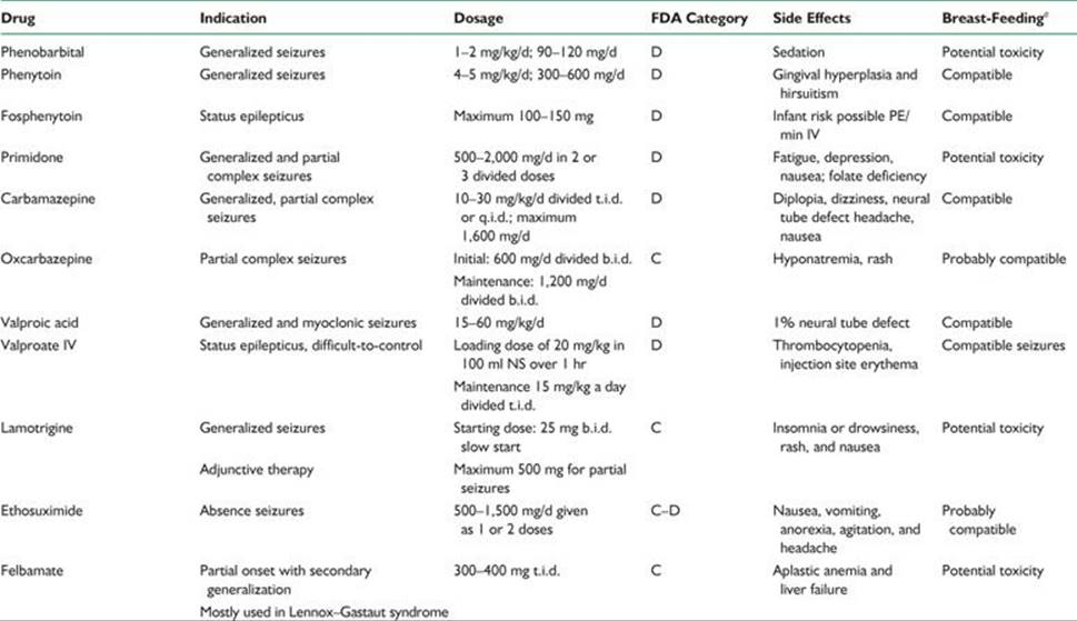
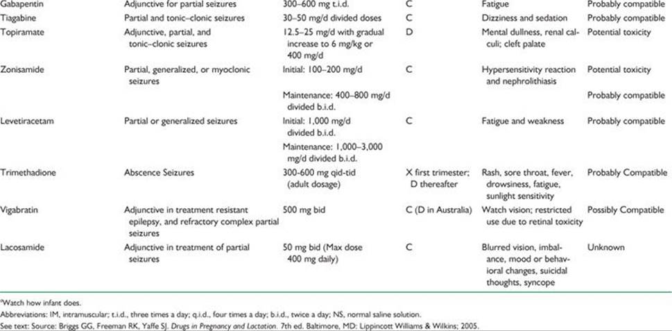
4. AEDs used in pregnancy (Table 57.2); breast-feeding while taking AEDs does not appear to affect cognition.
5. Other drugs to add or consider for patients with epilepsy.
a. Folic acid.
(1) Requirements may be further increased because of malabsorption, competitive metabolism, and increased hepatic metabolism.
TABLE 57.2 Medications Used in Epilepsy
(2) Increased supplementation may precipitate seizures by lowering anticonvulsant levels.
(3) Best advice is to maintain usual supplementation.
(4) Compelling evidence links the folic acid antagonism properties of AEDs to relatively increased risk of fetal neural tube defects in women taking anticonvulsants during the first trimester (neural tube defects form, or do not form, 26 to 28 days after conception). Women of reproductive potential should take continuous folic acid supplementation (400 mg per day) whether or not they are considering pregnancy.
b. Vitamin K should be administered (10 mg by mouth daily) to all pregnant women receiving AEDs beginning 4 weeks before expected delivery until birth to minimize the risk of neonatal hemorrhage. If a woman has not received vitamin K before delivery, consideration should be given to parenteral vitamin K administration.
c. Vitamin D not routinely supplemented.
6. Birth defects in infants of epileptic mothers.
a. Should be discussed with all epileptic women of reproductive age, irrespective of whether or not they are planning pregnancy (50% of pregnancies are unplanned).
b. Other factors that may explain the increased incidence of anomalies in infants of epileptic mothers are as follows:
(1) Increased incidence of anomalies in infants of epileptic mothers not taking AEDs. The only anomalies that are more common in phenytoin-exposed fetuses are hypertelorism and digital hypoplasia.
(2) Increased incidence of characteristic malformations in infants of epileptic fathers, described as being intermediate between treated and untreated epileptic mothers.
(3) A specific metabolic defect (epoxide hydrolase deficiency) more common in persons with epilepsy may predispose to damage in some cases. Autosomal codominant and increased fetal anomalies.
(4) Epilepsy may represent an underlying genetic disease.
(5) The defects may result from an AED-mediated relative folate deficiency. (Folate antagonists are known abortifacients and teratogens; see discussion above.)
7. AED teratogenesis should be discussed with all epileptic women of reproductive age.
a. Fetal anticonvulsant syndrome occurs in 3% to 5% of epileptic women and can occur in association with use of any anticonvulsant medication. The relative risk is dose dependent. This syndrome is being seen with decreasing frequency as fewer women receive polytherapy and more receive monotherapy.
(1) Craniofacial (cleft lip and palate) and digital dysmorphic changes.
(2) Growth deficiency.
(3) Microcephaly.
(4) Cardiac defects.
(5) Mental retardation.
b. AEDs and neural tube defects.
(1) Risk is 1% to 2% for valproic acid and slightly less for carbamazepine. It is <1% for other anticonvulsants. However, these risks are >0.1% population-wide risk in the United States.
(2) The relative risk is dose related.
(3) If the medications are necessary for seizure control, the patient should be offered maternal serum α-fetoprotein and ultrasound screening.
c. Trimethadione is clearly teratogenic and is contraindicated in pregnancy.
8. Breast-feeding.
a. Most AEDs cross into breast milk, although at low levels; the higher the protein binding of the AED, the less that is passed into breast milk. Recent studies show no cognitive change in babies breast fed while mother takes AED.
b. Contraindications to breast-feeding include poorly controlled maternal seizures, rapid somnolence on the part of an initially hungry infant, which suggests a drug effect.
F. Onset of seizures during pregnancy: differential diagnosis.
1. Rule out eclampsia. The most common multisystem disease in late pregnancy is preeclampsia or eclampsia.
2. Cortical venous thrombosis, especially late in pregnancy and in the immediate puerperium.
3. Tumors are especially likely to manifest in the first trimester, because this is when the pregnancy-associated increase in extracellular fluid begins. Meningioma tends to expand during pregnancy (response to the progressive increases in estrogen and progesterone).
4. Intracranial hemorrhage.
5. Gestational epilepsy is a diagnosis of exclusion and represents only a small fraction of all women who have initial seizures while pregnant.
G. Status epilepticus during pregnancy (follow guidelines for non-pregnant patients).
1. Less than 1% of all pregnant epileptic women.
2. Not an indication for termination of pregnancy.
3. Management should follow standard treatment of status epilepticus. Hospitalize, securing the airway, intravenous (IV) access for normal saline solution and B vitamins, baseline laboratory studies including electrolytes, CBC, glucose, calcium, and arterial blood gases. Maternal and fetal vital signs, including ECG and fetal heart rate monitoring. In addition administer the following:
a. Glucose bolus (50 ml of D50).
b. Thiamine (100 mg intramuscularly or intravenously).
c. Begin lorazepam (0.1 mg per kg IV, not to exceed 2 mg per minute) or diazepam (5 to 15 mg IV in 5 mg boluses) and fosphenytoin (150 mg per minute) or phenytoin 18 to 20 mg per kg IV, not to exceed 50 mg per minute, with ECG and blood pressure monitoring, administered in nonglucose-containing fluids).
d. If seizures persist, intubate and begin either phenobarbital (20 to 25 mg per kg IV, not to exceed 100 mg per minute). Alternatives include midazolam, propofol, levtriacetam, or IV valproic acid (if absolutely necessary).
e. If seizures still persist, institute general anesthesia with halothane and neuromuscular junction blockade. (See Chapters 38 and 39.)
III. HEADACHE
A. The most common headache diagnoses are as follows:
1. Migraine (with or without aura) occurs in 10% to 20% of women of childbearing age. Unilateral or bilateral throbbing headaches associated with photophobia, phonophobia, nausea, or vomiting may be exacerbated by activity.
2. Tension-type headache is very common. Mild-moderate headache, without nausea and vomiting, may be relieved by activity.
B. Genetics of migraine. Migraine is more common in affected families. Hemiplegic migraine is autosomal dominant associated with calcium channel genes located on chromosomes 1 and 19.
C. Course of migraine in pregnancy.
1. The condition of most women with migraine improves when they are pregnant. This is especially true with menstrual migraine and migraine whose onset was at menarche.
2. About 10% to 20% of headaches worsen or have the initial onset during pregnancy, usually in the first trimester. Many of these may be migraine aura without headache.
3. Migraineurs have no increased risk of complications during pregnancy, but headaches usually recur near term and in the puerperium.
4. Multiparous migraineurs may have an increase in headaches in the third trimester, whereas nulliparous women report less headache activity in pregnancy and the puerperium.
D. The differential diagnosis of headache or migraine occurring for the first time in pregnancy includes the following:
1. Severe preeclampsia—headache with hypertension should bring this diagnosis to the forefront.
2. Cerebral venous thrombosis.
3. Stroke (carotid or vertebral artery dissection).
4. Intracranial hypertension (increased intracranial pressure [ICP]).
5. Intracranial hemorrhage.
6. Brain tumor.
E. Therapeutic options.
1. Nonmedication treatment.
a. Adequate sleep.
b. Avoidance of dietary and environmental trigger factors.
c. Biofeedback, relaxation therapy, massage, physical therapy, and heat or ice packs.
2. Acute medication treatment principles.
a. Prevention of nausea (Table 57.3).
b. Management of pain (Table 57.4).
c. Sedation (Table 57.5).
3. Prophylactic treatment. In general, avoid daily medications, but if headaches are too severe or interfere excessively with life, daily treatment may be needed. In general, monotherapy should be attempted. The lowest dosage should be encouraged (Table 57.6).
TABLE 57.3 Acute Migraine Treatment in Pregnancy: Nausea Prevention
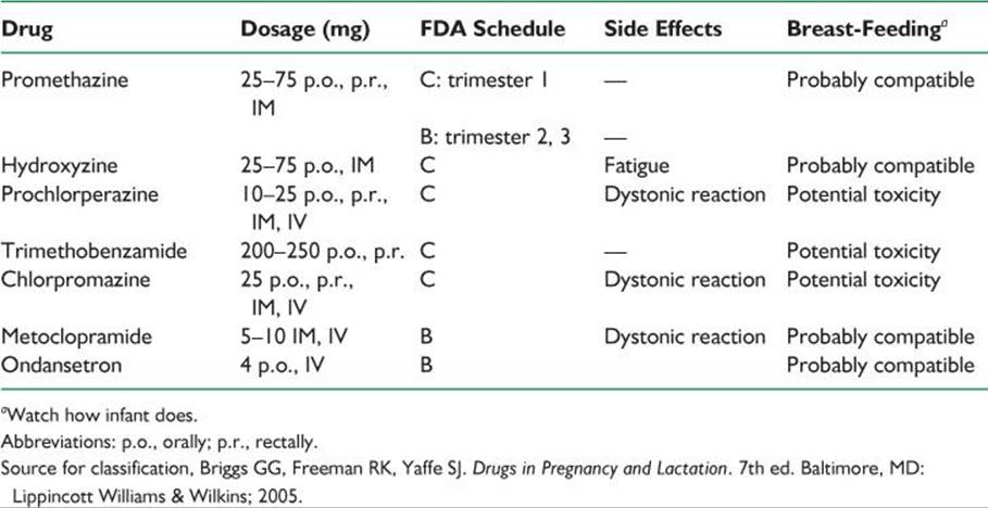
TABLE 57.4 Acute Migraine Treatment in Pregnancy: Pain Treatment
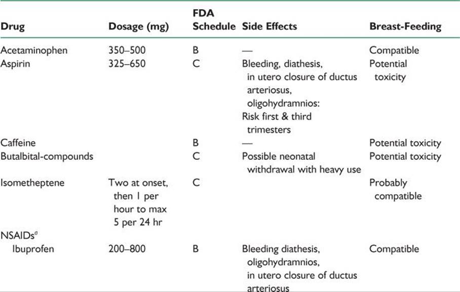
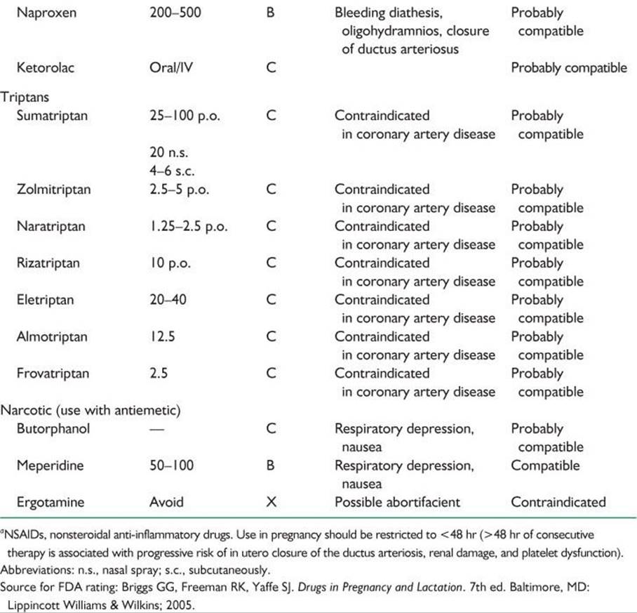
TABLE 57.5 Acute Migraine Treatment in Pregnancy: Sedation
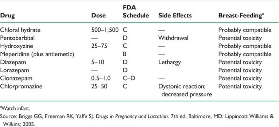
TABLE 57.6 Migraine Prophylaxis in Pregnancy
IV. TUMORS
A. Incidence.
1. Probably 100 per year nationwide.
2. Pregnancy does not increase the risk of brain tumors but does increase the likelihood of symptoms.
3. The types of tumors are identical to those observed in nonpregnant women of the same age, primarily glioma (32%), meningioma (29%), acoustic neuroma (15%), and others (24%).
B. Clinical features include headache, nausea and vomiting, papilledema, focal deficits, or seizures.
C. Diagnosis is made by imaging: MRI with contrast enhancement (gadolinium) or CT with contrast enhancement (Table 57.1).
D. Treatment.
1. Dexamethasone (risk factor C).
a. Dosage: 6 mg every 6 hours or 4 mg every 4 hours.
b. Problems: gastrointestinal; Cushingoid changes with prolonged use.
2. Mannitol (risk factor C) for acute brain swelling.
E. Pituitary tumors.
1. Course of disease.
a. Microadenoma rarely is symptomatic (5%).
b. Macroadenoma is symptomatic in 15% to 35% of cases.
2. Visual field evaluation must be performed for macroadenoma.
3. Treatment.
a. Bromocriptine (risk factor B) may be taken throughout pregnancy if the tumor enlarges. (Caution is advised in breast-feeding.)
b. If vision is threatened, surgical treatment is appropriate.
4. Sheehan’s syndrome is pituitary infarction, frequently associated with tumor or placental abruption.
a. Manifests as inability to lactate, hypopituitarism, and hypothyroidism.
b. Treatment involves steroid and thyroid replacement.
5. Lymphocytic hypophysitis mimics pituitary adenoma and suprasellar masses because it manifests as endocrinologic abnormalities, headaches, and a suprasellar mass at imaging. Lymphocytic hypophysitis occurs in pregnant and postpartum women. Biopsy often is needed to make the diagnosis. Steroid treatment with dexamethasone (category C) often is helpful.
V. PSEUDOTUMOR CEREBRI
Pseudotumor cerebri (idiopathic intracranial hypertension) is characterized by increased ICP not caused by an intracranial space-occupying lesion demonstrated at MRI or CT. Pregnancy does not cause pseudotumor cerebri. However, this disorder can occur in association with pregnancy. Pregnancy does not by itself cause visual loss. Pseudotumor cerebri does not cause miscarriage.
A. Symptoms and signs. Headache is the most common symptom (>90% of cases). Patients are otherwise alert and healthy. There are visual symptoms (transient visual obscurations) and auditory symptoms (whooshing noises). Signs include papilledema in almost all cases, cranial nerve VI palsy. Most women are obese.
B. Differential diagnosis of papilledema and no mass lesion in pregnancy.
1. Cerebral venous thrombosis (most important; most frequently needs to be excluded).
2. Venous hypertension.
3. Meningitis.
4. Syphilis.
C. Evaluation must include an imaging procedure (MRI and MR venography), CSF with opening pressure, and CSF constituents. Because the greatest threat to the patient is visual loss, visual acuity and visual field examinations must be performed frequently.
D. Treatment options.
1. Medical treatment.
a. Weight loss (restriction of weight gain is better than substantial weight loss).
b. Frequent lumbar puncture.
(1) Safe.
(2) Painful, often difficult.
c. Acetazolamide (500 to 2,000 mg) (risk C); compatible with breast-feeding.
d. Furosemide (Lasix; Aventis, Bridgewater, NJ, USA) (risk C).
e. Chlorthalidone (risk B); compatible with breast-feeding.
2. Surgical treatment.
a. Optic nerve sheath decompression is the preferred procedure to save vision.
b. Lumbar and ventriculo-peritoneal shunts can be difficult for pregnant patients due to displacement/compression from the enlarging uterus.
VI. CEREBROVASCULAR DISEASE
A. Attributable risk of ischemic stroke or intracerebral hemorrhage in pregnancy or the puerperium is 8.1/100,000 pregnancies. The causes of stroke during pregnancy are listed in Table 57.7.
1. Arterial stroke manifests as paresis but without altered consciousness or seizures; represents 90% of strokes during pregnancy.
2. Venous stroke manifests as headache, seizures, increased ICP, and alteration of consciousness; represents 80% of strokes during puerperium.
TABLE 57.7 Causes of Stroke in Pregnancy
Arterial occlusive disease
Thrombotic cause
Atherosclerotic
Cervicocephalic FMD
Cervicocephalic arterial dissections
Embolic source
Cardiac
Peripartum cardiomyopathy
Mitral valve prolapse
Rheumatic heart disease
Endocarditis (infective and nonbacterial)
Paradoxical embolism
Atrial fibrillation
Amniotic or air embolism
Venous occlusive disease
Hypercoagulable state (antithrombin, protein S or protein C deficiencies, factor V Leiden deficiency, hyperhomocysteinemia)
Infection
Drugs that can induce stroke:
Illicit drugs: cocaine, methamphetamine
Other drugs: sympathomimetics: phenylpropanolamine; ergotamine, bromocriptine, isometheptene
Hypotensive disorders
Watershed infarction
Sheehan’s pituitary necrosis
Hematologic disorders
Lupus anticoagulant, Sneddon’s syndrome
Thrombotic thrombocytopenic purpura
Sickle cell disease
Antithrombin deficiency, protein C and protein S deficiencies
Hyperhomocysteinemia
Factor V Leiden deficiency
Prothrombin G20210A mutation
MTHFR deficiency
Arteritis and angiopathy
SLE
Infectious arteritis (syphilis, tuberculosis, meningococcal)
Cerebral angiitis
Takayasu’s arteritis
Postpartum cerebral angiopathy
Cervicocephalic FMD and dissections
Intracerebral hemorrhage
Eclampsia and hypertensive disorders
Cerebral venous thrombosis
Choriocarcinoma
AVMs
Vasculitis
Infective endocarditis
Moyamoya disease
Tumors (primary and metastatic)
SAH
Aneurysm (saccular, mycotic, traumatic, etc.)
AVM (cerebral, spinal cord, and angiomas)
Eclampsia
Vasculitis
Choriocarcinoma
Cerebral venous thrombosis
Others
Carotid cavernous fistula
Dural vascular malformation
Carotid and vertebrobasilar arterial dissection (cervicocephalic FMD)
Abbreviations: FMD, fibromuscular dysplasia; SLE, systemic lupus erythematosus; AVM, arteriovenous malformations.
Modified from Digre KB, Varner MW. Diagnosis and treatment of cerebrovascular disorders in pregnancy. In: Adams HP, ed. Handbook of Cerebrovascular Diseases. New York, NY: Marcel Dekker; 2005:805–850.
3. Intracranial hemorrhage characteristically manifests as sudden onset of headache, loss of consciousness, and accompanying signs of neck stiffness and altered blood pressure.
4. Diagnosis.
a. CT and MRI (newer techniques of diffusion and perfusion may show early injury).
b. Angiography occasionally required.
c. Cardiac evaluation (transesophageal echocardiography—look also for right to left shunt).
d. Appropriate laboratory studies. The factor V Leiden mutation is now thought to be associated with at least one half of all cases of venous thromboses among white women. Consider protein C or protein S deficiency (may be falsely depressed simply because of pregnancy), antithrombin, antiphospholipid antibodies, platelets, fibrinogen, and homocysteine levels.
5. Treatment is directed at the underlying cause; treatment should be individualized.
a. Heparin, unfractionated or low-molecular weight, does not cross the placenta and can therefore be used safely during pregnancy. Low-molecular weight heparin (risk category B) has been used. Safe for breast-feeding.
b. Warfarin (risk category D; X in first trimester; compatible with breast-feeding) crosses the placenta and is contraindicated during pregnancy due to the embryopathy associated with use.
c. Low-dose aspirin (81 mg per day) (risk category C) can be used safely in pregnancy when clinically indicated. Other antithrombotic agents could be considered: Clopidrogel (risk category B) is an alternative to aspirin.
d. Management of acute ischemic stroke with tissue plasminogen activator (e.g., Alteplase [FDA C]; Urokinase [FDA B]) is not currently recommended although there are isolated case reports of benefit.
e. Manage elevation of homocysteine levels with folate.
B. Cerebral venous thrombosis.
1. Occurs primarily postpartum. The signs and symptoms include headache, seizures, hemiplegia, papilledema, and fluctuating obtundation and/or coma, especially in internal cerebral vein thrombosis.
2. Diagnosis optimally with MRI and MR venography; angiography, or venography occasionally is needed.
3. Treatment.
a. Correction of predisposing factors (infection and dehydration).
b. Control of seizures.
c. Use of antiedema agents when appropriate.
d. Anticoagulation (see sections VI.A.5.a. through c.).
4. Risk factors for cerebral venous thrombosis include cesarean delivery, hypertension, infection other than pneumonia or influenza, drug abuse, especially cocaine, methamphetamines, and IV drug abuse.
C. Postpartum cerebral angiopathy is a rare cause of a stroke-like syndrome characterized by seizure and focal neurologic deficits. Reversible cerebral vasoconstriction is found at angiography. Medications such as ergot alkaloids (e.g., ergonovine, bromocriptine, and ergotamine) and certain vasoconstrictive agents (isometheptene and sympathomimetic drugs) have been reported to cause the disorder. Treatment has been mainly supportive.
D. Hematologic disorders often manifest more frequently in pregnancy.
1. Antiphospholipid antibody syndrome is associated with recurrent pregnancy loss, fetal growth restriction, and severe preeclampsia and eclampsia.
2. Sickle cell disease.
3. Deficiencies of antithrombin, or protein C or S.
4. Thrombophilia, especially factor V Leiden mutation.
E. Subarachnoid hemorrhage (SAH). Causes include:
1. Intracranial aneurysm.
a. Thought to be present in 1% of all women of reproductive age; more likely in older, parous women.
b. A significant contributor to maternal mortality.
c. Rupture probably equally likely throughout pregnancy.
d. Diagnosis requires CT, lumbar puncture to look for RBCs, and angiography.
e. Optimum outcomes with surgical correction.
f. Avoid nitroprusside because of its cyanide effect on the fetus. Hypertension can be controlled with verapamil or nimodipine.
g. Vaginal delivery should be anticipated after successful clipping unless obstetric contraindications exist. If delivery occurs before clipping, cesarean section or forceps delivery with epidural anesthesia is indicated.
h. Vasospasm can be managed with nimodipine (FDA C). Volume expansion must be monitored, because pregnant women are relatively more prone to pulmonary edema (decreased osmotic pressure).
i. Outcome.
(1) Grades 1 through 3: with expedited surgery, 95% successful outcome expected.
(2) Grade 4: 45% to 75% mortality.
(3) Fetal outcome: 27% mortality rate without surgery.
j. Subsequent pregnancies after successful clipping have a good prognosis.
k. Asymptomatic aneurysm should be treated if >7 mm in diameter.
2. AVM.
a. Characteristically occurs in younger women who have had fewer pregnancies.
b. Diagnosis requires CT, lumbar puncture, and angiography.
c. The aneurysm should be corrected, if possible, surgically or with embolic therapy.
d. Stereotactic radiation therapy is not indicated during pregnancy.
e. Delivery is vaginal with epidural anesthesia and low-outlet forceps.
F. Eclampsia, severe preeclampsia.
1. Definition.
a. Preeclampsia (new onset hypertension and proteinuria beyond 20 weeks gestation) complicates 5% to 7% of pregnancies.
b. Severe preeclampsia. One or more of the following is present: persistent blood pressure 160 per 110 mm Hg, 5 g proteinuria in 24 hours, oliguria (500 ml per 24 hours), elevated results of liver function tests, thrombocytopenia, persistent visual disturbances or headache, epigastric pain, pulmonary edema, fetal growth restriction not explainable by other causes.
c. Eclampsia. Seizures or coma in a woman with preeclampsia in whom no other explanation can be found.
d. HELLP syndrome. A form of severe preeclampsia characterized by hemolysis, elevated results of liver function tests, and low platelet counts.
2. Symptoms and physical findings.
a. Headache, dizziness, scotomata, nausea, vomiting, and abdominal pain.
b. Generalized edema.
c. Funduscopic findings: segmental vasospasm, serous retinal detachment.
d. Neurologic finding: hyperreflexia; cortical blindness.
e. Bedside testing: visual acuity, Amsler’s grid for detection of scotomata.
3. CT and MRI findings.
a. CT. Edema and hypodense lesions 75%, hemorrhage 9%.
b. MRI.
(1) Severe preeclampsia. Deep white-matter signals on T2-weighted images
(2) Eclampsia. Signals on T2-weighted images at gray matter–white matter junctions, particularly in the parietal–occipital areas; cortical edema, hemorrhage; looks very much like hypertensive encephalopathy or posterior reversible encephalopathy.
4. Treatment.
a. Delivery.
b. Magnesium sulfate (FDA B; compatible with breast-feeding) is superior to IV diazepam and phenytoin in randomized controlled trials.
(1) Administered in a 4 to 6 g loading dose followed by 2 g per hour intravenously.
(2) Side effects include weakness, diplopia, ptosis, blurred vision, nausea, vomiting, and respiratory depression. Use with caution in the care of patients with reduced renal clearance or neuromuscular diseases such as myasthenia gravis.
(3) Neurologic findings of magnesium toxicity include diminished muscle stretch reflexes, ptosis and diminished accommodation, nausea, flushing, and respiratory depression.
c. Decrease blood pressure when necessary with an antihypertensive medication such as hydralazine or labetalol. Blood pressure needs to be controlled to minimize risk of maternal vascular accidents (usually below 160 per 110 mm Hg) but kept high enough to adequately perfuse mother and fetus.
d. Control seizures with an AED such as diphenylhydantoin (fosphenytoin) only if MgSO4 is unsuccessful.
e. Manage cerebral edema or herniation with hyperventilation, steroids, or mannitol after delivery.
6. Postpartum eclampsia (one-third of eclamptic convulsions do not begin until after delivery, usually within 24 to 48 hours after delivery), usually defined as within 7 days of delivery. Late postpartum eclampsia can occur up to 10 to 14 days after delivery. Consider the possibility of stroke, venous thrombosis, or reversible angiopathy if late postpartum eclampsia is being considered.
7. Outcome.
a. The maternal mortality rate in the United States is 1% to 2%.
b. The perinatal mortality rate is 13% to 30%.
8. Complications.
a. Intracranial hemorrhage, frequently from uncontrolled hypertension.
b. Congestive heart failure, frequently from iatrogenic fluid overload.
c. Intrahepatic hemorrhage.
VII. MULTIPLE SCLEROSIS
A. Multiple sclerosis (MS) does not affect pregnancy per se, or vice versa. Although recent studies do show that there may be increased relapses postpartum, especially in the first 6 months postpartum (particularly in the relapsing-remitting form), pregnancy does not affect the rate of disability.
1. Patients who have sphincter disturbances or paraplegia may experience increased difficulty during pregnancy.
2. There is no evidence of vertical transmission of MS.
3. MS does not occur more frequently in pregnancy.
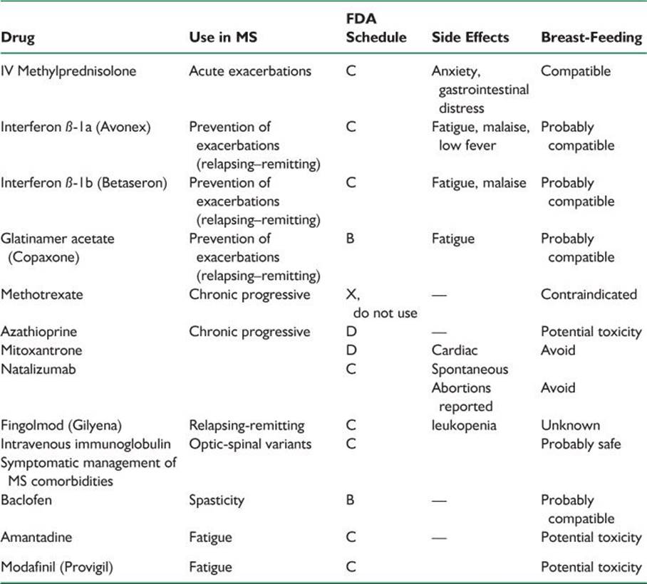
B. Management of acute MS in pregnancy (Table 57.8).
1. Steroids (see Chapter 40).
2. The interferons and copolymer are not yet recommended in pregnancy, although patients who were pregnant have used the medications without fetal harm.
TABLE 57.8 Drugs Used in the Management of MS
VIII. ROOT LESIONS AND PERIPHERAL NEUROPATHY
A. Lumbar disk.
1. Signs and symptoms are the same as in nonpregnant patients.
2. Generally treated nonoperatively. Consider surgery if there are bilateral symptoms or disturbance of sphincter function. Surgery frequently associated with increased blood loss due to increased collateral flow.
B. Carpal tunnel syndrome.
1. Often exacerbated during pregnancy due to increase in extracellular fluid.
2. Pain and paresthesia commonly are worse at night and tend to be worse in the dominant hand.
3. Symptoms usually respond to nocturnal wrist splinting and resolve within 3 months postpartum.
C. Bell’s palsy.
1. Facial paresis of lower-motor neuron type when no other specific etiologic agent can be found. Signs and symptoms include abrupt onset, often with pain around the ear; feeling of facial stiffness and pulling to one side; difficulty closing the eye on the affected side; taste disturbances; and hyperacusis.
2. Approximately three times more likely to occur during pregnancy, primarily in the third trimester or immediately postpartum.
3. Steroids are probably effective if given within the first 5 to 7 days. Surgery is ineffective.
D. Other forms of cranial nerve palsy.
1. Cranial nerve IV: reported rarely to occur; mechanism similar to cranial nerve VII or VI palsy.
2. Cranial nerve VI: similar to above; usually resolve postpartum.
E. Meralgia paresthetica.
1. Causes numbness in the lateral aspect of the thigh.
2. Usually resolves within 3 months postpartum.
F. Sciatica and back pain. Lumbosacral disk surgery should be reserved only for progressive atrophy or bowel or bladder dysfunction.
G. Guillain–Barré’s syndrome.
1. Causes are not generally affected by pregnancy.
2. Labor and delivery are otherwise normal.
IX. MYASTHENIA GRAVIS
A. Variable weakness and fatigability of skeletal muscles resulting from defective neuromuscular transmission (reduced acetylcholine receptors in the neuromuscular junction).
B. Does not affect labor progress, except for voluntary efforts in the second stage.
C. Certain drugs should be avoided, including the following:
1. Ester anesthetics: tetracaine (Pontocaine; Sanofi Winthrop, New York, NY, USA) and chloroprocaine (Nesacaine; AstraZeneca, Wilmington, DE, USA).
2. Curare (and other nondepolarizing muscle relaxants).
3. Halothane (Fluothane; Wyeth-Ayerst, Philadelphia, PA, USA).
4. Aminoglycoside antibiotics.
5. Quinine and quinidine.
6. Magnesium sulfate. The antidote with myasthenia is edrophonium (Tensilon; ICN, Costa Mesa, CA, USA), not calcium.
D. Treatment.
1. Antepartum.
a. Pregnancy per se does not affect the severity of preexisting disease.
b. Perinatal mortality is increased because of increased risk of premature delivery as well as neonatal myasthenia.
c. Pharmacologic management of myasthenia gravis is not altered by pregnancy.
2. Intrapartum.
a. Oral medications should be discontinued at the onset of labor and the intramuscular equivalents continued until oral medications can again be ingested. Equipotent dosages are as follows:
(1) Neostigmine 0.5 mg intravenously.
(2) Neostigmine 0.7 to 1.5 mg intramuscularly.
(3) Neostigmine 15 mg by mouth.
(4) Pyridostigmine 60 mg by mouth.
b. Analgesia and anesthesia for labor require the utmost caution because of the risks of respiratory depression and aspiration.
c. Myasthenia gravis does not affect the progress of labor and is not an indication for cesarean section.
3. Postpartum.
a. Exacerbations are more likely to occur postpartum; tend to be sudden and severe in onset.
b. Women with severe disease or whose babies have symptoms after nursing should not breast-feed.
c. Most women return to preconceptional oral dosage with modest increases in dose to allow for the additional stresses of early parenthood.
E. Neonatal myasthenia.
1. Occurs in 10% to 15% of cases.
2. Results from transplacental transfer of maternal antibody against acetylcholine receptors.
X. MYOTONIC DYSTROPHY
A. Clinical characteristics.
1. Autosomal dominant; weakness and wasting in muscles of face, neck, and distal limbs; myotonia of hands and tongue.
2. Variable age at onset. The condition sometimes is diagnosed in mothers only after an affected child is born (these pregnancies are frequently complicated by polyhydramnios that results from poor fetal swallowing).
3. Predisposition to cardiac arrhythmia.
4. Treatment. There is none for the dystrophy. Severe myotonia: phenytoin (Dilantin; Pfizer, New York, NY, USA), quinine (FDA D), and procainamide (FDA D).
B. Effects on pregnancy.
1. Increased risk of spontaneous abortion.
2. Increased risk of premature labor and polyhydramnios, particularly with fetal involvement.
3. Normal first stage of labor.
4. Normal response to oxytocin.
5. Prolonged second stage of labor.
C. Labor management includes outlet forceps, regional anesthesia; avoid succinylcholine (can cause hyperthermia); nonpolarizing agents are generally safe.
XI. MOVEMENT DISORDERS
A. Restless legs the most common movement disorder in pregnancy. Treat with iron or folate.
B. Chorea Gravidarum—look for unmasking of another disorder (e.g., systemic lupus, Sydenham’s chorea, etc.).
C. Parkinson’s disease in pregnancy is rare because the age at which most patients have the disease is past childbearing years. However, pregnancy has been successfully accomplished in patients with Parkinson’s disease.
1. Pregnancy may adversely affect Parkinson’s in that there may be exacerbations soon after pregnancy.
2. Drugs used in Parkinson’s disease:
a. Levodopa (FDA C), MAO-B (selegiline, rasagiline—FDA C), dopamine agonists (pramipexole, ropinirole FDA C), and COMT inhibitor (entacapone FDA C).
b. Amantadine (FDA C) can increase the risk of complications and malformations.

Recommended Readings
Adab N, Tudur SC, Vinten J, et al. Common antiepileptic drugs in pregnancy in women with epilepsy. Cochrane Database Syst Rev. 2004;(3):CD004848.
Argov Z, de Visser M. What we do not know about pregnancy in hereditary neuromuscular disorders. Neuromuscul Disord. 2009;19(10):675–679.
Briggs GG, Freeman RK, Yaffe SJ. Drugs in Pregnancy and Lactation. 7th ed. Baltimore, MD: Lippincott Williams & Wilkins; 2005.
Del Zotto E, Giossi A, Volonghi I, et al. Ischemic stroke during pregnancy and puerperium. Stroke Res Treat. 2011;2011:606780.
Dias MS, Sekhar LN. Intracranial hemorrhage from aneurysms and arteriovenous malformations during pregnancy and the puerperium. Neurosurgery. 1990;27:855–866.
Digre KB, Varner MW, Skalabrin E, et al. Diagnosis and treatment of cerebrovascular disorders in pregnancy. In: Adams HP, eds. Handbook of Cerebrovascular Diseases. New York, NY: Marcel-Dekker; 2005:805–850.
Goadsby PJ, Goldberg J, Silberstein SD. Migraine in pregnancy. BMJ. 2008;336:1502–1504.
Han IH. Pregnancy and spinal problems. Curr Opin Obstet Gynecol. 2010;22(6):477–481.
Harden CL, Hopp J, Ting TY, et al.; American Academy of Neurology; American Epilepsy Society. Management issues for women with epilepsy-focus on pregnancy (an evidence-based review): I. Obstetrical complications and change in seizure frequency: report of the Quality Standards Subcommittee and Therapeutics and Technology Assessment Subcommittee of the American Academy of Neurology and the American Epilepsy Society. Epilepsia. 2009;50(5):1229–1236.
Harden CL, Meador KJ, Pennell PB, et al.; American Academy of Neurology; American Epilepsy Society. Management issues for women with epilepsy-focus on pregnancy (an evidence-based review): II. Teratogenesis and perinatal outcomes: report of the Quality Standards Subcommittee and Therapeutics and Technology Subcommittee of the American Academy of Neurology and the American Epilepsy Society. Epilepsia. 2009;50(5):1237–1246.
Harden CL, Pennell PB, Koppel BS, et al.; American Academy of Neurology; American Epilepsy Society. Practice parameter update: management issues for women with epilepsy—focus on pregnancy (an evidence-based review): vitamin K, folic acid, blood levels, and breastfeeding: report of the Quality Standards Subcommittee and Therapeutics and Technology Assessment Subcommittee of the American Academy of Neurology and American Epilepsy Society. Neurology. 2009;73(2):142–149.
Kaplan PW. Neurologic aspects of eclampsia. Neurol Clin. 2004;22(4):841–861.
Lanska DJ, Kryscio RJ. Risk factors for peripartum and postpartum stroke and intracranial venous thrombosis. Stroke. 2000;31:1274–1282.
Lee AG, Pless M, Falardeau J, et al. The use of acetazolamide in idiopathic intracranial hypertension during pregnancy. Am J Ophthalmol. 2005;139(5):855–859.
Lee M, O’Brien P. Pregnancy and multiple sclerosis. J Neurol Neurosurg Psychiatry. 2008;79(12):1308–1311.
Loder E. Migraine in pregnancy. Semin Neurol. 2007;27(5):425–433.
Lynch JC, Gouvêa F, Emmerich JC, et al. Management strategy for brain tumour diagnosed during pregnancy. Br J Neurosurg. 2011;25(2):225–230.
Melhado EM, Maciel JA Jr, Guerreiro CA. Headache during gestation: evaluation of 1101 women. Can J Neurol Sci. 2007;34(2):187–192.
Menon R, Bushnell CD. Headache and pregnancy. Neurologist. 2008;14(2):108–119.
Micromedex. Version 2.0. Thompson Reuters. Nezvalová-Henriksen K, Spigset O, Nordeng H. Triptan exposure during pregnancy and the risk of major congenital malformations and adverse pregnancy outcomes: results from the Norwegian Mother and Child Cohort Study. Headache. 2010;50(4):563–575.
Oehm E, Hetzel A, Els T, et al. Cerebral hemodynamics and autoregulation in reversible posterior leukoencephalopathy syndrome caused by pre-/eclampsia. Cerebrovasc Dis. 2006;22(2–3):204–208.
Rudnik-Schoneborn S, Zerres K. Outcome in pregnancies complicated by myotonic dystrophy: a study of 31 patients and review of the literature. Eur J Obstet Gynecol Reprod Biol. 2004;114(1):44–53.
Sax TW, Rosenbaum RB. Neuromuscular disorders in pregnancy. Muscle Nerve. 2006;34(5):559–571.
Schlechte JA. Long-term management of prolactinomas. J Clin Endocrinol Metab. 2007;92(8):2861–2865.
Signore C, Spong CY, Krotoski D, et al. Pregnancy in women with physical disabilities. Obstet Gynecol. 2011;117:935–947.
Stafford IP, Dildy GA. Myasthenia gravis and pregnancy. Clin Obstet Gynecol. 2005;48(1):48–56.
Tang RA, Dorotheo EU, Schiffman JS, et al. Medical and surgical management of idiopathic intracranial hypertension in pregnancy. Curr Neurol Neurosci Rep. 2004;4(5):398–409.
Turan TN, Stern BJ. Stroke in pregnancy. Neurol Clin. 2004;22(4):821–840.
Wiese KM, Talkad A, Mathews M, et al. Intravenous recombinant tissue plasminogen activator in a pregnant woman with cardioembolic stroke. Stroke. 2006;37(8):2168–2169.
Winterbottom JB, Smyth RM, Jacoby A, et al. Preconception counselling for women with epilepsy to reduce adverse pregnancy outcome. Cochrane Database Syst Rev. 2008;(3):CD006645.