
[EPIDEMIOLOGY
Estimated prevalence: 1-1.5%.
Incidence of congenital hydrocephalus is ≈ 0.9-1.8/1000 births (reported range from 0.2 to 3.5/1000 births1).
FUNCTIONAL CLASSIFICATION
Two main functional subdivisions of hydrocephalus (HCP)
1. obstructive (AKA non-communicating): block proximal to the arachnoid granulations (AG). On CT or MRI: enlargement of ventricles proximal to block (e.g. obstruction of aqueduct of Sylvius → lateral and 3rd ventricular enlargement out of proportion to the 4th ventricle, sometimes referred to as triventricular hydrocephalus)
2. communicating (AKA non-obstructive): CSF circulation blocked at level of AG
SPECIAL FORMS OF HYDROCEPHALUS AND “PSEUDOHYDROCEPHALUS”
1. conditions that are not actually hydrocephalus “pseudohydrocephalus”
A. hydrocephalus ex vacuo: enlargement of the ventricles due to loss of cerebral tissue (cerebral atrophy), usually as a function of normal aging, but accelerated or accentuated by certain disease processes (e.g. Alzheimer’s disease, Creutzfeldt-Jakob disease, traumatic brain injury). For means of differentiating from true hydrocephalus, see page 906
B. otitic hydrocephalus: obsolete term used to describe the increased intracranial pressure seen in patients with otitis media (see see Idiopathic intracranial hypertension (IIH), page 713)
C. external hydrocephalus: seen in infancy, enlarged subarachnoid space with increasing OFCs and normal or mildly dilated ventricles see page 307
D. hydranencephaly: a post-neurulation defect (see page 243). Total or near-total absence of the cerebrum most commonly due to bilateral ICA infarcts. It is critical to differentiate this from severe (“maximal”) hydrocephalus (HCP) since shunting for true HCP may produce some re-expansion of the cortical mantle (see page 244 for means to differentiate)
2. normal pressure hydrocephalus (NPH): see page 329
3. entrapped fourth ventricle: see page 309
4. arrested hydrocephalus: see page 309
EXTERNAL HYDROCEPHALUS (AKA BENIGN EXTERNAL HYDROCEPHALUS)
 Key concepts:
• enlarged subarachnoid spaces over the frontal poles in the first year of life
• ventricles are normal or minimally enlarged
• may be distinguished from subdural hematoma by the “cortical vein sign”
• usually resolves spontaneously by 2 years of age
Enlarged subarachnoid space (usually over the cortical sulci of the frontal poles) seen in infancy (primarily in the first year of life) usually accompanied by abnormally increasing head circumference with normal or mildly dilated ventricles2. There are often enlarged basal cisterns and widening of the anterior interhemispheric fissure. No other symptoms or signs should be present (although there may be slight delay only in motor milestones due to the large head). Etiology is unclear, but a defect in CSF resorption is postulated. External hydrocephalus (EH) may be a variant of communicating hydrocephalus3. No predisposing factor may be found in some cases, although EH may be associated with some craniosynostoses4 (especially plagiocephaly) or it may follow intraventricular hemorrhage or superior vena cava obstruction.
Differential diagnosis: EH is probably distinct from benign subdural collections (or extra-axial fluid) of infancy (see page 904).  EH must be distinguished from symptomatic chronic extra-axial fluid collections (or chronic subdural hematoma), which may be accompanied by seizures, vomiting, headache… (see page 905) and may be the result of child abuse. With EH, MRI or CT may demonstrate cortical veins extending from the surface of the brain to the inner table of the skull coursing through the fluid collection (“cortical vein sign”), whereas the collections in subdural hematomas compress the subarachnoid space which apposes the veins to the surface to the brain5, 6.
Treatment: EH usually compensates by 12-18 mos age without shunting7. Recommend: follow serial ultrasound and/or CT to rule out abnormal ventricular enlargement. Emphasize to parents that this does not represent cortical atrophy. Due to increased risk for positional molding, parents may need to periodically reposition the head while the child is sleeping8.
A shunt may rarely be indicated when the collections are bloody (consider the possibility of child abuse) or for cosmetic reasons for severe macrocrania or frontal bossing.
X-LINKED HYDROCEPHALUS
Inherited hydrocephalus (HCP) with phenotypic expression in males passed on through carrier mothers who are phenotypically normal. Classical phenotypic expression will skip single generations.
Incidence: 1/25,000 to 1/60,000. Prevalence: ≈ 2 cases per 100 cases of hydrocephalus.
Gene located on Xq28151-153.
Pathophysiology
L1CAM membrane bound receptor plays a significant role in CNS development for axonal migration to appropriate target locations through Integrin cell adhesion molecules and MAP Kinase signal cascade151-153.
Abnormal gene expression results in poor differentiation and maturation of cortical neurons macroscopic anatomical abnormalities (bilateral absence of pyramidal tracts, see below)
Cytoplasmic domain loss of function mutations result in severe L1 syndrome, whereas mutations retaining expression of some functional protein (component imbedded in cell membrane) leads to mild L1 syndrome.
L1 syndromes
Classical syndromes include CRASH (corpus callosum hypoplasia, retardation, adducted thumbs (clasp thumbs), spastic paralysis, HCP), MSAS (mental handicap, aphasia, shuffling gate, adducted thumbs), HSAS (HCP with stenosis of the aqueduct of sylvius). Spectrum of disease also includes x-linked agenesis of the corpus callosum (ACC), and spastic paraparesis type 1151, 152.
Recent delineations153:
• mild L1 syndrome: adducted thumbs, spastic paralysis, hypoplasia of CC
• severe L1 syndrome: as in mild L1 syndrome plus anterior cerebellar vermis hypoplasia, large massa intermedia, enlarged quadrigeminal plate, rippled ventricular wall following VP shunt placement (pathognomonic for X-linked HCP). Profound mental retardation in virtually all cases
Radiographic findings likely present if severe L1152:
1. severe symmetric HCP with predominant posterior horn dilation
2. hypoplastic CC/ACC
3. hypoplastic anterior cerebellar vermis
4. large massa intermedia
5. large quadrigeminal plate
6. rippled ventricular wall following VP shunt placement (pathognomonic)
Treatment: no intervention demonstrates improvement in retardation status in observational papers.
1. VP shunt: main purpose is management of head size for improved care by care-giver. Does not improve neurologic outcome
2. there are no current genetic therapies for L1CAM protein abnormalities
3. prenatal U/S: early (≈ 20-24 weeks gestational age) with frequent repeat scan in known carrier mothers. May allow for medically indicated termination early on
4. male infants with HCP and ≥ 2 clinical/radiographic signs should undergo genetic testing for L1CAM mutation detection for future pregnancy counseling151
“ARRESTED HYDROCEPHALUS”
The exact definition of this term is not generally agreed upon, and some use the term compensated hydrocephalus interchangeably. Most clinicians use these terms to refer to a situation where there is no progression or deleterious sequelae due to hydrocephalus that would require the presence of a CSF shunt. Patients and families should be advised to seek medical attention if they develop symptoms of intracranial hypertension (decompensation): headaches, vomiting, ataxia or visual symptoms8.
Arrested hydrocephalus satisfies the following criteria in the absence of a CSF shunt:
1. near normal ventricular size
2. normal head growth curve
3. continued psychomotor development
Shunt independence
The concept of becoming independent of a shunt is not universally accepted9. Some feel that shunt independence occurs more commonly when the HCP is due to a block at the level of the arachnoid granulations (communicating hydrocephalus)10, but others have shown that it can occur regardless of the etiology11. These patients must be followed closely as there are reports of death as late as 5 years after apparent shunt independence, sometimes without warning10.
When to remove a disconnected or non-functioning shunt?
Note: a disconnected shunt may continue to function by CSF flow through an endothelialized subcutaneous tract. Recommendations on whether or not to repair vs. remove a disconnected or non-functioning shunt:
1. when in doubt, shunt
2. indications for shunt repair (vs. removal)
A. marginally functioning shunts
B. the presence of any signs or symptoms of increased ICP (vomiting, upgaze palsy, sometimes H/A alone12…)
C. changes in cognitive function, ↓ attention span, or emotional changes
D. patients with aqueductal stenosis or spina bifida: most are shunt dependent
3. because of risks associated with shunt removal, surgery for this purpose alone should be performed only in the situation of a shunt infection13
4. patients with a nonfunctioning shunt should be followed closely with serial CTs, and possibly with serial neuropsychological evaluations
ENTRAPPED FOURTH VENTRICLE
AKA isolated fourth ventricle: 4th ventricle that neither communicates with the 3rd ventricle (through sylvian aqueduct) nor with the basal cisterns (through foramina of Luschka or Magendie). Usually seen with chronic shunting of the lateral ventricles, especially with post-infectious hydrocephalus (fungal, in particular) or in those with repeated shunt infections. Possibly as a result of adhesions forming from prolonged apposition of the ependymal lining of the aqueduct due to the diversion of CSF through the shunt. Occurs in 2-3% of shunted patients14. May also occur in Dandy Walker malformation (see page 240) if the aqueduct is also obstructed. The choroid plexus of the 4th ventricle continues to produce CSF which enlarges the ventricle when there is 4th ventricular outlet obstruction or obstruction at the level of the arachnoid granulations.
Presentation may include:
1. headache
2. lower cranial nerve palsies: swallowing difficulties
3. pressure on the floor of the 4th ventricle may compress the facial colliculus (see page 844) → facial diplegia and bilateral abducens palsy
4. ataxia
5. reduced level of consciousness
6. nausea/vomiting
7. may also be an incidental finding (NB: some “atypical” findings, such as reduced attention span, may be related)
Treatment
Treatment of the entrapped 4th ventricle may alleviate associated slit ventricles15. Most surgeons advocate shunting the ventricle either with a separate VP shunt, or linking into an existing shunt. Options:
1. usual first choice: insertion from below the tonsils under direct vision. The catheter may be brought out at the dural suture line, and may be anchored here by use of an angle adapter sutured to the dura
2. passage through a cerebellar hemisphere: potential complications include delayed injury to the brainstem by the catheter tip as the brainstem moves into its normal position with drainage of the 4th ventricle. This may be avoided by bringing the catheter into the 4th ventricle at a slight angle through the cerebellar hemisphere
3. Torkildsen shunt (ventriculocisternal shunt) is an option for obstructive hydrocephalus if it is certain that the arachnoid granulations are functional (usually not the case with hydrocephalus of infantile onset)
4. an LP shunt may be considered when the 4th ventricle outlets are patent
Cranial nerve palsies may occur with shunting of the 4th ventricle usually as a result of penetration of the brainstem by the catheter either at the time of catheter insertion, or in a delayed fashion as the 4th ventricle decreases in size16, but also possibly as a result of overshunting causing traction on the lower cranial nerves as the brainstem shifts posteriorly14.
CT/MRI CRITERIA OF HYDROCEPHALUS
Numerous methods have been devised to attempt to quantitatively define hydrocephalus (HCP) (most date back to the early CT experience). Some are presented here for completeness. For radiologic features of chronic HCP, see page 312.
Hydrocephalus (HCP)
HCP is suggested when either17:
A. the size of both temporal horns (TH) is ≥ 2 mm in width (see Figure 15-1) (in the absence of HCP, the temporal horns should be barely visible), and the sylvian & interhemispheric fissures and cerebral sulci are not visible
OR
B. both TH are ≥ 2 mm, and the ratio  (where FH is the largest width of the frontal horns, and ID is the internal diameter from inner-table to inner-table at this level) (see Figure 15-1)

Figure 15-1 Linear ventricular measurement for CT, MRI or U/S Abbreviations: TH = temporal horns, FH = frontal horns, ID = internal diameter, BPD = biparietal diameter, OH = occipital horns
Other features suggestive of hydrocephalus (see Figure 15-1 for measurements):
1. ballooning of frontal horns of lateral ventricles (“Mickey Mouse” ventricles) and/or 3rd ventricle (the 3rd ventricle should normally be slit-like)
2. periventricular low density on CT, or periventricular high intensity signal on T2WI on MRI suggesting transependymal absorptionA of CSF
3. used alone, the ratio 
4. Evans ratio 18 (or index)B: ratio of FH to maximal biparietal diameter (BPD) measured in the same CT slice: > 0.3 suggests hydrocephalus
5. sagittal MRI may show thinning and/or upward bowing of the corpus callosum
A. a misnomer: CSF does not actually penetrate the ependymal lining (proven with CSF labeling studies), probably represents stasis of fluid in brain adjacent to ventricles
B. NB: measurements that rely on the frontal horn diameter tend to underestimate hydrocephalus in pediatrics possibly because of disproportionate dilatation of the occipital horns in peds19
ETIOLOGIES OF HYDROCEPHALUS
HCP is either due to subnormal CSF reabsorption, or rarely to CSF overproduction (as with some choroid plexus papillomas; even here, reabsorption is probably defective in some as normal individuals could probably tolerate the slightly elevated CSF production rate of these tumors). The etiologies in one series of pediatric patients is shown in Table 15-1.
• congenital
A. Chiari Type 2 malformation and/or myelomeningocele (MM) (usually occur together)
B. Chiari Type 1 malformation: HCP may occur with 4th ventricle outlet obstruction
C. primary aqueductal stenosis (usually presents in infancy, rarely in adult-hood)
D. secondary aqueductal gliosis: due to intrauterine infection or germinal matrix hemorrhage21
E. Dandy Walker malformation: atresia of foramina of Luschka & Magendie (see page 240). The incidence of this in patients with HCP is 2.4%
F. X-linked inherited disorder: rare
• acquired
A. infectious (the most common cause of communicating HCP)
1. post-meningitis (especially purulent and basal, including TB, cryptococcus (see page 374))
2. cysticercosis
B. post-hemorrhagic (2nd most common cause of communicating HCP)
1. post-SAH (see page 1037)
2. post-intraventricular hemorrhage (IVH): many will develop transient HCP. 20-50% of patients with large IVH develop permanent HCP
C. secondary to masses
1. non neoplastic: e.g. vascular malformation
2. neoplastic: most produce obstructive HCP by blocking CSF pathways, especially tumors around aqueduct, e.g. medulloblastoma. A colloid cyst can block CSF flow at the foramen of Monro. Pituitary tumor: suprasellar extension of tumor or expansion from pituitary apoplexy
D. post-op: 20% of pediatric patients develop permanent hydrocephalus (requiring shunt) following p-fossa tumor removal. May be delayed up to 1 yr
E. neurosarcoidosis: see page 71
F. “constitutional ventriculomegaly”: asymptomatic. Needs no treatment
G. associated with spinal tumors22
Table 15-1 Etiologies of HCP in 170 pediatric patients with HCP20
|
congenital (without myelomeningocele) |
38% |
|
congenital (with MM) |
29% |
|
perinatal hemorrhage |
11% |
|
trauma/subarachnoid hemorrhage |
4.7% |
|
tumor |
11% |
|
previous infection |
7.6% |
DIFFERENTIAL DIAGNOSIS OF HYDROCEPHALUS
For etiologies of HCP, see above. Conditions that may mimic HCP but are not due to inadequate CSF absorption include:
1. atrophy: sometimes referred to as “hydrocephalus ex vacuo”. Does not represent altered CSF hydrodynamics, but is rather loss of brain tissue (see page 307)
2. hydranencephaly: see page 243
3. developmental anomalies where the ventricles appear enlarged:
A. agenesis of the corpus callosum: see page 246 (may occasionally be associated with HCP, but more often merely represents expansion of the third ventricle and separation of the lateral ventricles)
B. septo-optic dysplasia: see page 247
SIGNS AND SYMPTOMS OF ACTIVE HCP
In young children
1. cranium enlarges at a rate > facial growth
2. irritability, poor head control, N/V
3. fontanelle full and bulging
4. enlargement and engorgement of scalp veins: due to reversal of flow from intracerebral sinuses due to increased intracranial pressure23
5. Macewen’s sign: cracked pot sound on percussing over dilated ventricles
6. 6th nerve (abducens) palsy: the long intracranial course is postulated to render this nerve very sensitive to pressure
7. “setting sun sign” (upward gaze palsy): Parinaud’s syndrome from pressure on region of suprapineal recess (see page 114)
8. hyperactive reflexes
9. irregular respirations with apneic spells
10. splaying of cranial sutures (seen on plain skull x-ray)
In older children/adults with rigid cranial vault
Symptoms of increased ICP, including: papilledema, H/A, N/V, gait changes, upgaze and/or abducens palsy. Slowly enlarging ventricles may initially be asymptomatic.
CHRONIC HCP
Features indicative of chronic hydrocephalus (as opposed to acute hydrocephalus):
1. beaten copper cranium (some refer to beaten silver appearance) on plain skull xray24. By itself, does not correlate with increased ICP, however when associated with #3 and #4 below, does suggest ↑ ICP. May be seen in craniosynostosis (see page 231 for description)
2. 3rd ventricle herniating into sella (seen on CT or MRI)
3. erosion of sella turcica (may be due to #2 above) which sometimes produces an empty sella, and erosion of the dorsum sella
4. the temporal horns may be less prominent on CT than in acute HCP
5. macrocrania: by convention, OFC greater than 98th percentile25 (pp 203)
6. atrophy of corpus callosum: best appreciated on sagittal MRI
7. in infants
A. sutural diastasis
B. delayed closure of fontanelles
C. failure to thrive or developmental delay
OCCIPITAL-FRONTAL CIRCUMFERENCE
The occipital-frontal circumference (OFC) should be followed in every growing child (as part of a “well-baby” check-up, and especially in infants with documented or suspected hydrocephalus (HCP)). As a rule of thumb, the OFC of a normal infant should equal the distance from crown to rump26 (rule #335). See page 1206 for the DDx of macrocephaly.
Normal head growth: parallels normal curves as seen on the graphs on the inside front cover, or in Figure 15-2 and Figure 15-3 for preemies. Any of the following may signify treatable conditions such as active HCP, subdural hematoma, or subdural effusions, and should prompt an evaluation of the intracranial contents (e.g. CT, head U/S …):
1. upward deviations (crossing curves)
2. continued head growth of more than 1.25 cm/wk
3. OFC approaching 2 standard deviations (SD) above normal
4. head circumference out of proportion to body length or weight, even if within normal limits for age (see Figure 15-3)27
These conditions may also be seen in the “catch-up phase” of brain growth in premature infants after they recover from their acute medical illnesses, see Catch-up phase of brain growthpage 1136). Deviations below the curves or head growth in the premature infant in the neonatal period of less than 0.5 cm/wk (excluding the first few weeks of life) may indicate microcephaly (see page 245).
Technique: measure circumference around forehead and occiput (excluding ears) three consecutive times, and use the largest value. OFC is then plotted on a graph of average values as a function of age28 and followed for each individual patient. Use the graphs on the inside front cover for most children and adolescents. The graph in Figure 15-2 shows the OFC for premature infants as a function of gestational age up to term.
The graph in Figure 15-3 shows the relationship of head circumference, weight and length for various gestational ages.

Figure 15-2 OFC for premature infants as a function of gestational age

Figure 15-3 Head circumference, weight and length*
*(Redrawn from Journal of Pediatrics, “Growth Graphs for the Clinical Assessment of Infants of Varying Gestational Age”, Babson S G, Benda G I, vol 89, pp 815, with permission)
15.1. Treatment of hydrocephalus
MEDICAL
HCP remains a surgically treated condition. Acetazolamide may be helpful for temporizing (see below).
Diuretic therapy
May be tried in premature infants with bloody CSF (as long as there is no evidence of active hydrocephalus) while waiting to see if there will be resumption of normal CSF absorption. However, at best this should only be considered as an adjunct to definitive treatment or as a temporizing measure.
Satisfactory control of HCP was reported in ≈ 50% of patients of age < 1 year who had stable vital signs, normal renal function and no symptoms of elevated ICP (apnea, lethargy, vomiting) using the following29:
• acetazolamide (a carbonic anhydrase inhibitor): 25 mg/kg/day PO divided TID x 1 day, increase 25 mg/kg/day each day until 100 mg/kg/day is reached
• simultaneously start furosemide: 1 mg/kg/day PO divided TID
• to counteract acidosis:
 tricitrate (Polycitra®) 4 ml/kg/day divided QID (each ml is equivalent to 2 mEq of bicarbonate, and contains 1 mEq K+ and 1 mEq Na+)
measure serial electrolytes, and adjust dosage to maintain serum HCO3 > 18 mEq/L
change to Polycitra-K® (2 mEq K+ per ml, no Na+) if serum potassium becomes low, or to sodium bicarbonate if serum sodium becomes low
• watch for electrolyte imbalance and acetazolamide side effects: lethargy, tachypnea, diarrhea, paresthesias (e.g. tingling in the fingertips)
• perform weekly U/S or CT scan and insert ventricular shunt if progressive ventriculomegaly occurs. Otherwise, maintain therapy for a 6 month trial, then taper dosage over 2-4 weeks. Resume 3-4 mos of treatment if progressive HCP occurs
SPINAL TAPS
HCP after intraventricular hemorrhage may be only transient Serial taps (ventricular or LP30) may temporize until resorption resumes (see page 1135) but LPs can only be performed for communicating HCP. If reabsorption does not resume when the protein content of the CSF is < 100 mg/dl, then it is unlikely that spontaneous resorption will occur (i.e. a shunt will usually be necessary).
SURGICAL
Goals of therapy:
Normal sized ventricles is not the goal of therapy. Goals are optimum neurologic function and a good cosmetic result.
Options include:
1. choroid plexectomy: described by Dandy in 1918 for communicating hydrocephalus31. May reduce the rate but does not totally halt CSF production (only a portion of CSF is secreted by the choroid plexus, other sources include the ependymal lining of the ventricles and the dural sleeves of spinal nerve roots). Open surgery was associated with a high mortality rate (possibly due to replacement of CSF by air). Endoscopic choroid plexus coagulation was originally described in 1910 and was recently resurrected32
2. eliminating the obstruction: e.g. opening a stenosed sylvian aqueduct. Often higher morbidity and lower success rate than simple CSF diversion with shunts, except perhaps in the case of tumor
3. third ventriculostomy: (see below)
4. shunting: various shunts are described below. The techniques of shunt placement are covered on page 209 for VP shunts, page 210 for VA shunt, page 210 for ventriculopleural shunts, and page 213 for LP shunt
Third ventriculostomy
Endoscopic third ventriculostomy (ETV) has energized a renewed interest in third ventriculostomy (see page 212 for technique).
Indications: ETV may be used in patients with obstructive HCP. May be an option in managing shunt infection (as a means to remove all hardware without subjecting the patient to increased ICP). ETV has also been proposed as an option for patients who developed subdural hematomas after shunting (the shunt is removed before the ETV is performed). ETV may also be indicated for slit ventricle syndrome (see page 326).
Contraindications: Communicating hydrocephalus has traditionally been considered a contraindication to ETV. However, it is used for NPH in some centers33. Relative contraindications to ETV would be the presence of any of the conditions associated with a low success rate (see below).
Complications:
1. hypothalamic injury
2. transient 3rd and 6th nerve palsies
3. uncontrollable bleeding
4. cardiac arrest34
5. traumatic basilar artery aneurysm35: possibly related to thermal injury from use of laser in performing ETV
Success rate: Overall success rate is ≈ 56% (range of 60-94% for nontumoral aqueductal stenosis35 (AqS)). Highest maintained patency rate is with previously untreated acquired AqS. Success rate in infants may be poor because they may not have a normally developed subarachnoid space. There is a low success rate (only ≈ 20% of TVs will remain patent) if there is pre-existing pathology including:
1. tumor
2. previous shunt
3. previous SAH
4. previous whole brain radiation (success with focal stereotactic radiosurgery is not known)
5. significant adhesions visible when perforating through the floor of the third ventricle at the time of performance of ETV
In one series, clinical improvement after ETV was achieved in 76% (72 of 95 patients), including 6 patients requiring second ETVs (three of which had partially functioning shunts that were left in place at the time of ETV).
15.1.1. Shunts
For surgical insertion techniques, see Ventricular shunts on page 208.
TYPES OF SHUNTS
SHUNT TYPE BY CATEGORY
1. ventriculoperitoneal (VP) shunt:
A. most commonly used shunt in modern era
B. lateral ventricle is the usual proximal location
C. intraperitoneal pressure: normal is near atmospheric
2. ventriculo-atrial (VA) shunt (“vascular shunt”):
A. shunts ventricles through jugular vein to superior vena cava, so-called “ventriculo-atrial” shunt because it shunts the cerebral ventricles to the vascular system with the catheter tip in the region of the right cardiac atrium)
B. treatment of choice when abdominal abnormalities are present (extensive abdominal surgery, peritonitis, morbid obesity, in preemies who have had NEC and may not tolerate VP shunt…)
C. shorter length of tubing results in lower distal pressure and less siphon effect than VP shunt, however pulsatile pressures may alter CSF hydrodynamics
3. Torkildsen shunt:
A. shunts ventricle to cisternal space
B. rarely used
C. effective only in acquired obstructive HCP, as patients with congenital HCP frequently do not develop normal subarachnoid CSF pathways
4. miscellaneous: various distal projections used historically or in patients who have had significant problems with traditional shunt locations (e.g. peritonitis with VP shunt, SBE with vascular shunts):
A. pleural space (ventriculopleural shunt): not a first choice, but a viable alternative if the peritoneum is not available36. To avoid symptomatic hydrothorax necessitating relocating distal end, it is recommended only for patients > 7 yrs age. Pressure in pleural space is less than atmospheric
B. gall bladder
C. ureter or bladder: causes electrolyte imbalances due to losses through urine
5. lumboperitoneal (LP) shunt (for insertion technique, see page 213):
A. only for communicating HCP: primarily pseudotumor cerebri or CSF fistula37. Useful in situations with small ventricles
B. over age 2 yrs, percutaneous insertion with Tuohy needle is preferred
6. cyst or subdural shunt: from arachnoid cyst or subdural hygroma cavity, usually to peritoneum
Disadvantages/complications of various shunts
1. those that may occur with any shunt:
A. obstruction: the most common cause of shunt malfunction
proximal: ventricular catheter (the most common site)
valve mechanism
distal: reported incidence of 12-34%38. Occurs in peritoneal catheter in VP shunt (see below), in atrial catheter in VA shunt
B. disconnection at a junction, or break at any point
C. infection
D. hardware erosion through skin, usually only in debilitated patients (especially preemies with enlarged heads and thin scalp from chronic HCP, who lay on one side of head due to elongated cranium). May also indicate silicone allergy (see below)
E. seizures (ventricular shunts only): there is ≈ 5.5% risk of seizures in the first year after placement of a shunt which drops to ≈ 1.1% after the 3rd year39 (NB: this does not mean that the shunt was the cause of all of these seizures). Seizure risk is questionably higher with frontal catheters than with parieto-occipital
F. act as a conduit for extraneural metastases of certain tumors (e.g. medulloblastoma). This is probably a relatively low risk40
G. silicone allergy41: rare (if it occurs at all). May resemble shunt infection with skin breakdown and fungating granulomas. CSF is initially sterile but later infections may occur. May require fabrication of a custom silicone-free device (e.g. polyurethane)
2. VP shunt:
A. 17% incidence inguinal hernia (many shunts are inserted while processus vaginalis is patent)42
B. need to lengthen catheter with growth: may be obviated by using long peritoneal catheter (see page 209)
C. obstruction of peritoneal catheter:
may be more likely with distal slit openings (“slit valves”) due to occlusion by omentum or by trapping debris from the shunt system38
by peritoneal cyst (or pseudocyst)43: usually associated with infection, may also be due to reaction to talc from surgical gloves (the omentum tends to “wall off” a nidus of irritation). It may rarely be necessary to differentiate a CSF collection from a urine collection in patients with overdistended bladders that have ruptured (e.g. secondary to neurogenic bladder). Fluid can be aspirated percutaneously and analyzed for BUN and creatinine (which should be absent in CSF)
severe peritoneal adhesions: reduce surface area for CSF resorption
malposition of catheter tip:
• at time of surgery: e.g. in preperitoneal fat
• tubing may pull out of peritoneal cavity with growth
D. peritonitis from shunt infection
E. hydrocele
F. CSF ascites
G. tip migration
into scrotum44
perforation of a viscus45: stomach46, bladder… More common with older spring-reinforced (Raimondi) shunt tubing
through the diaphragm47
H. intestinal obstruction (as opposed to perforation): rare
I. volvulus48
J. intestinal strangulation: occurred only in patients in whom attempt was made to remove peritoneal tubing using traction on the catheter applied at the cephalad incision with subsequent breakage of the tubing leaving a residual intraabdominal segment (immediate peritoneal exploration is recommended under these circumstances)49
K. overshunting: more likely than with VA shunt. Some recommend LP shunt for communicating hydrocephalus (see page 325)
3. VA shunt:
A. requires repeated lengthening in growing child
B. higher risk of infection, septicemia
C. possible retrograde flow of blood into ventricles if valve malfunctions (rare)
D. shunt embolus
E. vascular complications: perforation, thrombophlebitis, pulmonary micro-emboli may cause pulmonary hypertension50 (incidence ≈ 0.3%)
4. LP shunt:
A. if at all possible, should not be used in growing child unless ventricular access is unavailable (e.g. due to slit ventricles) because of:
laminectomy in children causes scoliosis in 14%51
risk of progressive cerebellar tonsillar herniation (Chiari I malformation)52 in up to 70% of cases53, 54
B. overshunting harder to control when it occurs (a special horizontal-vertical (H-V) valve increases resistance when upright, see below)
C. difficult access to proximal end for revision or assessment of patency (see Lumboperitoneal (LP) shunt evaluation, page 214)
D. lumbar nerve root irritation (radiculopathy)
E. leakage of CSF around catheter
F. pressure regulation is difficult
G. bilateral 6th and even 7th cranial nerve dysfunction from overshunting
H. high incidence of arachnoiditis and adhesions
MISCELLANEOUS SHUNT HARDWARE
1. tumor filter: used to prevent peritoneal or vascular seeding in tumors that may metastasize through CSF (e.g. medulloblastoma55, PNETs, ependymoma); may eventually become occluded by tumor cells and need replacement; may be able to radiate tumor filter to “sterilize” it. The risk of “shunt mets” appears to be low40
2. antisiphon device: prevents siphoning effect when patient is erect
3. “horizontal-vertical valve” (H-V valve) used with LP shunts to increase the valve resistance when the patient is vertical to prevent overshunting (see page 321)
4. there are a number of variable pressure valves on the market that may be externally programmed
5. on-off device: used to open or occlude shunt system by using external manipulation of shunt (e.g. Portnoy device)
Programmable shunt valves
3 externally programmable shunts are available in the U.S.: Strata by Medtronic (page 319), Polaris by Sophysa (page 319), and the Codman Hakim (page 320). All are programmed externally with a magnet, and can potentially be inadvertently reprogrammed by external magnetic fields including that encountered during an MRI (the Polaris valve may be less susceptible to inadvertent reprogramming see page 319).
 Therefore, valve settings should be rechecked after an MRI scan performed for any reason, or if there is ever a concern about shunt function. The pressure setting on all of these valves can be checked on a plain x-ray taken perpendicular to the shunt valve (see the section in this book for each shunt for interpretation), the Strata and Polaris can also be checked using a special hand-held compass-like device provided by the manufacturer to most hospitals and clinics that deal with their valves.
In all 3 systems, increasing the programmed number results in higher valve pressures and therefore less CSF drainage at any given CSF pressure.
SHUNT TYPE BY MANUFACTURER
Numerous shunt systems are on the market. The following describes the salient features of some commonly used shunts. Diagrams are not to scale.
X-RAY APPEARANCE OF SOME SHUNTS
The following figure depicts idealized x-ray appearances of some common shunts.

Figure 15-4 X-ray appearance of common shunts For x-ray appearance of programmable valves and the corresponding pressures, see the individual valve.
PS MEDICAL/MEDTRONIC
Medtronic
125 Cremona Dr.
Goleta, CA 93117 USA
(800) 826-5603
www.medtronic.com
Standard contoured valve
A single one-way membrane valve design. The radioopaque arrowhead points in the direction of flow (see Figure 15-4).
Pumping the valve
To pump the shunt in the “forward” direction, first occlude the inlet port (see Figure 15-6) with pressure from one finger on the “inlet occluder” (prevents back-flow into the ventricle during the next step). Then while maintaining this pressure, depress the reservoir dome with a second finger. Release both fingers, and repeat. The oneway valve regulates shunt pressure and prevents reflux of CSF during normal use and during the release phase of shunt pumping.
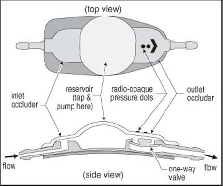
Figure 15-5 PS Medical standard contoured valve
X-ray characteristics
The three available valve pressures are indicated by radioopaque dots on the valve (allows x-ray identification of valve pressure): one dot = low pressure, two dots = medium, three dots = high.

Figure 15-6 Pumping the PS Medical valve
Strata® programmable valve
The Medtronic Strata valve is an externally adjustable valve that is programmed (using a magnet) to one of five performance level (“P/L”) settings (Figure 15-7).
Also, see general information regarding programmable valves on page 317.
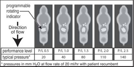
Figure 15-7 Performance level (P/L) settings for the regular size Strata valve as seen on x-ray
SOPHYSA USA
Sophysa USA, Inc.
1620 Sunflower Ave
Costa Mesa, CA 92626 USA
(714) 429-8801
www.sophysa.com
Polaris programmable valve
The Polaris valve is an externally programmable valve that uses two attracting Samarium-Cobalt magnets to lock the pressure setting and to resist inadvertent reprogramming by environmentally encountered magnets such as MRI scanners, cell phones, headphones…

Figure 15-8 Programmable settings for Polaris valve models as seen on x-ray (pressures in mm H2O)
Available in 4 models (different pressure ranges, each identified by a unique number of radioopaque dots), each with 5 externally adjustable positions. The x-ray appearance and corresponding pressures are shown in Figure 15-8.
CODMAN
Codman
325 Paramount Dr.
Raynham, MA 02767 USA
(800) 225-0460
www.codman.com
Codman Hakim programmable valve
18 pressure settings. Programmed by an AC-powered programming unit that requires confirmatory x-ray after reprogramming. Newer programming units with acoustic monitoring may obviate the need for x-ray. The manufacturer advises not to increase the pressure by > 40 mm H2O in a 24-hour period.
X-ray appearance for various settings are shown in Figure 15-9 (note: settings of 70, 120 & 170 mm H2O align with an arm of the central cross of the valve). NB: when x-rayed correctly, the x-ray beam passes first through the valve and then the patient, which causes the radioopaque marker to appear as a solid circle to the right of center as shown in Figure 15-9. If the marker is on the left side, the beam is passing from the bottom of the valve, and the actual pressure reading should be based on a mirror image of the x-ray.

Figure 15-9 X-ray appearance of Codman Hakim programmable valve at it’s various settings in mm Hg (e.g. the large central image shows a setting of 120 mm H2O)
NEUROCARE
Distributed in U.S. by:
NeuroCare Group
8401 102nd Street
Suite 200
Pleasant Prairie, WI 53158
(800) 997-4868
Heyer-Schulte
The LPV valve is shown in Figure 15-10. To pump the shunt, occlude inlet port with one finger, then depress reservoir with another finger (as for the PS Medical valve, see above). This valve may be injected in either direction by depressing the appropriate occluder while injecting into the reservoir.
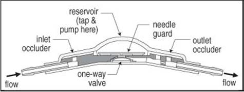
Figure 15-10 Heyer-Schulte LPV® (low-profile) valve (side view)
HAKIM SHUNT
Distributed by:
Integra Neurosciences
In the U.S.:
311 Enterprise Drive
Plainsboro, NJ 08536
(800) 654-2873
http://www.integra-ls.com

Figure 15-11 Hakim Standard Mechanism
A dual ball-valve mechanism. To pump shunt, depress the indicated portion of the valve. NB: do not tap here, as the silicone elastomer housing is not self-sealing. The antechamber is provided for this type of access.
INTEGRA (CORDIS) HORIZONTAL-VERTICAL LUMBAR VALVE
May be used in lumboperitoneal shunt to increase the transmission pressure when the patient is upright to prevent overshunting. Markings used to orient the device during implantation:
1. an arrow on the inlet side of the unit indicates direction of flow
2. inlet tubing is clear
3. inlet tubing has smaller diameter than outlet tubing
4. outlet tubing is white
5. before positioning the valve and fastening it to the fascia with permanent suture, the valve should be connected to both the subarachnoid catheter (inlet) and the peritoneal catheter (outlet). The arrow on the inlet valve should point towards the patient’s feet

Figure 15-12 Cordis H-V valve
HOLTER VALVE
A dual slit valve mechanism (see Figure 15-13). Usually used in combination with a Rickham or Salmon-Rickham reservoir (see Figure 15-14).
To pump the shunt, simply depress the indicated portion of the valve.
X-ray characteristics
The silastic tube between the two one-way valves is radiolucent (see Figure 15-4, page 318).
SALMON-RICKHAM RESERVOIR
Similar to standard Rickham reservoir except for lower profile (see Figure 15-14).

Figure 15-13 Holter valve

Figure 15-14 Salmon-Rick-ham Reservoir
15.2. Shunt problems
15.2.1. Problems associated with shunt insertion
1. intraparenchymal or intraventricular hemorrhage: ≈ 4% (in the absence of coagulopathy 56)
2. seizures
3. malposition
A. of ventricular catheter
B. of distal catheter
4. infection
15.2.2. Problems in patients with established CSF shunt
Shunt “problems” usually involve one or more of the following:
1. undershunting (see below): obstruction rate: ≈ 10% per year } accounts for most common shunt problems
2. infection (see page 345): range 1-40%. A serious complication. Having a shunt infection decrease IQ } accounts for most common shunt problems
3. overshunting: slit ventricle syndrome, subdural hematomas… (see page 325)
4. seizures: see page 316
5. problems related to the distal catheter
A. peritoneal: see page 316
B. atrial: see page 317
6. skin breakdown over hardware: infection or silicone allergy (see page 316)
7. hemorrhage at time of insertion: uncommon in pediatrics
TAPPING A SHUNT
Indications to tap a shunt or ventricular access device (e.g. Ommaya reservoir) include:
1. to obtain CSF specimen
A. to evaluate for shunt infection
B. for cytology: e.g. in PNET to look for malignant cells in CSF
C. to remove blood: e.g. in intraventricular hemorrhage
2. to evaluate shunt function
A. measuring pressures
B. contrast studies:
1. proximal injection of contrast (iodinated or radiolabeled)
2. distal injection of contrast
3. as a temporizing measure to allow function of a distally occluded shunt57, 58
4. to inject medication
A. antibiotics: for shunt infection or ventriculitis
B. chemotherapeutic (antineoplastic) agents
5. for catheters placed within tumor cyst (not a true shunt):
A. periodic withdrawal of accumulated fluid
B. for injection of radioactive liquid (usually phosphorous) for ablation
TECHNIQUE
Table 15-2 Steps in tapping a shunt
|
Step |
Information provided |
|
1. insert needle into reservoir and look for spontaneous flow into butterfly tubing; measure pressure in manometer |
• spontaneous flow indicates proximal end not completely occluded • pressure is that of ventricular system (should be < 15 cm of CSF in relaxed recumbent patient) |
|
2. also measure the pressure with distal occluder pressed if present |
• rise in pressure indicates some function of valve and distal shunt |
|
3. if no spontaneous flow, try to aspirate CSF with syringe |
• if CSF is easily aspirated, it may be that pressure seen by ventricular system is near 0 • if no CSF obtained or if difficult to aspirate, indicates proximal occlusion |
|
4. send CSF for: C&S, protein/glucose, cell count |
• checks for infection |
|
5. fill manometer with sterile saline, & occlude proximal (inlet) occluder (with Holter valve, tap the valve itself, although this is not recommended because the hole thus created may not seal) |
• measures forward transmission pressure (through valve and peritoneal catheter in presence of shunt with proximal occluder); forward pressure should be less than ventricular pressure |
|
6. repeat measurement after injecting 3-5 ml of saline |
• if peritoneal catheter is in a loculated compartment the pressure will be considerably higher after injection |
[(For LP shunt, see Lumboperitoneal (LP) shunt evaluation, page 214).
There is a risk of introducing infection with every entry into the shunt system. With care, this may be kept to a minimum.
1. shave area
2. povidone iodine solution prep x 5 minutes
3. use 25 gauge butterfly needle or smaller (ideally a noncoring needle should be used): for routine taps, the needle should only be introduced into shunt components specifically designed to be tapped
To measure pressures
Steps are outlined in Table 15-2.
INSTRUCTIONS TO PATIENTS
All patients and families of patients with hydrocephalus should be instructed regarding the following:
1. signs and symptoms of shunt malfunction or infection
2. not to pump the shunt unless instructed to do so for a specific purpose
3. prophylactic antibiotics: for the following situations (mandatory in vascular shunts, sometimes recommended in other shunts)
A. dental procedures
B. instrumentation of the bladder: cystoscopy, CMG, etc.
4. in a growing child: the need for periodic evaluation, including assessment of distal shunt length
UNDERSHUNTING
The shunt malfunction rate is ≈ 17% during the first year of placement in the pediatric population.
May be due to one or a number of the following:
1. blockage (occlusion)
A. possible causes of occlusion:
1. obstruction by choroid plexus
2. buildup of proteinaceous accretions
3. blood
4. cells (inflammatory or tumor)
5. secondary to infection
B. site of blockage
1. blockage of ventricular end (most common): usually by choroid plexus, may also be due to glial adhesions, intraventricular blood
2. blockage of intermediate hardware (valves, connectors, etc., tumor filters may become obstructed by tumor cells, antisiphon devices may close due to variable overlying subcutaneous tissue pressures59)
3. blocked distal end (see page 316 for VP shunt)
C. disconnection, kinking or breakage of system at any point: with age, silicone elastomers used in catheters calcify and break down, and become more rigid and fragile which may promote subcutaneous attachment60. Barium impregnation may accelerate this process. Tube fractures often occur near the clavicle, presumably due to the increased motion there
Signs and symptoms of undershunting
Signs and symptoms are those of active hydrocephalus, and include:
1. acute symptoms of increased intracranial pressure
A. H/A
B. N/V
C. diplopia
D. lethargy
E. ataxia
F. infants: apnea and/or bradycardia; irritability
G. seizures: new onset, increase in frequency, or difficulty in control
2. acute signs of increased intracranial pressure
A. upward gaze palsy (“setting sun sign”, see Parinaud’s syndrome, page 114)
B. abducens palsy: false localizing sign
C. field cut, or blindness (see Blindness from hydrocephalus, page 335)
D. papilledema (rare before age 2 yrs)
E. infants: bulging fontanelle, prominent scalp veins
3. swelling around shunt tubing: caused by CSF dissecting along shunt tract
4. chronic changes: before sutures close, OFCs crossing curves
EVALUATION OF SHUNT FOR UNDERSHUNTING
1. history and physical directed at determining presence of above signs and symptoms, also ascertain:
A. reason for initial insertion of shunt (MM, post-meningitis, etc.)
B. date of last revision and reason for revision
C. presence of accessory hardware in system (e.g. antisiphon device, etc.)
D. for children: OFC. Plot on graph of normal curves (use existing chart for that patient if available)
E. fontanelle tension (if open): a soft pulsating fontanelle varying with respirations is normal, a tense bulging fontanelle suggests obstruction, a sunken fontanelle may be normal or may represent overshunting
F. ability of shunt to pump and refill
1. caution: may exacerbate obstruction, especially if shunt is occluded by ependyma due to overshunting: controversial
2. difficult to depress: suggests distal occlusion
3. slow to refill (generally, any valve should refill in 15-30 secs): suggests proximal (ventricular) occlusion
G. evidence of CSF dissecting along tract outside of shunt tubing
H. in children presenting only with vomiting, especially those with cerebral palsy and feeding gastrostomy tubes, rule-out gastroesophageal reflux
2. radiographic evaluation
A. “shunt series” (plain x-rays to visualize entire shunt: for VP shunt, AP & lateral skull + “low” CXR and/or abdominal x-ray)
1. R/O disconnection or migration of tip by x-rays to visualize entire shunt; note: a disconnected shunt may continue to function by CSF flow through a fibrous tract; the following hardware may be radiolucent and can mimic disconnection:
a. the central silastic part of a Holter style valve
b. connectors (“Y” & “T” as well as straight)
c. antisiphon devices
d. tumor filters
2. obtain most recent x-rays available to compare for breaks (essential for “complicated” shunts involving multiple ventricular or cyst ends or accessory hardware)
B. in patients with open fontanelles, ultrasound is optimal method of evaluation (especially if previous U/S available)
C. CT required if fontanelles closed, may be desirable in complicated shunt systems (e.g. cyst shunts)
D. MRI: shunt hardware is difficult to see. May show transependymal absorption of CSF, loculations…
E. “shunt-o-gram” if it is still unclear if shunt is functioning
1. radionuclide: see below
2. x-ray: using iodinated contrast: see below
3. shunt tap: indications vary, generally performed if surgical exploration is considered or if infection is strongly suspected (see Tapping a shunt, page 322)
4. shunt exploration: sometimes even after thorough evaluation the only means to definitively prove or disprove the functioning of various shunt components is to operate and isolate and test each part of the system independently. Even when infection is not suspected, CSF and any removed hardware should be cultured.
“Shunt-o-gram”
Procedure: shave hair over reservoir & prep with Betadine. With patient supine tap the shunt by inserting a 25 gauge butterfly needle into the reservoir. Measure the pressure with a manometer. Patients with multiple ventricular catheters need to have each injected to verify its patency.
Radionuclide “shunt-o-gram” AKA radionuclide shuntography61: after tapping the shunt, drain 2-3 ml of CSF and send 1 ml of CSF for C&S. Inject radio-isotope (e.g. for VP shunt in an adult, use 1 mCi of 99m-Tc (technetium) pertechnetate (usable range: 0.5 to 3 mCi) in 1 cc of fluid) while occluding distal flow (by compressing valve or occluding ports). Flush in isotope with remaining CSF.
Immediately image the abdomen with the gamma camera to rule out direct injection into distal tubing. Image the cranium to verify flow into ventricles (proximal patency). If spontaneous flow into abdomen is not seen after 10 minutes the patient is sat up and res-canned. If flow is not seen after 10 minutes, then the shunt is pumped. Look for diffusion of the isotope within the abdomen to rule out pseudocyst formation around catheter.
X-ray “shunt-o-gram”: after tapping the shunt, drain ≈ 1 ml of CSF and send for C&S. Inject e.g. iohexol (Omnipaque 180) (see page 122) while occluding distal flow (by compressing valve or occluding ports).
PSEDUOCYST (PERITONEAL) WITH VP SHUNT
Usually a marker for infection.
Treatment algorithm
One of many viable surgical protocols to deal with this:
1. open abdominal incision over tubing, and divide tubing at this site
2. verify which cut end is the peritoneal end and which is the distal shunt (with a working shunt, pumping the valve should cause CSF to come out the distal shunt)
3. attempt to drain the cyst through the remaining peritoneal end
A. when you can’t draw any more fluid, or if you don’t get any to begin with, withdraw the catheter a little at a time and aspirate at each step
B. send any fluid obtained for culture
C. if tubing does not pull out smoothly, the abdomen may need to be opened (consider consulting general surgeon)
4. verify function of remaining shunt
A. if the remaining shunt is functioning
1. connect it to sterile collection system
2. monitor output volumes & send surveillance cultures of CSF qod
3. after 3 consecutive cultures are negative, internalize distal end of shunt (using fresh distal catheter). The choice of target for distal end (peritoneum, pleura, vein) depends on whether abdominal cyst fluid is infected and if the peritoneal cavity still seems suitable)
B. if the shunt is not functioning, a new external ventricular catheter should be inserted and connected to a collection system
1. monitor output volumes & send surveillance cultures of CSF qod
2. after 3 consecutive cultures are negative, remove the old shunt and place a totally shunt. The choice of target for distal end (peritoneum, pleura, vein) depends on whether abdominal cyst fluid is infected and if the peritoneal cavity still seems suitable)
SHUNT INFECTION
See Shunt infection on page 345 for evaluation and treatment.
“OVERSHUNTING”
POSSIBLE COMPLICATIONS OF OVERSHUNTING INCLUDE62
1. slit ventricles: including slit ventricle syndrome (see below)
2. intracranial hypotension: see below
3. subdural hematomas: see page 327
4. craniosynostosis and microcephaly: controversial (see page 328)
5. stenosis or occlusion of sylvian aqueduct
10-12% of long-term ventricular shunt patients will develop one of the above problems within 6.5 yrs of initial shunting62. Some experts feel that problems related to over-shunting could be reduced by utilizing LP shunts for communicating hydrocephalus, and reserving ventricular shunts for obstructive HCP62. VP shunts may also be more likely to overdrain than VA shunts because of the longer tubing → greater siphoning effect.
INTRACRANIAL HYPOTENSION
AKA low ICP syndrome. Very rare. Symptoms similar to those of spinal H/A (postural in nature, relieved by recumbency). Although usually not associated with the following symptoms63, they may occur62: N/V, lethargy, or neurologic signs (e.g. diplopia, upgaze palsy). Sometimes the symptoms resemble those of high ICP except that they are relieved when prostrate. Acute effects that may occur include62: tachycardia, loss of consciousness, other brain stem deficits due to a rostral shift of the intracranial contents or to low ICP.
Etiology is a siphoning effect due to the column of CSF in the shunt tube when the patient is erect64. Ventricles may be slit-like (as in slit ventricle syndrome (SVS)) or may be normal in appearance. Sometimes it is necessary to document a drop in ICP when going from supine to erect to diagnose this condition. These patients may also develop shunt occlusion and then the distinction from SVS blurs (see below).
With short-term symptoms, an ASD is the treatment of choice. However, patients with long-standing overshunting may not tolerate efforts to return intraventricular pressures to normal levels62, 65.
SLIT VENTRICLES
“Slit ventricles” refers to complete collapse of the ventricles. In a survey, a frontal-occipital horn ratio19 < 0.2 was most often interpreted as representing SVS. May be seen in:
1. overshunting
2. with entrapped (isolated) fourth ventricle: see page 309
3. some patients with idiopathic intracranial hypertension (AKA pseudotumor cerebri) (see page 713) have slit-like ventricles with consistently elevated ICP
May be one of the following:
1. asymptomatic:
A. slit ventricles (totally collapsed lateral ventricles) may be seen on CT in 3 -80% of patients after shunting63, 66, most are asymptomatic
B. these patients may occasionally present with symptoms unrelated to the shunt, e.g. true migraine
2. slit ventricle syndrome (SVS): seen in < 12% of all shunted patients. Subtypes:
A. intermittent shunt occlusion: overshunting leads to ventricular collapse (slit ventricles) which causes the ependymal lining to occlude the inlet ports of the ventricular catheter (by coaptation) producing shunt obstruction. With time, many of these patients develop low ventricular compliance67, where even minimal dilatation results in high pressure which produces symptoms. Expansion then eventually reopens the inlet ports allowing resumption of drainage (hence the intermittent symptoms). Symptoms may resemble shunt malfunction: intermittent headaches unrelated to posture, often with N/V, drowsiness, irritability and impaired mentation. Signs may include 6th cranial nerve palsy. Incidence in shunted patients: 2-5%63, 68. CT or MRI scans may also show evidence of transependymal absorption of CSF
B. total shunt malfunction (AKA normal volume hydrocephalus67): may occur and yet ventricles remain slit-like if the ventricles cannot expand because of subependymal gliosis, or due to the law of Laplace (which states that the pressure required to expand a large container is lower than the pressure required to expand a small container)
C. venous hypertension with normal shunt function: may result from partial venous occlusion that occurs in some conditions (e.g. at the level of the jugular foramen in Crouzon’s syndrome). Usually subsides by adulthood
3. intracranial hypotension: symptoms often relieved by recumbency (see above)
EVALUATION OF SLIT VENTRICLES
The shunt valve fills slowly if pumped when the ventricles are collapsed.
Monitoring CSF pressure: either via lumbar drain, or with a butterfly inserted into the shunt reservoir (with this method, pressure can be followed during postural changes to look for negative pressure when upright; possibly higher risk of infection with this). These patients are also monitored for pressure spikes, especially during sleep.
Alternatively, these patients may be evaluated by “shunt-o-gram” (see above).
TREATMENT
In treating a patient with slit ventricles in imaging studies, it is important to ascertain into which of the categories (see above) the patient falls. If the patient can be categorized, then the specific treatment listed below should be employed. Otherwise, it is probably most common to initially treat the patient empirically as intracranial hypotension, and then to move on to other methods for treatment failures.
Asymptomatic slit ventricles
Prophylactic upgrading to a higher pressure valve or insertion of an antisiphon device has largely been abandoned. However, this may be appropriate at the time of shunt revision when done for other reasons66.
Intracranial hypotension
Postural H/A due to intracranial hypotension (true overshunting) is usually self limited, however, if symptoms persist after ≈ 3 days of bed-rest and analgesics and a trial with a tight abdominal binder, the valve should be checked for proper closing pressure. If it is low, replace with a higher pressure valve. If it is not low, an antisiphon device (ASD) (which, by itself, also increases the resistance of the system) alone or together with a higher pressure valve may be needed69.
Slit ventricle syndrome
Patients with symptoms of SVS are actually suffering from intermittent high pressure. If total shunt malfunction is the cause, then shunt revision is indicated. For intermittent occlusion, treatment options include:
1. if symptoms occur early after shunt insertion or revision, initial expectant management may be indicated since symptoms will spontaneously resolve in many
2. revision of the proximal shunt. This may be difficult due to the small size of the ventricles. One can attempt to follow the existing tract and insert a longer or shorter length of tubing based on the pre-op imaging studies. Some advocate the placement of a second ventricular catheter, leaving the first one in place
3. patients may “respond” to either of the following interventions because the slight ventricular enlargement elevates the ependyma off of the inlet ports:
A. valve upgrade70 or
B. ASD insertion63, 69: the procedure of choice in some opinions62. First described in 197371
4. subtemporal decompression72-74 sometimes with dural incision72. This results in dilatation of the temporal horns (evidence for elevated pressure) in most, but not all74 cases
5. third ventriculostomy75: see page 315
Problems unrelated to shunting
For H/A consistent with migraine that are not postural, a trial with migraine-specific medications is warranted (Fiorinal®…). For treatment of idiopathic intracranial hypertension (pseudotumor cerebri), see page 717.
SUBDURAL HEMATOMAS
May be due to collapse of brain with tearing of bridging veins. In the pre-CT era, the incidence of subdural hematoma (SDH) formation following shunt insertion was probably underestimated at ≈ 1.2%. However, more recent estimates are 4-23% in adults, 2.8-5.4% in children, and is higher with normal pressure hydrocephalus (20-46%) than with “hypertensive hydrocephalus” (0.4-5%)76, 77. The risk of SDH is higher in the setting of longstanding hydrocephalus with a large head and little brain parenchyma (craniocerebral disproportion) with a thin cerebral mantle, as usually occurs in children with macrocephaly and large ventricles on initial evaluation. These patients have an “extremely delicate balance between subdural and intraventricular pressure”76. SDH can also follow shunting in elderly patients who have severe brain atrophy. The development of SDH may also be facilitated by negative pressures in the ventricles as a result of siphoning77, 78. There is also a low risk of epidural hematoma following CSF shunting77.
Characteristics of the fluid: The collections may be on the same side as the shunt in 32%, on the opposite side in 21%, and bilateral in 47%77.
At the time of discovery, the SDHs are usually subacute to chronic, and the previously large ventricles are usually collapsed. Only 1 of 19 cases showed colorless fluid77. In all cases tested, protein was elevated compared to CSF.
TREATMENT
Indications for treatment
Small (< 1-2 cm thick) asymptomatic collections in patients with closed cranial sutures may be followed with serial imaging. SDH were symptomatic in ≈ 40% of cases (symptoms often resemble those of shunt malfunction), and these require treatment. Treatment of SDH in children with open sutures has been advocated77 to prevent later symptoms and/or development of macrocrania. Many authors recommend not treating asymptomatic lesions regardless of appearance76, 79, whereas others vary their recommendations based on diverse criteria including size, appearance (chronic, acute, mixed…), etc.
Treatment techniques
A number of techniques have been described. Most involve evacuation of the SDHs by any of the usual methods (e.g. burr holes for chronic collections, craniotomy for acute collections) together with:
1. reducing the degree of shunting (i.e. to establish a lower pressure in the subdural space than in the intraventricular space, to cause the ventricles to re-expand and to prevent reaccumulation of the SDH)
A. in shunt dependent cases
1. replacing the valve with a higher pressure unit (upgrading the valve)
2. increasing the pressure on a programmable pressure valve80, 81
3. using a Portnoy device that can be turned off and on externally. Be sure that care providers can reliably open the device in an emergency
B. in non-shunt dependent cases
1. any of the methods outlined above for shunt dependent cases, or
2. temporarily tying off the shunt82
C. insertion of an anti-siphon device71
2. drainage of the subdural space to
A. the cisterna magna83
B. to the peritoneum with a low pressure valve (or no valve77). Some authors have the care-giver frequently pump the subdural valve
The goal is to achieve a delicate balance between undershunting (producing symptoms of active hydrocephalus) and overshunting (promoting the return of the SDH). Following surgery the patient should be mobilized slowly to prevent recurrence of the SDH.
MISCELLANEOUS SHUNT ISSUES
CRANIOSYNOSTOSIS, MICROCEPHALY & SKULL DEFORMITIES
Also see Craniosynostosis, page 228. A number of skull changes have been described in infants after shunting, including84: thickening and inward growth of the bone of the skull base and cranial vault, decrease in size of the sella turcica, reduction in size of the cranial foramina, and craniosynostosis. The most common skull deformity was dolichocephaly from sagittal synostosis85. Microcephaly accounted for ≈ 6% of skull deformities after shunting (about half of these had sagittal synostosis). Some of these changes were reversible (except when complete synostosis was present) if intracranial hypertension recurred.
LAPAROSCOPIC SURGERY IN PATIENTS WITH VP SHUNTS
Issues regarding safety of laparoscopic surgery in patients with VP shunts:
1. laparoscopic surgery: abdominal insufflation with CO2 is used to create a pneumoperitoneum permitting the general surgeon to work. Typical insufflation pressure: 15 mm Hg (see page 870 for conversion factors between mm Hg and cm of water). In thin patients, 10 mm Hg may suffice. Transient additional increases in pressure may occur, e.g. when the surgeon leans on the patient’s abdomen
2. concerns for patients with VP shunts:
A. in some cases insufflation → ↑ ICP86 which may be due to:
1. compression of vena cava → reduced venous return from head, as in valsalva maneuver (independent of presence of a shunt)
2. absorption of CO2 from the peritoneum → ↑ in arterial CO2 causing cerebral arterial dilatation thereby increasing ICP (see page 880)
3. ↓ CSF drainage due to ↑ pressure against which CSF must flow
4. retrograde passage of air/debris into intracranial compartment through an incompetent shunt valve (this also has potential for infection in the presence of peritonitis). This risk is minimal even with in vitro back-pressures up to 80 mm Hg87. Retrograde flow may also occur with a valveless shunt (rarely used)
• in one case report monitoring TCDs88, there was no change during laparoscopic surgery in a patient with a VP shunt (except during periods of very high pressure)
B. occlusion of the distal catheter by air, debris89 or soft tissue
C. extremely high intraabdominal pressures (> 80 mm Hg in vitro) may damage the valve87, which could cause malfunction after the laparoscopy
Prophylactic management options:
1. very controversial, special precautions may not be necessary90
2. one can temporarily occlude the peritoneal catheter (e.g. by a hemoclip applied by the general surgeon through the laparoscope under minimal initial insufflation pressure; the clip is removed at the end of the procedure), or, temporary externalization of the shunt by the neurosurgeon, with internalization at the end of the procedure (this engenders an increased risk of infection)
3. ICP monitoring during laparoscopy
4. using low insufflation pressures (e.g. < 10 mm Hg)
15.3. Normal pressure hydrocephalus
Key concepts:
• triad (not pathognomonic): dementia, gait disturbance, urinary incontinence
• communicating hydrocephalus on CT or MRI
• normal pressure on random LP
• symptoms may be remediable with CSF shunting
Normal pressure hydrocephalus (NPH), AKA Hakim-Adams syndrome, first described in 196591, is clinically important because it may cause treatable symptoms, including one of the few forms of remediable dementia.
As originally described, the hydrocephalus of NPH was considered to be idiopathic. However, in some cases a predisposing condition (“secondary NPH”) may be identified:
1. post-SAH
2. post-traumatic
3. post-meningitis
4. following posterior fossa surgery
5. tumors, including carcinomatous meningitis
6. also seen in ≈ 15% of patients with Alzheimer’s disease (AD)
7. deficiency of the arachnoid granulations
8. aqueductal stenosis may be an overlooked cause
It is becoming increasingly acknowledged that the ventricular enlargement is likely not the underlying pathologic entity. Interest persists in effects such as impaired pulsatility. The search continues to improve the understanding of this complicated condition.
Table 15-3 Comparison of cognitive deficits in Alzheimer’s disease (AD) and NPH* †
|
Feature |
AD |
NPH |
|
memory |
↓ |
± auditory memory |
|
executive function‡ |
↓ |
± |
|
attention concentration |
↓ |
± |
|
orientation |
↓ |
|
|
writing |
↓ |
|
|
learning |
↓ |
|
|
fine motor speed and accuracy |
± |
↓ |
|
psychomotor skills |
± |
slowed |
|
language and reading |
± |
|
|
behavioral or personality changes |
± |
* modified94
† Key: ↓ = impaired; ± = borderline impaired
‡ see Table 15-6 for definition of executive function
CLINICAL
Clinical triad92
The triad is not pathognomonic, and similar features may also be seen e.g. in vascular dementia93, Alzheimer’s dementia and Parkinson’s disease.
1. gait disturbance: usually precedes other symptoms. Wide based with short, shuffling steps and unsteadiness on turning. Patients often feel like they are “glued to the floor” (so-called “magnetic gait”) and may have difficulty initiating steps or turns. Absence of appendicular ataxia
2. dementia: primarily memory impairment with bradyphrenia (slowness of thought) and bradykinesia (Table 15-3 shows some differentiating features with Alzheimer’s disease)
3. urinary incontinence: usually unwitting (NB: a patient demented for any reason or with mobility impairment may have incontinence)
Other clinical features
Age usually > 60 yrs. Slight male preponderance. Also see Diagnosis below for other clinical information.
True aphasia is unusual, but speech output may be disturbed by impaired motivation or executive dysfunction94. As NPH progresses, cognitive impairment may become more generalized and less responsive to treatment94. Symptoms identical to those of idiopathic parkinsonism may occur in 11%95.
Case reports of a variety of psychiatric disturbances associated with NPH include: depression96, bipolar disorder97, aggressiveness98, paranoia99.
Symptoms not expected with NPH: Although a variety of clinical features have been demonstrated to occur infrequently (e.g. SIADH100, syncope…), clinical features not expected solely as a result of NPH include: papilledema, seizures (prior to shunting), headaches94.
Differential diagnosis
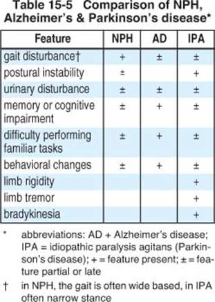
Table 15-4 shows conditions with presentations similar to findings in NPH in the differential diagnosis94, 101. Table 15-5 compares some features of NPH, Alzheimer’s disease, and Parkinson’s disease.
Table 15-4 Conditions with similar presentation to NPH
|
Neurodegenerative disorders • Alzheimer’s disease • Parkinson’s disease • Lewy body disease • Huntington’s disease • frontotemporal dementia • corticobasal degeneration • progressive supranuclear palsy • amyotrophic lateral stenosis • multisystem atrophy • spongiform encephalopathy |
|
Vascular dementia • cerebrovascular disease • multi-infarct dementia • Binswanger’s disease • CADASIL • vertebrobasilar insufficiency (VBI) |
|
Other hydrocephalic disorders • aqueductal stenosis • arrested hydrocephalus • long-standing overt ventriculomegaly syndrome • noncommunicating hydrocephalus |
|
Infectious disease • Lyme disease • HIV • syphilis |
|
Urological disorders • urinary tract infection • bladder or prostate cancer • benign prostatic hypertrophy (BPH) |
|
Miscellaneous • vitamin B12 deficiency • collagen vascular diseases • epilepsy • depression • traumatic brain injury • spinal stenosis • Chiari malformation • Wernicke’s encephalopathy • carcinomatous meningitis • spinal cord tumor |
DIAGNOSIS
PRACTICE GUIDELINE 15-1 DIAGNOSIS OF NPH
Level II94: Since strict diagnostic criteria cannot be formulated for NPH because of a lack of knowledge of the underlying pathophysiology at this time, it is recommended that the diagnosis be made in terms of Probable, Possible, and Unlikely NPH as described in Table 15-6
Table 15-6 Diagnostic guidelines for NPH94
|
PROBABLE NPH |
|
History*: must include: 1. insidious onset (vs. acute) 2. onset age ≥ 40 years 3. duration ≥ 3-6 months 4. no antecedent head trauma, ICH, meningitis or other known cause of secondary hydrocephalus 5. progression over time 6. no other neurological, psychiatric or general medical conditions that are sufficient to explain the presenting symptoms |
|
Brain imaging: CT or MRI after onset of symptoms 1. ventricular enlargement not attributable to cerebral atrophy or congenital enlargement (Evan’s index† > 0.3 or comparable measure) 2. no macroscopic obstruction to CSF flow 3. ≥ 1 of the following supportive features A. enlarged temporal horns not entirely attributable to hippocampal atrophy B. callosal angle ≥ 40° C. evidence of altered brain water content, including periventricular changes not attributable to microvascular, ischemic changes, or demyelination D. aqueductal or 4th ventricle flow void on MRI Other imaging findings that may support Probable designation but are not required: 1. pre-morbid study showing smaller or nonhydrocephalic ventricles 2. radionuclide cisternogram showing delayed clearance of radiotracer over the convexities after 48-72 hours 3. cine-MRI or other technique showing increased ventricular flow rate 4. SPECT showing decreased periventricular perfusion that is not altered by acetazolamide challenge |
|
Physiological CSF opening pressure (OP) on lateral decubitus LP: 5-18 mm Hg (70-245 mm H2O) |
|
Clinical: must show gait/balance disturbance, plus impairment in cognition and/or urinary function 1. gait/imbalance: ≥ 2 of the following (not entirely attributable to other conditions A. decreased step height B. decreased step length C. decreased cadence (speed of walking) D. increased trunk sway while walking E. widened standing base F. toes turn outward while walking G. retropulsion (spontaneous or provoked) must show: H. en bloc turning (≥ 3 steps to turn 180°) I. impaired walking balance: ≥ 2 corrections out of 8 tandem steps 2. cognition: documented impairment (adjusted for age & education) and/or decrease in performance on cognitive screening instrument (e.g. Monumental State examination), or evidence of ≥ 2 of the following not fully attributable to other conditions: A. psychomotor slowing (increased response latency) B. decreased fine motor speed C. decreased fine motor accuracy D. difficulty dividing or maintaining attention E. impaired recall, especially for recent events F. executive dysfunction: e.g. impairment in multistep procedures, working memory, formulation of abstractions/similarities, insight G. behavioral or personality changes 3. urinary dysfunction: A. any one of the following 1. episodic or persistent incontinence not attributable to primary urological disorder 2. persistent urinary incontinence 3. urinary and fecal incontinence B. or, any 2 of the following: 1. urinary urgency: frequent perception of a pressing need to void 2. ↑ urinary frequency (pollakiuria): voiding > 6 times in 12 hours with normal fluid intake 3. nocturia: needing to void > 2 times in an average night |
|
POSSIBLE NPH |
|
History: reported symptoms may: 1. have subacute or indeterminate mode of onset 2. onset at any age after childhood 3. duration: < 3 months or indeterminate 4. may follow events such as mild head trauma, remote history of ICH, or childhood or adult meningitis or other conditions judged not likely to be causally related 5. coexist with other neurological, psychiatric, or general medical disorders but judged not to be entirely attributable to these conditions 6. be nonprogressive or not clearly progressive |
|
Clinical: symptoms of either: 1. incontinence and/or cognitive impairment in the absence of observable gait/balance disturbance 2. gait disturbance or dementia alone |
|
Brain imaging: ventricular enlargement consistent with hydrocephalus but associated with any of the following: 1. cerebral atrophy of sufficient severity to potentially explain ventricular enlargement 2. structural lesions that may influence ventricular size |
|
Physiological OP not available or outside of the range delineated for Probable NPH |
|
UNLIKELY NPH |
|
1. no ventriculomegaly 2. signs of increased ICP (e.g. papilledema) 3. no component of the clinical triad of NPH 4. symptoms explained by other causes (e.g. spinal stenosis) |
* history should be verified by individual familiar with premorbid and current condition
† see page 310 for definition and illustration of Evan’s index
LUMBAR PUNCTURE (LP) - “TAP TEST”
Opening pressure: Normal LP opening pressure (OP) in the left lateral decubitus position averages 12.2 ± 3.4 cm H2O (8.8 ± 0.9 mm Hg)102 and should be < 180 mm H2OA. In NPH the average OP is 15 ± 4.5 cm H2O (11 ± 3.3 mm Hg), slightly higher than, but overlapping with, normal. Based on expert opinion, an upper limit of 24 cm H2O (17.6 mm Hg) is suggested for the definition of NPH. Patients with an initial OP > 10 cm H2O have a higher response rate to shunting.
Tap test: The tap test has not undergone rigorous prospective evaluation. A positive responseB to withdrawal of 40-50 ml of CSF has a PPV in the range of 73-100%104-106, but sensitivity is low (26-61%).
Send CSF for routine labs (see page 203).
RESISTANCE TESTING
CSF Ro is considered to be the impedance of CSF absorptive mechanisms. 1/Ro is the conductance. Techniques and thresholds are center-specific. No clinical study has adequately addressed the fact that Ro normally increases with age107.
Determination of CSF Ro may have a higher sensitivity (57-100%) but a similar PPV (75-92%) to the tap test.
Methodology
Numerous methods have been devised to measure Ro. Two illustrative methods:
1. bolus method108: inject ≈ 4 ml via LP at a rate of 1 ml/sec
2. Katzman test109: infuse saline through LP at a known rate, Ro is given by Eq 15-1 (up to 19% of patients experience H/A after infusion studies110)

AMBULATORY LUMBAR DRAINAGE104 (ALD)
A lumbar subarachnoid drain is placed with Tuohy needle, connected through a drip chamber to a closed drainage system (see page 202). The drip chamber is placed at the level of the patient’s ear when recumbent, or at the level of the shoulder when sitting or ambulating.
A properly functioning drain should put out ≈ 300 ml of CSF per day.
If symptoms of nerve root irritation develop during the drainage, the catheter should be withdrawn several millimeters. Daily surveillance CSF cell counts and cultures should be performed (NB: a pleocytosis of ≈ 100 cells/mm3is expected just with the presence of the drain).
A 5 day trial is recommended (mean time to improvement: 3 days).
CONTINUOUS CSF PRESSURE MONITORING
Some patients with normal OP on LP demonstrate pressure peaks > 270 mm H2O or recurrent B-waves111. These patients may have a higher response rate to shunting.
CT AND MRI
Features on CT112 and MRI113
1. prerequisite: communicating hydrocephalus
2. features that correlate with favorable response to shunt (these features suggest that the hydrocephalus is not due to atrophy aloneC):
A. periventricular low density on CT or high intensity on T2WI MRI: may represent transependymal absorption of CSF. May resolve with shunting
B. compression of convexity sulci (focal sulcal dilation may sometimes be seen and may represent atypical reservoirs of CSF which may diminish after shunting and should not be considered as atrophy115)
C. rounding of the frontal horns
A. OP > 24 cm H2O suggests noncommunicating hydrocephalus rather than NPH94, 103
B. what constitutes a “significant response” has not been standardized, most experts prefer demonstrating objective improvement in gait, taking into account the fact that NPH patients can have day-to-day fluctuations in symptoms
C. atrophy (hydrocephalus ex vacuo), as in conditions such as Alzheimer’s disease, lessens the chance of, but does not preclude, responding to a shunt (cortical atrophy is a common finding in healthy individuals of advanced age114)
Although some patients improve with no change in ventricles116, clinical improvement most often accompanies reduction of ventricular size.
RADIONUCLIDE CISTERNOGRAPHY
Usefulness remains controversial. One study found that the cisternogram does not increase the diagnostic accuracy of clinical and CT criteria117.
Technique: Lumbar subarachnoid injection of radio-isotope (e.g. 2.7 mCi of 99mTc-DTPA diluted to 1 cc with saline). Cisternograms are obtained by planar scintigraphy at 3, 6, and 24 hrs after injection (images may be obtained at 48 hrs if intraventricular activity is still seen at 24 hrs, however, an isotope other than 99mTc-DTPA must be used for such delayed images).
Conventional criteria for a normal study: Radioactivity is symmetrically distributed over the convexity 24 hrs after injection, with no intraventricular activity at any point. However, up to 41% of normals will demonstrate transient (up to 24 hrs, but not longer) activity in the ventricles118.
Findings that may indicate a better chance for response to shunting: Of the following, only #2 is a reliable marker for NPH.
1. early scan (4-6 hrs after injection): activity in ventricles (presumed reflux from obstructed outflow). May also occur in normals (see above) for < 24 hrs
2. late scan (48-72 hrs): persistence of ventricular activity. Patients with this finding are most likely to improve with shunting (≈ 75% chance)
3. retained activity over convexity: these patients are less likely to improve
4. quantitative cisternography
A. patients who clear over 50% of total intracranial radioactivity within 24 hrs are considered to have an adequate overall absorption rate, and are unlikely to improve with shunting. However others have found no correlation of clearance to shunt response
B. one study found that if the ratio of ventricular to total intracranial activity (V/T) at 24 hours is > 32%, there would be a response to shunting, whereas V/T < 32% did not exclude the possibility of improvement119
MISCELLANEOUS
Cerebral blood flow (CBF) measurements: Although some studies indicate otherwise, CBF measurements show no specific findings in NPH, and are not helpful in predicting who will respond to shunting. However, increased CBF after shunting correlates with clinical improvement120.
EEG: No specific findings on EEG in NPH.
TREATMENT
MANAGEMENT ALGORITHM
1. based on history, physical exam, and imaging studies, categorize as probable, possible, or unlikely NPH based on Table 15-6, page 331. For probable and possible NPH, without further testing, the degree of certainty of the diagnosis of NPH is ≈ 50-61%117, 121, 122. In an otherwise healthy patient in whom the diagnosis of NPH seems highly probable, it is not unreasonable to proceed to shunting103
2. otherwise, to increase the certainty of response to shunting, one or more of the following tests is recommended103
A. “tap test”: withdrawal of 40-50 ml of CSF via LP
1. positive response (see page 332) increases likelihood of responding to a shunt (PPV) to the range of 73-100%
2. due to low sensitivity (26-61%), a negative response does not rule out the possibility of responding, and a subsequent supplemental test should be performed103
3. if OP > 17.6 mm Hg (24 cm H2O), consider further search for cause of secondary hydrocephalus (does not rule-out shunting as a treatment)
B. resistance testing: sensitivity (57-100%) > tap test, similar PPV (75-92%)
C. external lumbar drainage
CSF DIVERSIONARY PROCEDURES
VP shunt is the procedure of choice. Lumbar-peritoneal shunts have been used, but disadvantages include: tendency to overshunt, difficult to tap, tendency to migrate. For most, use a medium pressure valve123(closing pressure 65-90 mm H2O) to minimize the risk of subdural hematomas (see below), although response rate may be higher with a low-pressure valve124. Gradually sit patient up over a period of several days; proceed more slowly in patients who develop low-pressure headaches. Alternatively, the risk of developing SDH may be decreased with use of a programmable shunt valve, set initially at a high pressure (to reduce risk of subdural hematoma) and gradually decreasing the pressure setting over a number of weeks.
Follow patients clinically and with CT for ≈ 6-12 months.
Patients who do not improve and whose ventricles do not change should be evaluated for shunt malfunction. If not obstructed, a lower pressure valve should be tried (or a lower pressure selected on a programmable shunt).
Endoscopic third ventriculostomy (ETV): Initially reported for NPH in 1999125. Mechanistically, it is difficult to explain why ETV would work for NPH, but it has been advocated by some33 in highly selected patients, using nonvalidated outcome measures, quoting post-op improvement in 69% of patients. At this time, ETV should not be considered a first line treatment for most cases of NPH.
POTENTIAL COMPLICATIONS OF SHUNTING FOR NPH
Complication rates may be as high as ≈ 35% (due to the frailty of the elderly brain)126, 127.
Potential complications include128:
1. subdural hematomas or hygroma (also see page 327): higher risk with low pressure valve and older patients who tend to have cerebral atrophy. Usually accompanied by headache, most resolve spontaneously or remain stable. Approximately one third require evacuation and tying off of shunt (temporarily or permanently). Risk may be reduced by gradual mobilization post-op
2. shunt infection
3. intracerebral hemorrhage
4. seizures: see page 316
5. delayed complications include: above, plus shunt obstruction or disconnection
OUTCOME
The most likely symptom to improve with shunting is incontinence, then gait disturbance, and lastly dementia. Black et al.123 give the following markers for good candidates for improvement with shunting:
• clinical: presence of the classic triad126 (see page 329). Also 77% of patients with gait disturbance as the primary symptom improved with shunting. Patients with dementia and no gait disturbance rarely respond to shunting
• LP: OP > 100 mm H2O
• isotope cisternogram: typical NPH pattern. The mixed or normal pattern has no correlation with response to shunting
• continuous CSF pressure recording: pressure > 180 mm H2O or frequent Lundberg B waves (see page 872)
• CT or MRI: large ventricles with flattened sulci (little atrophy)
Response is better when symptoms have been present for a shorter time.
NB: NPH patients with co-existing Alzheimer’s disease (AD) may still improve with VP shunts, thus AD should not exclude these patients from shunting129. However, patients with AD alone did not respond to shunting in a RPDB placebo-controlled trial130.
Some responders may subsequently deteriorate. Shunt malfunction and subdural collections must be ruled out before ascribing this to the natural course of the condition.
15.4. Blindness from hydrocephalus
A rare complication of hydrocephalus and/or shunt malfunction. Possible causes include:
1. occlusion of posterior cerebral arteries (PCA) caused by downward transtentorial herniation
2. chronic papilledema causing injury to optic nerve at the optic disc
3. dilatation of the 3rd ventricle with compression of optic chiasm
Ocular motility or visual field defects are more common with shunt malfunction than is blindness131-134. One series found 34 reported cases of permanent blindness in children attributed to shunt malfunction with concomitant increased ICP135 (these authors were based in a referral center for visually impaired children, thus incidence not estimated). Another series of 100 patients with tentorial herniation (most from acute EDH and/or SDH) proven by CT; 48 patients operated; only 19 of 100 survived > 1 month (all were in operated group); 9 of 100 developed occipital lobe infarct (2 died, 3 vegetative state, remaining 4 moderate to severe disability)136.
TYPES OF VISUAL DISTURBANCE
9 of 14 had pregeniculate (anterior visual pathway) blindness with marked optic nerve atrophy (early), and reduced pupillary light reflexes. 5 of 14 had postgeniculate (cortical) blindness with normal light responses and minimal or no optic nerve atrophy (or atrophy late). A few patients had evidence of damage in both sites.
Cortical blindness: due to lesions posterior to lateral geniculate bodies (LGB), may also be seen with hypoxic injuries or trauma137. Occasionally associated with Anton’s syndrome (denial of visual deficit) and with Ridoch’s phenomenon (appreciation of moving objects without perception of stationary stimuli).
PATHOPHYSIOLOGY
In patients with occipital lobe infarction
Occipital lobe infarctions (OLI) in PCA distribution are seen either bilaterally, or if unilateral are associated with other injuries to optic pathways posterior to LGB. The most often cited mechanism is compression of PCA resulting from brain herniating downward. Alternatively, upward cerebellar herniation (e.g. from ventricular puncture in face of a p-fossa mass) may impinge on PCA or branches with the same results138.
OLIs are more likely with a rapid rise in ICP (doesn’t allow compensatory shifts and collateral circulation to develop)139. Macular sparing is common.
Reported causes of OLI include: post traumatic edema, tumor, abscess, SDH, unshunted hydrocephalus, and shunt malfunction140-142.
The occipital poles are also particularly vulnerable to diffuse hypoxia143; attested to by cases of cortical blindness after cardiac arrest144. Hypotension superimposed on compromised PCA circulation (from herniation or elevated ICP) may thus increase the risk of postgeniculate blindness135, 139.
Both coup and contrecoup trauma may produce OLI. Unlike a PCA occlusion infarct, macular sparing is not expected in traumatic occipital lobe injury140.
In patients with pregeniculate blindness
Elevated ICP transmits pressure to retina → bloodflow stasis, as well as mechanical trauma to optic chiasm from enlarging third ventricle (latter more commonly thought to be responsible for bitemporal hemianopia131, but could, if unchecked, progress to complete visual loss). Also, if hypotension and anemia were present, consider the possibility of ischemic optic neuropathy145-147 which may be anterior, or posterior (the latter of which carries a poorer prognosis).
PRESENTATION
These deficits are frequency unsuspected (altered mental state and the youth of many of these patients135 makes detection difficult); an examiner must persevere to detect homonymous hemianopsias in an obtunded patient140.
Pregeniculate blindness is less often associated with depressed sensorium than is postgeniculate (where direct compression and vascular compromise of midbrain are more likely135).
PROGNOSIS
Cortical blindness after diffuse anoxia frequently improves (occasionally to normal); usually slowly (weeks to years quoted; several mos usually adequate)144. Many reports of blindness after shunt malfunction are pre-CT era, thus the presence or extent of occipital lobe infarction not ascertained. Some optimistic outcomes reported148, however, permanent blindness or severe visual handicap are described140, 142; no reliable predictor has been identified. As with infarcts elsewhere, younger patients fare better143, but extensive calcarine infarcts on CT are probably incompatible with significant visual recovery.
15.5. Hydrocephalus and pregnancy
Patients with CSF shunts may become pregnant, and there are at least 4 case reports of patients developing hydrocephalus during pregnancy requiring shunting149.
With VP shunts, distal shunt problems may be higher in pregnancy. The following are management suggestions modified from Wisoff et al.149.
Preconception management of patients with shunts
1. evaluation, including:
A. evaluation of shunt function: preconception baseline MRI or CT. Further evaluation of shunt patency if any suspicion of malfunction. Patients with slit ventricles may have reduced compliance and may become symptomatic with very small changes in volume
B. assessment of medications, especially anticonvulsants
2. counselling, including:
A. genetic counselling: if the HCP is due to a neural tube defect (NTD), then there is a 2-3% chance that the baby will have a NTD
B. other recommendations include early administration of prenatal vitamins, and avoiding teratogenic drugs and excessive heat (e.g. hottubs): see Neural tube defects, Risk factors on page 245.
Gravid management
1. close observation for signs of increased ICP: headache, N/V, lethargy, ataxia, seizures… Caution: these signs may mimic pre-eclampsia (which must also be ruled out). 58% of patients exhibit signs of increased ICP, which may be due to:
A. decompensation of partial shunt malfunction
B. shunt malfunction
C. some show signs of increased ICP in spite of adequate shunt function, may be due to increased cerebral hydration and venous engorgement
D. enlargement of tumor during pregnancy
E. cerebral venous thrombosis: including dural sinus thrombosis & cortical venous thrombosis
F. encephalopathy related to disordered autoregulation (see page 73)
2. patients developing symptoms of increased ICP should have CT or MRI
A. if no change from preconception study, puncture shunt to measure ICP and culture CSF. Consider radioisotope shunt-o-gram
B. if all studies are negative, then physiologic changes may be responsible. Treatment is bed rest, fluid restriction, and in severe cases steroids and/or diuretics. If symptoms do not abate, then early delivery is recommended as soon as fetal lung maturity can be documented (give prophylactic antibiotics for 48 hrs before delivery)
C. if ventricles have enlarged and/or shunt malfunction is demonstrated on testing, shunt revision is performed
1. in first two trimesters: VP shunt is preferred (do not use peritoneal trocar method after first trimester) and is tolerated well
2. in third trimester: VA or ventriculopleural shunt is used to avoid uterine trauma or induction of labor
Intrapartum management
1. prophylactic antibiotics are recommended during labor and delivery to reduce the incidence of shunt infection. Since coliforms are the most common pathogen in L&D, Wisoff et al. recommend ampicillin 2 gm IV q 6 hrs, and gentamicin 1.5 mg/kg IV q 8 hrs in labor and x 48 hrs post partum149
2. in patients without symptoms: a vaginal delivery is performed if obstetrically feasible (lower risk of forming adhesions or infection of distal shunt). A shortened second stage is preferred since the increase in CSF pressure in this stage is probably greater than during other valsalva maneuvers150
3. in the patient who becomes symptomatic near term or during labor, after stabilizing the patient a C-section under general anesthesia (epidurals are contraindicated with elevated ICP) is performed with careful fluid monitoring (e.g. PA catheter) and, in severe cases, steroids and diuretics
15.6. References
1. Lemire R J: Neural tube defects. JAMA 259: 558-62, 1988.
2. Alvarez L A, Maytal J, Shinnar S: Idiopathic external hydrocephalus: Natural history and relationship to benign familial macrocephaly. Pediatrics 77: 901-7, 1986.
3. Barlow C F: CSF dynamics in hydrocephalus - with special attention to external hydrocephalus. Brain Dev 6: 119-27, 1984.
4. Chadduck W M, Chadduck J B, Boop F A: The subarachnoid spaces in craniosynostosis. Neurosurgery 30: 867-71, 1992.
5. McCluney K W, Yeakley J W, Fenstermacher J W: Subdural hygroma versus atrophy on MR brain scans: “the cortical vein sign”. AJNR 13: 1335-9, 1992.
6. Kuzma B B, Goodman J M: Differentiating external hydrocephalus from chronic subdural hematoma. Surg Neurol 50: 86-8, 1998.
7. Ment L R, Duncan C C, Geehr R: Benign enlargement of the subarachnoid spaces in the infant. J Neurosurg 54: 504-8, 1981.
8. Sutton L N: Current management of hydrocephalus in children. Contemp Neurosurg 19 (21): 1-7, 1997.
9. Foltz E L, Shurtleff D B: Five-year comparative study of hydrocephalus in children with and without operation (113 cases). J Neurosurg 20: 1064-79, 1963.
10. Rekate H L, Nulsen F E, Mack H L, et al.: Establishing the diagnosis of shunt independence. Monogr Neural Sci 8: 223-6, 1982.
11. Holtzer G J, De Lange S A: Shunt-independent arrest of hydrocephalus. J Neurosurg 39: 698-701, 1973.
12. Hemmer R: Can a shunt be removed? Monogr Neural Sci 8: 227-8, 1982.
13. Epstein F: Diagnosis and management of arrested hydrocephalus. Monogr Neural Sci 8: 105-7, 1982.
14. Pang D, Zwienenberg-Lee M, Smith M, et al.: Progressive cranial nerve palsy following shunt placement in an isolated fourth ventricle: Case report. J Neurosurg 102: 326-31, 2005.
15. Oi S, Matsumoto S: Slit ventricles as a cause of isolated ventricles after shunting. Childs Nerv Syst 1 (4): 189-93, 1985.
16. Eder H G, Leber K A, Gruber W: Complications after shunting isolated IV ventricles. Childs Nerv Syst 13 (1): 13-6, 1997.
17. LeMay M, Hochberg F H: Ventricular differences between hydrostatic hydrocephalus and hydrocephalus ex vacuo by CT. Neuroradiology 17: 191-5, 1979.
18. Evans W A: An encepahlographic ratio for estimating ventricular and cerebral atrophy. Arch Neurol Psychiatry 47: 931-7, 1942.
19. O’Hayon B B, Drake J M, Ossip M G, et al.: Frontal and occipital horn ratio: A linear estimate of ventricular size for multiple imaging modalities in pediatric hydrocephalus. Pediatric Neurosurgery 29: 245-9, 1998.
20. Amacher A L, Wellington J: Infantile hydrocephalus: Long-term results of surgical therapy. Childs Brain 11: 217-29, 1984.
21. Hill A, Rozdilsky B: Congenital hydrocephalus secondary to intrauterine germinal matrix/intraventricular hemorrhage. Dev Med Child Neurol 26: 509-27, 1984.
22. Kudo H, Tamaki N, Kim S, et al.: Intraspinal tumors associated with hydrocephalus. Neurosurgery 21: 726-31, 1987.
23. Schmidek H H, Auer L M, Kapp J P: The cerebral venous system. Neurosurgery 17: 663-78, 1985.
24. Tuite G F, Evanson J, Chong W K, et al.: The beaten copper cranium: A correlation between intracranial pressure, cranial radiographs, and computed tomo-graphic scans in children with craniosynostosis. Neurosurgery 39: 691-9, 1996.
25. Section of Pediatric Neurosurgery of the American Association of Neurological Surgeons, (ed.) Pediatric neurosurgery. 1st ed., Grune and Stratton, New York, 1982.
26. Parker T: Never trust a calm dog: And other rules of thumb. Harper Perennial, New York, 1990.
27. Babson S G, Benda G I: Growth graphs for the clinical assessment of infants of varying gestational age. J Pediatr 89: 814-20, 1976.
28. Nelhaus G: Head circumference from birth to eighteen years. Pediatrics 41: 106-14, 1968.
29. Shinnar S, Gammon K, Bergman E W, et al.: Management of hydrocephalus in infancy: Use of acetazolamide and furosemide to avoid cerebrospinal fluid shunts. J Pediatr 107: 31-7, 1985.
30. Kreusser K L, Tarby T J, Kovnar E, et al.: Serial lps for at least temporary amelioration of neonatal posthemorrhagic hydrocephalus. Pediatrics 75: 719-24, 1985.
31. Dandy W E: Extirpation of the choroid plexus of the lateral ventricle in communicating hydrocephalus. Ann Surg 68: 569-79, 1918.
32. Griffith H B, Jamjoom A B: The treatment of childhood hydrocephalus by choroid plexus coagulation and artificial cerebrospinal fluid perfusion. Br J Neurosurg 4: 95-100, 1990.
33. Gangemi M, Maiuri F, Naddeo M, et al.: Endoscopic third ventriculostomy in idiopathic normal pressure hydrocephalus: An italian multicenter study. Neurosurgery 63 (1): 62-7; discussion 67-9, 2008.
34. Handler M H, Abbott R, Lee M: A near-fatal complication of endoscopic third ventriculostomy: Case report. Neurosurgery 35: 525-8, 1994.
35. McLaughlin M R, Wahlig J B, Kaufmann A M, et al.: Traumatic basilar aneurysm after endoscopic third ventriculostomy: Case report. Neurosurgery 41: 1400-4, 1997.
36. Jones R F C, Currie B G, Kwok B C T: Ventriculopleural shunts for hydrocephalus: A useful alernative. Neurosurgery 23: 753-5, 1988.
37. James H E, Tibbs P A: Diverse clinical application of percutaneous lumboperitoneal shunts. Neurosurgery 8: 39-42, 1981.
38. Cozzens J W, Chandler J P: Increased risk of distal ventriculoperitoneal shunt obstruction associated with slit valves or distal slits in the peritoneal catheter. J Neurosurg 87: 682-6, 1997.
39. Dan N G, Wade M J: The incidence of epilepsy after ventricular shunting procedures. J Neurosurg 65: 19-21, 1986.
40. Berger M S, Baumeister B, Geyer J R, et al.: The risks of metastases from shunting in children with primary central nervous system tumors. J Neurosurg 74: 872-7, 1991.
41. Jimenez D F, Keating R, Goodrich J T: Silicone allergy in ventriculoperitoneal shunts. Childs Nerv Syst 10: 59-63, 1994.
42. Moazam F, Glenn J D, Kaplan B J, et al.: Inguinal hernias after ventriculoperitoneal shunt procedures in pediatric patients. Surg Gynecol Obstet 159: 570-2, 1984.
43. Bryant M S, Bremer A M, Tepas J J, et al.: Abdominal complications of ventriculoperitoneal shunts. Am Surg 54: 50-5, 1988.
44. Ram Z, Findler G, Guttman I, et al.: Ventriculoperitoneal shunt malfunction due to migration of the abdominal catheter into the scrotum. J Pediatr Surg 22: 1045-6, 1987.
45. Rush D S, Walsh J W: Abdominal complications of CSF-peritoneal shunts. Monogr Neural Sci 8: 52-4, 1982.
46. Alonso-Vanegas M, Alvarez J L, Delgado L, et al.: Gastric perforation due to ventriculoperitoneal shunt. Pediatr Neurosurg 21: 192-4, 1994.
47. Lourie H, Bajwa S: Transdiaphragmatic migration of a ventriculoperitoneal catheter. Neurosurgery 17: 324-6, 1985.
48. Sakoda T H, Maxwell J A, Brackett C E: Intestinal volvulus secondary to a ventriculoperitoneal shunt. Case report. J Neurosurg 35: 95-6, 1971.
49. Couldwell W T, LeMay D R, McComb J G: Experience with use of extended length peritoneal shunt catheters. J Neurosurg 85: 425-7, 1996.
50. Pascual J M S, Prakash U B S: Development of pulmonary hypertension after placement of a ventriculoatrial shunt. Mayo Clin Proc 68: 1177-82, 1993.
51. Chumas P D, Kulkarni A V, Drake J M, et al.: Lumboperitoneal shunting: A retrospective study in the pediatric population. Neurosurgery 32: 376-83, 1993.
52. Welch K, Shillito J, Strand R, et al.: Chiari I “malformation”: An acquired disorder? J Neurosurg 55: 604-9, 1982.
53. Chumas P D, Armstrong D C, Drake J M, et al.: Tonsillar herniation: The rule rather than the exception after lumboperitoneal shunting in the pediatric population. J Neurosurg 78: 568-73, 1993.
54. Payner T D, Prenger E, Berger T S, et al.: Acquired Chiari malformations: Incidence, diagnosis, and management. Neurosurgery 34: 429-34, 1994.
55. Kessler L A, Dugan P, Concannon J P: Systemic metastases of medulloblastoma promoted by shunting. Surg Neurol 3: 147-52, 1975.
56. Savitz M H, Bobroff L M: Low incidence of delayed intracerebral hemorrhage secondary to ventriculoperitoneal shunt insertion. J Neurosurg 91 (1): J Neurosurg: 32-4, 1999.
57. Chan K H, Mann K S: Prolonged therapeutic external ventricular drainage: A prospective study. Neurosurgery 23: 436-8, 1988.
58. Mann K S, Yue C P, Ong G B: Percutaneous sump drainage: A palliation for oft-recurring intracranial cystic lesions. Surg Neurol 19: 86-90, 1983.
59. Hassan M, Higashi S, Yamashita J: Risks in using siphon-reducing devices in adult patients with normal-pressure hydrocephalus: Bench test investigations with delta valves. J Neurosurg 84: 634-41, 1996.
60. Boch A-L, Hermelin É, Sainte-Rose C, et al.: Mechanical dysfunction of ventriculoperitoneal shunts caused by calcification of the silicone rubber catheter. J Neurosurg 88: 975-82, 1998.
61. French B N, Swanson M: Radionuclide imaging shuntography for the evaluation of shunt patency. Monogr Neural Sci 8: 39-42, 1982.
62. Pudenz R H, Foltz E L: Hydrocephalus: Overdrainage by ventricular shunts. A review and recommendations. Surg Neurol 35: 200-12, 1991.
63. McLaurin R L, Olivi A: Slit-ventricle syndrome: Review of 15 cases. Pediat Neurosci 13: 118-24, 1987.
64. Gruber R, Jenny P, Herzog B: Experiences with the anti-siphon device (ASD) in shunt therapy of pediatric hydrpcephalus. J Neurosurg 61: 156-62, 1984.
65. Foltz E L, Blanks J P: Symptomatic low intraventricular pressure in shunted hydrocephalus. J Neurosurg 68: 401-8, 1988.
66. Teo C, Morris W: Slit ventricle syndrome. Contemp Neurosurg 21 (3): 1-4, 1999.
67. Engel M, Carmel P W, Chutorian A M: Increased intraventricular pressure without ventriculomegaly in children with shunts: “normal volume” hydrocephalus. Neurosurgery 5: 549-52, 1979.
68. Kiekens R, Mortier W, Pothmann R: The slit-ventricle syndrome after shunting in hydrocephalic children. Neuropediatrics 13: 190-4, 1982.
69. Hyde-Rowan M D, Rekate H L, Nulsen F E: Reexpansion of previously collapsed ventricles: The slit ventricle syndrome. J Neurosurg 56: 536-9, 1982.
70. Salmon J H: The collapsed ventricle: Management and prevention. Surg Neurol 9: 349-52, 1978.
71. Portnoy H D, Schult R R, Fox J L, et al.: Anti-siphon and reversible occlusion valves for shunting in hydrocephalus and preventing postshunt subdural hematoma. J Neurosurg 38: 729-38, 1973.
72. Epstein F J, Fleischer A S, Hochwald G M, et al.: Subtemporal craniectomy for recurrent shunt obstruction secondary to small ventricles. J Neurosurg 41: 29-31, 1974.
73. Holness R O, Hoffman H J, Hendrick E B: Subtemporal decompression for the slit-ventricle syndrome after shunting in hydrocephalic children. Childs Brain 5: 137-44, 1979.
74. Linder M, Diehl J, Sklar F H: Subtemporal decompressions for shunt-dependent ventricles: Mechanism of action. Surg Neurol 19: 520-3, 1983.
75. Reddy K, Fewer H D, West M, et al.: Slit ventricle syndrome with aqueduct stenosis: Third ventriculostomy as definitive treatment. Neurosurgery 23: 756-9, 1988.
76. Puca A, Fernandez E, Colosimo C, et al.: Hydrocephalus and macrocrania: Surgical or non-surgical treatment of postshunting subdural hematoma. Surg Neurol 45: 76-82, 1996.
77. Hoppe-Hirsch E, Sainte Rose C, Renier D, et al.: Pericerebral collections after shunting. Childs Nerv Syst 3: 97-102, 1987.
78. McCullogh D C, Fox J L: Negative intracranial pressure hydrocephalus in adults with shunts and its relationship to the production of subdural hematoma. J Neurosurg 40: 372-5, 1974.
79. Schut L: Comment on Puca A, et al.: Hydrocephalus and macrocrania: Surgical or non-surgical treatment of postshunting subdural hematoma. Surg Neurol 45: 82, 1996.
80. Dietrich U, Lumenta C, Sprick C, et al.: Subdural hematoma in a case of hydrocephalus and macrocrania: Experience with a pressure-adjustable valve. Childs Nerv Syst 3: 242-4, 1987.
81. Kamano S, Nakano Y, Imanishi T, et al.: Management with a programmable pressure valve of subdural hematomas caused by a ventriculoperitoneal shunt: Case report. Surg Neurol 35: 381-3, 1991.
82. Illingworth R D: Subdural hematoma after the treatment of chronic hydrocephalus by ventriculocaval shunts. J Neurol Neurosurg Psychiatry 33: 95-9, 1970.
83. Davidoff L M, Feiring E H: Subdural hematoma occurring in surgically treated hydrocephalic children with a note on a method of handling persistent accumulations. J Neurosurg 10: 557-63, 1963.
84. Kaufman B, Weiss M H, Young H F, et al.: Effects of prolonged cerebrospinal fluid shunting on the skull and brain. J Neurosurg 38: 288-97, 1973.
85. Faulhauer K, Schmitz P: Overdrainage phenomena in shunt treated hydrocephalus. Acta Neurochir 45: 89-101, 1978.
86. Al-Mufarrej F, Nolan C, Sookhai S, et al.: Laparoscopic procedures in adults with ventriculoperitoneal shunts. Surg Laparosc Endosc Percutan Tech 15 (1): 28-9, 2005.
87. Neale M L, Falk G L: In vitro assessment of back pressure on ventriculoperitoneal shunt valves. Is laparoscopy safe? Surg Endosc 13 (5): 512-5, 1999.
88. Ravaoherisoa J, Meyer P, Afriat R, et al.: Laparoscopic surgery in a patient with ventriculoperitoneal shunt: Monitoring of shunt function with transcranial doppler. Br J Anaesth 92 (3): 434-7, 2004.
89. Baskin J J, Vishteh A G, Wesche D E, et al.: Ventriculoperitoneal shunt failure as a complication of laparoscopic surgery. J Soc Laparoendosc Surg 2 (2): 177-80, 1998.
90. Collure D W, Bumpers H L, Luchette F A, et al.: Laparoscopic cholecystectomy in patients with ventriculoperitoneal (VP) shunts. Surg Endosc 9 (4): 409-10, 1995.
91. Hakim S, Adams R D: The special clinical problem of symptomatic hydrocephalus with normal CSF pressure. J Neurol Sci 2: 307-27, 1965.
92. Adams R D, Fisher C, Hakim S, et al.: Symptomatic occult hydrocephalus with ‘normal’ cerebrospinal fluid pressure. N Engl J Med 273: 117-26, 1965.
93. Thal L J, Grundman M, Klauber M R: Dementia: Characteristics of a referral population and factors associated with progression. Neurology 38: 1083-90, 1988.
94. Relkin N, Marmarou A, Klinge P, et al.: INPH guidelines, part II: Diagnosing idiopathic normal-pressure hydrocephalus. Neurosurgery 57 (3): S2-4 to 16, 2005.
95. Knutsson E, Lying-Tunell U: Gait apraxia in normal-pressure hydrocephalus. Neurology 35: 155-60, 1985.
96. Rosen H, Swigar M E: Depression and normal pressure hydrocephalus. A dilemma in neuropsychiatric differential diagnosis. J Nerv Ment Dis 163 (1): 35-40, 1976.
97. Schneider U, Malmadier A, Dengler R, et al.: Mood cycles associated with normal pressure hydrocephalus. Am J Psychiatry 153 (10): 1366-7, 1996.
98. Crowell R M, Tew J M, Jr., Mark V H: Aggressive dementia associated with normal pressure hydrocephalus. Report of two unusual cases. Neurology 23 (5): 461-4, 1973.
99. Bloom K K, Kraft W A: Paranoia--an unusual presentation of hydrocephalus. Am J Phys Med Rehabil 77 (2): 157-9, 1998.
100. Yoshino M, Yoshino Y, Taniguchi M, et al.: Syndrome of inappropriate secretion of antidiuretic hormone associated with idiopathic normal pressure hydrocephalus. Intern Med 38: 290-2, 1999.
101. Bech-Azeddine R, Waldemar G, Knudsen G M, et al.: Idiopathic nromal pressure hydrocephalus: Evaluation and findings in a multidisciplinary memory clinic. Eur J Neurol 8: 601-11, 2001.
102. Bono F, Lupo M R, Serra P, et al.: Obesity does not induce abnormal CSF pressure in subjects with normal cerebral MR venography. Neurology 59: 1641-3, 2002.
103. Marmarou A, Bergsneider M, Klinge P, et al.: INPH guidelines, part III: The value of supplemental prognostic tests for the preoperative assessment of idiopathic normal-pressure hydrocephalus. Neurosurgery 57 (3): S2-17 to 28, 2005.
104. Haan J, Thomeer R T W M: Predictive value of temporary external lumbar drainage in normal pressure hydrocephalus. Neurosurgery 22: 388-91, 1988.
105. Malm J, Kristensen B, Karlsson T, et al.: The predictive value of cerebrospinal fluid dynamic tests in patients with the idiopathic adult hydrocephalus syndrome. Arch Neurol 52 (8): 783-9, 1995.
106. Walchenbach R, Geiger E, Thomeer R T, et al.: The value of temporary external lumbar CSF drainage in predicting the outcome of shunting on normal pressure hydrocephalus. J Neurol Neurosurg Psychiatry 72 (4): 503-6, 2002.
107. Czosnyka M, Czosnyka Z H, Whitfield P C, et al.: Age dependence of cerebrospinal pressure-volume compensation in patients with hydrocephalus. J Neurosurg 94 (3): 482-6, 2001.
108. Marmarou A, Shulman K, Rosende R M: A nonlinear analysis of the cerebrospinal fluid system and intracranial pressure dynamics. J Neurosurg 48 (3): 332-44, 1978.
109. Katzman R, Hussey F: A simple constant-infusion manometric test for measurement of CSF absorption. I. Rationale and method. Neurology 20 (6): 534-44, 1970.
110. Meier U, Bartels P: The importance of the intrathecal infusion test in the diagnostic of normal-pressure hydrocephalus. Eur Neurol 46 (4): 178-86, 2001.
111. Symon L, Dorsch N W C, Stephens R J: Pressure waves in so-called low-pressure hydrocephalus. Lancet 2: 1291-2, 1972.
112. Vassilouthis J: The syndrome of normal-pressure hydrocephalus. J Neurosurg 61: 501-9, 1984.
113. Jack C R, Mokri B, Laws E R, et al.: MR findings in normal pressure hydrocephalus: Significance and comparison with other forms of dementia. J Comput Assist Tomogr 11: 923-31, 1987.
114. Schwartz M, Creasey H, Grady C L, et al.: Computed tomographic analysis of brain morphometrics in 30 healthy men, aged 21 to 81 years. Ann Neurol 17: 146-57, 1985.
115. Holodny A I, George A E, de Leon M J, et al.: Focal dilation and paradoxical collapse of cortical fissures and sulci in patients with normal-pressure hydrocephalus. J Neurosurg 89: 742-7, 1998.
116. Shenkin H A, Greenberg J O, Grossman C B: Ventricular size after shunting for idiopathic normal pressure hydrocephalus. J Neurol Neurosurg Psychiatry 38: 833-7, 1975.
117. Vanneste J, Augustijn P, Davies G A G, et al.: Normal-pressure hydrocephalus: Is cisternography still useful in selecting patients for a shunt? Arch Neurol 49: 366-70, 1992.
118. Bergstrand G, Oxenstierna G, Flyckt L, et al.: Radionuclide cisternography and computed tomography in 30 healthy volunteers. Neuroradiology 28: 154-60, 1986.
119. Larsson A, Moonen M, Bergh A-C, et al.: Predictive value of quantitative cisternography in normal pressure hydrocephalus. Acta Neurol Scand 81: 327-32, 1990.
120. Tamaki N, Kusunoki T, Wakabayashi T, et al.: Cerebral hemodynamics in normal-pressure hydrocephalus: Evaluation by 133Xe inhalation method and dynamic CT study. J Neurosurg 61: 510-4, 1984.
121. Vanneste J, Augustijn P, Tan W F, et al.: Shunting normal pressure hydrocephalus: The predictive value of combined clinical and CT data. J Neurol Neurosurg Psychiatry 56: 251-6, 1993.
122. Takeuchi T, Kasahara E, Iwasaki M, et al.: Indications for shunting in patients with idiopathic normal pressure hydrocephalus presenting with dementia and brain atrophy (atypical idiopathic normal pressure hydrocephalus). Neurol Med Chir (Tokyo) 40: 38-47, 2000.
123. Black P M, Ojemann R G, Tzouras A: CSF shunts for dementia, incontinence and gait disturbance. Clin Neurosurg 32: 632-51, 1985.
124. McQuarrie I G, Saint-Louis L, Scherer P B: Treatment of normal-pressure hydrocephalus with low versus medium pressure cerebrospinal fluid shunts. Neurosurgery 15: 484-8, 1984.
125. Mitchell P, Mathew B: Third ventriculostomy in normal pressure hydrocephalus. Br J Neurosurg 13: 382-5, 1999.
126. Black P M: Idiopathic normal-pressure hydrocephalus: Results of shunting in 62 patients. J Neurosurg 52: 371-7, 1980.
127. Peterson R C, Mokri B, Laws E R: Surgical treatment of idiopathic hydrocephalus in elderly patients. Neurology 35: 307-11, 1985.
128. Udvarhelyi G B, Wood J H, James A E: Results and complications in 55 shunted patients with normal pressure hydrocephalus. Surg Neurol 3: 271-5, 1975.
129. Golomb J, et al.: Alzheimer’s disease comorbidity in normal pressure hydrocephalus: Prevalence and shunt response. J Neurol Neurosurg Psychiatry 68: 778-81, 2000.
130. Silverberg G D, Mayo M, Saul T, et al.: Continuous CSF drainage in ad: Results of a double-blind, randomized, placebo-controlled study. Neurology 71 (3): 202-9, 2008.
131. Humphrey P R D, Moseley I F, Russell R W R: Visual field defects in obstructive hydrocephalus. J Neurol Neurosurg Psychiatry 45: 591-7, 1982.
132. Calogero J A, Alexander E: Unilateral amaurosis in a hydrocephalic child with an obstructed shunt. J Neurosurg 34: 236-40, 1971.
133. Kojima N, Kuwamura K, Tamaki N, et al.: Reversible congruous homonymous hemianopia as a symptom of shunt malfunction. Surg Neurol 22: 253-6, 1984.
134. Black P M, Chapman P H: Transient abducens paresis after shunting for hydrocephalus. J Neurosurg 55: 467-9, 1981.
135. Arroyo H A, Jan J E, McCormick A Q, et al.: Permanent visual loss after shunt malfunction. Neurology 35: 25-9, 1985.
136. Sato M, Tanaka S, Kohama A, et al.: Occipital lobe infarction caused by tentorial herniation. Neurosurgery 18: 300-5, 1986.
137. Joynt R J, Honch G W, Rubin A J, et al.: Occipital lobe syndromes. In Handbook of clinical neurology, Frederiks J A M, (ed.). Elsevier Science Publishers, Holland, 1985, Vol. 1: pp 49-62.
138. Rinaldi I, Botton J E, Troland C E: Cortical visual disturbances following ventriculography and/or ventricular decompression. J Neurosurg 19: 568-576, 1962.
139. Lindenberg R, Walsh F B: Vascular compressions involving intracranial visual pathways. Tr Am Acad Ophth Otol 68: 677-94, 1964.
140. Hoyt W F: Vascular lesions of the visual cortex with brain herniation through the tentorial incisura. Arch Ophthalm 64: 44-57, 1960.
141. Barnet A B, Manson J I, Wilner E: Acute cerebral blindness in childhood. Neurology 20: 1147-56, 1970.
142. Keane J R: Blindness following tentorial herniation. Ann Neurol 8: 186-90, 1980.
143. Hoyt W F, Walsh F B: Cortical blindness with partial recovery following cerebral anoxia from cardiac arrest. Arch Ophthalm 60: 1061-9, 1958.
144. Weinberger H A, van der Woude R, Maier H C: Prognosis of cortical blindness following cardiac arrest in children. JAMA 179: 126-9, 1962.
145. Slavin M L: Ischemic optic neuropathy after cardiac arrest. Am J Ophthalmol 104: 435-6, 1987.
146. Sweeney P J, Breuer A C, Selhorst J B, et al.: Ischemic optic neuropathy: A complication of cardiopulmonary bypass surgery. Neurology 32: 560-2, 1982.
147. Drance S M, Morgan R W, Sweeney V P: Shock-induced optic neuropathy. A cause of nonprogressive glaucoma. N Engl J Med 288: 392-5, 1973.
148. Lorber J: Recovery of vision following prolonged blindness in children with hydrocephalus or following pyogenic meningitis. Clin Pediatr 6: 699-703, 1967.
149. Wisoff J H, Kratzert K J, Handwerker S M, et al.: Pregnancy in patients with cerebrospinal fluid shunts: Report of a series and review of the literature. Neurosurgery 29: 827-31, 1991.
150. Marx G F, Zemaitis M T, Orkin L R: CSF pressures during labor and obstetrical anesthesia. Anesthesiology 22: 348-54, 1981.
151. Weller S, Gartner J: Genetic and clinical aspects of X-linked hydrocephalus (L1 disease): Mutations in the L1CAM gene. Hum Mutat 18 (1): 1-12, 2001.
152. Kanemura Y, Okamoto N, Sakamoto H, et al.: Molecular mechanisms and neuroimaging criteria for severe L1 syndrome with X-linked hydrocephalus. J Neurosurg 105 (5 Suppl): 403-12, 2006.
153. Yamasaki M, Arita N, Hiraga S, et al.: A clinical and neuroradiological study of X-linked hydrocephalus in Japan. J Neurosurg 83 (1): 50-5, 1995.