
14.1. General information
Cerebrospinal fluid (CSF) surrounds the brain and spinal cord, and may function as a shock absorber for the CNS. It may also serve an immunological function analogous to the lymphatic system1. It circulates within the subarachnoid space, between the arachnoid and the pial membranes.
CSF is normally a clear colorless fluid with a specific gravity of 1.007 and a pH of ≈ 7.33-7.35.
Production
80% of CSF is produced by the choroid plexuses, located in both lateral ventricles (accounts for ≈ 95% of CSF produced in the choroid plexuses) and in the 4th ventricle. Most of the rest of intracranial production occurs in the interstitial space2. A small amount may also be produced by the ependymal lining of the ventricles. In the spine, it is produced primarily in the dura of the nerve root sleeves. Table 14-1 shows properties of CSF production, volumes and pressures.
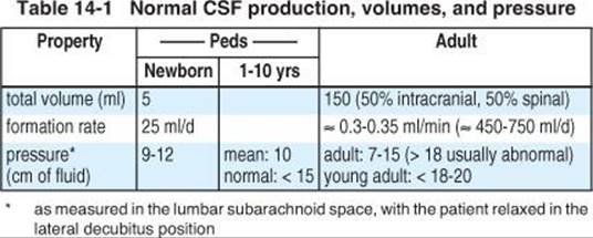
Production rate: In the adult, CSF is produced at a rate of about 0.3 ml/min (see Table 14-1). In terms that are clinically relevant, this approximates 450 ml/24hrs, which means that in an adult, the CSF is “turned over” ≈ 3 times every day. The rate of formation is independent of the intracranial pressure3 (except in the limiting case when ICP becomes so high that cerebral blood flow is reduced4).
Absorption
CSF is absorbed primarily by arachnoid villi (granulations) that extend into the dural venous sinuses. Other sites of absorption include the choroid plexuses and lymphatics. The rate of absorption is pressure dependent5.
14.2. CSF constituents
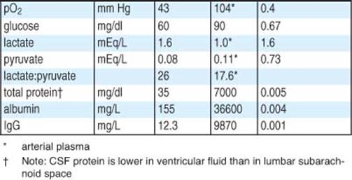
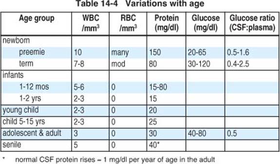
The composition of CSF differs slightly in the ventricles where the majority of it is produced compared to the lumbar subarachnoid space.
CELLULAR COMPONENT
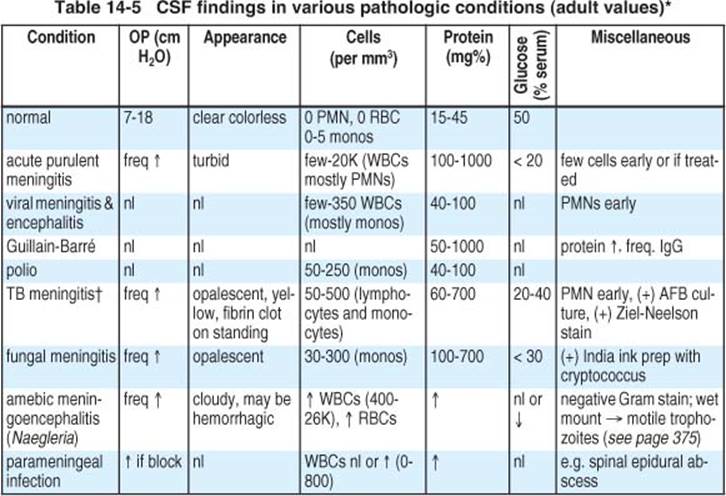
In normal adult CSF, there are 0-5 lymphocytes or mononuclear cells per mm3, and no polys (PMNs) or RBCs. In the absence of RBCs, 5-10 WBCs per mm3 is suspicious, and > 10 WBCs per mm3 is definitely abnormal.
TRAUMATIC TAP
Differentiating true leukocytosis from traumatic tap
When many RBCs and WBCs are present in the CSF due to a traumatic tap (TT), it may be important to tell if the WBCs are elevated or if they are present in the same ratio as in the peripheral blood. In non-anemic patients, there should be ≈ 1-2 WBCs for every 1000 RBCs (as a correction6 (p 176): subtract 1 WBC for every 700 RBCs6 (p 176)). In the presence of anemia or peripheral leukocytosis, use Fishman’s formula6 (p 176) shown in Eq 14-1 to estimate the original WBC count in the CSF before the TT,

where WBCCSF ORIGINAL = WBC count in the CSF before the TT, WBCCSF & RBCCSF = WBC & RBC counts measured in the CSF, and WBCBLOOD & RBCBLOOD = WBC & RBC per mm3 in the peripheral blood.
Estimating true total CSF protein content with a traumatic tap
If the hemogram and peripheral protein are normal, then have the cell count and protein content run on the same tube, and the correction is6 (p 176):
• subtract 1 mg per 100 ml of protein for every 1000 RBC per mm3
Differentiating SAH from traumatic tap
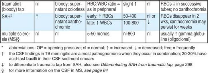
For typical findings in SAH, see page 1038. Some features helpful in differentiating SAH from TT are shown in Table 14-2.
Table 14-2 Features distinguishing traumatic tap from SAH
|
Feature |
Traumatic tap (TT) |
SAH |
|
RBC count (and gross appearance of bloodiness) |
declines as CSF drains (compare first tube to last tube) |
usually > 100,000 RBCs/mm3, changes little as CSF drains |
|
ratio of WBC:RBC |
similar to the ratio in peripheral blood (see Differentiating true leukocytosis from traumatic tap above) |
usually promotes a leukocytosis (elevated WBC count) |
|
supernatant |
clear |
xanthochromic* (rarely in < 2 hrs, present in 70% by 6 hrs, and > 90% by 12 hrs after SAH) |
|
clotting of fluid |
usually clots if erythrocyte count > 200,000/mm3 |
usually does not clot |
|
protein concentration |
fresh bleeding elevates CSF protein from normal by only ≈ 1 mg per 1000 RBC |
blood breakdown products elevate this more than TT (measured protein exceeds the sum of normal protein + 1 mg protein/1000 RBC) |
|
repeat LP at higher level |
usually clear |
remains bloody |
|
opening pressure |
usually normal |
usually elevated |
* NB: other conditions can cause xanthochromia
CSF CONSTITUENTS

14.3. Artificial CSF
A number of formulations of “artificial” CSF have been proposed over the years in order to more closely mimic the pH, osmolarity, CO2, and membrane active ion concentration of CSF. In many instances, normal saline (NS) has been used in brain surgery, probably without consequence. However, renewed interest in the subject of artificial CSF has been brought about by the use of neuroendoscopy, with possible reactions to nonphysiologic solutions when large volumes of fluid are exchanged, as occurs during some of these procedures. An actual reaction to NS, however, has never been proven.
In addition to simulating the constituents of CSF, it may also be well to insure a physiologic temperature of the solution8.
Elliott’s solution
AKA Solution B of Elliott and Jasper9, 10: an elaborate formulation that was widely used in the past.
14.4. CSF fistula (cranial)
 Key concepts:
• suspect in posttraumatic otorrhea/rhinorrhea or recurrent meningitis
• management strategy: 1) confirm the fluid is CSF, 2) identify the site of origin of the leak, 3) determine etiology/mechanism
• most bedside tests are unreliable and include: “reservoir sign”, target/halo sign, qualitative glucose
• most accurate confirmatory test is ß2-transferrin
• CT cisternography is the test of choice for localizing site of fistula
AKA CSF leak. Two major subgroups (omitting the ambiguous category of “spontaneous”)11:
1. traumatic (or posttraumatic): may occur acutely or may be delayed
A. post-procedure (iatrogenic). Including: post-transsphenoidal surgery and post skull base surgery
B. posttraumatic (more common): 67-77% of cases
2. nontraumatic
A. high pressure
1. hydrocephalus
2. tumor
B. normal pressure
1. congenital defects
2. bony erosion from infection or necrosis
3. focal atrophy (olfactory or sellar)
CSF fistula should be suspected in patients with otorrhea or rhinorrhea after head trauma, or in patients with recurrent meningitis.
Possible routes of egress of CSF
1. mastoid air cells (especially after p-fossa surgery, e.g. for vestibular schwannoma (VS), see page 630)
2. sphenoid air cells (especially post-transsphenoidal surgery)
3. cribriform plate/ethmoidal roof (floor of frontal fossa)
4. frontal air cells
5. herniation into empty sella and then into sphenoid air sinus
6. along path of internal carotid artery
7. Rosenmüller’s fossa: located just inferior to cavernous sinus, may be exposed by drilling off anterior clinoids to allow access to ophthalmic artery aneurysms
8. site of the opening of the transient lateral craniopharyngeal canal
9. percutaneously through a surgical or traumatic wound
10. petrous ridge or internal auditory canal: following temporal bone fracture or vestibular schwannoma surgery (see page 630). Then either:
A. rhinorrhea: through middle ear → eustachian tube → nasopharynx
B. otorrhea: via perforated tympanic membrane → external auditory canal
TRAUMATIC FISTULA
Occur in 2-3% of all patients with head injury, 60% occur within days of trauma, 95% within 3 months12. 70% of cases of CSF rhinorrhea stop within 1 wk, and usually within 6 mos in the rest. Non-traumatic cases cease spontaneously in only 33%. Adult:child ratio is 10:1, rare before age 2 yrs. In children the incidence of CSF leaks is less than 1% of closed head injuries13. Anosmia is common in traumatic leaks (78%), rare in spontaneous14. Most (80-85%) CSF otorrhea ceases in 5-10 days.
CSF fistula occurred in 8.9% of 101 cases of penetrating trauma, and increases the infection rate over those penetrating injuries without fistula (50% vs. 4.6%)15. It is reported to occur post-op in up to 30% of cases of skull-base surgery16.
NONTRAUMATIC CSF FISTULA
Nontraumatic leaks primarily occur in adults > 30 yrs. Often insidious. May be mistaken for allergic rhinitis. Unlike traumatic leaks, these tend to be intermittent, the sense of smell is usually preserved, and pneumocephalus is uncommon17.
Sometimes associated with the following18
1. agenesis of the floor of the anterior fossa (cribriform plate) or middle fossa
2. empty sella syndrome: primary or post transsphenoidal surgery (see page 660)
3. increased ICP and/or hydrocephalus
4. infection of the paranasal sinuses
5. tumor: including pituitary adenomas (see page 634), meningiomas
6. a persistent remnant of the craniopharyngeal canal19
7. AVM17
8. congenital anomalies: most involve dehiscence of bone
A. dehiscence of the footplate of the stapes (a congenital abnormality) which can produce CSF rhinorrhea via the eustachian tube17
B. dehiscence below foramen rotundum
Posterior fossa
1. pediatric: usually presents with either meningitis or hearing loss
A. preserved labyrinthine function (hearing and balance): these usually present with meningitis. 3 usual routes of fistula:
1. facial canal: can fistulize into middle ear
2. petromastoid canal: along path of arterial supply to mucosa of mastoid air sinuses
3. Hyrtl’s fissure (AKA tympanomeningeal fissure): links p-fossa to hypotympanum
B. anomalies of labyrinth (hearing lost): one of several types of Mundini dysplasias, usually presenting with rounded labyrinth/cochlea that permits CSF to erode through oval or round window into auditory canal
2. adult: usually presents with conductive hearing loss with serous effusion, meningitis (often following an episode of otitis media), or cerebral abscess. Occurs most commonly through middle fossa. May be due to arachnoid granulations eroding into air sinus compartment
Spinal
Often presents with postural headache associated with neck stiffness and tenderness20 (see page 305).
MENINGITIS IN CSF FISTULA
Incidence with posttraumatic CSF leak: 5-10%, increases as leak persists > 7 days. Meningitis is more common with spontaneous fistula. Risk may be higher in post-neurosurgical CSF fistula than in post-traumatic due to elevated ICP common in latter (forces CSF outward). If site of leak unidentified prior to attempted surgical treatment, 30% develop a recurrent leak post-op, with 5-15% of these developing meningitis before leak is stopped21.
Meningitis may promote inflammatory changes at the site of the leak, with a resultant cessation of the leak.
Pneumococcal meningitis is the most common pathogen (83% of cases22), mortality is lower than in pneumococcal meningitis without underlying fistula (< 10% vs. 50%), possibly because the latter is frequently seen in elderly debilitated patients. Prognosis in children is worse12.
EVALUATION
Determining if rhinorrhea or otorrhea is due to a CSF fistula
1. characteristics of the fluid suggesting the presence of CSF
A. fluid is as clear as water (unless infected or admixed with blood)
B. fluid does not cause excoriation within or outside the nose
C. patients with rhinorrhea describe the taste as salty
2. confirmatory tests
A. ß2-transferrin: present in CSF, but absent in tears, saliva, nasal exudates and serum (except for newborns and patients with liver disease)23, 24. The only other source is the vitreous fluid of the eye. detected by protein electrophoresis. ≈ 0.5 ml needs to be placed in a sterile container, packed in dry ice, and shipped to a lab that can perform this study. Very sensitive & specific
B. collect fluid and obtain quantitative glucose (urine glucose detection strips may be positive even with excess mucus). Test the fluid shortly after collection to minimize fermentation. Normal CSF glucose is > 30 mg% (usually lower with meningitis) whereas lacrimal secretions and mucus are usually < 5 mg%. A negative test is more helpful since it rules out CSF (except in hypoglycorrhachia), but there is a 45-75% chance of false positive25 (p 1638)
C. “ring sign”: when a CSF leak is suspected but the fluid is blood tinged, allow the fluid to drip onto linen (sheet or pillowcase). A ring of blood with a larger concentric ring of clear fluid (so called “double ring” or halo sign) suggests the presence of CSF. An old, but unreliable, sign
D. reservoir sign: a gush of fluid that occurs with a certain head position. Most commonly when first sitting up after a period of recumbency. Thought to indicate drainage of CSF pooled in the sphenoid sinus. Not reliable26
3. radiographic signs: pneumocephalus on CT or skull x-ray. Pneumocephalus occurs in ≈ 20% of patients with CSF leaks27 (p 280)
4. cisternogram: intrathecal injection of radionuclide tracer followed by scintigram or injection of radioopaque contrast followed by CT scan (see below)
5. anosmia is present in ≈ 5% of CSF leaks
6. following skull-base surgery (especially involving greater superficial petrosal nerve) there may be a pseudo-CSF rhinorrhea possibly due to nasal hypersecretion from imbalanced autonomic regulation of the nasal mucosa16ipsilateral to the surgery. Often accompanied by nasal stuffiness and absent ipsilateral lacrimation, and occasionally by facial flushing
TO LOCALIZE SITE OF CSF FISTULA
90% of the time, localization does not require water-soluble contrast CT cisternography (WS-CTC) (see below).
1. CT: to detect pneumocephalus, fractures, skull base defects, hydrocephalus and obstructive neoplasms. Include thin coronal cuts or reconstructions through anterior fossa all the way back to the sella turcica
A. non-contrast (optional): to demonstrate bony anatomy
B. with IV contrast: leak site is usually associated with abnormal enhancement of adjacent brain parenchyma (possibly from inflammation)
2. water-soluble contrast CT cisternography (procedure of choice): see below
3. plain skull x-ray (helpful in only 21%)
4. MRI: may provide additional information for localization and can R/O p-fossa mass, tumor, and empty sella better than CT. Both CT and MRI can R/O hydrocephalus. T2WI fast spin-echo sequences with fat suppression and video image reversal have been used to visualize CSF flow (sensitivity and specificity are 0.87 and 0.57 respectively)28
5. older tests (abandoned in favor of above):
A. radionuclide cisternography (RNC): may be useful in leaks too slow or small to show up on WS-CTC. Various radioactive agents have been used, including: radioiodinated human serum albumin (RIHSA)17, 29, and 500 μCi Indium111 DPTA. Cotton pledgets are packed intranasally (anterior nasal roof, posterior nasal roof, sphenoethmoidal recess, middle meatus, and posterior floor of the nose) and are marked so that their location is known. Radiotracer is then injected intrathecally usually by lumbar puncture. Scans are performed in lateral, AP and posterior view. A protocol using In111 DTPA is to obtain a scan shortly after injection. At 4 hours post-injection, the scan is repeated, and 0.5 ml of blood is drawn (to measure serum activity), and the pledgets are removed. The pledgets are then individually placed in a well-counter and a ratio is calculated for pledget radioactivity relative to serum. A ratio ≤ 1.3 is normal, and a ratio > 1.3 suggests leak. If no leak, the nose can be repacked and the study repeated the following morning.
Leaks into frontal sinus will empty into nasopharynx anterior to the middle concha, unlike leaks through cribriform plate. RNC identifies the site in only 50%. May be misleading14 with possible contamination after several hours from absorption of radioisotope into the bloodstream and accumulation in the mucosal glands of the turbinates. Patient positioning may also contaminate other pledgets
B. intrathecal (visible) dye studies: some success with indigo carmine or fluorescein (see page 144) with little or no complications (✖ methylene blue is neurotoxic and should not be used, see page 144)
WATER-SOLUBLE CONTRAST CT CISTERNOGRAPHY
Procedure of choice. This test is performed if:
1. no site identified on plain CT (with coronals)
2. when patient is leaking clinically (the site is only sometimes identified in the absence of an active leak)
3. when multiple bony defects are identified, and it is essential to determine which site is actively leaking
4. if a bony defect seen on plain CT does not have associated changes of abnormal enhancement of adjacent brain parenchyma
Technique30
Use iohexol (see page 122) 6-7 ml of 190-220 mg/ml) injected into lumbar subarachnoid space via 22 gauge spinal needle (or 5 ml via C1-2 puncture). Patient positioned in -70° Trendelenburg x 3 min prone with neck gently flexed, in CT they are kept prone with head hyperextended with 5 mm coronal cuts with 3 mm overlap (use 1.5 mm cuts if necessary). May need provocative maneuvers (coronal scans prone (brow up) or in position of leak, intrathecal saline infusion (requires Harvard pump)21…).
Look for accumulation of contrast in air sinuses. Apparent discontinuity of bone on CT without extravasation of contrast is probably not the site of leakage (bone discontinuities may be mimicked by partial volume averaging on CT).
TREATMENT
Acutely after trauma, observation is justified as most cases cease spontaneously.
Prophylactic antibiotics: Controversial. There was no difference in the incidence or morbidity of meningitis between treated and untreated patients31. Furthermore, the risk of selecting resistant strains appears real12 and is therefore usually avoided.
FOR PERSISTENT POSTTRAUMATIC OR POST-OP LEAKS
Non-surgical treatment
1. measures to lower ICP:
A. bed rest: although recumbency may ameliorate symptoms, there is no other benefit from bed rest32
B. avoid straining (stool softeners) and avoid blowing nose
C. acetazolamide (250 mg PO QID) to reduce CSF production
D. modest fluid restriction (caution post-transsphenoidal because of possible DI (see page 15): 1500 ml/day in adults, 75% of maintenance/day in peds
2. if leak persists (caution: first R/O obstructive hydrocephalus with CT or MRI)
A. LP: q d to BID (lower pressure to near atmospheric or until H/A)
OR
B. continuous lumbar drainage (CLD): via percutaneous catheter. Two (of many) management options:
1. keep HOB elevated 10-15° and place drip chamber at shoulder level (lower the chamber if leak persists)
2. allow 15-20 cc to drain, then clamp tubing. Repeat q 1 hour
• CLD may require ICU monitoring. If patient deteriorates with drain in place: immediately stop drainage, place patient flat in bed (or slight Trendelenburg), start 100% O2, get CT or bedside cross-table skull x-ray (to R/O tension pneumocephalus due to drawing in of air)
3. surgical treatment in persistent cases (see below)
SURGICAL TREATMENT
Indications for surgical intervention
1. traumatic CSF leak that persists > 2 weeks in spite of non-surgical measures
2. spontaneous leaks and those of delayed onset following trauma or surgery: usually require surgery because of a high incidence of recurrence
3. leaks complicated by meningitis
Petrous bone
May present as otorrhea or as rhinorrhea (via the eustachian tube).
1. following posterior fossa surgery: see page 630 for treatment following vestibular schwannoma surgery
2. following mastoid fractures: may be approached via extensive mastoidectomy17
3. due to dehiscence of the footplate of the stapes: may require obliteration of the middle ear and eustachian tube through a tympanomeatal flap17
Leaks through cribriform plate/ethmoidal roof
Extradural approach: Generally preferred by ENT surgeons33. If a frontal craniotomy is being performed, an intradural approach should be used since problems may arise in dissecting the dura off of the floor of the frontal fossa, wherein the dura almost always tears and then it is difficult to know if an identified tear is the cause of the leak or if it is iatrogenic. Fluorescein dye mixed with CSF injected intrathecally may help demonstrate the leak intraoperatively (CAUTION: must be diluted to reduce risk of seizures, see page 144).
Intradural approach: Generally the procedure of choice34. If the fistula site is unidentified preoperatively, use a bifrontal bone flap.
General techniques of intradural approach:
Close bone defects with fat, muscle, cartilage, or bone.
Close dural defect with fascia lata, temporalis muscle fascia, or pericranium. Fibrin glue may be used to help hold tissue in place.
If the leak is unidentified pre-op and intra-op, then pack both cribriform plates and sphenoid sinus (incise dura over tuberculum sellae, drill through bone to reach sphenoid sinus, remove mucosa or pack it inferiorly, pack with fat).
Post op: lumbar drain after craniotomy is controversial. Some feel CSF pressure may help enhance the seal35. If used, place the drip chamber at the level of shoulder for 3-5 days (for precautions, see above).
Consider shunt (LP or VP) if elevated ICP or hydrocephalus is demonstrated.
Leaks into sphenoid sinus (including post-transsphenoidal surgery leak)
1. LP BID or CLD: as long as pressure > 150 mm H2O or CSF xanthochromic
A. if leak persists > 3 days: repack sphenoid sinus and pterygoid recesses with fat, muscle, cartilage and/or fascia lata (must reconstruct floor of sella, packing alone is inadequate). Some recommend against muscle since it putrefies and shrinks. Continue LP or CLD as above for 3-5 days post-op
B. if leak persists > 5 days: lumboperitoneal shunt (first R/O obstructive hydrocephalus)
2. more difficult surgical approach: intracranial (intradural) approach to medial aspect of middle cranial fossa
3. consider transnasal sellar injection of fibrin glue under local anesthesia36
14.5. Intracranial hypotension
May be spontaneous (see below), post-traumatic (including iatrogenic, e.g. post-LP).
SPONTANEOUS INTRACRANIAL HYPOTENSION (SIH)
The syndrome of spontaneous intracranial hypotension is characterized by the following in the absence of antecedent trauma or LP (or epidural injection…)37:
1. orthostatic headache: dramatically worse when upright, improved in recumbency
2. low CSF pressure
3. diffuse pachymeningeal enhancement (cerebral and/or spinal) on MRI
In most cases, the underlying etiology is thought to be a spontaneous CSF leak from a spinal meningeal diverticulum or dural tear20.
Clinical features
Most patients have orthostatic headache. Onset is often sudden, and may be associated with spinal pain in a specific location. Atypical patients have been described without H/A, or H/A that is non-positional, without pachymeningeal enhancement on MRI38, with clinical signs of encephalopathy, cervical myelopathy, or parkinsonism39. Since some patients may have normal intracranial pressure, the term “CSF hypovolemia” has been suggested40.
Evaluation
1. brain MRI. Findings:
A.  diffuse pachymeningeal enhancement (cerebral and/or spinal) is common
B. brain descent with low lying cerebellar tonsils occurred in 36%39
C. reversible pituitary enlargement with a convex superior margin41
D. subdural hematomas (in 20%) and nonhemorrhagic subdural fluid collections (in 23%) in one series of 40 patients with SIH42
E. small ventricles and cisterns may be seen
2. spinal MRI: may show evidence of CSF leak. If there is focal spine pain, the leak will often be near this location
3. radioisotope cisternography: abnormal in 90%. Revealed the site of CSF leak in 40%39.
Treatment
Treatment includes:
1. bed rest
2. analgesics
3. hydration
4. caffeine
5. epidural blood patch (EBP) for appropriate cases: see page 59
Subdural fluid collections occasionally require intervention (usually drainage of the subdural and blood patching of the spinal leak, if identified).
Outcome
Complete resolution of H/A was achieved in 70% (usually in days to weeks), and was higher in patients receiving EBP, and was lower with multiple sites of CSF leak39.
14.6. References
1. Binhammer R T: CSF anatomy with emphasis on relations to nasal cavity and labyrinthine fluids. Ear Nose Throat J 71: 292-9, 1992.
2. Sato O, Bering E A: Extraventricular formation of cerebrospinal fluid. Brain Nerv 19: 883-5, 1967.
3. Lorenzo A V, Page L K, Wlaters G V: Relationship between cerebrospinal fluid formation, absorption, and pressure in human hydrocephalus. Brain 93: 679-92, 1970.
4. Bering E A, Sato O: Hydrocephalus: Changes in formation and absorption of cerebrospinal fluid within the cerebral ventricles. J Neurosurg 20: 1050-63, 1963.
5. Griffith H B, Jamjoom A B: The treatment of childhood hydrocephalus by choroid plexus coagulation and artificial cerebrospinal fluid perfusion. Br J Neurosurg 4: 95-100, 1990.
6. Fishman R A: Cerebrospinal fluid in diseases of the nervous system. W. B. Saunders, Philadelphia, 1980.
7. Felgenhauer K: Protein size and cerebrospinal fluid composition. Klin Wochenschr 52: 1158-64, 1974.
8. Oka K, Yamamoto M, Nonaka T, et al.: The significance of artificial cerebrospinal fluid as perfusate and endoneurosurgery. Neurosurgery 38: 733-6, 1996.
9. Elliott K A C, Jasper H H: Physiological salt solutions for brain surgery: Studies of local pH and pial vessel reactions to buffered and unbuffered isotonic solutions. J Neurosurg 6: 140-52, 1949.
10. Lewis R C, Elliott K A C: Clinical uses of an artificial cerebrospinal fluid. J Neurosurg 7: 256-60, 1950.
11. Ommaya A K: Spinal fluid fistulae. Clin Neurosurg 23: Clin Neurosurg: 363-92, 1975.
12. Spetzler R F, Zabramski J M: Cerebrospinal fluid fistula. Contemp Neurosurg 8: 1-7, 1986.
13. Shulman K: Later complications of head injuries in children. Clin Neurosurg 19: Clin Neurosurg: 371-80, 1971.
14. Manelfe C, Cellerier P, Sobel D, et al.: CSF rhinor-rhea: Evaluation with metrizamide cisternography. AJNR 3: 25-30, 1982.
15. Meirowsky A M, Ceveness W F, Dillon J D, et al.: CSF fistulas complicating missile wounds of the brain. J Neurosurg 54: 44-8, 1981.
16. Cusimano M D, Sekhar L N: Pseudo-cerebrospinal fluid rhinorrhea. J Neurosurg 80: 26-30, 1994.
17. Calcaterra T C: Cerebrospinal rhinorrhea. In Otolaryngology, English G M, (ed.). Lippincott-Raven, Philadelphia, 1992, Vol. 2: pp 1-7.
18. Nutkiewicz A, DeFeo D R, Kohout R I, et al.: Cerebrospinal fluid rhinorrhea as a presentation of pituitary adenoma. Neurosurgery 6: 195-7, 1980.
19. Jonhston W H: Cerebrospinal rhinorrhea: The study of one case and reports of twenty others collected from the literature published since nineteen hundred. Ann Otolaryngol 35: 1205, 1926.
20. Schievink W I, Meyer F B, Atkinson J L D, et al.: Spontaneous spinal cerebrospinal fluid leaks and intracranial hypotension. J Neurosurg 84: 598-605, 1996.
21. Naidich T P, Moran C J: Precise anatomic localization of atraumatic sphenoethmoidal CSF rhinorrhea by metrizamide CT cisternography. J Neurosurg 53: 222-8, 1980.
22. Hand W L, Sanford J P: Posttraumatic bacterial meningitis. Ann Int Medicine 72: 869-74, 1970.
23. Ryall R G, Peacock M K, Simpson D A: Usefulness of ß2-transferrin assay in the detection of cerebrospinal fluid leaks following head injury. J Neurosurg 77: 737-9, 1992.
24. Fransen P, Sindic C J M, Thauvoy C, et al.: Highly sensitive detection of beta-2 transferrin in rhinorrhea and otorrhea as a marker for cerebrospinal fluid (CSF) leakage. Acta Neurochir 109: 98-101, 1991.
25. Wilkins R H, Rengachary S S, (eds.): Neurosurgery. McGraw-Hill, New York, 1985.
26. Kaufman B, Nulsen F E, Weiss M H, et al.: Acquired spontaneous, nontraumatic normal-pressure cerebrospinal fluid fistulas originating from the middle fossa. Radiology 122 (2): Radiology: 379-87, 1977.
27. Bakay L: Head injury. Little Brown, Boston, 1980.
28. El Gammal T, Sobol W, Wadlington V R, et al.: Cerebrospinal fluid fistula: Detection with MR cisternography. AJNR Am J Neuroradiol 19 (4): AJNR Am J Neuroradiol: 627-31, 1998.
29. Oberson R: Radioisotope diagnosis of rhinorrhea. Radiol Clin Biol 41: 28-35, 1972.
30. Ahmadi J, Weiss M H, Segall H D, et al.: Evaluation of CSF rhinorrhea by metrizamide CT cisternography. Neurosurgery 16: 54-60, 1985.
31. Klastersky J, Sadeghi M, Brihaye J: Antimicrobial prophylaxis in patients with rhinorrhea or otorrhea: A double blind study. Surg Neurol 6: 111-4, 1976.
32. Allen C, Glasziou P, Del Mar C: Bed rest: A potentially harmful treatment needing more careful evaluation. Lancet 354: 1229-33, 1999.
33. Calcaterra T C: Extracranial repair of cerebrospinal rhinorrhea. Ann Otol Rhinol Laryngol 89: 108-16, 1980.
34. Lewin W: Cerebrospinal fluid rhinorrhea in closed head injuries. Br J Surgery 17: 1-18, 1954.
35. Dagi T F, George E D: Surgical management of cranial cerebrospinal fluid fistulas. In Operative neurosurgical techniques, Schmidek H H and Sweet W H, (eds.). W.B. Saunders, Philadelphia, 3rd ed., 1995, Vol. 1: pp 117-31.
36. Fujii T, Misumi S, Onoda K, et al.: Simple management of CSF rhinorrhea after pituitary surgery. Surg Neurol 26: 345-8, 1986.
37. Fishman R A, Dillon W P: Dural enhancement and cerebral displacement secondary to intracranial hypotension. Neurology 43: 609-11, 1993.
38. Schievink W I, Tourje J: Intracranial hypotension without meningeal enhancement on magnetic resonance imaging. J Neurosurg 92: 475-7, 2000 (case report).
39. Chung S J, Kim J S, Lee M C: Syndrome of cerebral spinal fluid hypovolemia: Clinical and imaging features and outcome. Neurology 55: 1321-7, 2000.
40. Mokri B: Spontaneous cerebrospinal fluid leaks, from intracranial hypotension to cerebrospinal fluid hypovolemia: Evolution of a concept. Mayo Clin Proc 74: 1113-23, 1999.
41. Alvarez-Linera J, Escribano J, Benito-Leon J, et al.: Pituitary enlargement in patients with intracranial hypotension syndrome. Neurology 55: 1895-7, 2000.
42. Lai T H, Fuh J L, Lirng J F, et al.: Subdural hematoma in patients with spontaneous intracranial hypotension. Cephalalgia 27: 133-8, 2007.