
4.1. Dementia
Definition: loss of intellectual abilities previously attained (memory, judgement, abstract thought, and other higher cortical functions) severe enough to interfere with social and/or occupational functioning1. Memory deficit is the cardinal feature, however, the DSM-IV definition requires impairment in at least one other domain (language, perception, visuospatial function, calculation, judgement, abstraction, problem-solving skills). Affects 3-11% of community-dwelling adults > 65 yrs of age, with a greater presence among institutionalized residents2.
Risk factors: advanced age, family history of dementia, and apolipoprotein E-4 allele.
Delerium: AKA acute confusional state. Distinct from dementia, however, patients with dementia are at increased risk of developing delerium3, 4. A primary disorder of attention that subsequently affects all other aspects of cognition5. Often represents life-threatening illness, e.g. hypoxia, sepsis, uremic encephalopathy (also see page 73), electrolyte abnormality, drug intoxication, MI. 50% of patients die within 2 yrs of this diagnosis.
Unlike dementia, delerium has acute onset, motor signs (tremor, myoclonus, asterixis), slurred speech, altered consciousness (hyperalert/agitated or lethargic, or fluctuations), hallucinations may be florid. EEG → pronounced diffuse slowing.
Brain biopsy for dementia
Clinical criteria are usually sufficient for the diagnosis of most dementias. Biopsy should be reserved for cases of a chronic progressive cerebral disorder with an unusual clinical course where all other possible diagnostic methods have been exhausted and have failed to provide adequate diagnostic certainty6. Biopsy may disclose CJD, low grade astrocytoma, and AD among others. The high incidence of CJD among patients selected for biopsy under these criteria necessitates appropriate precautions (see Creutzfeldt-Jakob disease, page 361). In a report of 50 brain biopsies performed to assess progressive neurodegenerative disease of unclear etiology7, the diagnostic yield was only 20% (6% were only suggestive of a diagnosis, 66% were abnormal but nonspecific, 8% were normal). The yield was highest in those with focal MRI abnormalities. Among the 10 patients with diagnostic biopsies, the biopsy result led to a meaningful therapeutic intervention in only 4.
Recommendations
Based on the above, the following recommendations are made for patients with an otherwise unexplained neurodegenerative disease:
1. those with a focal abnormality on MRI: stereotactic biopsy
2. those without focal abnormality (possibly including SPECT or PET scan): brain biopsy should only be performed within an investigative protocol
Recommendations for specimen
Ideally the biopsy specimen should8:
1. be large enough (usually 1 cm3)
2. be taken from an affected area
3. include grey and white matter
4. be handled carefully to minimize artifact (electrocautery should not be used on the specimen side of the incision)
4.2. Headache
Headache (H/A) may be broadly categorized as follows:
1. chronic recurring headaches
A. vascular type (migraine): see below
B. muscle contraction (tension) headaches
2. headache due to pathology
A. systemic pathology
B. intracranial pathology: a wide variety of etiologies including:
1. subarachnoid hemorrhage: sudden onset, severe, usually with vomiting, apoplexy, focal deficits possible (see page 1035 for differential diagnosis of paroxysmal H/A)
2. increased intracranial pressure from any cause (tumor, communicating hydrocephalus, inflammation, pseudotumor cerebri…)
3. irritation or inflammation of meninges: meningitis
4. tumor: with or without elevated ICP (see page 587)
C. local pathology of the eye, nasopharynx, or extracranial tissues (including giant cell arteritis, see page 74)
D. following head trauma (postconcussive syndrome): see page 910
E. following craniotomy (“syndrome of the trephined”): see page 149
A severe new H/A, or a change in the pattern of a longstanding or recurrent H/A (including developing associated N/V, or an abnormal neurologic exam) warrants further investigation with CT or MRI9.
 Unilateral H/A that never changes side over a period ≥ 1 year warrants an MRI (this would be atypical in migraine and may be a presentation of an occipital AVM - see page 1099).
4.2.1. Migraine
Migraine attacks usually occur in individuals predisposed to the condition, and may be activated by factors such as bright light, stress, diet changes, trauma, administration of radiologic contrast media (especially angiography) and vasodilators.
CLASSIFICATION
See also index under Headache, e.g. for: crash migraine (thunderclap headache) page 1035, post-myelogram headache page 58…
COMMON MIGRAINE
Episodic H/A with N/V and photophobia, without aura or neurologic deficit.
CLASSIC MIGRAINE
Common migraine + aura. May have H/A with occasional focal neurologic deficit(s) that resolve completely in ≤ 24 hrs.
Over half of the transient neurologic disturbances are visual, and usually consist of positive phenomena (spark photopsia, stars, complex geometric patterns, fortification spectra) which may leave negative phenomena (scotoma, hemianopsia, monocular or binocular visual loss…) in their wake. The second most common symptoms are somatosensory involving the hand and lower face. Less frequently, deficits may consist of aphasia, hemiparesis, or unilateral clumsiness. A slow march-like progression of deficit is characteristic. The risk of stroke is probably increased in patients with migraine10.
COMPLICATED MIGRAINE
Occasional attacks of classic migraine with minimal or no associated H/A, and complete resolution of neurologic deficit in ≤ 30 days.
MIGRAINE EQUIVALENT
Neurologic symptoms (N/V, visual aura, etc.) without H/A (acephalgic migraine). Seen mostly in children. Usually develops into typical migraine with age. Aura may be shortened by opening and swallowing contents of a 10 mg nifedipine capsule11.
HEMIPLEGIC MIGRAINE
H/A typically precedes hemiplegia which may persist even after H/A resolves.
CLUSTER HEADACHE
AKA histaminic migraine. Actually a neurovascular event, distinct from true migraine. Recurrent unilateral attacks of severe pain. Usually oculofrontal or oculotemporal with occasional radiation into the jaw, usually recurring on the same side of the head. Ipsilateral autonomic symptoms (conjunctival injection, nasal congestion, rhinorrhea, lacrimation, facial flushing) are common. Partial Horner’s syndrome (ptosis and miosis) sometimes occurs. Male:female ratio is ≈ 5:1. 25% of patients have a personal or family history of migraine.
Headaches characteristically have no prodrome, last 30-90 minutes, and recur one or more times daily usually for 4-12 weeks, often at a similar time of day, following which there is typically a remission for an average of 12 months12.
Treatment for cluster H/A (prophylaxis is only minimally effective):
Treatment is difficult because there is no prodrome and the H/A often stop after 1-2 hrs. Treatment of acute attacks includes:
• 100% O2 by face mask with patient sitting for ≤ 15 min or until attack aborted
• ergotamine
• SQ sumatriptan: usually aborts attack within 15 minutes
• steroids
• refractory cases may be considered for:
 percutaneous radiofrequency sphenopalatine ganglion blockade13
occipital nerve stimulation14
hypothalamic deep brain stimulation
BASILAR ARTERY MIGRAINE
Essentially restricted to adolescence. Recurrent episodes lasting minutes to hours of transient neurologic deficits in distribution of vertebrobasilar system. Deficits include: vertigo (most common), gait ataxia, visual disturbance (scotomata, bilateral blindness), dysarthria, followed by severe H/A and occasionally nausea and vomiting15. Family history of migraine is present in 86%.
4.2.2. Post LP (myelogram) H/A
AKA “postspinal headache” or “spinal headache”. May also follow procedures other than LP/myelogram, such as dural opening (see page 450). Can also occur with spontaneous intracranial hypotension (see page 305).
Clinical features
Important distinctive characteristic: H/A occurs when patient is erect, and is completely or partially (but significantly) relieved when recumbent. May be associated with nausea, vomiting, dizziness, or visual disturbances.
Time course: Most post-LP headaches (PLPHA) have a delayed onset 24-48 hrs after the LP, and although they may occur weeks post-LP, most also develop within 3 days. The duration of PLPHA varies, with a mean of 4 days16, and reports of duration of months17 and even > 1 year18.
Pathophysiology
Thought to be due to continued CSF leakage through the hole in the dura19, which reduces the CSF “cushion” of the brain. In the upright position, the pull of gravity on the brain produces traction on the blood vessels and any structures tethering the brain to the painsensitive dura. CSF may sometimes be demonstrable in the epidural space.
Epidemiology following LP
Reported incidence range is 2-40% (typically ≈ 20%), higher after diagnostic LP than for epidural anesthesia16.
For variables in LP that impact upon the risk of PLPHA, see page 204
TREATMENT FOR H/A FOLLOWING LP
Initial “conservative” measures include:
1. flat in bed for at least 24 hrs
2. hydration (PO or IV)
3. analgesics for H/A
4. tight abdominal binder
5. desoxycortisone acetate 5 mg IM q 8 hrs16
6. caffeine sodium benzoate 500 mg in 2 cc IV q 8 hrs up to 3 d max (70% of patients had relief with 1 or 2 injections)20
7. high-dose steroids: report of success in a case of intracranial hypotension associated with spontaneous slit ventricles tapering down from a starting dose of dexamethasone 20 mg/day21
8. blood patch if refractory (see below)
EPIDURAL BLOOD PATCH
For refractory post-lumbar puncture or post-myelogram H/A. Works in one application in over 90% of cases, may be repeated if ineffective17. Theoretical risks: infection, cauda equina compression, failure to relieve H/A.
Technique
Accessing epidural space (one of several techniques): proceed as routine LP. When ligaments are traversed, and needle tip is nearing spinal canal, stylet is removed. Then, either place drop of sterile saline in hub (hanging drop technique) and advance while watching for it to be drawn into needle as epidural space is entered, or gently try injecting air with small syringe (preferably glass) while advancing, when the epidural space is entered, resistance to injection disappears, but CSF cannot be aspirated.
A venipuncture site is prepared aseptically. 10 ml of the patient’s blood is with-drawn. After verifying CSF cannot be aspirated through the spinal needle, the blood is injected into the epidural space. After 30 minutes supine, patient may ambulate ad lib.
4.3. Parkinsonism
Parkinsonism may be primary or secondary to other conditions. All result from a relative loss of dopamine mediated inhibition of the effects of acetylcholine in the basal ganglia.
IDIOPATHIC PARALYSIS AGITANS (IPA)
Classical Parkinson’s disease AKA shaking palsy.
Clinical
Affects ≈ 1% of Americans > age 50 yrs22. Male:female ratio is 3:2. Not clearly environmentally or genetically induced, but may be influenced by these factors.
The classic triad is shown in Table 4-1. Other signs may include: postural instability, micrographia, mask-like facies. Gait consists of small, shuffling steps (marche á petits pas) or festinating gait.
Table 4-1 Classic triad of Parkinson’s disease
|
• tremor (resting, 4-7/second) • rigidity (cogwheel) • bradykinesia |
Clinically distinguishing IPA from secondary parkinsonism (see below): May be difficult early. IPA generally exhibits gradual onset of bradykinesia with tremor that is often asymmetrical, and initially responds well to levodopa. Other disorders are suggested with rapid progression of symptoms, when the initial response to levodopa is equivocal, or when there is early midline symptoms (ataxia or impairment of gait and balance, sphincter disturbance…) or the presence of other features such as early dementia, sensory findings, profound orthostatic hypotension, or abnormalities of extraocular movements23, 24.
Pathophysiology
Degeneration primarily of pigmented (neuromelaninladen) dopaminergic neurons of the pars compacta of the substantia nigra, resulting in reduced levels of dopamine in the neostriatum (caudate nucleus, putamen, globus pallidus). This decreases the activity of inhibitory neurons with predominantly D2 class of dopamine receptors which project directly to the internal segment of the globus pallidus (GPi), and also increases (by loss of inhibition) activity of neurons with predominantly D1 receptors which project indirectly to the globus pallidus externa (GPe) and subthalamic nucleus25. The net result is increased activity in GPi which has inhibitory projections to the thalamus which then suppresses activity in the supplemental motor cortex among other locations.
Histologically: Lewy bodies (eosinophilic intraneuronal hyaline inclusions) are the hallmark of IPA.
SECONDARY PARKINSONISM
The differential diagnosis includes the following etiologies of secondary parkinsonism or Parkinson-like conditions (some referred to as “Parkinson plus”):
1. olivopontocerebellar degeneration (OPC)
2. striato-nigral degeneration (SND): more aggressive than parkinsonism
3. postencephalitic parkinsonism: followed an epidemic of encephalitis lethargica (von Economo disease) in the 1920’s, victims are no longer living. Distinguishing features: oculogyric crisis, tremor involves not only extremities but also trunk and head, asymmetrical, no Lewy bodies
4. progressive supranuclear palsy (PSNP): impaired vertical gaze (see below)
5. multiple system atrophy (Shy-Drager syndrome): see below
6. drug induced: includes:
A. prescription drugs (elderly females seem more susceptible)
1. antipsychotics (AKA neuroleptics): haloperidol (Haldol®) which works by blocking postsynaptic dopamine receptors
2. phenothiazine antiemetics: prochlorperazine (Compazine®)
3. metoclopramide (Reglan®)
4. reserpine
B. MPTP (1-methyl-4-phenyl-1,2,3,6-tetrahydropyridine): a commercially available chemical intermediate which is also a by-product of the synthesis of MPPP (a meperidine analog) that was synthesized and self-injected by a graduate student26, and later produced by illicit drug manufacturers to be sold as “synthetic heroin” and unwittingly injected by some IV drug abusers in northern California in 198327. MPTP was subsequently discovered to be a potent neurotoxin for dopaminergic neurons. As a rule, the response to levodopa is dramatic, but short-lived
C. there is an as yet unproven assertion that methylenedioxymethamphetamine (MDMA) AKA “ecstasy” (on the street), may hasten the onset of Parkinsonism
7. toxic: poisoning with
A. carbon monoxide: symmetric low densities in the globus pallidus on CT
B. manganese: may be seen in miners, welders, and pyrotechnics workers. Manganese is excreted by the liver,  people with hepatic insufficiency are more susceptible. Imaging: symmetrical high signal abnormalities on T1WI primarily in the globus pallidus with essentially no findings on T2WI or GRASS (almost pathognomonic)
8. ischemic (lacunes in basal ganglia): produces so-called arteriosclerotic parkinsonism AKA vascular parkinsonism: “lower-half” parkinsonism (gait disturbance predominates28). Also causes pseudobulbar deficits, emotional lability. Tremor is rare
9. posttraumatic: parkinsonian symptoms may occur in chronic traumatic encephalopathy (dementia pugilistica, see page 911). There are usually other features not normally present in IPA (e.g. cerebellar findings)
10. normal pressure hydrocephalus (NPH): urinary incontinence… (see page 329)
11. neoplasm in the region of the substantia nigra
12. Riley-Day (familial dysautonomia)
13. parkinson-dementia complex of Guam: classic IPA + amyotrophic lateral sclerosis (ALS). Pathologically has features of parkinsonism and Alzheimer’s disease but no Lewy bodies nor senile plaques
14. Huntington’s disease (HD): whereas adults typically show chorea, when HD manifests in a young person it may resemble IPA
15. (spontaneous) intracranial hypotension may present with findings mimicking IPA (see page 305)
MULTIPLE SYSTEM ATROPHY (MSA)
AKA Shy-Drager syndrome. Parkinsonism (indistinguishable from IPA), PLUS idiopathic orthostatic hypotension, PLUS other signs of autonomic nervous system (ANS) dysfunction (ANS findings may precede parkinsonism and may include urinary sphincter disturbance and hypersensitivity to noradrenaline or tyramine infusions). Degeneration of preganglionic lateral horn neurons of thoracic spinal cord. Unlike IPA, most do not respond to dopa therapy. NB: classic IPA may eventually produce orthostatic hypotension from inactivity or as a result of progressive autonomic failure.
PROGRESSIVE SUPRANUCLEAR PALSY (PSNP)
AKA Steele-Richardson-Olszewski syndrome29.
Triad:
1. progressive supranuclear ophthalmoplegia (chiefly vertical gaze): paresis of voluntary vertical eye movement, but still moves to vertical doll’s eyes maneuver
2. pseudobulbar palsy (mask-like facies with marked dysarthria and dysphagia, hyperactive jaw jerk, emotional incontinence usually mild)
3. axial dystonia (especially of neck and upper trunk)
Associated findings: subcortical dementia (inconstant), motor findings of pyramidal, extrapyramidal and cerebellar systems. Average age of onset: 60 yrs. Males comprise 60%. Response to anti-parkinson drugs is usually very short lived. Average survival after diagnosis: 5.7 yrs.
Differentiating from Parkinson’s disease (IPA):
Patients with PSNP have a pseudo-parkinsonism. They have mask facies, but do not walk bent forward (they walk erect), and they do not have a tremor. They tend to fall backwards.
Course
1. early:
A. many falls: due to dysequilibrium + downgaze palsy (can’t see floor)
B. eye findings may be normal initially, subsequently may develop difficulty looking down (especially to command, less to following), calorics have normal tonic component but absent nystagmus (cortical component)
C. slurred speech
D. personality changes
E. difficulty eating: due to pseudobulbar palsy + inability to look down at food on plate
2. late:
A. eyes fixed centrally (no response to oculocephalics or oculovestibulars): ocular immotility is due to frontal lobe lesions
B. neck stiffens in extension (retrocollis)
SURGICAL TREATMENT FOR PARKINSON’S DISEASE
Before the introduction of L-dopa in the late 1960’s, stereotactic thalamotomy was widely used for Parkinson’s disease. The location ultimately targeted for lesioning was the ventrolateral nucleus. The procedure worked better for relieving the tremor than for the bradykinesia, however it was the latter symptom that was most disabling. This procedure cannot be done bilaterally without significant risk to speech function. The procedure fell out of favor when more effective drugs became available30.
See Surgical treatment of Parkinson’s disease on page 532 for further information.
4.4. Multiple sclerosis
 Key concepts:
• an idiopathic demyelinating disease of thc CNS producing exacerbating and remitting symptoms disseminated in space and time
• classic clinical findings: optic neuritis, paresthesias, INO and bladder symptoms
• diagnostic criteria (McDonald criteria) use clinical and/or lab results (MRI, CSF…) to stratify patients as: MS, probable MS, or not MS
• MRI: multiple usually enhancing lesions involving optic nerves & white matter of brain (especially periventricular white matter), cerebellum and spinal cord
An idiopathic demyelinating disease (affecting only white matter) of the cerebrum, optic nerves, and spinal cord (especially corticospinal tracts and the posterior columns). Does not affect peripheral myelin. Produces multiple plaques of various age in diffuse locations in the CNS, especially in the periventricular white matter. Lesions initially evoke an inflammatory response with monocytes and lymphocytic perivascular cuffing, but with age settle down to glial scars.
EPIDEMIOLOGY
Usual age of onset: 10-59 years, with the greatest peak between ages 20-40 years. Female to male ratio: 2:1.
Prevalence varies with latitude, and is < 1 per 100,000 near the equator, and is ≈ 30-80 per 100,000 in the northern U.S. and Canada.
CLINICAL
Causes exacerbations and remissions in various locations in the CNS (dissemination in space and time). Common symptoms: visual disturbances (diplopia, blurring, field cuts or scotoma), spastic paraparesis, and bladder disturbances. Nomenclature for the time course of MS is shown in Table 4-231. Relapsing-remitting MS is the most common pattern (≥ 70%) at onset, and has the best response to therapy, but > 50% of cases eventually become secondary progressive MS. Only 10% have primary progressive MS, and these patients tend to be older at onset (40-60 years) and frequently develop progressive myelopathy32. Progressive relapsing MS is very uncommon.
Deficits present > 6 months usually persist.
Table 4-2 Clinical categories of MS
|
Category |
Definition |
|
relapsingremitting |
episodes of acute worsening with recovery and a stable course between relapses |
|
secondary progressive |
gradual neurologic deterioration ± superimposed acute relapses in a patient who previously had relapsing-remitting MS |
|
primary progressive |
gradual, nearly continuous neurologic deterioration from the onset of symptoms |
|
progressive relapsing |
gradual neurologic deterioration from the onset of symptoms, but with subsequent superimposed relapses |
Differential diagnosis
The plethora of possible signs and symptoms in MS causes the differential diagnosis to extend to almost all conditions causing focal or diffuse dysfunction of the CNS. Conditions that may closely mimic MS clinically and on diagnostic testing include:
1. acute disseminated encephalomyelitis (ADEM): generally monophasic. May also have CSF-OCB. Corpus callosum involvement is uncommon
2. CNS lymphoma: see page 673
3. other closely related demyelinating diseases: e.g. Devic syndrome (see page 1187)
4. vasculitis
5. encephalitis: patients are usually very ill
6. chronic white matter changes: seen in older patients
Signs and symptoms
Visual disturbances: Disturbances of visual acuity may be caused by optic or retrobulbar neuritis which is the presenting symptom of MS in 15% of cases, and which occurs at some time in 50% of MS patients. The percentage of patients with an attack of optic neuritis and no prior attack that will go on to develop MS ranges from 17-87% depending on the series33. Symptoms: acute visual loss in one or both eyes with mild pain (often on eye movement).
Diplopia may be due to internuclear ophthalmoplegia (INO) (see page 834) from a plaque in the MLF. INO is an important sign because it rarely occurs in other conditions besides MS or brainstem stroke.
Motor findings: Extremity weakness (mono, para, or quadriparesis) and gait ataxia are among the most common symptoms of MS. Spasticity of the LEs is often due to pyramidal tract involvement. Scanning speech results from cerebellar lesions.
Sensory findings: Posterior column involvement often causes loss of proprioception. Paresthesias of extremities, trunk, or face occur. Lhermitte’s sign (electric shock-like pain radiating down the spine on neck flexion) is common, but is not pathognomonic. Trigeminal neuralgia occurs in ≈ 2%, and is more often bilateral and occurs at a younger age than the population in general34.
Mental disturbances: Euphoria (la belle indifference) and depression occur in ≈ 50% of patients.
Reflex changes: Hyperreflexia and Babinski signs are common. Abdominal cutaneous reflexes disappear in 70-80%.
GU symptoms: Urinary frequency, urgency, and incontinence are common. Impotence in males and reduced libido in either sex is often seen.
DIAGNOSTIC CRITERIA
No single clinical feature or diagnostic test is adequate for the accurate diagnosis of MS. Therefore, clinical information is integrated with paraclinical studies. Diagnosing MS after a single, acute remitting clinically isolated syndrome (CIS) is very risky. 50-70% of patients with a CIS suggestive of MS will have multifocal MRI abnormalities characteristic of MS. The presence of these MRI abnormalities increases the risk of developing MS in 1-3 years (with greater prognostic significance than CSF-OCB). The more MRI lesions, the higher the risk35. Criteria for the diagnosis of MS36 follows.
DEFINITIONS36, 37
1. attack (exacerbation, relapse): neurologic disturbance lasting > 24 hrs38 typical of MS when clinicopathological studies determine that the cause is demyelinating or inflammatory lesions
2. remission: ≥ 30 days should separate the onset of the first attack from the onset of a second
3. historical information: reporting of symptoms by the patient (confirmation by observer desirable), adequate to locate a lesion of MS, and has no other explanation (i.e. manifestations must not be attributable to another condition)
4. clinical evidence (signs): neuro dysfunction recorded by competent examiner
5. paraclinical evidence: tests or procedures demonstrating CNS lesion which has not produced signs; e.g. Uhthoff phenomenon or sign (worsening of symptoms with hot bath or shower), BAER, imaging procedures (CT, MRI), expert urological assessment
6. typical of MS: signs & symptoms (S/S) known to occur frequently in MS. Thus excludes gray matter lesions, peripheral nervous system lesions, and non-specific complaints such as H/A, depression, convulsive seizures, etc.
7. separate lesions: S/S cannot be explained on basis of single lesion (optic neuritis of both eyes simultaneously or within 15 days represents single lesion)
8. laboratory support: in this study, the only considerations were CSF oligoclonal bands (CSF-OCB) (see below) (OCB must not be present in serum) or increased CSF IgG production (CSF-IgG) (serum IgG must be normal). This assumes that syphilis, SSPE, sarcoidosis, etc. have been ruled out
Table 4-3 Diagnostic criteria for MS36
|
Clinical presentation |
Additional data needed to diagnose MS |
|
≥ 2 attacks; objective clinical evidence of ≥ 2 lesions |
none* |
|
≥ 2 attacks; objective clinical evidence of 1 lesion |
demonstrate dissemination in space by: • MRI† or • ≥ 2 MS-compatible lesions on MRI PLUS positive CSF‡ or • await additional clinical attack implicating another site |
|
1 attack; objective clinical evidence of ≥ 2 lesions |
demonstrate dissemination in time by • MRI§ or • second clinical attack |
|
1 attack; objective clinical evidence of 1 lesion (mono-symptomatic presentation; clinically isolated syndrome) |
demonstrate dissemination in space by: • MRI† or • ≥ 2 MS-compatible lesions on MRI PLUS positive CSF‡ or and demonstrate dissemination in time by: • MRI§ or • second clinical attack |
|
insidious neurological progression suggestive of MS |
positive MCSF‡ and demonstrate dissemination in space by A. ≥ 9 T2WI lesions on MRI or B. ≥ 2 lesions in spinal cord or C. 4-8 brain + 1 spinal cord lesion or D. abnormal VEPΔ + (4-8 brain lesions or< 4 brain lesions + 1 spinal cord lesion on MRI) and demonstrate dissemination in time by A. MRI§ or B. continued progression for 1 year |
* additional tests not required. If MRI or CSF test s are done and are negative, apply extreme caution in diagnosing MS
† must meet criteria in Table 4-4
‡ positive CSF showing oligoclonal bands, see Table 4-6
§ dissemination in time on MRI must meet the criteria in Table 4-5
Δ abnormal visual evoked potential as seen in MS (delay with well-preserved wave-form)
DIAGNOSIS OF MS
The “McDonald criteria” are shown in Table 4-336. Preferred terms:
• MS
• possible MS (at risk for MS but diagnosis is equivocal)
• not MS
MRI
MRI is the preferred imaging study in evaluating MS39 and can demonstrate dissemination of lesions in time and space. Recommended36 brain MRI criteria for diagnosing MS are shown in Table 4-440, 41. Lesions are normally > 3 mm diameter36. MRI shows multiple white matter abnormalities in 80% of patients with MS (compared to 29% for CT)42, 43. Lesions are high signal on T2WI, and acute lesions tend to enhance with gadolinium more than old lesions do. Periventricular lesions may blend in with the signal from CSF in the ventricles on T2WI, these lesions are shown to better advantage on proton density images as higher intensity than CSF. These lesions are ovoid and are oriented perpendicular to the ependymal surface and are sometimes called Dawson’s fingers.
Spinal cord lesions normally show little or no swelling, should be ≥ 3 mm but < 2 vertebral segments, occupy only a portion of the cross-section of the cord, and must be hyperintense on T2WI44.
Specificity of MRI is ≈ 94%45, however, encephalitis as well as UBOs seen in aging may mimic MS lesions. DWI should be normal, however plaques can sometimes exhibit “shine through” (see page 132) so the ADC map must be checked to rule-out infarct.
Table 4-4 Brain MRI criteria for MS
|
3 of the following 4 criteria* |
|
1. 1 gadolinium-enhancing lesion or, if no gadolinium enhancing lesions, then 9 T2WI lesions 2. ≥ 1 infratentorial lesion 3. ≥ 1 juxtacortical lesion (i.e. involving subcortical u fibers) 4. ≥ 3 periventricular lesions |
* 1 spinal cord lesion can be substituted for 1 brain lesion
Focal tumefactive demyelinating lesions (TDL) may occur in isolation or, more commonly, in patients with established MS (concentric sclerosis of Balo). TDL may represent an intermediate position between MS and ADEM46. TDLs tend to be symmetric. TDLs may enhance, and show perilesional edema (but less than MS) and thus be mistaken for neoplasms. Biopsy results may be confusing. MRS may not be able to differentiate from neoplasm47.
Table 4-5 MRI criteria for dissemination of lesions in time
|
1. if first MRI occurs ≥ 3 months after the onset of the clinical event, then a gadolinium enhancing lesion in a different location than that implicated by the event meets the criteria. If there is no enhancing lesion, a follow-up MRI is required*. A new enhancing or T2WI lesion meets the criteria |
|
2. if first MRI is < 3 months from onset, a second MRI ≥ 3 months from onset showing a new gadolinium enhancing lesion meets the criteria. If no enhancing lesion, a 3rd MRI ≥ 3 months from the first showing a new enhancing or T2WI lesion meets the criteria |
* timing not critical, but 3 months is recommended48
CSF
CSF analysis can support the diagnosis in some cases, but cannot document dissemination of lesions in time or space. The CSF in MS is clear and colorless. The OP is normal. Total CSF protein is < 55 mg/dl in ≈ 75% of patients, and < 108 mg/dl in 99.7% (values near 100 should prompt a search for an alternative diagnosis). The WBC count is ≤ 5 cells/μl in 70% of patients, and only 1% have a count > 20 cells/μl (high values may be seen in the acute myelitis).
In ≈ 90% of patients with MS, CSF-IgG is increased relative to other CSF proteins, and a characteristic pattern occurs. Agarose gel electrophoresis shows a few IgG bands in the gamma region (oligoclonal bands) that are not present in the serum. CSF-OCB are not specific for MS, and can occur in CNS infections and less commonly with strokes or tumors. The predictive value of the absence of IgG in a patient with suspected MS has not been satisfactorily elucidated.
Recommended criteria have been published49, most of which pertain to specifics of laboratory analysis, pertinent clinical excerpts are shown in Table 4-6.
Table 4-6 CSF criteria for MS
|
1. qualitative assessment of IgG is the most informative analysis 2. analysis should be performed on unconcentrated CSF and must be compared to simultaneously run serum sample in the same assay 3. quantitative analysis should be made in terms of one of the 5 recognized staining patterns for OCB 4. all other tests performed on the CSF (including WBC, protein & glucose, lactate) should be taken into consideration 5. if clinical suspicion is high but CSF results are equivocal, negative or show only a single band, consider repeating the LP 6. quantitative IgG is a complementary test, but is not a substitute for qualitative IgG testing |
4.5. Motor neuron diseases
Degenerative diseases of motor neurons. For comparison of upper motor neuron (UMN) with lower motor neuron (LMN) and the paralysis they produce, see page 786.
Three patterns of involvement:
1. mixed UMN & LMN degeneration: amyotrophic lateral sclerosis (ALS) (see below). The most common of the motor neuron diseases
2. UMN degeneration: primary lateral sclerosis. Rare, onset after age 50. No LMN signs. Slower progression than ALS (yrs to decades). Pseudobulbar palsy is common50. Usually does not shorten longevity. May present with falling due to balance problems or low back and neck pain due to axial muscle weakness
3. LMN degeneration: progressive muscular atrophy (PMA) and spinal muscular atrophy (SMA)
4.5.1. Amyotrophic lateral sclerosis
Key concepts:
• degeneration of anterior horn cells and corticospinal tracts in the cervical spine and medulla (bulb) of unknown etiology
• a mixed upper and lower motor neuron disease (UMN → mild spasticity in LEs; LMN → atrophy and fasciculations in UEs)
• clinically: progressive muscle wasting, weakness, and fasciculations
• no cognitive, sensory, nor autonomic dysfunction
In the U.S. amyotrophic lateral sclerosis (ALS) is AKA Lou Gehrig’s disease. Some-times AKA motor neuron disease (singular).
EPIDEMIOLOGY33
Prevalence: 4-6/100,000. Incidence: 0.8-1.2/100,000.
Familial in 8-10% of cases. Familial cases usually follow autosomal dominant inheritance, but occasionally demonstrate a recessive pattern.
Onset usually after 40 years of age.
PATHOLOGY
Etiology is not known with certainty. Histology: degeneration of anterior horn alpha-motoneurons (in the spinal cord and in brain stem motor nuclei) (LMNs) and corticospinal tracts (UMNs). Produces mixed UMN & LMN findings, with a great deal of variability depending on which predominates at any given time.
CLINICAL
Characterized by progressive muscle wasting, weakness, and fasciculations.
Involvement is of voluntary muscles, sparing the voluntary eye muscles and urinary sphincter.
Classically, presents initially with weakness and atrophy of the hands (lower motor neuron) with spasticity and hyperreflexia of the lower extremities (upper motor neuron). However, LEs may be hyporeflexic if the lower motor neuron deficits predominate.
Dysarthria and dysphagia are caused by a combination of upper and lower motor neuron pathology. Tongue atrophy and fasciculations may also occur.
Although cognitive deficits are generally considered to be absent in ALS, in actuality 1-2% of cases are associated with dementia, and cognitive changes may occasionally predate the usual features of ALS51.
DIFFERENTIAL DIAGNOSIS
At times, it may be very difficult to distinguish ALS from cervical spondylotic myelopathy. See page 489 for a discussion of differentiating features.
DIAGNOSTIC STUDIES
EMG: Not absolutely necessary to make diagnosis in most cases. Fibrillations and positive sharp waves are found in advanced cases (may be absent early, especially if upper motor neuron pathology predominates). LMN findings in the LE in the absence of lumbar spine disease, or fibrillation potentials in the tongue are suggestive of ALS.
LP (CSF): May have slightly elevated protein.
TREATMENT
Ongoing trials with riluzole (Rilutek®), which inhibits the presynaptic release of glutamate, indicate that doses of 50-200 mg/d increases tracheostomy-free survival at 9 & 12 months, but the improvement is more modest or may be non-existent by ≈ 18 months52-54. At the time of this writing, the drug is available only for premarketing trials, and cannot be procured commercially.
Much of care is directed towards minimizing disability:
1. aspiration may be treated with
A. tracheostomy
B. gastrostomy tube to allow continued feeding
C. vocal cord injection with Teflon
2. spasticity that occurs when upper motor neuron deficits predominate may be treated (usually with short-lived response) with:
A. baclofen: also may relieve the commonly occurring cramps (see page 537)
B. diazepam
PROGNOSIS
Most patients die within 5 years of onset (median survival: 3-4 yrs). Those with prominent oropharyngeal symptoms may have a shorter life-span usually due to complications of aspiration.
4.6. Guillain-Barré syndrome
Key concepts:
• acute onset of peripheral neuropathy with progressive muscle weakness (more severe proximally) with areflexia, reaches maximum over 3 days to 3 weeks
• cranial neuropathy: also common, may include facial diplegia, ophthalmoplegia
• little or no sensory involvement (paresthesias are not uncommon)
• onset often 3 days-5 weeks following viral URI, immunization, Campylobacter jejuni enteritis, or surgery
• pathology: focal segmental demyelination with endoneurial monocytic infiltrate
• elevated CSF protein without pleocytosis (albuminocytologic dissociation)
Guillain-Barré syndrome (GBS) AKA acute polyradiculoneuritis, among others, is actually a collection of syndromes having inflammatory polyradiculoneuropathy in common. Its most frequent form is acute inflammatory demyelinating polyradiculoneuropathy (AIDP). First described as an ascending paralysis, most forms are characterized by symmetric weakness and areflexia. Mild cases may present only with ataxia, whereas fulminant cases may ascend to complete tetraplegia with paralysis of respiratory muscles and cranial nerves. There are also a number of variants (see page 67).
GBS is the most common acquired demyelinating neuropathy. Incidence is ≈ 1-3/100,000. The lifetime risk for any one individual getting GBS is ≈ 1/1,000.
GBS is triggered by both humoral and cell mediated autoimmune response to an immune sensitizing event. Frequent (but not essential) antecedents: viral infection, surgery, immunization, mycoplasma infection, enteral infection with Campylobacter jejuni (≈ 4 days of intense diarrhea). Higher frequency in the following conditions than in general population: Hodgkin’s disease, lymphoma, lupus.
Most cases involve antibodies to gangliosides and glycolipids in peripheral myelin (axon antibodies occur in some forms). For unknown reasons serum creatine kinase can be mildly elevated, and may correlate with muscle type pain55.
DIAGNOSTIC CRITERIA56
1. features required for diagnosis:
A. progressive motor weakness of more than 1 limb (from minimal weakness ± ataxia to paralysis, may include bulbar or facial or EOM palsy). Unlike most neuropathies, proximal muscles are affected more than distal
B. areflexia (usually universal, but distal areflexia with definite hyporeflexia of biceps and knee jerks suffices if other features consistent)
2. features strongly supportive of diagnosis:
A. clinical features (in order of importance)
1. progression: motor weakness peaks at 2 wks in 50%, by 3 wks in 80%, and by 4 wks in > 90%
2. relative symmetry
3. mild sensory symptoms/signs (e.g. mild paresthesias in hands or feet)
4. cranial nerve involvement: facial weakness in 50%, usually bilateral. GBS presents initially in EOMs or other Cr. N. in < 5% of cases. Oropharyngeal muscles may be affected
5. recovery usually by 2-4 wks after progression stops, may be delayed by months (most patients recover functionally)
6. autonomic dysfunction (may fluctuate): tachycardia and other arrhythmias, postural hypotension, HTN, vasomotor symptoms
7. afebrile at onset of neuritic symptoms
8. variants (not ranked):
a. fever at onset of neuritic symptoms
b. severe sensory loss with pain
c. progression > 4 wks
d. cessation of progression without recovery
e. sphincter dysfunction (usually spared): e.g. bladder paralysis
f. CNS involvement (controversial): e.g. ataxia, dysarthria, Babinski signs
B. CSF: albuminocytologic dissociation (↑ protein without pleocytosis)
1. protein: elevated after 1 wk of symptoms, > 55 mg/dl
2. cells: 10 or fewer mononuclear leukocytes/ml
3. variants
a. no CSF protein rise 1-10 wks after onset (rare)
b. 11-50 monocytes/ml
c. electrodiagnostics: 80% have NCV slowing or block at some time (may take several weeks in some). NCV usually < 60% of normal, but not in all nerves
3. features casting doubt on diagnosis:
A. marked, persistent, asymmetry of weakness
B. persistent bowel or bladder dysfunction
C. > 50 monocytes/ml CSF
D. PMNs in CSF
E. sharp sensory level
4. features of conditions in the differential diagnosis (see below)
GUILLAIN-BARRÉ VARIANTS
A number of variants have been described (some may simply be incomplete forms of typical Guillain-Barré). Autonomic dysfunction may occur in some.
Miller-Fisher variant of GBS: Ataxia, areflexia and ophthalmoplegia. May also have ptosis. 5% of cases of GBS. Serum marker: anti-GQ1b antibodies.
Acute motor axonal neuropathy (AMAN): This variant and AIDP are the most common to follow Campylobacter jejuni enteritis.
Pharyngeal-cervical-brachial variant: Facial, oropharyngeal, cervical, and UE weakness, sparing the LEs.
Pure sensory variant: Sensory loss accompanied by areflexia.
Atypical GBS: May be accompanied by rhabdomyolysis57.
DIFFERENTIAL DIAGNOSIS
Also see conditions in the differential diagnosis under Myelopathy on page 1185
1. Guillain-Barré syndrome (including one of its variants)
2. critical illness polyneuropathy: EMG: ↓ CMAP & SNAP (see page 794)
3. current hexacarbon abuse: volatile solvents (n-hexane, methyl n-butyl ketone), glue sniffing
4. acute intermittent porphyria (AIP): a disorder of porphyrin metabolism. CSF protein is not elevated in AIP. Recurrent painful abdominal crises are common. Check urine delta-aminolevulinic acid or porphobilinogen
5. recent diphtheritic infection: diphtheritic polyneuropathy has a longer latency and a slower crescendo of symptoms
6. lead neuropathy: UE weakness with wrist drop. May be asymmetrical
7. poliomyelitis: usually asymmetric, has meningeal irritation
8. hypophosphatemia (may occur in chronic IV hyperalimentation)
9. botulism: difficult to distinguish clinically from GBS. Normal NCV and a facilitating response to repetitive nerve stimulation on electrodiagnostics
10. toxic neuropathy (e.g. from nitrofurantoin, dapsone, thallium or arsenic)
11. tick paralysis: may cause an ascending motor neuropathy without sensory impairment. Careful examination of the scalp for tick(s)
12. chronic immune demyelinating polyradiculoneuropathy (CIDP) AKA chronic relapsing GBS, chronic relapsing polyneuritis58. Similar to GBS, but long time course (symptoms must be present > 2 mos). CIDP produces progressive, symmetrical, proximal & distal weakness, depression of muscle stretch reflexes, and variable sensory loss. Cranial nerves are usually spared (facial muscles may be involved). Balance difficulties are common. Need for respiratory support is rare. Peak incidence: age 40-60 yrs. Electrodiagnostics and nerve biopsy findings are indicative of demyelination. CSF findings are similar to GBS (see above). Most respond to immunosuppressive therapy (especially prednisolone & plasmapheresis) but relapses are common. Refractory cases may be treated with IV gamma-globulin, cyclosporin-A59, total body lymphoid irradiation or interferon-α60
13. critical illness myopathy: Muscles not excitable with direct stimulation. EMG: low or normal CMAP with normal SNAP. Muscle biopsy: abnormalities may range from Type II fiber atrophy to necrosis (severe necrosis may not recover)
14. motor neuron disease: AKA ALS. Hyperreflexia in LEs (see page 65)
15. myasthenia gravis: weakness worsens towards the end of the day and with repeat efforts. Positive assay for circulating anti-acetylcholine receptor antibodies
16. spinal cord injury
IMAGING
No characteristic finding, however, diffuse enhancement of cauda equina and nerve roots occurs in up to 95% of cases61 (for Differential diagnosis, see page 1230). Thought to be due to disruption of the blood-nerve barrier from inflammation. Conspicuous nerve root enhancement correlates with pain, GBS disability grade, and duration of recovery61.
TREATMENT
Immunoglobulins may be helpful. In severe cases, early plasmapheresis hastens the recovery and reduces the residual deficit. Its role in mild cases is uncertain. Steroids are not helpful62. Mechanical ventilation and measures to prevent aspiration are used as appropriate. In cases of facial diplegia, the eyes must be protected from exposure (neuroparalytic) keratitis.
OUTCOME
Recovery may not be complete for several months. 35% of untreated patients have residual weakness and atrophy. Recurrence of GBS after achieving maximal recovery occurs in ≈ 2%.
4.7. Myelitis
AKA acute transverse myelitis (ATM). The terminology is confusing: myelitis overlaps with “myelopathy”. Both are pathologic conditions of the spinal cord. Myelitis indicates inflammation, and etiologies include: infectious/post-infectious, autoimmune, and idiopathic. Myelopathy is generally reserved for compressive, toxic, or metabolic etiologies63 (see page 1185 for differential diagnosis).
ETIOLOGY
Many so-called “causes” remain unproven. Immunologic response against the CNS (most likely via cell mediated component) is the probable common mechanism. Animal model: experimental allergic encephalomyelitis (requires myelin basic protein of CNS, not peripheral).
Generally accepted etiologies include:
1. infectious and post-infectious
A. primary infectious myelitis
1. viral: poliomyelitis, myelitis with viral encephalomyelitis, herpes zoster, rabies
2. bacterial: including tuberculoma of spinal cord
3. spirochetal: AKA syphilitic myelitis. Causes syphilitic endarteritis
4. fungal (aspergillosis, blastomycosis, cryptococcosis)
5. parasitic (Echinococcus, cysticercosis, paragonimiasis, schistosomiasis)
B. post-infectious: including post-exanthematous, influenza
2. post-traumatic
3. physical agents
A. decompression sickness (dysbarism)
B. electrical injury*
C. post-irradiation
4. paraneoplastic syndrome (remote effect of cancer): most common primary is lung, but prostate, ovary and rectum have also been described64
5. metabolic
A. diabetes mellitus*
B. pernicious anemia*
C. chronic liver disease*
6. toxins
A. cresyl phosphates*
B. intraarterial contrast agents*
C. spinal anesthetics
D. myelographic contrast agents
E. following chemonucleolysis65
7. arachnoiditis
8. autoimmune
A. multiple sclerosis (MS), especially Devic syndrome (see page 1187)
B. following vaccination (smallpox, rabies)
9. collagen vascular disease
A. systemic lupus erythematosus
B. mixed connective tissue disease
* items with an asterisk may be more properly associated with myelopathy rather than myelitis
CLINICAL
PRESENTATION
34 patients with ATM66: age of onset ranged 15-55 yrs (66% occurred in 3rd & 4th decade). 12 patients (35%) had a viral-like prodrome. Presenting symptoms are shown in Table 4-7. Other presenting symptoms of unspecified frequency68: fever and rash.
Table 4-7 Presenting symptoms in myelitis
|
Symptom |
Series A* |
Series B† |
|
pain (back or radicular) |
35% |
35% |
|
muscle weakness |
32% |
13% |
|
sensory deficit or paresthesias |
26% |
46% |
|
sphincter disturbance |
12% |
6% |
* series A: 34 patients with ATM66
† series B: 52 patients with acute or subacute transverse myelitis67
Table 4-8 Level of sensory deficit
|
Level |
% |
|
cervical |
8% |
|
high thoracic |
36% |
|
low thoracic |
32% |
|
lumbar |
8% |
|
unknown |
16% |
Presenting level
The levels at presentation in 62 patients with ATM are shown in Table 4-868. The thoracic level is the most common sensory level. ATM is rarely the presenting symptom of MS (≈ 3-6% of patients with ATM develop MS).
PROGRESSION
Progression is usually rapid, with 66% reaching maximal deficit by 24 hrs, however the interval between first symptom and maximal deficit varies from 2 hrs-14 days68. Findings at the time of maximal deficit are shown in Table 4-9.
EVALUATION
Myelogram, CT & MRI: no characteristic finding. One paper reports 2 patients with fusiform cord enlargement69. High resolution MRI with thin cuts may be able to demonstrate area of involvement within the cord. Patient should have imaging to R/O compressive lesion.
CSF: normal during acute phase in 38% of LPs. Remainder (62%) had elevated protein (usually > 40 mg%) or pleocytosis (lymphocytes, PMNs, or both) or both.
Table 4-9 Symptoms at time of maximal deficit (62 patients with ATM68)
|
Symptom |
% |
|
sensory deficit or paresthesias |
100% |
|
muscle weakness |
97% |
|
sphincter disturbance (hesitancy, retention, overflow incontinence) |
94% |
|
pain in back, abdomen, or limbs |
34% |
|
fever |
27% |
|
nuchal rigidity |
13% |
EVALUATION SCHEME
In a patient developing acute myelopathy/paraplegia, especially when ATM is considered likely, the first test of choice is an emergency MRI. If not readily available, a myelogram (with CT to follow) directed at the region of the sensory level is performed (CSF may be sent in this circumstance once block is ruled out).
TREATMENT
Suggested efficacy of high-dose steroid treatment in 1 patient with ATM70 (methylprednisolone 250 mg IV q 6 hrs x 24 hrs, 125 mg IV q 6 hrs x 24 hrs, 125 mg IV q 12 hrs x 48 hrs, then 30 mg PO q 6 hrs, tapered gradually. Regimen should probably be individualized based on response).
PROGNOSIS
In a series of 34 ATM patients with ≥ 5 yrs follow-up (F/U)66: 9 patients (26%) had good recovery (ambulate well, mild urinary symptoms, minimal sensory and UMN signs); 9 (26%) had fair recovery (functional gait with some degree of spasticity, urinary urgency, obvious sensory signs, paraparesis); 11 (32%) poor (paraplegic, absent sphincter control); 5 (15%) died within 4 mos of illness. 18 patients (62% of survivors) became ambulatory (in these cases, all could walk with support by 3-6 mos).
In a series of 59 patients68 (F/U period unspecified): 22 (37%) had good recovery; 14 (24%) poor; 3 died in acute stage (respiratory insufficiency in 2, sepsis in 1). Recovery occurred between 4 weeks and 3 mos after onset (no improvement occurred after 3 mos).
4.8. Neurosarcoidosis
Key concepts:
• neurologic involvement of sarcoidosis (a systemic granulomatous disease)
• may produce multiple cranial nerve palsies
• the most common neurologic manifestation is diabetes insipidus
• corticosteroids are beneficial for systemic as well as neurologic involvement
Sarcoidosis is a granulomatous disease that is usually systemic, and may include the CNS (so-called neurosarcoidosis AKA neurosarcoid). Only 1-3% of cases have CNS findings without systemic manifestations71. The cause of the disease is unknown. An exaggerated cellular immune response for unknown reasons is the currently favored hypothesis. Organs commonly involved include lungs, skin, lymph nodes, bones, eyes, muscles, and parotid glands33.
PATHOLOGY
CNS sarcoidosis primarily involves the leptomeninges, however parenchymal invasion often occurs. Adhesive arachnoiditis with nodule formation may also occur (nodules have a predilection for the posterior fossa). Diffuse meningitis or meningoencephalitis may occur, and may be most pronounced at the base of the brain (basal meningitis) and in the subependymal region of the third ventricle (including the hypothalamus).
Constant microscopic features of neurosarcoidosis include noncaseating granulomas with lymphocytic infiltrates. Langhans giant cells may or may not be present.
EPIDEMIOLOGY
Incidence of sarcoidosis is ≈ 3-50 cases/100,000 population; neurosarcoidosis occurs in ≈ 5% of cases (reported range: 1-27%). In one series, the median age of onset of neurologic symptoms was 44 years.
CLINICAL FINDINGS
Clinical findings include multiple cranial nerve palsies in 50-70% (particularly facial n., including diplegia), peripheral neuropathy, and myopathy72. Occasionally the lesions may produce mass effect73, and hydrocephalus may result from adhesive basal arachnoiditis. Patients may have low grade fever. Intracranial hypertension is common and may be dangerous. Hypothalamic involvement may produce disorders of ADH (diabetes insipidus, disordered thirst). Rare involvement of the pituitary may produce pituitary insufficiency. Seizures occur in 15%.
0.4% of patients with sarcoidosis develop spinal cord involvement74, and in 16% of these, the spinal cord was the only identifiable site of involvement.
LABORATORY
CBC: mild leukocytosis and eosinophilia may occur.
Serum angiotensin-converting enzyme (ACE): abnormally elevated in 83% of patients with active pulmonary sarcoidosis, but in only 11% with inactive disease75. False positive rate: 2-3%; may also be elevated in primary biliary cirrhosis.
CSF: similar to any subacute meningitis: elevated pressure, mild pleocytosis (10-200 cells/mm3) mostly lymphocytes, elevated protein (up to 2,000 mg/dl), mild hypoglycorrhachia (15-40 mg/dl), CSF ACE is elevated in ≈ 55% of cases with neurosarcoidosis (normal in patients with sarcoidosis not involving the CNS)76. No organisms are recovered on culture or gram stain.
IMAGING
CXR
Usually demonstrates characteristic findings of sarcoidosis (hilar adenopathy, mediastinal lymph nodes…).
MRI
Gadolinium enhancement of the leptomeninges and/or optic nerve may be the only abnormal finding(s). Lesions may be solitary or multiple, and may be located intra- or extraparenchymal, periventricular, and/or in basal cisterns. Lesions may be seen on FLAIR that would otherwise have been missed. Hydrocephalus may occur.
GALLIUM SCAN
Nuclear medicine scan with 67Ga citrate (see page 141). Described findings include:
1. Panda sign77: uptake in lacrimal glands, parotid glands & nasopharynx (normal). Not specific for sarcoidosis
2. lambda distribution78: uptake in hilar lymph nodes
3. leopard man sign79: diffuse dappled pattern due to uptake in soft tissues, skin, muscles, mediastinum, and lacrimal glands
DIAGNOSIS
Differentiating granulomatous angiitis (GA) from neurosarcoidosis that involves only the CNS can be done on histologic criteria: the inflammatory reaction in sarcoidosis is not limited to the region immediately surrounding blood vessels as it is in GA, where extensive disruption of the vessel wall may occur.
Making the diagnosis is relatively easy when systemic involvement occurs: characteristic findings on CXR, biopsy of skin or liver nodules, muscle biopsy, serum ACE assay.
Isolated neurosarcoidosis may be more difficult to diagnose, and may require biopsy (see below).
Table 4-10 Differential diagnosis of neurosarcoidosis
|
1. Hodgkin’s disease 2. chronic granulomatous meningitis: A. Hansen’s disease (leprosy) B. syphilis C. cryptococcosis D. tuberculosis 3. multiple sclerosis 4. CNS lymphoma 5. pseudotumor cerebri 6. granulomatous angiitis |
BIOPSY
In uncertain cases, biopsy may be indicated. Whenever possible, MRI should be used to localize a supratentorial region of involvement, and biopsy should include all layers of meninges and cerebral cortex. Cultures and stains for fungus and acid-fast bacteria (TB) should be performed in addition to microscopic examination.
TREATMENT
Antibiotics have not been proven to be of benefit. Immunosuppression primarily with corticosteroids are beneficial for systemic as well as neurologic involvement. Therapy may be initiated with prednisone 60 mg PO qd in adults, and tapered based on response. Therapy with cyclosporine may allow a reduction in steroid dosage in refractory cases80. Treatment for unresponsive cases include: methotrexate, cytoxan, cyclophosphamide, azathioprine, low dose XRT. CSF shunting is indicated if hydrocephalus develops.
PROGNOSIS
Usually a benign disease. Peripheral and cranial nerve palsies recover slowly.
4.9. Vascular dysautoregulatory encephalopathy
POSTERIOR REVERSIBLE ENCEPHALOPATHY SYNDROME (PRES)
AKA reversible posterior leukoencephalopathy syndrome (RPLS). A group of encephalopathies with characteristic pattern of widespread vasogenic brain edema seen on CT or MRI with some predominance in the parietal and occipital regions81. The most common PRES pattern involves watershed zones with involvement of the cortex, subcortical and deep white matter to a variable extent81. A small number of patients with PRES will go on to infarction.
Patients may present with headache, seizures, mental status changes and focal neurologic deficit. Intracerebral hemorrhage (ICH) and SAH may occur in up to 15%81.
Associated findings and conditions: Includes:
1. hypertensive encephalopathy: commonly seen in the setting of subacute blood pressure elevations (as may occur with malignant hypertension). Imaging studies show symmetric confluent lesions with mild mass effect and patchy enhancement primarily in the subcortical white matter of the occipital lobes82 which may produce cortical blindness
A. moderate to severe hypertension is seen in ≈ 75% of patients with PRES81 although the upper limits of autoregulation are often not reached
B. in addition to hemispheric patterns of edema isolated brain stem and cerebellar edema have been described. Posterior fossa edema has been reported to cause obstructive hydrocephalus in a severe case83
2. preeclampsia/eclampsia associated with cerebral edema84. The condition is often temporary, but (permanent) infarctions also occur. Restricted diffusion on MR imaging is seen in 11-26% of cases. Abnormal DWI areas on MRI may be associated with a worse prognosis85
A. may present (e.g. with blindness) during pregnancy complicated by preeclampsia or eclampsia86
B. may develop 4-9 days post-partum and may be associated with vasospasm87
C. toxemia is attributed to the placenta. Delivery and removal of the placenta is felt to be curative88
3. infection, sepsis and shock: blood pressure was normal in 40% (edema was greater in the normotensive patients). Gram positive organisms predominate89
4. autoimmune disease: PRES has been described in patients with lupus, scleroderma, Wegener’s granulomatosis and polyarteritis nodosa81. These patients often receive regimens of immunosuppressive medications (tacrolimus, cyclosporine), which have also been linked to cases of PRES
5. cancer chemotherapy: PRES occurs in patients receiving multi-drug high dose chemotherapy most commonly for hematopoietic malignancies
6. transplantation: PRES has been reported both with bone marrow and solid organ transplantation
A. incidence: 3-16% with bone marrow transplantation depending on the preconditioning regimen and whether or not it is myeloablative81
B. highest incidence in the first month following allogeneic bone marrow transplant81
C. lower incidence following solid organ transplants. Occurs earlier following liver transplantation, usually within 2 months. Occurs later in renal transplants81
7. cyclosporine post-transplant neurotoxicity89
Treatment
Disordered autoregulation mandates tight control of blood pressure to reduce the risk of ICH. The underlying cause needs to be addressed (i.e. control HTN, hold immunosuppressives or chemotherapeutics, delivery of the placenta, etc.).
UREMIC ENCEPHALOPATHIES
Imaging studies show multiple areas of symmetric edema in the basal ganglia, with severe cases developing focal infarcts with or without hemorrhage82. These disorders are associated with elevated BUN and include:
1. uremia
2. glomerulonephritis
3. hemolytic-uremic syndrome (HUS)
4. thrombotic thrombocytic purpura (TTP)
CROSSED CEREBELLAR DIASCHISIS
Hypometabolism of cerebellar cortex contralateral to a cerebral hemispheric lesion (lesions include: stroke, brain tumor…). Lesions in the motor cortex, anterior corona radiata, and thalamus produce the most marked suppression of metabolism. Theory: hypometabolism is due to disconnection of cerebro-ponto-cerebellar pathways → decreased oxygen and glucose consumption → decreased CO2 production → local arterial constriction (down-regulation of cerebellar blood flow).
4.10. Vasculitis and vasculopathy
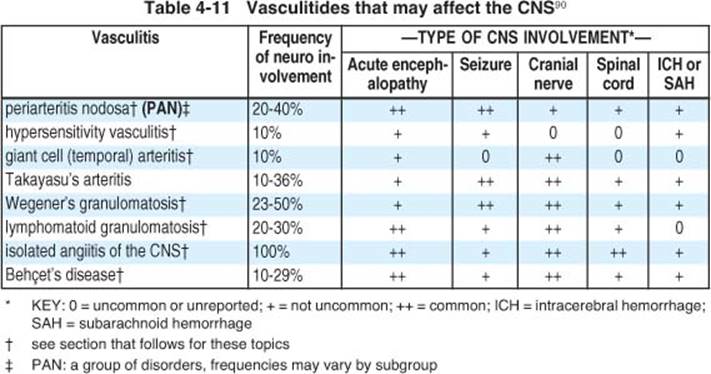
The vasculitides are a group of disorders characterized by inflammation and necrosis of blood vessels. Vasculitis may be primary or secondary. Those that may affect the CNS are listed in Table 4-11, all of these cause tissue ischemia (even after the inflammation is quiescent) that may range in effect from neuropraxia to infarction.
4.10.1. Giant cell arteritis (GCA)
Key concepts:
• formerly often referred to as temporal arteritis
• a chronic vasculitis of large and medium caliber vessels, primarily involving cranial branches of the arteries arising from the aortic arch
• age > 50 years; affects women twice as often as men
• important possible late complications: blindness, stroke, thoracic aortic aneurysms and aortic dissections
• temporal artery biopsy is recommended for all patients suspected of GCA
• corticosteroids are the drug of choice for treatment
AKA temporal arteritis (TA), AKA cranial arteritis. A chronic granulomatous arteritis of unknown etiology involving primarily the cranial branches of the aortic arch (especially the external carotid artery (ECA))91, which if untreated, may lead to blindness. Takayasu’s arteritis is similar to GCA, but tends to affect large arteries in young women; it has 2 phases: inflammatory (treated with corticosteroids) and stenotic (treated with arterial bypasses).
EPIDEMIOLOGY
Seen almost exclusively in Caucasians > 50 yrs age (mean age of onset is 70). Incidence: 17.8 per 100,000 people ≥ 50 years old92 (range: 0.49-23). Prevalence: ≈ 223 (autopsy incidence may be much higher)93. More common in northern latitudes and among individuals of Scandinavian descent91. Female:male ratio is ≈ 2:1 (reported range: 1.05-7.4:1). 50% of GCA patients also have polymyalgia rheumatica (PMR) (see page 77).
PATHOLOGY
Discontinuous (so-called “skip lesions”) inflammatory reaction of lymphocytes, plasma cells, macrophages, ± giant cells (if absent, intimal proliferation may be prominent); predominantly in media of involved arteries. Arteries preferentially involved include the ophthalmic and posterior ciliary branches and the entire distribution of the external carotid system (of which the STA is a terminal branch). Other arteries in the body may be involved (reported involvement of abdominal aorta, femoral, brachial and mesenteric arteries are rarely symptomatic). Unlike PAN, GCA generally spares the renal arteries.
CLINICAL
Various combinations of symptoms of giant cell arteritis are listed in Table 4-12. Onset is usually insidious, although occasionally it may be abrupt95.
Table 4-12 Signs and symptoms of GCA91, 94
|
Frequent (> 50% of cases) |
Occasional (10-50% of cases) |
Rare (< 10% of cases) |
|
H/A: 66% temporal artery tenderness |
visual symptoms weight loss fever (low grade) proximal myalgias jaw claudication facial pain scalp tenderness |
blindness extremity claudication tongue claudication ear pain synovitis stroke angina |
Details of some findings:
1. H/A: the most common presenting symptom. May be nonspecific or located in one or both temporal areas, forehead, or occiput. May be superficial or burning with paroxysmal lancinating pain
2. symptoms relating to ECA blood supply (strongly suggestive of GCA, but not pathognomonic96): jaw claudication, tongue, or pharyngeal muscles
3. ophthalmologic symptoms: due to arteritis and occlusion of branches of ophthalmic artery or posterior ciliary arteries
A. symptoms include: amaurosis fugax (precedes permanent visual loss in 44%), blindness, visual field cuts, diplopia, ptosis, ocular pain, corneal edema, chemosis
B. blindness: incidence is ≈ 7%, and once it occurs, recovery of sight is unlikely
4. systemic symptoms
A. nonspecific constitutional symptoms: fever (may present as FUO in 15% of cases), anorexia, weight loss, fatigue, malaise
B. 30% have neurologic manifestations. 14% are neuropathies including mononeuropathies and peripheral polyneuropathies of the arms or legs97
C. musculoskeletal symptoms
1. PMR is the most common (occurs in 40% of patients): see page 77
2. peripheral arthritis, swelling & pitting edema of hands & feet in 25%
3. arm claudication from stenosis of subclavian and axillary arteries
D. thoracic aortic aneurysms: 17 times as likely in GCA. Annual CXRs are adequate for screening
5. temporal arteries on physical examination may exhibit tenderness, swelling, erythema, reduced pulsations, or nodularity. Normal in 33%
6. the presence of systemic symptoms correlates with a lower incidence of blindness or stroke
Differential diagnosis:
1. periarteritis nodosa (PAN): see page 77
2. hypersensitivity vasculitis
3. atherosclerotic occlusive disease
4. malignancy: symptoms of low grade fever, malaise and weight loss
5. infection
6. trigeminal neuralgia: see page 5517. ophthalmoplegic migraine
8. dental problems
EVALUATION
Laboratory studies
1. ESR > 40 mm/hr (usually > 50) by Westergren method (if > 80 mm/hr with above clinical syndromes, highly suggestive of GCA). ESR is normal in up to 22.5%98
2. C-reactive protein: another acute phase reactant that is more sensitive than ESR. Has the advantage that it can be performed on frozen sera
3. CBC: may show mild normochromic anemia99
4. rheumatoid factor, ANA, and serum complement usually normal
5. LFTs abnormal in 30% (usually elevated alkaline phosphatase)
6. tests for rheumatoid factor and ANA are usually negative
7. temporal artery angiography not helpful (angiography elsewhere indicated if suspicion of large artery involvement exists)
8. CT: usually not helpful, one report described calcified areas corresponding to the temporal arteries100
9. temporal artery biopsy: see below
TEMPORAL ARTERY BIOPSY
Sensitivity and specificity are shown in Table 4-13.
Table 4-13 Temporal artery biopsy
|
sensitivity |
≈ 90% (reported range94, 101 is 9-97%) |
|
specificity |
near 100% |
|
predictive value |
≈ 94% |
Indications and timing
Current recommendations: temporal artery biopsy in all patients suspected of having GCA91.
Preferably, biopsy should be done before treatment is initiated91. However, pathologic changes be seen after more than 2 weeks of therapy102, therefore do not withhold steroids to await biopsy.
Technique of temporal artery biopsy
Biopsy side of involvement if laterality exists. The yield is increased by removing a portion of artery that is involved clinically (a tender or inflamed segment)103. Mark the frontal branch of the STA with a skin marker (spare the main trunk and parietal branch if possible). Infiltrate local anesthetic. The incision is made parallel to the artery and if possible behind the hairline. The incision is taken down to the fascia of the temporalis muscle, to which the STA is superficial104. Optimal length of STA biopsy: 4-6 cm (if an abnormal segment of STA can be palpated, some say that a smaller biopsy to include this area may be sufficient, but this is probably unreliable as the muscle may be tender, etc.). Step-sectioning by pathologist through the entire length of the biopsy specimen also increases the yield.
Frozen sections can be performed. Biopsy of the contralateral side if the first side is negative in cases where clinical suspicion is high increases the yield by 5-10%.
TREATMENT
No known cure. Steroids can produce symptomatic relief and usually prevent blindness (progression of ocular problems 24-48 hrs after institution of adequate steroids is rare). Totally blind patients or those with longstanding partial visual loss are unlikely to respond to any treatment.
1. for most cases:
A. start with prednisone, 40-60 mg/d PO divided BID-QID (qod dosing is usually not effective in initial management) (see page 31 for dosing forms…)
B. if no response after 72 hrs, and diagnosis certain, ↑ to 10-25 mg QID
C. once response occurs (usually within 3-7 days), give entire dose as q AM dose for 3-6 weeks until symptoms resolved and ESR normalizes (occurs in 87% of patients within ≈ 4 weeks) or stabilizes at < 40-50 mm/hr
D. once quiescent, a gradual taper is performed to prevent exacerbations: reduce by 10 mg/d q 2-4 weeks to 40 mg/d, then by 5 mg/d q 2-4 wks to 20 mg/d, then by 2.5 mg/d q 2-4 wks to 5-7.5 mg/d which is maintained for several months, followed by 1 mg/d decrements q 1-3 mos (usual length of treatment is 6-24 mos; do not D/C steroids when ESR normalizes)
E. if symptoms recur during treatment, prednisone dose is temporarily increased until symptoms resolve (isolated rise in ESR is not sufficient reason to increase steroids91)
F. patients should be followed closely for ≈ 2 years
2. in severely ill patients: methylprednisolone, 15-20 mg IV QID
3. anticoagulant therapy: controversial
4. acute blindness (onset within 24-36 hrs) in a patient with giant cell arteritis:
A. consider up to 500 mg methylprednisolone IV over 30-60 mins (no controlled studies show reversal of blindness)
B. some have used intermittent inhalation of 5% carbon dioxide and oxygen
OUTCOME
Complications of steroid therapy occur in ≈ 50% of patients (most are not life threatening, and include vertebral compression fractures in ≈ 36%, peptic ulcer disease in ≈ 12%, proximal myopathy, cataracts, exacerbation of diabetes; also see Possible deleterious side effects of steroids, page 33).
30-50% of patients will have spontaneous exacerbations of GCA (especially during the first 2 years) regardless of the corticosteroid regimen91.
Survival parallels that of the general population. Onset of blindness after initiation of steroid therapy is rare.
4.10.2. Polymyalgia rheumatica (PMR)
PMR and giant cell arteritis (GCA) (see page 74) may be different points on a continuum of the same disease. Both have in increased frequency of HLA-DR4 and systemic monocyte activation. 15% of patients with PMR eventually develop GCA.
Epidemiology91
Both GCA & PMR occur in people ≥ 50 years old. The incidence increases with age and peaks between 70-80 years and is higher at higher latitudes91.
PMR is more common than GCA. Prevalence: 500/100,000)105. Incidence: 52.5 per 100,000 people ≥ age 50, higher in females (61.7) than males (39.9)106.
Features91
• an inflammatory condition of unknown etiology
• clinical characteristics
A. aching and morning stiffness in the cervical region and shoulder & pelvic girdles lasting > 1 month. The pain usually increases with movement
1. shoulder pain: present in 70-95% of patients. Radiates toward elbow
2. hip & neck pain: 50-70%. Hip pain radiates towards knees
B. age ≥ 50 years
C. ESR ≥ 40 mm/hr (7-20% have normal ESR107)
D. usually responds rapidly to low dose corticosteroids (≤ 20 mg prednisone/day) see below
E. systemic symptoms (present in ≈ 33%): fever, malaise or fatigue, anorexia and weight loss
• favorable prognosis: usually remits in 1-3 years
Treatment
PMR responds to either to low doses of steroids105 (10-20 mg prednisone/day) or sometimes to NSAIDs (response to steroids is much more rapid). The initial dose of steroids is maintained for 2-4 weeks, and then by ≤ 10% of the daily dose every 1-2 weeks91 while observing for signs of GCA.
4.10.3. Other vasculitides
PERIARTERITIS NODOSA
AKA polyarteritis nodosa. Actually a group of necrotizing vasculitides, including:
• classic periarteritis nodosa (PAN): a multisystem disease with inflammatory necrosis, thrombosis (occlusion), and hemorrhage of arteries and arterioles in every organ except lung & spleen. Nodules may be palpated along medium sized muscular arteries. Commonly produces mononeuritis multiplex, weight loss, fever, and tachycardia. Peripheral nerve manifestations are attributed to arteritic occlusion of vasa nervorum. CNS manifestations are uncommon and include H/A, seizures, SAH, retinal hemorrhages, and stroke in ≈ 13%
• allergic angiitis and granulomatosis (Churg-Strauss syndrome)
• systemic necrotizing vasculitis
These patients do better when treated with cyclophosphamide rather than steroids.
WEGENER’S GRANULOMATOSIS
A systemic necrotizing granulomatous vasculitis involving the respiratory tract (lung → cough/hemoptysis, and/or nasal airways → serosanguinous nasal drainage ± septal perforation → characteristic “saddle nose deformity”) and frequently the kidneys (no reported cases of kidney involvement without respiratory)108.
Nasal obstruction and crusting are the usual initial findings. Arthralgia (not true arthritis) is present in > 50%.
Neurologic involvement usually consists of cranial nerve dysfunction (usually II, III, IV, & VI; less often V, VII, & VIII; and least commonly IX, X, XI, & XII) and peripheral neuropathies, with diabetes insipidus (occasionally preceding other symptoms by up to 9 months). Focal lesions of the brain and spinal cord occur less frequently.
Differential diagnosis includes:
• “lethal midline granuloma” (may be similar or identical to polymorphic reticulosis) may evolve into lymphoma. May cause fulminant local destruction of the nasal tissue. Differentiation is crucial as this condition is treated by radiation; one should avoid immune suppression (e.g. cyclophosphamide). Probably does not involve true granulomas. Renal and tracheal involvement do not occur
• fungal disease: Sporothrix schenckii & Coccidioides may cause identical syndrome
• other vasculitides: especially Churg-Strauss syndrome (asthma and peripheral eosinophilia usually seen), and PAN (granulomas usually lacking)
LYMPHOMATOID GRANULOMATOSIS
Rare; affects mainly the lungs, skin (erythematous macules or indurated plaques in 40%) and nervous system (CNS in 20%, peripheral neuropathies in 15%). Sinuses, lymph nodes, and spleen are usually spared.
BEHÇET’S SYNDROME
Relapsing ocular lesions and recurrent oral and genital ulcers, with occasional skin lesions, thrombophlebitis, and arthritis 90. H/A occur in > 50%. Neurologic involvement includes pseudotumor, cerebellar ataxia, paraplegia, seizures, and dural sinus thrombosis. Only 5% have neurologic symptoms as the presenting complaint.
86% have CSF pleocytosis and protein elevation. Cerebral angiography is usually normal. CT may show focal areas of enhancing low density.
Steroids usually ameliorate ocular and cerebral symptoms, but usually have no effect on skin and genital lesions. Uncontrolled trials of cytotoxic agents → some benefit. Thalidomide may be effective (uncontrolled studies), but carries risk of serious adverse effects (teratogenicity, peripheral neuropathy…)109.
Although painful, the disease is usually benign. Neurologic involvement portends a worse prognosis.
ISOLATED CNS VASCULITIS
AKA isolated angiitis of the CNS. Rare (≈ 20 cases reported110 as of 1983); limited to vessels of CNS. Small vessel vasculitis is ≈ always present → segmental inflammation and necrosis of small leptomeningeal and parenchymal blood vessels with surrounding tissue ischemia or hemorrhage90.
PRESENTATION
Combinations of H/A, confusion, dementia, and lethargy. Occasionally seizures. Focal and multifocal brain disturbance occurs in > 80%. Visual symptoms are frequent (secondary either to involvement of choroidal and retinal arteries, or to involvement of visual cortex → visual hallucinations).
EVALUATION
ESR & WBC count are usually normal. CSF may be normal or have pleocytosis and/or elevated protein. CT may show enhancing areas of low density.
Angiography (required for diagnosis): characteristically shows multiple areas of symmetrical narrowing (“string of pearls” configuration). If normal, it does not exclude diagnosis.
Histological diagnosis (recommended): all biopsy material should be cultured. Brain parenchyma biopsy infrequently shows vasculitis. Leptomeningeal biopsy invariably shows involvement.
HYPERSENSITIVITY VASCULITIS
Neurologic involvement is not a prominent feature of this group of vasculitides, which include:
• drug induced allergic vasculitis
• cutaneous vasculitis
• serum sickness: may → encephalopathy, seizures, coma, peripheral neuropathy and brachial plexopathy
• Henoch-Schönlein purpura
DRUG INDUCED VASCULITIS
A number of drugs are associated with the development of cerebral vasculitis. These include methamphetamines (“speed”), cocaine (frank vasculitis occurs111 but is rare), heroin and ephedrine.
4.10.4. Fibromuscular dysplasia
A vasculopathy (angiopathy) affecting primarily branches of the aorta, with renal artery involvement in 85% of cases (the most common site) and commonly associated with hypertension. The disease has an incidence of ≈ 1%, and results in multifocal arterial constrictions and intervening regions of aneurysmal dilatation.
The second most commonly involved site is the cervical internal carotid (primarily near C1-2), with fibromuscular dysplasia (FMD) appearing on 1% of carotid angiograms, making FMD the second most common cause of extracranial carotid stenosis112. Bilateral cervical ICA involvement occurs in ≈ 80% of cases. 50% of patients with carotid FMD have renal FMD. Patients with FMD have an increased risk of intracranial aneurysms and neoplasms, and are probably at higher risk of carotid dissection.
Table 4-14 Previous symptoms in 37 cases of aortocranial FMD113
|
Symptom |
% |
|
H/A |
78% |
|
mental distress |
48% |
|
tinnitus |
38% |
|
vertigo |
34% |
|
cardiac arrhythmia |
31% |
|
TIA |
31% |
|
syncope |
31% |
|
carotidynia |
21% |
|
epilepsy |
15% |
|
hearing impairment |
12% |
|
abdominal angina |
8% |
|
angina/MI |
8% |
ETIOLOGY
The actual etiology remains unknown, although congenital defects of the media (muscular layer) and internal elastic layer of the arteries has been identified which may predispose the arteries to injury from otherwise well-tolerated trauma. A high familial rate of strokes, HTN, and migraine have supported the suggestion that FMD is an autosomal dominant trait with reduced penetrance in males113.
ANEURYSMS AND FIBROMUSCULAR DYSPLASIA
The reported incidence of aneurysms with FMD114 ranges from 20-50%.
PRESENTATION
Most patients have recurrent, multiple symptoms shown in Table 4-14.
Up to 50% of patients present with episodes of transient cerebral ischemia or infarction. However, FMD may also be an incidental finding and some cases have been followed for 5 years without recurrence of ischemic symptoms suggesting that FMD may be a relatively benign condition.
Headaches are commonly unilateral and may be mistaken for typical migraine. Syncope may be caused by involvement of the carotid sinus.
Horner’s syndrome occurs in ≈ 8% of cases. T-wave changes on EKG may be seen in up to one third of cases, and may be due to involvement of the coronary arteries.
DIAGNOSIS
The “gold-standard” for the diagnosis of FMD is the angiogram. The three angiographic types of FMD115 are shown in Table 4-15.
TREATMENT
Medical therapy including antiplatelet medication (e.g. aspirin) has been recommended.
Direct surgical treatment is problem ridden due to the difficult location (high carotid artery, near the base of the skull), and the friable nature of the vessels making anastomosis or arteriotomy closure difficult.
Transluminal angioplasty has achieved some degree of success. Carotid cavernous fistulas and arterial rupture have been reported as complications.
Table 4-15 Angiographic classification of FMD
|
Type |
Findings |
|
1 |
most common (80-100% of reported cases). Multiple, irregularly spaced, concentric narrowings with normal or dilated intervening segments giving rise to the so-called “string of pearls” appearance. Corresponds with arterial medial fibroplasia |
|
2 |
focal tubular stenosis, seen in ≈ 7% of cases. Less characteristic for FMD than Type 1, and may also be seen in Takayasu’s arteritis and other conditions |
|
3 |
“atypical FMD”. Rare. May take on various appearances, most commonly consisting of diverticular outpouchings of one wall of the artery |
4.10.5. Miscellaneous vasculopathies
CADASIL
Key concepts:
• clinical: migraines, dementia, TIAs, psychiatric disturbances
• MRI: white matter abnormalities
• autosomal dominant inheritance
• anticoagulants controversial, generally discouraged
An acronym for Cerebral Autosomal Dominant Arteriopathy with Subcortical Infarcts and Leukoencephalopathy116. A familial disease with onset in early adulthood (mean age at onset: 45 ± 11 yrs), mapped to chromosome 19. Clinical and neuroradiologic features are similar to those seen with multiple subcortical infarcts from HTN, except there is no evidence of HTN. The vasculopathy is distinct from that seen in lipohyalinosis, arteriosclerosis and amyloid angiopathy, and causes thickening of the media of leptomeningeal and perforating arteries measuring 100-400 μm in diameter.
Clinical involvement: recurrent subcortical infarcts (84%), progressive or stepwise dementia (31%), migraine with aura (22%), and depression (20%). All symptomatic and 18% of asymptomatic patients had prominent subcortical white-matter and basal ganglia hyperintensities on T2WI MRI.
Treatment: Coumadin® is used by some.
4.11. References
1. Consensus Conference: Differential diagnosis of dementing diseases. JAMA 258: 3411-6, 1987.
2. Fleming K C, Adams A C, Petersen R C: Dementia: Diagnosis and evaluation. Mayo Clin Proc 70: 1093-107, 1995.
3. Lipowski Z J: Delerium (acute confusional states). JAMA 258: 1789-92, 1987.
4. Pompei P, Foreman M, Rudberg M A, et al.: Delerium in hospitalized older persons: Outcomes and predictors. J Am Geriatr Soc 42: 809-15, 1994.
5. Petersen R C: Acute confusional state: Don’t mistake it for dementia. Postgrad Med 92: 141-8, 1992.
6. Hulette C M, Earl N L, Crain B J: Evaluation of cerebral biopsies for the diagnosis of dementia. Arch Neurol 49: 28-31, 1992.
7. Javedan S P, Tamargo R J: Diagnostic yield of brain biopsy in neurodegenerative disorders. Neurosurgery 41: 823-30, 1997.
8. Groves R, Moller J: The value of the cerebral cortical biopsy. Acta Neurol Scand 42: 477-82, 1966.
9. Forsyth P A, Posner J B: Headaches in patients with brain tumors: A study of 111 patients. Neurology 43: 1678-83, 1993.
10. Welch K M A, Levine S R: Migraine-related stroke in the context of the international headache society classification of head pain. Arch Neurol 47: 458-62, 1990.
11. Lance J W: Treatment of migraine. Lancet 339: 1207-9, 1992.
12. Kittrelle J P, Grouse D S, Seybold M E: Cluster headache: Local anesthetic abortive agents. Arch Neurol 42: 496-8, 1985.
13. Sanders M, Zuurmond W W A: Efficacy of spheno-palatine ganglion blockade in 66 patients suffering from cluster headache: A 12- to 70-month follow-up evaluation. J Neurosurg 87: 876-80, 1997.
14. Burns B, Watkins L, Goadsby P J: Treatment of medically intractable cluster headache by occipital nerve stimulation: Long-term follow-up of 8 patients. Lancet 369 (9567): 1099-106, 2007.
15. Lapkin M L, Golden G S: Basilar artery migraine: A review of 30 cases. Am J Dis Child 132: 278-81, 1978.
16. DiGiovanni A J, Dunbar B S: Epidural injections of autologous blood for postlumbar-puncture headache. Anesth and Analg 49: 268-71, 1970.
17. Seebacher J, Ribeiro V, Le Guillou J L, et al.: Epidural blood patch in the treatment of post dural puncture headache: A double blind study. Headache 29: 630-2, 1989.
18. Lance J W, Branch G B: Persistent headache after lumbar puncture. Lancet 343: 414, 1994 (letter).
19. Gass H, Goldstein A S, Ruskin R, et al.: Chronic postmyelogram headache. Arch Neurol 25: 168-170, 1971.
20. Sechzer P H, Abel L: Post-spinal anesthesia headache treated with caffeine: Evaluation with demand method. Part 1. Cur Ther Res 24: 307-12, 1978.
21. Murros K, Fogelholm R: Spontaneous intracranial hypotension with slit ventricles. J Neurol Neurosurg Psychiatry 46: 1149-51, 1983.
22. Mitchell S L, Kiely D K, Kiel D P, et al.: The epidemiology, clinical characteristics, and natural history of older nursing home residents with a diagnosis of Parkinson’s disease. J Am Geriatr Soc 44: 394-9, 1996.
23. Koller W C, Silver D E, Lieberman A: An algorithm for the management of Parkinson’s disease. Neurology 44: S5-52, 1994.
24. Young R: Update on Parkinson’s disease. Am Fam Physician 59: 2155-67, 1999.
25. Kondziolka D, Bonaroti E A, Lunsford L D: Pallidotomy for Parkinson’s disease. Contemp Neurosurg 18 (6): 1-6, 1996.
26. Davis G C, Williams A C, Markey S P, et al.: Chronic parkinsonism secondary to intravenous injection of meperidine analogues. Psychiatry Res 1: 249-54, 1979.
27. Langston J W, Ballard P, Tetrud J W, et al.: Chronic parkinsonism in humans due to a product of meperidine-analog synthesis. Science 219: 979-80, 1983.
28. Lang A E, Lozano A M: Parkinson’s disease. First of two parts. N Engl J Med 339: 1044-53, 1998.
29. Kristensen M O: Progressive supranuclear palsy - 10 years later. Acta Neurol Scand 71: 177-89, 1985.
30. Gildenberg P L: Whatever happened to stereotactic surgery? Neurosurgery 20: 983-7, 1987.
31. Lublin F D, Reingold S C: Defining the clinical course of multiple sclerosis: Results of an international survey. Neurology 46: 907-11, 1996.
32. Rudick R A, Cohen J A, Weinstock-Guttman B, et al.: Management of multiple sclerosis. N Engl J Med 22: 1604-11, 1997.
33. Rowland L P, (ed.) Merritt’s textbook of neurology. 8th ed., Lea and Febiger, Philadelphia, 1989.
34. Jensen T S, Rasmussen P, Reske-Nielsen E: Association of trigeminal neuralgia with multiple sclerosis. Arch Neurol 65: 182-9, 1982.
35. Filippi M, Horsfield M A, Morrissey S P, et al.: Quantitative brain MRI lesion load predicts the course of clinically isolated syndromes suggestive of multiple sclerosis. Neurology 44: 635-41, 1994.
36. McDonald W I, Compston A, Edan G, et al.: Recommended diagnostic criteria for multiple sclerosis: Guidelines from the international panel on the diagnosis of multiple sclerosis. Ann Neurol 50: 121-7, 2001.
37. Polman C H, Reingold S C, Edan G, et al.: Diagnostic criteria for multiple sclerosis: 2005 revisions to the “mcdonald criteria”. Ann Neurol 58 (6): 840-6, 2005.
38. Poser C M, Paty D W, Scheinberg L, et al.: New diagnostic criteria for multiple sclerosis: Guidelines for research protocols. Ann Neurol 13: 227-31, 1983.
39. Swanson J W: Multiple sclerosis: Update in diagnosis and review of prognostic factors. Mayo Clin Proc 64: 577-86, 1989.
40. Barkhof F, Filippi M, Miller D H, et al.: Comparison of MR imaging criteria at first presentation to predict conversion to clinically definite multiple sclerosis. Brain 120: 2059-69, 1997.
41. Tintore M, Rovira A, Martinez M, et al.: Isolated demyelinating syndromes: Comparison of different MR imaging criteria to predict conversion to clinically definite multiple sclerosis. AJNR 21: 702-6, 2000.
42. Stewart J M, Houser O W, Baker H L, et al.: Magnetic resonance imaging and clinical relationships in multiple sclerosis. Mayo Clin Proc 62: 174-84, 1987.
43. Mushlin A I, Detsky A S, Phelps C E, et al.: The accuracy of magnetic resonance imaging in patients with suspected multiple sclerosis. JAMA 269: 3146-51, 1993.
44. Kidd C, Thorpe J W, Thompson A J, et al.: Spinal cord imaging MRI using multi-array coils and fast spin echo. II. Findings in multiple sclerosis. Neurology 43: 2632-7, 1993.
45. Kent D L, Larson E B: Magnetic resonance imaging of the brain and spine. Ann Intern Med 108: 402-24, 1988.
46. Kepes J J: Large focal tumor-like demyelinating lesions of the brain: Intermediate entity between multiple sclerosis and acute disseminated encephalomyelitis? A study of 31 patients. Ann Neurol 33 (1): 18-27, 1993.
47. Law M, Meltzer D E, Cha S: Spectroscopic magnetic resonance imaging of a tumefactive demyelinating lesion. Neuroradiology 44 (12): 986-9, 2002.
48. Brex F A, Miszkiel K A, O’Riordan J I, et al.: Asessing the risk of early MS in patients with clinically isolated syndromes: The role of follow-up MRI. J Neurol Neurosurg Psychiatry 70: 390-3, 2001.
49. Freedman M S, Thompson E J, Deisenhammer D, et al.: Recommended standard of cerebrospinal fluid analysis in the diagnosis of multiple sclerosis: A consensus statement. Arch Neurol 62: 865-70, 2005.
50. Rowland L P: Diagnosis of amyotrophic lateral sclerosis. J Neurol Sci 160: S6-24, 1998.
51. Peavy G M, Herzog A G, Rubin N P, et al.: Neuropsychological aspects of dementia of motor neuron disease: A report of two cases. Neurology 42: 1004-8, 1992.
52. Bensimon G, Lacomblez L, Meininger V, et al.: A controlled trial of riluzole in amyotrophic lateral sclerosis. N Engl J Med 24: 585-91, 1994.
53. Riluzole for amyotrophic lateral sclerosis. Med Letter 37: 113-4, 1995.
54. Lacomblez L, Bensimon G, Guillet P, et al.: Riluzole: A double-blind randomized placebo-controlled dose-range study in amyotrophic lateral sclerosis (ALS). Electroenceph Clin Neurophysiol 97: S68, 1995 (abstract).
55. Ropper A H, Shahani B T: Pain in Guillain-Barre syndrome. Arch Neurol 41 (5): 511-4, 1984.
56. Asbury A K, Arnaso B G W, Karp H R, et al.: Criteria for diagnosis of Guillain-Barre syndrome. Ann Neurol 3: 565-6, 1978.
57. Scott A J, Duncan R, Henderson L, et al.: Acute rhabdomyolysis associated with atypical Guillain-Barre syndrome. Postgrad Med J 67 (783): 73-4, 1991.
58. Mendell J R: Chronic inflammatory demyelinating polyradiculoneuropathy. Annu Rev Med 44: 211-9, 1993.
59. Mahattanakul W, Crawford T O, Griffin J W, et al.: Treatment of chronic inflammatory demyelinating polyneuropathy with cyclosporin-a. J Neurol Neurosurg Psychiatry 60: 185-7, 1996.
60. Gorson K C, Ropper A H, Clark B D, et al.: Treatment of chronic inflammatory demyelinating polyneuropathy with interferon-α 2a. Neurology 50: 84-7, 1998.
61. Gorson K C, Ropper A H, Muriello M A, et al.: Prospective evaluation of MRI lumbosacral nerve root enhancement in acute Guillain-Barre syndrome. Neurology 47 (3): 813-7, 1996.
62. Guillain-Barré Syndrome Steroid Trial Group: Double-blind trial of intravenous methylprednisolone in Guillain-Barré syndrome. Lancet 341: 586-90, 1993.
63. Kincaid J C, Dyken M L: Myelitis and myelopathy. In Clinical neurology, Baker A B and Joynt R J, (eds.). Harper and Row, Hagerstown, 1984: pp 1-32.
64. Altrocchi P H: Acute transverse myelopathy. Arch Neurol 9: 111-9, 1963.
65. Eguro H: Transverse myelitis following chemonucleolysis: Report of a case. J Bone Joint Surg 65A: 1328-9, 1983.
66. Lipton H L, Teasdall R D: Acute transverse myelopathy in adults: A follow-up study. Arch Neurol 28: 252-7, 1973.
67. Ropper A H, Poskanzer D C: The prognosis of acute and subacute transverse myelopathy based on early signs and symptoms. Ann Neurol 4: 51-9, 1978.
68. Berman M, Feldman S, Alter M, et al.: Acute transverse myelitis: Incidence and etiologic considerations. Neurology 31: 966-71, 1981.
69. Merine D, Wang H, Kumar A J, et al.: CT myelography and MRI of acute transverse myelitis. J Comput Assist Tomogr 11: 606-8, 1987.
70. Dowling P C, Bosch V V, Cook S D: Possible beneficial effect of high-dose IV steroid therapy in acute demyelinating disease and transverse myelitis. Neurology 30: 33-6, 1980.
71. Stern B J, Krumholz A, Johns C, et al.: Sarcoidosis and its neurological manifestations. Arch Neurol 42: 909-17, 1985.
72. Oksanen V: Neurosarcoidosis: Clinical presentation and course in 50 patients. Acta Neurol Scand 73: 283-90, 1986.
73. de Tribolet N, Zander E: Intracranial sarcoidosis presenting angiographically as a subdural hematoma. Surg Neurol 9: 169-71, 1978.
74. Saleh S, Saw C, Marzouk K, et al.: Sarcoidosis of the spinal cord: Literature review and report of eight cases. J Natl Med Assoc 98 (6): J Natl Med Assoc: 965-76, 2006.
75. Rohrbach M S, DeRemee R A: Pulmonary sarcoidosis and serum angiotensin-converting enzyme. Mayo Clin Proc 57: 64-6, 1982.
76. Oksanen V: New cerebrospinal fluid, neurophysio-logical and neuroradiological examinations in the diagnosis and follow-up of neurosarcoidosis. Sarcoidosis 4: 105-10, 1987.
77. Kurdziel K A: The panda sign. Radiology 215: 884-5, 2000.
78. Sulavik S B, Spencer R P, Weed D A, et al.: Recognition of distinctive patterns of gallium-67 distribution in sarcoidosis. J Nucl Med 31 (12): 1909-14, 1990.
79. Fayad F, Duet M, Orcel P, et al.: Systemic sarcoidosis: The “leopardman” sign. Joint Bone Spine 73 (1): 109-12, 2006.
80. Stern B J, Schonfeld S A, Sewell C, et al.: The treatment of neurosarcoidosis with cyclosporine. Arch Neurol 49: 1065-72, 1992.
81. Bartynski W S: Posterior reversible encephalopathy syndrome, part 1: Fundamental imaging and clinical features. AJNR Am J Neuroradiol 29 (6): 1036-42, 2008.
82. Port J D, Beauchamp N J: Reversible intracerebral pathologic entities mediated by vascular autoregulatory dysfunction. Radiographics 18: 353-67, 1998.
83. Lin K L, Hsu W C, Wang H S, et al.: Hypertension-induced cerebellar encephalopathy and hydrocephalus in a male. Pediatr Neurol 34 (1): Pediatr Neurol: 72-5, 2006.
84. Schaefer P W, Buonanno F S, Gonzalez R G, et al.: Diffusion-weighted imaging discriminates between cytotoxic and vasogenic edema in a patient with eclampsia. Stroke 28: 1082-5, 1997.
85. Covarrubias D J, Luetmer P H, Campeau N G: Posterior reversible encephalopathy syndrome: Prognostic utility of quantitative diffusion-weighted MR images. AJNR Am J Neuroradiol 23 (6): AJNR Am J Neuroradiol: 1038-48, 2002.
86. Beeson J H, Duda E E: Computed axial tomography scan demonstration of cerebral edema in eclampsia preceded by blindness. Obstet Gynecol 60: 529-32, 1982.
87. Raps E C, Galetta S L, Broderick M, et al.: Delayed peripartum vasculopathy: Cerebral eclampsia revisited. Ann Neurol 33: 222-5, 1993.
88. Dekker G A, Sibai B M: Etiology and pathogenesis of preeclampsia: Current concepts. Am J Obstet Gynecol 179 (5): Am J Obstet Gynecol: 1359-75, 1998.
89. Bartynski W S, Boardman J F, Zeigler Z R, et al.: Posterior reversible encephalopathy syndrome in infection, sepsis, and shock. AJNR Am J Neuroradiol 27 (10): AJNR Am J Neuroradiol: 2179-90, 2006.
90. Moore P M, Cupps T R: Neurologic complications of vasculitis. Ann Neurol 14: 155-67, 1983.
91. Salvarani C, Cantini F, Boiardi L, et al.: Polymyalgia rheumatica and giant-cell arteritis. N Engl J Med 347: 261-71, 2002.
92. Salvarani C, Gabriel S E, O’Fallon W M, et al.: The incidence of giant cell arteritis in Olmstead county, Minnesota: Apparent fluctuations in a cyclic pattern. Ann Intern Med 123: 192-4, 1995.
93. Machado E B, Michet C J, Ballard D J, et al.: Trends in incidence and clinical presentation of temporal arteritis in Olmstead county, Minnesota, 1950-1985. Arthritis Rheum 31: 745-9, 1988.
94. Allen N B, Studenski S A: Polymyalgia rheumatica and temporal arteritis. Med Clin N Amer 70: 369-84, 1986.
95. Hunder G G: Giant cell (temporal) arteritis. Rheum Dis Clin N Amer 16: 399-409, 1990.
96. Hall S, Hunder G G: Is temporal artery biopsy prudent? Mayo Clin Proc 59: 793-6, 1984.
97. Caselli R J, Danube J R, Hunder G G, et al.: Peripheral neuropathic syndromes in giant cell (temporal) arteritis. Neurology 38: 685-9, 1988.
98. Salvarani C, Hunder G G: Giant cell arteritis with low erythrocyte sedimentation rate: Frequency of occurrence in a population-based study. Arthritis Rheum 45: 140-5, 2001.
99. Baumel B, Eisner L S: Diagnosis and treatment of headache in the elderly. Med Clin N Amer 75: 661-75, 1991.
100. Karacostas D, Taskos N, Nikolaides T: CT findings in temporal arteritis: A report of two cases. Neurorad 28: 373, 1986.
101. McDonnell P J, Moore G W, Miller N R, et al.: Temporal arteritis: A clinicopathologic study. Ophthalmology 93: 518-30, 1986.
102. Achkar A A, Lie J T, Hunder G G, et al.: How does previous corticosteroid treatment affect the biopsy findings in giant cell (temporal) arteritis? Ann Intern Med 120: 987-92, 1994.
103. Hunder G G: Giant cell arteritis and polymyalgia rheumatica. In Textbook of rheumatology, Kelley W N, Harris E D, Ruddy S, et al., (eds.). W. B. Saunders, Philadelphia, 4th ed., 1993, Vol. 2: pp 1103-12.
104. Kent R B, Thomas L: Temporal artery biopsy. Am Surg 56: 16-21, 1989.
105. Chuang T Y, Hunder G G, Ilstrup D M, et al.: Polymyalgia rheumatica: A 10-year epidemiologic and clinical study. Ann Intern Med 97: 672-80, 1982.
106. Salvarani C, Gabriel S E, O’Fallon W M, et al.: Epidemiology of polymyalgia rheumatica in Olmstead county, Minnesota, 1970-1991. Arthritis Rheum 38: 369-73, 1995.
107. Cantini F, Salvarani C, Olivieri I, et al.: Erythrocyte sedimentation rate and C-reactive protein in the evaluation of disease activity and severity in polymyalgia rheumatica: A prospective follow-up study. Semin Arthritis Rheum 30: 17-24, 2000.
108. McDonald T J, DeRemee R A: Wegener’s granulomatosis. Laryngoscope 93: 220-31, 1983.
109. New uses of thalidomide. Med Letter 38: 15-6, 1996.
110. Cupps T R, Moore P M, Fauci A S: Isolated angitis of the central nervous system: Prospective diagnostic and therapeutic experience. Am J Med 74: 97-105, 1983.
111. Kaye B R, Fainstat M: Cerebral vasculitis associated with cocaine abuse. JAMA 258: 2104-6, 1987.
112. Hasso A N, Bird C R, Zinke D E, et al.: Fibromuscular dysplasia of the internal carotid artery: Percutaneous transluminal angioplasty. AJR 136: 955-60, 1981.
113. Mettinger K L, Ericson K: Fibromuscular dysplasia and the brain: Observations on angiographic, clinical, and genetic characteristics. Stroke 13: 46-52, 1982.
114. Mettinger K L: Fibromuscular dysplasia and the brain II: Current concept of the disease. Stroke 13: 53-8, 1982.
115. Osborn A G, Anderson R E: Angiographic spectrum of cervical and intracranial fibromuscular dysplasia. Stroke 8: 617-26, 1977.
116. Chabriat H, Vahedi K, Iba-Zizen M T, et al.: Clinical spectrum of CADASIL: A study of seven families. Lancet 346: 934-9, 1995