
5.1. Surface anatomy
5.1.1. Cortical surface anatomy
Lateral cortical surface (Figure 5-1)
For abbreviations, see Table 5-2 and Table 5-1. The MFG is usually more sinuous than the IFG or SFG, and it often connects to the pre-central gyrus via a thin isthmus1. The central sulcus joins the Sylvian fissure in only 2% of cases (i.e. in 98% of cases there is a “subcentral” gyrus). The intraparietal sulcus (ips) separates the superior and inferior parietal lobules. The IPL is composed primarily of the AG and SMG. The Sylvian fissure terminates in the SMG (Brodmann’s area 40). The superior temporal sulcus terminates in the AG.
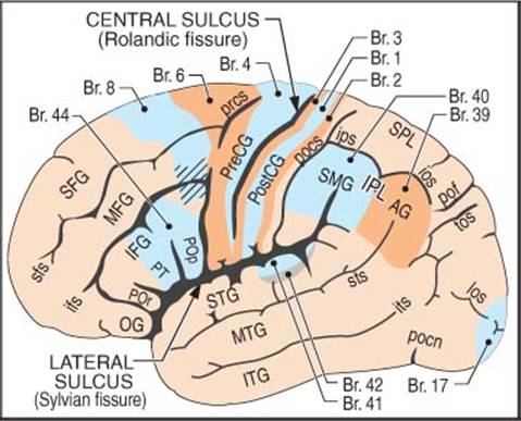
Figure 5-1 Left lateral cerebral cortical surface anatomy*
* Br. = Brodmann’s area (shaded). See Table 5-1 and Table 5-2 for abbreviations (lowercase = sulci, UPPERCASE = gyri)
Brodmann’s areas
Figure 5-1 also identifies the clinically significant areas of Brodmann’s (Br.) map of the cytoarchitectonic fields of the human brain. Functional significance of these areas is as follows:
• Br. areas 3,1,2: primary somatosensory cortex
• Br. areas 41 & 42: primary auditory areas (transverse gyri of Heschl)
• Br. area 4: precentral gyrus, primary motor cortex (AKA “motor strip”). Large concentration of giant pyramidal cells of Betz
• Br. area 6: premotor area or supplemental motor area. Immediately anterior to motor strip, it plays a role in contralateral motor programming
• Br. area 44: (dominant hemisphere) Broca’s area (motor speech)A
• Br. area 17: primary visual cortex
• Wernicke’s area (language)A: in the dominant hemisphere, most of Br. area 40 and a portion of Br. area 39 (may also include ≈ posterior third of STG)
• the striped portion of Br. area 8 in Figure 5-1 (frontal eye field) initiates voluntary eye movements to the opposite direction
A. language function cannot be reliably localized on anatomic grounds due to individual variability in its exact location; in order to perform maximal brain resections with minimal risk of aphasia, techniques such as intraoperative brain mapping2 or looking for phase reversal on intraoperative cortical SSEP3should be employed (see page 150)
Medial surface
(Figure 5-2)
The cingulate sulcus terminates posteriorly in the pars marginalis (pM) (plural: partes marginales). On axial imaging, the pMs: are visible on 95% of CTs and 91% of MRIs4, are usually the most prominent of the paired grooves straddling the midline, and they extend a greater distance into the hemispheres4. On axial CT, the pM is located slightly posterior to the widest biparietal diameter4; on the typically more horizontally oriented MRI slices the pM assumes a more posterior position. The pMs curve posteriorly in lower slices and anteriorly in higher slices (here, the paired pMs form the “pars bracket” - a characteristic “handlebar” configuration straddling the mid-line).
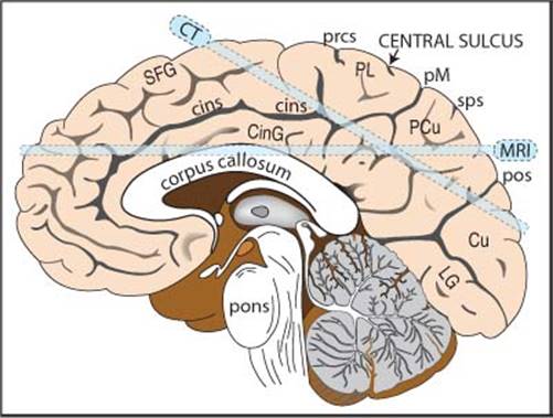
Figure 5-2 Medial aspect of the right hemisphere “CT” & “MRI” bars depict typical axial slice orientation for CT & MRI scans. See Table 5-1 and Table 5-2 for abbreviations
Central sulcus on axial imaging
See Figure 5-3. Identification is important to localize the motor strip (contained in the PreCG). The central sulcus (CS) is visible on 93% of CTs and 100% of MRIs4. It curves posteriorly as it approaches the interhemispheric fissure (IHF), and often terminates in the paracentral lobule, just anterior to the pars marginalis (pM) within the pars bracket (see above)4 (i.e. the CS often does not reach the midline).
Pointers:
• parieto-occipital sulcus (pos) (or fissure): more prominent over the medial surface, and on axial imaging is longer, more complex, and more posterior than the pars marginalis5
• post-central sulcus (pocs): usually bifurcates and forms an arc or parenthesis (“lazy-Y”) cupping the pM. The anterior limb does not enter the pM-bracket and the posterior limb curves behind the pM to enter the IHF

Figure 5-3 CT scan (upper cut) showing gyri/sulci. See Table 5-1 and Table 5-2 for abbreviations
Table 5-1 Cerebral sulci (abbreviations)
|
cins |
cingulate sulcus |
|
cs |
central sulcus |
|
ips-ios |
intraparietal-intraoccipital sulcus |
|
los |
lateral occipital sulcus |
|
pM |
pars marginalis |
|
pocn |
pre-occipital notch |
|
pocs |
post-central sulcus |
|
pof |
parieto-occipital fissure |
|
pos |
parieto-occipital sulcus |
|
prcs |
pre-central sulcus |
|
sfs, ifs |
superior, inferior frontal sulcus |
|
sps |
superior parietal sulcus |
|
sts, its |
superior, inferior temporal sulcus |
|
tos |
trans occipital sulcus |
Table 5-2 Cerebral gyri and lobules (abbreviations)
|
AG |
angular gyrus |
|
CinG |
cingulate gyrus |
|
Cu |
cuneus |
|
LG |
lingual gyrus |
|
MFG, SFG |
middle & superior frontal gyrus |
|
OG |
orbital gyrus |
|
PCu |
precuneous |
|
PreCG, PostCG |
pre- and post-central gyrus |
|
PL |
paracentral lobule (upper SFG and PreCG and PostCG) |
|
IFG POp PT POr |
inferior frontal gyrus pars opercularis pars triangularis pars orbitalis |
|
STG, MTG, ITG |
superior, middle & inferior temporal gyrus |
|
SPL, IPL |
superior & inferior parietal lobule |
|
SMG |
supramarginal gyrus |
5.1.2. Surface anatomy of the cranium
CRANIOMETRIC POINTS
See Figure 5-4.
Pterion: region where the following bones are approximated: frontal, parietal, temporal and sphenoid (greater wing). Estimated as 2 fingerbreadths above the zygomatic arch, and a thumb’s breadth behind the frontal process of the zygomatic bone (blue circle in Figure 5-4).
Asterion: junction of lambdoid, occipitomastoid and parietomastoid sutures. Usually lies within a few millimeters of the posterior-inferior edge of the junction of the transverse and sigmoid sinuses (not always reliable6 - may overlie either sinus).
Vertex: the top-most point of the skull.
Lambda: junction of the lambdoid and sagittal sutures.
Stephanion: junction of coronal suture and superior temporal line.
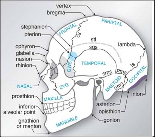
Figure 5-4 Craniometric points & cranial sutures.
Named bones appear in all upper case letters.
Abbreviations: GWS = greater wing of sphenoid bone, NAS = nasal bone, stl = superior temporal line, ZYG = zygomatic.
Sutures: cs = coronal, ls = lambdoid, oms = occipitomastoid, pms = parietomastoid, sms = squamomastoid, sqs = squamosal
Glabella: the most forward projecting point of the forehead at the level of the supraorbital ridge in the midline.
Opisthion: the posterior margin of the foramen magnum in the midline.
Bregma: the junction of the coronal and sagittal sutures.
Sagittal suture: midline suture from coronal suture to lambdoid suture. Although often assumed to overlie the superior sagittal sinus (SSS), the SSS lies to the right of the sagittal suture in the majority of specimens7 (but never by > 11 mm).
The most anterior mastoid point lies just in front of the sigmoid sinus8.
RELATION OF SKULL MARKINGS TO CEREBRAL ANATOMY
Taylor-Haughton lines
Taylor-Haughton (T-H) lines can be constructed on an angiogram, CT scout film, or skull x-ray, and can then be re-constructed on the patient in the O.R. based on visible external landmarks9. T-H lines are shown as dashed lines in Figure 5-5.
1. Frankfurt plane, AKA baseline: line from inferior margin of orbit through the upper margin of the external auditory meatus (EAM) (as distinguished from Reid’s base line: from inferior orbital margin through the center of the EAM)10 (p 313)
2. the distance from the nasion to the inion is measured across the top of the calvaria and is divided into quarters (can be done simply with a piece of tape which is then folded in half twice)
3. posterior ear line: perpendicular to the baseline through the mastoid process
4. condylar line: perpendicular to the baseline through the mandibular condyle
5. T-H lines can then be used to approximate the sylvian fissure (see below) and the motor cortex (also see below)

Figure 5-5 Taylor-Haughton lines and other localizing methods
Sylvian fissure AKA lateral fissure
Approximated by a line connecting the lateral canthus to the point 3/4 of the way posterior along the arc running over convexity from nasion to inion (T-H lines).
Angular gyrus
Located just above the pinna, important on the dominant hemisphere as part of Wernicke’s area. Note: there is significant individual variability in the location2.
Angular artery
Located 6 cm above the EAM.
Motor cortex
Numerous methods utilize external landmarks to locate the motor strip (pre-central gyrus) or the central sulcus (Rolandic fissure) which separates motor strip anteriorly from primary sensory cortex posteriorly. These are just approximations since individual variability causes the motor strip to lie anywhere from 4 to 5.4 cm behind the coronal suture11. The central sulcus cannot even be reliably identified visually at surgery12.
• method 1: the superior aspect of the motor cortex is almost straight up from the EAM near the midline
• method 213: the central sulcus is approximated by connecting:
A. the point 2 cm posterior to the midposition of the arc extending from nasion to inion (illustrated in Figure 5-5), to
B. the point 5 cm straight up from the EAM
• method 3: using T-H lines, the central sulcus is approximated by connecting:
A. the point where the “posterior ear line” intersects the circumference of the skull (see Figure 5-5) (usually about 1 cm behind the vertex, and 3-4 cm behind the coronal suture), to
B. the point where the “condylar line” intersects the line representing the sylvian fissure
• method 4: a line drawn 45° to Reid’s base line starting at the pterion points in the direction of the motor strip14 (p 584-5)
RELATIONSHIP OF VENTRICLES TO SKULL
Figure 5-6 shows the relationship of non-hydrocephalic ventricles to the skull in the lateral view. Some dimensions of interest are shown in Table 5-315.
In the non-hydrocephalic adult, the lateral ventricles lie 4-5 cm below the outer skull surface. The center of the body of the lateral ventricle sits in the midpupillary line, and the frontal horn is intersected by a line passing perpendicular to the calvaria along this line16. The anterior horns extend 1-2 cm anterior to the coronal suture.
Average length of third ventricle ≈ 2.8 cm.

Figure 5-6 Relationship of ventricles to skull landmarks*
* Abbreviations: (F = frontal horn, B = body, A = atrium, O = occipital horn, T = temporal horn) of lateral ventricle. FM = foramen of Monro. Aq = sylvian aqueduct. V3 = third ventricle. V4 = fourth ventricle. cs = coronal suture. Dimensions D1-4 → see Table 5-3

5.1.3. Surface landmarks of spine levels
Estimates of cervical levels for anterior cervical spine surgery may be made using the landmarks shown in Table 5-4. Intraoperative C-spine x-rays are essential to verify these estimates.
The scapular spine is located at about T2-3.
The inferior scapular pole is ≈ T6 posteriorly.
Intercristal line: a line drawn between the highest point of the iliac crests across the back will cross the midline either at the interspace between the L4 and L5 spinous processes, or at the L4 spinous process itself.
Table 5-4 Cervical levels17
|
Level |
Landmark |
|
C1-2 |
angle of mandible |
|
C3-4 |
1 cm above thyroid cartilage (≈ hyoid bone) |
|
C4-5 |
level of thyroid cartilage |
|
C5-6 |
crico-thyroid membrane |
|
C6 |
carotid tubercle |
|
C6-7 |
cricoid cartilage |
5.2. Cranial foramina & their contents
Table 5-5 Cranial foramina and their contents*
|
Foramen |
Contents |
|
nasal slits |
anterior ethmoidal nn., a. & v |
|
superior orbital fissure |
Cr. Nn. III, IV, VI, all 3 branches of V1 (ophthalmic division divides into nasociliary, frontal, and lacrimal nerves); superior ophthalmic vv.; recurrent meningeal br. from lacrimal a.; orbital branch of middle meningeal a.; sympathetic filaments from ICA plexus |
|
inferior orbital fissure |
Cr. N. V-2 (maxillary div.), zygomatic n.; filaments from pterygopalatine branch of maxillary n.; infraorbital a. & v.; v. between inferior ophthalmic v. & pterygoid venous plexus |
|
foramen lacerum |
usually nothing (ICA traverses the upper portion but doesn’t enter, 30% have vidian a.) |
|
carotid canal |
internal carotid a., ascending sympathetic nerves |
|
incisive foramen |
descending septal a.; nasopalatine nn. |
|
greater palatine foramen |
greater palatine n., a., & v. |
|
lesser palatine foramen |
lesser palatine nn. |
|
internal acoustic meatus |
Cr. N. VII (facial); Cr. N. VIII (stato-acoustic) - (see text & Figure 5-7 below) |
|
hypoglossal canal |
Cr. N. XII (hypoglossal); a meningeal branch of the ascending pharyngeal a. |
|
foramen magnum |
spinal cord (medulla oblongata); Cr. N. XI (spinal accessory nn.) entering the skull; vertebral aa.; anterior & posterior spinal arteries |
|
foramen cecum |
occasional small vein |
|
cribriform plate |
olfactory nn. |
|
optic canal |
Cr. N. II (optic); ophthalmic a. |
|
foramen rotundum |
Cr. N. V2 (maxillary div.), a. of foramen rotundum |
|
foramen ovale |
Cr. N. V3 (mandibular div.) + portio minor (motor for CrN V) |
|
foramen spinosum |
middle meningeal a. & v. |
|
jugular foramen |
internal jugular v. (beginning); Cr. Nn. IX, X, XI |
|
stylomastoid foramen |
Cr. N. VII (facial); stylomastoid a. |
|
condyloid foramen |
v. from transverse sinus |
|
mastoid foramen |
v. to mastoid sinus; branch of occipital a. to dura mater |
* Abbreviations: a. = artery, aa. = arteries, v. = vein, vv. = veins, n. = nerve, nn. = nerves, br. = branch, Cr. N. = cranial nerve, fmn. = foramen, div. = division
Porus acusticus
AKA internal auditory canal (see Figure 5-7)
The filaments of the acoustic portion of VIII penetrate tiny openings of the lamina cribrosa of the cochlear area18.
Transverse crest: separates superior vestibular area and facial canal (above) from the inferior vestibular area and cochlear area (below)18.
Vertical crest (AKA Bill’s bar): separates the meatus to facial canal anteriorly (containing VII and nervus intermedius) from the vestibular area posteriorly (containing the superior division of vestibular nerve).
The “5 nerves” of the IAC:
1. facial nerve (VII) (mnemonic: “7-up” as VII is in superior portion)
2. nervus intermedius: the somatic sensory branch of the facial nerve primarily innervating mechanoreceptors of the hair follicles on the inner surface of the pinna and deep mechanoreceptors of nasal and buccal cavities and chemoreceptors in the taste buds on the anterior 2/3 of the tongue
3. acoustic portion of the VIII nerve (mnemonic: “Coke down” for cochlear portion)
4. superior branch of vestibular nerve: passes through the superior vestibular area to terminate in the utricle and in the ampullæ of the superior and lateral semi-circular canals
5. inferior branch of vestibular nerve: passes through inferior vestibular area to terminate in the saccule

Figure 5-7 Right internal auditory canal (porus acusticus) & nerves
* NI = nervus intermedius
5.3. Cerebellopontine angle anatomy
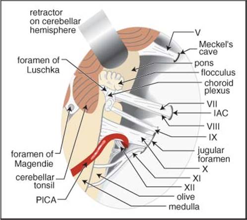
Figure 5-8 Normal anatomy of right cerebellopontine angle viewed from behind (as in a suboccipital approach)18
5.4. Occiptoatlantoaxial-complex anatomy
≈ 50% of head rotation occurs at the C1-2 (atlantoaxial) joint.
Ligaments of the occipito-atlanto-axial complex

Figure 5-9 Sagittal view of the ligaments of the craniovertebral junction Modified with permission from “In Vitro Cervical Spine Biomechanical Testing” BNI Quarterly, Vol.9, No. 4, 1993
Stability of this joint complex is primarily due to ligaments, with little contribution from bony articulations and joint capsules (see Figure 5-9 through Figure 5-11):
1. ligaments that connect the atlas to the occiput:
A. anterior atlanto-occipital membrane: cephalad extension of the anterior longitudinal ligament. Extends from anterior margin of foramen magnum (FM) to anterior arch of C1
B. posterior atlanto-occipital membrane: connects the posterior margin of the FM to posterior arch of C1
C. the ascending band of the cruciate ligament
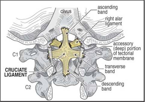
Figure 5-10 Dorsal view of the cruciate and alar ligaments Viewed with tectorial membrane removed. Modified with permission from “In Vitro Cervical Spine Biomechanical Testing” BNI Quarterly, Vol.9, No. 4, 1993
2. ligaments that connect the axis (viz. the odontoid) to the occiput:
A. tectorial membrane: some authors distinguish 2 components
1. superficial component: cephalad continuation of the posterior longitudinal ligament. A strong band connecting the dorsal surface of the dens to the ventral surface of the FM above, and dorsal surface of C2 & C3 bodies below
2. accessory (deep) portion: located laterally, connects C2 to occipital condyles
B. alar (“check”) ligaments19
1. occipito-alar portion: connects side of the dens to occipital condyle
2. atlanto-alar portion: connects side of the dens to the lateral mass of C1
C. apical odontoid ligament: connects tip of dens to the FM. Little mechanical strength
3. ligaments that connect the axis to the atlas:
A. transverse (atlanto-axial) ligament: the horizontal component of the cruciate ligament. Traps the dens against the anterior atlas via a strap-like mechanism (see Figure 5-11). Provides the majority of the strength (“the strongest ligament of the spine”20)
B. atlanto-alar portion of the alar ligaments (see above)
C. descending band of the cruciate ligament
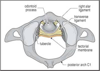
Figure 5-11 C1 viewed from above, showing the transverse and alar ligaments Modified with permission from “In Vitro Cervical Spine Biomechanical Testing” BNI Quarterly, Vol.9, No. 4, 1993
The most important structures in maintaining atlanto-occipital stability are the tectorial membrane and the alar ligaments. Without these, the remaining cruciate ligament and apical dentate ligament are insufficient.
5.5. Spinal cord anatomy
5.5.1. Spinal cord tracts
Figure 5-12 depicts a cross-section of a typical spinal cord segment, combining some elements from different levels (e.g. the intermediolateral grey nucleus is only present from T1 to ≈ L1 or L2 where there are sympathetic (thoracolumbar outflow) nuclei). It is schematically divided into ascending and descending halves, however, in actuality, ascending and descending paths coexist on both sides.
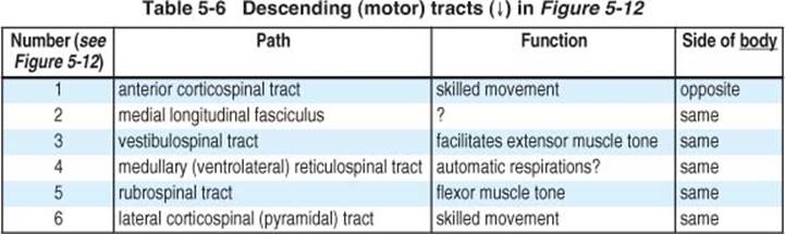
Table 5-7 Bi-directional tracts in Figure 5-12
|
Number (see Figure 5-12) |
Path |
Function |
|
7 |
dorsolateral fasciculus (of Lissauer) |
|
|
8 |
fasciculus proprius |
short spinospinal connections |
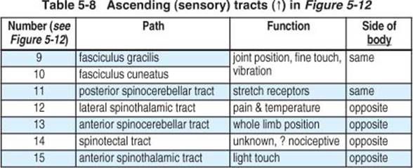
Figure 5-12 also depicts some of the laminae according to the scheme of Rexed. Lamina II is equivalent to the substantia gelatinosa. Laminae III and IV are the nucleus proprius. Lamina VI is located in the base of the posterior horn.
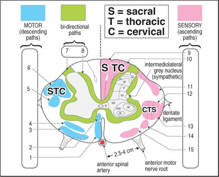
Figure 5-12 Schematic cross-section of cervical spinal cord
SENSATION
PAIN & TEMPERATURE: BODY
Receptors: free nerve endings (probable).
1st order neuron: small, finely myelinated afferents; soma in dorsal root ganglion (no synapse). Enter cord at dorsolateral tract (zone of Lissauer). Synapse: substantia gelatinosa (Rexed II).
2nd order neuron axon cross obliquely in the anterior white commissure ascending ≈ 1-3 segments while crossing to enter the lateral spinothalamic tract.
Synapse: VPL thalamus. 3rd order neurons pass through IC to postcentral gyrus (Brodmann’s areas 3, 1, 2).
FINE TOUCH, DEEP PRESSURE & PROPRIOCEPTION: BODY
Fine touch AKA discriminative touch. Receptors: Meissner’s & pacinian corpuscles, Merkel’s disks, free nerve endings.
1st order neuron: heavily myelinated afferents; soma in dorsal root ganglion (no synapse). Short branches synapse in nucleus proprius (Rexed III & IV) of posterior gray; long fibers enter the ipsilateral posterior columns without synapsing (below T6: fasciculus gracilis; above T6: fasciculus cuneatus).
Synapse: nucleus gracilis/cuneatus (respectively), just above pyramidal decussation. 2nd order neuron axons form internal arcuate fibers, decussate in lower medulla as medial lemniscus.
Synapse: VPL thalamus. 3rd order neurons pass through IC primarily to postcentral gyrus.

Figure 5-13 Dermatomal and sensory nerve distribution
(Redrawn from “Introduction to Basic Neurology”, by Harry D. Patton, John W. Sundsten, Wayne E. Crill and Phillip D. Swanson, © 1976, pp 173, W. B. Saunders Co., Philadelphia, PA, with permission)
LIGHT (CRUDE) TOUCH: BODY
Receptors: as fine touch (see above), also peritrichial arborizations.
1st order neuron: large, heavily myelinated afferents (Type II); soma in dorsal root ganglion (no synapse). Some ascend uncrossed in post. columns (with fine touch); most synapse in Rexed VI & VII.
2nd order neuron axons cross in anterior white commissure (a few don’t cross); enter anterior spinothalamic tract.
Synapse: VPL thalamus. 3rd order neurons pass through IC primarily to postcentral gyrus.
5.5.2. Dermatomes and sensory nerves
Figure 5-13 shows anterior and posterior view, each schematically separated into sensory dermatomes (segmental) and peripheral sensory nerve distribution.
5.5.3. Spinal cord vasculature
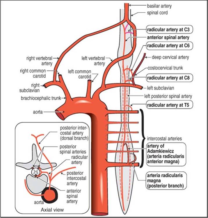
Figure 5-14 Schematic diagram of spinal cord arterial supply
Modified from Diagnostic Neuroradiology, 2nd ed., Volume II, pp. 1181, Taveras J M, Woods EH, editors, © 1976, the Williams and Wilkins Co., Baltimore, with permission)
Although a radicular artery from the aorta accompanies the nerve root at many levels, most of these contribute little flow to the spinal cord itself. The anterior spinal artery is formed from the junction of two branches, each from one of the vertebral arteries. Major contributors of blood supply to the anterior spinal cord is from 6-8 radicular arteries at the following levels (“radiculomedullary arteries”, the levels listed are fairly consistent, but the side varies21 (p 1180-1)):
• C3 - arises from vertebral artery
• C6 - usually arises from deep cervical artery } ≈ 10% of population lack an anterior radicular artery in lower cervical spine22
• C8 - usually from costocervical trunk } ≈ 10% of population lack an anterior radicular artery in lower cervical spine22
• T4 or T5
• artery of Adamkiewicz AKA arteria radicularis anterior magna
A. the main arterial supply for the spinal cord from ≈ T8 to the conus
B. located on the left in 80%23
C. situated between T9 & L2 in 85% (between T9 & T12 in 75%); in remaining 15% between T5 & T8 (in these latter cases, there may be a supplemental radicular artery further down)
D. usually fairly large, gives off cephalic and caudal branch (latter is usually larger) giving a characteristic hair-pin appearance on angiography
The paired posterior spinal arteries are less well defined than the anterior spinal artery, and are fed by 10-23 radicular branches.
The midthoracic region has a tenuous vascular supply (“watershed zone”), possessing only the above noted artery at T4 or T5. It is thus more susceptible to vascular insults.
ANATOMIC VARIANTS
Arcade of Lazorthes: normal variant where the anterior spinal artery joins with the paired posterior spinal arteries at the conus medullaris.
5.6. Cerebrovascular anatomy
5.6.1. Cerebral vascular territories

Figure 5-15 Vascular territories of the cerebral hemispheres
Figure 5-15 depicts approximate vascular distributions of the major cerebral arteries. There is considerable variability of the major arteries24 as well as the central distribution. The lenticulostriates may have origins off of different segments of the middle or anterior cerebral artery). Recurrent artery of Heubner (RAH) (AKA medial striate artery) origin: junction of the ACA and a-comm in 62.3%, proximal A2 in 23.3%, A1 in 14.3%25.
5.6.2. Cerebral arterial anatomy
The symbol “⇒” is used to denote a region supplied by the indicated artery. See Angiography (cerebral) on page 134 for angiographic diagrams of the following anatomy.
CIRCLE OF WILLIS
A balanced configuration of the Circle of Willis is present in only 18% of the population. Hypoplasia of 1 or both p-comms occurs in 22-32%, absent or hypoplastic A1 segments occurs in 25%.

Figure 5-16 Circle of Willis viewed from in front of and below the brain
Key point: the anterior cerebral arteries pass over the superior surface of the optic chiasm.
Anatomical segments of intracranial cerebral arteries
• carotid artery: the traditional numbering system26 was from rostral to caudal (counter to the direction of flow, and to the numbering scheme of the other arteries). A number of systems have been described to addresses this inconsistency and also to identify anatomically important segments of the ICA that were not originally delineated (e.g. see Table 5-927). Also see below for more detail
• anterior cerebral28:
 A1: ACA from origin to ACoA
A2: ACA from ACoA to branch-point of callosomarginal
A3: from branch-point of callosomarginal to superior surface of corpus callosum 3 cm posterior to the genu
A4: pericallosal
A5: terminal branch
• middle cerebral28:
M1: MCA from origin to bifurcation (horizontal segment on AP angiogram). A classical bifurcation into relatively symmetrical superior and inferior trunks is seen in 50%, no bifurcation occurs in 2%, 25% have a very proximal branch (middle trunk) arising from the superior (15%) or the inferior (10%) trunk creating a “pseudo-trifurcation”, a pseudo-tetrafurcation occurs in 5%
1. lateral fronto-orbital and prefrontal branches arise from M1 or superior M2 trunk
2. precentral, central, anterior and posterior parietal arteries arise from a superior (60%) or middle (25%) or inferior (15%) trunk
3. the superior M2 trunk does not give any branches to the temporal lobe
M2: MCA trunks from bifurcation to emergence from Sylvian fissure
M3-4: distal branches
M5: terminal branch
• posterior cerebral (PCA) (several nomenclature schemes exist29, 30):
P1: PCA from the origin to posterior communicating artery (AKA mesencephalic, precommunicating, circular, peduncular, basilar…). The long and short circumflex and thalamoperforating arteries arise from P1
P2: PCA from origin of p-comm to the origin of inferior temporal arteries (AKA ambient, postcommunicating, perimesencephalic), P2 traverses the ambient cistern, Hippocampal, anterior temporal, peduncular perforating and medial posterior choroidal arteries arise from P2
P3: PCA from the origin of the inferior temporal branches to the origin of the terminal branches (AKA quadrigeminal segment). P3 traverses the quadrigeminal cistern
P4: segment after the origin of the parieto-occipital and calcarine arteries, includes the cortical branches of the PCA
Table 5-9 Segments of the ICA
|
Cincinnati system |
System of Fischer |
|
C1 (cervical) |
not described |
|
C2 (petrous) |
|
|
C3 (lacerum) |
C5 |
|
C4 (cavernous) |
C4 + part of C5 |
|
C5 (clinoid) |
C3 |
|
C6 (ophthalmic) |
C2 |
|
C7 (communicating) |
C1 |
ANTERIOR CIRCULATION
ANATOMIC VARIANTS
Bovine circulation: the common carotids arise from a common trunk off the aorta.
EXTERNAL CAROTID
1. superior thyroid a.: 1st anterior branch
2. ascending pharyngeal a.
A. neuromeningeal trunk of the ascending pharyngeal a.: supplies IX, X & XI
B. pharyngeal branch: usually the primary feeder for jugular foramen tumors (essentially the only cause of hypertrophy of the ascending pharyngeal a.)
3. lingual a.
4. facial a.: branches anastamose with ophthalmic a. (important in collateral flow with ICA occlusion - see page 1027)
5. occipital a. ⇒ posterior scalp
6. posterior auricular
7. superficial temporal
A. frontal branch
B. parietal branch
8. (internal) maxillary a. - initially within parotid gland
A. middle meningeal a.
1. anterior branch
2. posterior branch
B. accessory meningeal
C. inferior alveolar
D. infraorbital
E. others: distal branches of which may anastomose with branches of ophthalmic artery in the orbit
INTERNAL CAROTID ARTERY (ICA)
Lies posterior & medial to the external carotid (ECA).
Segments of the ICA and its branches27
• C1 (cervical): begins in neck at carotid bifurcation where the common carotid artery divides into internal and external carotids. Travels in carotid sheath with IJV and vagal nerve, encircled with postganglionic sympathetic nerves (PGSN). C1 ends where the ICA enters carotid canal of petrous bone. No branches
• C2 (petrous): still surrounded by PGSNs. Ends at the posterior edge of the foramen lacerum (f-Lac) (inferomedial to the edge of the Gasserian ganglion in Meckel’s cave). Three divisions:
A. vertical segment: ICA ascends then bends as the…
B. posterior loop: anterior to cochlea, bends antero-medially becoming the…
C. horizontal segment: deep and medial to greater and lesser superficial petrosal nerves, anterior to tympanic membrane (TM)
• C3 (lacerum): the ICA passes over (but not through) the f-Lac forming the lateral loop. Ascends in the canalicular portion of the f-Lac to juxtasellar position, piercing the dura as it passes the petrolingual ligament to become the cavernous segment. Branches (usually not visible angiographically):
A. caroticotympanic (inconsistent) ⇒ tympanic cavity
B. pterygoid (vidian) branch: passes through foramen lacerum, present in only 30%, may continue as artery of pterygoid canal
• C4 (cavernous): covered by vascular membrane lining sinus, still surrounded by PGSNs. Passes anteriorly then supero-medially, bends posteriorly (medial loop of ICA), travels horizontally, and bends anteriorly (part of anterior loop of ICA) to anterior clinoid process. Ends at the proximal dural ring (incompletely encircles ICA). Many branches, main ones include:
A. meningohypophyseal trunk (MHT) (largest & most proximal). 2 causes of a prominent MHT: 1) tumor (usually petroclival meningioma - see below), 2) dural AVM (see page 1109)
1. a. of tentorium (AKA artery of Bernasconi & Cassinari): the blood supply of petroclival meningiomas
2. dorsal meningeal a. (AKA dorsal clival a.)
3. inferior hypophyseal a. (⇒ posterior lobe of pituitary): post-partum occlusion causes pituitary infarcts (Sheehan’s necrosis), however, DI is rare because the stalk is spared
B. anterior meningeal a.
C. a. to inferior portion of cavernous sinus (present in 80%)
D. capsular aa. of McConnell (in 30%): supply the capsule of the pituitary31
• C5 (clinoid): begins at proximal dural ring, ends at distal dural ring (which completely encircles ICA) where the ICA becomes intradural
• C6 (ophthalmic): begins at distal dural ring, ends just proximal to p-comm. Branches:
A. ophthalmic a.: the origin from the ICA is distal to the cavernous sinus in 89% (intracavernous in 8%, absent in 3%32) and can vary from 5 mm anterior to 7 mm posterior to the anterior clinoid31. Passes through the optic canal into the orbit. Has a characteristic bayonet-like “kink” on lateral angiogram
B. superior hypophyseal a. branches ⇒ anterior lobe of pituitary & stalk (1st branch of supraclinoid ICA)
• C7 (communicating): begins just proximal to p-comm origin, travels between Cr. N. II & III, terminates just below anterior perforated substance where it bifurcates into the ACA & MCA
A. posterior communicating a. (p-comm)
1. few anterior thalamoperforators (⇒ optic tract, chiasm & posterior hypothalamus): see Posterior circulation below
2. plexal segment: enters supracornual recess of temporal horn, ⇒ only this portion of choroid plexus
3. cisternal segment: passes through crural cistern
B. anterior choroidal artery 33: takeoff 2-4 mm distal to p-comm ⇒ (variable) portion of optic tract, medial globus pallidus, genu of internal capsule (IC) (in 50%), inferior half of posterior limb of IC, uncus, retrolenticular fibers (optic radiation), lateral geniculate body (see page 1028 for occlusion syndromes)
Differentiating p-comm from ACh on arteriogram
1. p-comm origin is proximal to that of the anterior choroidal artery (ACh)
2. p-comm is usually larger than ACh
3. p-comm usually goes up or down a little, then straight back & usually bifurcates
4. ACh usually has a superior “hump” (plexal point) where it pass through the choroidal fissure to enter the ventricle
“Carotid siphon”: not a segment, but a region incorporating the cavernous, ophthalmic and communicating segments. Begins at the posterior bend of the cavernous ICA, and ends at the ICA bifurcation
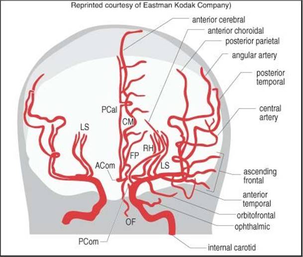
Figure 5-17 Internal carotid arteriogram (AP view)
ANTERIOR CEREBRAL ARTERY (ACA)
Passes between Cr. N. II and anterior perforated substance. See Figure 5-18. Branches:
1. recurrent artery (of Heubner): 80% arise from A1 (one of the larger medial lenticulostriates, remainder of lenticulostriates may arise from this artery) ⇒ head of caudate, putamen, and anterior internal capsule
2. medial orbitofrontal artery
3. frontopolar artery
4. callosomarginal
A. internal frontal branches
1. anterior
2. middle
3. posterior
B. paracentral artery
5. pericallosal artery (continuation of ACA)
A. superior internal parietal (precuneate) artery
B. inferior internal parietal artery
Abbreviations from Figure 5-17
|
ACom |
anterior communicating artery |
|
CM |
callosomarginal artery |
|
FP |
frontopolar artery |
|
LS |
lenticulostriate arteries |
|
OF |
orbitofrontal artery |
|
PCal |
pericallosal artery |
|
PCom |
posterior communicating artery |
|
RH |
recurrent artery of Heubner |
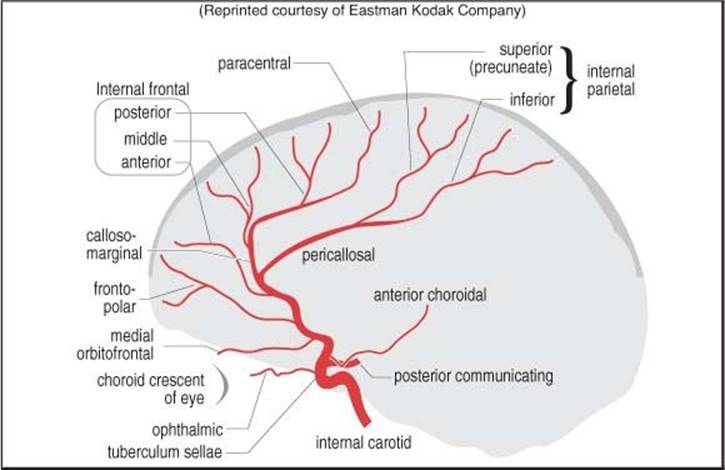
Figure 5-18 Anterior cerebral arteriogram (lateral view)
ANATOMIC VARIANTS
Hypoid: having only one anterior cerebral artery (as in a horse).
MIDDLE CEREBRAL ARTERY (MCA)
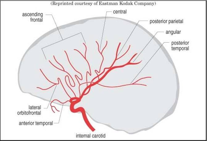
Figure 5-19 Middle cerebral arteriogram (lateral view)
See Figure 5-19 (see page 98 for anatomy). Branches vary widely, 10 common ones:
1. medial (3-6 per side) and lateral lenticulostriate arteries
2. anterior temporal
3. posterior temporal
4. lateral orbitofrontal
5. ascending frontal (candelabra)
6. precentral (prerolandic)
7. central (rolandic)
8. anterior parietal (postrolandic)
9. posterior parietal
10. angular
POSTERIOR CIRCULATION
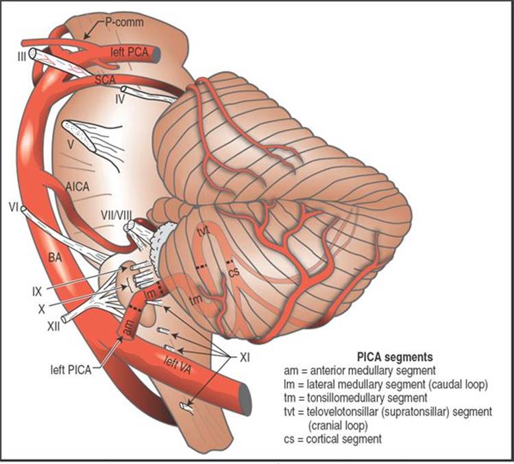
Figure 5-20 Intradural VA and PICA segments (lateral view) Modified with permission from: Lewis SB, Chang DJ, Peace DA, Lafrentz PJ, Day AL. Distal posterior inferior cerebellar artery aneurysms: clinical features and management. J Neurosurg 2002;97(4):756-66.
VERTEBRAL ARTERY (VA)
The VA is the first and usually the largest branch of the subclavian artery. Variant: the left VA arises off the aortic arch in ≈ 4%. Diameter ≈ 3 mm. Mean blood flow ≈ 150 ml/min. The left VA is dominant in 60%. The right VA will be hypoplastic in 10%, and the left will be hypoplastic in 5%. The VA is atretic and does not communicate with the BA on the left in 3%, and on the right in 2% (the VA may terminate in PICA).
Four segments:
V1 prevertebral: from subclavian artery, courses superiorly and posteriorly and enters the foramen transversarium, usually of the 6th vertebral body
V2 ascends vertically within the transverse foramina of the cervical vertebrae surrounded by sympathetic fibers (from the stellate ganglion) and a venous plexus. It is situated anterior to the cervical roots. It turns laterally to enter the foramen within the transverse process of the axis
V3 exits the foramen of the axis and curves posteriorly and medially in a groove on the upper surface of the atlas and enters the foramen magnum
V4 pierces the dura (location somewhat variable) and immediately enters the subarachnoid space. Joins the contralateral VA at the vertebral confluens located at the lower pontine border to form the basilar artery (BA)
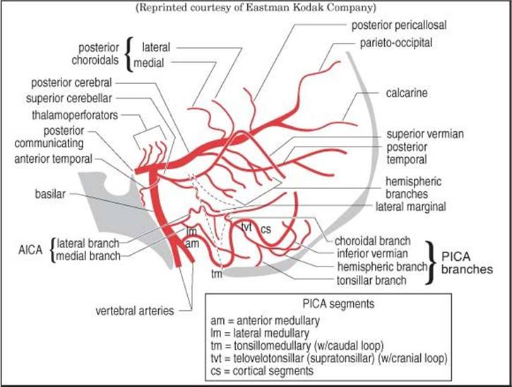
Figure 5-21 Vertebrobasilar arteriogram (lateral view)
Branches:
1. anterior meningeal: arises at body of C2 (axis), may feed chordomas or foramen magnum meningiomas, may also act as collateral in vascular occlusion
2. posterior meningeal: may a source of blood for some dural AVMs (see page 1109)
3. medullary (bulbar) aa.
4. posterior spinal
5. posterior inferior cerebellar artery (PICA) (largest branch): (see Figure 5-20) usually arises ≈10 mm distal to point where VA becomes intradural, ≈ 15 mm proximal to the vertebrobasilar junction
A. anatomic variants:
1. in 5-8% the PICA has an extradural origin
2. “AICA-PICA”: origin is off basilar trunk (where AICA would usually originate)
B. 5 segments34 (some systems some describe only 4). During surgery, the first three must be preserved, but the last 2 may usually be sacrificed with minimal deficit35:
1. anterior medullary: from PICA origin to inferior olivary prominence. 1 or 2 short medullary short circumflex branches ⇒ ventral medulla
2. lateral medullary: to origin of nerves IX, X & XI. Up to 5 branches that supply brainstem
3. tonsillomedullary: to tonsillar midportion (contains caudal loop on angio)
4. telovelotonsillar (supratonsillar): ascends in tonsillomedullary fissure (contains cranial loop on angio)
5. cortical segments
C. 3 branches
1. choroidal a. (BRANCH 1) arises from cranial loop (choroidal point), ⇒ choroid plexus of 4th ventricle
2. terminal branches:
a. tonsillohemispheric (BRANCH 2)
b. inferior vermian (BRANCH 3) inferior inflection = copular point on angio
6. anterior spinal
ANATOMIC VARIANTS
Fetal circulation: 15-35% of patients supply their posterior cerebral artery on one or both sides from the carotid (via p-comm) instead of via the vertebrobasilar system.
BASILAR ARTERY (BA)
Formed by the junction of the 2 vertebral arteries. Branches:
1. anterior inferior cerebellar artery (AICA): from lower part of BA, runs postero-laterally anterior to VI, VII & VIII. Often gives off a loop that runs into the IAC and gives off the labyrinthine artery and then emerges to supply the anterolateral inferior cerebellum and then anastomoses with PICA
2. internal auditory (labyrinthine)
3. pontine branches
4. superior cerebellar a. (SCA)
A. sup. vermian
5. posterior cerebral: joined by p-comms ≈ 1 cm from origin (the p-comm is the major origin of the PCA in 15% and is termed “fetal” circulation, bilateral in 2%). 3 segments (named for surrounding cistern) and their branches:
A. peduncular segment (P1)
1. mesencephalic perforating aa. (⇒ tectum, cerebral peduncles, and these nuclei: Edinger-Westphal, oculomotor and trochlear)
2. interpeduncular thalamoperforators (1st of 2 groups of posterior thalamoperforating aa.)
3. medial post. choroidal (most from P1 or P2)
4. “artery of Percheron”: a rare anatomic variant36 in which a solitary arterial trunk arising from the proximal segment of one PCA supplies the paramedian thalami and rostral midbrain bilaterally
B. ambient segment (P2)
1. lateral post. choroidal (most from P2)
2. thalamogeniculate thalamoperforators (2nd of 2 groups of posterior thalamoperforating aa.) ⇒ geniculate bodies + pulvinar
3. anterior temporal (anastomoses with anterior temporal br. of MCA)
4. posterior temporal
5. parieto-occipital
6. calcarine
C. quadrigeminal segment (P3)
1. quadrigeminal & geniculate branches ⇒ quadrigeminal plate
2. post. pericallosal (splenial) (anastomoses with pericallosal of ACA)
5.6.3. Cerebral venous anatomy
SUPRATENTORIAL VENOUS SYSTEM
See Figure 5-22, page 105 for angiogram and branches.
The left and right internal jugular veins (IJVs) are the major source of outflow of blood from the intracranial compartment. The right IJV is usually dominant. Other sources of outflow include orbital veins and the venous plexuses around the vertebral arteries. Diploic and scalp veins may act as collateral pathways, e.g. with superior sagittal sinus obstruction38. The following outline traces the venous drainage back from the IJVs.
A. inferior petrosal sinus: terminates (i.e. drains to) ≤ 1 cm of junction of sigmoid and transverse sinuses
B. sigmoid sinus
1. superior petrosal sinus: drains to IJV near junction with sigmoid sinus
2. transverse sinus (R > L in 65%)
A. v. of Labbe (inferior anastomotic v.)
B. confluens of sinuses (torcular herophili)
1. occipital sinus
2. superior sagittal sinus
a. v. of Trolard (superior anastomotic v.): the prominent superficial vein on the non-dominant side (Labbé is more prominent on the dominant side)
3. straight sinus
a. inferior sagittal sinus
b. great cerebral v. (of Galen)
i. pre-central cerebellar v.
ii. basal vein of Rosenthal
iii. internal cerebral v.: joined at the foramen of Monro (venous angle) by:
1. anterior septal v.
2. thalamostriate v.
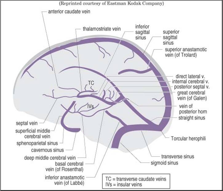
Figure 5-22 Internal carotid venogram (lateral view)
CAVERNOUS SINUS
Although classical teaching depicts the cavernous sinus as a large venous space with multiple trabeculations, injection studies39 and surgical experience40 supports the concept of the cavernous sinus as a plexus of veins. It is highly variable between individuals and from side-to-side. Figure 5-23 is an oversimplified schematic of one section through the right cavernous sinus.
1. inflowing veins:
A. superior & inferior ophthalmic veins
B. superficial middle cerebral veins
C. sphenoparietal sinus
D. superior & inferior petrosal sinus
2. outflow:
A. sphenoparietal sinus
B. superior petrosal sinus
C. basilar plexus (which drains to the inferior petrosal sinus)
D. pterygoid plexus
E. the right and left cavernous sinuses communicate anteriorly and posteriorly via the circular sinus
3. contents41
• Oculomotor n. (III)
• Trochlear n. (IV)
• Ophthalmic division of trigeminal (V1)
• Maxillary division of trigeminal (V2): the only nerve of the cavernous sinus that doesn’t exit the skull through the superior orbital fissure (it exits through foramen rotundum)
• Carotid artery (ICA). 3 segments within the cavernous sinus
1. posterior ascending segment: immediately after ICA enters the sinus
2. horizontal segment: after ICA turns anteriorly (the longest segment of the intracavernous ICA)
3. anterior ascending segment: ICA turns superiorly
• Abducens n. (VI): the only nerve NOT attached to lateral dural wall, some-times referred to as the only cranial nerve inside the cavernous sinus
3. triangular space (of Parkinson): superior border formed by Cr. N. III & IV, and the lower margin formed by V1 & VI (a landmark for surgical entrance to the cavernous sinus)42, 43 (p 3007)
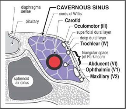
Figure 5-23 Right cavernous sinus (coronal section) Modified from the Journal of Neurosurgery, Umansky F and Nathan H, Vol. 56, pp. 228-34, 1982, with permission)
POSTERIOR FOSSA VENOUS ANATOMY
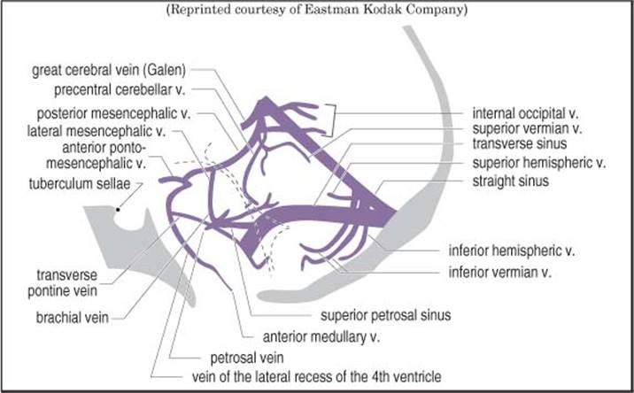
Figure 5-24 Vertebrobasilar venogram (lateral view)
5.6.4. Carotid-vertebrobasilar anastomoses
P-comm artery: the “normal” (most common) anastomosis.
Persistent fetal anastomoses37: (see Figure 5-25) result from failure to involute as the VAs and p-comms develop (order of involution: otic, hypoglossal, primitive trigeminal, proatlantal). Most are asymptomatic. However, some may be associated with vascular anomalies such as aneurysms or AVMs, and occasionally cranial nerve symptoms (e.g. trigeminal neuralgia with PPTA) can occur.
Four types (from cranial to caudal - the 1st 3 are named for the associated cranial nerve):
1. persistent primitive trigeminal artery (PPTA): seen in ≈ 0.6% of cerebral angiograms. The most common of the persistent fetal anastomoses (83%). May be associated with trigeminal neuralgia (see page 551). Connects the cavernous carotid to the basilar artery. Arises from the ICA proximal to the origin of the meningohypophyseal trunk (50% go through sella, 50% exit the cavernous sinus & course with the trigeminal nerve) and connects to the upper basilar artery between AICA & SCA. The VAs may be small. Saltzman type 1 variant: the p-comms are hypoplastic and the PPTA provides significant blood supply to the distributions of the distal BA, PCA and the SCAs (the basilar artery is often hypoplastic). Saltzman type 2: p-comm supplies PCA. Saltzman type 3: PPTA joins the SCA (instead of the BA). It is critical to recognize a PPTA before doing a Wada test (see page 421) because of the risk anesthetizing the brainstem, and in doing transsphenoidal surgery because of risk of arterial injury. May rarely be an explanation of posterior fossa symptoms in a patient with carotid disease
2. otic: the first to involute, and the rarest to persist (8 cases reported). Passes through IAC to connect petrous carotid to basilar artery
3. hypoglossal: connects petrous or distal cervical ICA (origin usually between C1-C3) to VA. Traverses the hypoglossal canal. Does not cross foramen magnum
4. proatlantal intersegmental: connects cervical ICA to VA. May arise from: bifurcation of common carotid, ECA, or ICA from C2-C4. Anastomosis with VA in suboccipital region. 50% have hypoplastic proximal VA. 40 cases reported

Figure 5-25 Carotid-vertebrobasilar anastomoses
5.7. Internal capsule
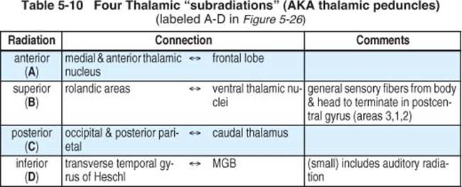
For a schematic diagram, see Figure 5-26. Table 5-10 delineates the thalamic sub-radiations. Most IC lesions are caused by vascular accidents (thrombosis or hemorrhage).
Vascular supply of the internal capsule (IC)
1. anterior choroidal: ⇒ all of retrolenticular part (includes optic radiation) and ventral part of posterior limb of IC
2. lateral striate branches (AKA capsular branches) of middle cerebral artery: ⇒ most of anterior AND posterior limbs of IC
3. genu usually receives some direct branches of the internal carotid artery

Figure 5-26 Internal capsule schematic diagram (left side shows tracts, right side shows radiations)
5.8. Miscellaneous
OBERSTEINER-REDLICH ZONE (ORZ)
Transition from CNS myelin to peripheral myelin of cranial nerves = area where pressure from intracranial structures can cause cranial nerve symptoms (trigeminal neuralgias, hemifacial spasm, disabling positional vertigo, etc.)44. Also, zone where neoplasms tend to occur, especially vestibular schwannoma. On Cr. N. VIII, the ORZ is 8-12 mm distal to exit point from brainstem, and is close to porus acusticus (especially common on vestibular division)14 (p 695).
DENTATE LIGAMENT
The dentate ligament separates dorsal from ventral nerve roots in the spinal nerves. The spinal accessory nerve (Cr. N. XI) is dorsal to the dentate ligament.
5.9. Neurophysiology
5.9.1. Blood-brain barrier
The passage of water-soluble substances from the blood to the CNS is limited by tight junctions (zonulae occludentes) which are found between cerebral capillary endothelial cells, limiting penetration of the cerebral parenchyma (blood-brain barrier, (BBB)), as well as between choroid plexus epithelial cells (blood-CSF barrier)45. A number of specialized mediated transport systems allow transmission of, among other things, glucose and certain amino acids (especially precursors to neurotransmitters).
The efficacy of the BBB is compromised in certain pathological states (e.g. tumor, infection, trauma, stroke, hepatic encephalopathy…), and can also be manipulated pharmacologically (e.g. hypertonic mannitol increases the permeability, whereas steroids reduce the penetration of small hydrophilic molecules).
The BBB is absent in the following areas: choroid plexus, hypophysis, tuber cinereum, area postrema, pineal and preoptic recess.
CEREBRAL EDEMA
Three basic types (diffusion-weighted MRI may be able to differentiate, see page 132):
1. cytotoxic: BBB is closed, therefore no protein extravasation, therefore no enhancement on CT or MRI. Cells swell then shrink. Seen e.g. in head injury
2. vasogenic: BBB disrupted. Protein (serum) leaks out of vascular system, and therefore may enhance on imaging. Extracellular space (ECS) expands. Cells are stable. Responds to corticosteroids (e.g. dexamethasone). Seen e.g. surrounding metastatic brain tumor
3. ischemic: a combination of the above. BBB closed initially, but then may open. ECS shrinks then expands. Fluid extravasates late. May cause delayed deterioration following intracerebral hemorrhage (see page 1125)
5.9.2. Pituitary embryology & neuroendocrinology
The posterior pituitary (neurohypophysis) derives from downward evagination of neural crest cells (brain neuroectoderm) from the floor of the third ventricle. The residual recess in the floor of the third ventricle is called the median eminence. The anterior pituitary gland (adenohypophysis) develops from an evagination of epithelial ectoderm of the oropharynx, the evagination is known as Rathke’s pouch and is eventually separated from the oropharynx by the sphenoid bone. Cleft-like remnants of Rathke’s pouch separates the adenohypophysis and neurohypophysis. The adenohypophysis is comprised of the pars distalis (anterior lobe), the pars intermedia (intermediate lobe) and the pars tuberalis (extension of adenohypophyseal cells on the anterior aspect of the pituitary stalk). The pituitary gland is functionally outside the blood-brain barrier.
PITUITARY HORMONES AND THEIR CONTROLS
The pituitary gland releases 8 hormones, 6 from the anterior pituitary, 2 from the posterior pituitary (see Figure 5-27). The anterior pituitary is one of only two sites in the body having a portal circulation (the other being the liver). 6 hypothalamic hormones released in a pulsatile fashion are conveyed in blood from hypothalamic capillaries through this portal circulation via the pituitary stalk to a second capillary bed in the anterior pituitary where they control release of hormones by adenohypophyseal gland cells.
Hormones released from the posterior pituitary (ADH & oxytocin) are synthesized in neurons in the hypothalamus (not gland cells) and are conveyed along their axons also in the pituitary stalk to the posterior pituitary gland where they are released.
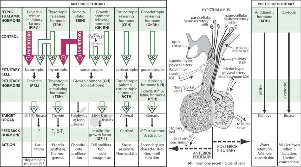
Figure 5-27 Pituitary neuroendocrinology
The complete homeostatic loop (including negative feedback of the hypothalamic hormones) will not be covered here, and the reader is referred to physiology texts.
Propiomelanocortin (POMC) AKA proopiomelanocortin
241 amino acid polypeptide hormone precursor synthesized primarily in corticotroph cell of the anterior pituitary (but also found in the hypothalamus). Contains amino acid sequences for ACTH, alpha–melanocyte–stimulating hormone (α-MSH), ß-lipotropin, γ-lipotropin, ß-endorphin and met-enkephalin.
Corticotropin AKA adrenocorticotrophic hormone (ACTH)
A 39 amino acid trophic hormone synthesized from POMC (see above). The first 13 amino acids at the amino terminal of ACTH are identical to α-MSH. Active half-life is ≈ 10 minutes. Produces a diurnal peak in cortisol (the highest peak occurs in the early morning, with a second, lesser peak in the late afternoon) and also increases in response to stress.
Control: CRH from the hypothalamus stimulates the release of ACTH.
Prolactin (PRL)
AKA somatomammotropin. 199 amino-acid protein weighing 23,000 daltons. Levels are higher in females than males, and are higher still in pregnancy (see page 643). Secreted in pulsatile fashion with a frequency and amplitude that varies during menstrual cycle (range: 5-27 ng/ml). There is also diurnal variation: levels begin to rise 1 hour after the onset of sleep, peak ≈ 5:00-7:00 AM, and nadir in midmorning after awakening. Heterogeneity of the molecule may produce different results between bioassays and immunoassays.
Control: PRL is the only pituitary hormone predominantly under inhibitory control from the hypothalamus by prolactin inhibitory factors (PRIFs), with dopamine being the primary PRIF. Prolactin releasing factors (PRFs)include: thyrotropin-releasing hormone (TRH) and vasoactive intestinal peptide (VIP). The physiologic role of PRFs is not established. For differential diagnosis of hyperprolactinemia see page 644.
Growth hormone (GH)
A 191 amino-acid polypeptide trophic hormone. GH normally has pulsatile secretion (≈ 5-10 pulses/24 hours, primarily at night, up to 30 μg/L), levels may be undetectable (< 0.2 μg/L) by standard assays between pulses46. Insulin-like growth factor-1 (IGF-1) (formerly AKA somatomedin-C) is the protein secreted primarily by the liver in response to GH that is responsible for most of GH’s systemic effects (see page 648 for levels). GH also acts directly on epiphyseal end-plates of long bone to stimulate chondrocyte proliferation.
Control: GH is under dual hypothalamic control via the hypophysial portal system. GH-releasing hormone (GHRH) from the arcuate nucleus stimulates pituitary secretion and synthesis of GH and induces GH gene transcription. Somatostatin from the periventricular nucleus suppresses GH release only. GH release is also stimulated by ghrelin47, a peptide synthesized primarily in the GI tract in response to certain nutrients (may act partially or totally via hypothalamic GHRH).
Thyrotropin AKA thyroid stimulating hormone (TSH)
Glycoprotein trophic hormone secreted by thyrotroph cells of the anterior pituitary.
Control: TSH is also under dual hypothalamic control. TRH stimulates production and release of TSH. Somatostatin inhibits the release of TSH.
Gonadotropins
Follicle stimulating hormone (FSH) and leuteinizing hormone (LH) (AKA lutropin) are released from the pituitary in response to gonadotropin releasing hormone 1 (GnRH) (formerly leuteinizing hormone releasing hormone (LH-RH)) synthesized primarily in the preoptic area of the hypothalamus.
Antidiuretic hormone (ADH)
AKA arginine vasopressin (AVP). The major source of this nanopeptide hormone is the magnocellular portion of the supraoptic nucleus of the hypothalamus. It is conveyed along axons in the supraoptic-hypophyseal tract to the posterior pituitary gland where it is released into the systemic circulation. All actions of ADH result from binding of the hormone to specific membrane bound receptors on the surface of target cells48. One of the major effects of ADH is to increase the permeability of the distal renal tubules resulting in increased reabsorption of water, diluting the circulating blood and producing a concentrated urine. The most powerful physiologic stimulus for ADH release is an increase in serum osmolality, a less potent stimulus is a reduction of intravascular volume. ADH is also released in glucocorticoid deficiency, and is inhibited by exogenous glucocorticoids and adrenergic drugs. ADH is also a potent vasoconstrictor.
Oxytocin
A nonapeptide. The hypothalamus is the main source of pituitary oxytocin which is stored in nerve endings in the neurohypophysis and is involved in the milk letdown reflex for breastfeeding as well as in uterine contraction during labor.
5.9.3. Regional brain syndromes
This section serves to briefly describe typical syndrome associated with lesions in various areas of the brain. Unless otherwise noted, lesions considered are destructive.
1. frontal lobe
A. unilateral injury:
1. may produce few clinical findings except with very large lesions
2. bilateral or large unilateral lesions: apathy, abulia
3. the frontal eye field (for contralateral gaze) is located in the posterior frontal lobe (Br. area 8, shown as the striped area in Figure 5-1, page 84). Destructive lesions impair gaze to the contralateral side (patient looks towards the side of the lesion), whereas irritative lesions (i.e. seizures) cause the center to activate, producing contralateral gaze (patient looks away from the side of the lesion). Also see page 834
B. bilateral injury: may produce apathy, abulia
C. olfactory groove region: may produce Foster Kennedy syndrome (see below)
D. prefrontal lobes control “executive function”: planning, prioritizing, organizing thoughts, suppressing impulses, understanding the consequences of decisions
2. parietal lobe: major features (see below for details)
A. either side: cortical sensory syndrome, sensory extinction, contralateral homonymous hemianopia, contralateral neglect
B. dominant parietal lobe lesion (left in most): language disorders (aphasias), Gerstmann’s syndrome (see page 113), bilateral astereognosis
C. non-dominant parietal lobe lesions: topographic memory loss, anosognosia and dressing apraxia
3. occipital lobe: homonymous hemianopsia
4. cerebellum
A. lesions of the cerebellar hemisphere cause ataxia in the ipsilateral limbs
B. lesions of the cerebellar vermis cause truncal ataxia
5. brainstem: usually produces a mixture of cranial nerve deficits and long tract findings (see below for some specific brainstem syndromes)
6. pineal region
A. Parinaud’s syndrome: see page 114
FOSTER KENNEDY SYNDROME
Usually from olfactory groove or medial third sphenoid wing tumor (usually meningioma). Now rare due to earlier detection by CT scan. Classic triad:
1. ipsilateral anosmia
2. ipsilateral central scotoma (with optic atrophy due pressure on optic nerve)
3. contralateral papilledema (from elevated ICP)
Occasionally ipsilateral proptosis will also occur due to orbital invasion of tumor.
5.9.3.1. Parietal lobe syndromes49 (p 308-12)
PARIETAL LOBE ANATOMY
The parietal lobe is located behind the central sulcus, above the Sylvian fissure, merging posteriorly into the occipital lobe (the border on the medial surface of brain is defined by a line connecting the parieto-occipital sulcus to the pre-occipital notch).
PARIETAL LOBE NEUROPHYSIOLOGY
• either side: anterior parietal cortex organizes tactile precepts (probably contralateral) and integrates with visual and auditory sensation to build awareness of body and its spatial relations
• dominant side (on left in 97% of adults): understanding language, includes “cross-modal matching” (auditory-visual, visual-tactile, etc.). Dysphasia present with dominant lobe lesions often impedes assessment
• non-dominant side: integrates visual and proprioceptive sensation to allow manipulation of body and objects, and for certain constructional activities
CLINICAL SYNDROMES OF PARIETAL LOBE DISEASE
1. unilateral parietal lobe disease (dominant or non-dominant):
A. cortical sensory syndrome (see below) and sensory extinction (neglecting 1 of 2 simultaneously presented stimuli). Large lesion → hemianesthesia
B. congenital injury → mild hemiparesis & contralateral muscle atrophy
C. homonymous hemianopia or visual inattentiveness
D. occasionally: anosognosia
E. neglect of contralateral half of body and visual space (more common with right side lesions)
F. abolition of optokinetic nystagmus to one side
2. additional effects of dominant parietal lobe lesion (left in most):
A. language disorders (aphasias)
B. speech-related or verbally mediated functions, e.g. cross-modal matching (e.g. patient understands spoken words and can read, but cannot understand sentences with elements of relationships)
C. Gerstmann’s syndrome, classically:
1. agraphia without alexia (patients can read but cannot write)
2. left-right confusion
3. digit agnosia: inability to identify finger by name
4. acalculia
D. tactile agnosia (bilateral astereognosis)
E. bilateral ideomotor apraxia (inability to carry out verbal commands for activities that can otherwise be performed spontaneously with ease)
3. additional effects of non-dominant parietal lobe lesions (usually right):
A. topographic memory loss
B. anosognosia and dressing apraxia
CORTICAL SENSORY SYNDROME
Lesion of postcentral gyrus, especially area that maps to hand.
• sensory deficits:
A. loss of position sense and of passive movement sense
B. inability to localize tactile, thermal, and noxious stimuli
C. astereognosis (inability to judge object size, shape, and identity by feel)
D. agraphesthesia (cannot interpret numbers written on hand)
E. loss of two point discrimination
• preserved sensations: pain, touch, pressure, vibration, temperature
• other features
A. easy fatigability of sensory perceptions
B. difficulty distinguishing simultaneous stimulations
C. prolongation of superficial pain with hyperpathia
D. touch hallucinations
ASOMATAGNOSIAS
ANTON-BABINSKI SYNDROME
Unilateral asomatagnosia. May seem more common with non-dominant (usually right) parietal lesions because it may be obscured by the aphasia that occurs with dominant (left) sided lesions.
1. anosognosia (indifference or unawareness of deficits, patient may deny that paralyzed extremity is theirs)
2. apathy (indifference to failure)
3. allocheiria (one-sided stimuli perceived contralaterally)
4. dressing apraxia: neglect of one side of body in dressing and grooming
5. extinction: patient is unaware of contralateral stimulus when presented with double-sided simultaneous stimulation
6. inattention to an entire visual field (with or without homonymous hemianopia), with deviation of head, eyes, and torsion of body to unaffected side
APHASIAS
As related to parietal lobe lesions:
1. Wernicke’s aphasia: lesion of auditory association areas or their separation from angular gyrus and primary auditory cortex. A fluent aphasia (normal sentence length & intonation, devoid of meaning). May include paraphasias. Lesion in region of Wernicke’s area (Brodmann areas 40 & 39, see Figure 5-1, page 84)
2. Broca’s (motor) aphasia: in reality, “apraxia” of motor sequencing for speech (speech and phonation muscles aren’t paralyzed, and function for other activities), producing faltering, dysarthric speech. Lesion in region of Broca’s area (Brodmann area 44, see Figure 5-1, page 84)
3. global aphasia: usually due to lesion that destroys large portion of language center; all aspects of speech and language affected
A. unable to speak except for some clichés, habitual phrases, or expletives
B. anomia (inability to name objects or parts of objects)
C. verbal and motor perseveration
D. unable to understand all except for a few words
E. inability to read or write
4. conduction aphasia: due to disruption of connections between frontal and temporal speech areas, usually involving supramarginal gyrus. Similar to Wernicke’s (fluent spontaneous speech and paraphasias), but patients understand spoken or written words, and are aware of their deficit. Repetition is severely affected
5. pure word blindness: AKA alexia without agraphia (rare) due to lesion in parieto-occipital lobe that interrupts connections between left angular gyrus and both occipital lobes. Patients can write, but are unable to read what they’ve written, and frequently seem unconcerned about this. Often accompanied by loss of ability to name colors. Reading and naming numbers usually preserved
5.9.3.2. Brain stem and related syndromes
WEBER’S SYNDROME
Cr. N. III palsy with contralateral hemiparesis (also see Lacunar strokes, page 1026). Third nerve palsies from parenchymal lesions may be relatively pupil sparing.
BENEDIKT’S SYNDROME
Similar to Weber’s, plus red nucleus lesion. Cr. N. III palsy with contralateral hemiparesis except arm which has hyperkinesia, ataxia, & a coarse intention tremor. Lesion: midbrain tegmentum involving red nucleus, brachium conjunctivum, and fascicles of III.
MILLARD-GUBLER SYNDROME
Facial (VII) & abducens (VI) palsy + contralateral hemiplegia (corticospinal tract) from lesion in base of pons (usually ischemic infarct, occasionally tumor).
PARINAUD’S SYNDROME
AKA dorsal midbrain syndrome, AKA pretectal syndrome. As originally described, a supranuclear paralysis of vertical gaze resulting from damage to the mesencephalon50. There are a number of variations, most include:
1. supranuclear upward gaze palsy (i.e. upgaze palsy affecting both voluntary saccadic and pursuit movements, with preservation of vestibulo-ocular or oculocephalic (doll’s eyes) reflexes in most cases). Horizontal eye movements are spared
2. lid retraction (Collier’s sign): NB: upgaze palsy + lid retraction produces the “setting sun sign”
3. convergence palsy
4. accommodation palsy
5. less common associations: pseudoabducens palsy (AKA thalamic esotropia), seesaw nystagmus, fixed pupils, dissociated light-near response (pseudo-Argyll Robertson), convergence spasm, nystagmus retractorius, internuclear ophthalmoplegia (INO)
Skew deviation may be a unilateral variant.
When combined with downgaze palsy, Parinaud’s syndrome (PS) is known as the syndrome of the Sylvian aqueduct.
Differential diagnosis
Etiologies
1. masses pressing directly on quadrigeminal plate (e.g. pineal region tumors)
2. elevated ICP: secondary to compression of mesencephalic tectum by dilated suprapineal recess, e.g. in hydrocephalus
3. stroke or hemorrhage in upper brainstem
4. multiple sclerosis (MS)
5. occasionally seen with toxoplasmosis
Conditions affecting ocular motility that could mimic the upgaze palsy of PS:
1. Guillain-Barré syndrome
2. myasthenia gravis
3. botulism
4. hypothyroidism
5. there may be a gradual benign loss of upgaze with senescence
5.9.4. Jugular foramen syndromes
Contents of jugular foramen (JF): Cr. N. IX, X, XI, petrosal sinus, sigmoid sinus, some meningeal branches from the ascending pharyngeal and occipital arteries51.
Nearby: Cr. N. XII passes through the hypoglossal canal just above the occipital condyle. The carotid artery with the sympathetic plexus enters the carotid canal.
See Table 5-11 for a summary and Figure 5-28 for a schematic diagram of deficits in various JF syndromes.
AKA syndrome of the jugular foramen. Damage of nerves in JF itself (IX, X, XI), usually due to intracranial lesion.
Collet-Sicard syndrome: More likely with lesion outside skull. If caused by an intracranial lesion, it would have to be of such a large size that it would usually produce brain stem compression → long tract findings.
Vernet’s syndrome: Villaret’s syndrome: AKA posterior retropharyngeal syndrome.

Figure 5-28 Schematic diagram of jugular foramen syndromes
(coronal section through left jugular foramen viewed from the front) Solid line through a nerve indicates a deficit, dashed line indicates ± involvement.

5.9.5. Babinski sign
Although regarded as the most famous sign in neurology, there is still disagreement over what constitutes a normal response and when abnormal responses should occur52. The following represents one interpretation.
The plantar reflex (PR) (AKA Babinski sign) is a primitive reflex, present in infancy, consisting of extension of the great toe in response to a noxious stimulus applied to the foot. The small toes may fan, but this is not consistent nor clinically important. The PR disappears usually at ≈ 10 months age (range: 6 mos to 12 yrs), presumably under inhibitory control as myelination of the CNS occurs, and the normal response then converts to plantarflexion of the great toe. An upper motor neuron (UMN) lesion anywhere along the pyramidal (corticospinal) tract from the motor strip down to ≈ L4 will result in a loss of inhibition, and the PR will be “unmasked” producing extension of the great toe. With such an UMN lesion, there may also be exaggeration of flexor synergy resulting in dorsiflexion of the ankle, and flexion of the knee and hip (AKA triple flexor response).
Neuroanatomy
The afferent limb of the reflex originates in cutaneous receptors restricted to the first sacral dermatome (S1) and travels proximally via the tibial nerve. The spinal cord segments involved in the reflex-arc lie within L4-S2. The efferent limb to the toe extensors travels via the peroneal nerve.
Etiologies
Lesions producing a PR need not be structural, but may be functional and reversible. etiologies are listed in Table 5-12.
Table 5-12 Differential diagnosis of the PR
|
• spinal cord injuries* • cervical spinal myelopathy • lesions in motor strip or internal capsule (CVA, tumor, contusion…) • subdural or epidural hematoma • hydranencephaly • toxic-metabolic coma • seizures • trauma • TIAs • hemiplegic migraine • motor neuron disease (ALS) |
* in spinal cord injuries, the PR may initially be absent during the period of spinal “shock” (see page 930)
Eliciting the PR, and variations
The optimal stimulus consists of stimulation of the lateral plantar surface and transverse arch in a single movement lasting 5-6 seconds53. Other means for applying noxious stimuli may also elicit the plantar reflex (even outside the S1 dermatome, although these do not produce toe flexion in normals). Described maneuvers include: Chaddock (scratch the lateral foot; positive in 3% where plantar stimulation was negative), Schaeffer (pinch the Achilles tendon), Oppenheim (slide knuckles down shin), Gordon (momentarily squeeze lower gastrocnemius), Bing (light pinpricks on dorsolateral foot), Gonda or Stronsky (pull the 4th or 5th toe down and out and allow it to snap back).
Hoffman’s (or Hoffmann’s or Hoffmann) sign
May signify a similar UMN interruption to the upper extremities. Elicited by flicking downward on the nail of the middle or ring finger: a positive (pathologic) response consists of involuntary flexion of the adjacent fingers and/or thumb (may be weakly present in normals)54. Differs from the plantar reflex since it is monosynaptic (synapse in Rexed lamina IX).
Can sometimes be seen as normal in young individual with diffusely brisk reflexes & positive jaw jerk, usually symmetric. When present pathologically, represents disinhibition of a C8 reflex,  indicates lesion above C8.
Was observed in 68% of patients operated for cervical spondylotic myelopathy54. In 11 patient’s presenting with lumbar symptoms but no myelopathy, a bilateral Hoffman sign was associated with occult cervical spinal cord compression in 10 (91%)54.
5.9.6. Bladder neurophysiology
CENTRAL PATHWAYS
The primary coordinating center for bladder function resides within the nucleus locus coeruleus of the pons. This center synchronizes bladder contraction with relaxation of the urethral sphincter during voiding55.
Voluntary cortical control primarily involves inhibition of the pontine reflex, and originates in the anteromedial portion of the frontal lobes and in the genu of the corpus callosum. In an uninhibited bladder (e.g. infancy) the pontine voiding center functions without cortical inhibition and the detrusor muscle contracts when the bladder reaches a critical capacity. Voluntary suppression from the cortex via the pyramidal tract may contract the external sphincter and may also inhibit detrusor contraction. Cortical lesions in this location → urgency incontinence with inability to suppress the micturition reflex43 (p 1031).
Efferents to the bladder travel in the dorsal portion of the lateral columns of the spinal cord (blue areas in Figure 5-29).
MOTOR
There are two sphincters that prevent the flow of urine from the bladder: internal (autonomic, involuntary control), and external (striated muscle, voluntary control).

Figure 5-29 Location of spinal cord bladder efferents (blue)
Parasympathetics (PSN): the detrusor muscle of the bladder contracts and the internal sphincter relaxes under PSN stimulation. PSN preganglionic cell bodies reside in the intermediolateral grey of spinal cord segments S2-4. Fibers exit as ventral nerve roots and travel via pelvic splanchnic nerves (nervi erigentes) to terminate on ganglia within the wall of the detrusor muscle.
Somatic nerves: somatic voluntary control descends in the pyramidal tract to synapse on motor nerves in S2-4, and then travels via the pudendal nerve to the external sphincter. This sphincter may be voluntarily contracted, but relaxes reflexly with opening of the internal sphincter at the initiation of micturition. Primarily maintains continence during ↑ vesical pressure (e.g. valsalva).
Sympathetics: sympathetic cell bodies lie within the intermediolateral gray column of lumbar spinal cord from segments T12 - L2. Preganglionic axons pass through the sympathetic chain (without synapsing) to the inferior mesenteric ganglion. Postganglionic fibers pass through the inferior hypogastric plexus to the bladder wall and internal sphincter. Sympathetics heavily innervate the bladder neck and trigone. Sympathetics have little effect on bladder motor activity, but alpha adrenergic stimulation results in bladder neck closure which is necessary for bladder filling.
Pelvic nerve stimulation → increased sympathetic tone → detrusor relaxation & increased bladder neck tone (allowing larger volume to be accommodated).
SENSORY
Less well understood than motor innervation. Bladder wall stretch receptors sense bladder filling and send afferent signals via pelvic, pudendal and hypogastric nerves to spinal cord segments T10-L2 & S2-4. Fibers ascend primarily in the spinothalamic tract.
URINARY BLADDER DYSFUNCTION
The term neurogenic bladder describes bladder dysfunction due to lesions within the central or peripheral nervous systems. Some use the term synonymously with detrusor areflexia.
Dorsal (sensory) roots lesions interrupt the afferent limb, producing an atonic bladder that fills until dribbling and overflow incontinence occur. No sensation of bladder fullness is appreciated. Voluntary voiding is still possible, but is usually incomplete.
Detrusor hyperreflexia: Can result from interruption of efferents anywhere from cortex to sacral cord. When a critical volume is attained, reflex bladder emptying occurs. Clinically associated with frequent, uncontrollable, precipitous voiding. Cerebral lesions include: CVA, head injury, brain tumors, hydrocephalus, Parkinson’s disease, various dementias, and MS. Cord lesions include anything that causes myelopathy (see Myelopathy, page 1185).
Detrusor areflexia: Clinically correlates with difficulty initiating micturition, interrupted flow, and significant residual urine. Incontinence may result from over-distention of the bladder (overflow incontinence), or may be associated with absence of sphincter tone. Etiologies include: chronic infection, long-term bladder catheterization, certain drugs (especially phenothiazines), injury or tumor of the cauda equina or conus medullaris, myelomeningocele, and diabetes mellitus (autonomic neuropathy).
In general, regarding discrete neurologic lesions affecting the bladder56:
1. supraspinal (lesions above the brain stem): loss of centrally mediated inhibition of the pontine voiding reflex. Usually produces involuntary bladder contractions with smooth and striated sphincter synergy, often with preserved sensation and voluntary striated sphincter function. Symptoms: urinary frequency or urgency, urgency incontinence, and nocturia55. If sensory pathways are interrupted, unconscious incontinence occurs (incontinence of the unawares type). Since muscles are coordinated, normal bladder pressures are maintained and there is low risk of high-pressure related renal dysfunction. Voluntary bladder emptying is usually maintained, and timed voiding together with anticholinergic medications are used in management. Areflexia may sometimes occur
2. complete (or near complete) spinal cord lesions:
A. suprasacral (lesion above the S2 spinal cord level, which is ≈ T12/L1 vertebral body level in an adult): the sacral voiding center is located in the conus medullaris. Etiologies: spinal cord injuries (after spinal shock has subsidedA), tumors, transverse myelitis. Usually develop detrusor hyperreflexia → involuntary bladder contractions without sensation (automatic bladder), smooth sphincter synergy, but striated dyssynergy (involuntary contraction of the external sphincter during voiding which produces a functional outlet obstruction with poor emptying and high vesical pressures). Bladder fills and empties spontaneously (or in response to lower extremity cutaneous stimulation). Bladder compliance is often reduced. Managed by intermittent catheterizations + anticholinergics
B. infrasacral lesions (lesion below the S2 spinal cord level): includes injury to conus medullaris, cauda equina or peripheral nerves (formerly referred to as lower motor neuron lesions). Etiologies: large HLD, trauma with compromise of spinal canal. Usually develop detrusor areflexia, and do not have involuntary bladder contractions. Reduced urinary flow rate or retention results, and voluntary voiding may be lost. Overflow incontinence develops. There may be reduced compliance during filling, and paralysis of the smooth sphincter. Usually associated with loss of bulbocavernosus and anal wink reflex (preserved in suprasacral lesions) and perineal sensory loss
3. interruption of the peripheral reflex arc: may produce disturbances similar to low spinal cord injury with detrusor areflexia, low compliance and inability to relax the striated sphincter
4. herniated lumbar disc: (see page 443) most consist initially of difficulty voiding, straining, or urinary retention. Later, irritative symptoms may develop
5. spinal stenosis (lumbar or cervical): urologic symptoms vary, and depend on the spinal level(s) involved and the type of involvement
6. cauda equina syndrome: usually produces urinary retention, although sometimes incontinence may occur (some cases are overflow incontinence) (see page 446)
7. peripheral neuropathies: e.g. with diabetes, usually → impaired detrusor activity
8. neurospinal dysraphism: most myelodysplastic patients have an areflexic bladder with an open bladder neck. The bladder usually fills until the resting residual fixed external sphincter pressure is exceeded and the leakage occurs
9. multiple sclerosis: 50-90% of patients develop voiding symptoms at some time. The demyelination primarily involves the posterior and lateral columns of the cervical spinal cord. Detrusor hyperreflexia is the most common urodynamic abnormality (in 50-99% of cases), with bladder areflexia being less common (5-20%)
A. during spinal shock (see page 930), the bladder is acontractile and areflexic (detrusor areflexia); sphincter tone usually persists and urinary retention is the rule (urinary incontinence generally does not occur except with overdistention)
URINARY RETENTION
Etiologies of urinary retention:
1. bladder outlet obstruction (a brief differential diagnosis list is presented here)
A. urethral stricture: retention tends to be progressive over time
B. prostatic enlargement in males:
1. benign prostatic hypertrophy (BPH) & prostate cancer: retention tends to be progressive over time
2. acute prostatitis: onset of retention may be sudden
3. rare: extruded prostatic stone
C. women may develop a cystocele which can produce a urethral kink
D. rare: urethral cancer
2. detrusor areflexia (see page 117) or hypotonia
A. spinal cord injury
B. cauda equina syndrome (see page 446)
C. chronic infectinon
D. long-term bladder catheterization
E. certain drugs (narcotics, phenothiazines)
F. injury of the cauda equina or conus medullaris, or of the spinal cord at or below the sacrum
1. trauma
2. tumor
3. myelomeningocele
G. diabetes mellitus (autonomic neuropathy)
H. herpes zoster at the level of the sacral dorsal root ganglia56 (p 967)
I. incomplete opening of the bladder neck: occurs almost exclusively in young males with longstanding obstructive and irritative symptoms56 (p 968)
J. following severe bladder over distention from any of the above
3. postoperative retention: well-recognized but poorly understood. More common after lower urinary tract, perineal, gynecologic and anorectal operations. Anesthesia and analgesia may contribute to a number of factors56 (p 969)
4. psychogenic
EVALUATION OF BLADDER FUNCTION
URODYNAMICS
Usually combined with x-ray (cystometrogram (CMG)) or fluoro (videourodynamics). Measures intravesicular pressures during retrograde bladder filling through a urethral catheter, usually combined with sphincter electromyography. Presence or absence (detrusor areflexia, see below) of detrusor reflex is detected. If present, procedure is repeated, asking patient to suppress the urge to void. Inability to suppress is called an un-inhibited detrusor reflex (AKA detrusor hyperreflexia, see above).
SPHINCTER ELECTROMYOGRAPHY (EMG)
Either via needle electrodes, or with externally mounted surface electrodes. Voluntary sphincter contraction tests intactness of supraspinal innervation. When combined with CMG, detects electrical activity in sphincters during associated phases of detrusor contraction.
VOIDING CYSTOURETHROGRAM AND INTRAVENOUS PYELOGRAPHY (IVP)
Voiding cystourethrogram (VCUG) detects urethral pathology (diverticula, strictures…), abnormalities of bladder (diverticula, detrusor trabeculations associated with longstanding contractions against high resistance…), and vesical-ureteral reflux.
TREATMENT
Goals are to preserve renal function (which usually involves prevention of UTIs, renal calculi, and ureteral reflux due to high intravesicular pressures) and optimization of urinary continence. Patients with inadequate emptying or increased bladder pressure are often managed by intermittent catheterizations and anticholinergics. Anticholinergics and behavioral therapy are used for patients with maintained voluntary bladder emptying with urinary frequency or urgency incontinence.
5.10. References
1. Naidich T P: MR imaging of brain surface anatomy. Neuroradiology 33 (Suppl): S95-9, 1991.
2. Ojemann G, Ojemann J, Lettich E, et al.: Cortical language localization in left, dominant hemisphere. An electrical stimulation mapping investigation in 117 patients. J Neurosurg 71: 316-26, 1989.
3. Suzuki A, Yasui N: Intraoperative localization of the central sulcus by cortical somatosensory evoked potentials in brain tumor: Case report. J Neurosurg 76: 867-70, 1992.
4. Naidich T P, Brightbill T C: The pars marginalis, I: A “bracket” sign for the central sulcus in axial plane CT and MRI. Int J Neuroradiol 2: 3-19, 1996.
5. Valente M, Naidich T P, Abrams K J, et al.: Differentiating the pars marginalis from the parieto-occipital sulcus in axial computed tomography sections. Int J Neuroradiol 4: 105-11, 1998.
6. Day J D, Tschabitscher M: Anatomic position of the asterion. Neurosurgery 42 (1): 198-9, 1998.
7. Tubbs R S, Salter G, Elton S, et al.: Sagittal suture as an external landmark for the superior sagittal sinus. J Neurosurg 94 (6): 985-7, 2001. NEUROSURGERY 5.10. References 119
8. Barnett S L, D’Ambrosio A L, Agazzi S, et al.: Petroclival and upper clival meningiomas III: Combined anterior and posterior approach. In Meningiomas, Lee J H, (ed.). Springer-Verlag, London, 2009: pp 425-32.
9. Willis W D, Grossman R G: The brain and its environment. In Medical neurobiology. C V Mosby, St. Louis, 3rd ed., 1981: pp 192-3.
10. Warwick R, Williams P L, (eds.): Gray’s anatomy. 35th ed., W.B. Saunders, Philadelphia, 1973.
11. Kido D, LeMay M, Levinson A, et al.: Computed tomographic localization of the precentral gyrus. Radiology 135: 373-7, 1980.
12. Martin N, Grafton S, Viñuela F, et al.: Imaging techniques for cortical functional localization. Clin Neurosurg 38: 132-65, 1990.
13. Anderson J E: Grant’s atlas of anatomy. Vol. 7, Williams and Wilkins, Baltimore, 1978.
14. Wilkins R H, Rengachary S S, (eds.): Neurosurgery. McGraw-Hill, New York, 1985.
15. Lusted L B, Keats T E: Atlas of roentgenographic measurement. 3rd ed. Year Book Medical Publishers, Chicago, 1972.
16. Ghajar J B G: A guide for ventricular catheter placement: Technical note. J Neurosurg 63: 985-6, 1985.
17. Watkins R G: Anterior cervical approaches to the spine. In Surgical approaches to the spine. Springer-Verlag, New York, 1983: pp 1-6.
18. Rhoton A L, Jr.: The cerebellopontine angle and posterior fossa cranial nerves by the retrosigmoid approach. Neurosurgery 47: S93-129, 2000.
19. Dvorak J, Panjabi M M: Functional anatomy of the alar ligaments. Spine 12: 183-89, 1987.
20. Dickman C A, Crawford N R, Brantley A G U, et al.: In vitro cervical spine biomechanical testing. BNI Quarterly 9 (4): 17-26, 1993.
21. Taveras J M, Wood E H: Diagnostic neuroradiology. 2nd ed. Williams and Wilkins, Baltimore, 1976.
22. Turnbull I M, Breig A, Hassler O: Blood supply of the cervical spinal cord in man. A microangiographic cadaver study. J Neurosurg 24: 951-65, 1966.
23. El-Kalliny M, Tew J M, van Loveren H, et al.: Surgical approaches to thoracic disk herniations. Acta Neurochir 111: 22-32, 1991.
24. van der Zwan A, Hillen B, Tulleken C A F, et al.: Variability of the territories of the major cerebral arteries. J Neurosurg 77: 927-40, 1992.
25. Loukas M, Louis R G, Jr., Childs R S: Anatomical examination of the recurrent artery of Heubner. Clin Anat 19 (1): 25-31, 2006.
26. Fischer E: Die lageabweichungen der vorderen hirnarterie im gefässbild. Zentralbl Neurochir 3: 300-13, 1938.
27. Bouthillier A, van Loveren H R, Keller J T: Segments of the internal carotid artery: A new classification. Neurosurgery 38: 425-33, 1996.
28. Krayenbühl H, Yasargil M G: Rontgenanatomie und topographie der hirngefasse. In Zerebrale angiographie fur klinik und praxis, Huber P, (ed.). Georg Thieme Verlag, Stuttgart, 1979: pp 38-246.
29. Ecker A, Riemenschneider P A: In Angiographic localization of intracranial masses. Charles C. Thomas, Springfield, Illinois, 1955: pp 433.
30. Krayenbühl H A, Yasargil M G: In Cerebral angiography. Butterworths, London, 2nd ed., 1968: pp 80-1.
31. Gibo H, Lenkey C, Rhoton A L: Microsurgical anatomy of the supraclinoid portion of the internal carotid artery. J Neurosurg 55: 560-74, 1981.
32. Renn W H, Rhoton A L: Microsurgical anatomy of the sellar region. J Neurosurg 43: 288-98, 1975.
33. Rhoton A L, Jr.: The supratentorial arteries. Neurosurgery 51: S53-120, 2002.
34. Lister J R, Rhoton A L, Matsushima T, et al.: Micro-surgical anatomy of the posterior inferior cerebellar artery. Neurosurgery 10: 170-99, 1982.
35. Getch C C, O’Shaughnessy B A, Bendok B R, et al.: Surgical management of intracranial aneurysms involving the posterior inferior cerebellar artery. Contemp Neurosurg 26 (9): 1-7, 2004.
36. Percheron G: The anatomy of the arterial supply of the human thalamus and its use for the interpretation of the thalamic vascular pathology. Z Neurol 205: Z Neurol: 1-13, 1973.
37. Luh G Y, Dean B L, Tomsick T A, et al.: The persistent fetal carotid-vertebrobasilar anastomoses. AJR Am J Roentgenol 172 (5): 1427-32, 1999.
38. Schmidek H H, Auer L M, Kapp J P: The cerebral venous system. Neurosurgery 17: 663-78, 1985.
39. Taptas J N: The so-called cavernous sinus: A review of the controversy and its implications for neurosurgeons. Neurosurgery 11: 712-7, 1982.
40. Sekhar L N: Operative management of tumors involving the cavernous sinus. In Tumors of the cranial base: Diagnosis and treatment, Sekhar L N and Schramm V L, (eds.). Futura Publishing, Mount Krisco, 1987: pp 393-419.
41. Umansky F, Nathan H: The lateral wall of the cavernous sinus: With special reference to the nerves related to it. J Neurosurg 56: 228-34, 1982.
42. van Loveren H R, Keller J T, El-Kalliny M, et al.: The Dolenc technique for cavernous sinus exploration (cadaveric prosection). J Neurosurg 74: 837-44, 1991.
43. Youmans J R, (ed.) Neurological surgery. 2nd ed., W. B. Saunders, Philadelphia, 1982.
44. Jannetta P J, Moller M B, Moller A R: Disabling positional vertigo. N Engl J Med 310: 1700-5, 1984.
45. Neuwelt E A, Barnett P A, McCormick C I, et al.: Osmotic blood-brain barrier modification: Monoclonal antibody, albumin, and methotrexate delivery to cerebrospinal fluid and brain. Neurosurgery 17: 419-23, 1985.
46. Peacey S, Toogood A, Veldhuis J, et al.: The relationship between 24-hour growth hormone secretion and insulin-like growth factor I in patients with successfully treated acromegaly: Impact of surgery or radiotherapy. J Clin Endocrinol Metab 86: 259-66, 2001.
47. Tannenbaum G, Epelbaum J, Bowers C: Interrelationship between the novel peptide ghrelin and somatostatin/growth hormone-releasing hormone in regulation of pulsatile growth hormone secretion. Endocrinology 144: 967-74, 2003.
48. Thibonnier M: Antidiuretic hormone: Regulation, disorders, and clinical evaluation. In Neuroendocrinology, Barrow D L and Selman W, (eds.). Concepts in neurosurgery. Williams and Wilkins, Baltimore, 1992, Vol. 5: pp 19-30.
49. Adams R D, Victor M: Principles of neurology. 2nd ed. McGraw-Hill, New York, 1981.
50. Pearce J M: Parinaud’s syndrome. J Neurol Neurosurg Psychiatry 76 (1): 99, 2005.
51. Svien H J, Baker H L, Rivers M H: Jugular foramen syndrome and allied syndromes. Neurology 13: 797-809, 1963.
52. Marcus J C: Flexor plantar responses in children with upper motor neuron lesions. Arch Neurol 49: 1198-9, 1992.
53. Dohrmann G J, Nowack W J: The upgoing great toe: Optimal method of elicitation. Lancet 1: 339-41, 1973.
54. Houten J K, Noce L A: Clinical correlations of cervical myelopathy and the Hoffmann sign. J Neurosurg Spine 9: 237-42, 2008.
55. MacDiarmid S A: The ABCs of neurogenic bladder for the neurosurgeon. Contemp Neurosurg 21 (4): 1-8, 1999.
56. Wein A J: Neuromuscular dysfunction of the lower urinary tract and its treatment. In Campbell’s urology, Walsh P C, Retik A B, Vaughan E D, et al., (eds.). W.B. Saunders, Philadelphia, 7th ed., 1998, Vol. 1: pp 953-1006.