
HEAD INJURY
INTRODUCTION
Many patients attend accident and emergency departments with head injury. Approximately 300 per 100000 of the population per year require hospital admission; of these 9 per 100 000 die, i.e. 5000 patients per year in Britain. Some of these deaths are inevitable, some are potentially preventable.
The principal causes of head injury include road traffic accidents, falls, assaults and injuries occurring at work, in the home and during sports. The relative frequency of each cause varies between different age groups and from place to place throughout the country.
Head injuries from road traffic accidents are most common in young males; alcohol is frequently involved. Road traffic accidents, although only constituting about 25% of all patients with head injury, are the cause of more serious injuries. This cause contributes to 60% of the deaths from head injury; of these, half die before reaching hospital.
In many countries preventative and punitive measures controlling alcohol levels and the use of seat belts, air bags and crash helmets have reduced the incidence. Once a head injury has occurred, nothing can alter the impact damage. The aim of head injury management is to minimise damage arising from secondary complications.
PATHOLOGY
Imaging permits the categorisation of brain damage into focal and diffuse, although often both types co-exist. Alternatively brain damage can be classified as primary occurring at impact, or secondary from ongoing neuronal damage, haematoma, brain swelling, ischaemia or infection.
FOCAL DAMAGE
Cortical contusions and lacerations
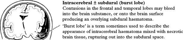
These may occur under or opposite (contre-coup) the site of impact, but most commonly involve the frontal and temporal lobes. Contusions are usually multiple and may occur bilaterally. Multiple contusions do not in themselves contribute to depression of conscious level, but this may arise when bleeding into the contusions produces a space-occupying haematoma.
Intracranial haematoma
Intracranial bleeding may occur either outside (extradural) or within the dura (intradural).
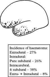
Intradural lesions usually consist of a mixture of both subdural and intracerebral haematomas although pure subdurals occur in a proportion. Brain damage is caused directly or indirectly as a result of tentorial or tonsillar herniation.
Subdural
In some patients impact may rupture bridging veins from the cortical surface to the venous sinuses producing a pure subdural haematoma with no evidence of underlying cortical contusion or laceration.

Extradural
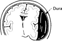
A skull fracture tearing the middle meningeal vessels bleeds into the extradural space. This usually occurs in the temporal or temporoparietal region. Occasionally extradural haematomas result from damage to the sagittal or transverse sinus.
Tentorial/tonsillar herniation (syn. ‘cone”)
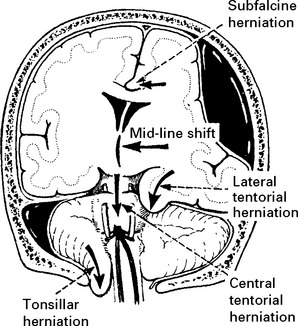
It is unlikely that high intracranial pressure alone directly damages neuronal tissue, but brain damage occurs as a result of tonsillar or tentorial herniation (see page 81). A progressive increase in intracranial pressure due to a supratentorial haematoma initially produces midline shift. Herniation of the medial temporal lobe through the tentorial hiatus follows (lateral tentorial herniation), causing midbrain compression and damage. Uncontrolled lateral tentorial herniation or diffuse bilateral hemispheric swelling will result in central tentorial herniation. Herniation of the cerebellar tonsils through the foramen magnum (tonsillar herniation) and consequent lower brain stem compression may follow central tentorial herniation or may result from the infrequently occurring traumatic posterior fossa haematoma.
Infection

The presence of a dural tear provides a potential route for infection. This seldom occurs within 48 hours of injury. Meningitis may develop after several months or years.
DIFFUSE DAMAGE
Diffuse axonal injury
Shearing forces cause immediate mechanical damage to axons. Over the subsequent 48 hours, further damage results from release of excitotoxic neurotransmitters which cause Ca2+ influx into cells and triggers the phospholipid cascade (page 246). Genetic susceptibility conferred by the presence of the APOE ͛4 gene may also play a part. Depending on the severity of the injury, effects may range from mild coma to death.
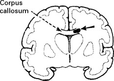
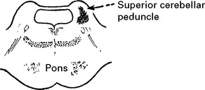
The macrosopic appearance may appear entirely normal but in some patients pathological sections reveal small haemorrhagic tears, particularly in the corpus callosum or in the superior cerebellar peduncle.
Microscopic evidence of neuronal damage depends on the duration of survival and on the severity of the injury. After a few days, retraction balls and microglial clusters are seen in the white matter.

If the patient survives 5 weeks or more after injury then appropriate staining demonstrates Wallerian degeneration of the long tracts and white matter of the cerebral hemispheres. Even a minor injury causing a transient loss of consciousness produces some neuronal damage. Since neuronal regeneration is limited, the effects of repeated minor injury are cumulative.
Cerebral swelling

This may occur with or without focal damage. It results from either vascular engorgement or an increase in extra- or intracellular fluid. The exact causative mechanism remains unknown
Cerebral ischaemia
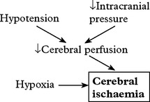
Cerebral ischaemia commonly occurs after severe head injury and is caused by either hypoxia or impaired cerebral perfusion. In the normal subject, a fall in blood pressure does not produce a drop in cerebral perfusion since ‘auto-regulation’ results in cerebral vasodilatation. After head injury, however, autoregulation is often defective and hypotension may have more drastic effects. Glutamate excess and free radical accumulation may also contribute to neuronal damage (see page 246).
MULTIPLE INJURY – PRIORITIES OF ASSESSMENT
Patients admitted in coma with multiple injuries require urgent care and the clinician must be aware of the priorities of assessment and management.
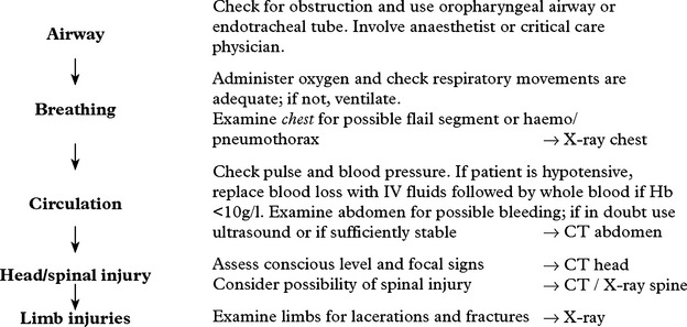
When intracranial haematoma is suspected, a CT scan is essential, especially before clinical signs are masked by a general anaesthetic required for the management of limb or abdominal injuries. However, if difficulty occurs in maintaining blood pressure, then urgent laparotomy or thoracotomy would take precedence over investigation of a possible intracranial haematoma.
HEAD INJURY – ASSESSMENT
Some patients may describe the events leading to and following head injury, but often the doctor depends on descriptions from witnesses.
Points to determine:
Period of loss of consciousness: relates to severity of diffuse brain damage and may range from a few seconds to several weeks.
Period of post-traumatic amnesia: the period of permanent amnesia occurring after head injury. This reflects the severity of damage and in severe injuries may last several weeks.
Period of retrograde amnesia: amnesia for events before the injury.
Cause and circumstances of the injury: the patient may collapse, or crash his vehicle as a result of some preceding intracranial event, e.g. subarachnoid haemorrhage or epileptic seizure. The more “violent” the injury, the greater the risk of associated extracranial injuries.
Presence of headache and vomiting: these are common symptoms after head injury. If they persist, the possibility of intracranial haematoma must be considered.
HEAD INJURY – CLINICAL ASSESSMENT
EXAMINATION

1. Lacerations and bruising
The presence of these features confirms the occurrence of a head injury, but traumatic intracranial haematoma can occur in patients with no external evidence of injury.
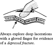
Beware of falling into the trap of diagnosing a depressed fracture when only scalp haematoma is present.

Consider the possibility of a hyperextension injury to the cervical spine if frontal laceration or bruising is present.
2. Basal skull fracture
Clinical features indicate the presence of a basal skull fracture which may be hard to detect on CT scan or skull X-ray. If present, a potential route of infection exists with the concomitant risk of meningitis.
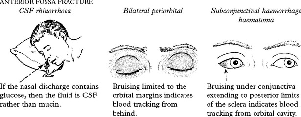
PETROUS FRACTURE
Bleeding from the external auditory meatus or CSF otorrhoea:
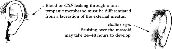
3. Conscious level – Glasgow Coma Score (GCS)
Assess patient’s conscious level in terms of eye opening, verbal and motor response on admission (see page 5) and record at regular intervals thereafter. An observation chart incorporating these features is essential and clearly shows the trend in the patient’s condition. Deterioration in conscious level indicates the need for immediate investigation and action where appropriate.

Note: This chart shows a ‘14 point scale’ with a maximum score of ‘14’ in a fully conscious patient. Many centres use a 15 point coma scale where ‘Flexion to pain’ is divided into ‘normal’ or ‘spastic’ flexion (see page 29).
4. Pupil response
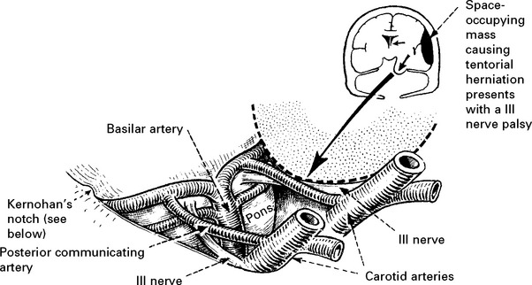
The light reflex (page 142) tests optic (II) and oculomotor (III) nerve function. Although II nerve damage is important to record and may result in permanent visual impairment, it is the III nerve function which is the most useful indicator of an expanding intracranial lesion. Herniation of the medial temporal lobe through the tentorial hiatus may damage the III nerve directly or cause midbrain ischaemia, resulting in pupil dilatation with impaired or absent reaction to light. The pupil dilates on the side of the expanding lesion and is an important localising sign. With a further increase in intracranial pressure, bilateral pupillary dilatation may occur.
5. Limb weakness
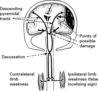
Determine limb weakness by comparing the response in each limb to painful stimuli (page 30). Hemiparesis or hemiplegia usually occurs in the limbs contralateral to the side of the lesion. Indentation of the contralateral cerebral peduncle by the edge of the tentorium cerebelli (Kernohan’s notch) may produce an ipsilateral deficit, a false localising sign more often seen with chronic subdural haematomas. Limb deficits are therefore of limited value in lesion localisation.
6. Eye movements
Evaluation of eye movements does not help in immediate management, but provides a useful prognostic guide.
Eye movements may occur spontaneously, or can be elicited reflexly (page 30) by head rotation (oculocephalic reflex) or by caloric stimulation (oculovestibular reflex).

Abnormal eye movements may result from: brain stem dysfunction, damage to the nerves supplying the extraocular muscles or damage to the vestibular apparatus. Absent eye movements relate to low levels of responsiveness and indicate a gloomy prognosis.
Vital signs
At the beginning of the century, the eminent neurosurgeon Harvey Cushing noted that a rise in intracranial pressure led to a rise in blood pressure and a fall in pulse rate and produced abnormal respiratory patterns. In the past, much emphasis has been placed on close observation of these vital signs in patients with head injury, but these changes may not occur and when present are usually preceded by deterioration in conscious level. Close observation of consciousness is therefore more relevant.
Cranial nerve lesions
Basal skull fracture or extracranial injury can result in damage to the cranial nerves. Evidence of this damage must be recorded but, with the exception of a III nerve lesion, does not usually help immediate management. Full cranial nerve examination is difficult in the comatose patient and this can await patient co-operation.
Clinical assessment cannot reliably distinguish the type or even the site of intracranial haematoma, but is invaluable in indicating the need for further investigation and in providing a baseline against which any change can be compared.
HEAD INJURY – INVESTIGATION AND REFERRAL CRITERIA
IN THE ACCIDENT AND EMERGENCY DEPARTMENT (A&E)
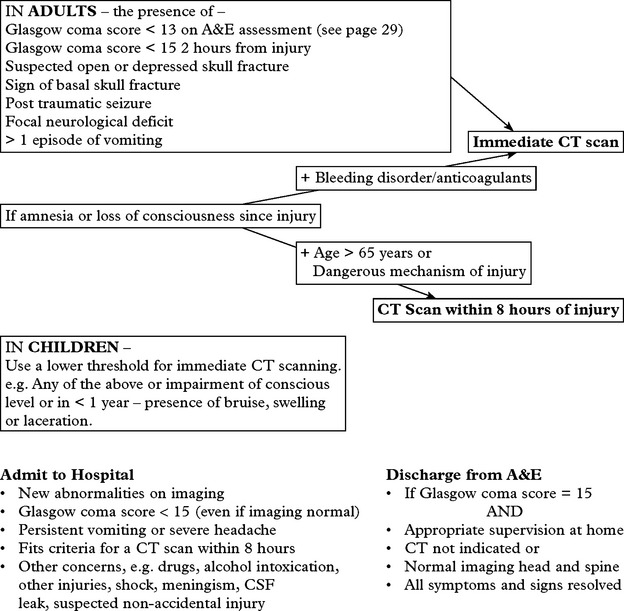
Various guidelines now exist. The following are based on those from the National Institute for Health and Clinical Excellence (NICE). (Further details available at http://www.nice.org.uk).
|
Admit to Hospital |
Discharge from A&E |
|
• New abnormalities on imaging • Glasgow coma score < 15 (even if imaging normal) • Persistent vomiting or severe headache • Fits criteria for a CT scan within 8 hours • Other concerns, e.g. drugs, alcohol intoxication, other injuries, shock, meningism, CSF leak, suspected non-accidental injury |
• If Glasgow coma score = 15 AND • Appropriate supervision at home • CT not indicated or • Normal imaging head and spine • All symptoms and signs resolved |
Referral to Neurosurgical Unit
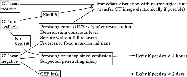
Transfer to the neurosurgical unit
Prior to the transfer, ensure that resuscitation is complete, and that more immediate problems have been dealt with (see page 221). Insert an oropharyngeal airway. Intubate and ventilate if the patient is in coma or if the blood gases are inadequate (PO2 <8 kPa on air, 13 kPa on O2 or CO2 > 6 kPa). If the patient’s conscious level is deteriorating, an intravenous bolus infusion of 100 ml of 20% mannitol should ‘buy time’ by temporarily reducing the intracranial pressure.
NOTE: for comatose patients with an unstable systemic state from multiple injuries, a negative CT scan in the local hospital may avoid a dangerous transfer to the neurosurgical unit.
Cervical spine injury may accompany head injury. Guidelines also exist with criteria for investigation. (For full details see http://www.nice.org.uk/guidance/index.jsp?action=download&o=36259).
For ADULTS and CHILDREN 10 years and over
|
AP, Lateral and Odontoid Peg X-rays if |
CT cervical spine if |
|
• Impaired neck rotation to right or left • No indication for CT scanning • Not safe to assess clinically • Neck pain/midline tenderness + ≥ 65 years or dangerous mechanism of injury • To exclude injury urgently e.g. prior to surgery |
• Patient intubated • Continued suspicion despite X-rays • Inadequate X-rays • Undergoing CT scanning for another reason e.g. Glasgow coma score < 13 or multi-region trauma |
HEAD INJURY – INVESTIGATION
CT scan: the investigation of choice for head injury (and cervical spine injury incertain circumstances – see page 227).
Scans must extend from the posterior fossa to the vertex, otherwise haematomas in these sites will be missed.
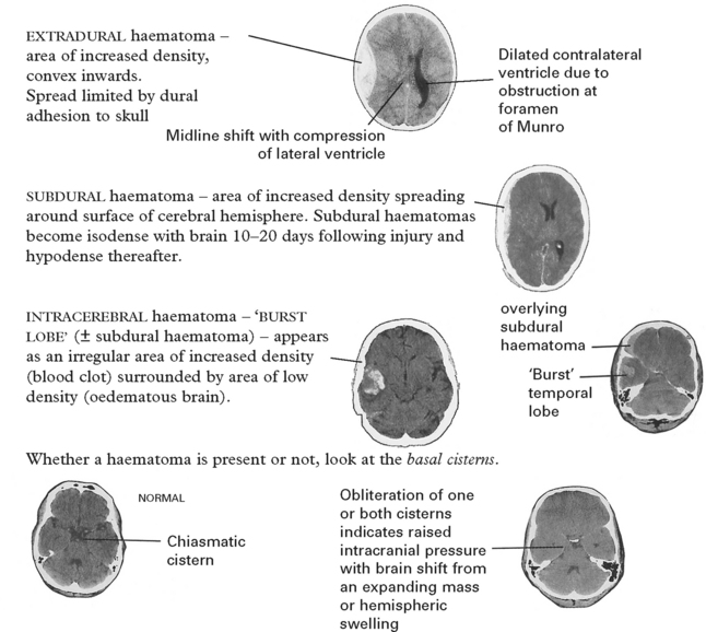
With diffuse axonal shearing injuries, small haematomas may be seen on CT scan scattered throughout the white matter, particularly in the corpus callosum, the subcortical white matter and in the brain stem adjacent to the cerebellar peduncles.
If hydrocephalus is present on the upper scan cuts, look carefully for a haematoma (extradural, subdural or intracerebral) in the posterior fossa, compressing and obstructing the 4th ventricle.
Further investigation may be required to exclude other coincidental or contributory causes of the head injury, e.g. drugs, alcohol, postictal state, encephalitis (Cause of coma, see page 86).
If a CT scan is not available, a skull fracture on X-ray identifies those at high risk of intracranial haematoma. In those patients, referral to a neurosurgical unit for a CT scan is essential (see page 227).

HEAD INJURY – MANAGEMENT
Management aims at preventing the development of secondary brain damage from intracranial haematoma, ischaemia, raised intracranial pressure with tentorial or tonsillar herniation and infection.
– Ensure the airway is patent and that blood oxygenation is adequate. Intubation is advisable in patients ‘flexing to pain’ or worse. Ventilation may be required if respiratory movements are depressed or lung function is impaired, e.g. ‘flail’ segment, aspiration pneumonia, pulmonary contusion or fat emboli. Hypoxia can cause direct cerebral damage, but in addition causes vasodilatation resulting in an increase in cerebral blood volume with subsequent rise in ICP.
– A space-occupying haematoma requires urgent evacuation (see over). If the patient’s conscious level is deteriorating, give an initial or repeat i.v. bolus of mannitol (100 ml of 20%). Coagulation should be checked and any deficits corrected.
– Scalp lacerations require cleaning, inspection to exclude an underlying depressed fracture and suturing.
– Correct hypovolaemia following blood loss – but avoid fluid overload as this may aggravate cerebral oedema. In adults, 2 litres/day of fluid is sufficient. Commence nasogastric fluids or oral fluids when feasible.
– Anticonvulsants (e.g. phenytoin) must be given intravenously if seizures occur; further seizures and in particular status epilepticus significantly increase the risk of cerebral anoxia.
– Monitor intracranial pressure (ICP), blood pressure and cerebral perfusion pressure (CPP) in selected patients with diffuse swelling or after evacuation of an intracranial haematoma. Maintain CPP either by raising blood pressure if low or by treating raised intracranial pressure.
– Brain protective agents include corticosteroids, free radical scavengers, calcium channel blockers, and glutamate antagonists. The evolution of axonal damage after a diffuse shearing injury provides a potential window of opportunity for treatment. Despite experimental animal studies revealing encouraging results, trials of these agents in head-injured patients have failed to show efficacy, perhaps due to insufficient patient numbers or a failure to target treatment at appropriate patients. A recent study of corticosteroids involving 10 000 patients has shown a worse outcome in the treatment group (the CRASH study).
– Operative repair of a dural defect is required if CSF leak persists for more than 7 days. (Many still use prophylactic antibiotics in patients with a CSF leak, but there is no conclusive evidence of their efficacy and they may do more harm than good by encouraging the growth of resistant organisms.) The development of meningitis requires prompt treatment with an empirical antibiotic.
INTRACRANIAL HAEMATOMA
Most intracranial haematomas require urgent evacuation – evident from the patient’s clinical state combined with the CT scan appearance of a space-occupying mass.
Extradural haematoma
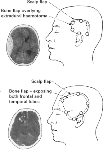
Using the CT scan the position of the extradural haematoma is accurately delineated and a ‘horse shoe’ craniotomy flap is turned over this area, allowing complete evacuation of the haematoma. For low temporal extradural haematomas, a ‘question mark’ flap may be more suitable. If patient deterioration is rapid, a burr hole and craniectomy positioned centrally over the haematoma may provide temporary relief, but this seldom provides adequate decompression.
Subdural/intracerebral haematoma (‘burst lobe’)
Subdural and intracerebral haematomas usually arise from lacerations on the under-surface of the frontal and/or temporal lobes. Again the CT scan is useful in demonstrating the exact site. A ‘question mark’ flap permits good access to both frontal and temporal ‘burst’ lobes. The subdural collection is evacuated and any underlying intracerebral haematoma is removed along with necrotic brain.
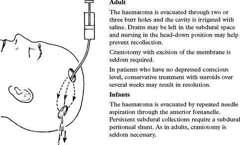
N.B. Burr holes are insufficient to evacuate an acute subdural haematoma or to deal with any underlying cortical damage.
Conservative management of traumatic intracranial haematomas
Not all patients with traumatic intracranial haematomas deteriorate. In some, the haematomas are small and clearly do not require evacuation. In others, however, the decision to operate proves difficult, e.g. the CT scan may reveal a moderate-sized haematoma with minimal or no mass effect in a conscious but confused patient.
If conservative management is adopted, careful observation in a neurosurgical unit is essential. Any deterioration indicates the need for immediate operation. In this group of patients, intracranial pressure monitoring may serve as a useful guide. An intracranial pressure of 25 mmHg or more suggests that haematoma evacuation is required as the likelihood of subsequent deterioration with continued conservative management would be high.
TREATMENT OF RAISED INTRACRANIAL PRESSURE (ICP)
Raised ICP in the absence of any easily treatable condition (e.g. intracranial haematoma or raised pCO2) requires careful management. The various techniques used to lower ICP have already been described (pages 83–84) but these must not be applied indiscriminately.
Recent studies show that even in a modern ITU head injured patient are still at risk of sustaining potentially harmful “insults” to the brain in the first few days after head injury from high ICP, low BP, low cerebral perfusion pressure (CPP), hypoxaemia, hypoglycaemia or raised temperature.
Most believe that both raised ICP and reduced cerebral perfusion pressure (CPP) can exacerbate brain damage. What is less clear is whether treatment should focus on lowering ICP or increasing CPP. When autoregulation is impaired, raising CPP beyond 70 mmHg could cause harm. The blind use of hyperventilation in the past to lower ICP by causing vasoconstriction and reduced intracranial blood volume has now been recognised to produce worse outcomes by aggravating cerebral ischaemia.
Patient selection for ICP monitoring: Monitoring ICP and CPP is most relevant in patients with a flexion response to painful stimuli or worse (a response of ‘localising to pain’ signifies a milder degree of injury and spontaneous recovery is likely). Such patients may have already undergone removal of an intracranial haematoma or may have had no mass lesion on CT scan (i.e.: diffuse injury or contusional damage). Each neurosurgical unit is likely to have its own policy for ICP monitoring but the following outline may serve as a guide for patients with no intracranial mass lesion –

DIFFUSE BRAIN DAMAGE/NEGATIVE CT SCAN
A proportion of patients have no intracranial haematoma on CT scan or have only a small haematoma or contusion without mass effect.
In these patients, coma or impairment of conscious level may be due to:

Several of these factors may coexist and contribute to brain damage in patients with intracranial haematoma.
The management principles outlined above apply; in particular it is essential to ensure that respiratory function is adequate and that cerebral perfusion pressure is maintained.
Fat emboli usually occur a few days after injury and may be related to fracture manipulation; deterioration of respiratory function usually accompanies cerebral damage and most patients require ventilation.
Meningitis may occur several days after injury in the presence of basal fractures.
Cerebral swelling may occur at any time after injury and cause a rise in intracranial pressure.
REPEAT CT SCANNING

Occasionally, small areas of ‘insignificant’ contusion on an initial CT scan may develop into a space-occupying haematoma requiring evacuation. Following haematoma evacuation, recollection may occur in 5–10% of cases.
DEPRESSED SKULL FRACTURE
This injury is caused by a blow from a sharp object. Since diffuse ‘deceleration’ damage is minimal, patients seldom lose consciousness.
SIMPLE DEPRESSED FRACTURE (closed injury)
There is no overlying laceration and no risk of infection. Operation is not required except for cosmetic reasons. Removal of any bone spicules imbedded in brain tissue does not reverse neuronal damage.
COMPOUND DEPRESSED FRACTURE (open injury)
A scalp laceration is related to (but does not necessarily overlie) the depressed bone segments. A compound depressed fracture with an associated dural tear may result in meningitis or cerebral abscess.
Investigation
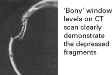
Double density appearance on skull X-ray suggests depression but tangential views may be required to establish the diagnosis. Impairment of conscious level or the presence of focal signs indicate the need for a CT scan to exclude underlying extradural haematoma or severe cortical contusion. Selecting bone window levels on CT scan will clearly demonstrate any depressed fragments.
Management
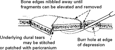
Treatment aims to minimise the risk of infection. The wound is debrided and the fragments elevated within 24 hours from injury. Bone fragments are either removed or replaced after washing with antiseptic. Antibiotics are not essential unless the wound is excessively dirty.
If the venous sinuses are involved in the depressed fracture, then operative risks from excessive bleeding may outweigh the risk of infection and antibiotic treatment alone is given.
Complications
Most patients make a rapid and full recovery, but a few develop complications:
Infection: May lead to meningitis or abscess formation. Some believe that operation does not reduce the infection risk and advocate a conservative approach unless contamination is severe.
Epilepsy: Early epilepsy (in the first week) occurs in 10% of patients with depressed fracture. Late epilepsy develops in 15% overall, but is especially common when the dura is torn, when focal signs are present, when post-traumatic amnesia exceeds 24 hours or when early epilepsy has occurred (the risk ranges from 3 to 60%, depending on the number of the above factors involved). Elevation of the bone fragments does not alter the incidence of epilepsy.
DELAYED EFFECTS OF HEAD INJURY
POST-TRAUMATIC EPILEPSY
Early epilepsy (occurring within the first week from injury)
Early epilepsy occurs in 5% of patients admitted to hospital with non-missile (i.e. deceleration) injuries. It is particularly frequent in the first 24 hours after injury. Focal seizures are as common as generalised seizures. Status epilepticus occurs in 10%.
The risk of early epilepsy is high in
– children under 5 years.
– patients with prolonged post-traumatic amnesia
– patients with an intracranial haematoma
– patients with a compound depressed fracture.
Late epilepsy (occurring after the first week from injury)
Late epilepsy also occurs in about 5% of all patients admitted to hospital after head injury. It usually presents in the first year, but in some the first attack occurs as long as 10 years from the injury. Late epilepsy is prevalent in patients with
– early epilepsy (25%)
– intracranial haematoma (35%)
– compound depressed fracture (17%).
Prophylactic anticonvulsants appear to be of little benefit in preventing the development of an epileptogenic focus. Management is discussed on page 102.
CEREBROSPINAL FLUID (CSF) LEAK
After head injury a basal fracture may cause a fistulous communication between the CSF space and the paranasal sinuses or the middle ear. Profuse CSF leaks (rhinorrhoea or otorrhoea) are readily detectable, but brain may partially plug the defect and the leak may be minimal or absent. Patients risk developing meningitis particularly in the first week, but in some this occurs after several years. When this is associated with anterior fossa fractures, it is usually pneumococcal; when associated with fractures through the petrous bone, a variety of organisms may be involved.
Clinical signs of a basal fracture have previously been described (page 222). The patient may comment on a ‘salty taste’ in the mouth. Anosmia suggests avulsion of the olfactory bulb from the cribriform plate.
Management
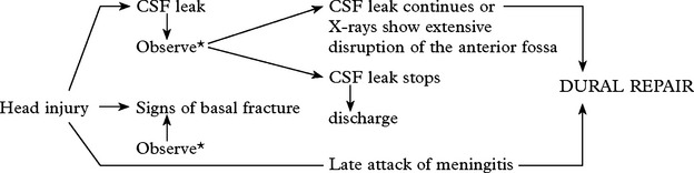 *A Cochrane Review has concluded that evidence does not support the use of prophylactic antibiotics. Prophylactic antibiotics only encourage resistance and late attacks of meningitis may still occur despite their use.
(Lancet (1994) 344:1547–1551)
Preoperative investigations
Coronal high definition CT scanning should identify the fracture site.
CT cisternography – CT scanning after running contrast injected into the lumbar theca, up to the basal cisterns may identify the exact site of the leak.
CSF isotope infusion studies combined with pledget insertion into the nasal recesses may also be of value, but results can be misleading.
Operation
As fractures of the anterior fossa often extend across the midline, a bifrontal exploration is required. The dural tear is repaired with fascia lata, pericranium or synthetic dural substitute. A CSF leak through the middle ear requires a subtemporal approach.
Failure to repair a CSF fistula may result from impaired CSF absorption with an intermittent or persistent elevation of ICP. In these patients a CSF shunt may be required.
POSTCONCUSSIONAL SYMPTOMS
Even after relatively minor head injury, patients may have persistent symptoms of:
– headache, dizziness and increased irritability
– difficulty in concentration and in coping with work
– fatigue and depression.
This condition was once thought to have a purely psychological basis, but it is now recognised that in an injury of sufficient severity to cause loss of consciousness, or a period of post-traumatic amnesia, some neuronal damage occurs; studies show a distinct delay in information processing in these patients, requiring several weeks to resolve. Vestibular ‘concussion’ (end-organ damage) may contribute to the symptomatology (‘dizziness’ and vertigo).
CUMULATIVE BRAIN DAMAGE
The effects of repeated neuronal damage are cumulative; when this exceeds the capacity for compensation, permanent evidence of brain damage ensues. The ‘punch-drunk’ state is well recognised in boxers; dementia may also occur from repeated head injury in jockeys.
CRANIAL NERVE DAMAGE
Cranial nerve damage occurs in about one-third of patients with severe head injury, but treatment is seldom of benefit. These lesions may contribute towards the patient’s residual disability.

OUTCOME AFTER SEVERE HEAD INJURY
Head injury remains a major cause of disability and death, especially in the young. Of those patients who survive the initial impact and remain in coma for at least 6 hours, approximately 40% die within 6 months. The extent of recovery in the remainder depends on the severity of the injury. Residual disabilities include both mental (impaired intellect, memory and behavioural problems) and physical defects (hemiparesis and dysphasia). Most recovery occurs within the first 6 months after injury, but improvement may continue for years. Physiotherapy and occupational therapy play an important role not only in minimising contractures and improving limb power and function but also in stimulating patient motivation.
Outcome is best categorised with the Glasgow Outcome Scale (GOS – see page 214) which uses dependence to differentiate between intermediate grades. After severe injury, about 40% regain an independent existence and may return to premorbid social and occupational activities. Inevitably some remain severely disabled requiring long term care, but few (< 2%) are left in a vegetative state with no awareness or ability to communicate with their environment (see page 214). Prognosis in this group is marginally better than for non-traumatic coma – with about one-third of those vegetative at one monthregaining consciousness within one year; of those who regain consciousness, over two-thirds either subsequently die or remain severely disabled. Of those vegetative at 3 months after the injury, none regain an independent existence.
Prognostic features following traumatic coma
The duration of coma relates closely to the severity of injury and to the final outcome, but in the early stages after injury the clinician must rely on other features – age, eye opening, verbal and motor responses, pupil response and eye movements.
|
Poor outcome (GOS 1–3) |
Favourable outcome (GOS 4–5) |
|
|
Patients in coma for > 6 hours |
61% |
39% |
|
Best Glasgow Coma Score > 11 |
18% |
82% |
|
Best Glasgow Coma Score 8–10 |
32% |
68% |
|
Best Glasgow Coma Score < 8 |
73% |
27% |
|
Pupillary response – reacting |
50% |
50% |
|
Pupillary response – non-reacting |
96% |
4% |
|
Age < 20 years |
41% |
59% |
|
Age > 60 years |
94% |
6% |
CHRONIC SUBDURAL HAEMATOMA
Subdivision of subdural haematomas into acute and subacute forms serves no practical purpose. Chronic subdural haematoma however is best considered as a separate entity, differing both in presentation and management.
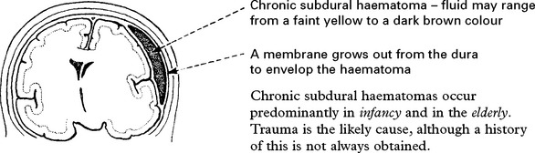
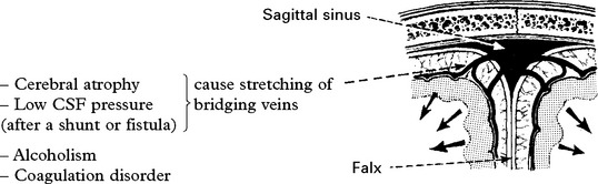
Predisposing factors
Breakdown of protein within the haematoma and a subsequent rise in osmotic pressure was originally believed to account for the gradual enlargement of the untreated subdural haematoma. Studies showing equality of osmotic pressures in blood and haematoma fluid cast doubt on this theory and recurrent bleeding into the cavity is now known to play an important role.
Clinical features tend to be non-specific.
– Dementia.
– Deterioration in conscious level, occasionally with fluctuating course.
– Symptoms and signs of raised ICP.
– Focal signs occasionally occur, especially limb weakness. This may be ipsilateral to the side of the lesion, i.e. a false localising sign (see page 224).
Diagnosis
CT Scan appearances depend on the time between the injury and the scan.
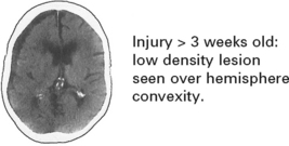
With injuries 1–3 weeks old, the subdural haematoma may be isodense with brain tissue. In this instance, i.v. contrast enhancement may delineate the cortical margin.
Beyond 3 weeks subdural haematomas appear as a low density lesion.
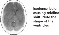
If CT scan shows midline shift without any obvious extra- or intracerebral lesion, look at the shape of the ventricles.

Management
CEREBROVASCULAR DISEASES
Vascular diseases of the nervous system are amongst the most frequent causes of admission to hospital. The annual incidence in the UK varies regionally between 150–200/100 000, with a prevalence of 600/100 000 of which one-third are severely disabled.
Better control of hypertension, reduced incidence of heart disease and a greater awareness of all risk factors have combined to reduce mortality from stroke. Despite this, stroke still ranks third behind heart disease and cancer as a cause of death in affluent societies.
RISK FACTORS
Prevention of cerebrovascular disease is more likely to reduce death and disability than any medical or surgical advance in management. Prevention depends upon the identification of risk factors and their correction. Increasing age is the strongest risk factor (but is not amenable to correction).
Hypertension
Hypertension is a major factor in the development of thrombotic cerebral infarction and intracranial haemorrhage.
There is no critical blood pressure level; the risk is related to the height of blood pressure and increases throughout the whole range from normal to hypertensive. A 6 mmHg fall in diastolic blood pressure is associated in relative terms with a 40% fall in the fatal and non-fatal stroke rate.
Systolic hypertension (frequent in the elderly) is also a significant factor and not as harmless as previously thought.
Cardiac disease
Cardiac enlargement, failure and arrhythmias, as well as rheumatic heart disease, patent foramen ovale and, rarely, cardiac myxoma are all associated with an increased risk of stroke.
Diabetes
The risk of cerebral infarction is increased twofold in diabetes. More effective treatment of diabetes has not reduced the frequency of atherosclerotic sequelae.
Heredity
Close relatives are at only slightly greater risk than non-genetically related family members of a stroke patient. Diabetes and hypertension show familial propensity thus clouding the significance of pure hereditary factors.
Blood lipids, cholesterol, smoking, diet/obesity
These factors are much less significant than in the genesis of coronary artery disease.
Race
Alterations in life style, diet and environment probably explain the geographical variations more than racial tendencies.
Haematocrit
A high blood haemoglobin concentration (or haematocrit level) is associated with an increased incidence of cerebral infarction. Other haematological factors, such as decreased fibrinolysis, are important also.
Oral contraceptives
Combined oral contraception (COC) containing high dose oestrogen increased the risk of thrombosis, including stroke. The effect of low dose oestrogen COC is less clear.
CEREBROVASCULAR DISEASE – MECHANISMS
‘Stroke’ is a generic term, lacking pathological meaning. Cerebrovascular diseases can be defined as those in which brain disease occurs secondary to a pathological disorder of blood vessels (usually arteries) or blood supply.

CEREBROVASCULAR DISEASE – NATURAL HISTORY
Approximately one-third of all ‘strokes’ are fatal. The age of the patient, the anatomical size of the lesion, the degree of deficit and the underlying cause all influence the outcome.
Immediate outcome
In cerebral haemorrhage, mortality approaches 50%.
Cerebral infarction fares better, with an immediate mortality of less than 20%, fatal lesions being large with associated oedema and brain shift.
Embolic infarction carries a better outcome than thrombotic infarction.
Fatal cases of infarction die either at onset, within a few days because of cytotoxic cerebral oedema or later from cardiovascular or respiratory complications.
The level of consciousness on admission to hospital gives a good indication to immediate outcome. The deeper the conscious level the graver the prognosis.
Long-term outcome
The prognosis following infarction due to thrombosis or embolisation from diseased neck vessels or heart is dependent on the progression of the underlying atherosclerotic disease. Recurrent cerebral infarction rates vary between 5% and 15% per year. Symptoms of coronary artery disease and/or peripheral vascular disease may also ensue. Five year mortality is 44% for males and 36% for females.
The long-term prognosis following survival from haemorrhage depends upon the cause and the treatment.
CEREBROVASCULAR DISEASE – CAUSES
OCCLUSION (50%)
Atheromatous/thrombotic

Non-atheromatous diseases of the vessel wall
1. Collagen disease e.g. rheumatoid arthritis systemic lupus erythematosus (SLE)
2. Vasculitis e.g. polyarteritis nodosa temporal arteritis
3. Granulomatous vasculitis e.g. Wegener’s granulomatosis
4. Miscellaneous e.g. trauma fibromuscular dysplasia syphilitic vasculitis
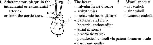
EMBOLISATION (25%) from:
DISEASES OF BLOOD
e.g. Coagulopathies or Haemoglobinopathies
CEREBRAL VENOUS THROMBOSIS
Thrombosis of cerebral veins may occur with infection, dehydration or in association with oestrogen excess, either post-partum or combined oral contraceptive use.
DECREASED CEREBRAL PERFUSION
Hypotension, from cardiac arrhythmia or GI bleed, can lead to infarction in the watershed between arterial territories.
HAEMORRHAGE (20%)
|
Into the brain substance – parenchymal (15%) and/or subarachnoid space (5%) |
Neoplasm |
|
Coagulation disorder e.g. haemophilia |
|
|
Hypertension |
Anticoagulant therapy |
|
Amyloid vasculopathy |
Vasculitis |
|
Aneurysm |
Drug abuse e.g. cocaine |
|
Arteriovenous malformation |
Trauma |
OCCLUSIVE AND STENOTIC CEREBROVASCULAR DISEASE
PATHOLOGY
The normal vessel wall comprises:

Within brain and spinal cord tissue the adventitia is usually very thin and the elastic lamina between media and adventitia less apparent.
The intima is an important barrier to leakage of blood and constituents into the vessel wall. In the development of the atherosclerotic plaque, damage to the endothelium of the intima is the primary event.
The atherosclerotic plaque
Following intimal damage:
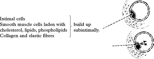
Haemorrhage may occur within the plaque or the plaque may ulcerate into the lumen of the vessel forming an intraluminal mural thrombus. Either way, the lumen of the involved vessel is narrowed (stenosed) or blocked (occluded).
The plaque itself may give rise to emboli. Cholesterol is present partly in crystal form and fragments following plaque rupture may be sufficiently large to occlude the lumen of distal vessels. The cholesterol esters, lipids and phospholipids each play a role in the aggregation of such emboli.
The carotid bifurcation in the neck is a frequent site at which the antheromatous plaque causes stenosis or occlusion.

Platelet emboli arise from thrombus developed over the damaged endothelium. This thrombus is produced partly by platelets coming into contact with exposed collagen fibres. Endothelial cells synthesise PROSTACYCLIN which is a potent vasodilator and inhibitor of platelet aggregation. THROMBOXANE A2, synthesised by platelets, has opposite effects. In thrombus formation these two PROSTAGLANDINS actively compete with each other.
CEREBROVASCULAR DISEASE – PATHOPHYSIOLOGY
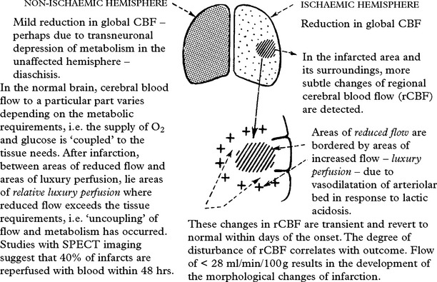
Standard techniques of cerebral blood flow (CBF) measurement provide information on both global and regional flow in patients with cerebral ischaemia or infarction. Recent availability of positron emission tomography (PET), recording oxygen and glucose metabolism, as well as blood flow and blood volume, gives a more detailed and accurate understanding of pathophysiological changes after stroke.
Changes in cerebral infarction
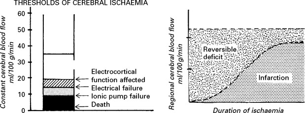
Pathophysiology of ischaemia
Progression from reversible ischaemia to infarction depends upon the degree and duration of the reduced blood flow.
Ischaemic cascade
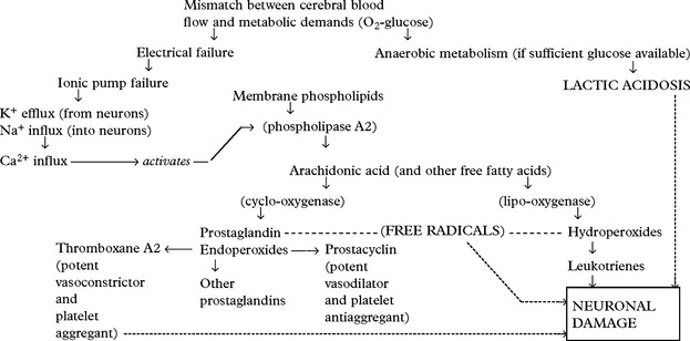
A significant fall in cerebral blood flow produces a cascade of events which, if unchecked, lead to the production and accumulation of toxic compounds and apoptosis (programmed cell death).
Role of neurotransmitters
In addition to the cascade outlined above one of the amino acid excitatory neurotransmitters, Glutamate, in excess is a powerful neurotoxin, which plays an important role in ischaemic brain damage.
There have been numerous agents that interfere with different steps in this complicated series of interactions with an ultimate aim of providing neuroprotection and limiting the size of the stroke. Despite numerous trials of more than 100 different agents no drug has been developed that provides neuroprotection in man.
TRANSIENT ISCHAEMIC ATTACKS (TIAs)
Transient ischaemic attacks are episodes of focal neurological symptoms due to inadequate blood supply to the brain. Attacks are sudden in onset, resolve within 24 hours or less and leave no residual deficit. These attacks are important as warning episodes or precursors of cerebral infarction.
Before diagnosing TIAs, consider other causes of transient neurological dysfunction – migraine, partial seizures, hypoglycaemia, syncope and hyperventilation.
The pathogenesis of transient ischaemic attacks
A reduction of cerebral blood flow below 20–30 ml/100 g/min produces neurological symptoms. The development of infarction is a consequence of the degree of reduced flow and the duration of such a reduction. If flow is restored to an area of brain within the critical period, ischaemic symptoms will reverse themselves. TIAs may be due to:
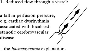
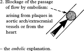
Both mechanisms occur. Emboli are accepted as the cause of the majority of TIAs.
The symptomatology of TIAs
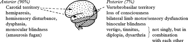
A small number of transient ischaemic attacks are difficult to fit convincingly into either anterior or posterior circulation, e.g. dysarthria with hemiparesis.
The natural history of TIAs
Following a TIA, 5% of patients will develop infarction within 1 week and 12% within 3 months. The risk of infarction is greatest in older patients with more risk factors (hypertension and diabetes) who have had longer hemispheric TIAs. About 10% of patients who have a stroke have had a warning TIA.
CLINICAL SYNDROMES – LARGE VESSEL OCCLUSION
OCCLUSION OF THE INTERNAL CAROTID ARTERY – may present in a ‘stuttering’ manner due to progressive narrowing of the lumen or recurrent emboli.
The degree of deficit varies – occlusion may be asymptomatic and identified only at autopsy, or a catastrophic infarction may result.
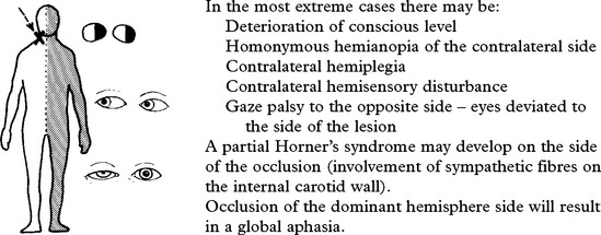
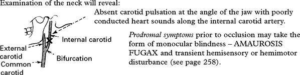
The origins of the vessels from the aortic arch are such that an innominate artery occlusion will result not only in the clinical picture of carotid occlusion but will produce diminished blood flow and hence blood pressure in the right arm.
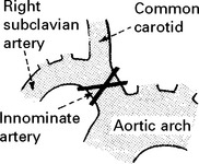
The outcome of carotid occlusion depends on the collateral blood supply primarily from the circle of Willis, but, in addition, the external carotid may provide flow to the anterior and middle cerebral arteries through meningeal branches and retrogradely through the ophthalmic artery to the internal carotid artery.

ANTERIOR CEREBRAL ARTERY
Anatomy
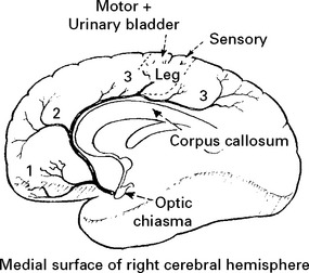
The anterior cerebral artery is a branch of the internal carotid and runs above the optic nerve to follow the curve of the corpus callosum. Soon after its origin the vessel is joined by the anterior communicating artery. Deep branches pass to the anterior part of the internal capsule and basal nuclei.
Cortical branches supply the medial surface of the hemisphere:
1. Orbital
2. Frontal
3. Parietal
Clinical features
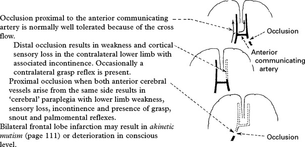
The anterior cerebral artery may be occluded by embolus or thrombus. The clinical picture depends on the site of occlusion (especially in relation to the anterior communicating artery) and anatomical variation, e.g. both anterior cerebral arteries may arise from one side by enlargement of the anterior communicating artery.
MIDDLE CEREBRAL ARTERY
Anatomy
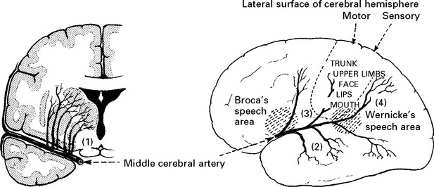
The middle cerebral artery is the largest branch of the internal carotid artery. It gives off (1) deep branches (perforating vessels – lenticulostriate) which supply the anterior limb of the internal capsule and part of the basal nuclei. It then passes out to the lateral surface of the cerebral hemisphere at the insula of the lateral sulcus. Here it gives off cortical branches (2) temporal, (3) frontal, (4) parietal.
Clinical features
The middle cerebral artery may be occluded by embolus or thrombus. The clinical picture depends upon the site of occlusion and whether dominant or non-dominant hemisphere is affected.
Occlusion at the insula

When cortical branches are affected individually, the clinical picture is less severe, e.g. involvement of parietal branches alone may produce Wernicke’s dysphasia with no limb weakness or sensory loss.
The deep branches (perforating vessels) of the middle cerebral artery may be a source of haemorrhage or small infarcts (lacunes – see later).
VERTEBRAL ARTERY OCCLUSION
Anatomy
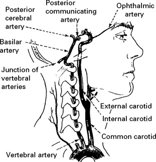
The vertebral artery arises from the subclavian artery on each side. Underdevelopment of one vessel occurs in 10%.
The vertebral artery runs from its origin through the foramen of the transverse processes of the mid-cervical vertebrae. It then passes laterally through the transverse process of the axis, then upwards to the atlas accompanied by a venous plexus and across the suboccipital triangle to the vertebral canal. After piercing the dura and arachnoid matter, it enters the cranial cavity through the foramen magnum. At the lower border of the pons, it unites with its fellow to form the basilar artery.
The vertebral artery and its branches supply the medulla and the inferior surface of the cerebellum before forming the basilar artery.
Clinical features
Occlusion of the vertebral artery, when low in the neck, is compensated by anastomotic channels.
When one vertebral artery is hypoplastic, occlusion of the other is equivalent to basilar artery occlusion.
Only the posterior inferior cerebellar artery (PICA) depends solely on flow through the vertebral artery. Vertebral artery occlusion may therefore present as a PICA syndrome (page 255).
The close relationship of the vertebral artery to the cervical spine is important. Rarely, damage at intervertebral foramina or the atlanto-axial joints following subluxation may result in intimal damage, thrombus formation and embolisation.
Vertebral artery compression during neck extension may cause symptoms of intermittent vertebrobasilar insufficiency.

BASILAR ARTERY OCCLUSION
Anatomy

The basilar artery supplies the brain stem from medulla upwards and divides eventually into posterior cerebral arteries as well as posterior communicating arteries which run forward to join the anterior circulation (circle of Willis).
Branches can be classified into:
1. Posterior cerebral arteries
2. Long circumflex branches
3. Paramedian branches.
Clinical features
Prodromal symptoms are common and may take the form of diplopia, visual field loss, intermittent memory disturbance and a whole constellation of other brain stem symptoms:
– vertigo
– ataxia
– paresis
– paraesthesia
The complete basilar syndrome following occlusion consists of:
– impairment of consciousness → coma
– bilateral motor and sensory dysfunction
– cerebellar signs
– cranial nerve signs indicative of the level of occlusion.
The clinical picture is variable. Occasionally basilar thrombosis is an incidental finding at autopsy.
‘Top of basilar’ occlusion: This results in lateral midbrain, thalamic, occipital and medial temporal lobe infarction. Abnormal movements (hemiballismus) are associated with visual loss, pupillary abnormalities, gaze palsies, impaired conscious level and disturbances of behaviour.
Paramedian perforating vessel occlusion gives rise to the ‘LOCKED-IN’ SYNDROME (page 256) and LACUNAR infarction (page 257).
POSTERIOR CEREBRAL ARTERY
Anatomy
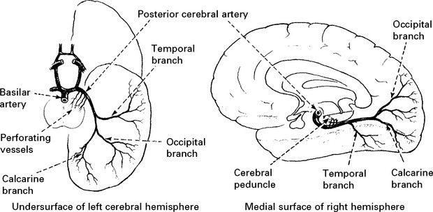
The posterior cerebral arteries are the terminal branches of the basilar artery. Small perforating branches supply midbrain structures, choroid plexus and posterior thalamus. Cortical branches supply the undersurface of the temporal lobe – temporal branch; and occipital and visual cortex – occipital and calcarine branches.
Clinical features
Proximal occlusion by thrombus or embolism will involve perforating branches and structures supplied:
Midbrain syndrome – III nerve palsy with contralateral hemiplegia – WEBER’s SYNDROME
Thalamic syndromes – chorea or hemiballismus with hemisensory disturbance.
Occlusion of cortical vessels will produce a different picture with visual field loss (homonymous hemianopia) and sparing of macular vision (the posterior tip of the occipital lobe, i.e. the macular area, is also supplied by the middle cerebral artery).
Posterior cortical infarction in the dominant hemisphere may produce problems in naming colours and objects.
CLINICAL SYNDROMES – BRANCH OCCLUSION
BASILAR ARTERY – LONG CIRCUMFLEX BRANCH OCCLUSION
Anatomy
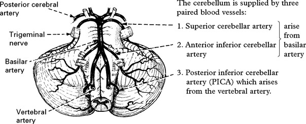
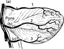
It can be seen that a vascular lesion in the territory of these vessels will produce, not only cerebellar, but also brain stem symptoms and signs localising to:
(a) superior pontine,
(b) inferior pontine and
(c) medullary levels.
Clinical features
Superior cerebellar artery syndrome results in:

Anterior inferior cerebellar artery syndrome results in:

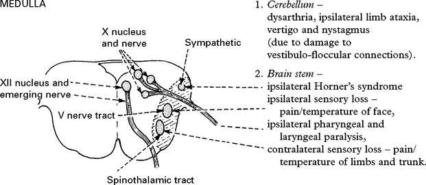
Posterior inferior cerebellar artery syndrome (lateral medullary syndrome) results in:
BASILAR ARTERY – PARAMEDIAN BRANCH OCCLUSION
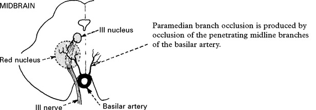
At the midbrain level damage to the nucleus or the fasciculus of the oculomotor nerve (III) will result in a complete or partial III nerve palsy; damage to the red nucleus (outflow from opposite cerebellar hemisphere) will also produce contralateral tremor – referred to as BENEDIKT’S SYNDROME.

At the pontine level an abducens nerve (VI) palsy will occur with ipsilateral facial (VII) weakness and contralateral sensory loss – light touch, proprioception (medial lemniscus damage)when the lesion is more basal.
Abducens and facial palsy may be accompanied by contralateral hemiplegia – MILLARD-GUBLER SYNDROME.
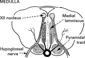
At the medullary level, bilateral damage usually occurs and results in the ‘LOCKED-IN’ SYNDROME. The patient is paralysed and unable to talk, although some facial and eye movements are preserved.
Spinothalamic sensation is retained, but involvement of the medial lemniscus produces loss of ‘discriminatory’ sensation in the limbs. The syndrome usually follows basilar artery occlusion and carries a grave prognosis.
CLINICAL SYNDROMES – LACUNAR STROKE (LACI)
Occlusion of deep penetrating arteries produces subcortical infarction characterised by preservation of cortical function – language, other cognitive and visual functions.
Clinical syndromes are distinctive and normally result from long-standing hypertension. In 80%, infarcts occur in periventricular white matter and basal ganglia, the rest in cerebellum and brain stem. Areas of infarction are 0.5 –1.5cm in diameter and occluded vessels demonstrate lipohyalinosis, microaneurysm and microatheromatous changes. Lacunar or subcortical infarction accounts for 17% of all thromboembolic strokes and knowledge of commoner syndromes is essential.
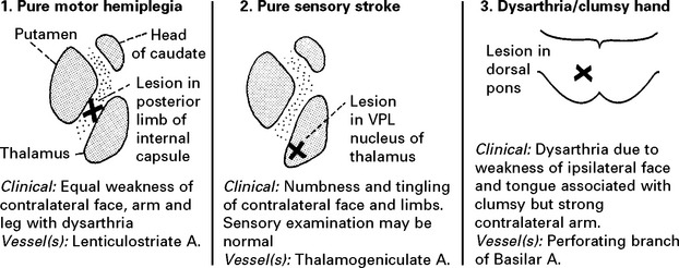
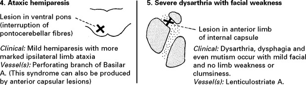
Sensorimotor syndromes are common although anatomical basis is obscure. A recent Stroke Data Bank survey showed the commonest presentations to be:
Pure motor hemiplegia 57%
Sensorimotor 20%
Ataxic hemiparesis 10%
Pure sensory 7%
Dysarthria/Clumsy hand 6%
Investigations MRI is superior to CT demonstrating lacunae, although either may occasionally misdiagnose a small resolving haematoma. Confirmation of lacunar stroke may save patients from unnecessary investigations for carotid and cardiac embolic source.
Prognosis For all syndromes this is encouraging. Careful control of blood pressure and the use of aspirin usually prevents recurrence. Multiple lacunar infarctions – ‘état lacunaire’ – results in shuffling gait, pseudobulbar palsy and subcortical dementia.
CLASSIFICATION OF SUBTYPES OF CEREBRAL INFARCTION
A recently devised classification of infarction has proved simple and of practical value in establishing diagnosis and in predicting outcome –
|
Clinical features |
Outcome |
|
|
Total Anterior Circulation |
motor and sensory deficit, hemianopia and disturbance of higher cerebral function |
Poor |
|
Syndrome (TACS) |
||
|
Partial Anterior Circulation |
any two of above |
Variable |
|
Syndrome (PACS) |
or isolated disturbance of cerebral function |
|
|
Posterior Circulation |
signs of brain stem dysfunction |
Variable |
|
Syndrome (POCS) |
or isolated hemianopia |
|
|
Lacunar Anterior |
pure motor stroke |
Good |
|
Circulation Syndrome (LACS) |
or pure sensory stroke |
|
|
or pure sensorimotor stroke |
||
|
or ataxic hemiparesis |
||
EMBOLISATION
Emboli consist of friable atheromatous material, platelet-fibrin clumps or well formed thrombus.
The diagnosis of embolic infarction depends on:
• The identification of an embolic source, e.g. cardiac disease.
• The clinical picture of sudden onset.
• Infarction in the territory of a major vessel or large branch.
Clinical picture – depends on the vessel involved. Emboli commonly produce transient ischaemic attacks (TIA) as well as infarction.
Symptoms are referable to the eye (retinal artery) and to the anterior and middle cerebral arteries, and take the form of:
Visual loss – transient, i.e. amaurosis fugax or permanent.
Hemisensory and hemimotor disturbance.
Disturbance of higher function, e.g. dysphasia.
Focal or generalised seizures – may persist for some time after the ischaemic episode.
Depression of conscious level if major vessel occlusion occurs.
Emboli less frequently affect the posterior circulation.
EMBOLI FROM THE INTERNAL CAROTID ARTERY AND AORTA
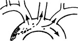
Emboli from these sources are commonest outwith the heart. The majority of all cerebral emboli arise from ulcerative plaques in the carotid arteries (see page 244).
Emboli arising from the aorta (atheromatous plaque or aortic aneurysm) often involve both hemispheres and systemic embolisation (e.g. affecting limbs) may coexist.
EMBOLI OF CARDIAC ORIGIN

Non-bacterial endocarditis (marantic endocarditis): associated with malignant disease due to fibrin and platelet deposition on heart valves.
Atrial myxoma is a rare cause of recurrent cerebral embolisation. Bihemisphere episodes with a persistently elevated ESR should arouse suspicion which may be confirmed by cardiac ultrasound.
Patent foramen ovale may result in paradoxical embolisation; suspect in patient with deep venous thrombosis who develops cerebral infarction. Emboli can also arise from intracardiac thrombus.
New cardiac imaging techniques especially Transoesophageal Echocardiography (TOE) allow a more accurate detection of potential embolic source. Transcranial Doppler (TCD) may characterise emboli by analysing their signals and help quantify risk of recurrence.
EMBOLI FROM OTHER SOURCES
Fat emboli: following fracture, especially of long bones and pelvis, fat appears in the bloodstream and may pass into the cerebral circulation, usually 3 –6 days after trauma. Emboli are usually multiple and signs are diffuse.
Air emboli follow injury to neck/chest, or follow surgery. Rarely, air emboli complicate therapeutic abortion. Again the picture is diffuse neurologically. Onset is acute; if the patient survives the first 30 minutes, prognosis is excellent.
Nitrogen embolisation or decompression sickness (the ‘bends’) produces a similar picture, but if the patient survives, neurological disability may be profound.
Tumour emboli result in metastatic lesions; the onset is usually slow and progressive. Acute stroke-like presentation may occur, followed weeks or months later by the mass effects.

STENOTIC/OCCLUSIVE DISEASE – INVESTIGATIONS
1. CONFIRM THE DIAGNOSIS
Computerised tomography (CT scan)
All patients should have a CT scan, urgently if
– conscious level depressed
– diagnosis uncertain
– on anticoagulants
– before commencing/resuming antithrombotics
– if thrombolysis is considered.
– severe headache at onset.
Infarction is evident as a low-density lesion which conforms to a vascular territory, i.e. usually wedge shaped. Subtle changes occur within 3 hours in some patients; most scans become abnormal within 48 hours.
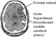
CT scan also identifies:
– the site and size of the infarct, providing a prognostic guide
– the presence of haemorrhagic infarction where bleeding occurs into the infarcted area
– intracerebral haemorrhage or tumour.
Magnetic resonance imaging (MRI)
T2 prolongation (hyperintensity in relation to white and grey matter) occurs within hours of onset of ischaemic symptoms. Advanced techniques, diffusion weighted imaging (DWI) and perfusion imaging (PWI) show respectively early infarction (cytotoxic oedema) and ischaemic tissue at risk (the ischaemic penumbra). These advanced techniques are valuable predictors of outcome and guide treatments directed as ‘ischaemic salvage’ e.g. thrombolysis.
2. DEMONSTRATE THE SITE OF PRIMARY LESION
(a) Non-invasive investigation
Ultrasound – Doppler/Duplex scanning: assesses extra- and intracranial vessels (page 44). A normal study precludes the need for angiography.
Cardiac ultrasound (transthoracic or transoesophageal): this often reveals a cardiac embolic source in young people with stroke, e.g. prolapsed mitral valve, patent foramen ovale.
Magnetic resonance angiography (MRA)
‘Time of flight’ or contrast enhanced techniques are used. Whilst of value in patients with heavily calcified carotid plaques, resistant to Doppler, it tends to overestimate the severity of stenosis. When assessing the carotid arteries it is best used in combination with Doppler. Its non-invasive nature makes it helpful in investigating the intracranial circulation.
Computed tomographic angiography (CTA)
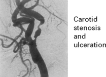
Dynamic helical CT, following bolus injection of non-ionic contrast, can be used to investigate both intracranial and extracranial vasculature. CTA compared with DSA correctly classifies the degree of carotid stenosis in 96% of cases but is insensitive to ulcerative plaque. Again it is best used in conjunction with Doppler.
(b) Digital intravenous subtraction angiography (DSA)
The combination of the above techniques has decreased the need for invasive investigation but cerebral angiography may still be required to make a definitive diagnosis.
Indications of angiography
1. In those patients with anterior circulation TIA or minor stroke if non-invasive techniques have not clarified the nature and degree of carotid stenosis.
2. In patients where unusual aetiologies are suspected and less invasive imaging has not been diagnostic – for example young patients or when cerebral vasculitis is suspected.
3. IDENTIFY FACTORS WHICH MAY INFLUENCE TREATMENT AND OUTCOME
General investigations identify conditions which may predispose towards premature cerebrovascular disease. These are essential in all patients.
Chest x-ray – cardiac enlargement – hypertension/valvular heart disease
ECG – ventricular enlargement and/or arrhythmias – hypertension/embolic disease recent myocardial infarct – embolic disease sinoatrial conduction defect – embolic disease/output failure
Blood glucose – diabetes mellitus
Serum lipids and cholesterol – hyperlipidaemia

VDRL-TPHA – neurosyphilis
Prothrombin time – circulating auto-anticoagulants
Partial thromboplastin time (PTT) – prolonged by lupus anticoagulant
Note drug history – oral contraceptives, amphetamines, opiates
Following the interpretation of these preliminary investigations, more detailed studies may be required, e.g.
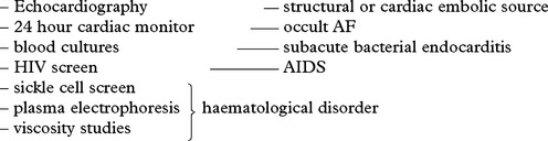
– anticardiolipin antibodies – antiphospholipid syndrome
– muscle biopsy – mitochondrial disease
CEREBRAL INFARCTION – MANAGEMENT
THE ACUTE STROKE
Clinical history, examination and investigation will separate infarction and haemorrhage. Once the nature of the ‘stroke’ has been confidently defined, treatment should be instigated. The treatment of stroke has been the subject of many clinical trials and the following is a digest of the current advice based on those studies.
Treatment aims
– Recanalise blocked vessels
– Prevent progression of present event
– Prevent immediate complication
– Prevent the development of subsequent events
– Rehabilitate the patient.
General measures
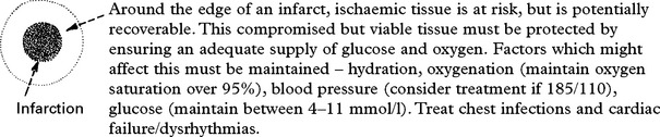
Specific measures
Thrombolysis
Intravenous recombinant tissue plasminogen activator (alteplase) given within 3 hours of an anterior circulation ischaemic stroke improves outcome despite the increased risk of iatrogenic intracranial haemorrhage. Thus patients who might be candidates need urgent assessment and CT scanning to exclude cerebral haemorrhage.
Key contraindications to thrombolysis –
Uncertain time of onset
Spontaneously improving
Head injury or previous stroke in last 3 months
GI surgery in last 21 days
BP >180/110
On anticoagulant
Seizure
Hypodensity on CT
Patients not eligible for thrombolysis
Give aspirin 300 mg daily for 2 weeks or clopidogrel in aspirin intolerant patients. Anticoagulants should be avoided if possible as they increase the risk of deterioration from haemorrhagic transformation.
Transfer to stroke unit
There is good evidence that multidisciplinary care on a stroke unit improves the outcome of patients with stroke.
Assess swallow
Aspiration pneumonia is a significant complication after stroke. Minimise this risk by assessing swallowing and using a nasogastric tube for fluids and food if swallowing unsafe.
Early mobilisation
Help patients sit up when possible and mobilise early.
Special situations
Decompressive hemicraniectomy
A small number of young patients (<60) with large middle cerebral artery strokes deteriorate after 24–72 hours from massive cytotoxic cererbral oedema which is resistant to medical therapy. Surgical decompression can save life, allowing many patients to make reasonable recoveries.
Other neurosurgical interventions
Patients with large cerebellar infarcts can deteriorate 24–48 hours after their stroke when oedema leads to compression of the posterior fossa and associated hydrocephalus. Posterior fossa decompression can be life saving and many patients then make good recoveries.
Prevention of further stroke
The recognition of risk factors and their correction to minimise the risk of further events forms a necessary and important step in long-term treatment.
The strategies here are the same as those used for treatment for patients with TIA (see below).
– Control hypertension
– mphasise the need to stop cigarette smoking
– Correct lipid abnormality
– Give platelet antiaggregation drugs (aspirin or in selected cases Dipyridamole or Clopidogrel) to reduce the rate of reinfarction
– Remove or treat embolic source (long term anticoagulation in atrial fibrillation). Defer anticoagulation in disabling stroke for 2 weeks as risk of haemorrhage outweighs benefit.
– Treat inflammatory or vascular inflammatory diseases
– Stop thrombogenic drugs, e.g. oral contraceptives.
TIAs AND MINOR INFARCTION – MANAGEMENT
The aim of treatment is to prevent subsequent cerebral infarction:
Establish diagnosis and exclude other pathologies causing transient neurological symptoms, e.g. migraine.

Correct predisposing condition.
Examine patient for evidence of extracranial vascular disease:
Palpate carotids, upper limb pulses. Auscultate the neck for bruits.
Check blood pressure in both arms. Examine heart.
Medical treatment
Prevention of a further cerebrovascular event (secondary prevention) depends on the cause, which for almost all patients is atherosclerosis – Stop smoking, control diabetes, reduce, cholesterol or blood pressure, even if the levels are in the normal range.
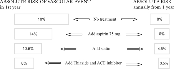
Adding dipyridamole with aspirin reduces the risk further. Clopidogrel has a similar effect to aspirin.
Special situations:
Atrial fibrillation – high risk of recurrence (12%) is reduced to approximately 4% by oral anticoagulation.
Surgical and other interventional treatments
Carotid stenosis – high quality surgical studies demonstrated that patients with carotid stenosis >70% (though not with occlusion) and a TIA or small stroke in carotid territory have a lower risk of further stroke if they undergo a carotid endarterectomy. This benefit depends on the procedure being done by an experienced surgeon with low rate of complications. The risk of stroke, and thus the benefit of surgery is highest with higher grades of stenosis and in patients with hemisphere TIA (as opposed to amaurosis fugax). The risk of complications for patients with lower degrees of stenosis outweighs the benefit.
Carotid angioplasty and stenting is an alternative to carotid endarterectomy in patients with stenosis of >70% but recent studies have found a higher risk of late recurrence than for surgery, The role for these interventions in vertebrobasilar stenosis is not yet established.
Other surgical interventions, for example the superficial temporal to middle cerebral artery bypass provide no benefit.
HYPERTENSION AND CEREBROVASCULAR DISEASE
Next to age, the most important factor predisposing to cerebral infarction or haemorrhage is hypertension. The risk is equal in males and females and is proportional to the height of blood pressure (diastolic and systolic).

The pathological effects of sustained hypertension are:
– Charcot Bouchard microaneurysms → INTRACEREBRAL HAEMORRHAGE (from perforating vessels)
– Accelerated atheroma and thrombus formation → INFARCTION (large vessels)
– Hyalinosis and fibrin deposition → INFARCTION (lacunes – small vessels)
HYPERTENSIVE ENCEPHALOPATHY
An acute, usually transient, cerebral syndrome precipitated by sudden severe hypertension. The excessive blood pressure may be due to malignant hypertension from any cause, or uncontrolled hypertension in glomerulonephritis, pregnancy (eclampsia) or phaeochromocytoma.
The mechanism is complex: Cerebral resistance vessels

Clinical features: Headache and confusion precede convulsions and coma. Papilloedema with haemorrhages and exudates are invariably found. Proteinuria and signs of renal and cardiac failure are common.
Diagnosis: CT scanning shows a widespread white matter low attenuation and excludes other pathology. MRI confirms increased brain water content and SPECT shows hyperperfusion adjacent to these changes.
Treatment: a precipitous fall in blood pressure can result in retinal damage and watershed infarction. Gradually reduce blood pressure with i.v. nitroprusside or hydralazine. Reserve peritoneal dialysis for resistant cases.
N.B. With treatment full recovery is usual. Without treatment death occurs.
BINSWANGER’S ENCEPHALOPATHY (Subcortical arteriosclerotic encephalopathy – SAE)
A rare disorder in which progressive dementia and pseudobulbar palsy are associated with diffuse hemisphere demyelination. The CT scan shows areas of periventricular low attenuation, often also involving the external capsule. The pathological changes were previously attributed to chronic diffuse oedema, but the recent finding of a high plasma viscosity in these patients suggests that this, in conjunction with hypertensive small vessel disease, could produce chronic ischaemic change in central white matter.
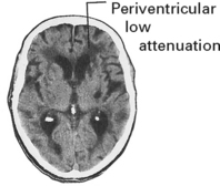
Subclinical forms of this disease may exist as this CT scan appearance is occasionally found in asymptomatic patients.
MRI appears more sensitive in establishing radiological diagnosis.
NON-ATHEROMATOUS CEREBROVASCULAR DISEASE
ABNORMALITIES OF EXTRACRANIAL VESSELS
SPONTANEOUS AND TRAUMATIC ARTERIAL DISSECTION
Extracranial and intracranial dissections are an underdiagnosed cause of stroke in young persons. Spontaneous dissections occur in Marfan’s syndrome or collagen disorders (Ehlers–Danlos), but more frequently in patients with no clear risk factors. Whilst there may be a clear history of neck trauma, often the trauma is minor (eg a sneeze). This may lead to dissection and stenosis or occlusion. The vertebral arteries are particularly susceptible to trauma in view of their close relationship to the cervical spine at intervertebral foramina, the atlanto-axial joint and the occipito-atlantal joint. Carotid dissection may present with a painful isolated Horner’s syndrome or lower cranial nerve palsies. Angiography or CT/MR angiography will confirm. Treatment is with anticoagulation or antiplatelet agents. No studies are available to determine the best treatment strategy.

FIBROMUSCULAR DYSPLASIA
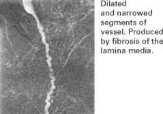
This disease involves intracranial as well as extracranial vessels which appear like a ‘string of beads’. The patient presents with infarction as a result of thrombotic occlusion or from an associated saccular aneurysm, of which there is an increased risk. Transluminal angioplasty can be used to dilate a stenotic segment.
INFLAMMATION VESSEL OCCLUSION
Infection in structures close to the carotid artery can result in inflammatory change in the vessel wall and secondary thrombosis. In children, infection in the retropharyngeal fossa (tonsillar infection) may cause cerebral infarction. Meningitis (especially pneumococcal) may result in secondary arteritis and occlusion of intracerebral vessels as they cross the subarachnoid space.
MOYAMOYA DISEASE
Bilateral occlusion of the carotid artery at the siphon is followed by the development of a fine network of collateral arteries and arterioles at the base of the brain. This may be a congenital or acquired disorder associated with previous meningitis, oral contraception or granulomatous disease (e.g. sarcoidosis). Children present with alternating hemiplegia, adults with subarachnoid haemorrhage. There is no specific treatment though some use surgical revascularisation procedures.
DISEASES OF THE VESSEL WALL
VASCULITIS AND COLLAGEN VASCULAR DISEASES
These disorders have systemic as well as neurological features. Occasionally only the nervous system is diseased. All are rare causes of stroke but need different treatments.
Collagen vascular diseases:
– Systemic lupus erythematosus
– Rheumatoid arthritis
– Other connective tissue disorders.
Vasculitis
– Vasculitis associated with connective tissue disease.
– Micropolyangiitis (previously called polyarteritis nodosa).
– Allergic angiitis (hypersensitivity vasculitis).
– Takayasu’s arteritis.
– Isolated angiitis of the central nervous system (IAC).
– Giant cell arteritis/Temporal arteritis
– Churg-Strauss angiitis.
All the above conditions can result in infarction or haemorrhage.
Granulomatous vasculitis e.g. Wegener’s granulomotosis.
Mechanism
An immune basis for these disorders is likely.
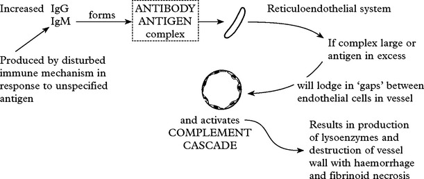
This is termed IMMUNE COMPLEX VASCULITIS.
Indirect immunofluorescent microscopy on biopsy material will demonstrate the presence of immune complexes.
In giant cell arteritis and granulomatous vasculitis, cellular immune mechanisms are probably to blame and vessels are directly attacked. A reaction of antigen with sensitised lymphocytes results in lymphokine release – attracted mononuclear cells release lysosomal enzymes with resultant granuloma formation.
In all vasculitides affecting predominantly large and medium size vessels, angiography is important in establishing diagnosis. On MRI the presence of bilateral cortical and subcortical infarction is suggestive.
SYSTEMIC LUPUS ERYTHEMATOSUS: in 75% of patients nervous system involvement occurs and may predate systemic manifestation.
– Psychiatric change
– Dementia
– Seizures
– HEMIPLEGIA
– Cranial or peripheral nerve involvement
– SPINAL stroke
– Involuntary movements.
Pathology
The predominant CNS finding is microvascular injury with hyalinisation, perivascular lymphocytosis, endothelial proliferation and thrombosis. Active vasculitis is rare. Cardiogenic embolism and coagulopathy (antiphospholipid antibodies) are alternative mechanisms of stroke.
Investigations
Blood
Elevated ESR and C-reactive protein
Circulating antibodies to nucleoproteins e.g. anti-DNA(ANA)
Elevated immunoglobulins
Depressed serum complement levels
Prolonged prothrombin time and antiphospholipid antibodies (60%)
Other
EEG – diffuse disturbance
CT/MRI – multiple small intraparenchymal haemorrhages or infarcts
CSF – protein elevated (Ig), mononuclear cells
Angiography – vessels have beaded appearance
Treatment
Corticosteroids in moderate dosage. In patients with severe or fulminant disease, immunosuppressants and plasma exchange may help.
POLYARTERITIS NODOSA
Neurological involvement is common (80%): Small and medium size arteries are affected.
– HEMIPLEGIA – microinfarction
– INTRACRANIAL HAEMORRHAGE – aneurysm formation
– SPINAL INFARCTION or HAEMORRHAGE
– Peripheral nerve involvement (mononeuritis multiplex)

Hypertension and renal involvement are common.
Investigations
|
Blood Elevated ESR and C-reactive protein Anaemia Leukocytosis Eosinophilia Antinuclear cytoplasmic antibodies (ANCA) Circulating immune complexes IgM rheumatoid factor |
Other Biopsy – renal or peripheral nerve – necrotic vessel – lumen diminished – leucocytes and eosinophils in necrotic media and adventitia CT/MRI as in systemic lupus erythematosus Angiography. Multiple irregularities and micro-aneurysm formation. These changes can be visible on MRA |
Treatment
Steroids and immunosuppressant therapy have dramatically improved outcome (60% 5-year survival).
Plasmapheresis is successful in acute cases.
ALLERGIC ANGIITIS (Hypersensitivity vasculitis)
Intercurrent illnesses (infection or neoplasia) trigger immune complex deposition and basement membranes of capillaries and venules. Systemic symptoms – rash, fever and arthralgia are associated with multiorgan involvement. Neurological features – neuropathy, stroke-like syndromes – occur in 30% of patients. Investigations suggest systemic upset – elevated ESR, anaemia, leukopaenia. Skin biopsy confirms peri-venular inflammation. Treatment of underlying infection and steroids produce rapid improvement.
TAKAYASU’S (PULSELESS) DISEASE
A giant cell arteritis involving the aorta and its major branches. Predominantly affects Asian females in third or fourth decades.
|
Symptoms: – Non-specific – fever, arthralgias and myalgia – Vascular – myocardial ischaemia, peripheral vascular disease – Neurological vascular TIAs (including subclavian steal), strokes and dementia. |
Diagnosis: Steroids are useful initially. The role of surgical reconstruction of occluded vessels is uncertain |
ISOLATED ANGIITIS OF CENTRAL NERVOUS SYSTEM
Systemic symptoms and laboratory evidence of generalised vasculitis are absent.
Presentation with headaches/seizures/encephalopathy and stroke
|
Diagnosis: Condition should be borne in mind in atypical stroke – CSF shows lymphocytes – MRI, multiple ischaemic changes – Angiography, beading (multiple narrow segments) on intracranial arteries – Meningeal biopsy. |
Treatment: Prognosis often dismal. Steroids and cyclophosphamide may produce remission. |
GIANT CELL ARTERITIS (see page 73)
CHURG-STRAUSS ANGIITIS
A distinctive syndrome of eosinophilia, pulmonary infiltrates, neuropathy and encephalopathy or stroke. Related to polyarteritis nodosa, steroid responsive. Other immunosuppressants e.g. cyclophosphamide in resistant cases.
GRANULOMATOUS VASCULITIS/WEGENER’S GRANULOMATOSIS
A rare disorder, most frequent in males aged 20–50 years.

|
Diagnosis: – Elevated ESR and C-reactive protein (CRP) – Elevated immunoglobulins – Impaired renal function – Radiological findings: Chest and sinuses: granuloma mass MRI (cranium): granuloma mass or vasculitis. |
Treatment: – Immunosuppression: steroids and cyclophosphamide – Surgical decompression of granulomas occasionally required. |
DISEASES OF THE BLOOD
Disorders of the blood may manifest themselves as ‘stroke-like’ syndromes. Examination of the peripheral blood film is an important investigation in cerebrovascular disease. Where indicated, more extensive haematological investigation is necessary.
DISSEMINATED INTRAVASCULAR COAGULATION (DIC)

Neurological involvement – a diffuse fluctuating encephalopathy, subarachnoid or subdural haemorrhage.
Diagnosis confirmed by – low platelet count – prolonged prothrombin time, elevated fibrin degradation products and reduced fibrinogen levels.
Treatment
Heparin. Fresh frozen plasma/vitamin K. Treatment of underlying cause.
HAEMOGLOBINOPATHIES
These are genetically determined disorders in which abnormal haemoglobin is present in red blood cells.
Sickle cell disease
This disorder is common in people of African origin but also occurs sporadically throughout the Mediterranean and Middle East region.
The patient is of small stature, usually with chronic leg ulcers, cardiomegaly and hepatosplenomegaly. When arterial oxygen saturation is reduced, ‘sickling’ will occur, manifested clinically by abdominal pain/bone pain.
Neurological involvement – hemiparesis, optic atrophy, subarachnoid haemorrhage.
Diagnosis is confirmed in vitro by the ‘sickling’ of cells when O2 tension is reduced and by haemoglobin electrophoresis.
Treatment
Analgesics for pain O2 therapy, or hyperbaric O2. Exchange transfusion should be carried out for those with a severe or progressive deficit.
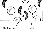
ANTIPHOSPHOLIPID ANTIBODIES
These IgG or IgM antibodies prolong APTT and appear to be associated with thrombotic stroke. There remains uncertainty as to whether they are caused by or represent a transient non-specific ‘acute phase’ reaction to illness. Such antibodies can be found in patients with systemic lupus erythematosus.
ANTITHROMBIN III, PROTEIN C AND PROTEIN S DEFICIENCY
Deficiency of any of these circulating antithrombotic fibrinolytic agents can result in deep venous thrombosis, pulmonary embolism or cerebral venous sinus thrombosis.
POLYCYTHAEMIA
Both polycythaemia rubra vera (primary) and secondary polycythaemia may result in neurological involvement – increased viscosity results in reduced cerebral blood flow and an increased tendency towards thrombosis.
Headaches, visual blurring and vertigo are common neurological symptoms.
Transient ischaemic attacks and thrombotic cerebral infarction occur.
Diagnosis
Hb and PCV are elevated.
Primary polycythaemia is confirmed by increased red cell count, white blood count and platelets.
Secondary polycythaemia – respiratory, renal or congenital heart disease are causal.
Treatment
Venesection with replacement of volume with low molecular weight dextran.
Antimitotic drugs may also be used when polycythaemia is due to myeloproliferative disease.
HYPERGAMMAGLOBULINAEMIA
An increase in serum gamma globulin may arise as a primary event or secondary to leukaemia, myeloma, amyloid.
Neurological involvement develops in 20% of cases – due to increased viscosity.
Clinical features are similar to those of polycythaemia – peripheral nervous system involvement may also occur.
Diagnosis is confirmed by protein electrophoresis.
Treatment – underlying cause – plasmapheresis.
THROMBOTIC THROMBOCYTOPENIC PURPURA (syn: Moschkowitz’s syndrome)
This is a fibrinoid degeneration of the subintimal structures of small blood vessels. Lesions occur in all organs including the brain.
Clinical features – fever with purpura and multiorgan involvement and neurological features of diffuse encephalopathy or massive intracranial haemorrhage.
Haemolytic anaemia, haematuria and thrombocytopenia are the main laboratory features.
Treatment
Heparin, steroids and platelet inhibitors may be of value.
THROMBOCYTOPENIA
Whether idiopathic, drug-induced or due to myeloproliferative disorders, this condition may be associated with intracranial haemorrhage.
THROMBOCYTOSIS
This is an elevation in platelet count above 600 000 per mm3. It may be part of a myeloproliferative disorder, or ‘reactive’ to chronic infection. Patients present with recurrent thrombotic episodes.
Treatment
Aspirin in mild cases; plasmapheresis and antimitotic drugs if more severe.
HYPERFIBRINOGENAEMIA
Serum fibrinogen is occasionally elevated in people with cerebrovascular disease. This enhances coagulation and raises blood viscosity. Infection, pregnancy, malignancy and smoking all raise fibrinogen and may explain in part the increased risk of cerebral infarction. Arvin (Malayan viper venom) acutely lowers serum levels.
METABOLIC DISORDERS
HOMOCYSTINURIA
A recessively inherited disorder. Accumulation of homocystine in blood damages endothelium and induces premature occlusive arterial disease. The significance of the heterozygote state is uncertain.
MELAS
See Mitochondrial disorders (page 481).
CEREBROVASCULAR DISEASE – VENOUS THROMBOSIS
The venous sinuses are important in CSF absorption, with arachnoid villi invaginating the sagittal sinus in particular. Thrombotic occlusion of the venous system occurs with
— infection (especially ear or sinus infection)
— dehydration
— pregnancy, puerperium and pill
— coagulation disorders
— malignant meningitis
— miscellaneous disorders e.g. sarcoid, Behçets
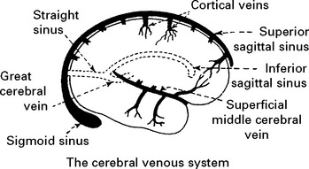
Improved imaging (MRI) has resulted in increased recognition. Venous infarction accounts for 1% of all ‘strokes’.
Superior sagittal and lateral sinus thrombosis (85% of cases)
Impaired CSF drainage results in headache, papilloedema and impaired consciousness. Venous infarction produces seizures and focal deficits (e.g. hemiplegia).
Diagnosis is suggested by venous (nonarterial territory) infarction and ‘empty delta’ sign (following contrast the wall of the sinus enhances but not the central thrombus on CT) and confirmed by occlusion of filling deficit on MR or CT venography. Outcome is variable; intracranial hypertension may develop (p. 378). A thorough search for causation – coagulation screen, drug history and underlying systemic illness – essential.

Treatment:
Correct causative factors (dehydration/infection etc)
Anticoagulation with heparin or alternative.
Deep cerebral venous thrombosis (10% of cases)
This produces venous infarction of the basal ganglion and other subcortical structures. Presentation with similar features; diagnosis can only be established by imaging (CT/MRI and MRV). Treatment as above.
Cavernous sinus thrombosis (5% of cases)
Commonly results from infection spreading from the jaw through draining veins or paranasal sinuses. Painful ophthalmoplegia, proptosis and chemosis with oedema of periorbital structures are associated with facial numbness and fever. The disorder may be bilateral. Base diagnosis on clinical suspicion supported by venography. Treatment with antibiotics and if indicated, sinus drainage.
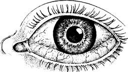
CEREBROVASCULAR DISEASE – INTRACEREBRAL HAEMORRHAGE
By definition, ‘intracerebral haemorrhage’ occurs within the brain substance, but rupture through to the cortical surface may produce associated ‘subarachnoid’ bleeding. When the haemorrhage occurs deep in the hemisphere, rupture into the ventricular system is common.
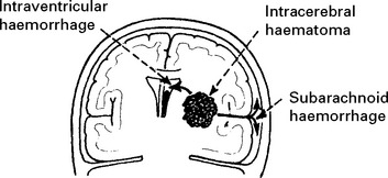
CAUSES
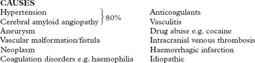
In autopsy series, hypertension accounts for 40–50% of patients dying from non-traumatic haematomas. In hypertensive patients, degenerative changes weaken the walls of small intraparenchymal perforating vessels. Rupture usually occurs near a vessel bifurcation. The ‘microaneurysms’ originally described by Charcot and Bouchard are more likely to be small subadventitial haemorrhages or extravascular clots. In normotensive patients without any evident underlying pathology the cause remains unknown, but cryptic arteriovenous malformations are suspect especially in younger patients (i.e. less than 40 years) and when the haematoma is ‘lobar’ (i.e. frontal, temporal, parieto-occipital). In these patients, the haematoma may temporarily or permanently obliterate the lesion. Reinvestigation following haematoma resolution occasionally reveals previously undetected malformations. In the normotensive elderly patient, subcortical haematomas are commonly associated with amyloid vasculopathy, a degenerative disorder affecting the walls of arteries.
PATHOLOGICAL EFFECTS

Within 48 hours the blood and plasma act on surrounding brain causing disruption of the blood–brain barrier, vasogenic and cytotoxic oedema, neuronal damage and necrosis.
Haematoma resolution occurs in 4–8 weeks, leaving a cystic cavity.
INTRACEREBRAL HAEMORRHAGE
SITES
In hypertensive patients, up to 70% occur in the basal ganglia/thalamic region.
In normotensive patients:
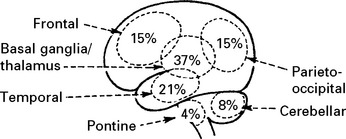
CLINICAL EFFECTS
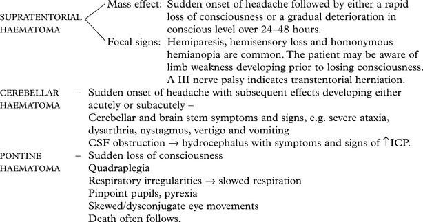
INVESTIGATIONS
A CT scan determines the exact site and size of the haematoma and excludes other pathologies.
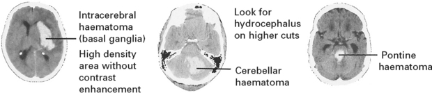
Angiography/CT angiography
– Performed immediately if clinical state requires urgent operation, to identify a secondary cause i.e. arteriovenous malformation, aneurysm or vasculitis.
– Otherwise delayed until condition improves and the haematoma resolves, unless age and medical condition preclude further management.
In patients with negative angiography, a late MRI may demonstrate a CAVERNOUS ANGIOMA (see page 299).
|
MANAGEMENT |
PROGNOSIS |
|
For patients with intracerebral haemorrhage on anticoagulants, reverse using intravenous prothrombin concentrate and vitamin K. Supratentorial haematoma There is still no evidence to show that early evacuation of an intracerebral haematoma improves outcome. |
Poor prognostic features – Increasing age – Large, deep lesions (basal ganglia/thalamic) – Intraventricular blood – Depth of conscious level (flexion or extension to painful stimuli). |
|
In general, haematoma evacuation is indicated in patients who deteriorate gradually from the ‘mass’ effect, especially when the lesion lies superficially; operation will not benefit moribund patients, i.e. patients extending to painful stimuli with no pupil reaction. |
Good prognostic factors – Small superficial lesions (i.e. frontal, temporal or parieto-occipital) – Conscious patients or patients localising to painful stimuli. The overall mortality ranges from 25–60% (90% if the patient is in coma) and is improved by an integrated ‘Stroke Unit’ |
Cerebellar haematoma:
Small haematomas causing minimal effects may be managed conservatively. Otherwise, urgent evacuation through a suboccipital craniectomy is required. Relief of brain stem compression may be life saving and operative morbidity is low.
The overall mortality is approximately 30%.
Pontine haemorrhage
The mortality from pontine haemorrhage is high. A conservative approach is usually adopted although some advocate operative exploration.
INTRAVENTRICULAR HAEMORRHAGE
Haemorrhage into the ventricles causes a sudden loss of consciousness. With a large bleed, death may follow from the pressure transmission from within the ventricular system. Blood in the ventricles does not in itself cause damage and, following clot resolution, complete recovery may occur.
No treatment is required; attempts at flushing out the ventricles usually fail. If the blood ‘cast’ causes obstructive hydrocephalus, then ventricular drainage (although hampered by the presence of blood) is indicated. Infusion of thrombolytic agents awaits evaluation.
SUBARACHNOID HAEMORRHAGE (SAH)
Intracranial vessels lie in the subarachnoid space and give off small perforating branches to the brain tissue. Bleeding from these vessels or from an associated aneurysm occurs primarily into this space. Some intracranial aneurysms are embedded within the brain tissue and their rupture causes intracerebral bleeding with or without subarachnoid haemorrhage.

Occasionally the arachnoid layer gives way and a subdural haematoma results.
INCIDENCE
Subarachnoid haemorrhage occurs in approximately 8–10 per 100 000 per year.
CAUSE
Cerebral aneurysms are the most frequent cause of subarachnoid haemorrhage, with arteriovenous malformations accounting for up to 5%.
In about 20% of patients detailed investigation fails to reveal a source of the haemorrhage. Small thrombosed or undetected aneurysms or cryptic arteriovenous malformations may account for some.
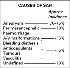
SYMPTOMS AND SIGNS
The severity of the symptoms is related to the severity of the bleed.
Usually the headache is severe and the onset usually instantaneous (often described as a ‘blow to the head’). A transient or prolonged loss of consciousness or epileptic seizure may immediately follow. Nausea and vomiting commonly occur. Symptoms continue for many days.
Occasionally, the headache is mild (although still sudden onset) and may represent a ‘warning leak’ of blood before a major bleed.
Signs of meningism develop after 3–12 hours
Neck stiffness is present in most patients on passive neck flexion.
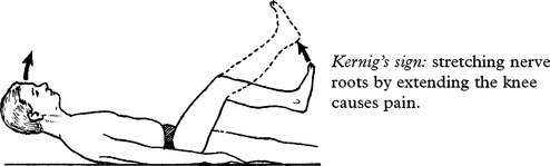
Kernig’s sign: stretching nerve roots by extending the knee causes pain.
Coma or depression of conscious level may result from the direct effect of the subarachnoid haemorrhage or from the mass effect of an associated intracerebral haematoma.
Focal damage from a haematoma will produce focal signs, e.g. limb weakness, dysphasia. The presence of a III nerve palsy indicates either transtentorial herniation or direct nerve damage from a posterior communicating artery aneurysm (or rarely from a basilar artery aneurysm).
Seizures frequently occur and may mask other features.
Fundus examination may reveal papilloedema or a subhyaloid or vitreous haemorrhage caused by the sudden rise in intracranial pressure.
A ‘reactive hypertension’ commonly develops, i.e. a rise in BP in patients with no evidence of pre-existing hypertension, and takes several days to return to normal levels.
Pyrexia is also a common finding; if severe and fluctuating, it may reflect ischaemic hypothalamic damage.
INVESTIGATIVE APPROACH
CT scan is the investigation of choice, perfomed as soon as possible after the headache onset. Lumbar puncture establishes the diagnosis of subarachnoid haemorrhage, but in patients with a mass lesion, lumbar puncture could precipitate transtentorial herniation.
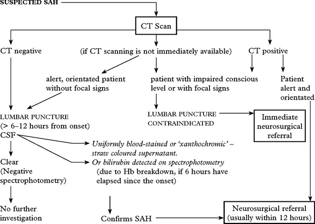
Age limit for neurosurgical referral: Although mortality and morbidity increase with age, with the option of endovascular aneurysm treatment, age limitations no longer apply provided the patient’s clinical state is satisfactory.
CT scan
Confirms the diagnosis of SAH in 95% (if within 48 hours of the bleed).
Blood may be widely distributed
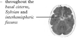
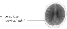
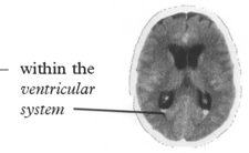
or more localised aiding identification of the site of the ruptured aneurysm
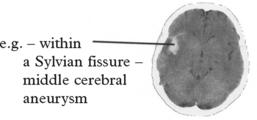
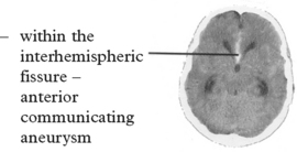
CT also identifies other associated lesions
– hydrocephalus
– intracerebral haematoma
– tumour
– arteriovenous malformation
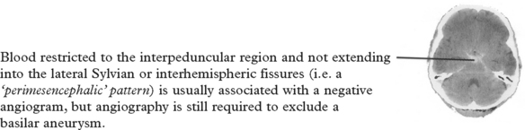
MRI scan
Not routinely used, but in patients with multiple aneurysms, MRI performed several days after the bleed may provide greater sensitivity than CT in detecting small areas of subarachnoid clot and help determine the particular lesion responsible.
N.B. Spinal arteriovenous malformations can also cause SAH – if the patient’s pain begins in the back before spreading to the head, or if any features of cord compression exist, then MRI of the cervical or thoracic spine should be the preliminary investigation (see page 424).
CT/MR angiography
These non invasive techniques, particularly when combined with 3-D or 4-D imaging (colour as the 4th dimension), will detect up to 95% of intracranial aneurysms, but those < 3mm in diameter may be missed. Both MRA and in particular CTA can provide more information than conventional angiography about the aneurysm shape and size of the neck.
Demonstration of an aneurysm which matches the distribution of blood on standard CT, permits planning of treatment on the assumption that this is the source of the haemorrhage.
CT angiogram showing carotid aneurysm
Digital angiography
Four-vessel angiography is performed in patients with a negative CTA or MRA, in patients where further clarification of the vessels or the aneurysm is required to aid decision making or immediately prior to endovascular treatment.
With the latest equipment, 3-D rotational techniques are employed, combined with cropping of unwanted data, to permit the radiologist to focus on specific regions. Without this aid, antero-posterior, lateral and oblique views are required for each vessel.
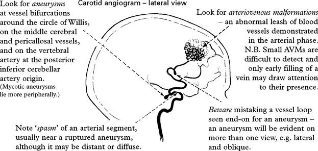
Negative angiography
Angiography fails to reveal a source of the subarachnoid haemorrhage in approximately 20% of patients. In the presence of arterial spasm, reduction in flow may prevent the demonstration of an aneurysm and repeat angiography may be required at a later date.
Prognosis: In patients with a ‘perimesencephalic’ pattern of haemorrhage on CT scan and with negative angiography, the outlook is excellent; those patients with an ‘aneurysmal’ pattern with blood lying in the interhemispheric or Sylvian fissure still run a risk of rebleeding.
CEREBRAL ANEURYSMS
INCIDENCE
At autopsy intracranial aneurysms are found in approximately 2% of the population.
Aneurysm rupture occurs in 6–8 per 100 000 per year
Female:male = 3.2; but this ratio varies with age: < 40 years, male > females
> 40 years, females > males
Risk factors: atherosclerotic diseases (2.3x), family history (6x), polycystic kidney disease (4.4x).
Inheritance: investigations reveal aneurysms in 10% of relatives with two or more affected 1st degree family members. The genetic basis remains unknown. Procollagen III deficiency may play a role in some patients.
MORPHOLOGY
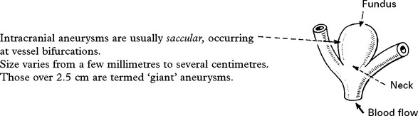
Intracranial aneurysms are usually saccular, occurring at vessel bifurcations. Size varies from a few millimetres to several centimetres. Those over 2.5 cm are termed ‘giant’ aneurysms.

Aneurysm rupture: usually occurs at the fundus of the aneurysm and the risk is related to size. Smoking, hypertension and alcohol excess also play a part. In some patients, rupture occurs during exertion, straining or coitus, but in most there is no associated relationship.
Sites of saccular aneurysm

Multiple aneurysms: in approximately 30% of patients with aneurysmal SAH, more than one aneurysm is demonstrated on angiography.
PATHOGENESIS

Hypertension may play a role; more than half the patients with ruptured aneurysm have pre-existing evidence of raised blood pressure.
CLINICAL PRESENTATION
Of those patients with intracranial aneurysms presenting acutely, most have had a subarachnoid haemorrhage. A few present with symptoms or signs due to compression of adjacent structures. Others present with an aneurysm found incidentally.
1. Rupture
The features of SAH have already been described in detail (page 276); they include sudden onset of headache, vomiting, neck stiffness, loss of consciousness, focal signs and epilepsy.
Since the severity of the haemorrhage relates to the patient’s clinical state and this in turn relates to outcome, much emphasis has been placed on categorising patients into 5 level grading systems, e.g. Hunt and Hess. A scale incorporating the Glasgow Coma scale (page 29) has been adopted by the World Federation of Neurosurgical Societies:

This grading scale correlates well with final outcome and provides a prognostic index for the clinician. In addition, it enables matching of patient groups before comparing the effects of different management techniques.
2. Compression from aneurysm sac
A large internal carotid artery aneurysm (or anterior communicating artery aneurysm) may compress –

3. Incidental finding
The improved availability of sensitive high quality, non-invasive MR or CT imaging techniques has greatly increased the number of patients in whom an intracranial aneurysm is detected incidentally, during investigation for other disease.
NATURAL HISTORY OF RUPTURED ANEURYSM
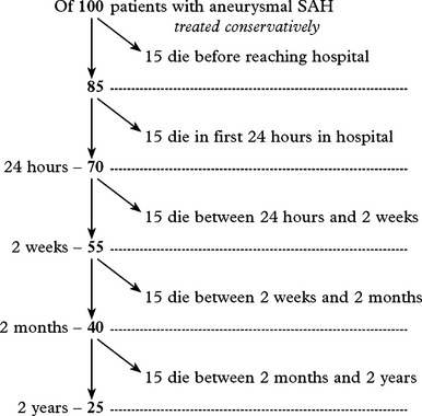
SAH from ruptured aneurysm carries a high initial mortality risk which gradually declines with time. Of those who survive the initial bleed, rebleeding and cerebral infarction (see below) are the major causes of death.
These figures are based on studies of conservative treatment carried out in the 1960s, at a time when the risks of operation were greater and benefits uncertain.
COMPLICATIONS OF ANEURYSMAL SAH
|
INTRACRANIAL – Rebleeding – Cerebral ischaemia/infarction – Hydrocephalus – ‘Expanding’ haematoma – Epilepsy. |
EXTRACRANIAL – Myocardial infarction – Cardiac arrhythmias – Pulmonary oedema – Gastric haemorrhage (stress ulcer). |
CEREBRAL ANEURYSMS – COMPLICATIONS
REBLEEDING
Rebleeding is a major problem following aneurysmal SAH. In the first 28 days (in untreated patients), approximately 30% of patients would rebleed; of these 70% die. In the following few months the risk gradually falls off but it never drops below 3.5% per year.
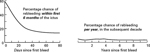
Adapted from Winn, Richardson, Jane 1977 Annals of Neurology
If, for example, a patient survives the first 30 days after a bleed, there is still a 20% chance of a rebleed occurring in the next 5 months. Even if patients survive the ‘high risk’ period in the first 6 months, there is still a considerable chance of rebleeding and death in the subsequent years.
The clinical picture of rebleeding is that of SAH, but usually the effects are more severe than the initial bleed. Most patients lose consciousness; the risk of death from a rebleed is more than twice that from the initial bleed.
Investigation
All patients deteriorating suddenly require a CT scan. This helps in establishing the diagnosis of rebleeding and excludes a remediable cause of the deterioration, e.g. acute hydrocephalus.
CEREBRAL ISCHAEMIA/INFARCTION
Following subarachnoid haemorrhage, patients are at risk of developing cerebral ischaemia or infarction and this is an important contributory factor to mortality and morbidity. Cerebral ischaemia/infarction may occur as an immediate and direct result of the haemorrhage, but more often develops 4–12 days after the onset, either before or after operation – hence the term ‘delayed cerebral ischaemia’. Approximately 25% of patients develop clinical evidence of delayed ischaemia/infarction; of these 25% die as a result. About 10% of the survivors remain permanently disabled.
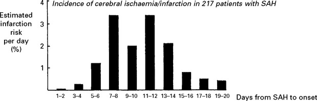
Adapted from Vermeulen, Lindsay, Murray et al 1984 New England Journal of Medicine 311: 432–437
Aetiology of cerebral ischaemia/infarction
Several factors probably contribute to the development of cerebral ischaemia or infarction: ‘Vasospasm’: arterial narrowing on angiography occurs in up to 60% of patients after SAH and is either focal or diffuse. The development of ‘vasospasm’ shows a similar pattern of delay to that of cerebral ischaemia.
The angiogram appearance was initially thought to result from arterial constriction; this may be so, but the pathogenesis of ‘vasospasm’ now seems more complex. Many vasoconstrictive substances either released from the vessel wall or from the blood clot appear in the CSF after SAH, e.g. serotonin, prostaglandin, oxyhaemoglobin, endothelin-1 and endothelial synthesis of the vasodilator nitric oxide is reduced, but numerous studies with vasoconstrictor antagonists have failed to reverse the angiographic narrowing. This failure may be a result of the arteriopathic changes which have been observed in the vessel wall. Only calcium antagonists appear to have a beneficial effect (see page 291).
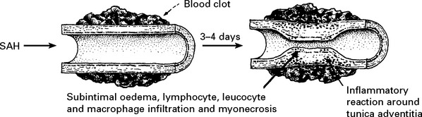
The greater the amount of blood in the basal cisterns (as shown on CT scan), the higher the incidence of arterial narrowing and associated ischaemic deficits.
Hypovolaemia
Hyponatraemia develops after SAH in many patients due to excessive renal secretion of sodium rather than a dilutional effect from inappropriate antidiuretic hormone secretion. Fluid loss and a fall in plasma volume lead to a raised blood viscosity with an increased risk of developing cerebral ischaemia.
Reduced cerebral perfusion pressure
Following SAH, intracranial haematoma or hydrocephalus may cause a rise in intracranial pressure (ICP). Since cerebral perfusion pressure = mean BP – ICP, a subsequent reduction in cerebral perfusion may occur.
Clinical effects of cerebral ischaemia/infarction
This may affect one particular arterial territory producing characteristic signs:
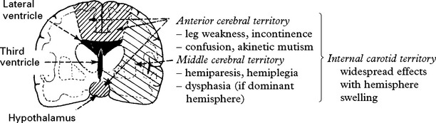
Commonly the ischaemia occurs in multiple areas, often in both hemispheres. This correlates with the pattern of arterial ‘spasm’.
Transcranial Doppler: a significant increase in flow velocity within an intracranial vessel may indicate developing ‘vasospasm’, even before clinical problems develop, and allow the early introduction of prophylactic measures (see page 291).
HYDROCEPHALUS
Following SAH, cerebrospinal fluid drainage may be impaired by:

Acute hydrocephalus occurs in about 20% of patients, usually in the first few days after the ictus; occasionally this is a late complication. In only one-third are symptoms of headache, impaired conscious level, dementia, incontinence, or gait ataxia severe enough to warrant treatment.
In a further 10% of patients, hydrocephalus develops late – months or even years after the haemorrhage.
‘EXPANDING’ INTRACEREBRAL HAEMATOMA
Brain swelling around an intracerebral haematoma may aggravate the mass effect of the haematoma; this may cause a progressive deterioration in conscious level or progression of focal signs.
EPILEPSY
Epilepsy may occur at any stage after SAH, especially if a haematoma has caused cortical damage.
Seizures may be generalised or partial (focal).
EXTRACRANIAL COMPLICATIONS
Myocardial infarction/cardiac arrhythmias: electrocardiographic and pathological changes in the myocardium are occasionally evident after SAH, and ventricular fibrillation has been recorded. These problems are likely to occur secondarily to catecholamine release following ischaemic damage to the hypothalamus.
Pulmonary oedema: this occasionally occurs after SAH, probably as a result of massive sympathetic discharge; note the ‘pink, frothy’ sputum and typical auscultatory and chest X-ray findings.
Gastric haemorrhage: bleeding from gastric erosions occasionally occurs after SAH but rarely threatens life.
CEREBRAL ANEURYSMS – MANAGEMENT FOLLOWING SAH
Headache requires analgesia – codeine or dihydrocodeine. Stronger analgesics may depress conscious level and mask neurological deterioration. Management is otherwise aimed at preventing complications –
PREVENTION OF REBLEEDING
Bed rest: Often enforced after SAH, although there is no evidence that this reduces the rebleed risk. Allowing gentle mobilisation and using the toilet may induce less ‘stress’ than using a bedpan.
Aneurysm repair: Both surgical (clipping of the aneurysm neck) and endovascular (coil embolisation of the aneurysm sac) techniques are used. Aneurysm repair, whether coiling or clipping, is performed within 48 hours of the bleed, but this is not always feasible. Operative risks are greater the earlier the procedure, but the greater the delay, the greater the risk of rebleeding. Despite this, in past years operation was often deferred in patients in poor clinical condition. Endovascular techniques appear to be less dependent on timing and avoid potentially harmful effects of brain retraction and vessel dissection. This supports their use in the elderly or in patients in poor clinical condition, but other factors must also be considered – see page 290. Once the aneurysm is clipped, aggressive methods of treating ischaemia with induced hypertension can be applied – see page 291.
OPERATIVE TECHNIQUES
Direct clipping of the aneurysm neck: Through a craniotomy and using the operating microscope, the surgeon dissects out the aneurysm and applies a clip across the neck without compromising the proximal or distal vessels. Clipping prevents rebleeding; clip slippage rarely occurs. If any part of the neck lies outwith the clip, this may occasionally lead to recurrent growth.
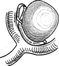
Wrapping: If the width of the aneurysm neck or its involvement with adjacent vessels prevents clipping, then muslin gauze may be wrapped around the fundus. This provides some protection, but rebleeding may still occur.
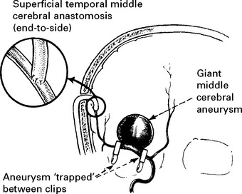
Trapping: Used for giant aneurysms (>25mm diameter) where other methods have failed due to the width of the aneurysm neck. Should be combined with cerebral revascularisation to minimize the risk of ischaemia. Techniques include either superficial temporal–middle cerebral artery anastomosis or insertion of a saphenous vein or radial artery bypass graft from the carotid to the middle cerebral artery.
ENDOVASCULAR TECHNIQUES
Coil embolisation: this endovascular technique, where multiple helical platinum coils are packed into the aneurysm fundus, has been developed and refined over the last two decades. A tracker catheter is inserted via a femoral puncture and guided up through the arterial system into the aneurysm sac.
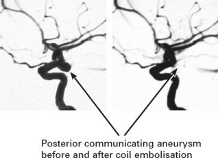 Posterior communicating aneurysm before and after coil embolisation
The coil attached to the end of a delivery wire is then guided into the fundus and after accurate placement, the passage of an electric current causes electrochemical release. On average, 4–5 coils are required to pack each aneurysm.
The radiologist aims to completely obliterate the fundus, but this is not always feasible and to avoid occluding the adjacent vessel, a portion of the neck may remain. In either case, a small risk of rebleeding persists, even when completely obliterated. Thrombotic complications may also occur during the procedure.
Balloon remodelling: the wider the aneurysm neck, the greater the risk that coils will project into and occlude the vessel lumen. A balloon is attached to a second catheter and periodically inflated across the aneurysm neck during coil insertion to preserve the vessel lumen.
Basilar bifurcation aneurysm with wide neck –

Stent assisted coil embolisation: for very wide-necked aneurysms or for those where balloon remodelling has failed, one or more stents can be manouvred through the parent vessel alongside the aneurysm neck. Coils are then packed into the fundus via a tracker catheter passed through the interstices of the stent. Such patients require long-term anti-platelet therapy to prevent thrombotic complications and this may create difficulties in the acute phase of SAH management.
Balloon occlusion: On rare occasions the above operative or endovascular techniques fail to treat ‘giant’ or fusiform aneurysms arising from the carotid artery. Temporary balloon occlusion of the carotid artery for a 30 minute period tests whether the patient’s collateral circulation from the Circle of Willis is sufficient to sustain flow through the hemisphere. Similarly temporary occlusion of the vertebro-basilar system is possible. If tolerated, intra-arterial inflation of a detachable balloon can provide permanent occlusion. Intra-arterial balloon inflation can also provide temporary intra-operative protection when proximal control is difficult to achieve.
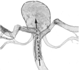 Wide necked basilar aneurysm with one stent inserted into the left posterio rcerebral artery, and another stent passing through the interstices of the first and inserted into the right posterior cerebral artery. Coils will be passed beyond the stent into the aneurysm fundus.
SELECTION OF TREATMENT
Until recent years direct surgical clipping was the standard method of aneurysm treatment. Coil embolisation was reserved for aneurysms technically difficult to repair, particularly those in the posterior circulation. A large multicentre randomized trial of clipping vs. endovascular treatment (the International Subarachnoid Aneurysm Trial – ISAT) demonstrated a 23% reduction in the proportion of patients with a poor outcome (dependent or dead) at one year in those patients undergoing coil embolisation, despite more rebleeds occurring in this group. Following publication in 2002, the proportion of patients undergoing coil embolisation as the first line of treatment dramatically increased, reaching 85–90% in some centres. This swing occurred despite the trial being weighted towards small anterior circulation aneurysms in patients in good clinical condition. Concern persisted that coil treatment would not eliminate rebleeding. Long-term follow up (mean 9 years after treatment) of the trial patients has shown that although rebleeding was higher in the coil treatment group, the risk of death was still significantly lower in coiled patients. In young patients e.g. 30–40 years of age, with 40 years of expectant life, the rebleeding concern after coil treatment persists and clipping may be the preferred option.
Aneurysm treatment requires a team approach involving interventional radiologists and neursurgeons. Treatment selection must take a variety of factors into account including the nature and location of the aneurysm, the relative difficulties of the endovascular or operative approach and the patients age and clinical condition. Some patients e.g. those with basilar, carotid and anterior cerebral aneurysms, the elderly or those in poor clinical condition are more likely to require coil embolisation, whereas others, such as those with large middle cerebral aneurysms are more likely to require direct operative treatment. Unfortunately aneurysms that are difficult to treat with one technique are often difficult to treat with both methods. Patients undergoing coil treatment require check angiography/CT angiography follow-up, e.g. 6 months and 2 years, to ensure recanalisation has not occurred. Up to 10% will require retreatment.
PREVENTION OF CEREBRAL ISCHAEMIA/INFARCTION
Despite considerable clinical and experimental research, cerebral ischaemia is still a major cause of morbidity and mortality after subarachnoid haemorrhage. In recent years some advances have proved beneficial.
Calcium antagonists: several large studies and a meta-analysis have confirmed that Nimodipine reduces the incidence of cerebral infarction by about one third and improves outcome. Whether this acts by improving collateral circulation, by reducing the harmful effect of calcium flooding into brain cells or by reducing cerebral ‘vasospasm’ remains uncertain.
Avoidance of antihypertensive therapy: after SAH, autoregulation (page 79) is often impaired; a drop in BP causes a reduction in cerebral blood flow with a subsequent risk of cerebral ischaemia. Patients on long-term antihypertensive treatment can continue with this therapy, but ‘reactive’ hypertension should not be treated.
High fluid intake (haemodilution): maintenance of a high fluid input (3 litres per day) may help prevent a fall in plasma volume from sodium and fluid loss. If hyponatraemia develops do not restrict fluids (this significantly increases the risk of cerebral infarction). If sodium levels fall below 130 mmol/1, give hypertonic saline or fludrocortisone.
Plasma volume expansion (hypervolaemia): expanding the plasma volume with colloid, e.g. plasma proteins, dextran 70, Haemacel, increases blood pressure and improves cerebral blood flow. This should be given either prophylactically in high risk patients (heavy cisternal blood load on CT scan or with high Doppler velocities) or at the first clinical sign of ischaemia. If clinical evidence of ischaemia develops despite this treatment, then (if the aneurysm has been repaired) combine with:
Hypertensive therapy: treatment with inotropic agents, e.g. dobutamine, increases cardiac output and blood pressure. Since cerebral autoregulation commonly fails after subarachnoid haemorrhage, increasing blood pressure increases cerebral blood flow. Up to 70% of ischaemic neurological deficits developing after aneurysm operations can be reversed by inducing hypertension; often a critical level of blood pressure is evident.
Early recognition and treatment of a developing neurological deficit may prevent progression from ischaemia to infarction. Delayed treatment may merely aggravate vasogenic oedema in an ischaemic area. This technique of induced hypertension is now widely applied, with good results, but requires careful, intensive monitoring. In view of the risk of precipitating aneurysm rupture, it is reserved until after aneurysm repair.
Transluminal angioplasty/papaverine infusion: this involves balloon dilatation of the vasospastic segment of the vessel. It is usually combined with an intra-arterial infusion of the antispasmodic agent papaverine. Although no controlled studies exist, many small studies report a beneficial effect on cerebral blood flow and on clinical state. Timing is difficult. If used too early, the patient may be unnecessarily exposed to an invasive procedure; if too late, the ischaemia may be irreversible. Consider angiography and angioplasty if other measures (haemodilution/hypervolaemia/hypertension) have failed to reverse a significant clinical deterioration within a few hours.
Brain protective agents: to date, studies of neuroprotective drugs (antioxidants and anti-inflammatory agents) other than calcium antagonists, have failed to demonstrate a beneficial effect. Some recent studies assessing magnesium sulphate infusion, pravastatin and the endothelin-1 antagonist clazosentan have had encouraging results, but await further evaluation.
Antifibrinolytic agents: i.e.tranexamic acid, epsilon aminocaproic acid should not be used.
These agents prevent rebleeding by delaying clot dissolution around the aneurysm fundus, but any beneficial effect is offset by an increased incidence of cerebral ischaemia.
HYDROCEPHALUS
Hydrocephalus causing acute deterioration in conscious level requires urgent CSF drainage with a ventricular catheter (in ‘communicating’ hydrocephalus a lumbar drain provides an alternative. Lumbar puncture may provide temporary benefit).
Gradual deterioration or failure to improve in the presence of enlarged ventricles indicates the need for permanent CSF drainage with either a ventriculoperitoneal or lumboperitoneal shunt.
EXPANDING INTRACEREBRAL HAEMATOMA
Intracerebral haematomas from ruptured aneurysms do not require specific treatment unless the ‘mass’ effect causes a deterioration of conscious level. This necessitates urgent CT or digital angiography followed by evacuation of the haematoma with or without simultaneous clipping of the aneurysm; under these circumstances, operative mortality is high.
OUTCOME AFTER SUBARACHNOID HAEMORRHAGE
The National Study of Subarachnoid Haemorrhage collected information on patients admitted to all neurosurgical units in the UK and Ireland between September 2001 and September 2002 and provides useful information on outcome. The study included 3174 patients, of which 2397 had a confirmed aneurysm. (Published by the Royal College of Surgeons, 2006 – available online)
Of those patients surviving the initial bleed and admitted to the neurosurgical unit with a confirmed aneurysm, 11% died in hospital. Of those undergoing aneurysm repair, 40% made a good recovery; a further 21% had moderate disability and were independent.
Factors associated with unfavourable outcome were: age, clinical condition on admission, quantity of subarachnoid blood on CT scan and the presence of pre-existing medical illness.
Table showing relationship of admission grade to outcome
|
Neurological grade on admission (WFNS) |
No. of patients undergoing aneurysm repair |
Unfavourable outcome – death/severe disability (percentage) |
|
I |
1214 |
24.7 |
|
II |
378 |
37.6 |
|
III |
88 |
48.9 |
|
IV |
164 |
64.0 |
|
V |
118 |
71.2 |
Of all patients with a confirmed aneurysm, 92% underwent repair, 53% by surgical clipping and 38% by coil embolisation. No difference was noted in outcome between the two groups even after case mix adjustment (unfavourable outcome 35% for clipped group: 34% for coiled group).
Comparing different operative or management policies: Comparison of different treatments for ruptured aneurysms is difficult, unless conducted under the confines of a randomised controlled trial. ‘Operative mortality’ provides limited information unless patient groups are carefully matched for age, clinical condition and timing of operation. ‘Management mortality’ (e.g. outcome of all admitted patients up to 3 months from the ictus) is of more practical value, but even then, admission policies require careful scrutiny.
CEREBRAL ANEURYSMS – UNRUPTURED
UNRUPTURED ANEURYSMS
Identification of unruptured aneurysms may result from –
– Investigation of unrelated neurological symptoms with CT, MRI or angiography
– Investigation of symptoms arising as a result of aneurysmal compression of adjacent structures
– Investigation of patients with a family history of aneurysmal SAH (see page 295)
– Investigation of SAH when multiple aneurysms exist (about 25% of patients)
Management: depends on the above circumstances and on balancing the risk of rupture (and death) in future years against the risk of aneurysm repair; the decision is often difficult. The International Study of Unruptured Intracranial Aneurysms (ISUIA), by far the largest study of its kind, examined both the natural history and the results of treatment of unruptured aneurysms. The data suggested that the risk of rupture related to the size, site and the occurrence of a SAH from a previously treated source. For small aneurysms < 7 mm in diameter and no previous SAH, the annual risk of rupture was 0.1% (far lower than the 1–2% suggested from previous smaller studies). For aneurysms > 12 mm in diameter the annual risk of rupture ranged from from 3–10% depending on the site and size. For those treated, the study also reported a combined mortality and morbidity of from 7–10% for the coiled patients and from 10–13% for the operated patients, a figure higher than surgeons had previously liked to admit. The operative risk increased with age, aneurysm size and a site on the posterior circulation.
When determining appropriate management, the following factors must be taken into account
– patient’s age, life expectancy and the occurence of a previous haemorrhage
– aneurysm size and site
– the possibility of future growth (about 1/3 expand > 3 mm over 20 years)
– the patient’s view (a conservative approach can create considerable stress)
– the operative/endovascular results of unruptured aneurysm treatment for that centre
In general treatment would not be recommended for anterior circulation aneurysms < 7 mm in size and without a previous SAH, although this view may change with continued improvement in endovascular techniques.
For those undergoing a conservative approach, it is essential to ensure that they do not smoke, since this doubles the risk of aneurysm rupture.
When aneurysms present with compressive symptoms such as a III nerve palsy, it is assumed that recent expansion has occurred and that rupture could be imminent. Such patients normally receive urgent treatment.
Cumulative 5 year rupture rates for unruptured aneurysms at different sites (ISUIA, Lancet 2003)
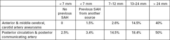
CEREBRAL ANEURYSMS – SCREENING
SCREENING FOR INTRACRANIAL ANEURYSMS
When two or more first-degree relatives have a history of cerebral aneurysms or SAH, then other members of that family (over the age of 25 years) have an increased risk of harbouring an intracranial aneurysm (about 10% or 6x greater than the rest of the population). A similar increased risk occurs for patients with a genetic predisposition, e.g. polycystic kidney disease, Type IV Ehlers-Danlos. Before undergoing screening to detect whether such an aneurysm exists, several important facts should be considered –
– We do not know how rapidly aneurysms form. They may develop over a few hours, days or weeks. A negative screening investigation will fail to provide the reassurance that a subarachnoid haemorrhage from a ruptured aneurysm will never occur.
– The ISUIA study described above, suggests that for small aneurysms (< 7 mm), the rupture risk is extremely low. Even if a small aneurysm is found, treatment risks may preclude any action.
– Aneurysm repair, either by direct operation or by coil embolisation, carries a risk of death or disability, which depends on the patient’s age and the size and site of the aneurysm.
– The presence of an aneurysm may carry implications for life insurance, mortgage applications and even for future pregnancies (since the risk of aneurysm rupture is increased during pregnancy).
– It is impractical to consider screening in relatives < 20–25 years (due to the rarity of aneurysms in this age group) and in those > 60–70 years since the risks outweigh any potential benefit.
– In those with only one affected first-degree relative the low, albeit slightly increased risk of finding an unruptured aneurysm, is still insufficient to justify screening.
If after consultation and consideration of these issues the patient wishes to proceed with screening, then CT angiography would be the most appropriate technique, accepting that this may fail to detect aneurysms 3 mm or less in diameter. For those who decide not to undergo screening, other measures may minimise the risk of aneurysm formation in the future – avoid smoking and treat elevated blood pressure and cholesterol.
After a negative investigation, the patient may wish to consider the possibility of a further screen in 3–5 years time.
VASCULAR MALFORMATIONS
Vascular malformations vary in size and different forms exist:
Arteriovenous malformations (AVMs) are developmental anomalies of the intracranial vasculature; they are not neoplastic despite their tendency to expand with time and the descriptive term ‘angioma’ occasionally applied.
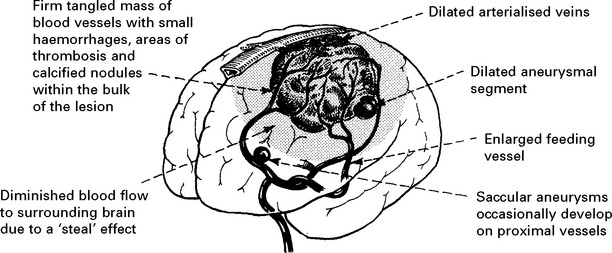
Dilated arteries feed directly into a tangled mass of blood vessels of varying calibre; they bypass capillaries and shunt oxygenated blood directly into the venous system. Due to high intraluminal pressure, veins may adopt an ‘aneurysmal’ appearance. Arteriovenous malformations occur at any site but are commonest in the middle cerebral artery territory.
Capillary telangiectasis: an area of dilated capillaries, like a small petechial patch on the brain surface – especially in the pons. These lesions are often only revealed at autopsy.
Cavernous malformation/angioma: plum coloured sponge-like mass composed of a collection of blood filled spaces with no intervening brain tissue. No enlargement of feeding or draining vessels.
ARTERIOVENOUS MALFORMATIONS
CLINICAL PRESENTATION
Haemorrhage
About 40–60% of patients with an AVM present with haemorrhage – often with an intracerebral or intraventricular component. In comparison with saccular aneurysms, AVMs tend to bleed in younger patients, i.e. 20–40 years, and are less likely to have a fatal outcome. Vasospasm and delayed ischaemic complications rarely develop. Small AVMs, those with high intranidal pressure and those draining exclusively to deep veins have an increased risk of haemorrhage.
Annual risk of haemorrhage: patients with no history of haemorrhage have an annual risk of bleeding of 2–4%. For those presenting with haemorrhage, the risk of rebleeding may be higher, particularly in the first year. One study reported an annual risk of 17%.
Mortality from haemorrhage: in contrast to the high mortality following aneurysm rupture, haemorrhage from an AVM carries the relatively low mortality rate of approximately 10%.
Epilepsy
Generalised or partial seizures commonly occur in patients with arteriovenous malformation, especially if the lesion involves the cortical surface. Of patients presenting with haemorrhage, 30% have a history of epilepsy.
Neurological deficit
Large AVMs, especially those involving the basal ganglia, may present with a slowly progressive dementia, hemiparesis or visual field defect, probably as a result of a ‘steal’ effect. The infrequent brain stem AVM may also produce a motor or sensory deficit, with or without cranial nerve involvement.
Headache
Attacks of well localised headache – unilateral and throbbing – occur in a proportion of patients subsequently shown to have a large AVM.
Cranial bruit
Auscultation, especially over the eyeball, occasionally reveals a bruit.
INVESTIGATIONS
CT scan
Most AVMs are evident on CT scan unless masked by the presence of an intracranial haematoma. A double dose of intravenous contrast may aid visualisation, especially with small ‘cryptic’ lesions.
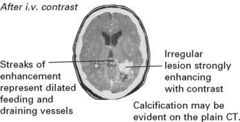
MRI
Conventional MRI will clearly demonstrate the AVM as a region of flow voids, with associated signal change within or around the lesion from areas of old haemorrhage or gliosis.
The MRI provides exact anatomical detail and helps surgical planning. Functional MRI (page 42) aids identification of any adjacent eloquent areas.
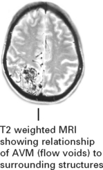
Angiography
Both CT and MR angiography should confirm the presence of an AVM but digital subtraction four-vessel angiography is required to delineate the feeding and draining vessels. Occasionally small AVMs are difficult to detect and only early venous filling may draw attention to their presence.
N.B. In the presence of a haematoma, digital subtraction angiography should be delayed until the haematoma resolves, otherwise local pressure may mask demonstration of an AVM. If the angiogram is subsequently negative, then MRI is required to exclude the presence of a cavernous malformation.
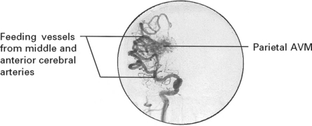
MANAGEMENT
Various methods of treating arteriovenous malformations are available. All risk further damage and a team comprised of the neurosurgeon and neuroradiologist should decide on the optimal method or combination of methods for each patient. The urgency of the patient’s clinical condition and the risks of treatment must be weighed against the risk of a conservative approach. The Spetzler-Martin grading system provides a useful guide to operative risk.

Indications for intervention
|
Indications for intervention – ‘Expanding’ haematoma associated with AVM – Progressive neurological deficit – Risk of haemorrhage especially – young patients with many years at risk – AVMs < 3 cm |
Take into account operative risk low in – Grade 1–2 – AVMs in ‘non-eloquent’ sites – AVMs < 3 cm diameter – with superficial venous drainage high in – Grade 4–5 – AVMs > 3 cm diameter – AVMs in ‘eloquent’ sites – with drainage to deep veins |
Methods of treatment
|
Operation: |
Excision – complete excision of the AVM (confirmed by per-or postoperative angiography) is the most effective method of treatment particularly for small AVMs in non-eloquent areas. Image guidance (page 386) may aid localisation. |
|
Larger lesions (> 6 cm) have a greater risk of postoperative hyperperfusion syndrome and brain swelling and carry a 40% risk of permanent neurological deficit. |
|
|
Stereotactic |
|
|
radiosurgery: |
Focused beams from multiple cobalt sources or from a linear accelerator (25Gy) obliterates about 75% of AVMs < 3 cm in diameter, but this may take up to 3 years during which time the risk of haemorrhage persists. In smaller lesions < 1 cm the obliteration rate with 25 Gy approaches 100%. For lesions greater than 3 cm, the lower dose required to minimise the damaging effect of local tissue destruction, makes obliteration unlikely. Pre-treatment with embolisation helps only if this produces a segmental reduction in size. Suboptimal embolisation may merely hinder radiosurgical treatment. Despite the delay in action, radiosurgery may prove ideal for small deeply seated lesions. |
|
Embolisation: |
Skilled catheterisation permits selective embolisation of feeding vessels with isobutyl-cyanoacrylate, although this technique is not without risk. |
|
Embolisation may cure up to 40% of AVMs when small particularly if supplied by a single feeding vessel, but filling may persist from collaterals. When used preoperatively, it may significantly aid operative removal. |
|
CAVERNOUS MALFORMATIONS (syn. cavernous angioma, cavernoma)
Cavernous malformations occur in 0.5% of the population. They are occasionally multiple and in a few patients, have a familial basis. A cavernous malformation may present with epilepsy, haemorrhage or with focal neurological signs. MRI is the investigation of choice as cavernous malformations, are often missed on CT scanning and rarely seen on angiography. Most lesions show marked signal change around this lesion due to a rim of haemosiderin deposition.
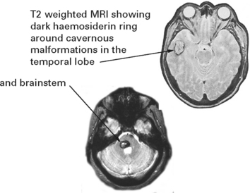
The annual risk of haemorrhage is about 1% per year, but this varies depending on whether the lesion lies deeply (i.e. brain stem or basal ganglia) or superficially. With deep lesions in critical sites, a small bleed causes damage more readily. For deep lesions the risk of a bleed sufficiently severe to cause neurological signs is about 5% per year, whereas for superficial lesions, this is almost zero. Unfortunately the high risk, deep lesions are more hazardous to surgically remove, although this may be the appropriate management in selected patients.
VEIN OF GALEN MALFORMATION
The term ‘vein of Galen’ malformation is actually a misnomer since this is a type of arteriovenous malformation in which arteries feed directly into the embryonic precurser of the great vein of Galen (the prosencephalic vein of Markowski) causing massive aneurysmal dilatation. Patients present either in the neonatal period with severe high output cardiac failure due to the associated arteriovenous shunt, in infancy with cranial enlargement due to an obstructive hydrocephalus, or in childhood with subarachnoid haemorrhage. A cranial bruit is always evident. Cardiac failure usually develops in the neonatal period and is usually fatal. In the other groups the treatment of choice is now endovascular obliteration of the feeding vessels followed by ventricular drainage if required. As a result, the high mortality and morbidity experienced with direct operative repair has been considerably reduced.
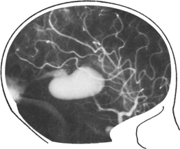
STURGE-WEBER SYNDROME
Angiomatosis affecting the facial skin, eyes and leptomeninges produces the characteristic features of the Sturge-Weber syndrome – a capillary naevus over the forehead and eye, epilepsy and intracranial calcification. (See page 563.)
DURAL ARTERIOVENOUS FISTULA
In contrast to AVMs these fistulous communications are usually acquired rather than developmental in origin. Arterial blood drains directly into either a venous sinus, cortical veins or a combination of both (see carotid-cavernous fistula page 301). The aetiology remains unknown, but sinus thrombosis or trauma may play a part. In a benign form, no reversal of flow occurs and no treatment is required. When retrograde venous flow occurs, venous hypertension results and haemorrhage may follow. For this type, treatment requires ligation and division of the draining vein, often combined with endovascular occlusion.
CAROTID–CAVERNOUS FISTULA
A fistulous communication between the internal carotid artery and the cavernous sinus may follow skull base trauma either immediately or after a delay of several days or weeks. Less often carotid-cavernous fistulae occur spontaneously, perhaps from rupture of a small intracavernous meningeal artery or a saccular carotid aneurysm.
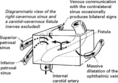
Clinical features
Symptoms develop suddenly (cf. cavernous sinus thrombosis) – the patient becomes aware of pulsating tinnitus as a ‘noise’ inside the head. Pain may follow. Examination reveals characteristic signs:

Methods of fistula repair
Spontaneous closure occurs in up to 60%. Provided symptoms do not progress, for the first few months, treatment should be conservative.
Endovascular: with detachable balloon catheterization, either through the transvenous or intra-arterial route, or by stent assisted coil embolisation. Recent reports include the use of covered stents alone.
INTRACRANIAL TUMOURS
INCIDENCE
Primary brain tumours occur in approximately 6 persons per 100 000 per year. Fewer patients with metastatic tumours reach a neurosurgical centre, although the actual incidence must equal, if not exceed that of primary tumours. It is estimated that 25% of patients with a malignancy have a CNS metastasis. About 1 in 12 primary brain tumours occur in children under 15 years.
SITE
In adults, the commonest tumours are gliomas, metastases and meningiomas; most lie in the supratentorial compartment.
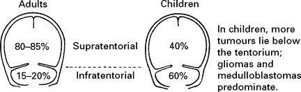
PATHOLOGY
Intracranial tumours are often described as ‘benign’ or ‘malignant’, but these terms cannot be directly compared with their extracranial counterparts:
A benign intracranial tumour may have devastating effects if allowed to expand within the rigid confines of the skull cavity. A benign astrocytoma may infiltrate widely throughout brain tissue preventing complete removal, or may occupy a functionally critical site preventing even partial removal.
A malignant intracranial tumour implies rapid growth, poor differentiation, increased cellularity, mitosis, necrosis and vascular proliferation, but metastases to extracranial sites rarely occur.
Pathological classification
In 2000, the World Health Organization drew up an internationally agreed classification of intracranial tumours based on the tissue of origin. This system avoids the term ‘glioma’ – previously encompassing astrocytoma, oligodendroglioma, ependymoma and glioblastoma multiforme. The cell origin of the highly malignant glioblastoma is now recognizable as astrocytic rather than embryonal as previously classified.
INTRACRANIAL TUMOURS – PATHOLOGICAL CLASSIFICATION
NEUROEPITHELIAL
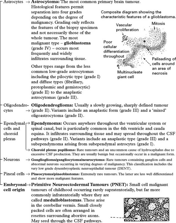
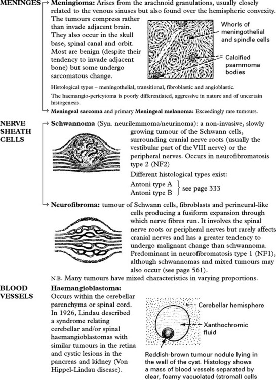

Primary central nervous system lymphoma (PCNSL): Forms around periventricular parenchymal blood vessels. May be solitary or multifocal. It generally occurs in immuno-compromised patients, e.g. AIDS. Metastatic spread from systemic lymphoma (e.g. non-Hodgkin’s lymphoma) is less common, involves the meninges and is rarely intraparenchymal.
Metastatic tumours: May arise from any primary site but most commonly spread from the bronchus or breast. Nervous system metastases occur in 25% of patients with disseminated cancer.
Tumour markers
Immunohistochemical techniques permit identification of antigens specific for certain cell or tissue characteristics and aid the histological diagnosis of tumours.
e.g. Glial fibrillary acidic protein (GFAP) – for astrocytic tumours
Cytokeratin – for metastatic carcinoma
Synaptophysin – for neuronal tumours
HMB 45 – for malignant melanoma
Some markers also indicate the degree of proliferation in various tumours (e.g. Ki-67). The identification of growth factors (e.g. Epidermal growth factor (EGRF)) may help distinguish between a primary glioblastoma (arising de novo) from a secondary glioblastoma (dedifferentiating from a previous lower grade tumour). Molecular techniques are increasingly used to identify loss of heterozygositye.g. 1p,19q in oligodendroglioma.
INTRACRANIAL TUMOURS – CLASSIFICATION ACCORDING TO SITE
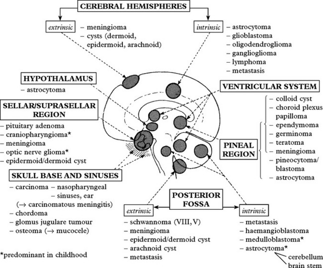
AETIOLOGY
Genetic factors: Over recent years, the role of genetic factors in tumour development has gained increasing prominence. Transformation of normal cells to malignant growth probably results from a variety of different processes –
(a) Normal cell growth and differentiation controlled by –

INTRACRANIAL TUMOURS – AETIOLOGY/INCIDENCE
(b) Inactivation of expression of tumour suppressor genes (e.g. mutation of the p53 gene with loss of heterozygosity on the 17p chromosome in many patients with low grade astrocytoma).
(c) Over expression of genes controlling growth factor (e.g. amplification of EGFR in primary glioblastoma).
Clearly defined inherited factors play a minor role. Only 5% of patients have a family history of brain tumour and with the exception of tuberous sclerosis (related to the formation of subependymal astrocytomas) and neurofibromatosis (linked to an increased incidence of schwannoma, optic nerve glioma and meningioma) do not fall into an obvious autosomal recessive or dominant pattern. Others include von Hippel-Lindau disease, Cowden’s disease and Li-Fraumeni syndrome.
Cranial irradiation: long term follow-up of patients undergoing whole head irradiation for treatment of tinea capitis and childhood leukemia shows an increased incidence of both benign and malignant tumours – e.g. astrocytoma, meningioma.
Immunosuppression: increased incidence of lymphoma.
INCIDENCE
The table below shows the approximate incidence of intracranial tumours extracted from large series.
|
Adults |
|
|
Glioblastoma |
15% |
|
Low grade glioma |
5% |
|
Meningioma |
25% |
|
Pituitary adenoma |
25% |
|
Primary CNS Lymphoma |
4% |
|
Peripheral Nerve Sheath Tumour (schwannoma) |
8% |
|
Others |
18% |
Adapted from Louis et al. WHO Classification of Tumours of the CNS, IARC Press 2007
|
Children |
|
|
Medulloblastoma/PNET |
16% |
|
Low grade glioma |
33% |
|
Malignant glioma |
14% |
|
Ependymoma |
10% |
|
Craniopharyngioma |
6% |
|
Germ cell tumours |
2.5% |
|
Meningioma |
2.5% |
|
Others |
16% |
Adapted from Rickert & Paulus Child’s Nervous System 2001 17.503-511
INTRACRANIAL TUMOURS – CLINICAL FEATURES
Symptoms tend to develop insidiously, gradually progressing over a few weeks or years, depending on the degree of malignancy (cf. acute onset of a cerebrovascular accident followed by a gradual improvement if the patient survives). Occasionally tumours present acutely due to haemorrhage or the development of hydrocephalus.

CLINICAL EFFECTS
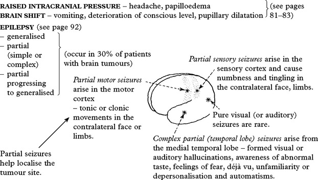
DISTURBED FUNCTION
Supratentorial – see higher cortical dysfunction, pages 109–117
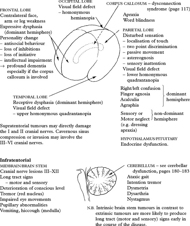
INTRACRANIAL TUMOURS – INVESTIGATION
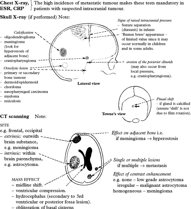
HIGH DEFINITION SCANS (1 mm slice width) – useful in the detection of pituitary, orbital and posterior fossa tumours.

MRI Note: SITE, MASS EFFECT and LESION MULTIPLICITY as for CT scanning.
Of particular value in tumours of the skull base, cranio-cervical junction and brain stem.
Flow voids show the relationship of adjacent blood vessels to the tumour.
Coronal and sagittal scanning provide additional information, showing the exact anatomical relationship of the tumour to the sulci and gyri, the ventricles, the falx and the tentorium cerebelli.

Paramagnetic enhancement: intravenous gadolinium increases sensitivity of detection and clarifies the site of origin, i.e. intrinsic or extrinsic, and may delineate the border between tumour and surrounding oedema.
Single or multiple lesions: MRI appears more sensitive than CT scanning in identifying small tumours and improves the detection of multiple lesions, e.g. metastasis.
Angiography/CTA/MRA: although angiography may reveal a tumour ‘blush’ or vessel displacement, it is only occasionally required to supplement other investigations. In some patients, it provides useful preoperative information, e.g. identifies feeding vessels to a vascular tumour or tumour involvement and constriction of major vessels.
Thallium SPECT: helps identify sites of high grade activity within a tumour. Useful to exclude if proposing conservative management or in planning stereotactic biopsy.
Functional MRI: Shows the relationship of eloquent areas to the tumour and may aid resection.
MR Diffusion Tensor Imaging: used to indentify fibre tracts running adjacent to or through the tumour. Of potential value in planning operative resection.
CSF examination: lumbar puncture is contraindicated if the clinician suspects intracranial tumour. If CSF is obtained by another source, e.g. ventricular drainage or during shunt insertion, then cytological examination may reveal tumour cells.
Tumour markers: although useful as an aid to histological diagnosis (see page 305), attempts to find a substance in blood or CSF which reflects growth of a specific tumour have been limited – only the link between elevated alpha fetoprotein and human chorionic gonadotrophins with yolk sac tumours and choriocarcinoma of the third ventricle helps diagnosis.
DIFFERENTIAL DIAGNOSIS OF MASS LESIONS (other than tumour)
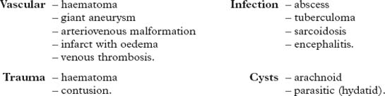
INTRACRANIAL TUMOURS – MANAGEMENT
STEROID THERAPY
Steroids dramatically reduce oedema surrounding intracranial tumours, but do not affect tumour growth.
A loading dose of 12 mg i.v. dexamethasone followed by 4 mg q.i.d. orally or by injection often reverses progressive clinical deterioration within a few hours. After several days treatment, gradual dose reduction minimises the risk of unwanted side effects.
Sellar/parasellar tumours occasionally present with steroid insufficiency. In these patients, steroid cover is an essential prerequisite of any anaesthetic or operative procedure.
OPERATIVE MANAGEMENT
Most patients with intracranial tumours require one or more of the following approaches:
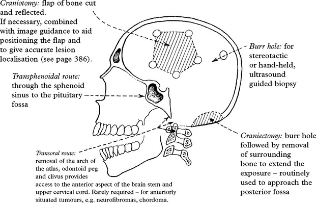
The subsequent procedure – biopsy, partial tumour removal/internal decompression or complete removal – depends on the nature of the tumour and its site. The infiltrative nature of primary malignant tumours prevents complete removal and often operation is restricted to biopsy or tumour decompression. Prospects of complete removal improve with benign tumours such as meningioma or craniopharyngioma; if any tumour tissue is overlooked, or if fragments remain attached to deep structures, then recurrence will result.
Image Guided Surgery
It is essential to accurately identify the tumour site on pre-operative imaging and to be able to use this information to guide the surgeon to the tumour whether for biopsy or for resection. Various techniques are available –
Stereotactic surgery: by rigidly attaching the frame to the patient’s head and using a CT or MR to identify the position of the locating rods, coordinates are determined for a selected target allowing accurate placement of a biopsy needle to within 1 mm (see page 384). This technique is routinely used to biopsy selected points within the tumour. It is possible to perform a craniotomy and tumour resection with the frame in place, but the frame tends to impede access and after opening the bone flap, the brain may shift introducing errors of localisation. When a craniotomy is planned, most now use neuronavigation (‘frameless’ stereotaxy) if available.
Neuronavigation: this technique requires rigid fixation of the head in a standard three pin head holder, but avoids the use of a cumbersome frame (see page 386). The system accurately detects the position of the handheld probe in relation to the skull and allows the surgeon to see where the probe tip lies in relation to pre-operative imaging. Although often routinely used, this technique also fails to take into account brain shift which can occur on opening the bone flap or if cerebrospinal fluid is drained off thus limiting accuracy.
Real-time intra-operative imaging: some centres have now acquired CT or MR imaging available within the operative theatre. Although costly, this real-time imaging overcomes problems encountered with brain shift and not only helps to locate the tumour, but also shows the extent of tumour resection as the operation progresses. Ultrasound has also been combined with neuronavigation to provide real-time imaging at a more realistic expense.
Surgery in Eloquent Areas
When intrinsic tumours lie adjacent to or within eloquent areas within the brain, i.e. speech area, motor strip, basal ganglia and internal capsule, resection is potentially hazardous. Various techniques have been developed to try to minimise this risk
fMRI/Diffusion Tensor Imaging (Tractography): Superimposing speech and/or motor strip areas seen on fMRI and white matter tracts seen on tractography on to the standard MR image, demonstrates the relationship of the tumour to these crucial structures. When these images are incorporated into the neuro-navigation system it enables the surgeon to avoid extending the tumour resection into these areas and causing irreversible neurological deficits. The reliability of each technique, however, is still in question and benefits remain uncertain.
‘Awake’ craniotomy: by either performing the surgery wholly under sedation with local anaesthetic, or by giving an anaesthetic for opening and closing the craniotomy and waking the patient up in between, gives the surgeon the opportunity to identify eloquent areas by applying electrical stimulation direct to the cortical surface and observing the functional effect. Studies show that patients tolerate the technique well and maximal tumour resection is possible with a low risk of deficit.
RADIOTHERAPY
Treatment of intracranial tumours with radiotherapy utilises one of the following:
– megavoltage X-rays (by far the most common method)
– electron beam from a linear accelerator (which can also produce megavoltage X-rays)
– accelerated particles from a cyclotron, e.g. nuclei of helium, protons (awaits full evaluation)
– γ rays from cobalt60.
In contrast to older methods, these modern techniques produce greater tissue penetration and avoid radiation damage to the skin surface. The effect of radiotherapy depends on the total dose – usually up to 60 Gy, and the treatment duration. This must be balanced against the risk to normal structures. Treatment aims to provide the highest possible dose to a specified region whilst minimising irradiation to adjacent normal brain. Various methods have been developed to achieve this –
• Conformal therapy where standard radiotherapy is administered, but the beams are shaped by the use of variable collimators or blocks which conform with the shape of the tumour, thereby eliminating normal brain.
• Stereotactic radiosurgery (SRS) where multiple converging beams from a linear accelerator or from multiple cobalt60 sources are focused on a selected target in a single treatment. Stereotactic radiotherapy (SRT) uses the same localisation method but with fractionated treatment as used in conventional radiotherapy (see page 385).
• Interstitial techniques where the tumour is treated from within (brachytherapy) by the implantation of multiple radioactive seeds, e.g. iodine125.
• Beam intensity modulated radiotherapy (IMRT) uses non-uniform beams of varying intensity (in contrast to the conventional uniform dose intensity) to complex tumour volumes. This helps protect surrounding structures, yet allows a higher dose.
• Proton therapy is available in only a few centres. It allows the delivery of high doses of radiation to very localised regions adjacent to vital structures such as the skull base.
Radiotherapy is of particular value in the management of malignant tumours – malignant astrocytoma, metastasis, medulloblastoma and germinoma, but also plays an important part in the management of some benign tumours – pituitary adenoma, craniopharyngioma. With some tumours that seed throughout the CSF pathways, e.g. medulloblastoma, whole neural axis irradiation minimises the risk of a distant recurrence.
Complications of radiotherapy: following treatment, deterioration in a patient’s condition may occur for a variety of reasons:
• Increased oedema – during treatment – reversible.
• Demyelination – after weeks, months – usually reversible.
• Radionecrosis – in usually 1–2 years (range 6 months–10 years) – irreversible.
• Cognitive impairment – whole brain irradiation causes dementia, ataxia and incontinence in over 10% at one year. Radiotherapy should be avoided in children under 3 years of age.
• Radiation induced tumours e.g. meningioma, may result many years after the treatment.
Oedema, demyelination and radionecrosis may involve the spinal cord after irradiation of spinal tumours. Other harmful effects include hair loss, skin reactions and endocrine disturbance.
CHEMOTHERAPY
Chemotherapeutic agents have been used for many years in the management of malignant brain tumours, but their benefits remain limited. Historically drugs most commonly used include nitrosoureas (e.g. BCNU, CCNU), procarbazine, vincristine and methotrexate (for lymphoma).
Temozolomide, an oral alkylating agent with excellent blood brain barrier penetration and modest toxicity is established as an alternative treatment for patients with recurrent high grade gliomas. It has also been shown to improve survival for patients with newly diagnosed glioblastoma when given concomitantly with radiotherapy. A combination of maximal safe surgery followed by combined chemoradiotherapy is now the standard of care for good performance patients with glioblastoma. Patients with methylation of the MGMT gene in the tumour appear particularly to benefit. Carmustine impregnated wafers (Gliadel) may also be considered both as a primary treatment or for tumour recurrence (see below).
Patients with anaplastic oligodendrogliomas and oligoastrocytoma with loss of heterozygosity on chromosomes 1p and 19q have a good prognosis and respond well to both radiation and to alkylating agent based chemotherapy (nitrosoureas, Temozolomide). Chemotherapy may be used either at initial diagnosis or at relapse in these patients. Other tumours where chemotherapy plays an important role include medullo-blastomas, primary CNS lymphomas and germ cell tumours.
Traditionally, chemotherapy has had a lesser role in low grade glial tumours but current studies are examining its use in both astrocytomas and oligodendrogliomas as an alternative to radiation in newly diagnosed patients.
Problems of drug administration
Toxicity: The ideal cytotoxic drug selectively kills tumour cells; but tumour cell response relates directly to the dose. High drug dosage frequently causes bone marrow suppression which may limit cytotoxic activity before an adequate therapeutic dose is reached.
Drug access: ‘Toxic’ doses are usually required before sufficient amounts penetrate the blood–brain barrier and gain access to the tumour cells.
Intrinsic resistance: Some tumour cells appear to have an inbuilt resistance to certain drugs. The vast array of available cytotoxic drugs and the infinite permutations of combined therapy creates difficulties in drug selection.
IMPROVING ACCESS: Many attempts to improve the access of cytotoxic drugs have been made with little success. Approaches such as blood brain barrier opening with mannitol, liposome delivery and direct intra-carotid injection have either failed to deliver or proved too toxic. Slow release preparations of BCNU (Gliadel) inserted directly into the surgical cavity has shown a modest increase in survival in some patients with GBM.
TARGETTED THERAPY
Greater understanding of the changes in gene expression and their effects on patients with brain tumours has allowed the development of drugs targeted specifically against these aberrant areas of the cell cycle. Agents directed against epidermal growth factor (EGF), platelet derived growth factor (PDGF) and angiogenetic factors (VEGF) have all shown some activity. Others are now in trial either as single agents or in combination with other targeted agents or conventional therapy. To date only Bevacizumab (Avastin) has been granted a licence in the US.
TUMOURS OF THE CEREBRAL HEMISPHERES – INTRINSIC
Intrinsic tumours arise within the brain substance.
ASTROCYTOMA (and glioblastoma multiforme) Astrocytomas may occur in any age group, but are commonest between 40 and 60 years.
Male:female = 2:1
Primary sites: Found in equal incidence throughout the frontal, temporal, parietal and thalamic regions, but less often in the occipital lobe. Microscopic classification defines 4 grades (WHO I–IV), but this is of limited accuracy and gene array technology may play a future role. A practical description divides tumours into either ‘malignant’ or ‘low grade’.
Anaplastic astrocytoma/glioblastoma multiforme
Anaplastic astrocytomas (grade III) and glioblastoma multiforme (grade IV) constitute up to 20% of all primary intracranial tumours. Glioblastoma occurs 4x more commonly than anaplastic astrocytoma. Median age at diagnosis is 64 years and 45 years respectively. These tumours widely infiltrate adjacent brain; growth is rapid. At autopsy, histology often reveals spread to multiple distant sites.
Genetic analysis differentiates ‘primary’ glioblastoma arising de novo (e.g. amplification of EGFR gene, loss of p16, mutation of PTEN and loss of heterozygosity of 10q), from a ‘secondary’ glioblastoma where dedifferentiation has occurred from a lower grade tumour (loss of p53, overexpression of PDGFR, loss of heterozygosity of 10q and abnormalities in the p16 and Rb pathways).
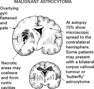
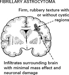
‘Low grade’ astrocytoma
Grade I/II astrocytomas make up 5% of all primary intracranial tumours in adults. The more frequent grade II tumours occur on average around 35 years. They are diffuse and slowly growing, and composed of well differentiated astrocytic cells subdivided into fibrillary, protoplasmic and gemistocytic types. Up to 90% show loss of the p53 gene. Although benign, these tumours widely infiltrate surrounding brain and lack a definitive edge or capsule.
The pilocytic (grade I) astrocytoma occurs in children and young adults in the hypothalamic region, the optic nerve in association with NF1 (page 561) and in the cerebellum and brain stem (pages 331,332). They grow very slowly, can often stabilise and even regress. Even partial resection can result in a cure.
CLINICAL FEATURES
Astrocytomas may present with:
– epilepsy – more common with low grade tumours
– signs and symptoms of focal brain damage – dysphasia, hemiparesis, personality change
– signs and symptoms of raised intracranial pressure – headache, vomiting, depression of conscious level.
Symptoms usually develop gradually, progressing over several weeks, months or years, the rate depending on the degree of malignancy. Sudden deterioration suggests haemorrhage into a necrotic area. In a patient with long standing epilepsy, the rapid development of further symptoms may result from malignant change within a previously ‘low grade’ lesion.
INVESTIGATIONS
CT scan: appearances vary considerably; in general, malignant and low grade lesions show different characteristics:
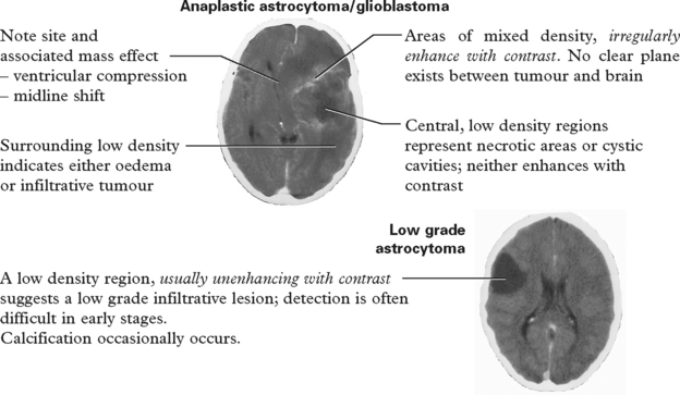
MRI: with and without gadolinium identifies the tumour location, size and degree of surrounding oedema more clearly. MRI is a more sensitive investigation for detecting low grade astrocytomas.
The MR spectroscopy profile (page 43) may suggest a pathological diagnosis.

MANAGEMENT
The management of glial tumours varies depending on a number of factors –
• the lesion site
• the degree of malignancy
• the presence or absence of a raised ICP
• the degree of disability and the effect of steroid therapy
• the suspected nature of the tumour on imaging
• the patient’s age
• the patient’s wishes
TREATMENT OPTIONS
Steroid therapy: For patients presenting with symptoms of raised intracranial pressure and/or focal neurological signs, a loading dose of dexamethasone 12 mg i.v. followed by 4mg q.i.d., by injection or orally, reduces surrounding oedema and leads to rapid improvement. Steroid treatment is an essential prerequisite to operation. Its introduction has significantly reduced the perioperative mortality. After several days, a gradual reduction in dosage avoids side effects.
Biopsy: Imaging is insufficient to conclusively establish the diagnosis. If not proceeding to an open operation, failure to confirm the nature of the lesion risks omitting treatment in benign conditions such as abscess, tuberculoma or sarcoidosis. Identification of tumour type and grade gives a prognostic guide and aids further management.
METHODS:
Framed or frameless stereotactic methods (see page 386) – permit accurate placement of a fine cannula at a predetermined site selected on CT scan or MRI. Stereotactic guidance is essential for small and/or deep inaccessible lesions (e.g. hypothalamus) and enables biopsy of specific regions in larger tumours e.g. enhancing areas on the MRI or CT scan. Prior selection of the needle path avoids vessels and important structures, thus minimising the risks. Since the degree of malignancy varies from region to region within a single lesion, several samples are taken from different sites to increase accuracy. If findings vary, then the region of greatest malignancy dictates the tumour grade. These techniques are now frequently used, even for more accessible lesions, due to the low mortality and morbidity. Provided patients receive preoperative steroid cover the risks are small, but occasionally biopsy produces or increases a focal deficit or causes a fatal haemorrhage.
Ultrasound guided – a brain cannula inserted into the abnormal region permits aspiration of a small quantity of tissue for immediate (smear and frozen section) and later (paraffin section) examination.

Tumour resection: Often combined with neuronavigation (frameless stereotaxy) or ideally real time CT, MRI or ultrasound if available, to aid tumour localisation and determine the extent of tumour resection as the operation progresses (see page 313).
Through a craniotomy, the surgeon performs an ‘open’ biopsy under direct vision, or resects as much tumour tissue as is safely feasible. The difficulty lies in the absence of a plane of cleavage between tumour tissue and brain. Neuronavigation can identify the boundaries seen on CT scan or MRI but this is limited by the resolution of the imaging. When adjacent to eloquent regions, the surgeon can merge fMRI or tractography to the neuronavigation image to help guide the resection, but reliability is limited and is still the subject of research. Alternatively performing the procedure in an awake patient and observing the direct effect of electrical stimulation may minimise the risk of causing an irreversible neurological deficit (see page 313). Large resections are most safely performed in the frontal, occipital or non-dominant temporal lobes. Most believe that the greater the reduction of the tumour mass, i.e. the greater the cytoreduction, the greater the effect of adjuvant therapy. A multicentre randomised study has shown that administration of an agent (5-ALA) orally prior to surgery causes tumour tissue to fluoresce during the operative procedure and improves the extent of resection without increasing the risk of deficit.
Radiotherapy: Most effective in rapidly growing tumours – grade III and IV. Radiotherapy extends survival, but does not cure. Studies show a dose–effect relationship – the greater the dose to the tumour area, the longer the survival. Usually up to 60 Gy is delivered in fractionated doses (see page 314). Methods of ‘conformal’ therapy and ‘interstitial radiotherapy’ (see page 314) aim to achieve this.
Chemotherapy: Concomitant Temozolomide based chemoradiotherapy is the standard of care following maximal safe resection in patients with glioblastoma of good performance status (WHO 0,1 = can do anything except heavy physical work). This improves survival from 10% to 26% at 2 years compared to radiation alone. The value of combined therapy in anaplastic astrocytoma and oligodendroglioma is under investigation. Chemotherapy, including Gliadel, is also used to palliate patients at recurrence. Bevacizumab is increasingly used as second or third line treatment in the US and some parts of Europe but has not been approved in the UK.
Treatment selection and prognosis
Since treatment cannot cure, the clinician must aim to produce maximal benefit to the patient with minimal burden, taking quality of survival into account as well as the duration.
Malignant astrocytoma/glioblastoma multiforme: Modern techniques have extended survival, but these tumours still carry a grave prognosis. Complete removal is impossible; even the formidable ‘hemispherectomy’ fails due to interhemispheric spread.
|
Median survival (months) |
|
|
Burr hole biopsy |
3–4 |
|
Tumour resection |
6 |
|
Burr hole biopsy + radiotherapy |
6–8 |
|
Tumour resection + radiotherapy |
12–14 |
|
Tumour resection + radiotherapy + concomitant chemotherapy |
14–18 |
In patients undergoing surgery, extensive tumour resection extends average survival by only 2 months, but when combined with radiotherapy and concomitant chemotherapy gains a further 12 months. With this treatment, 25% of patients survive 2 years; for patients with silencing of the MGMT gene by promoter methylation, over 40% survive beyond 2 years.
Management policies vary widely, but in general, maximal tumour resection with radiotherapy and chemotherapy is considered in most patients except:
– patients over 70 years of age (older patients tolerate radiotherapy less well)
– patients with extensive, deep lesions, e.g. involving basal ganglia or corpus callosum
– patients with severe disability, unresponsive to steroid therapy
In such patients, a diagnostic biopsy may be the only appropriate treatment.
Tumour recurrence may warrant a further resective procedure, perhaps combined with carmustine wafer implantation or other chemotherapy, particularly in younger patients who responded well to the initial treatment.
Low grade astrocytoma: A poorly defined region of low density on CT/MR scan without contrast enhancement suggests a low grade tumour (grade I or II) with a better prognosis. As with malignant astrocytomas, since these tumours infiltrate surrounding brain, they tumours cannot be ‘eradicated’ by surgical resection. The dilemma in such patients who often present with epilepsy and no other symptoms, is whether to proceed with radical surgery or to wait and watch. The argument for intervention is that at some point in the future the tumour will become malignant; many therefore opt for resecting as much tumour as is safely possible at an early stage in the hope that this defers malignant change. There is however no evidence that active intervention with operation and/or radiotherapy or chemotherapy changes outcome. The issues involved should be discussed with the patient, but if a conservative approach is adopted, the surgeon should advise intervention if subsequent CT or MR scanning shows definitive tumour progression (expansion or contrast enhancement), or if clinical symptoms supervene. A positive Thallium SPECT scan (page 49) would also indicate the need for action. If proceeding to surgery, the operative techniques used to avoid damaging eloquent regions apply (page 313).
About 50–60% of patients with grade II astrocytomas survive 5 years; about 40% survive 10 years. Of those patients with pilocytic (grade I) astrocytomas, 80% survive 20 years.
OLIGODENDROGLIOMA
Oligodendrogliomas are far less common than astrocytomas. They occur in a slightly younger age group – 30–50 years, and usually involve the frontal lobes. Occasionally involvement of the ventricular wall results in CSF seeding. Calcification occurs in 40%.

In contrast to astrocytomas, the tumour margin often appears well defined. Both low grade and anaplastic forms exist. Genetic analysis of anaplastic oligodendrogliomas has revealed that almost 80% have 1p and 19q allelic losses (i.e. loss of heterozygosity) and these patients respond well to chemotherapy.
Management and prognosis: when imaging suggests a low grade tumour, the approach is similar to astrocytomas with the option of delaying treatment until symptoms appear. Patients can expect to survive for 12–16 years. For patients with anaplastic oligodendrogliomas, resection followed by chemotherapy is combined with either immediate or delayed radiotherapy. Ideally treatment should depend on the patient’s genetic profile. Those patients with loss of 1p and 19q alleles respond well to chemotherapy and survive over 10 years. The 27% with a genetic profile similar to primary glioblastoma (page 316) seldom respond to chemotherapy and survive on average about 16 months.
Mixed oligoastrocytoma: those with a mixed form of astrocytoma/oligodendroglioma have a prognosis lying between that for each type.
HYPOTHALAMIC ASTROCYTOMA
Hypothalamic tumours usually occur in children; they are usually astrocytomas of the pilocytic (juvenile) type. The clinical effect of hypothalamic damage takes different forms. Initially the child fails to thrive and becomes emaciated. Signs of panhypopituitarism may develop. Eventually an anabolic phase results in obesity accompanied by diabetes insipidus and delayed puberty. Disturbance of affect and of sleep–wake rhythms may occur.
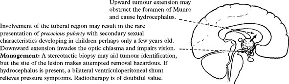
METASTATIC TUMOURS
|
Common primary sites |
|
|
Any malignant tumour may metastasise to the brain. Malignant melanomas show the highest frequency (of those with metastasis, 66% are in the brain); this contrasts with tumours of the cervix and uterus where < 3% develop intracranial metastasis. The most commonly encountered metastatic intracranial tumours arise from the bronchus and the breast; of patients with carcinomas at these sites, 25% develop intracranial metastasis. In over 50% of patients, metastases are multiple. |
–bronchus – breast – kidney – thyroid – stomach – prostate – testis –melanoma |
Spread is usually haematogenous to the grey/white matter junction. Occasionally a metastasis to the skull vault may result in a nodule or plaque forming over the dural surface from direct spread.
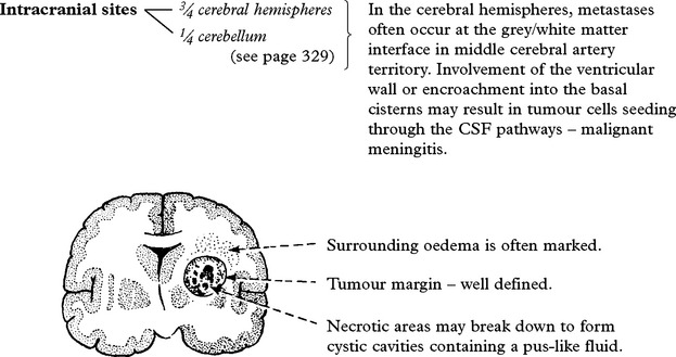
Clinical features
Patients with supratentorial metastatic tumours may present with epilepsy, or with signs and symptoms occurring from focal damage or raised intracranial pressure. Cerebellar metastases are discussed onpage 329. Malignant meningitis causes single or multiple cranial nerve palsies and may obstruct CSF drainage (see page 517). About 10% of diagnosed intracranial metastases are asymptomatic, detected on screening patients with known malignancy.
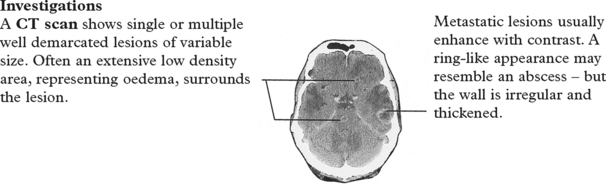
MRI scanning, with and without paramagnetic enhancement, is even more sensitive than CT in detecting small metastatic lesions.
The search for a primary lesion if not already established must include a thorough clinical examination and a chest X-ray. Other investigations including barium studies, intravenous pyelogram (IVP), abdominal CT scans, ultrasound and sputum and urine cytology have questionable value, unless clinically indicated. Whole body PET scanning, if available, is the most sensitive method of detecting the primary lesion.
Management and prognosis:
Corticosteriods (dexamethasone) have a dramatic, rapid effect, producing clinical improvement in most patients.
– Solitary lesions: If the tumour lies in an accessible site, complete excision followed by radiotherapy provides good results – survival usually depends on the extent of extracranial disease and its ability to respond to treatment rather than on intracranial recurrences. Stereotactic radiosurgery provides a valuable alternative, particularly for lesions less than 3 cm in diameter and for deep-seated lesions.
– Multiple lesions: Operative removal is seldom practical. Provided no doubt exists about the diagnosis (abscesses or tuberculomata may resemble metastasis) radiosurgery may be administered to two or even three lesions. For other patients whole brain irradiation may be considered.
Prognosis: Patients < 65 years, with a good performance status and no evidence of systemic metastasis have the best prognosis. In the absence of evidence of systemic cancer, the median survival period approaches 2 years. In those with systemic disease, results are less good with a median survival of 8 months.
PRIMARY CNS LYMPHOMA (PCNSL) (syn. HIGH GRADE NON HODGKIN’S B-CELL LYMPHOMA) Single or multiple lymphomas usually lie deep within the basal ganglia or in the periventricular region. Some are discrete lesions, others extensively invade surrounding brain. Histology shows sleeves of primitive reticulum cells extending outwards from the blood vessels. The incidence is significantly increased in AIDS and in immunocompromised patients.
CSF examination is important; 30% of patients with PCNSL show positive cytology. A positive Epstein-Barr test within tumour cells in CSF is diagnostic of AIDS lymphoma.

Management: In AIDS patients, CT and MRI finding of PCNSL appear similar to toxoplasmosis; antiprotozoal therapy should be tried first. Failure to respond indicates the need for biopsy. Steroidscan cause dramatic shrinkage, and the imaging should be repeated if any delay occurs prior to biopsy. Radiotherapy also has dramatic effects, but with this treatment alone, the median survival period is only 10–12 months. Methotrexate based chemotherapy (in patients with a normal immune system) can increase median survival to up to 44 months. Some advocate delaying radiotherapy treatment until a recurrence occurs. AIDS patients, who receive radiotherapy, have a median survival of about 4 months, but chemotherapy can improve this in selected patients.
GANGLIOGLIOMA
This a rare tumour occurring in the younger age group (< 30 years), composed of abnormal neuronal growth mixed with a glial component. The proportion of each component varies from patient to patient. Growth is slow and malignant change uncommon; when this occurs it probably develops in the glial component.
Management follows that of low grade astrocytomas.
NEUROBLASTOMA
Rarely occurs intracranially in children < 10 years. Highly cellular, malignant lesion composed of small round cells, some showing neuronal differentiation.
TUMOURS OF THE CEREBRAL HEMISPHERES – EXTRINSIC
Extrinsic tumours arise outwith the brain substance.
MENINGIOMA
Meningiomas constitute about one-quarter of all primary intracranial tumours. They are slow growing and arise from the arachnoid granulations. These lie in greatest concentration around the venous sinuses, but they also occur in relation to surface tributary veins. Meningiomas may therefore develop at any meningeal site. Occasionally they are multiple.
Meningiomas present primarily in the 40–60 age group and have a slight female preponderance. They are principally benign tumours, although 1–3% show malignant change.
Pathology
Various histological types are described – syncytial, transitional, fibroblastic and angioblastic; different types may coexist within the same tumour. These distinctions serve little clinical value, although it is important to identify the anaplastic (malignant) form, as this indicates the likelihood of rapid growth and a high rate of recurrence following removal.

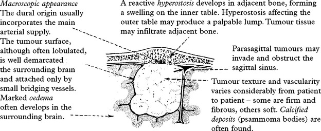
En-plaque meningioma: In some patients, rather than developing a spherical form, the meningioma spreads ‘en-plaque’ over the dural surface. This type often arises from the outer aspect of the sphenoid wing.
MENINGIOMA Clinical features:
Approximately a quarter of patients with meningioma present with epilepsy – often with a focal component. In the remainder, the onset is insidious with pressure effects (headache, vomiting, papilloedema) often developing before focal neurological signs become evident.
Notable characteristic features occur, dependent on the tumour site – PARASAGITTAL/PARAFALCINE tumours lying near the vertex affect the ‘foot’ and ‘leg’ area of the motor or sensory strip.Partial seizures or a ‘pyramidal’ weakness may develop in the leg (i.e. primarily affecting foot dorsiflexion, then knee and hip flexion). Extension of the lesion through the falx can produce bilateral leg weakness. Posteriorly situated parasagittal tumours may present with a homonymous hemianopia. Tumours arising anteriorly may grow to extensive proportions before causing focal signs; eventually minorimpairment of memory, intellect and personality may progress to a profound dementia.
INNER SPHENOIDAL WING tumours may compress the optic nerve and produce visual impairment. Examination may reveal a central scotoma or other field defect with optic atrophy.
N.B. The FOSTER KENNEDY syndrome denotes a tumour causing optic atrophy in one fundus from direct pressure and papilloedema in the other due to increased intracranial pressure.
Involvement of the cavernous sinus or the superior orbital fissure may produce ptosis and impaired eye movements (III, IV and VI nerve palsies) or facial pain and anaesthesia (V1 nerve damage) – see diagram on page 153. Proptosis occasionally results from venous obstruction or tumour extension into the orbit.
OLFACTORY GROOVE tumours destroy the olfactory bulb or tract causing unilateral followed by bilateral anosmia. Often unilateral loss passes unnoticed by the patient; with tumour expansion, dementia may gradually ensue.
SUPRASELLAR tumours – see page 348.
ASYMPTOMATIC TUMOURS: With the increased availability of imaging, small meningiomas are frequently detected incidentally. Conservative management of such patients has shown that over a 5 year period about 40% show expansion and one in six develop symptoms.
Investigations:
CT SCAN

MRI: On T1 weighted images most meningiomas are isointense with brain, but after gadolinium injection, they diffusely and strikingly enhance. T2 weighted images give useful preoperative information by identifying major vessels and showing their relationship with the tumour.
ANGIOGRAPHY: Characteristically shows a highly vascular lesion with a typical tumour ‘blush’, but with the availability of CT angiography, its main value is in selective catheterisation and embolisation of external carotid feeding vessels to reduce tumour vascularity and diminish operative risks from excessive haemorrhage.
3-D CT ANGIOGRAPHY: can show the relationship of surrounding blood vessels to the tumour.
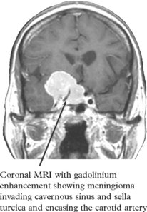
Management
Management aims at complete removal of both the tumour and its origin without damaging adjacent brain; but this depends on the tumour site and its nature. Even with ‘convexity’ tumours, where complete excision of the dural origin is possible, overlooking a small fragment of tumour may result in recurrence. This is more likely with malignant meningiomas where the plane of cleavage is often obscured.
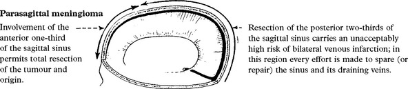
When the tumour is asymptomatic or when the patient’s age or the tumour site prevents operation or allows only a limited removal, a conservative approach may be more appropriate, only intervening if the tumour progresses or causes disabling symptoms. Alternatively stereotactic radiosurgery could be considered for small tumours or for residual fragments. Benefits of standard radiotherapy are uncertain unless histology reveals evidence of malignant change.
Operative results: with modern techniques, operative mortality has fallen to less than 3%, but this varies depending on the size and position of the tumour. Although in vitro studies have demonstrated numerous hormonal receptors (e.g. progesterone and oestrogen) in meningioma tissue, clinical studies of hormonal therapy have failed to show any benefit.
Tumour recurrence: depends predominantly on the completeness of removal and on the duration of follow-up. With ‘total’ resection, about 20% recur after 10 years. With sub-total resection over 50% require a further operation within 10 years.
HAEMANGIOPERICYTOMA
A tumour arising from the meninges, but of uncertain cell of origin. It presents with similar clinical features and CT/MRI appearance to meningiomas, but is fifty times less common. Angiography may show a more prominent vascular supply. Calcification does not occur. Haemangiopericytomas tend to invade adjacent bone and to recur even after apparent complete surgical removal. Post-operative radiotherapy should delay recurrence.
ARACHNOID CYSTS

Treatment: These are common findings and in the vast majority, no treatment is indicated. Rarely patients present with mass effect and require marsupialisation (via a craniotomy) or cystoperitoneal shunting. Some believe that prophylactic treatment in young children aids normal brain development.
EPIDERMOID/DERMOID CYSTS
These cysts, more commonly found in the posterior fossa (page 337), occasionally develop in the Sylvian or interhemispheric fissure. They are either of congenital or acquired origin due to implantation and sequestration of ectoderm. They may present with epilepsy, features of raised intracranial pressure or with focal neurological signs. Rupture into the subarachnoid space causes a chemical meningitis.
On CT scan, the extreme low attenuation of the cyst contents is characteristic. Symptoms may necessitate operative evacuation of the cyst contents. Complete removal of the cyst wall is difficult and reaccumulation may occur.
Lipomas
Rarely occur intracranially. They are usually found incidentally on imaging or at autopsy and are often associated with other developmental anomalies such as agenesis of the corpus callosum. They are located in midline structures e.g. corpus callosum, dorsal midbrain and cerebellar vermis. They require no treatment.
TUMOURS OF THE POSTERIOR FOSSA – INTRINSIC
CEREBELLAR METASTASIS
In adults, metastasis is the commonest tumour of the cerebellar hemisphere. Primary tumour sites match those of supratentorial lesions (page 322).
Clinical features: may present acutely or progress over several months.

Management
Operative removal of a single metastasis through a suboccipital craniectomy is worthwhile, provided the patient has a reasonable prognosis from the primary tumour. Risks are small – extensive cerebellar hemisphere resection (on one side) seldom produces any significant permanent deficit. A course of radiotherapy can follow operation if resection is incomplete. Radiosurgery provides a possible alternative to surgical resection. Persistence of obstructive hydrocephalus requires a ventriculoperitoneal shunt.
HAEMANGIOBLASTOMA
This benign tumour of vascular origin occurs primarily in the middle-aged; it is slightly more prevalent in males and is the commonest primary cerebellar tumour of adults. In some patients, haemangioblastomas occur at other sites, e.g. the spinal cord and retina and may be associated with other pathologies e.g. polycythaemia and cysts in the pancreas and kidneys – Von Hippel-Lindau disease(page 563).
The tumour is usually highly vascular. In 70% there is an associated cyst, the lining of which does not contain tumour.

Clinical features
Cerebellar signs and symptoms or the effects of CSF obstruction usually develop insidiously. Occasionally subarachnoid haemorrhage occurs. In female patients, symptoms often appear during pregnancy. Polycythaemia due to increased erythropoietin production is common.
Investigations
CT scan/MRI shows either a strongly enhancing solid tumour in the cerebellum or a tumour nodule lying in the wall of a well defined cystic region. Occasionally, multiple lesions are evident. Enhancing vessels on CT or tortuous flow voids on MRI reflect the high vascularity.
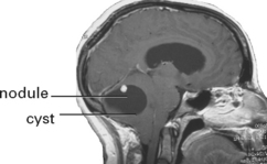
Management
In some patients operative removal of the tumour nodule is straightforward, but recurrences (or further tumours at other sites, e.g. spine) develop in 20%. Patients with highly vascular solid tumours can present a formidable surgical challenge, particularly if they involve the medulla. Pre-operative embolisation may greatly reduce the surgical risks.
MEDULLOBLASTOMA
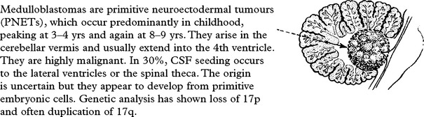
Clinical features
Destruction of the cerebellar vermis causes truncal and gait ataxia often developing over a few weeks.
Alternatively, the patient presents with signs and symptoms of raised intracranial pressure due to blockage of CSF drainage. In the very young, failure to recognise these features has resulted in permanent visual loss from severe papilloedema.
Investigations
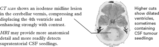
Management
Staging is essential due to the high incidence of leptomeningeal spread and bone marrow involvement. Assess this with spinal MRI with gadolinium, CSF analysis and bone marrow examination. CSF obstruction may require urgent relief, preferably by 3rd ventriculostomy.
Operation: The aim is to remove as much tumour as possible (particularly if staging has excluded disseminated disease), without damaging crucial structures in the floor of the 4th ventricle.
Radiotherapy: the most effective post-operative treatment. Whole neural axis irradiation attempts to cover any CSF seeding, but this is unacceptable in children < 3 years due to severe side effects. In this group and in recurrent tumours radiosurgery may help.
Chemotherapy: routinely used, but the extent to which chemotherapy alters the quality or duration of survival is less certain.
Prognosis
Five-year survival ranges from 50–90% depending on the extent of tumour removal, dissemination and age (<3 years poor risk).
CEREBELLAR ASTROCYTOMA
In contrast to astrocytomas of the cerebral hemispheres, cerebellar astrocytomas are usually low grade tumours of the fibrillary or pilocytic types. They are particularly common in children and carry an excellent prognosis. Occasionally a more diffuse or anaplastic type occurs with a less favourable outcome. They usually lie in the cerebellar hemisphere or vermis but occasionally extend through a peduncle into the brain stem. Many have cystic components.
Clinical features
Cerebellar signs and symptoms tend to develop gradually over many months; if CSF obstruction occurs, the patient may present acutely with headache, papilloedema and deteriorating conscious level.
Investigations
CT scan – density changes and the degree of contrast enhancement are variable.
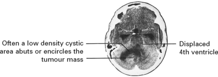
MRI – may provide more anatomical definition.
Management
Ideally, complete operative removal is attempted provided the brain stem is not involved. With pilocytic tumours, 80% survive 20 years. Even after partial removal ‘cures’ have been reported. Persistent hydrocephalus may require 3rd ventriculostomy or a ventriculoperitoneal shunt.
BRAIN STEM ASTROCYTOMA
Rarely, astrocytomas arise within the brain stem. Most are of the fibrillary or pilocytic types and diffusely expand the pontine region although they can be malignant. They develop mainly in children or young adults.
Clinical features
Cranial nerve palsies and long tract signs gradually develop as the tumour progresses. Eventually conscious level is impaired. More malignant gliomas are associated with a rapidly progressing course, often with signs of raised intracranial pressure.
Investigations
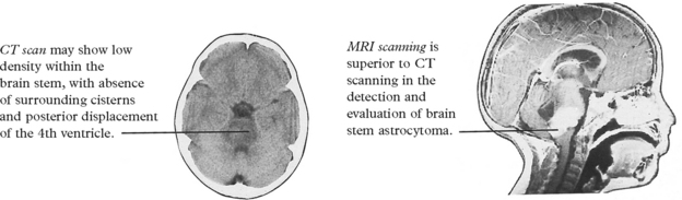
Management
Operative exploration is seldom indicated. Radiotherapy is often administered, usually after a stereotactic biopsy, with occasional palliation of symptoms and uncertain effect on survival. Chemotherapy is of no value.
Prognosis
At best, the 5-year survival following radiotherapy is 35%, although some patients may survive for up to 20 years with minimum disability.
TUMOURS OF THE POSTERIOR FOSSA – EXTRINSIC
VESTIBULAR SCHWANNOMA
Nerve sheath tumours are the commonest infratentorial tumours, constituting 8% of all primary intracranial tumours and 80% of cerebellopontine angle lesions. They usually present in middle age (40–50 years) and occur more frequently in women. Bilateral schwannomas occur in 5% of patients and are characteristic of type 2 neurofibromatosis (NF2) (page 561).
They are benign, slowly growing tumours which arise primarily from the vestibular portion of the VIII cranial nerve and lie in the cerebellopontine angle – a wedge shaped area bounded by the petrous bone, the pons and the cerebellum. Rarely these tumours arise from the V cranial nerve.
Schwannomas expand at an average rate of 2 mm/year, but about 50% show no growth on serial investigation.
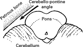
Pathology: The other type of nerve sheath tumour – neurofibroma (page 304) – does not occur intracranially.
Different histological types exist, often within the same tumour:
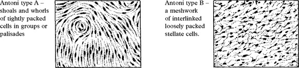
Clinical features
Patients with acoustic tumours often complain of occipital pain on the side of the tumour. In addition:
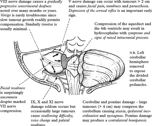
Investigations

– brain stem auditory evoked potential (BAEP) – perhaps the most sensitive of these tests shows a delay of the wave V latency on the affected side.
CT scan

MRI
The investigation of choice, particularly for small intracanalicular tumours. On a T1 weighted image, the lesion enhances strongly after i.v. gadolinium.

MANAGEMENT OPTIONS
Conservative approach
Since about 50% of tumours show no growth on yearly follow-up and since treatment carries risk, a ‘wait and watch’ policy is a sensible option for small to medium-sized tumours (< 20 mm), particularly in the elderly.
Stereotactic radiosurgery
This single dose technique (see page 314), initially reserved for elderly patients, is now used more widely for schwannomas up to 3 cm in size. Centres report ‘control’ of tumour growth in up to 90%, with preservation of hearing in about 75% and facial nerve function in 98%. A 10-year follow-up study suggests that growth control is maintained.
SURGICAL RESECTION
Techniques
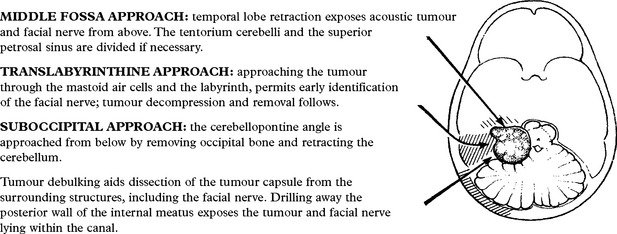
Results
Outcome relates to tumour size. With a tumour diameter of 5–20 mm, some hearing can be preserved in > 50% and facial nerve function in > 95%. With tumours of > 3 cm, all lose hearing and 25–50% sustain facial nerve damage. When present, incomplete eye closure may require tarsorrhaphy to prevent corneal ulceration. When facial nerve palsy persists, hypoglossal-facial anastomosis may improve the cosmetic result. Mortality ranges from 1–3% and usually results from damage to important vascular structures (e.g. anterior inferior cerebellar artery), haemorrhage, aspiration pneumonia or pulmonary embolus.
Treatment selection: For tumours < 2 cm in diameter a conservative approach is the most appropriate option. If serial scans show tumour growth or if the tumour is > 2 cm on diagnosis, treatment is required aimed at removal or control of growth, preservation of facial nerve function and retention of useful hearing unless this is already lost. The options of radiosurgery and surgical removal should be discussed with the patient along with the pros and cons (i.e. removal with increased risk or ‘control’ with less risk). For tumours > 3 cm in diameter only surgical resection is feasible.
TRIGEMINAL SCHWANNOMA
Rarely schwannomas arise from the trigeminal ganglion or nerve root. These lie in the middle fossa or extend into the cerebellopontine angle, compress surrounding structures – cavernous sinus, midbrain and the pons – and erode the apex of the petrous bone.
Clinical features are usually long-standing – facial pain, paraesthesia and numbness. Compression of posterior fossa structures results in nystagmus, ataxia and hemiparesis.
CT scan or MRI with contrast demonstrates an enhancing lesion eroding the petrous apex and extending into the middle and/or posterior fossa.
Management: Operative removal, even if subtotal, should provide long-lasting benefit. The tumour is approached either from above via a subtemporal route across the middle fossa floor, from below via a suboccipital craniectomy, or via a combination of these approaches.
MENINGIOMA
Approximately 8% of all intracranial meningiomas arise in the posterior fossa.
Clinical features
These depend on the exact tumour site. Those arising over the cerebellar convexity may not present until the mass obstructs CSF drainage. Meningiomas arising in the cerebellopontine angle may involve any cranial nerve from V to XII. A clivus meningioma may cause bilateral VI nerve palsies before pontine pressure causes long tract signs.
Tumours growing at the foramen magnum, compressing the cervico-medullary junction, produce characteristic effects – pyramidal weakness initially affecting the ipsilateral arm, followed by the ipsilateral leg, spreading to the contralateral limbs with further tumour growth.
Investigations
CT scan with intravenous contrast will identify the tumour site, but MRI with gadolinium enhancement shows more anatomical detail.
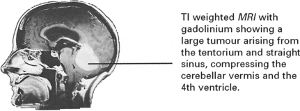
Management
As with supratentorial meningiomas, treatment aims at complete tumour removal. In the posterior fossa, cranial nerve involvement makes this difficult and exacting; excision of the tumour origin is seldom possible. For some, stereotactic radiosurgery is an alternative method of controlling tumour growth. Radiotherapy or radiosurgery may be considered when residual tumour persists.
EPIDERMOID/DERMOID CYSTS
These rare cysts of embryological origin develop from cells predestined to become either epidermis or dermis. They most commonly arise in the cerebellopontine angle but may also occur around the suprasellar cisterns, in the lateral ventricles and in the Sylvian fissures, often extending deeply into brain tissue.
Pathology: Depends on cell of origin:
Epidermoid (epidermis) – a thin transparent cyst wall often adheres firmly to surrounding tissues; the contents – keratinised debris and cholesterol crystals – produce a ‘pearly’ white appearance.
Dermoid (dermis) – as above, but thicker walled and, in addition, containing hair follicles and glandular tissue. Midline dermoid cysts lying in the posterior fossa often connect to the skin surface through a bony defect. This presents a potential route for infection.
Clinical features
When lying in the cerebellopontine angle, epidermoid/dermoid cysts often cause trigeminal neuralgia (see page 163). Neurological findings may range from a depressed corneal reflex to multiple cranial nerve palsies. Rupture and release of cholesterol into the subarachnoid space produces a severe and occasionally fatal chemical meningitis. The presence of a suboccipital dimple combined with an attack ofinfective meningitis should raise the possibility of a posterior fossa dermoid cyst with a cutaneous fistula.
Investigations
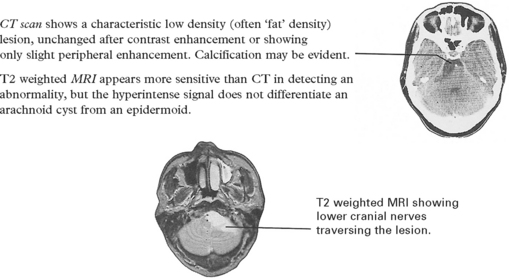
Treatment
Adherence of the cyst wall to important structures often prevents complete removal, but evacuation of the contents provides symptomatic relief. Aseptic meningitis in the postoperative period requires prompt treatment with steroids. Even when removal is incomplete, recollection of the keratinised debris is uncommon and may take many years.
SELLAR/SUPRASELLAR TUMOURS – PITUITARY ADENOMA
Tumours of the pituitary gland constitute about 5–10% of intracranial tumours. They arise from the anterior portion of the gland and are usually benign.
‘CLASSIC’ classification
Previously based on the light microscopic appearance of the tumour cell type.
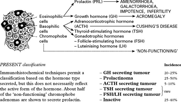
CLINICAL PRESENTATION

LOCAL MASS EFFECT
Headache
Occurs in most patients with enlargement of the pituitary fossa. It is not specific in site or nature.
Visual field defects
Pressure on the inferior aspect of the optic chiasma usually causes superior temporal quadrantanopia initially, with progression to bitemporal hemianopia, but any pattern can occur.
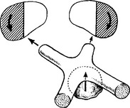
Cavernous sinus compression
In some pituitary tumours, lateral expansion may compress nerves lying within the walls of the cavernous sinus. The III nerve is especially vulnerable.
Rarely vertical expansion obstructs the foramen of Munro causing hydrocephalus and/or hypothalamic compression (page 346).
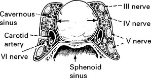
ENDOCRINE EFFECT
1. HYPERSECRETION
The clinical syndrome produced is dependent on the hormone secreted.
Growth hormone (GH)
Stimulates growth and plays a part in control of protein, fat and carbohydrate metabolism. Excess GH in the adult causes ACROMEGALY.
In childhood, prior to fusion of bone epiphyses, GH excess causes GIGANTISM.
GH levels are usually increased to > 10 mU/l. Increased serum levels of insulin growth factor-1 enhances the effect of growth hormone on target organs.
Hyperglycaemia normally suppresses GH secretion. GH samples are taken in conjunction with blood glucose during a glucose tolerance test. The lack of GH suppression after glucose administration confirms the presence of a tumour.
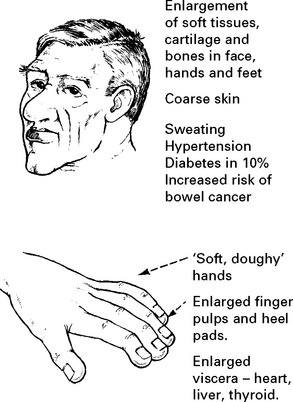
Prolactin
This hormone helps promote lactation. Prolactinoma is the commonest type of pituitary tumour. Immunoassay techniques aid early detection. Female:male ratio – 4:1
This tumour may present with
– INFERTILITY
– AMENORRHOEA
– GALACTORRHOEA
In males, the tumour may present with IMPOTENCE or remain undetected until local pressure effects occur.
In most centres, a serum prolactin of 500 mU/l is considered abnormal, but before assuming the presence of a prolactin secreting tumour, other causes must be excluded.
Causes of hyperprolactinaemia
– Stress
– Pregnancy
– Drugs (phenothiazines, oestrogens)
– Hypothyroidism
– Renal disease
– Pituitary adenoma
– Hypothalamic lesion (e.g. sarcoid, craniopharyngioma) or the pituitary stalk syndrome
– Seizures
Prolactin differs from other anterior pituitary hormones in that it is under tonic inhibitory control from the hypothalamus. Hypothalamic lesions or raised intrasellar pressure, compromising hypothalamic–pituitary perfusion (i.e. the ‘pituitary stalk syndrome’) produce a rise in serum prolactin, but levels seldom exceed 2000 mU/l. Prolactin levels above 4000 mU/l invariably indicate prolactinoma.
Adrenocorticotrophic hormone (ACTH)
ACTH stimulates secretion of cortisol and androgens. Hypersecretion from a pituitary adenoma or hyperplasia causes CUSHING’S DISEASE (bilateral adrenal hyperplasia) which presents with the characteristic features of CUSHING’S SYNDROME.
This syndrome may also be caused by excessive oral corticosteroids, but also by an adrenal tumour or by ectopic secretion of ACTH from a bronchial carcinoma.
Features of Cushing’s syndrome
– Moon face
– Acne
– Hirsutism and baldness
– Buffalo-type obesity
– Purple striae over flanks and abdomen
– Bruising
– Muscle weakness and wasting
– Osteoporosis
– Hypertension
– Increased susceptibility to infection
– Diabetes mellitus
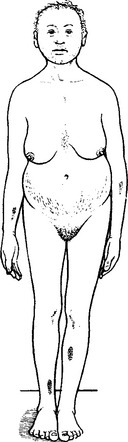
A loss of normal diurnal variation of plasma free cortisol and an increase in 24 hour urinary free cortisol indicates excess secretion. The diagnosis of a pituitary cause is suggested by finding normal or moderately raised ACTH levels which suppress with high doses of dexamethasone.
Ectopic ACTH production does not suppress with dexamethasone and with adrenal tumours, ACTH levels are virtually undetectable.
Other tests include
– the effect of corticotrophin releasing factor (↑ACTH and cortisol if pituitary origin)
– petrosal versus peripheral venous sampling to identity the source of the ACTH.
Bilateral adrenalectomy for Cushing’s syndrome is sometimes followed by the development of Nelson’s syndrome – high ACTH levels, pituitary enlargement and marked skin pigmentation.

2. HYPOSECRETION
Many pituitary tumours are diagnosed before panhypopituitarism develops, but large tumours may cause gradual impairment of pituitary hormone secretion. Growth hormone and the gonadotrophins are first affected, followed by TSH and ACTH. Panhypopituitarism only occurs when more than 80% of the anterior pituitary is destroyed.
|
Impaired secretion |
Adults |
Children |
|
GH – |
‘Adult GH deficiency syndrome’ – weight gain, loss of libido, fatigue |
Pituitary dwarfism – (diminished somatic growth, retarded sexual development, hypoglycaemic episodes, normal intelligence) |
|
Gonadotrophins – |
Amenorrhoea, sterility, loss of libido |
|
|
ACTH – |
Glucocorticoid and androgen deficiency, muscle weakness and fatigue |
|
|
TSH – |
Secondary hypothyroidism – sensitivity to cold, dry skin, physical and mental sluggishness, coarseness of hair |
|
|
Prolactin* – |
Failure of lactation |
* Prolactin secretion is most resistant to pituitary damage. Deficiency is seldom evident, usually only presenting after postpartum haemorrhage (Sheehan’s syndrome) as a failure of lactation associated with the other features of panhypopituitarism.
Pituitary hormone assay cannot distinguish low ‘normal’ levels from impaired secretion, but low levels of pituitary hormone in the presence of low target gland hormones confirm hyposecretion, e.g. low TSH levels despite a low serum thyroxine. Basal levels guide replacement therapy.
The lack of response to tests designed to increase specific pituitary hormones provides additional confirmation of hypofunction:
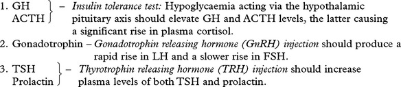
The above tests can be carried out simultaneously as the Combined pituitary stimulation test. Insulin, GnRH and TRH are injected intravenously and all anterior pituitary hormones measured from repeated blood samples taken over a 2-hour period. Glucose levels are also checked to ensure adequacy of the hypoglycaemia.
PITUITARY APOPLEXY
This is an uncommon complication of pituitary tumours due to the occurrence of infarction followed by haemorrhage into the tumour. Severe headache of sudden onset simulating subarachnoid haemorrhage, rapidly progressive visual failure and extraocular nerve palsies accompany acute pituitary insufficiency. Death may follow unless urgent steroid treatment is instituted.
NEURORADIOLOGICAL INVESTIGATION
LARGE TUMOURS
Skull X-ray
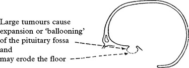
CT scan with contrast enhancement demonstrates tumours filling the pituitary fossa and expanding into the suprasellar compartment, but MRI gives more anatomical detail, clearly delineating any suprasellar extension and the effect on adjacent structures.

MICROADENOMAS

MANAGEMENT
A variety of different forms of treatment are available:
Drug therapy
Dopamine agonists lower abnormal hormone concentrations, especially prolactin. In prolactinoma, the prolactin levels usually fall and the tumour shrinks, but patients require long-term therapy as the source persists. Cessation of treatment can result in rapid tumour re-expansion. Agents used include bromocriptine and cabergoline, a long acting preparation.
Somatostatin analogues: e.g. octreotide, inhibit growth hormone production and cause some tumour shrinkage in a proportion of patients. No longer used for long-term therapy.
GH receptor antagonists: pegvisomant may be of value in GH secreting tumours with an inadequate response to surgery, radiation or octreotide.
Operative approach
From BELOW:
1. Trans-sphenoidal
Through an incision in the upper gum the nasal mucosa is stripped from the septum and the pituitary fossa approached through the sphenoid sinus. The microscope aids vision and either traditional intraoperative fluoroscopy, neuronavigation or real-time MRI (page 386) is used for guidance. Through this route the pituitary gland can be directly visualised and explored for microadenomas. Even large tumours with suprasellar extensions may be removed from below, avoiding the need for craniotomy.
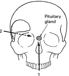
Many centres now use a transnasal endoscopic approach to remove the tumour. This avoids the sublabial incision and minimises septal retraction and post-operative discomfort. It greatly improves visualisation of the cavernous sinus and intrasellar structures.
From ABOVE
2. Transfrontal
Through a craniotomy flap the frontal lobe is retracted to provide direct access to the pituitary tumour. This approach is usually reserved for tumours with large frontal or lateral extensions.
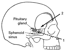
N.B. Patients may require steroid cover before any anaesthetic or operative procedure.
Radiotherapy
Pituitary adenomas are radiosensitive and external irradiation is commonly employed. Stereotactic radiosurgery is also used, but may not provide additional benefit. Occasionally, radioactive seeds of yttrium or gold are implanted into the pituitary fossa.
Several months elapse before hormone levels begin to fall. Pituitary function gradually declines over a 5–10 year period after treatment and most patients eventually require replacement hormone therapy to prevent symptoms of hypopituitarism developing.
Treatment selection
Treatment choice depends on:
– presenting problems and patient’s requirements,
e.g. incidental finding, restoring fertility, halting visual deterioration.
– patient’s age.
– preference and experience of the treatment centre.
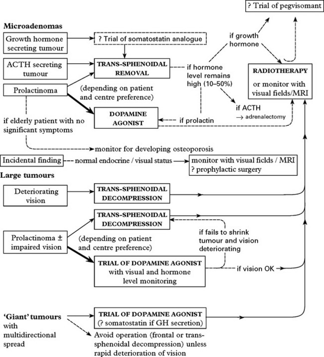
SELLAR/SUPRASELLAR TUMOURS
CRANIOPHARYNGIOMA
These tumours arise from remnants of Rathke’s cleft and constitute about 3% of all primary intracranial tumours. They may present at any age, but occur predominantly in children from 5–14 years (adamantinomatous type) and in adults from 50–60 years (papillary type). Although benign, proximity to crucial structures poses complex problems of management. About 40% of craniopharyngiomas have solid components of squamous epithelium with calcified debris and one or more cystic regions containing greenish cholesteatomatous fluid. In 20% the tumour is solid throughout. Although the tumour capsule appears well defined, histological examination reveals finger-like projections extending into adjacent tissue with marked surrounding gliosis.
Sites: growth usually begins near the pituitary stalk, but may extend in any direction.
Clinical features: depend on the exact site and size of the tumour. Growth is slow and most signs and symptoms develop insidiously.

Investigations
Skull X-ray: shows calcification above or within the pituitary fossa in most children and in 25% of adults.

Pituitary function studies (page 342): often demonstrate the need for hormone replacement.
Management
Several options exist; the more aggressive the treatment, the higher the risks, but the lower the recurrence rate.
All patients require pre-operative ophthalmological and endocrine assessment and steroid cover before any anaesthetic or operative procedure.
Operative removal usually involves a subfrontal or subtemporal craniotomy, perhaps combined with a transcallosal approach (i.e. splitting the anterior corpus callosum from above and approaching the tumour through the 3rd ventricle). The trans-sphenoidal route permits removal of purely intrasellar tumours.
Methods

Although total excision avoids the immediate need and associated risks of radiotherapy to a developing brain, it carries a risk of life-threatening hypothalamic damage. Accepting a subtotal resection is often the safest option. Operative mortality lies between 0–10% and depends on the tumour site and the extent of the attempted removal. Some report a recurrence rate of up to 30% within 10 years of an apparent ‘total’ removal. This presumably results from residual tumour extensions lying beyond the capsule.
Within subtotal removal the recurrence rate approaches 90%, but with radiotherapy this falls to 30–50% after 5 years. The decision to aim for total or subtotal removal requires careful judgement. Preoperative investigations help but the final decision often awaits direct exploration.
Coronal or sagittal MRI helps by demonstrating the exact relationships of
OPTIC NERVE (GLIOMA) ASTROCYTOMA
This rare tumour usually presents in children under 10 years. Up to one-third are associated with neurofibromatosis (NFI) where the tumour may be bilateral. Tumour growth expands the nerve in a fusiform manner. Some extend anteriorly into the orbit, others posteriorly to involve the optic chiasma. All are of the pilocytic type and growth is slow. Spontaneous regression may occur, particularly in NF1 patients.
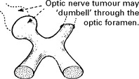
Clinical features
Visual field scotomas gradually progress to complete visual loss.
Orbital extension causes proptosis.
In some patients posterior expansion beyond the chiasma causes hypothalamic damage (precocious puberty and other endocrine disturbance) and/or hydrocephalus.
CT scans demonstrate an enhancing mixed attenuation mass within the orbit or lying in the suprasellar region. MRI is more sensitive for chiasmatic extensions.
|
Management |
Prognosis |
|
|
Unilateral within orbit |
– Conservative approach but if imaging shows progression towards chiasma, complete excision (with orbital enucleation if necessary) |
– Long-term survival expected. |
|
Lesion involving the optic chiasma |
– Conservative approach (the value of radiotherapy is not known and may risk vasculitis and intellectual deterioration). |
– Patients may retain vision for many years; survival is often long-term. Those with hypothalamic damage have a poor prognosis. |
SUPRASELLAR MENINGIOMA
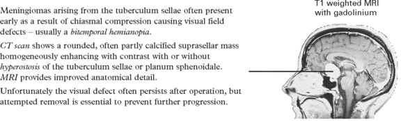
MENINGIOMA OF THE OPTIC NERVE SHEATH
Rarely, meningiomas arise from the optic nerve sheath, usually extending in dumbbell fashion through the optic foramen. Some penetrate the orbital dura and invade the orbital contents. Total excision is impossible without sacrificing the adjacent optic nerve.
SUPRASELLAR EPIDERMOID/DERMOID (see page 337).
Note: large aneurysms or granulomas (TB, sarcoid) may simulate a sellar/suprasellar tumour on CT scan or MRI. If in doubt, perform CTA or MRA prior to operative exploration.
PINEAL REGION TUMOURS

PATHOLOGICAL TYPES
Germ cell tumours: Germinoma is the commonest pineal region tumour of germ cell origin. It is malignant in nature and adheres firmly to surrounding tissues and cells may spread to the floor and anterior wall of the third ventricle. Teratomas are usually well differentiated, occurring predominantly in males, and formed from various cell types – muscle, bone, cartilage, dermis. Tumour consistency depends on the predominant cell type. In most the tumour margin is well defined, but malignant, poorly differentiated forms occasionally occur.
Other germ cell tumours include the highly malignant yolk sac tumour, choriocarcinoma and embryonal carcinoma.
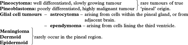
CLINICAL FEATURES Develop due to:
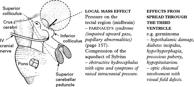
INVESTIGATIONS
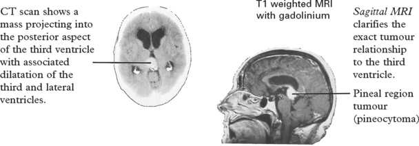
Pineocytomas – may appear calcified.
Teratomas – may contain mixed densities from fat to calcification.
Tumour markers – Serum/CSF human chorionic gonadotrophin – ↑ in choriocarcinoma (and slight ↑ in some germinomas)
– Serum/CSF alpha fetoprotein – ↑ in yolk sac tumours
CSF cytology: Malignant pineal region tumours can metastasise through CSF and cytology is important in planning treatment.
MANAGEMENT
Hydrocephalus often requires urgent treatment with a ventriculoperitoneal shunt or 3rd ventriculostomy. Large tumours may obstruct the foramen of Munro, making bilateral ventricular drainage necessary.
If biopsy, either via an endoscope or by stereotaxy, confirms a germinoma, or if serum/CSF markers are significantly raised suggesting choriocarcinoma or a yolk sac tumour, then radiotherapy ± chemotherapy is the treatment of choice.
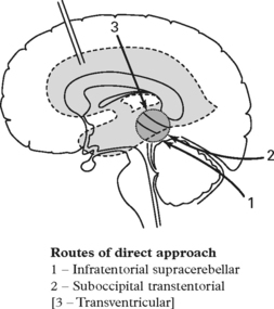
Teratomas, pineocytomas, dermoid or epidermoid cysts and meningiomas require direct operative exploration and excision, usually via either the supracerebellar or the transtentorial approach as shown. Pineoblastomas, certain pineocytomas and ependymomas require a combination of excision + radiotherapy. Chemotherapy may be added for the more malignant tumours.
When imaging shows disseminated tumour or when CSF cytology is positive, the entire craniospinal axis should be irradiated.
Outcome depends on tumour type. For germinomas and resectable tumours, the outlook is excellent and long-term survival is the rule.
TUMOURS OF THE VENTRICULAR SYSTEM
EPENDYMOMA
Intracranial ependymomas originate from cells lining the ventricular cavities. The majority arise in the 4th ventricle and in this site occur predominantly in children. Most are low grade (grade II), but an anaplastic form (grade III) exists and in about 10% tumour cells seed throughout CSF pathways.
In the 4th ventricle, ependymomas present with cerebellar signs or, more commonly, with signs and symptoms of raised intracranial pressure from CSF obstruction. Vomiting is often an early feature from direct brain stem involvement.
CT scanning shows an isodense mass, with or without calcification, lying within the 4th ventricle and usually enhancing with contrast. MRI more clearly delineates the anatomical relationships.
Management
The aim is complete operative removal, although infiltration of the floor of the 4th ventricle may prevent this. Most clinicians advise radiotherapy postoperatively, but its value is limited in the low grade tumours.
CSF metastases are treated by total neuraxis irradiation.
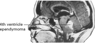
Prognosis
Despite relatively slow growth, results are often disappointing with 5-year survival ranging from 20–50%. Poor prognostic factors include incomplete resection and age < 2 years.
CHOROID PLEXUS PAPILLOMA
Rare, benign tumour with a granular surface and a gritty texture.
They develop from the choroid plexus – in the 4th ventricle – adults, – in the lateral ventricle – children.
Malignant forms occasionally occur in children. Most patients present with hydrocephalus, either due to obstruction or to excessive CSF secretion from the tumour. CT scanning shows a hyperdense mass within the ventricular system. Operative removal gives good results.
COLLOID CYST OF THE THIRD VENTRICLE
A benign cyst, containing a mucoid fluid may arise from embryological remnants in the roof of the third ventricle. When of sufficient size (about 2 cm) it occludes CSF drainage from both lateral ventricles through the foramen of Munro.
Clinical features: Many patients exhibit no symptoms. In others, symptoms occur intermittently, possibly due to a ball-valve effect – headaches, episodes of loss of consciousness or even sudden death.
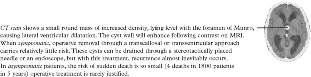
MENINGIOMA: rarely arises in the lateral ventricles. Often symptoms are mild and long standing. Operative removal only becomes necessary when symptoms and signs appear.

TUMOURS OF THE ORBIT
The orbital cavity is bounded –

PATHOLOGY
Tumours may arise from any of the structures lying within or around the orbit.
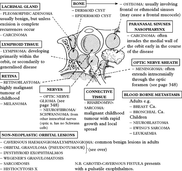
CLINICAL SYMPTOMS AND SIGNS
Orbital pain: prominent in rapidly growing malignant tumours, but also a characteristic feature of orbital granuloma and carotid-cavernous fistula.
Proptosis: forward displacement of the globe is a common feature, progressing gradually and painlessly over months or years (benign tumours) or rapidly (malignant lesions).
Lid swelling: may be pronounced in orbital granuloma, dysthyroid exophthalmos or carotid-cavernous fistula.
Palpation: may reveal a mass causing globe or lid distortion – especially with lacrimal gland tumours or with a mucocele. Pulsation indicates a vascular lesion – carotid-cavernous fistula or arteriovenous malformation – listen for a bruit.
Eye movements: often limited for mechanical reasons, but if marked, may result from a dysthyroid ophthalmoplegia or from III, IV or VI nerve lesions in the orbital fissure (e.g. Tolosa Hunt syndrome) or cavernous sinus.
Visual acuity: may diminish due to direct involvement of the optic nerve or retina, or indirectly from occlusion of vascular structures.
INVESTIGATIONS
CT scan with a fast helical scanner is the investigation of choice for bone lesions. It will demonstrate the exact relationship of the lesion to surrounding structures and will show the presence of any intracranial extension.
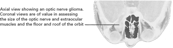
MRI shows the orbital anatomy in detail, but eye movements may cause artefacts.
MANAGEMENT
BENIGN tumours: require excision, but if visual loss would inevitably result, the clinician may adopt a conservative approach.
MALIGNANT tumours: require biopsy plus radiotherapy. Lymphomas may also benefit from chemotherapy. Occasionally localised lesions (e.g. carcinoma of the lacrimal gland) require radical resection.
Operative approach
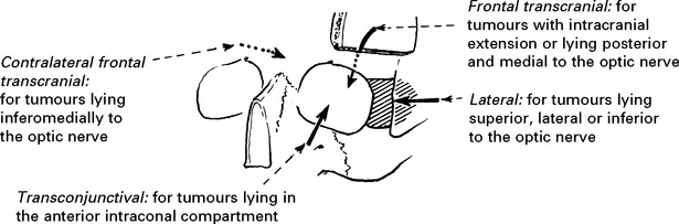
NON-NEOPLASTIC ORBITAL LESIONS
ORBITAL GRANULOMA (pseudotumour)
Sudden onset of orbital pain with lid oedema, proptosis and chemosis due to a diffuse granulomatous infiltrate of lymphocytes and plasma cells involving multiple structures within the orbit.
This condition usually occurs in middle age and seldom occurs bilaterally. CT scanning or MRI shows a diffuse orbital lesion, although one structure may be predominantly involved, e.g. optic nerve, extraocular muscles or the lacrimal gland. If diagnostic doubt remains, a biopsy is required. Most patients show a dramatic response to high dose steroid therapy. If symptoms persist, the lesion should respond well to radiotherapy.
DYSTHYROID EXOPHTHALMOS
The thyrotoxic patient with bilateral exophthalmos presents no diagnostic difficulty, but dysthyroid exophthalmos, with marked lid oedema, lid retraction and ophthalmoplegia may occur unilaterallywithout evidence of thyroid disease.
Coronal CT scanning establishes the diagnosis by demonstrating enlargement of the extraocular muscles – primarily the medial and inferior recti. MRI shows a similar appearance.
Circulating thyroid hormone levels are often normal. Thyroid releasing hormone stimulation or thyroid suppression tests may support the diagnosis.
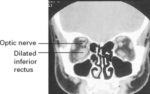
Management
Steroids should help. A few patients require orbital decompression in an attempt to prevent corneal ulceration, papilloedema and blindness.
TUMOURS OF THE SKULL BASE
MALIGNANT
CARCINOMA
Carcinoma of the nasopharynx, paranasal sinuses or ear may extend intracranially either by direct erosion or through the skull foramina. It frequently penetrates the dura (in contrast to metastatic carcinoma of the spine) and may involve almost any cranial nerve. Symptoms of nasopharyngeal or sinus disease are often associated with facial pain and numbness. Spread to the CSF pathways leads tocarcinomatous meningitis and may cause multiple cranial nerve palsies. Skull X-rays, CT scan and MRI scan will demonstrate a lesion involving the skull base. CT scanning most clearly shows the bone involvement. Treatment is usually restricted to retropharyngeal biopsy plus radiotherapy.
CHORDOMA
Rare tumours of notochordal cell rests arising predominantly in the sphenoido-occipital (clivus) and sacrococcygeal regions. Although growth begins in the midline, they often expand asymmetrically into the intracranial cavity. Chordomas may present at any age, but the incidence peaks in the 4th decade. They are locally invasive and rarely metastasise.
Clinical: most patients develop nasal obstruction. Cranial nerve palsies usually follow and depend on the exact tumour site.
Skull X-ray shows a soft tissue mass with an osteolytic lesion of the sphenoid, basi-occiput or petrous apex.
CT scan confirms the presence of a partly calcified mass causing marked bone destruction and extending into the nasopharyngeal space.
MRI scan more clearly demonstrates the structural relationships.
Management: the tumour site usually prevents complete removal. Extensive debulking (often through the transoral route) is combined with radiotherapy. Most patients die within 10 years of the initial presentation.
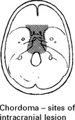
BENIGN
GLOMUS JUGULARE TUMOUR (syn: chemodectoma, paraganglioma)
Rare tumour arising from chemoreceptor cells in the jugular bulb or from similar cells in the middle ear mucosa. This tumour extensively erodes the jugular foramen and petrous bone; many patients present with cranial nerve palsies, especially IX–XII. Chemodectomas occasionally arise at other sites and metastasis may occur.
X-ray and CT scan demonstrate an osteolytic lesion expanding the jugular foramen.
MRI shows the anatomical relationships.
Angiography reveals a vascular tumour, usually only filling from the external carotid artery, but occasionally from vertebral branches.
Management: tumour vascularity makes excision difficult. Selective embolisation may considerably reduce the operative risks or provide an alternative treatment. The value of radiotherapy is uncertain, but radiosurgery could be considered for tumours < 3 cm in size.
OSTEOMA
Rare tumours, usually occurring in the frontal sinus and eroding into the orbit, nasal cavity or anterior fossa. If sinus drainage becomes obstructed, a mucocele develops, often with infected contents. These lesions require excision, either through an ethmoidal approach or through a frontal craniotomy.
INTRACRANIAL ABSCESS
The advent of antibiotics and improved treatment of ear and sinus infection has led to a reduction in intracranial abscess formation but the incidence still lies at 2–3 patients per million per year.
CEREBRAL ABSCESS
Source of infection
Haematogenous spread
– Subacute bacterial endocarditis
– Congenital heart disease (especially right to left shunt)
– Bronchiectasis or pulmonary abscess
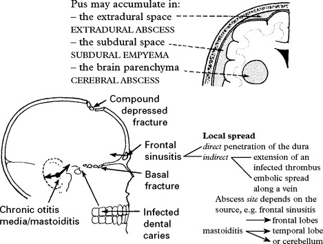
Organisms: Improved aerobic and anaerobic culture techniques now reveal the responsible organism in over 80% of patients. These depend on the source –
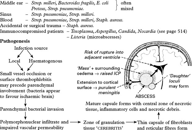
Clinical effects
Symptoms and signs usually develop over 2–3 weeks and progress. Occasionally the onset is more gradual, but features may develop acutely in the immunocompromised patient. Clinical features arise from:
– Toxicity – pyrexia, malaise (although systemic signs often absent).
– Raised intracranial pressure – headache, vomiting → deterioration of conscious level.
– Focal damage – hemiparesis, dysphasia, ataxia, nystagmus – epilepsy – partial or generalised, occurring in over 30%
– Infection source – tenderness over mastoid or sinuses, discharging ear. bacterial endocarditis – cardiac murmurs, petechiae, splenomegaly.
– Neck stiffness due to coexistent meningitis or tonsillar herniation occurs in 25%.
N.B. Beware attributing patient’s deteriorating clinical state to the primary condition, e.g. otitis media, thus delaying essential investigations.
Investigations
X-rays of the sinuses and mastoids: opacities indicate infection.
CT scan: in the stage of ‘cerebritis’ the CT scan may appear normal or only show an area of low density. As the abscess progresses, a characteristic appearance emerges:
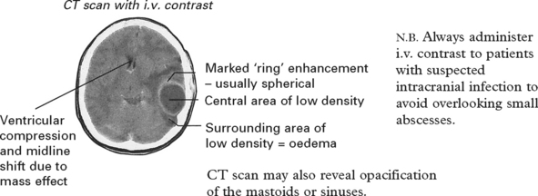
If abscesses occur at multiple sites, suspect a haematogenous source.
MRI: will more readily detect the ‘cerebritic’ stage, but does not distinguish infection from other pathologies.
Lumbar puncture is contraindicated in the presence of a suspected mass lesion, but if CSF is obtained inadvertently, this will show ↑ protein e.g. 1 g/l, ↑ white cell count (several hundred/ml) – polymorphs or lymphocytes. The Gram stain is occasionally positive.
Peripheral blood – may show ↑ ESR, leucocytosis. Blood culture is positive in 10%.
Management:
1. Antibiotics
Commence i.v. antibiotics on establishing the diagnosis (prior to determining the responsible organism and its sensitivities). Antibiotics are selected on an empirical basis depending on the likely source of the infection and their ability to cross the blood–brain barrier and to achieve therapeutic concentrations in intracranial pus.
Use combined therapy: (note adult doses indicated)
|
– CEFTRIAXONE |
i.v. 3–4 g/day |
|
– METRONIDAZOLE |
i.v. 500 mg tds |
|
for a middle ear source |
|
|
+ AMOXICILLIN |
i.v. 2 g 4 hourly |
|
if endocarditis or congenital heart disease |
|
|
+ BENZYLPENICILLIN |
i.v. 1.8–2.4 g 6 hourly |
If a penetrating trauma source
|
– FLUCLOXACILLIN |
i.v. 2 g 4 hourly |
|
± GENTAMICIN |
i.v. 5 mg/kg/day (+ monitor levels) |
In immunocompromised patients – see page 514.
Later determination of the organism and its sensitivities permits alteration to more specific drugs. Intravenous antibiotics should continue for 2–3 weeks followed by oral medication for a further 3–4 weeks.
2. Abscess drainage
Various methods exist:
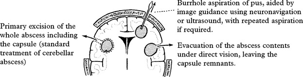
Burr hole aspiration is simple and relatively safe. Persistent reaccumulation of pus despite repeated aspiration requires secondary excision. Primary excision removes the abscess in a single procedure, but carries the risk of damage to surrounding brain tissue. Open evacuation of the abscess contents requires a craniotomy, but minimises damage to surrounding brain.
3. Treatment of the infection site
Mastoiditis or sinusitis requires prompt operative treatment, otherwise this acts as a persistent source of infection.
Steroids help reduce associated oedema but they may also reduce antibiotic penetration and impede formation of the abscess capsule. Their value in management remains controversial.
Conservative management: In some situations the risks of operative intervention outweigh its benefits. In those patients, treatment depends on i.v. antibiotics.
Indications:
– small deep abscesses, e.g. thalamic (although stereotactic aspiration may help).
– multiple abscesses.
– early ‘cerebritic’ stage.
Prognosis
The use of CT scanning in the diagnosis and management of intracranial abscesses and the recognition and treatment of pathogenic anaerobic organisms have led to a reduction in the mortality rate from 40% to 10%. In survivors, focal deficits usually improve dramatically with time. Persistent seizures occur in 50%.
SUBDURAL EMPYEMA
Subdural empyema occurs far less frequently than intracerebral abscess formation. Infection usually spreads from infected sinuses or mastoids, but may arise from any of the aforementioned sources. The responsible organism is usually Strep. pneumoniae, Strep. milleri or Staph. aureus. Clinical features match those of intracerebral abscess but since rapid extension occurs across the subdural space, overwhelming symptoms often develop suddenly. Seizures occur in 70% at onset.
CT scan shows a low density extracerebral collection with mass effect, often with enhancement on the cortical surface; occasionally isodense lesions make identification difficult.
Management: Intravenous antibiotic treatment is combined with evacuation of pus either through multiple burr holes or a craniotomy flap. Despite active treatment, the mortality rate still runs at approximately 20%.
GRANULOMA
TUBERCULOMA
Although tuberculomas still constitute an important cause of mass lesions in underdeveloped countries (20% in India), they are now rare in Britain. The lesions may be single or multiple. They often lie in the cerebellum, especially in children.
Clinical features are those of any intracranial mass; alternatively tuberculoma may present in conjunction with tuberculous meningitis.
CT scan clearly demonstrates an enhancing lesion – but this often resembles astrocytoma or metastasis; tuberculomas have no distinguishing features. MRI is even more sensitive and may show additional lesions.
Other investigations: ESR, chest X-ray often fail to confirm the diagnosis. A Mantoux (PPD) test is usually positive but a negative test does not eliminate the diagnosis.
Management: When tuberculoma is suspected, a trial of antituberculous therapy is worthwhile. Follow up CT scans should show a reduction in the lesion size. Other patients require an exploratory operation and biopsy followed by long-term drug treatment.
SARCOIDOSIS
Sarcoidosis is a multisystem disease process of unknown cause whose pathogenesis involves formation of an inflammatory lesion known as a granuloma. Nervous system involvement occurs in 8% and may dominate the presentation.
When sarcoid infiltrates the central nervous system it usually involves the meninges. In some patients mass lesions may arise from the dura, but more commonly signs and symptoms relate to an adhesive arachnoiditis involving the skull base, cranial nerves and pituitary stalk. Mass lesions may occasionally arise within the brain and spinal cord without obvious meningeal involvement.
Investigation: MRI (T1 weighted) shows either a hyperintense mass or multiple periventricular foci. The use of gadolinium and FLAIR (fluid-attenuated inversion recovery) increases the sensitivity of MRI. A definitive diagnosis is based on clinical and radiological evidence of multisystem disease confirmed by characteristic histology.
The diagnosis is often elusive and suggested by clinical presentation supported by some of the following.
– elevated serum and CSF angiotensin converting enzyme (ACE),
– elevated serum immunoglobulins,
– elevated serum calcium,
– elevated CSF cell count (monocytes), IgG, Ig index, and presence of oligoclonal bands.
Management: Immunosuppression with corticosteroids is usually indicated and long-term therapy required. In exacerbation, intravenous pulsed methylprednisolone is used. Success in resistant cases is reported with each of the following – azathioprine, cyclophosphamide, methotrexate, cyclosporin or irradiation.
MOVEMENT DISORDERS – EXTRAPYRAMIDAL SYSTEM
The control of voluntary movement is effected by the interaction of the pyramidal, cerebellar and extrapyramidal systems interconnecting with each other as well as projecting to the anterior horn region or cranial nerve motor nuclei.
The extrapyramidal system consists of paired subcortical masses or nuclei of grey matter basal ganglia.
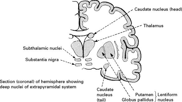
The caudate nucleus and putamen are collectively referred to as the STRIATUM.
Interconnections of the deep nuclei
The connections between components of the extrapyramidal system and other parts of the brain are complex. However, certain simple observations can be made:
 The thalamus plays a vital role in projecting information from the basal ganglia to the motor cortex and back
 The cortex projects through the striatum to other basal ganglia
 The final common pathway for basal ganglia motor function is the corticospinal or pyramidal tract
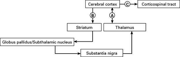
NEUROPHARMACOLOGY
The observation that drugs such as reserpine and phenothiazines regularly produce extrapyramidal syndromes has clarified the neurochemical basis of movement disorders and delineated the role of neurotransmitters.
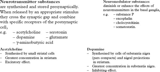
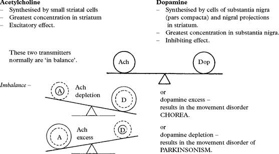
γ-Aminobutyric acid (GABA) is synthesised from glutamate in the striatum and globus pallidus. It has inhibitory actions and deficiency is associated with Huntington’s disease.
Drugs may produce movement disorders by interfering with neurotransmission in the following ways:
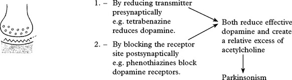
CLINICAL FEATURES
The effects of disease of the extrapyramidal system on movement can be regarded as negative (hypokinetic) and positive (hyperkinetic).
Negative features
Bradykinesia: - a loss or slowness of voluntary movement.
A major feature of Parkinson’s disease and produces:
– reduced facial expression (mask-like)
– reduced blinking
– reduced adjustments of posture when seated.
When agitated the patient will move swiftly – ‘kinesia paradoxica’.
Postural disturbance: most commonly seen in Parkinson’s disease.
Flexion of limbs and trunk is associated with a failure to make quick postural or ‘righting’ adjustments to correct imbalance. The patient falls whilst turning or if pushed.
Positive features
Involuntary movements:
– tremor
– chorea (irregular, repetitive, jerking movements).
– athetosis (irregular, repetitive, writhing movements).
– dystonia (slow, sustained, abnormal movement).
– ballismus (explosive, violent movement).
– myoclonus (shock-like jerks).
Chorea and athetosis may merge into one another – choreoathetosis.
Rigidity
Stiffness felt by the examiner when passively moving a limb. This ‘resistance’ is present to the same degree throughout the full range of movement, affecting flexor and extensor muscle groups equally and is described as PLASTIC or LEAD PIPE rigidity. When tremor is superimposed upon rigidity it produces a COGWHEELING quality.
In Parkinson’s disease both positive features, e.g. tremor, and negative features, e.g. bradykinesia, occur.
In Huntington’s disease positive features, e.g. chorea, predominate.
PARKINSON’S DISEASE
Described by James Parkinson (1817) in ‘An essay on the shaking palsy’.
Recognised as an extrapyramidal disorder by Kinnier Wilson (1912).
Annual incidence: 20 per 100 000. Prevalence: 190 per 100 000.
Sex incidence: male:female – 3:2
Age of onset: 50 years upwards. Incidence peaks in mid-70s then declines.
Familial incidence occurs in 5%.
AETIOLOGY
The cause(s) of Parkinson’s disease is unknown. Gene mutations have been identified in young onset and familial cases (synuclein, parkin and LRRK2).
The observation that 1-methyl-4-phenyl-1,2,3,6-tetrahydropyridine (MPTP), a meperidine analogue derived during illicit drug production, produces Parkinson’s disease in humans and animals has resulted in increased interest in the role of toxins and an animal model for developing new treatments.
Parkinsonian features may be present in many disorders and are not always treatment (L Dopa) responsive. These disorders usually share features of slowness and rigidity (akinetic rigid syndromes).
|
Parkinson’s disease |
Mimics |
– Multiple system atrophy (MSA) – Progressive supranuclear palsy (PSP) – Corticobasal ganglionic degeneration (CBD) – Diffuse Lewy body disease (DLBD) |
Secondary Parkinsonism
– Drug induced (dopamine receptor blockers-antipsychotics/antiemetics; sodium valproate)
– Post traumatic (pugilist’s encephalopathy)
– Vascular disease (small vessel multi-infarct state)
– Infectious (post encephalitic/prion disease/HIV)
– Miscellaneous: hydrocephalus/parathyroid/paraneoplastic
PATHOLOGY of idiopathic Parkinson’s disease
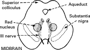
The substantia nigra contains pigmented cells (neuromelanin) which give it a characteristic ‘black’ appearance (macroscopic). These cells are lost in Parkinson’s disease and the substantia nigra becomes pale.
Remaining cells contain atypical eosinophilic inclusions in the cytoplasm – Lewy bodies – although these are not specific to Parkinson’s disease. Lewy bodies may be found in the cerebral cortex especially when dementia is present (diffuse Lewy body disease).
Changes are seen in other basal nuclei – striatum and globus pallidus.
Radiolabelled ligand studies have identified two dopamine receptors on striatal cell membranes – D1 – D2 receptors.
CLINICAL FEATURES
Initial symptoms are vague, the patient often complains of aches and pains.
A coarse TREMOR at a rate of 4–7 Hz usually develops early in the disease. It begins unilaterally in the upper limbs and eventually spreads to all four limbs. The tremor is often ‘pill rolling’, the thumb moving rhythmically backwards and forwards on the palm of the hand. It occurs at rest, improves with movement and disappears during sleep.
RIGIDITY is detected by examination. It predominates in the flexor muscles of the neck, trunk and limbs and results in the typical ‘flexed posture’.
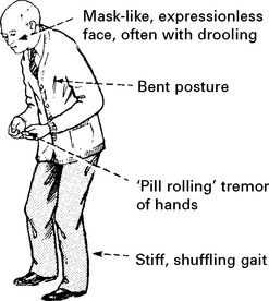
BRADYKINESIA: This slowness or paucity of movement affects facial muscles of expression (mask-like appearance) as well as muscles of mastication, speech, voluntary swallowing and muscles of the trunk and limbs. Dysarthria, dysphagia and a slow deliberate gait with little associated movement (e.g. arm swinging) result.
Tremor, rigidity and bradykinesia deteriorate simultaneously, affecting every aspect of the patient’s life:
Handwriting reduces in size.
The gait becomes shuffling and festinant (small rapid steps to ‘keep up with’ the centre of gravity) and the posture more flexed.
Rising from a chair becomes laborious with progressive difficulty in initiating lower limb movement from a stationary position.
Eye movements may be affected with loss of ocular convergence and upward gaze.
Excessive sweating and greasy skin (seborrhoea) can be troublesome.
Depression occurs in about 50%.
As the disease progresses the frequency of drug-induced confusional states and dementia increases, with 80% developing dementia after 20 years of disease (if they survive).
Autonomic features occur – postural hypotension, constipation.
REM sleep behaviour disorder – where patient acts out dreams and may hurt themselves or their sleep partner. May precede onset of motor symptoms.
Time of onset is mid–late fifties with increasing incidence with increasing age. Juvenile presentation can occur, when presentation and disease progression is often atypical; a genetic basis is more often found.
DIAGNOSIS
The diagnosis of PD in the early stages is difficult. Post-mortem data from the London Brain Bank shows this to be incorrect in 25% of those diagnosed in life.
New tremor in middle age causes particular difficulty – senile/essential & metabolic tremor is generally absent at rest and worsened by voluntary movement.
The diagnostic use of a L-dopa or dopamine agonist (apomorphine) challenge has declined due to concerns that it may increase the risk of subsequent drug induced dyskinesia.
Functional imaging (SPECT & PET) should improve diagnostic accuracy and ensure that persons with conditions unresponsive to treatments (PD mimics) are not unnecessarily exposed to them.

The ligand I-IBZM demonstrates the degree of D2 receptor binding. This is normal in PD but reduced in its mimics (MSA/PSP)
The ligand FP-CIT demonstrates the integrity of the presynaptic dopamine terminals. These are normal in essential tremor and reduced in PD and its mimics (MSA/PSP).
PD MIMICS
Multiple system atrophy occurs in two forms, a relatively symmetrical extrapyramidal syndrome associated with autonomic failure, usually postural hypotension and bladder symptoms, and a cerebellar syndrome with bilateral upper motor neuron signs. Both progress over 5–10 years.
Progressive supranuclear palsy (PSP) is characterised by gaze palsies, extrapyramidal features, axial dystonia (truncal dystonia), progressive upper motor neuron syndrome and dementia. Onset in the 5th to 6th decade. The key feature is the supranucelar gaze palsy: downward eye movement is impaired followed by all other voluntary eye movement which can be overcome by doll’s head manoeuvre (a supranuclear palsy). Lid retraction is common. Levodopa gives disappointing results. Progression is relentless with death in 3–7 years.
Vascular Parkinsonism usually presents with gait disturbance with step wise deterioration. It tends to predominantly involve the lower limbs and have a partial response to L-dopa. Brain imaging is helpful in diagnosis.
Wilson’s disease (see page 373)
Corticobasal degeneration (CBD) is rare and presents with an asymetric akinetic-rigid syndrome associated with marked dyspraxia, myoclous and dementia. Patients may have an ‘alien hand’, where the hand moves purposely without conscious control. There are no specific treatments.
TREATMENT is symptomatic and does not halt the pathological process. No agents have yet demonstrated convincing neuroprotective effect.
Levodopa/Dopamine agonists

Exogenous dopa
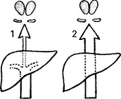
Levodopa is given with a decarboxylase inhibitor, which prevents peripheral breakdown in the liver (as in 1) allowing a higher concentration of dopa to reach the blood–brain barrier (as in 2) and reduces the peripheral side effects (nausea, vomiting, hypotension).
Central side effects: confusion, depression, dyskinetic movements and following long-term treatment – ‘On/Off’ phenomenon (see later).
Rapid onset or longer action can be achieved using dispersible or controlled-release preparations.
Exogenous dopa improves bradykinesia, rigidity and, to a lesser extent, tremor, but in 20% the response is poor. Dopa has relatively less effect on non-motor symptoms.
A new preparation of dopa is available for continuous infusion via jejunostomy in severe disease.
Dopamine agonists: Now used earlier in disease management, they act directly on the dopamine receptor independent of degenerating dopaminergic neurons. It is not clear if patients do better in the long term if dopamine agonists or dopa are used first. There are two types of dopamine agonists, ergot derived, including pergolide, cabergoline, apomorphine, and non-ergot derived, such as ropinerole, pramipexole, rotigotine (available as a transdermal patch). Ergot agonists are now avoided because of the high rate of fibrotic reactions, with up to 25% of patients developing cardiac valve fibrosis. Apomorphine is given by continuous infusion or intermittent injection and is useful late in the disease.
Side effects: postural hypotension, hallucinations & psychosis, sedation and agonist specific complications (erythromelalgia/pulmonary fibrosis).
COMT inhibitors: Entacapone reduces the metabolism of levodopa and is used as adjunctive treatment. Tolcapone is an alternative that can cause hepatic toxicity; it requires close monitoring.
Selegiline and Resagiline are monoamine oxidase (MAO) type B inhibitors which slow breakdown of dopa. Its usage results in increased dopamine levels.
Amantidine, is useful in reducing dyskinesias late in the disease.
Deep brain stimulation: for patients with normal cognitive function who remain responsive to medication but have significant on/off phenomenon despite optimum medical therapy, the insertion of deep brain electrodes into the subthalamic nucleus can provide useful clinical benefits. Long-term studies are ongoing to determine how best to use surgery. Complications include dysarthria and visual field defects.
Human fetal and medullary transplantation: experimental evidence shows that transplantation to the striatum of tissue capable of synthesising and releasing dopamine reverses the motor symptoms of Parkinson’s disease. This treatment remains experimental.
Regime of treatment (Drug therapy becomes more complex as disease progresses)

Additional measures
|
Nausea: _______________ |
domperidone (peripheral dopamine antagonist) |
|
Hypotension: _____________ |
tilt bed head, elastic stockings + mineralocorticoid |
|
Peak dose dyskinesia: ___________ |
lower levodopa dose |
|
End dose dyskinesia: _________ |
add dopamine agonist |
|
Nocturnal pain/immobility: _______ |
add controlled-release levodopa at night |
|
Confusion/aggravated dementia: _____ |
reduce dopamine agonist first, then levodopa consider anticholinesterase (rivastigmine) or quetiapine or clozapine (dopa antagonists) |
CHOREA
An involuntary, irregular, jerking movement affecting limb and axial muscle groups. These movements are suppressed with difficulty and are incorporated into voluntary gestures resulting in a ‘semipurposeful’ appearance, e.g. crossing and uncrossing of legs.
Causes of chorea
|
Hereditary: – Huntington’s disease – Benign chorea |
Metabolic: – Hyperthyroidism – Hypocalcaemia |
|
Drugs: – Antiparkinsonian drugs – oral contraceptives |
Immunological: – Systemic lupus erythematosus – Polyarteritis nodosa |
|
Toxins: – alcohol – carbon monoxide poisoning |
Miscellaneous: – Chorea gravidarum – Polycythaemia rubra vera |
|
Infections: – Sydenham’s chorea – encephalitis |
HUNTINGTON’S DISEASE
Huntington disease (HD) is inherited as an autosomal dominant disease that gives rise to progressive, selective (localized) neural cell death associated with choreic movements and dementia. It is associated with increases in the length of a CAG triplet repeat present in a gene called ‘huntingtin’ located on chromosome 4p16.3. Huntington disease has a frequency of 4 to 7 per 100000 persons. The condition shows ‘anticipation’, becoming more severe in each succeeding generation.
Pathology: Neuronal loss in the striatum is associated with a reduction in projections to other basal ganglia structures. In addition, cells of the deep layers of the frontal and parietal cortex are lost (corticostriatal projections). The neurochemical basis of this disorder involves deficiency of gamma aminobutyric acid (GABA) and acetylcholine with reduced activity of enzymes glutamic acid decarboxylase (GAD) and choline acetyltransferase (CAT).
Symptoms and signs: The classic signs of Huntington disease are progressive chorea, rigidity, and dementia. Typically, there is a prodromal phase of mild psychotic and behavioural symptoms, which precedes frank chorea by up to 10 years.
Chorea – may be the initial symptom. This progressess from mere fidgetiness to gross involuntary movements which interrupt voluntary movement and make feeding and walking impossible.
Dementia – this is of a subcortical type (see page 126).
Behavioural disturbance – personality change, affective disorders and psychosis occur.
Hypotonicity often accompanies fidgety, choreiform movements.
Primitive reflexes – grasp, pout and palmomental – are usually elicited. Eye movements are disturbed with impersistence of gaze.
Diagnosis: MRI shows an increase in the T2 signal in the caudate nucleus. Positron-emission tomography (PET scanning) demonstrates loss of uptake of glucose in the caudate nuclei. Genetic testing is diagnostic, but given the significance of the diagnosis to both patients and their family, usually requires informed consent. If the patient is too demented to consent discussion with family members is advised.
Prediction of disease: Identifying the CAG repeat provides a reliable method of detecting the disease. Presymptomatic testing is now available in many centres. These tests raise ethical issues but also the possibility of neuroprotective therapy.
Treatment: Mainly supportive. Phenothiazines (risperidone), haloperidol or tetrabenazine, may reduce abnormal movements in early disease. SSRIs help affective disturbance.
SYDENHAM’S CHOREA
Rare in an age of antibiotic therapy, this condition (also known as St Vitus’ dance) followed streptococcus pneumoniae infection. Unlike arthritis and carditis, symptoms developed weeks or months after primary infection. Movements are diffuse and often associated with florid behavioural changes.
Pathology: Necrotising arteritis in thalamus, caudate nucleus and putamen.
Diagnosis is confirmed by elevated ESR and ASO (antistreptolysin) titre.
Treatment: symptomatic with phenothiazines. The condition may recur during pregnancy, or with intercurrent infection.
CHOREA GRAVIDARUM
Acute onset in pregnancy, usually the first trimester or whilst on oral contraceptive. It can be restricted to face or generalised and may represent a reactivation of Sydenham’s chorea. If occurring whilst on the oral contraceptive this should be stopped; risperidone can be used to control symptoms.
SENILE CHOREA
Begins in late middle age unaccompanied by family history or behavioural change. Some patients do have caudate or putaminal atrophy and occasionally test positive for Huntington’s disease.
DYSTONIA
Dystonia manifests as a sustained abnormal posture produced by contraction of large trunk and limb muscles, e.g. sustained head retraction …
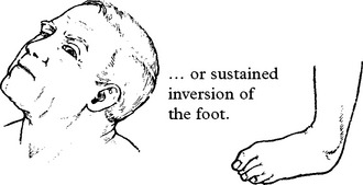
Dystonias may be classified by distribution:
generalised, focal, when limited to one area of the body or task-specific such as writer’s cramp.
And by aetiology:
Primary, often genetically proven, or secondary, to drugs, metabolic disorders and other neurodegenerative disorders.
PRIMARY DYSTONIA
Primary Generalised: Idiopathic Torsion Dystonia
The first gene identified for idiopathic torsion dystonia, DYT 1, is located on 9q34. The disorder is inherited as an autosomal dominant with reduced penetrance. It is responsible for early-onset generalized dystonia in Ashkenazi Jews. Initially, a flexion deformity of leg develops when walking.
Movements then become generalised but ultimately constant. Despite eventual gross contortion the postures disappear during sleep.
Diagnosis is made on clinical grounds and by exclusion of other disorders. – EMG studies show inappropriate co-contraction of antagonistic muscle groups.
Pathology: No known pathological substrate.
Treatment: levodopa or carbamezapine are of benefit in some patients; anticholinergics help in others. A small proportion are dramatically dopa-responsive.
Pallidal stimulation may benefit (see page 387).

DYSTONIAS – FOCAL AND SEGMENTAL IDIOPATHIC
PRIMARY FOCAL DYSTONIAS
CERVICAL DYSTONIA OR SPASMODIC TORTICOLLIS
Unilateral deviation of the head.
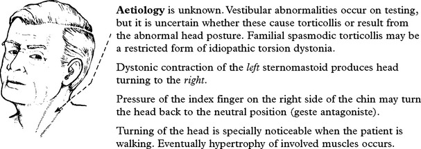
Pathology: unknown. Diagnosis is based on clinical findings.
Treatment: anticholinergics produce limited benefit in a few patients. Regular injection of Botulinum toxin into the overactive muscles gives good symptomatic control.
OROMANDIBULAR DYSTONIA
Constant involuntary prolonged tight eye closure (blepharospasm) is associated with dystonia of mouth, tongue or jaw muscles (jaw clenching and tongue protrusion). Response to treatment is poor though phenothiazines should be tried. Section of the nerves to orbicularis oculi muscles will relieve blepharospasm. Botulinum toxin injection is also effective.
When oromandibular dystonia occurs with cervical dystonia this segmental dystonia is called Meige’s syndrome.
PRIMARY TASK SPECIFIC DYSTONIA: WRITER’S CRAMP
Muscles of the hand and forearm tighten on attempting to write and pain may occur in the forearm muscles. Previously regarded as an ‘occupational neurosis’ but now classified as a partial dystonia.
Treatment: may respond to botulinum toxin injection. Avoiding the activity is most successful.
Other task specific dystonias relate to other repeated movements and include golfer’s yips.
SECONDARY DYSTONIAS
DOPAMINE RESPONSIVE DYSTONIA (DRD)
This disorder presents in childhood and generally involves the legs only. Falls are frequent and the response to levodopa is maintained over many years. DRD may be the result of a developmental reduction in the number of dopaminergic nerve endings in the striatum and maps to the same region of 14q as does the gene for the enzyme GTP cyclohydrolase 1 (GCH1) implicated in a hyperphenylalaninaemia.
DRUG INDUCED DYSTONIA
Acute adoption of abnormal dystonic posture – usually head and neck or oculogyric crisis (upward deviation of eyes) – caused by phenothiazines, e.g. haloperidol, metoclopramide.
Anticholinergics, e.g. benzotropine for 24–48 hours helps symptoms settle.
OTHERS
Dystonia is a feature of other conditions, most commonly Parkinson’s disease on treatment, but also PSP, Wilson’s disease (see page 366).
OTHER MOVEMENT DISORDERS
TICS
Abrupt jerky movements affecting head, neck and trunk. Tics can be voluntarily suppressed and often take the form of winking, grimacing, shoulder shrugging, sniffing and throat clearing.
Gilles de la Tourette syndrome is characterised by motor and vocal tics, copropraxia (making obscene gestures), coprolalia (obscene utterances) and obsessive behaviour. Onset is in childhood, males are more often affected and the condition may be inherited. However results of a systematic genome screen were negative. A population study showed that 3% of all children and that up to 25% of children requiring special education may have mild to moderate Tourette’s syndrome.
The dopaminergic systems in the basal ganglia appear involved, dopamine D2 receptor antagonists improving and dopamimetic agents worsening symptoms. Clonidine helps control tics with few adverse effects.
TARDIVE DYSKINESIA
This is a consequence of long-term treatment with neuroleptic drugs – phenothiazines, butyrophenones – and results from the development of drug-induced supersensitive dopamine receptors.
Involuntary movements in the face, mouth and tongue (orofacial dyskinesia) as well as limb movements of a choreoathetoid nature occur.
This movement disorder may commence even after stopping the responsible drug and can persist indefinitely.
Prevention
Incidence may be reduced by:
1. Using newer atypical antipsychotic agents.
2. Early recognition and drug withdrawal.
The practice of increasing the dose of the offending drug when movements occur should be avoided. This will improve movements initially, but they will ‘break through’ later.
Treatment
Discontinue neuroleptic. If not possible, continue on lowest possible dose. Drugs which increase acetylcholine (anti-cholinesterases), reduce catecholamine release (lithium), or deplete dopamine (reserpine) are variably effective.
ATHETOSIS
Athetosis presents in childhood and appears as a slow writhing movement disorder with a rate of movement between that of chorea and dystonia. It usually involves the digits, hands and face on each side.
These abnormal movements may result from:
– Hypoxic neonatal brain damage,
– Kernicterus,
– Lipid storage diseases.
Response to anticholinergics is variable and occasionally dramatic.
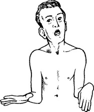
HEMIBALLISMUS
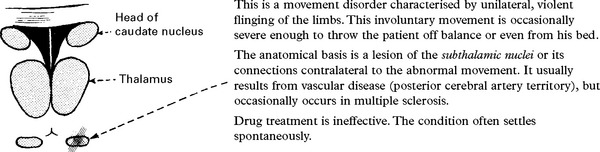
MALIGNANT NEUROLEPTIC SYNDROME AND SEROTONIN SYNDROME – see page 534.
WILSON’S DISEASE (hepatolenticular degeneration)
An autosomal recessive disorder characterised by the build-up of intracellular copper with hepatic and neurological consequences.
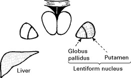
Pathology
Cavitation and neuronal loss occurs within the putamen and the globus pallidus.
The liver shows the appearances of coarse cirrhosis. Copper accumulates in all organs, especially in Descemet’s membrane in the eye, nail beds and kidney.
Biochemistry
There is deficiency of α2 globulin – Ceruloplasmin – which normally binds 98% of copper in the plasma and transfers copper to enzyme (cytochrome oxidase). This results in an increase in loosely bound copper/albumin, and deposition occurs in all organs. Urinary copper is increased.
Clinical features
There are two clinical forms:
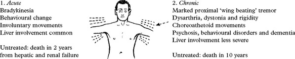

Diagnosis
Should be considered in any patient with unusual hepatic and/or neurological features.
Supported by biochemical evidence of abnormal copper metabolism:
– Low ceruloplasmin (less than 20 mg/dl)
– Elevated unbound serum copper
– High urinary copper excretion
– Liver biopsy and copper metabolism tests with radioactive 64Cu.
– MRI (T2) shows thalamic and putaminal hyperintensity.
In families, biochemical tests will identify low ceruloplasmin in carries and in presymptomatic patients. Over 20 mutations in copper transporting ATPase have been identified. Diagnostic genetic testing is not available.
Treatment
Low copper diet and a chelating agent, e.g. penicillamine 1–1.5g daily. Side effects such as anaphylaxis, skin rash, bone marrow suppression and glomerulonephritis are common in which case trientine is an effective alternative.
Therapy is necessary for the rest of the patient’s life. Adequate treatment is compatible with normal life expectancy. Kayser-Fleischer rings will disappear with time.
HYDROCEPHALUS
DEFINITION
Hydrocephalus is an active distension of the ventricular system of the brain arising when an imbalance exists between cerebrospinal fluid (CSF) production and absorption. This definition excludes ventricular expansion secondary to brain shrinkage from a diffuse atrophic process (hydrocephalus ex vacuo).
CSF FORMATION AND ABSORPTION
CSF forms at a rate of 500 ml/day (0.35 ml/min), secreted predominantly by the choroid plexus of the lateral, third and fourth ventricles. CSF flows in a caudal direction through the ventricular system and exits through the foramina of Luschka and Magendie into the subarachnoid space. After passing through the tentorial hiatus and over the hemispheric convexity, absorption occurs through the arachnoid granulations into the venous system.
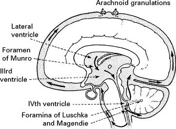
CLASSIFICATION
‘Obstructive’ hydrocephalus – obstruction of CSF flow within the ventricular system.
‘Communicating’ hydrocephalus – obstruction to CSF flow outwith the ventricular system i.e. ventricular CSF ‘communicates’ with the subarachnoid space.
CAUSES OF HYDROCEPHALUS
|
Obstructive |
Communicating |
|
|
Acquired |
– Acquired aqueduct stenosis (adhesions following infection or haemorrhage) – Supratentorial masses causing tentorial herniation – Intraventricular haematoma – Tumours – ventricular, e.g. colloid cyst – pineal region – posterior fossa – Abscesses/granuloma – Arachnoid cysts |
Thickening of the leptomeninges and/or involvement of the arachnoid granulations – infection (pyogenic, TB, fungal) – subarachnoid haemorrhage – spontaneous – trauma – postoperative – carcinomatous meningitis Increased CSF viscosity, e.g. high protein content Excessive CSF production – choroid plexus papilloma (rare) |
|
Congenital |
– Aqueduct stenosis or forking – Dandy-Walker syndrome (atresia of foramina of Magendie and Luschka) – Chiari malformation |
|
PATHOLOGICAL EFFECTS
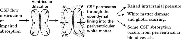
In the infant, prior to suture fusion, head expansion and massive ventricular dilatation may occur, often leaving only a thin rim of cerebral ‘mantle’. Untreated, death may result, but in many cases the hydrocephalus ‘arrests’; although the ventricles remain dilated, intracranial pressure (ICP) returns to normal and CSF absorption appears to balance production. When hydrocephalus arrests, normal developmental patterns resume, although pre-existing mental or physical damage may leave a permanent handicap. In these patients, the rapid return of further pressure symptoms following a minor injury or infection suggests that the CSF dynamics remain in an unstable state.
CLINICAL FEATURES
Infants and young children
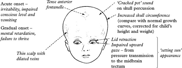
Juvenile/adult type hydrocephalus
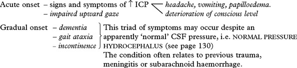
INVESTIGATIONS
Skull X-ray
Note:
– skull size and suture width.
– evidence of chronic raised pressure – posterior clinoid erosion, ‘copper beating’.
– associated defects – platybasia, basilar invagination.
CT scan
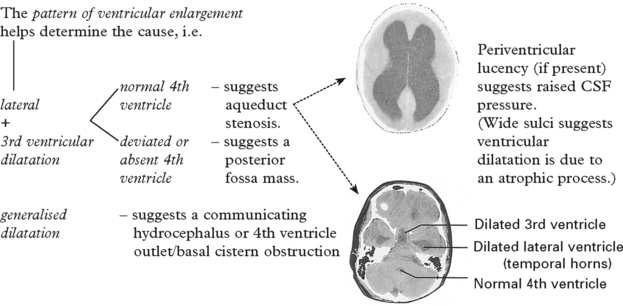
Ultrasonography through the anterior fontanelle, usefully demonstrates ventricular enlargement in infants and allows safe serial measurements.
MRI shows similar ventricular expansion, but may more clearly demonstrate periventricular lucency or a neoplastic cause of the obstruction.
ICP monitoring: used in some patients to determine whether symptoms relate to the enlarged ventricular size and to investigate patients with suspected normal pressure hydrocephalus (see page 131).
Developmental assessment and psychometric analysis detect impaired cerebral function and provide a baseline for future comparison.
MANAGEMENT
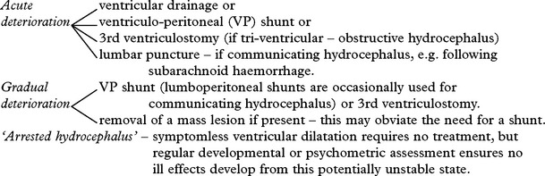
Shunt techniques
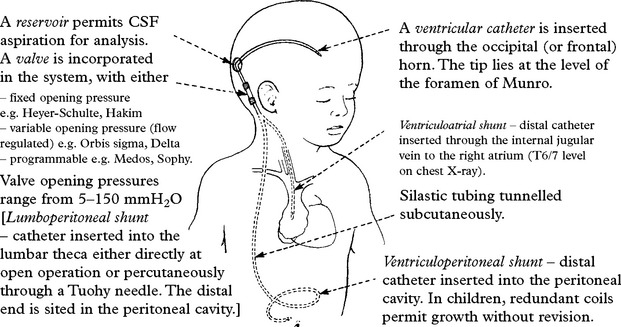
Complications of shunting
Infection: results in meningitis, peritonitis or inflammation extending along the subcutaneous channel. With a V-A shunt, bacteraemia may lead to shunt ‘nephritis’. Staphylococcus epidermidis or aureus are usually involved, with infants at particular risk. Minimise the risk of infection with prophylactic antibiotics and in neonates, with antibiotic impregnated shunt systems. When established, eradication usually requires shunt removal.
Subdural haematoma: ventricular collapse pulls the cortical surface from the dura and leaves a subdural CSF collection or tears bridging veins causing subdural haemorrhage. The risk may be reduced with a variable pressure or programmable valve.
Shunt obstruction: blockage of the shunt system with choroid plexus, debris, omentum or blood clot results in intermittent or persistent recurrence of symptoms. Demonstration of an increase in ventricular size compared to a previous baseline CT scan confirms shunt malfunction. Over a third require revision within 1 year and 80% within 10 years.
Low pressure state: following shunting, some patients develop headache and vomiting on sitting or standing. This low pressure state usually resolves with a high fluid intake and gradual mobilisation. If not, insertion of an antisyphon device or conversion to a high pressure valve is required.
Third ventriculostomy: Suitable for patients with tri-ventricular hydrocephalus e.g. obstructive hydrocephalus caused by aquaduct stenosis or a pineal or posterior fossa tumour occluding the posterior end of the 3rd ventricle/aqueduct. By using a flexible or rigid endoscope introduced through a frontal burrhole, a fistula is created in the floor of the 3rd ventricle. This provides an alternative method of treatment, which if successful, avoids the above problems of shunt insertion. About ⅔ of patients obtain permanent benefit.
Prognosis: Provided treatment precedes irreversible brain damage, results are good with most children attaining normal IQs. Repeated complications, however, particularly prevalent in infancy and in young children carry a significant morbidity.
IDIOPATHIC INTRACRANIAL HYPERTENSION
Idiopathic intracranial hypertension (previously benign intracranial hypertension or pseudotumour cerebri) is characterised by increased intracranial pressure without evidence of an intracranial space-occupying lesion, obstruction to CSF pathways, infection, or hypertensive encephalopathy.

TREATMENT
Indicated to prevent visual loss, which may develop insidiously if untreated:

PROGNOSIS
Generally improves with interventions descibed above, particularly weight loss. Minority of patients have visual loss.
CHIARI MALFORMATION
Although the names of two authors (Arnold and Chiari) were originally linked to the description of malformations at the medullary-spinal junction, Chiari must take most credit for providing a detailed description of this condition.
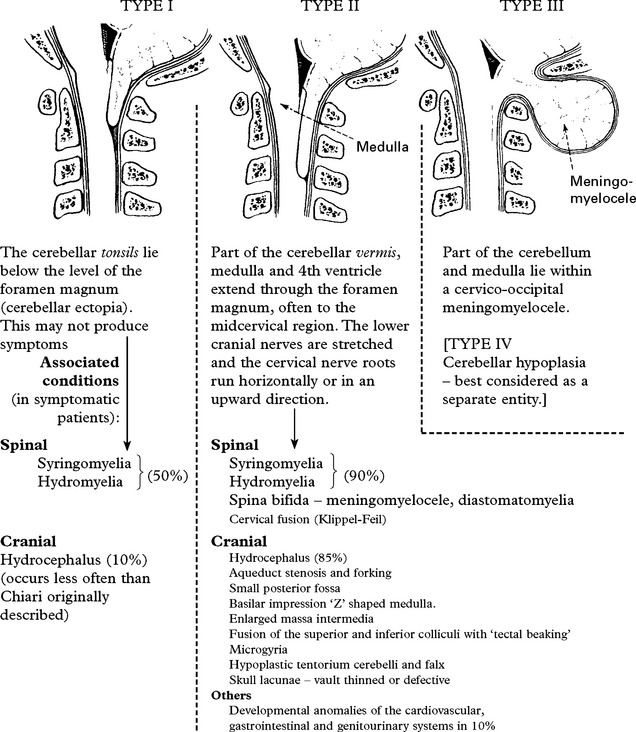
PATHOGENESIS
Several hypotheses have been proposed to explain the pathological findings of these malformations. Gardner suggested that downward pressure from hydrocephalus played an important role in displacing the posterior fossa structures and, when associated with a patent central canal, explained the high incidence of syringomyelia (page 401). Others supposed that traction from a tethered spinal cord (dysraphism), or a CSF leak through a myelocele into the amniotic sac in fetal life resulted in caudal displacement of the posterior fossa structures. Of these theories, none provides an entirely satisfactory explanation; a more realistic view attributes the hindbrain deformity to maldevelopment during early fetal life. This would explain the presence of other developmental anomalies.
CLINICAL PRESENTATION
Depends on age
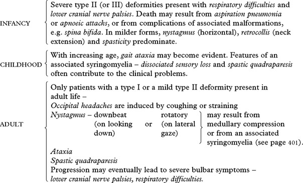
INVESTIGATIONS
Magnetic resonance imaging (MRI) is the investigation of choice. T1 weighted sagittal and axial scans most clearly demonstrate cerebellar ectopia and the presence or absence of an associated syringomyelia.


Myelography (if MRI unavailable)
CT scan: difficult to interpret at the cervico-medullary junction, but shows soft tissue filling the spinal canal at this level.

MANAGEMENT (see also syringomyelia, page 401)
|
In patients with hydrocephalus and signs and symptoms of raised intracranial pressure. |
→ |
Ventriculoperitoneal or atrial shunt may significantly improve signs and symptoms attributed to the Chiari malformation. |
|
In patients with other symptoms and signs |
→ |
Posterior fossa decompression – by removing the posterior rim of the foramen magnum and the arch of the atlas. For more severe cases, the dura is opened and a graft is inserted. Attempts at freeing tonsillar adhesions should be resisted. An apnoea monitor in the initial postoperative period helps detect potentially fatal apnoea, especially during sleep. In some instances, patients with minimal symptoms or with no evidence of progression may warrant a conservative approach. |
PROGNOSIS
Patients with mild symptoms and signs often respond well to operation, but those with long-standing neurological deficits rarely improve. Treatment should aim at preventing further progression.
Further deterioration eventually occurs in one-third, despite operative measures.
SYRINGOBULBIA
Extension of a syringomyelic cavity upwards into the medulla may produce signs and symptoms which are difficult to distinguish from those of medullary compression in the Chiari malformation:
– difficulty in swallowing, dysphonia, dysarthria, vertigo, facial pain
– nystagmus, palatal and vocal cord weakness, occasional facial and tongue weakness.
DANDY-WALKER SYNDROME
This rare developmental anomaly comprises:
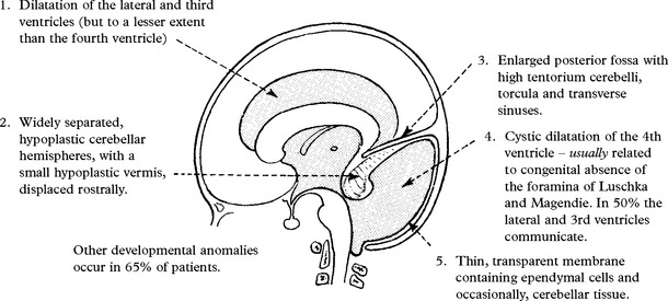
CLINICAL PRESENTATION
Infancy: Symptoms and signs of hydrocephalus (page 375) combined with a prominent occiput.
Childhood: Signs of cerebellar dysfunction with or without signs of hydrocephalus.
INVESTIGATIONS
Skull X-ray: Usually shows elevation of the transverse sinuses and occipital bulging, confirming the presence of an enlarged posterior fossa.
CT scan or MRI:
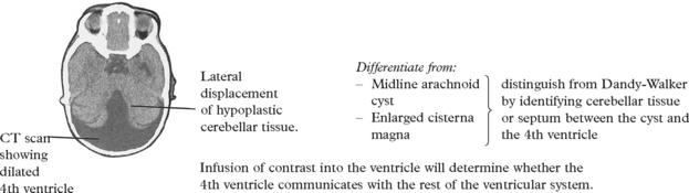
MANAGEMENT
When the dilated 4th ventricle communicates with the rest of the ventricular system, a cystoperitoneal shunt suffices and helps maintain a patent aqueduct. When a ‘two-compartment’ hydrocephalus exists, both the encysted 4th ventricle and the other ventricles require drainage (i.e. with a cysto-peritoneal and a ventriculo-peritoneal shunt).
Excision of the cyst membrane (‘marsupialising’ the 4th ventricle) is no longer thought to normalize CSF flow.
PROGNOSIS
Marked neurological impairment prior to treatment carries a poor outlook. In less impaired patients, the prognosis relates more to the presence of other developmental anomalies.
CRANIOSYNOSTOSIS
In normal childhood development, the cranial sutures allow skull enlargement as the brain grows. Premature fusion of one or more sutures results in restricted growth of bone perpendicular to the suture and exaggerated growth parallel to the suture. The effect depends on the site and number of sutures involved. Sagittal synostosis is the most frequently occurring deformity.
SAGITTAL SYNOSTOSIS
Lateral growth is restricted, resulting in a long narrow head with ridging sagittal suture (scaphocephaly). Treatment: wide excision alone does not allow for lateral expansion of the vault. Either removal of horizontal strips of bone or the use of a ‘helmet’ aids the remodeling process.

CORONAL SYNOSTOSIS
Bilateral or unilateral.
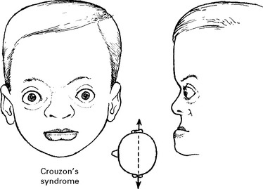
Expansion occurs in a superior and lateral direction (brachiocephaly). This produces a short anterior fossa, shallow orbits and hypertelorism (widening of the interocular distance). Exophthalmos, elevated ICP and visual impairment from papilloedema may result. Bilateral coronal synostosis commonly occurs as one of several congenital defects incorporated in Crouzon’s and Apert’s syndromes.
Involvement of several sutures (oxycephaly) results in skull expansion towards the vertex, the line of least resistance.
PANSYNOSTOSIS (all sutures affected) results in failure of skull growth with a symmetrical abnormally small head and raised intracranial pressure. ICP monitoring or a progressive reduction in normal circumferential growth distinguishes pansynostosis from microcephaly due to inadequate brain development.
Treatment of coronal, metopic and pansynostosis involves extensive craniofacial surgery correcting both cranial and orbital deformities.
Indication for operative treatment is primarily cosmetic when only one suture is involved, but with involvement of two or more sutures operation is also aimed at prevention of visual and cerebral damage from raised ICP.
Posterior plagiocephaly (flattening of the back of the head) An increasing number of infants present with this condition perhaps resulting from the ‘back to sleep’ campaign. Now thought to be due to benign positional moulding rather than a true lambdoid synostosis. Very few of those who develop a progressive skull deformity require surgical treatment.
STEREOTACTIC SURGERY
Stereotactic techniques developed initially for lesion making, enable precise placement of the tip of a cannula or electrode to a predetermined target site within the brain with the least risk.
Many different stereotactic frames have been developed, e.g. Leksell, Todd-Wells, Guiot. These, combined with radiological landmarks (the third ventricle) and a brain atlas, provide anatomical localisation to within ± 1mm. Since some functional variability occurs at each anatomical site, electrode localisation is also based on the recorded neuronal activity and on the effects of electrical stimulation.
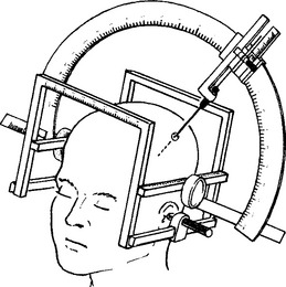
CT/MRI STEREOTACTIC SYSTEM
CT and MRI compatible stereotactic systems allow cannula insertion to any point selected on the image. They are all based on the concept of identifiable external reference (fiducial) markers, e.g. Codman-Robert-Wells (CRW) system:

CT/MRI stereotactic surgery provides the optimal method for the biopsy or aspiration of small, deeply situated tumours or abscesses. Many now use stereotactic biopsy, for larger tumours. It carries lower risk than handheld biopsy and allows selection of specific areas within the tumour. Although improved resolution now available with CT/MRI scanning has led to sufficient anatomical detail for accurate lesion making, functional stereotaxy, e.g. thalamotomy, deep brain stimulation, still requires electrical stimulation for the final target localisation.
METHODS OF LESION MAKING

USES OF STEREOTACTIC SURGERY
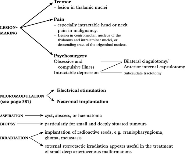
IMAGE GUIDED ‘FRAMELESS’ STEREOTAXY
Neuronavigation uses a combination of modern imaging and elaborate computer software to permit the surgeon to determine how the direction and tip of a pointer lying outwith or within the skull, relates to a two or three dimensional CT or MR image.
The accuracy of the technique depends on the quality of the digitised image and on the methods used to register the patient’s head to the image. The registration of recognisable skin points (e.g. nasion, orbital margins, inner canthus) on the patient to the CT/MR image provides an accuracy of 2–3 mm and this is sufficient for most purposes.
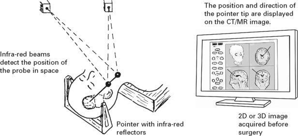
Uses of ‘frameless’ stereotaxy
Aids accurate positioning of burrholes and bone flap, and planning the safest approach to the lesion.
|
TUMOURS |
ARTERIOVENOUS MALFORMATION |
|
– biopsy |
– localisation of lesion and the feeding vessels |
|
– resection: locates, then identifies the tumour margins and the position of important adjacent structures |
|
|
ABSCESS |
|
|
– brachytherapy (see page 314) |
– aspiration |
|
EPILEPSY |
ORBIT |
|
– defining the extent of resected tissue |
– location of intraorbital lesion |
|
– placement of depth electrodes |
|
|
SPINE |
|
|
– pedicle screw fixation |
Deficiencies of ‘frameless’ stereotaxy
The accuracy averages 2–3 mm and although adequate for the above, it is insufficient for most functional procedures.
On opening the skull, brain shift can occur, adding to any registration innaccuracy. Only real time imaging (ultrasound or CT/MRI) can overcome this difficulty (see page 313).
NEUROMODULATION
Definition: Neuromodulation is the alteration of the central, peripheral or autonomic nervous system for therapeutic benefit by electrical or pharmacological stimulation. In the central nervous system, the definition should also include the experimental implantation of foetal or stem cells.
For many years electrical stimulation of the dorsal columns has been used in the treatment of chronic pain, but with technological improvements in implantable devices – stimulators and pumps, the field of neuromodulation has rapidly developed, particularly in relation to deep brain stimulation. In contrast to ablative procedures previously forming the basis of ‘functional’ neurosurgery, neuromodulation techniques are reversible and by using external computors the amount of stimulation or the dose of drug can be tailored to the individual patient’s needs.

PSYCHOSURGERY
In 1935, observation of behavioural changes in chimpanzees following bilateral ablation of the frontal association area, led to the introduction of lesion-making for psychiatric disease (Moniz). In Britain, between 1940 and 1955, neurosurgeons performed over 10 000 prefrontal leucotomy operations. It became evident that patients with affective problems – depression, anxiety and obsessive compulsive disorder – showed better results than those with schizophrenia. Due to the introduction of chlorpromazine in the 1950s, and the operative complications prefrontal leucotomy fell into disrepute, but despite pharmacological improvements, some patients developed chronically disabling conditions and the need for a surgical procedure persisted in those where drugs had little effect.
Stereotactic surgery provides a low risk method of lesion-making and is now generally accepted as a suitable treatment in selected patients where drug treatment has failed. Since issues of ‘informed consent’ for such procedures in the mentally ill are often ethically difficult, careful assessment by a multidisciplinary team of psychiatrists and neurosurgeons is essential. In recent years, interest has focussed on deep brain stimulation (DBS) as an alternate to an irreversible ablative lesion and early results are encouraging (see page 387).
INDICATIONS FOR STEREOTACTIC SURGERY AND LESION SITE

Results
|
Depression/anxiety states |
– up to two-thirds benefit from subcaudate tractotomy. |
|
Obsessional neurosis |
– 80% improve following limbic leucotomy and deep brain stimulation. |