
HEADACHE – GENERAL PRINCIPLES
Headache is a common symptom arising from psychological, otological, ophthalmological, neurological or systemic disease. In clinical practice tension-type headache is encountered most frequently.
Definition: Pain or discomfort between the orbits and occiput, arising from pain-sensitive structures.
Intracranial pain-sensitive structures are:
venous sinuses, cortical veins, basal arteries, dura of anterior, middle and posterior fossae.

Extracranial pain-sensitive structures are:
Scalp vessels and muscles, orbital contents, mucous membranes of nasal and paranasal spaces, external and middle ear, teeth and gums.
Estimated prevalence of headache in the general population
|
Type |
Percentage |
|
Tension type headache |
50–70 |
|
Migraine |
10–15 |
|
Medication overuse headache |
4 |
|
Cluster headache |
0.1 |
|
Raised intracranial pressure |
< 0.01 |
Examination
Full general examination, including:
Ocular – acuity, tenderness, strabismus
Teeth and scalp
Percussion over frontal and maxillary sinuses
Full neurological examination.
HEADACHE – DIAGNOSTIC APPROACH
History: most information is derived from determining:
– the first attack or previous attacks
– whether onset is acute or gradual (days or weeks)
– whether attacks have recurred for many years (chronic)
– site of headache
– accompanying symptoms
– precipitating factors
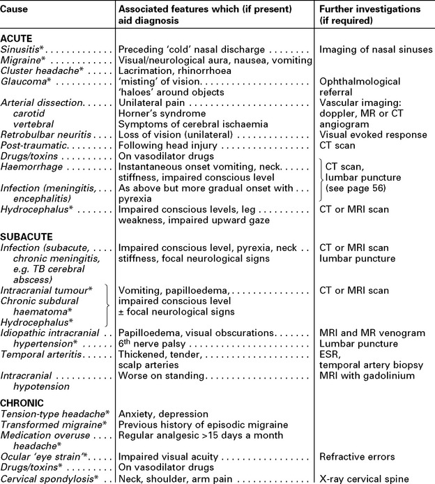
The following table classifies causes in these categories:
(*) Indicates that attacks can be recurrent
Headache in children
Most causes of adult headache may occur in children. In this age group, the commonest type of headache is that accompanying any febrile illness or infection of the nasal passages or sinuses.
The clinician must not take a complaint of headache lightly; the younger the child, the more likely the presence of an underlying organic disease. Pyrexia may not only represent a mild ‘constitutional’ upset, but may result from meningitis, encephalitis or cerebral abscess. The presence of neck stiffness and/or impaired conscious level indicates the need for urgent investigation.
Although intracranial tumours are uncommon in childhood, when they occur they tend to lie in the midline (e.g. medulloblastoma, pineal region tumours). As a result, obstructive hydrocephalus often develops acutely with headache as a prominent initial symptom.
In a child with ‘unexplained’ headache, CT or MRI scan should be performed if the headache is acute or progressive or if there are other features (increase in head circumference, change in personality or decline in school performance) or in children under 5.
HEADACHE – SPECIFIC CAUSES
TENSION TYPE HEADACHE
This is the commonest form of headache experienced by 70% of males and 90% of females at some time in their lives.

Characteristics: Diffuse, dull, aching, ‘band-like’ headache, worse on touching the scalp and aggravated by noise; associated with ‘tension’ but not with other physical symptoms. Attacks may be chronic or episodic. Depression commonly co-exists.
Duration: Many hours–days.
Frequency: Infrequent or daily; worse towards the end of the day. May persist over many years.
Mechanism: ‘Muscular’ due to persistent contraction, e.g. clenching teeth, head posture, furrowing of brow. Some overlap with transformed migraine (see below).
Treatment: Reassurance. Attempt to reduce psychological stress and analgesic over-use (see medication-overuse headache). Amitriptyline and other tricyclic antidepressants or β-blockers.
MIGRAINE
Migraine is a common, often familial disorder characterised by unilateral throbbing headache.
Onset: Childhood or early adult life.
Incidence: Affects 5–10% of the population.
Female:male ratio: 2:1
Family history: Obtained in 70% of all sufferers.
Two recognisable forms exist:
Specific diagnostic criteria are required for migraine with and without aura.
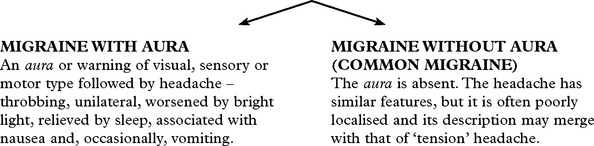
The aura is absent. The headache has similar features, but it is often poorly localised and its description may merge with that of ‘tension’ headache.
The aura of migraine may take many forms. The visual forms comprise: flashing lights, zig-zags (fortifications), scintillating scotoma (central vision) and may precede visual field defects. Such auras are of visual (occipital) cortex origin.
The headache is recurrent, lasting from 2 to 48 hours and rarely occurring more frequently than twice weekly. In migraine equivalents the aura occurs without ensuing headache.
Specific types of migraine with aura
Basilar: Characterised by bilateral visual symptoms, unsteadiness, dysarthria, vertigo, limb paraesthesia, even tetraparesis. Loss of consciousness may ensue and precede the onset of headache. This form of migraine affects young women.
Hemiplegic: Characterised by an aura of unilateral paralysis (hemiplegia) which unusually persist for some days after the headache has settled. Often misdiagnosed as a ‘stroke’. When familial, mendelian dominant inheritance is noted. Recovery is the rule.
Retinal
Unilateral (monocular) visual loss which is reversible and followed by headache. Ophthalmological examination between episodes is normal.
Precipitating factors in migraine
– Dietary: alcohol, chocolate and cheese (contain tyramine).
– Hormonal: often premenstrual or related to oral contraceptive (fluctuations in oestrogen).
– Stress, physical fatigue, exercise, sleep deprivation and minor head trauma.
Diagnosis
|
Clinical history with |
– occasional positive family history – travel sickness or migraine variants (abdominal pains) in childhood – onset in childhood, adolescence, early adult life or menopause |
|
Distinguish |
– partial (focal) epilepsy (in hemiplegic or hemisensory migraine) – transient ischaemic attack (in hemiplegic or hemisensory migraine) – arteriovenous malformation – gives well localised but chronic headache) – hypoglycaemia |
Management
(i) Identification and avoidance of precipitating factors
(ii) Treatment of acute attacks:
Simple analgesics (e.g. aspirin) with metoclopramide to enhance reduced absorption during an attack. If vomiting is prominent anti-emetic (domperidone or prochlorperazine) and analgesic can be helpful.
Sumatriptan (a selective 5HT1 agonist) and other triptans e.g. Naratriptan, Rizatriptan and Zolmitriptan – effectively reverse dilatation in extracranial vessels. Given orally or subcutaneously.
Ergotamine – widespread action on 5HT receptors reversing dilatation. Give orally or by inhalation, injection or by suppository.
Methylprednisolone i.m. or i.v. will halt the attack when prolonged (status migrainosus).
(iii) Prophylaxis: use only for frequent and severe attacks
Pizotifen (5HT2 receptor blocker)
Propranolol (beta adrenergic receptor blocker)
Calcium antagonists (verapamil), antidepressants(amitriptyline) and anticonvulsants, (topiramate or sodium valproate).
Medication Overuse Headaches
Some patients with episodic tension headache or migraine find their headache pattern changes so that they have headaches most days. Many such patients take regular analgesics and/or triptans and this overuse (>14 days a month) can cause medication overuse headaches (MOH). These do not respond to prophylactic agents and will improve on stopping the regular analgesics; this can take some weeks and headaches can be worse in the short-term.
Transformed migraine
If patients with migraine go on to develop chronic daily headache without overusing medication this is ‘transformed migraine’. It usually responds to migraine prophylactic agents.
POST-TRAUMATIC HEADACHE
A ‘common migraine’ or ‘tension-like’ headache may arise after head injury and accompany other symptoms including light-headedness, irritability, difficulty in concentration and in coping with work. This will often respond to amitriptyline or migraine prophylaxis.
CLUSTER HEADACHES (Histamine cephalgia or migrainous neuralgia)
‘Cluster headaches occur less frequently than migraine, and more often in men, with onset in middle age. Charecterised by episodes of severe unilateral pain, lasting 10 minutes to 2 hours, around one eye, associated with conjunctival injection, lacrimation, rhinorrhea and occasionally a transient Horner’s syndrome. The episodes occur between once and many times per day, often wakening from sleep at night. ‘Clusters’ of attacks separated by weeks or even many months. Alcohol may precipitate the attacks.’
Other Trigeminal Autonomic Cephalagias
Cluster headache is the most common form of trigeminal autonomic cephalalgia, where there is a combination of facial pain and autonomic dysfunction. Other rarer combinations of facial pain and antonomic symptoms include:
Hemicrania continua: continuous unilateral moderately severe head pain with exacerbations and variable tearing and partial Horner’s syndrome. More common in women than men (3:1). Responds dramatically to indometacin.
Paroxysmal hemicrania: Same pain but lasts 2-45 minutes multiple times a day. Responds to indometacin.
Short-lasting Unilateral Neuralgiform pain with Conjunctival injection and Tearing (SUNCT): brief pain lasting seconds to 3 minutes with associations described in its name. Women:men, 2:1. Does not respond to indometacin. Lamotrigine has some effect.
GIANT CELL (TEMPORAL) ARTERITIS
Giant cell arteritis, an autoimmune disease of unknown cause, presents with throbbing headache in patients over 60 often with general malaise. The involved vessel, usually the superficial temporal artery, may be tender, thickened, and but nonpulsatile.

Neurological symptoms: strokes, hearing loss, myelopathy and neuropathy.
Jaw claudication: pain when chewing or talking due to ischaemia of the masseter muscles is pathognomonic.
Visual symptoms are common with blindness (transient or permanent) or diplopia.
Associated systemic symptoms – weight loss, lassitude and generalised muscle aches – polymyalgia rheumatica in one-fifth of cases.
Duration: the headache is intractable, lasting until treated.
Mechanism:
Large and medium-sized arteries undergo intense ‘giant cell’ infiltration, with fragmentation of the lamina and narrowing of the lumen, resulting in distal ischaemia as well as stimulating pain sensitive fibres. Occlusion of important end arteries, e.g. the ophthalmic artery, may result in blindness; occlusion of the basilar artery may cause brain stem or bilateral occipital infarction.

Diagnosis: ESR usually high. Blood film shows anaemia or thrombocytosis. C-reactive protein and hepatic alkaline phosphatase elevated. Biopsy of 1 cm length of temporal artery is often diagnostic.
Treatment: Urgent treatment, prednisolone 60 mg daily, prevents visual loss or brain-stem stroke, as well as relieving the headache. If complications have already occurred e.g. blindness, give parenteral high dose steroids. Monitoring the ESR allows gradual reduction in steroid dosage over several weeks to a maintenance level, e.g. 5 mg daily. Most patients eventually come off steroids; 25% require long-term treatment and if so, complications commonly occur.
HEADACHE FROM RAISED INTRACRANIAL PRESSURE
|
Characteristics: |
Associated features: |
|
– generalised. – aggravated by bending or coughing. – worse in the morning on awakening; may awaken patient from sleep. – the severity of the headache gradually progresses. |
– vomiting in later stages. – transient loss of vision (obscuration) with sudden change in posture. – eventual impairment of conscious level Management: further investigations are essential – CT or MRI |
Low pressure headache
Low pressure headache occurs most commonly after lumbar puncture but can arise spontaneously (Spontaneous intracranial hypotension). Headache is worse on standing and improves lying flat. After LP no investigation is needed. If no cause is apparent MRI will show downward displacement cerebellar tonsils and meningeal enhancement with contrast (Gd). Spontaneous improvement is usual. Occasionally a dural ‘blood patch’ at the site of CSF leak (post LP or epidural anaesthesia) is necessary.
HEADACHE DUE TO INTRACRANIAL HAEMORRHAGE
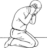
Characteristics:
– instantaneous onset.
– severe pain, spreading over the vertex to the occiput, or described as a ‘sudden blow to the back of the head’.
– patient may drop to knees or lose consciousness.
Associated features:
– usually accompanied by vomiting.
– focal neurological signs suggest a haematoma.
Management: further investigation – CT scan/lumbar puncture (see Meningism, page 75).
N.B. Consider sudden severe headaches to be due to subarachnoid haemorrhage until proved otherwise.
NON-NEUROLOGICAL CAUSES OF HEADACHE
Local causes:
Sinuses: Well localised. Worse in morning. Affected by posture, e.g. bending.
X-ray – sinus opacified. Treatment – decongestants or drainage.
Ocular: Refraction errors may result in ‘muscle contraction’ headaches
– resolves when corrected with glasses.
Acute glaucoma can produce headache but is accompanied by other symptoms, e.g. misting of vision, ‘haloes’.
Dental disease: Discomfort localised to teeth. Check for malocclusion.
Check temporomandibular joints.
Systemic causes:
Headache may accompany any febrile illness or may be the presenting feature of accelerated hypertension or metabolic disease, e.g. hypoglycaemia, hypercalcaemia.
Many drugs produce headache
– through vasodilatation, e.g. bronchodilators, antihistamines
– on withdrawal, e.g. amphetamines, benzodiazepines, caffeine.
MENINGISM
Evidence of meningeal irritation caused by infection or subarachnoid haemorrhage results in characteristic clinical features (though not all will necessarily be present):
SYMPTOMS
1. Headache
2. Vomiting
3. Photophobia
SIGNS

RAISED INTRACRANIAL PRESSURE
The skull is basically a rigid structure. Since its contents – brain, blood and cerebrospinal fluid (CSF) – are incompressible, an increase in one constituent or an expanding mass within the skull results in an increase in intracranial pressure (ICP) – the ‘Monro-Kellie doctrine’.
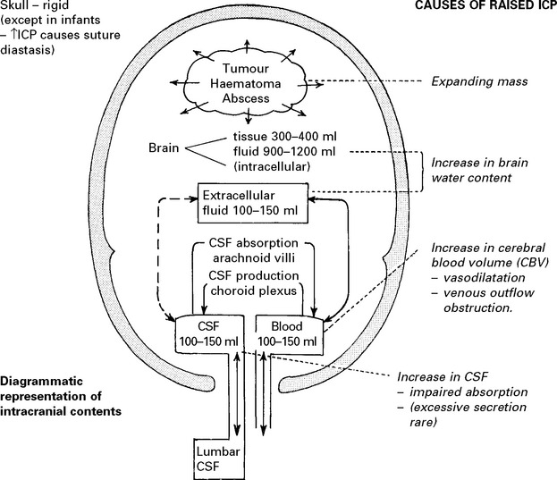
Compensatory mechanisms for an expanding intracranial mass lesion:

CEREBROSPINAL FLUID (CSF)
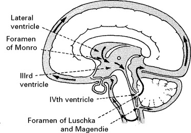
Secreted at a rate of 500 ml per day from the choroid plexus, CSF flows through the ventricular system and enters the subarachnoid space via the 4th ventricular foramina of Magendie and Luschka.
Under normal conditions, CSF flows freely through the subarachnoid space and is absorbed into the venous system through the arachnoid villi. If flow is obstructed at any point in the pathway,hydrocephalus with an associated rise in intracranial pressure develops, as a result of continued CSF production. With an expanding intracranial mass lesion, normal pressure is initially maintained by CSF expulsion to the expandable lumbar theca. Further expansion and subsequent brain shift may obstruct the free flow of CSF not only to the lumbar theca but also to the arachnoid villi, causing an acute rise in intracranial pressure.
BRAIN WATER/OEDEMA
Cerebral oedema – an excess of brain water – may develop around an intrinsic lesion within the brain tissue, e.g. tumour or abscess or in relation to traumatic or ischaemic brain damage, and contribute to the space-occupying effect.
Different forms of cerebral oedema exist:

Vasogenic: excess fluid (protein rich) passes through damaged vessel walls to the extracellular space – especially in the white matter. The extracellular fluid gradually infiltrates throughout normal brain tissue towards the ventricular CSF and this drainage route may aid clearance. e.g. adjacent to tumour.
Cytotoxic: fluid accumulates within cells – neurons and glia i.e. intracellular, e.g. toxic or metabolic states.
Interstitial: when obstructive hydrocephalus develops, CSF is forced through to the extracellular space especially in the periventricular white matter.
With ischaemic damage, as cell metabolism fails, intracellular Na+ and Ca2+ increase and the cells swell i.e. cytotoxic oedema. Capillary damage follows and vasogenic oedema supervenes.
CEREBRAL BLOOD FLOW (CBF)/CEREBRAL BLOOD VOLUME (CBV)
Blood flow is dependent on blood pressure and the vascular resistance:
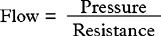
Inside the skull, intracranial pressure must be taken into account:

Under normal conditions the cerebral blood flow is coupled to the energy requirements of brain tissue. Various regulatory mechanisms acting on the arterioles maintain a cerebral blood flow sufficient to meet the metabolic demands.
FACTORS AFFECTING THE CEREBRAL VASCULATURE
Chemoregulation
– Change in extracellular pH or an accumulation of metabolic by-products directly affects the vessel calibre.
– Any change in arteriolar PCO2 has a direct effect on cerebral vessels, but only a reduction of PO2 to < 50 mmHg has a significant effect.
Autoregulation
– A change in the cerebral perfusion pressure results in a compensatory change in vessel calibre.
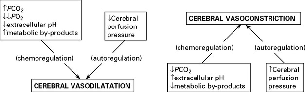
Any change in blood vessel diameter results in considerable variation in cerebral blood volume and this, in turn, directly affects intracranial pressure.
Energy requirements differ in different parts of the brain. To meet such needs in the white matter, flow is 20 ml/100 g/min, whereas in the grey matter flow is as high as 100ml/100g/min.
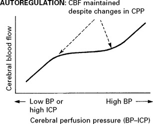
Autoregulation is a compensatory mechanism which permits fluctuation in the cerebral perfusion pressure within certain limits without significantly altering cerebral blood flow.
A drop in cerebral perfusion pressure produces vasodilation (probably due to a direct ‘myogenic’ effect on the vascular smooth muscle) thereby maintaining flow; a rise in the cerebral perfusion pressure causes vasoconstriction.
Neurogenic influences appear to have little direct effect on the cerebral vessels but they may alter the range of pressure changes over which autoregulation acts.
Autoregulation fails when the cerebral perfusion pressure falls below 60 mmHg or rises above 160 mmHg. At these extremes, cerebral blood flow is more directly related to the perfusion pressure.
In damaged brain (e.g. after head injury or subarachnoid haemorrhage), autoregulation is impaired; a drop in cerebral perfusion pressure is more likely to reduce cerebral blood flow and cause ischaemia. Conversely, a high cerebral perfusion may increase the cerebral blood flow, break down the blood–brain barrier and produce cerebral oedema as in hypertensive encephalopathy.
INTRACRANIAL PRESSURE (ICP)
Intracranial pressure, measured relative to the foramen of Monro, under normal conditions ranges from 0–135 mm CSF (0–10 mmHg) although very high pressure, e.g. 1000 mm CSF may occur transiently during coughing or straining.

When intracranial pressure is monitored with a ventricular catheter, regular waves due to pulse and respiratory effects are recorded (page 53). As an intracranial mass expands and as the compensatory reserves diminish, transient pressure elevations (pressure waves) are superimposed. These become more frequent and more prominent as the mean pressure rises.
Eventually the rise in intracranial pressure and resultant fall in cerebral perfusion pressure reach a critical level and a significant reduction in cerebral blood flow occurs. Electrical activity in the cortex fails at flow rates about 20 ml/100 g/min. If autoregulation is already impaired these effects develop even earlier. When intracranial pressure reaches the mean arterial blood pressure, cerebral blood flow ceases.
INTERRELATIONSHIPS
Many factors affect intracranial pressure and these should not be considered in isolation. Inter-relationships are complex and feedback pathways may merely serve to compound the brain damage.
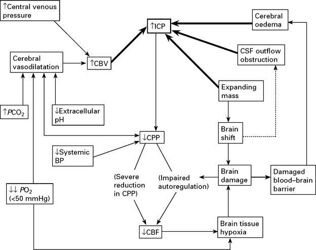
CLINICAL EFFECTS OF RAISED INTRACRANIAL PRESSURE
A raised ICP will produce symptoms and signs but does not cause neuronal damage provided cerebral blood flow is maintained. Damage does, however, result from brain shift – tentorial or tonsillar herniation.
Clinical features due to ↑ICP:
1. Headache – worse in the mornings, aggravated by stooping and bending.
2. Vomiting – occurs with an acute rise in ICP.
3. Papilloedema – occurs in a proportion of patients with ICP. It is related to CSF obstruction and does not necessarily occur with brain shift alone. Increased CSF pressure in the optic nerve sheath impedes venous drainage and axoplasmic flow in optic neurons. Swelling of the optic disc and retinal and disc haemorrhages result. Vision is only at risk when papilloedema is both severe and prolonged.
BRAIN SHIFT – TYPES
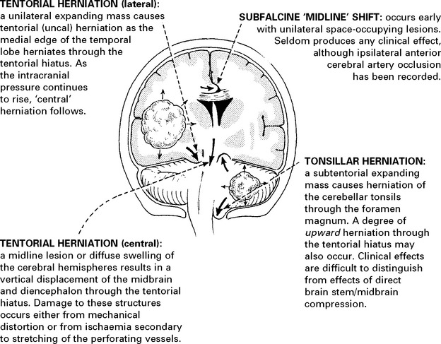
Unchecked lateral tentorial herniation leads to central tentorial and tonsillar herniation, associated with progressive brain stem dysfunction from midbrain to medulla.
CLINICAL EFFECTS OF BRAIN SHIFT

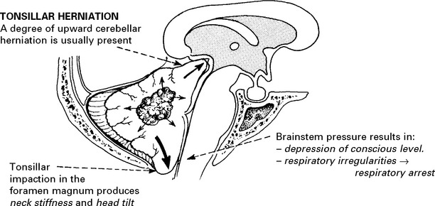
An injudicious lumbar puncture in the presence of a subtentorial mass may create a pressure gradient sufficient to induce tonsillar herniation.
N.B. Harvey Cushing described cardiovascular changes – an increase in blood pressure and a fall in pulse rate, associated with an expanding intracranial mass, and probably resulting from direct medullary compression. The clinical value of these observations is often overemphasised. They are often absent; when present they are invariably preceded by a deterioration in conscious level.
INVESTIGATIONS
Patients with suspected raised intracranial pressure require an urgent CT/MRI scan. Intracranial pressure monitoring where appropriate (see page 53).
TREATMENT OF RAISED INTRACRANIAL PRESSURE
When a rising intracranial pressure is caused by an expanding mass, or is compounded by respiratory problems, treatment is clear-cut; the mass must be removed and blood gases restored to normal levels – by ventilation if necessary.
In some patients, despite the above measures, cerebral swelling may produce a marked increase in intracranial pressure. This may follow removal of a tumour or haematoma or may complicate a diffuse head injury. Artificial methods of lowering intracranial pressure may prevent brain damage and death from brain shift, but some methods lead to reduced cerebral blood flow, which in itself may cause brain damage (see page 84).
Intracranial pressure is monitored with a ventricular catheter or surface pressure recording device (see page 52). Treatment may be instituted when the mean ICP is > 25 mmHg. Ensure cause is not due to constriction of neck veins.
Methods of reducing intracranial pressure
Mannitol infusion: An i.v. bolus of 100 ml of 20% mannitol infused over 15 minutes reduces intracranial pressure by establishing an osmotic gradient between the plasma and brain tissue. This method ‘buys’ time prior to craniotomy in a patient deteriorating from a mass lesion. Mannitol is also used 6 hourly for a 24–48 hour period in an attempt to reduce raised ICP. Repeated infusions, however, lead to equilibration and a high intracellular osmotic pressure, thus counteracting further treatment. In addition, repeated doses may precipitate lethal rises in arterial blood pressure and acute tubular necrosis. Its use is therefore best reserved for emergency situations.
CSF withdrawal: Removal of a few ml of CSF from the ventricle immediately reduces the intracranial pressure. Within minutes, however, the pressure will rise and further CSF withdrawal will be required. In practice, this method is of limited value, since CSF outflow to the lumbar theca results in a diminished intracranial CSF volume and the lateral ventricles are often collapsed. Continuous CSF drainage may make most advantage of this method.
Sedatives: If intracranial pressure fails to respond to standard measures then sedation may help under carefully controlled conditions.
Propofol, a short acting anaesthetic agent, reduces intracranial pressure but causes systemic vasodilatation. If this occurs pressor agents may be required to prevent a fall in blood pressure and a reduction in cerebral perfusion. Avoid high doses of Propofol; rhabdomyolysis may result and carries a 70% mortality.
Barbiturates (thiopentone) reduce neuronal activity and depress cerebral metabolism; a fall in energy requirements theoretically protects ischaemic areas. Associated vasoconstriction can reduce cerebral blood volume and intracranial pressure but systemic hypotension and myocardial depression also occur. Clinical trials of barbiturate therapy have not demonstrated any improvement in outcome.
Controlled hyperventilation: Bringing the PCO2 down to 3.5kPa by hyperventilating the sedated or paralysed patient causes vasoconstriction. Although this reduces intracranial pressure, the resultant reduction in cerebral blood flow may aggravate ischaemic brain damage and do more harm than good (see page 232). Maintaining the blood pressure and the cerebral perfusion pressure (CPP) (>60 mmHg) appears to be as important as lowering intracranial pressure.
Decompressive craniectomy: This technique is gaining renewed interest in treating raised ICP unresponsive to other methods. The principal concern is that although reducing mortality, unacceptable levels of morbidity may result. A randomised trial of decompressive craniectomy in head injury is currently underway.
Hypothermia: Cooling to 34°C lowers ICP. Although hypothermia after cardiac arrest with slow rewarming has been reported to improve outcome, trials in head injured patients have failed to demonstrate significant benefit.
Steroids: By stabilising cell membranes, steroids play an important role in treating patients with oedema surrounding intracranial tumours. Trials have found no evidence of benefit after traumatic or ischaemic damage.
COMA AND IMPAIRED CONSCIOUS LEVEL
Consciousness is regarded as a state of awareness of self and surroundings. Impaired consciousness is due to disturbed arousal or content of mental function.
Many pathological processes may impair conscious level and numerous terms have been employed to describe the various clinical states which result, including obtundation, stupor, semicoma and deep-coma. These terms result in ambiguity and inconsistency when used by different observers. Recording conscious level with the Glasgow coma scale (page 5) avoids these difficulties and clearly describes the level of arousal. With this scale:
COMA = NO SPEECH, NO EYE OPENING, NO MOTOR RESPONSE
In this section we describe conditions which may present with, or lead to, coma. Patients experiencing ‘transient disturbance of conscious level’ require a different approach.
Pathophysiology of coma
A ‘conscious’ state depends on intact cerebral hemispheres, interacting with the ascending reticular activating system in the brain stem, midbrain, hypothalamus and thalamus. Lesions diffusely affecting the cerebral hemispheres, or directly affecting the reticular activating system cause impairment of conscious level:


CAUSES
INTRACRANIAL
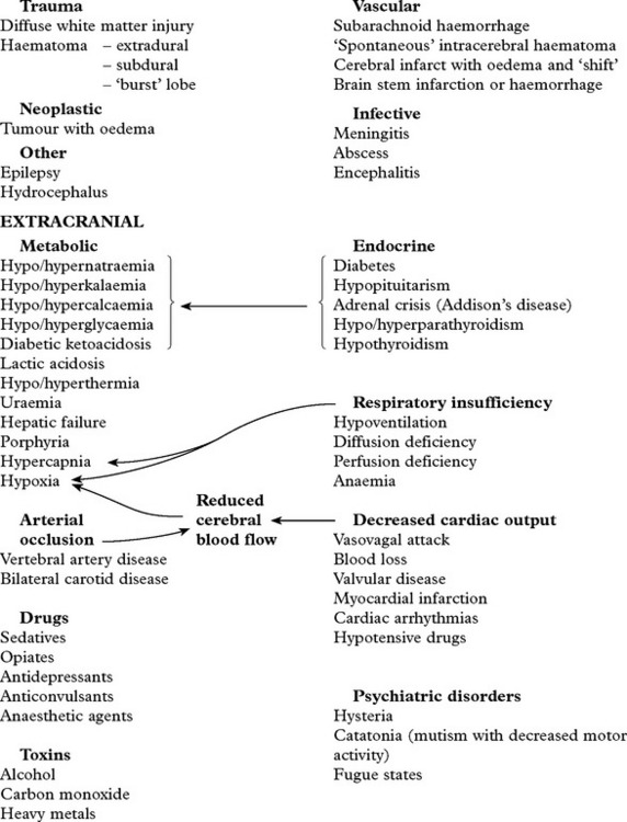
Examination of the unconscious patient (see pages 29, 30)
DIAGNOSTIC APPROACH
Questioning friends, relatives or the ambulance team, followed by general and neurological examination all provide important diagnostic information.
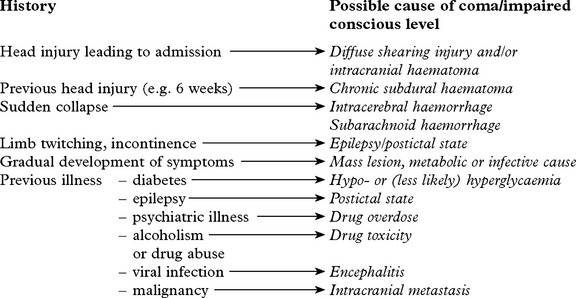
General examination
Note the presence of:


Neurological examination

Investigations
The sequence of investigations depends on clinical suspicion:

In addition
CHEST X-RAY – may reveal a bronchial carcinoma.
ELECTROENCEPHALOGRAPHY – may provide evidence of – subclinical epilepsy
– herpes simplex encephalitis
– metabolic encephalopathy.
MRI – has a limited role in the investigation of coma. More sensitive than CT scan in demonstrating small ischaemic changes and early encephalitis.
Prognosis
Although conscious level examination does not aid diagnosis, it plays an essential role in patient management and along with the duration of coma, pupil response and eye movements provides valuable prognostic information. Non-traumatic coma tends to carry a better prognosis (see page 214).
TRANSIENT LOSS OF CONSCIOUSNESS
Many conditions causing coma may also transiently affect a patient’s conscious level. This results from:
Syncope:
Reduction in cerebral arterial oxygen supply can be caused by cardiac arrhythmias, cardiac outflow obstruction or vasovagal attack.
Seizure:
Pseudo-seizure (non-epileptic attack disorder) – see below
Acute toxic or metabolic coma:
– Drug abuse – alcohol, solvents or barbiturates – may cause transient, intermittent confusion.
– Hypoglycaemia
DIAGNOSTIC APPROACH
History
Try to obtain a history from eye-witness as well as from the patient themselves.
History from the patient:
Context: may suggest likely cause – a collapse when having blood taken suggests syncope; an episode arising from sleep suggests a seizure.
Prodrome: a brief sensation of déjà vu before the episode indicates a focal onset seizure; a feeling of lightheadness, sweatiness and visual fading suggests syncope.
Recovery: a rapid recovery suggest syncope; waking in the ambulance suggest seizure.
History from witness (find them; phone them):
How long the patient was out for; – syncope is typically less than 1 minute; seizures usually longer.
What they did; brief asynchronous jerking movements occur in syncope; more prolonged synchronous tonic clonic movements occur in seizures.
Any colour change; ‘ashen’ suggests syncope; cyanosed suggests seizure.
How quickly they recovered; rapid recovery suggest syncope.
Silent witnesses:
Incontinence is common in all forms of loss of consciousness and does not distinguish between a seizure and syncope. Tongue biting strongly suggests a seizure as do other much less common injuries – posterior dislocation of the shoulder or vertebral fracture.
Investigation is directed by the clinical history:
Electroencephalography (EEG) may reveal a focal or generalized disturbance – epilepsy.
Electrocardiography (ECG) and 24 hour ECG may reveal a cardiac arrythmia.
Head up tilt-table testing may reveal neurocardiogenic syncope or orthostatic hypotension.
Echocardiography may reveal cardiomyopathy.
Blood glucose may indicate hypoglycaemia.
EEG telemetry is occassionally needed.
Often attacks of unconsciousness remain unexplained and possibly have a psychological basis. The circumstances of the attack (e.g. during an argument), the non-stereotyped nature of the episode suggest a non-organic explanation. Such attacks are often mistaken for a seizure and are referred to as pseudo-seizures or non-epileptic attacks (see page 99).
CONFUSIONAL STATES AND DELIRIUM
Of all acute medical admissions, 5–10% present with a confused verbal response, i.e. disorientation in time and/or place. Most patients are easily distracted, have slowed thought processes and a limited concentration span. Some may lose interest in the examination to the point of drifting off to sleep.
Perceptual disorders (illusions and hallucinations) may accompany the confused state – delirium. This is often associated with withdrawal and lack of awareness or with restlessness and hyperactivity.
Primary neurological disorders contribute to only 10% of those patients presenting with an acute confusional state. In the elderly, postoperative disorientation is particularly common and multiple factors probably apply; in these patients the prognosis is good.
The Confusion Assessment Method (CAM) is used to confirm delirium.
Feature 1 – Acute onset and fluctuating course.
Feature 2 – Inattention.
Feature 3 – Disorganised thinking.
Feature 4 – Altered level of consciousness.
The presence of features 1 and 2 and either 3 or 4 are diagnostic.
DIAGNOSTIC APPROACH
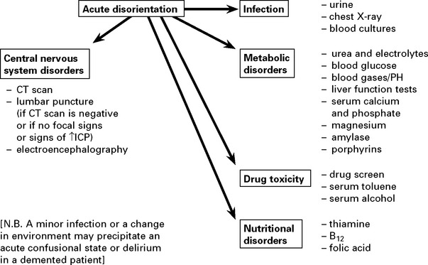
EPILEPSY
Definitions
A seizure or epileptic attack is the consequence of a paroxysmal uncontrolled discharge of neurons within the central nervous system. The clinical manifestations range from a major motor convulsion to a brief period of lack of awareness.
The prodrome refers to mood or behavioural changes which may precede the attack by some hours.
The aura refers to the symptom immediately before a seizure and will localise the attack to its point of origin within the nervous system.
The ictus refers to the attack or seizure itself.
The postictal period refers to the time immediately after the ictus during which the patient may be confused, disorientated and demonstrate automatic behaviours.
The stereotyped and uncontrollable nature of the attack is characteristic of epilepsy.
A patient is said to have epilepsy when they have had more than one seizure. It is important to remember that epilepsy is not a single condition; epilepsy can be the symptom of other disorders and there are numerous different epilepsy syndromes.
Pathogenesis
Epilepsy has been described since ancient times. The 19th century neurologist Hughlings-Jackson suggested ‘a sudden excessive disorderly discharge of cerebral neurons’ as the causation of the attack. Berger (1929) recorded the first electroencephalogram (EEG) and not long after, it was appreciated that certain seizures were characterised by particular EEG abnormalities.
Recent studies in animal models of focal epilepsy suggest a central role for the excitatory neurotransmitter glutamate. This produces a depolarisation shift by activating receptors which in turn facilitate cellular influx of Na+, K+ and Ca2+. Gamma amino butyric acid (GABA) has an important inhibitory influence in containing abnormal cortical discharges and preventing the development of generalised seizures.
Epilepsies have complex inheritance; molecular genetics studies in rarer syndromes with autosomal dominant features have identified genes that code for ion channel subunits, either ligand or voltage gated (Channelopathies).
Incidence and course
Epilepsy presents most commonly in childhood and adolescence or in those over 65, but may occur for the first time at any age.
5% of the population suffer a single seizure at some time.

Though there is considerable variability depending on seizure type, 6 years after diagnosis 40% of patients have had a substantial remission; after 20 years – 75%.
SEIZURE CLASSIFICATION
The classification of epilepsy involves two steps:
1. The classification of the seizure types
2. Integration of seizure type with history, family history, EEG and imaging (as needed)
CLASSIFICATION OF SEIZURE TYPE
Attacks which begin focally from a single location within one hemisphere are distinguished from those of a generalised nature which probably commence in deeper midline structures and project to both hemispheres simultaneously.
1. PARTIAL (focal, localisation related) SEIZURE
Classified by site of onset (frontal, temporal, parietal or occipital lobe) and by severity:
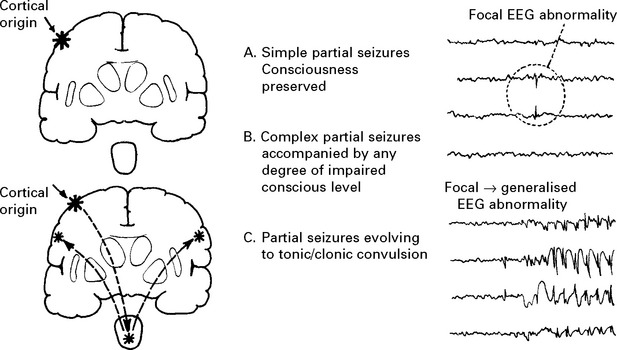
2. GENERALISED SEIZURES (convulsive or non-convulsive)
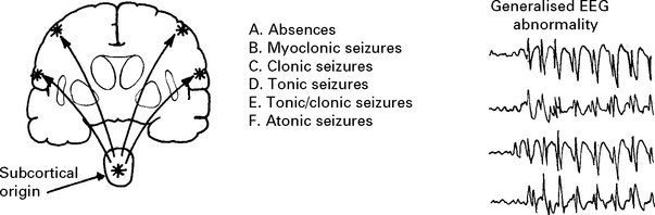
3. UNCLASSIFIED SEIZURES, There may be insufficient information to classify a seizure.
THE PARTIAL SEIZURES
Partial seizures are classified according to both their:
Severity – simple; complex partial; evolving to tonic/clonic convulsion
Semiology – what happens during the seizure, which reflects the site of origin, in order of frequency: temporal, frontal, parietal and occipital lobes.
FRONTAL LOBE SEIZURES
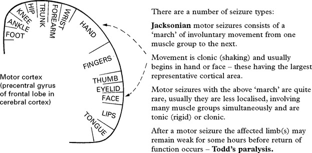
Adversive seizures

PARIETAL LOBE SEIZURES
These arise in the sensory cortex (parietal lobe), the patient describing paraesthesia or tingling in an extremity or on the face sometimes associated with a sensation of distortion of body image. A ‘march’ similar to the Jacksonian motor seizure may occur. Motor symptoms occur concurrently – the limb appears weak without involuntary movement.
The representation of limbs, trunk, etc. in the post-Rolandic sensory cortex is similar to that of the motor cortex.
VISUAL, AUDITORY and AUTONOMIC simple partial seizures occur, but are rare.
Frontal and Parietal seizures indicate structural brain disease, the focal onset localising the lesion. Full investigation is mandatory.
TEMPORAL LOBE SEIZURES
These attacks are characterised by a complex aura (initial symptom) often with some impairment of consciousness.
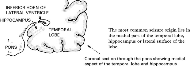
The nature of the attack
The content of attacks may vary in an individual patient. Commonly encountered symptoms include:
Visceral disturbance: Gustatory (taste) and olfactory (smell) hallucinations, lip smacking, epigastric fullness, choking sensation, nausea, pallor, pupillary changes (dilatation), tachycardia.
Memory disturbance: Déjà vu (‘something has happened before’), jamais vu (‘feeling of unfamiliarity’), depersonalisation, derealisation, flashbacks, formed visual or auditory hallucinations.
Motor disturbance: Fumbling movement, rubbing, chewing, semi-purposeful limb movements.
Affective disturbance: Displeasure, pleasure, depression, elation, fear.
A constellation of these symptoms associated with subtle clouding of consciousness characterises a temporal lobe onset seizure.
AUTOMATISM occurs during the state of clouding of consciousness either during or after the attack (postictal) and takes the form of involuntary, often complicated, motor activity. In ambulatory automatism, subjects may ‘wander off’.
Confusion and headache after an attack are common. The whole episode may last for seconds but occasionally may be prolonged and a rapid succession or cluster of attacks may occur. Attacks show an increased incidence in adolescence and early adult life. A history of birth trauma or febrile convulsions in infancy may be obtained. Lesions in the hippocampus occur as a result of anoxia or from the convulsion itself and act as a source of further epilepsy. When surgery is carried out, hippocampal sclerosis is often found. Occasionally other pathologies are identified, such as dysembryoplastic neuroepithelial tumours (DNET), vascular malformations and low-grade astrocytomas.
OCCIPITAL LOBE SEIZURES
These are uncommon. Typically there is an elementary visual hallucination – a line or flash – prior to a tonic-clonic seizure.
PARTIAL SEIZURES EVOLVING TO TONIC/CLONIC CONVULSION
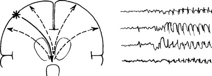
Seizure discharges have the capacity to spread from their point of origin and excite other structures. When spread occurs to the subcortical structures (thalamus and upper reticular formation) their excitation releases a discharge which spreads back to the cerebral cortex of both hemispheres, resulting in a tonic/clonic seizure. This chain of events is reflected in the electroencephalogram (EEG).
The symptoms before the tonic/clonic convulsion give a clue to the site of the initial discharge (simple partial or complex partial).
An eyewitness account is important because retrograde amnesia may prevent recall of the onset.
TONIC/CLONIC ATTACKS
Loss of consciousness; falls to the ground.

The patient then sleeps with stertorous respiration and cannot be roused. On regaining consciousness, confusion and headache are present. He may feel exhausted for hours or even days afterwards. Muscles may ache as a result of violent movement and muscle damage occurs with elevation of the muscle enzyme creatinine phosphokinase (CPK). Trauma occurs frequently, either as a result of the fall, or as a result of the movements, e.g. posterior dislocation of the shoulder. Very rarely sudden death may occur from inhalation or an associated cardiac arrhythmia.
The differentiation of these attacks from pseudoseizures will be discussed later.
GENERALISED SEIZURES
Generalised seizure attacks arise from subcortical structures and involve both hemispheres. Consciousness may be impaired and motor manifestations are bilateral.
ABSENCES (previously called Petit mal)
The patient (usually a child) stares vacantly, eyes may blink. The absence may occur many times a day with a duration of 5–15 seconds and may be induced by hyperventilation.
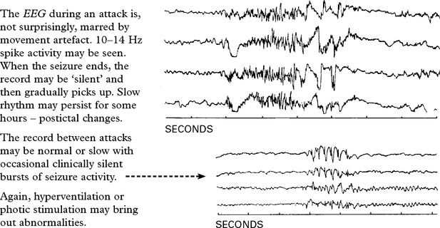
The ELECTROENCEPHALOGRAM (EEG) is diagnostic.
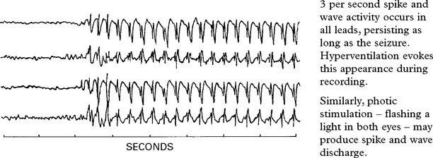
ABSENCE STATUS
Long periods of clouding of consciousness with continuing ‘spike and wave’ activity on the EEG.
MYOCLONIC SEIZURES
Sudden, brief, generalised muscle contractions. They often occur in the morning and are occasionally associated with tonic/clonic seizures. The commonest disorder is benign juvenile myoclonic epilepsy (JME) with onset after puberty. Myoclonus on the edge of sleep is normal. Myoclonus also occurs in degenerative and metabolic disease (see page 190).
TONIC SEIZURES
Sudden sustained muscular contraction associated with immediate loss of consciousness.
Tonic episodes occur as frequently as tonic/clonic episodes in children and should alert the physician to a possible anoxic aetiology.
In adults, tonic attacks are rare.
GENERALISED SEIZURES
TONIC/CLONIC SEIZURES (previously called Grand mal)
Primary tonic/clonic seizures occur without warning or aura. The epileptic cry at onset results from tonic contraction of respiratory muscles with partial closure of vocal cords. The tonic phase is associated with rapid neuronal discharge. The clonic phase begins as neuronal discharge slows.
ATONIC SEIZURES
These are rare and almost always occur in patients with other types of seizure. They are characterised by a loss of muscle tone and a sudden fall. Consciousness may only be lost briefly. The EEG shows polyspike activity or low voltage fast activity.
SYMPTOMATIC SEIZURES
Seizures can be symptoms of acute brain pathology. If the patient goes on to develop recurrent seizures this is symptomatic epilepsy (see later).
The age of onset gives a clue to the causation.
|
Newborn |
Infancy and Childhood |
Adult |
|
Asphyxia |
Febrile convulsions |
Trauma |
|
Intracranial haemorrhage |
CNS infection |
Drugs and alcohol |
|
Hypocalcaemia |
Trauma |
CNS infection |
|
Hypoglycaemia |
Congenital defects |
Intracranial haemorrhage |
|
Hyperbilirubinaemia |
Inborn errors of metabolism |
Tumours |
|
Water intoxication |
Tumours |
Vascular disease |
|
Inborn errors of metabolism |
Hypoglycaemia |
Seizures occur in about 5% of patients following stroke and in 5% of patients with multiple sclerosis.
SEIZURES – DIFFERENTIAL DIAGNOSIS
The following should be considered in the differential diagnosis of seizures – SYNCOPE (VASOVAGAL) ATTACKS
Syncope usually occurs when the patient is standing and result from a global reduction of cerebral blood flow.
Prodromal pallor, nausea and sweating occur associated with a feeling of lightheadness and often fading of vision. If the patient sits down, the attack may pass off or proceed to a brief loss of consciousness.
Brief asynchronous jerks are common as is urinary incontinence. Tonic and clonic movements may develop if impaired cerebral blood flow is prolonged (‘anoxic’ seizures).
Mechanism: Peripheral vasodilatation with drop in blood pressure followed by vagal overactivity with fall in heart rate.
Syncopal attacks occur in hot, crowded rooms (e.g. classroom) or in response to pain or emotional disturbance.
‘Reflex’ syncope from cardiac slowing may occur with carotid sinus compression. Similarly, cough syncope may result from vigorous coughing.
CARDIAC ARRHYTHMIAS
Seen in situations such as complete heart block (Adams-Stokes attacks).
Prolonged arrest of cardiac rate or critical reduction will progressively lead to loss of consciousness – tonic jerks – cyanosis/stertorous respiration – fixed pupils and extensor plantar responses.
On recovery of normal cardiac rhythm, the degree of persisting neurological damage depends upon the duration of the episode and the presence of pre-existing cerebrovascular disease. In suspected patients, electrocardiography is mandatory. Continuous (24 hours) ECG monitoring may be necessary.
HYPOGLYCAEMIA
Amongst other neuroglycopenic manifestations, seizures or intermittent behavioural disturbances may occur. A rapid fall of blood sugar is associated with symptoms of catecholamine release, e.g. palpitations, sweating, etc. In ‘atypical’ seizures exclude a metabolic cause by blood sugar estimation when symptomatic.
EPISODIC CONFUSION
Intermittent confusional episodes caused by drugs (e.g. barbiturates) or toxins (e.g. solvents).
PANIC ATTACKS Hyperventilation can induce focal motor and sensory symptoms.
NARCOLEPSY
Inappropriate sudden sleep episodes may easily be confused with epilepsy (see page 107).
DISSOCIATIVE SEIZURES (pseudoseizures, non-epileptic attack disorder, NEAD)
A difficult distinction lies between epileptic seizures and dissociative seizures. The latter are heterogeneous comprising episodes in which shaking/thrashing and apparent loss of consciousness occur. The episodes are often variable (rather than stereotyped), prolonged, with a rapid recovery. Often patients with epilepsy will also manifest such attacks. Patients may have a history of other functional illness and have an increased frequency of preceding sexual or physical trauma (about 30%). Dissociative seizures are usually thought to be a subconscious disorder. Rarely some patients do have insight and the episodes are part of a facticious disorder or malingering. EEG studies, particularly with video telemetry, may help discriminate. Management depends on helping the patient understand and manage the episodes, for example with cognitive behavioural therapy, managing any associated depression or anxiety and stopping unnecessary anticonvulsants.
EPILEPSY – CLASSIFICATION
The classification of epilepsy brings together the seizure semiology and other aspects of the history and investigations. The International League Against Epilepsy classified epilepsies as:
• Idiopathic – thought to be primarily genetic with generalised seizures, sometimes grouped as more specific syndromes (see below). Account for about 10–20% of cases.
• Symptomatic – partial onset seizures associated with a structural lesion, such as tumour, cortical dysplasia, infection, head injury or trauma – about 30–40% of cases. The combination of the site of seizure onset and the underlying pathology leads to the diagnosis: for example ‘post traumatic frontal lobe epilepsy’ or ‘temporal lobe epilepsy due to mesial temporal sclerosis’ or ‘symptomatic occipital lobe epilepsy secondary to an arteriovenous malformation’.
• Cryptogenic – partial onset seizures for which no cause has been found. Account for about 50% of patients.
With developments in understanding, particularly in genetics, limitations with this generally practical classification have arisen – for example familial frontal onset epilepsy (associated with a mutation in the gene encoding the neuronal nicotinic acetylcholine receptor (nAChR) alpha-4 subunit) is an idiopathic yet partial onset epilepsy. Newer proposals under consideration suggest the classification should move to ‘genetic’, ‘structural/metabolic’ or ‘of unknown cause’ rather than the groups given above.
Selected Idiopathic Epilepsy Syndromes (by age of onset)
Childhood absence epilepsy (common)
Absence seizures begin between 4 and 12 years of age. Family history in 40% of patients. The absence may occur many times a day with a duration of 5–15 seconds.
Frequent episodes lead to falling off in scholastic performance.
Attacks rarely present beyond adolescence.
In 30% of children, adolescence may bring tonic/clonic seizures.
Distinction of absences from complex partial seizures is straightforward; the latter are longer – 30 seconds or more – and followed by headache, lethargy, confusion and automatism.
EEG finds 3 Hz spike and wave (page 97)
Juvenile myoclonic epilepsy (common)
Myoclonic jerks begin in teenage years, typically in the morning. Develop tonic/clonic seizures, often with sleep deprivation, in late teens. Occasionally have absence seizures. EEG frequently finds 4-5 polyspike and wave discharges.
West Syndrome (rare)
Infants present with diffusely abnormal EEGs, tonic clonic convulsions, myoclonic jerks and mental retardation following perinatal trauma or asphyxia. The seizures are sometimes called infantile spasms and the abnormal EEG pattern between events – hypsarrhythmia. Mortality or severe disability is high.
Lennox-Gastaut Syndrome (rare)
This similar syndrome presents later between 1–7 years of age. The response to anticonvulsant treatment and the degree of retardation is variable. The condition is associated with a large number of disorders including hypoxia, intracranial haemorrhage, toxoplasmosis, cytomegalovirus infection and tuberous sclerosis.
The REFLEX EPILEPSIES are a rare group of seizure disorders in which tonic/clonic or complex partial seizures are evoked by sensory stimuli. These stimuli can be certain pieces of music (Musicogenic epilepsy), reading (reading epilepsy) or performing calculations (arithmetical epilepsy).
EPILEPSY – INVESTIGATION
Investigations are directed at:
• corroborating the diagnosis of epilepsy
• classifying the type of epilepsy
• looking for an underlying cause
• eliminating alternative diagnoses
The relative emphasis of these elements will depend on the clinical situation.
For most patients the clinical diagnosis of a seizure is secure and the emphasis is to seek the cause and to classify the epilepsy to direct treatment. In others the main concern is whether the episodes are seizures or an alternative diagnosis.
Neuroimaging
All adults and all with focal onset seizures should be scanned. MRI brain imaging is more sensitive than CT and many lesions, for example small tumours, cortical dysplasia or hippocampal sclerosis will be missed on CT.
EEG
Standard interictal EEG is relatively insensitive – though this varies according to the type of epilepsy (it is very sensitive in childhood absence epilepsy). The interpretation of abnormalities requires caution; 0.5% of the normal population have inter-ictal spikes or sharp waves (epileptic discharges) as compared to 30% of patients after their first seizure.
The pattern of abnormalites can point towards a focal or generalised onset and can supplement the clinical classification.
Sleep deprived EEG increases the yield but with the risk of provoking a seizure. EEG shortly after a seizure is more likely to find an abnormality.
Ambulatory EEG recording increases the chance of finding an abnormality and of recording a clinical event. The ‘gold standard’ investigation is simultaneous EEG monitoring and video monitoring (videotelemetry).
Eliminating alternatives
ECG should be done in all patients with seizures. This is a simple cheap test and a small number of epilepsy mimics can be identified this way, e.g. prolonged QT syndrome.
Prolonged ECG may be useful in patients with possible cardiac syncope – especially in patients with sleep associated events. Implantable loop recorders can be used when patients have infrequent events.
Head up tilt table testing is often helpful in the diagnosis of neurocardiogenic syncope.
Metabolic investigation to consider include fasting glucose for insulinoma and synacthen test for Addison’s disease.
Advanced investigation
Volumetric MRI can identify hippocampal sclerosis not apparent on conventional imaging.
Functional imaging, using ictal and inter-ictal SPECT may be helpful in identifying an epileptogenic focus when evaluating patients for surgery.
Advanced EEG techniques for example using sphenoidal electrodes or recording from surgically inserted intracranial grid or depth electrodes can help localise a focus before surgery.
EPILEPSY – TREATMENT
Basic principles: Most patients respond to anticonvulsant drug therapy. Drug treatment should be simple, preferably using one anticonvulsant (monotherapy). Polytherapy should be avoided to minimise adverse effects and drug interactions.
Treatment aims to prevent seizures without side effects though this is not always achieved. Surgery is an option in a small number on non-responders.
Teratogenicity: it is important to consider the teratogenetic risks when starting any anticonvulsant in a woman of childbearing age. Large prospective studies have established rates of major congenital malformations for widely used drugs: those on no medication, carbamazepine or lamotrigine had similar rates of around 3%; in valproate monotherapy the rate was significantly higher at 6%; polytherapy overall was about 6%, and 9% if valproate was one of the drugs.
Interactions: many anticonvulsants (especially carbamazepine, phenytoin, phenobarbitone) induce liver enzymes to increase metabolism of other drugs (notably the oral contraceptive, warfarin and other anticonvulsants); valproate inhibits liver enzymes.
Blood levels: monitoring levels is useful for phenytoin because of the difficult pharmacokinetics. Other blood levels can occasionally be useful to check the patient is taking the medication or for toxicity.
Drug choice:
Idiopathic generalised epilepsy: sodium valproate*; lamotrigine*; topiramate; levetiracetam; phenytoin.
Partial (focal) epilepsy: lamotrigine*; carbamazepine*; sodium valproate*; Phenytoin*; Phenobarbitone; Levetiracetam; Topiramate; Tiagabine; Zonisamide; Oxcarbazepine; Gabapentin; pregabalin; lacosamide.
Those drugs asterisked are typically used for monotherapy others as ‘add-on’ therapy when control sub-optimal. The choice of anticonvulsant will be a balance between efficacy, adverse effects, teratogenicity and drug interactions and the patient should be involved in this decision.
Main adverse effects of main anticonvulsants:
Lamotrigine; rash – can produce Stevens–Johnson syndrome; drowsiness.
Carbamazepine and oxcarbazepine; rash; dose related drowsiness, ataxia, diplopia; hyponatraemia; thrombocytopenia.
Sodium valproate; abdominal pain, hair loss, weight gain, tremor, thrombocytopenia.
Phenytoin; gum hypertrophy, acne; ataxia, diplopia, skin thickening, neuropathy.
Phenobarbitone; sedation, behavioural changes, withdrawal seizures.
Gabapentin and pregabalin; drowsiness, ataxia, weight gain.
Topiramate and zonisamide; drowsiness, weight loss, renal stones, paraesthesiae.
Levetiracetam; irritability, weight loss.
Lifestyle issues: Generally there should be as few restrictions as possible (see driving regulations). Patient should be made aware of potential triggers to avoid – sleep deprivation, excess alcohol, and, where relevant flashing lights (though most patients are not photosensitive). Sensible precautions – showering rather than taking a bath, avoiding heights – should be suggested.
EPILEPSY – SURGICAL TREATMENT
In some patients, particularly those with complex partial epilepsy, seizures remain intractable despite adequate drug administration and prevent a normal lifestyle; of those, a proportion will benefit from surgery.
Operation is contraindicated in patients with severe mental retardation or with an underlying psychiatric problem.
Investigations: Videotelemetry (24–48 hr EEG), in some after electrode grid or depth electrode insertion and imaging with MRI, SPECT or PET scanning help identify the primary focus. Coronal MRI may show ‘mesial temporal sclosis’ or a structural abnormality (e.g. tumour, AVM, hamartoma or a neuronal migration disorder). The presence of such a lesion improves the chance of a good result with resective surgery.

Operative techniques

Vagal nerve stimulation (VNS): involves periodic stimulation of the left vagus nerve by an implanted stimulator. Considered in patients with intractable epilepsy not suited to the resective procedures. VNS appears to reduce neuronal excitability, but the exact mechanism remains obscure. About 30% of patients show a 50% seizure reduction within two years.
EPILEPSY – SPECIFIC ISSUES
WITHDRAWAL OF DRUG TREATMENT
Withdrawal of medication can be considered when the patient has been seizure free for 2 or more years. The decision to come off medication rests with the patient. There is a risk of recurrence (about 40% on average) with a temporary loss of driving licence (and risk of loss if seizures recur). The benefit depends on circumstances but will be greatest where there are drug side effects or in a woman planning pregnancy.
Several factors increase the likelihood of relapse of epilepsy after drug withdrawal:
– epilepsy associated with known cerebral disease
– response to starting treatment
– seizure type
– early childhood onset
EPILEPSY AND PREGNANCY
Seizures developing during pregnancy: The patient may present with the first seizure during pregnancy (when investigation is limited) or during the puerperium. Tumours and arteriovenous malformations can enlarge in pregnancy and produce such seizures; however, these causes are rare and most attacks are idiopathic. In late pregnancy seizures occur in association with hypertension and proteinuria as eclampsia. This is an emergency which needs to be managed in association with obsteticians. Intravenous magnesium sulphate and delivery is the recommended management.
When seizures present post-partum consider cortical venous thrombosis.
In patients with established epilepsy folic acid is recommended, preferably preconceptually, to reduce congenital malfomations (on little evidence). The risks of teratogenicity should be discussed with all women of childbearing ages before they become pregnant. Patients should be offered early detailed scans. Over 90% of pregnant women with epilepsy will deliver a normal child.
Strategies to best minimise the risk when nursing the baby need to be discussed, including any potential problems with breast feeding.
FEBRILE CONVULSIONS
Febrile convulsions occur in the immature brain as a response to high fever, probably as a result of water and electrolyte disturbance.
Usually occurs between 6 months and 3 years of age.
Long-term follow up suggests a liability to develop seizures in later life (unassociated with fever) especially in males, when seizures are prolonged and have focal features.
Treatment is aimed at preventing a prolonged seizure by sponging the patient and using rectal diazepam. The role of prophylaxis after one seizures is debatable.
SUDDEN UNEXPLAINED DEATH IN EPILEPSY (SUDEP)
The Standardised Mortality Ratio (SMR) compares mortality in a group with a specific illness to age and sex matched controls. The SMR is increased 2–3 times in epilepsy. When accidental death and suicide are excluded it appears that some persons with epilepsy die abruptly of no clear cause. Such deaths could be seizure related (cardiac arrhythmias/suffocation); autopsy is usually uninformative. A community-based study suggests 1 SUDEP/year/370 persons with epilepsy. Patients and carers should be compassionately informed of this small risk.
DRIVING AND EPILEPSY (DVLA UK regulations for type 1 licence (cars))
|
Off treatment |
Isolated (single) seizure: 1 year off driving; if MRI and EEG are normal DVLA will consider reducing this to 6 months. |
|
Withdrawal of treatment: 6 months off driving (excluding period of drug withdrawal) |
|
|
On treatment |
Patients must be free of attacks (whilst awake) for 1 year |
|
Patients must be free of attacks whilst asleep for 1 year unless they have a 3 year history of sleep related attacks alone. |
STATUS EPILEPTICUS
A succession of tonic/clonic convulsions, one after the other with a gap between each, is referred to as serial epilepsy.
When consciousness does not return between attacks the condition is then termed status epilepticus. This state may be life-threatening with the development of pyrexia, deepening coma and circulatory collapse.
Status epilepticus may occur with frontal lobe lesions, following head injury, on reducing drug therapy (especially phenobarbitone), with alcohol or other sedation withdrawal, drug intoxications (tricyclic antidepressants), infections, metabolic disturbances (hyponatraemia) or pregnancy.
TREATMENT
There is no completely satisfactory approach.
Death occurs in 5–10%

General
Establish an airway.
O2 inhalation 10 litres/minute.
I.V. infusion: 500 ml 5% dextrose/0.9N saline.
Vital signs recorded regularly – especially temperature.
Prevent hyperthermia (sponging, etc.).
Monitor and treat acidosis
During assessment consider:
Potential causes of status (i.e. infection, intracranial event, metabolic factors)
Potential complications (i.e. aspiration, rhabdomyolysis and renal failure)
Specific
Pre-hospital: Diazepam 10–20 mg rectally or midazolam 10 mg buccally Effective for 10–20 minutes then seizures may return.
Early status: Lorazepam 4 mg i.v.
Beware respiratory depression with repeated injections.
If not controlled then proceed to longer acting drug.
Established status: Phenytoin 15–18 mg/kg or Fosphenytoin 15–20 mg/kg intravenously. Needs to be given at 50 mg/minute with cardiac monitoring.
At this point status should be controlled and oral maintenance therapy re-established.
Refractory status: If control has not been achieved, the stage of refractory status is reached and general anaesthesia with Propofol should be commenced immediately (2 mg/kg i.v. bolus followed by continuous infusion of 5–10 mg/kg/h). Alternatively Thiopentone can be used (100–250 mg i.v. bolus over 20 sec with further 50 mg boluses every 2–3 min until control is achieved. This is then followed by continuous infusion.) These treatments where possible should be used under EEG control to induce and maintain a ‘burst suppression’ pattern.
DISORDERS OF SLEEP
PHYSIOLOGY
Sleep results from activity in certain sleep producing areas of the brain rather than from reduced sensory input to the cerebral cortex. Stimulation of these areas produces sleep; damage results in states of persistent wakefulness.

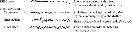
The electroencephalogram shows characteristic patterns which correspond to the type and death of sleep.
The sleep pattern
In adults non-REM and REM sleep alternate throughout the night.

In view of the important role of serotonin and noradrenaline (norepinephrine) in sleep, it is understandable that drugs may affect the duration and/or content of sleep.
NARCOLEPSY AND CATAPLEXY

Males are affected more than females. Prevalence 1:2000.
Onset is in adolescence/early adult life. The disorder is life long, but becomes less troublesome with age. It may have a familial incidence, or may occur after head injury, with multiple sclerosis, or with hypothalamic tumours. Pathological studies have found an early loss of hypothalamic neurons producing hypocretin/orexin, a wakefulness associated neurotransmitter.
Diagnosis
The suggestive history is supported by EEG studies. The multiple sleep latency test (MSLT) is diagnostic in showing onset of REM within 15 min of sleep onset in 2 of 4 naps (short sleeps).
Treatment
The non-amphetamine stimulant Modafinil, a wake promoting agent, reduces daytime sleepiness. Amphetamines are more potent but carry the risk of habituation. Sodium oxybate is a newer agent that improves night-time sleep and reduces cataplexy. Selegilene, metabolised in part to amphetamine, has a stimulant effect and may help. Clomipramine and SSRIs are also worth trying. Occasionally modifying life-style alone by ‘cat-napping’ is sufficient.
OTHER SLEEP DISORDERS (PARASOMNIAS)
NIGHT TERRORS (pavor nocturnus)
These occur in children, shortly after falling asleep and during deep to intermediate non-REM sleep. The child awakes in a state of fright with a marked tachycardia, yet in the morning cannot recollect the attack. Such attacks are not associated with psychological disturbance, are self limiting and if necessary will respond to diazepam.
NIGHTMARES
These occur during REM sleep. Drug or alcohol withdrawal promotes REM sleep and is often associated with vivid dreams.
SOMNAMBULISM (sleep walking)
Sleep walking varies from just sitting up in bed to walking around the house with the eyes open, performing complex major tasks. Episodes occur during intermediate or deep non-REM sleep. In childhood, somnambulism is associated with night terrors and bed wetting, but not with psychological disturbance. In adults, there is an increased incidence of psychoneurosis. Prevention of injury is important.
In REM sleep-behaviour disorder patients physically act out their dreams sometimes hurting themselves or their sleeping partner. This is associated with Parkinson’s disease and other dementias and may be the earliest symptom.
SLEEP STARTS (HYPNIC JERKS)
On entering sleep, sudden jerks of the arms or legs commonly occur and are especially frequent when a conscious effort is made to remain awake, e.g. during a lecture. This is a physiological form of myoclonus.
Other movement disorders in sleep: Restless legs, Dystonia, Bruxism (teeth grinding) and head banging.
HYPERSOMNIA
Rarely lesions (e.g. tumours or encephalitis) in the floor of the third ventricle may produce excessive sleepiness, often associated with diabetes insipidus.
Systemic disease such as hypothyroidism may result in hypersomnia, as may conditions which produce hypercapnia – chronic bronchitis, or primary muscle disease, e.g. dystrophia myotonica.

SLEEP APNOEA SYNDROMES
Respiratory rate fluctuates during REM sleep with occasional short episodes of apnoea. These are normal physiological events and are brief and infrequent.
Prolonged sleep apnoea results from central reduction of respiratory drive, a mechanical obstruction of the airway or a mixture of both.
|
Central causes: |
Mechanical causes: |
|
Brain stem medullary infarction or following cervical/foramen magnum surgery. |
Obesity. Tonsillar enlargement. |
|
Myxoedema. Acromegaly. |
When breathing ceases, the resultant hypercapnia and hypoxia eventually stimulate respiration.
Patients may present with daytime sleepiness, nocturnal insomnia and early morning headache. Snoring and restless movements are characteristic. In severe cases of sleep apnoea, hypertension may develop with right heart failure secondary to pulmonary arterial hypertension. Polycythaemia and left heart failure may ensue.
Evaluation requires sleep oximetry and video recording with low level illumination. Fall in oxygen saturation may be as much as 50%.
Treatment depends on aetiology. Mechanical airway obstruction should be relieved; drugs such as theophylline are occasionally helpful. Continuous positive airway pressure (CPAP) applied to the nose may help. Surgical reconstruction of palate and oropharynx is offered in extreme cases.
The Pickwickian syndrome: sleep apnoea associated with obesity, named after the Dickens’ fat boy who repeatedly fell asleep.
INSOMNIA
The most common sleep disorder, difficult to evaluate and of multiple causation including psychiatric, alcohol, drug related or due to systemic illness. Treatment depends on cause e.g. antidepressant.
HIGHER CORTICAL DYSFUNCTION
Specific parts of the cerebral hemispheres are responsible for a certain aspect of function. In normal circumstances these functions are integrated and the patient operates as a whole. Damage to part of the cortex will result in a characteristic disturbance of function. Interruption by disease of ‘connections’ between one part of the cortex and another will ‘disconnect’ function.
GENERAL ANATOMY
Brodmann, on the basis of histological differences, divided the cortex into 47 areas. Knowledge of these areas is not practical, though they are referred to often in some texts.
Six layers can be recognized in the cerebral cortex superficial to the junction with the underlying white matter.
The relative preponderance of each layer varies in different regions of the cortex and appears to be related to function.

The frontal motor cortex, dominated by pyramidal rather than granular layers, is termed the AGRANULAR CORTEX.
The parietal sensory cortex, dominated by granular layers, is termed the GRANULAR CORTEX.
The largest cells of the granular cortex are the giant cells of Betz. These give rise to some of the motor fibres of the corticospinal tract.
RIGHT AND LEFT HEMISPHERE FUNCTION
Unilateral brain damage reveals a difference in function between hemispheres. The left hemisphere is ‘dominant’ in right-handed people. In left-handed subjects the left hemisphere is dominant in the majority (up to 75%).
Hand preference may be hereditary, but in some cases disease of the left hemisphere in early life determines left-handedness.

Hemisphere dominance may be demonstrated by the injection of sodium amytal into the internal carotid artery. On the dominant side this will produce an arrest of speech for up to 30 seconds – the WADA TEST. Such a test may be important before temporal lobectomy for epilepsy when handedness/hemisphere dominance is in doubt.
FRONTAL LOBES

FRONTAL LOBE FUNCTION
1. Precentral gyrus – motor cortex contralateral movement – face, arm, leg, trunk.
2. Broca’s area – dominant hemisphere – expressive centre for speech.
3. Supplementary motor area – contralateral head and eye turning.
4. Prefrontal areas – ‘personality’, initiative.
5. Paracentral lobule – cortical inhibition of bladder and bowel voiding.
IMPAIRMENT OF FRONTAL LOBE FUNCTION
1. Precentral gyrus Monoplegia or hemiplegia depending on extent of damage.
2. Broca’s area (inferior part of dominant frontal lobe) Results in Broca’s dysphasia (see page 124) (motor or expressive).
3. Supplementary motor area Paralysis of head and eye movement to opposite side. Head turns ‘towards’ diseased hemisphere and eyes look in the same direction.

4. Prefrontal areas (the vast part of the frontal lobes anterior to the motor cortex as well as undersurface – orbital – of frontal lobes)
Damage is often bilateral, e.g. infarction, following haemorrhage from anterior communicating artery aneurysm, neoplasm, trauma or anterior dementia, resulting in a change of personality with antisocial behaviour/loss of inhibitions.
Three pre-frontal syndromes are recognised
|
Orbitofrontal syndrome |
Frontal convexity syndrome |
Medial frontal syndrome |
|
Disinhibition |
Apathy |
Akinetic |
|
Poor judgement |
Indifference |
Incontinent |
|
Emotional lability |
Poor abstract thought |
Sparse verbal output |
Pre-frontal lesions are also associated with:
1. Primitive reflexes – grasp, pout, etc. (see page 127).
2. Disturbance of gait – ‘gait apraxia’.
3. Resistance to passive movements of the limbs – paratonia.
Unilateral lesions may show minor degrees of such change.
5. Paracentral lobule
Damage to the posterior part of the superior frontal gyrus results in incontinence of urine and faeces – ‘loss of cortical inhibition’. This is particularly likely with ventricular dilatation and is an important symptom of normal pressure hydrocephalus.
PARIETAL LOBES
PARIETAL LOBE FUNCTION
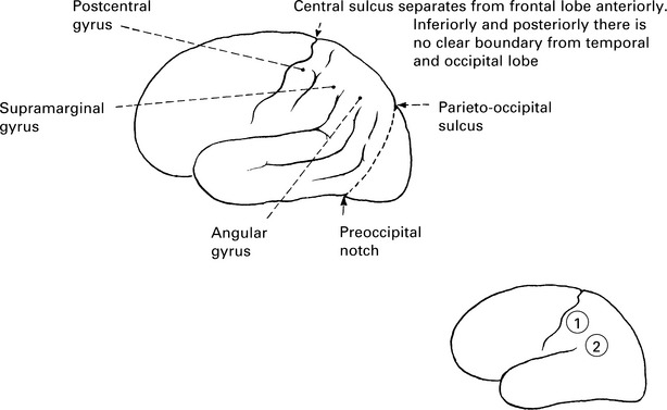
1. Postcentral gyrus (granular cortex)
The sensory cortex (representation similar to the motor cortex) receives afferent pathways for appreciation of posture, touch and passive movement.
2. Supramarginal and angular gyri (dominant hemisphere) make up part of Wernicke’s language area.
This is the receptive area where auditory and visual aspects of comprehension are integrated.
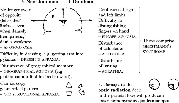
The non-dominant parietal lobe is important in the concept of body image and the awareness of the external environment. The ability to construct shapes, etc. results from such visual/proprioceptive skills.
The dominant parietal lobe is implicated in the skills of handling numbers/calculation.
The visual pathways – the fibres of the optic radiation (lower visual field) – pass deep through the parietal lobe.
IMPAIRMENT OF PARIETAL LOBE FUNCTION
Disease of either dominant or non-dominant sensory cortex (postcentral gyrus) will result in contralateral disturbance of cortical sensation:
Postural sensation disturbed.
Sensation of passive movement disturbed.
Accurate localization of light touch may be disturbed.
Discrimination between one and two points (normally 4 mm on finger tips) is lost.
Appreciation of size, shape, texture and weight may be affected, with difficulty in distinguishing coins placed in hand, etc. (astereognosis).
Perceptual rivalry (sensory inattention) is characteristic of parietal lobe disease. Presented with two stimuli, one applied to each side (e.g. light touch to the palm of the hand) simultaneously, the patient is only aware of that one contralateral to the normal parietal lobe.
As the gap between application of stimuli is increased (approaching 2–4 seconds) the patient becomes aware of both.
2. Supramarginal and angular gyri – Wernicke’s dysphasia (see page 124).
TEMPORAL LOBES
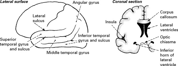
Anteriorly, the temporal lobe is separated from the frontal lobe by the lateral sulcus. Posteriorly and superiorly, separation from occipital and parietal lobes is less clearly defined.
The lateral sulcus is deep and contains ‘buried’ temporal lobe. The buried island of cortex is referred to as the INSULA.
The temporal lobe also has a considerable inferior and medial surface in contact with the middle fossa.

TEMPORAL LOBE FUNCTION
1. The auditory cortex lies on the upper surface of the superior temporal gyrus, buried in the lateral sulcus (Heschl’s gyrus).
The dominant hemisphere is important in the hearing of language.
The non-dominant hemisphere is important in the hearing of sounds, rhythm and music. Close to the auditory cortex labyrinthine function is represented.
2. The middle and inferior temporal gyri are concerned with learning and memory (see later).
3. The limbic lobe: the inferior and medial portions of the temporal lobe, including the hippocampus and parahippocampal gyrus.
The sensation of olfaction is mediated through this structure as well as emotional/affective behaviour.
Olfactory fibres terminate in the uncus.
The limbic lobe or system also incorporates inferior frontal and medial parietal structures and will be discussed later.
4. The visual pathways pass deep in the temporal lobe around the posterior horn of the lateral ventricle.
IMPAIRMENT OF TEMPORAL LOBE FUNCTION
1. Auditory cortex
Cortical deafness: Bilateral lesions are rare but may result in complete deafness of which the patient may be unaware.
Lesions which involve surrounding association areas may result in difficulty in hearing spoken words (dominant) or difficulty in appreciating rhythm/music (non-dominant) – AMUSIA. Auditory hallucinations may occur in temporal lobe disease.
2. Middle and inferior temporal gyri
Disturbance or memory/learning will be discussed later.
Disordered memory may occur in complex partial seizures either after the event – postictal amnesia – or in the event – déjà vu, jamais vu.
3. Limbic lobe damage may result in:
Olfactory hallucination with complex partial seizures.
Aggressive or antisocial behaviour.
Inability to establish new memories (see later).

4. Damage to optic radiation will produce an upper homonymous quadrantanopia. Dominant hemisphere lesions are associated with Wernicke’s dysphasia.
OCCIPITAL LOBE

The striate cortex is the primary visual cortex and when stimulated by visual input relays information to the parastriate – association visual cortex. This, in turn, connects with the parietal, temporal and frontal lobes both on the same side and on the opposite side (through the posterior part of the corpus callosum) so that the meaning of a visual image may be interpreted, remembered, etc.
The visual field is represented upon the cortex in a specific manner (page 140).
IMPAIRMENT OF OCCIPITAL LOBE FUNCTION
A cortical lesion will result in a homonymous hemianopia with or without involvement of the macula, depending on the posterior extent of the lesion.
When only the occipital pole is affected, a central hemianopia field defect involving the macula occurs with a normal peripheral field of vision.
Cortical blindness
Extensive bilateral cortical lesions of the striate cortex will result in cortical BLINDNESS. In this, the pupillary light reflex is normal despite the absence of conscious perception of the presence of illumination (light reflex fibres terminate in the midbrain).
Anton’s syndrome
Involvement of both the striate and the parastriate cortices affects the interpretation of vision. The patient is unaware of his visual loss and denies its presence. This denial in the presence of obvious blindness characterizes Anton’s syndrome.
Cortical blindness occurs mainly in vascular disease (posterior cerebral artery), but also following hypoxia and hypertensive encephalopathy or after surviving tentorial herniation.
Balint’s syndrome
Inability to direct voluntary gaze, associated with visual agnosia (loss of visual recognition) due to bilateral parieto-occipital lesions.
Visual hallucinations are common in migraine when the occipital lobe is involved; also in epilepsy when the seizure source lies here.
Hallucinations of occipital origin are elementary – unformed – appearing as patterns (zig-zags, flashes) and fill the hemianopic field, whereas hallucinations of temporal lobe origin are formed, complex and fill the whole of the visual field.
Visual illusions also may occur as a consequence of occipital lobe disease. Objects appear smaller (MICROPSIA) or larger (MACROPSIA) than reality. Distortion of a shape may occur or disappearance of colour from vision.
These illusions are more common with non-dominant occipital lobe disease.
Prosopagnosia: the patient, though able to see a familiar face, e.g. a member of the family, cannot name it. This is usually associated with other disturbances of ‘interpretation’ and naming with intact vision such as colour agnosia (recognition of colours and matching of pairs of colours). Bilateral lesions at occipito-temporal junction are responsible.
APRAXIA
A loss of ability to carry out skilled movement despite adequate understanding of the task and normal motor power.
Constructional and dressing apraxia: See page 113, non-dominant parietal disease.
Gait apraxia: Difficulty in initiating walking – frontal lobe/anterior corpus callosum disease.
Oculomotor apraxia: Impaired voluntary eye movement – parieto-occipital disease.
Ideamotor apraxia: Separation of idea of movement from execution – cannot carry out motor command but can perform the required movement under different circumstances – dominant hemisphere (see later).
Ideational apraxia: Inability to carry out a sequence of movements each of which can be performed separately – frontal lobe disease.
HIGHER CORTICAL DYSFUNCTION – DISCONNECTION SYNDROMES
Cortical function is described, on the previous pages, ‘lobe by lobe’. These functions integrate by means of connections between hemispheres and lobes. Lesions of these connecting pathways disorganise normal function, resulting in recognizable syndromes – the disconnection syndromes. APRAXIA is a feature of some of these disorders.
The connecting pathways may be divided into:
Intra hemispheric: lying in the subcortical white matter and linking parts of the same hemisphere.
Inter hemispheric: traversing the corpus callosum and linking related parts of the two hemispheres.
THE INTRAHEMISPHERIC DISCONNECTION SYNDROMES

THE INTERHEMISPHERIC DISCONNECTION SYNDROMES

HIGHER CORTICAL FUNCTION MEMORY
Normal memory involves the recognition, registering and cataloguing of a stimulus – acquisition, as well as the skill of appropriate recall – retrieval.
|
Verbal memory: |
refers to material presented in the verbal form. |
|
Visual memory: |
denotes material presented without words or verbal mediation. |
|
Episodic memory: |
|
|
Short term: |
immediate recall of a short message. |
|
Long term: |
retrieval of recent or remote events. |
|
Semantic memory: |
refers to long established factual knowledge. |
Disordered memory may be confused with disturbances of attention, motivation and concentration and requires detailed neuropsychological examination to properly assess.
THE ANATOMICAL BASIS OF MEMORY
The structures of the limbic system involved in the memory process are inferred from the pathological examination of diseases that disorder function. The hippocampus, a deep structure in the temporal lobe, ridges the floor of the lateral ventricle. Fimbriae of the hippocampus connect this structure to the fornix. There appears to be a loop from hippocampus → fornix → mamillary body → thalamus → cingulate gyrus → back to hippocampus.


TESTS OF MEMORY (see examination, page 8)
These aim to distinguish loss of immediate, recent or remote memory.
Disorders may be further classified into those which affect memories established before the injury or damage – RETROGRADE AMNESIA – and those which affect memory of events following the injury or damage – ANTEROGRADE or POST-TRAUMATIC AMNESIA.
DISORDERS OF MEMORY
THE AMNESIC SYNDROME is characterised by –
Retrograde amnesia – impairment of memory for events that antedate illness or injury
Anterograde amnesia – inability to learn new verbal or non verbal information from onset of the illness or injury
Intact retrieval of old information
Intact intellectual function
Intact personality
Tendency to confabulate
CAUSES
Korsakoff’s syndrome: results from – alcoholism, encephalitis, and head injury
Lesions occur within the thalamus and the mamillary bodies. Commonly associated with confabulation – a false rationalization of events and circumstances.
Post-traumatic amnesia: after trauma, retrograde amnesia may span several years, but with recovery, this gradually diminishes. The duration of post-traumatic amnesia on the other hand remains fixed and relates directly to the severity of the injury.
Amnesic stroke: bilateral medial temporal lobe infarction from a posterior circulation stroke is usually associated with hemiplegia and visual disturbance or loss e.g. Anton’s or Balint’s syndrome (page 115).
Amnesia with tumours: tumours that compress thalamic structures or the fornix may produce amnesia – e.g. colloid cyst of the 3rd ventricle.
Temporal lobectomy: amnesia will only occur if function in the unoperated temporal lobe is abnormal. Pre-operative assessment during a unilateral carotid injection of sodium amytal minimises this risk.
Transient global amnesia: typically a single episode lasting between 1 and 10 hours; the patient is bewildered, typically repeatedly asking the same questions, but with clear consciousness and often able to carry out complex tasks such as driving or cooking. Benign phenomenon probably associated with migraine. May be triggered by stress or exercise.
Transient epileptic amnesia: recurrent episodes of amnesia lasting 15 minutes to 1 hour, often on waking.
Psychogenic amnesia: affects overlearned and personally relevant aspects of memory e.g. ‘What is my name?’, while less well learned memory remains unaffected. Clinically evident acute mental stress may precipitate this. This inadequate defence mechanism suggests a serious underlying psychiatric or personality disorder.
DISORDERS OF MEMORY RETRIEVAL
Senescence – as part of normal aging, rapid retrieval of stored memory becomes defective.
Depression – impaired memory is a common complaint in depressive illness. The disorder is one of motivation and concentration.
Subcortical dementia – This will be described later (page 126). The major abnormality is that of a slowed (but correct) response rate to questions of memory function.
NB DEMENTIA, TUMOURS and CEREBROVASCULAR DISEASE are all often associated with memory loss but this is usually combined with evidence of more widespread disordered cognitive function.
DISORDERS OF SPEECH AND LANGUAGE
Introduction
Disturbed speech and language are important symptoms of neurological disease. The two are not synonymous. Language is a function of the dominant cerebral hemisphere and may be divided into (a)emotional – the instinctive expression of feelings representing the earliest forms of language acquired in infancy and (b) symbolic or prepositional – conveying thoughts, opinion and concepts. This language is acquired over a 20-year period and is dependent upon culture, education and normal cerebral development.
An understanding of disorders of speech and language is essential, not just to the clinical diagnosis but also to improve communication between patient and doctor. All too often patients with language disorders are labelled ‘confused’ as a consequence of superficial evaluation.
DYSARTHRIA
Dysarthria is a disturbance of articulation in which the content of speech – language – is unaffected.

DISORDERS OF SPEECH – DYSARTHRIA
DIAGNOSTIC APPROACH
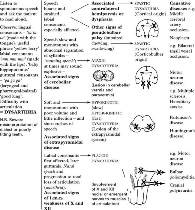
Many diseases affect multiple sites and a ‘mixed’ dysarthria occurs.
For example, multiple sclerosis with corticobulbar and cerebellar involvement will result in a mixed spastic/ataxic dysarthria.
DISORDERS OF SPEECH – DYSPHONIA
Sound is produced by the passage of air over the vocal cords.
Respiratory disease or vocal cord paralysis results in a disturbance of this facility – dysphonia. A complete inability to produce sound is referred to as aphonia. Dysarthria often co-exists.
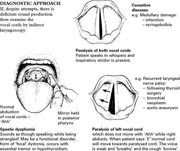
OTHER DISORDERS OF SPEECH
Mutism: An absence of any attempt at oral communication. It may be associated with bilateral frontal lobe or third ventricular pathology (see Akinetic mutism).
Echolalia: Constant repetition of words or sentences heard in dementing illnesses.
Palilalia: Repetition of last word or words of patient’s speech. Heard in extrapyramidal disease.
Logorrhoea: Prolonged speech monologues; associated with Wernicke’s dysphasia.
DISORDERS OF SPEECH – DYSPHASIA
Dysphasia is an acquired loss of production or comprehension of spoken and/or written language secondary to brain damage.
Hand preference is associated with ‘hemisphere dominance’ for language. In right-handed people the left hemisphere is dominant; in left-handed people the left hemisphere is dominant in most, though 25% have a dominant right hemisphere.
The cortical centres for language reside in the dominant hemisphere.

Receptive and expressive areas must be linked in order to integrate function. The link is provided by (4), the arcuate fasciculus, a fibre tract which runs forward in the subcortical white matter.
Dysphasia may develop as a result of vascular, neoplastic, traumatic, infective or degenerative disease of the cerebrum when language areas are involved.
DIAGNOSTIC APPROACH

DEMENTIAS
Definition
Progressive deterioration of intellect, behaviour and personality as a consequence of diffuse disease of the cerebral hemispheres, maximally affecting the cerebral cortex and hippocampus.
Distinguish from delirium which is an acute disturbance of cerebral function with impaired conscious level, hallucinations and autonomic overactivity as a consequence of toxic, metabolic or infective conditions.
Dementia may occur at any age but is more common in the elderly, increasing with age (approximate prevalence 1% in 60s, 5% in 70s, 15% in 80s). Dementia is a symptom of disease rather than a single disease entity. When occurring under the age of 65 years it is labelled ‘presenile’ dementia. This term is artificial and does not suggest a specific aetiology.
Clinical course:
The rate of progression depends upon the underlying cause.
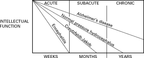
The duration of history helps establish the cause of dementia; Alzheimer’s disease is slowly progressive over years, whereas encephalitis may be rapid over weeks. Dementia due to cerebrovascular disease appears to occur ‘stroke by stroke’.
All dementias show a tendency to be accelerated by change of environment, intercurrent infection or surgical procedures.
Development of symptoms
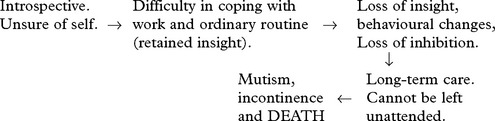
This initial phase of dementia may be inseparable from the pseudodementia of depressive illness.
DEMENTIAS – CLASSIFICATION
Based on cause

It is important to investigate all patients with dementia as many causes are treatable in practice 10–15% can be reversed.
Based on site
Subdividing dementia depending upon the site of predominant involvement is useful in clinical classification but has only limited value in predicting underlying pathology:

DEMENTIAS – HISTORY AND CLINICAL EXAMINATION
When obtaining a history from a patient with dementia and relative or carer, establish:
– Rate of intellectual decline – Nutrition status
– Impairment of social function – Drug history
– General health and relevant disorders, e.g. stroke, head injury
– Nutrition status
– Drug history
– Family history of dementia.
|
Tests to assess intellectual function are designed to check |
The Mini Mental Status Examination (MMSE) |
|
|
– memory – abstract thought – judgement – specific focal cortical functions |
Date orientation Place orientation Register 3 objects Obeying verbal command |
Serial sevens Naming Repeating |
|
Obeying written command Writing/drawing This is the standard tool of evaluation. Top Score = 30; score >24 normal; <24 suggests dementia Folstein at el J. Psych Res 12:196–198 1975 |
||
On neurological examination note:
– Focal signs
– Involuntary movements
– Pseudobulbar signs
– Primitive reflexes:

DEMENTIAS – SPECIFIC DISEASES
ALZHEIMER’S DISEASE
This is the commonest cause of dementia with an estimated half million sufferers in the UK. The disorder rarely occurs under the age of 45 years. The incidence increases with age. Up to 30% of cases are familial.
Pathology
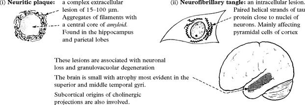
Diagnosis This may be established during life by early memory failure, slow progression and exclusion of other causes. Specific clinical criteria have been established, mainly for research purposes. Whilst certain blood tests can identify populations at risk (i.e. APOE genotyping) these are of no diagnostic value in individual cases.
CT scanning: aids diagnosis by excluding multiple infarction or a mass lesion.

Causation
The cause of Alzheimer’s disease is not known. Some genetic forms have been identified. The most common are mutations in the presenilin-1 gene (on chromosome 14). Patient’s with Down’s syndrome (trisomy 21) develop Alzheimer’s pathology. The role of environmental toxins, especially aluminium, is uncertain. Early research suggested selective lesions of neurotransmitter pathways occurred and a disorder of cholinergic innervation was postulated. It is now known that many neurotransmitter pathways are defective.
Treatment
Centrally acting drugs such as acetylcholinesterase inhibitors (e.g. Donepezil, Rivastigmine, Galantamine) have been shown in trials to enhance cognitive performance in early disease. However they do not cure. Memantine is an NMDA antagonist that also provides some symptomatic relief.
DEMENTIAS – SPECIFIC DISEASES
MULTI-INFARCT (arteriosclerotic dementia)
This is an overdiagnosed condition which accounts for less than 10% of cases of dementia.
Dementia occurs ‘stroke by stroke’, with progressive focal loss of function. Clinical features of stroke profile – hypertension, diabetes, etc. – are present.
Diagnosis is obtained from the history and confirmed by CT scan.
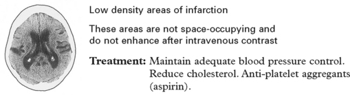
FRONTOTEMPORAL DEMENTIA
This progressive condition accounts for 5% of all dementias, but about 20% of those under age of 65. There are three clinical patterns of presentation that are associated with differing areas of focal atrophy:
Behavioural variant – frontal lobe atrophy – change in personality; impaired judgement; apathy; stereotyped behaviours; loss of appropriate emotional response. Relatively preserved memory.
Progressive non-fluent aphasia – dominant temporal lobe atrophy – loss of verbal fluency, relatively preserved understanding.
Sematic dementia – bilateral temporal lobe atrophy – loss of knowledge of the meaning of words, and knowledge about the world.
The pathology is heterogeneous some have tau-positive inclusions (including Pick’s disease) while others do not, some of whom have ubiquitin inclusions.
About 40% of patients have a family history of dementia and a number of the responsible genes have been identified, the most common being the progralulin (PRGN) mutation.
Cerebral autosomal dominant arteriopathy with subcortical infarcts and leukoencephalopathy (CADASIL)
This inherited disorder presents with migraine (often hemiplegic) in early adult life, progessing through TIAs and subcortical strokes to early dementia. The advent of MRI with its characteristic appearance has led to increasing recognition of what was previously only identified at autopsy. CADASIL has been mapped to the ‘Notch 3’ gene on chromosome 19 in many (though not all) cases allowing diagnostic testing. The role of the gene is uncertain and specific treatments not available.
AIDS DEMENTIA COMPLEX (see pages 515–516)
Approximately two-thirds of persons with AIDS develop dementia, mostly due to AIDS dementia complex. In some patients HIV is found in the CNS at postmortem. In others an immune mechanism or an unidentified pathogen is blamed. Dementia is initially of a ‘subcortical’ type. CT shows atrophy; MRI shows increased T2 signal from white matter. Imaging excludes other infections and neoplastic causes of intellectual decline.
Treatment with Zidovudine (AZT) halts and partially reverses neuropsychological deficit.
METABOLIC DEMENTIA
General medical examination is important in suggesting underlying systemic disease. B12 deficiency may produce dementia rather than subacute combined degeneration of the spinal cord. In alcoholics, consider not only Wernicke Korsakoff syndrome but also chronic subdural haematoma.
NORMAL PRESSURE HYDROCEPHALUS
Normal pressure hydrocephalus (NPH) is the term applied to the triad of:
|
Dementia |
occurring in conjunction with |
|
Gait disturbance |
hydrocephalus and normal |
|
Urinary incontinence |
CSF pressure. |
Two types occur:
|
– NPH with a preceding cause |
– subarachnoid haemorrhage – meningitis – trauma – radiation-induced |
(This must be distinguished from hydrocephalus with raised intracranial pressure associated with these causes.)
– NPH with no known preceding cause – idiopathic (50%).
Aetiology is unclear. It is presumed that at some preceding period, impedence to normal CSF flow causes raised intraventricular pressure and ventricular dilatation. Compensatory mechanisms permit a reduction in CSF pressure yet the ventricular dilatation persists and causes symptoms:

Normal pressure hydrocephalus must be differentiated from patients whose ventricular enlargement is merely the result of shrinkage of the surrounding brain, e.g. Alzheimer’s disease. These patients do not respond to CSF shunting, whereas a proportion of patients with NPH (but not all) show a definitive improvement with shunting.
Investigations
Numerous tests have been assessed to predict those most likely to benefit from operation.
The most frequently used are –

Other tests include the presence of periventricular lucency or disproportionate sulcal width on CT scan, isotope cisternography and CSF infusion studies but predictive accuracy is low. Some believe that the risks of treatment are not warranted in the ‘ideopathic’ group.
Operation: Ventriculo-peritoneal shunting; although a small procedure, not without risk (see page 377).
Results: Improvement occurs in 50–70% of those patients with a known preceding cause e.g. subarachnoid haemorrhage. At best, 30% of the idiopathic group respond to shunting.
TRAUMA
Reduction of intellectual function is common after severe head injury. Chronic subdural haematoma can also present as progressive dementia, especially in the elderly. Punch-drunk encephalopathy(dementia pugilistica) is the cumulative result of repeated cerebral trauma. It occurs in both amateur and professional boxers and is manifest by dysarthria, ataxia and extrapyramidal signs associated with ‘subcortical’ dementia. There is no treatment for this progressive syndrome.
TUMOUR presenting as dementia
Concern is always expressed at the possibility of dementia being due to intracranial tumour. This is rare, but may happen when tumours occur in certain sites.
Mental or behavioural changes occur in 50–70% of all brain tumours as distinct from dementia which is associated with frontal lobe tumours (and subfrontal tumours), III ventricle tumours andcorpus callosum tumours.

DEMENTIA – DIAGNOSTIC APPROACH
It is neither practical nor essential to perform all the screening tests in every patient with dementia. The presenting features should guide investigations.

When the reason for dementia is unclear, comprehensive investigation is essential to ensure that treatable nutritional, infective, metabolic and structural causes are not overlooked.
ANATOMY AND PHYSIOLOGY
Anatomically the visual system is contained in the supratentorial compartment. It is composed of peripheral receptors in the retina, central pathways and cortical centres. The control of ocular movement and pupillary responses are closely integrated.
The retina: three distinct layers of the retina are identified:

The macular region of the retina is its most important area for visual acuity. Here, cones lie in the greatest concentration whereas rods are more numerous in the surrounding retina.
The optic nerve leaves the orbit through the optic foramen and passes posteriorly to unite with the opposite optic nerve at the optic chiasma. Here, partial decussation occurs (axons from ganglion cells on the nasal side of the retina cross over to the opposite side).
The optic tract consisting of ipsilateral temporal and contralateral nasal fibres passes to the lateral geniculate body. A few fibres leave the tract before the lateral geniculate body and pass to thesuperior colliculus (fibres concerned with pupillary light reflex).
Axons of cell bodies in the lateral geniculate body make up the optic radiation. This enters the hemisphere in the most posterior part of the internal capsule, courses deep in parietal and temporal lobes and terminates in the calcarine cortex of the occipital lobe.
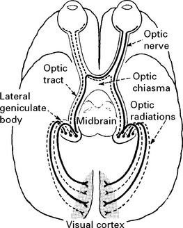
IMPAIRMENT OF VISION
CLINICAL APPROACH AND DIFFERENTIAL DIAGNOSIS
Patients presenting with visual impairment require a systematic examination, not only of vision, but also of the pupillary response, eye movements, and, unless the cause clearly lies within the globe, a full neurological examination.
The findings aid localisation of the lesion, e.g.

Refractive errors are excluded by testing visual acuity through a pinhole or by correcting a lens deformity (page 9).
Four types of refractive error exist:
PRESBYOPIA – failure of accommodation with age
HYPERMETROPIA (long sightedness) – short eyeball
MYOPIA (short sightedness) – long eyeball
ASTIGMATISM – variation in corneal curvature
If this examination is normal, then the lesion lies in the retina, visual pathways or visual cortex.
Examine the globe and anterior chamber

Examine the lens with an ophthalmoscope
Opacification indicates CATARACT.
CLINICAL APPROACH AND DIFFERENTIAL DIAGNOSIS
Examine the posterior segment of the eye with an ophthalmoscope. Pupil dilatation may be required.
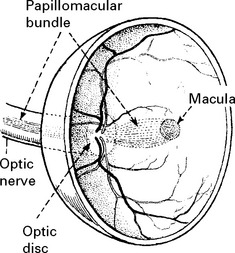
In the normal fundus, the disc is pale with a central cup and reddish-brown surrounding retina. Arteries and veins emerge from the optic disc. The macula is darker than the rest of the fundus and lies on the temporal side of the disc. One-third of all retinal fibres arise from the small macular region and pass to the optic nerve head (disc) as the papillomacular bundle. The macula is the region of sharpest vision (cone vision), whereas peripheral vision (rod vision) serves the purpose of perception of movement and directing central/macular vision. The optic nerve head contains no rods or cones and accounts for the physiological blind spot in normal vision. The macular fibres being so functionally active, are the most susceptible to damage and produce a specific defect in the visual field – a scotoma.
Retinal abnormality with acute impairment of vision
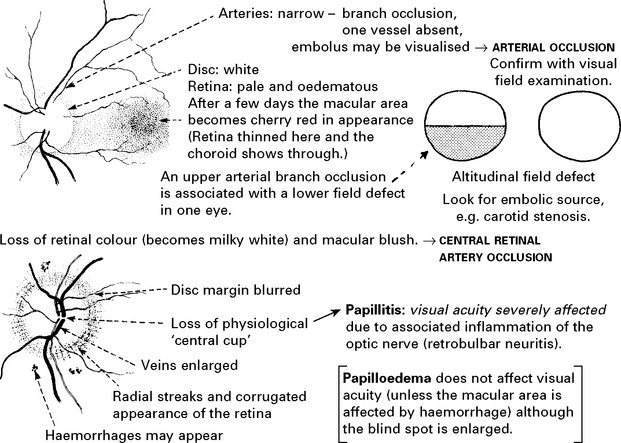
N.B. Distinguish:
HYPERMETROPIC patients who have a pale indistinct disc often difficult to differentiate from early papilloedema.
HYPERTENSIVE RETINOPATHY – superficial haemorrhages and ‘cotton wool’ exudates.
PSEUDOPAPILLOEDEMA – ‘DRUSEN’ – hyaline bodies near the optic disc which raise the disc and blur the margin. This normal variant may be inherited.
Separation of the superficial retina from the pigment layer → RETINAL DETACHMENT (traumatic or spontaneous)
Retinal abnormalities with gradual impairment of vision
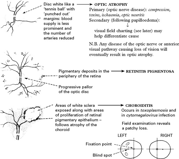

Examine the visual fields
If ophthalmoscopic examination is normal, or if optic atrophy is evident, then visual field examination is essential. Visual confrontation is useful for detecting large defects, but smaller defects require visual field charting with a Goldmann perimeter (page 10).
In interpreting the results of examination it is important to remember that the ocular system reverses the image. The nasal side of the fundus picks up the temporal image and vice versa. Damage, therefore, to the nasal side of the retina will produce a temporal visual field defect.

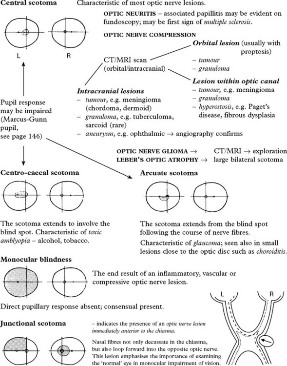
Bitemporal hemianopia/quadrantanopia
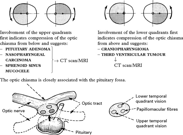
Homonymous hemianopia
An incongruous homonymous hemianopia (i.e. one eye more affected than the other) suggests a compressive lesion of the optic tract near the chiasma.

Congruous homonymous hemianopia (fields can be exactly superimposed)

At the temporo-parietal junction where fibres meet, lesions produce a complete ‘homonymous hemianopia’.

The interpretation of the visual image and its integration with other cortical functions is discussed under ‘Higher cortical function’.
DISORDERS OF SMELL
OLFACTORY (I) cranial nerve conveys the sensation of smell.

Differential diagnosis

OLFACTORY HALLUCINATIONS – occur in complex partial seizures and migraine.
PUPILLARY DISORDERS
ANATOMY/PHYSIOLOGY

Pathway of pupillary constriction and the light reflex (parasympathetic)

A stimulus, such as a bright light shone in the left eye, will send an afferent impulse along the optic nerve to the midbrain (superior colliculus); here a second order fibre passes to the Edinger-Westphal nucleus (part of the III nerve nucleus) on the same and opposite side (through the posterior commissure). Efferent fibres leave in the oculomotor nerve, pass to the ciliary ganglion and thence, in the short ciliary nerve, to the constrictor fibres of the sphincter pupillae muscle.
If all pathways are intact, shining a light in one eye will constrict both pupils at an equal rate and to a similar degree.
Pathway of pupillary dilatation (sympathetic)
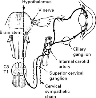
Sympathetic fibres descend from the ipsilateral hypothalamus through the lateral aspect of the brain stem into the spinal cord. The pupillary fibres pass out in the anterior roots of C8 and T1, enter the sympathetic chain and, in the superior cervical ganglion, give rise to postganglionic fibres which ascend on the wall of the internal carotid artery to enter the cranium. The fibres eventually leave the intracranial portion of the internal carotid artery and pass directly through the ciliary ganglion to the iris or join the cranial nerves III, IV, V and VI, running to the eye and iris. Sudomotor fibres (concerned with sweating) run up the external carotid artery to the dermis of the face.
Interruption of sympathetic supply affects:
1. Dilator pupillae causing a small pupil (miosis)
2. Levator palpebrae muscle (30% supplied by sympathetic) causing drooping of eyelid (ptosis)
3. Vasoconstrictor fibres to orbit, eyelid and face causing absence of sweating.
Interruption of parasympathetic supply affects:
Sphincter pupillae causing a large pupil (mydriasis)
Mechanism of accommodation
When gaze is focused on a near object the medial rectus muscles contract, producing convergence, the ciliary muscles contract enabling the lens to produce a more convex shape and the pupil constricts (accommodation for near vision).
The pathway is poorly understood but must involve the visual cortex, Edinger-Westphal nuclei and both medial rectus components of the III nerve nucleus in the midbrain.
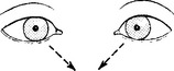
Inability of the pupil to constrict during accommodation need not always be associated with impairment of convergence, though usually this is the case.
Pupillary inequality (anisocoria)
A difference in pupil size occurs in 20% of the normal population and is distinguished from pathological states by a normal response to bright light.
PUPIL DILATATION – CAUSES
III nerve lesion
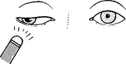
Examination of the light reflex (page 11) distinguishes lesions of the optic (II) and oculomotor (III) nerves. Failure of the pupil to constrict when light is shone into either the affected or the contralateral eye indicates a lesion of the parasympathetic component of the III nerve.
Look for – ptosis – 70% of levator palpebrae muscle is supplied by the oculomotor nerve
– impaired eye movements.
Causes of a III nerve lesion are described on page 153.
In comatose patients, pupil dilatation and failure to react to light is the simplest way of detecting a III nerve lesion; after head injury or in patients with raised intracranial pressure this is an important sign of transtentorial herniation.
The tonic pupil – Adie’s pupil
This is a benign condition usually affecting young women. Onset is usually acute and unilateral in 80%.
The pupil dilates and the patient complains of mistiness in the affected eye.

Occasionally the pupil appears completely unreactive to both light and accommodation. When the pupil is associated with reduced or absent limb reflexes this is termed the Holmes-Adie syndrome. More widespread autonomic dysfunction – orthostatic hypotension, segmental disturbance of sweating and diarrhoea can co-exist.
Diagnosis: confirmed by pupillary response to pilocarpine (0.1% or 0.05%) – the tonic pupil will constrict (denervation hypersensitivity); the normal eye is not affected.
The cause is unknown; the lesion probably lies in the midbrain or ciliary ganglion.
Migraine: Mydriasis persisting for some hours can accompany headache.
Drugs: Mydriasis occurs with anticholinergic drugs (atropine), tricyclic antidepressants, non-steroidal anti-inflammatories, antihistamines and oral contraceptives. Mydriasis can precipitate an attack of acute angle-closure glaucoma.
PUPIL CONSTRICTION – CAUSES
Horner’s syndrome
MIOSIS: the affected pupil is smaller than the opposite pupil. It does not dilate when the eye is shaded.

PTOSIS: the affected eyelid droops and may be slightly raised voluntarily. Ptosis is less marked than with a III nerve palsy.
DISTURBANCE OF SWEATING: depends on the site of the lesion. Absence of sweating occurs when the lesion is proximal to fibre separation along the internal and external carotid arteries.
Horner’s syndrome may result from sympathetic damage at the following sites:

The congenital or familial form exists, often associated with lack of pigmentation of the iris. The lesion site is unknown.
Distinguish peripheral and central lesions by instilling drugs, e.g. 1% cocaine in eyes.

Investigative approach: depends on associated signs. Chest X-ray is mandatory to exclude an apical lung tumour.
The Argyll-Robertson pupil
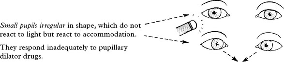
The Argyll-Robertson pupil has also been described in diabetes and in alcoholic neuropathy as well as following infectious mononucleosis. The lesion could lie in the midbrain, involving fibres passing to the Edinger-Westphal nucleus, in the posterior commissure, or alternatively, in the ciliary ganglion. A central lesion seems most likely.
Investigative approach: – look for associated signs of neurosyphilis – blood serology – VDRL, Captia G.
Drugs
Parasympathomimetic drugs – Carbachol, phenothiazines and opiates produce miosis.
N.B. Do not confuse with small pupils, normally occurring in the elderly.
OTHER PUPILLARY DISORDERS
Failure of accommodation and convergence
Impaired accommodation and convergence are of limited diagnostic value since other clinical features are usually more prominent
Causes – extrapyramidal disease, e.g. Parkinson’s
– tumours of the pineal region.
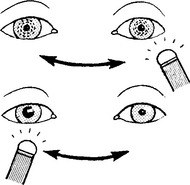
The Marcus Gunn pupil (pupillary escape)
Illumination of one eye normally produces pupillary constriction with a degree of waxing and waning (hippus).
When afferent transmission in the optic nerve is impaired, this ‘escape’ becomes more evident.
If the light source is ‘swung’ from eye to eye, dwelling 2–3 seconds on each, the affected pupil may eventually, paradoxically, dilate – a ‘Marcus Gunn’ pupil.
The swinging light test is a sensitive test of optic nerve damage but is also abnormal in retinal or macular disease.
DIPLOPIA – IMPAIRED OCULAR MOVEMENT
Diplopia or double vision results from impaired ocular movement.
RELATED ANATOMY AND PHYSIOLOGY

The line of action of individual ocular muscles
Eye movements result from a continuous interplay of all the ocular muscles, but each muscle has a direction of maximal efficiency. The oblique muscles move the eye up and down when it is turned in. The superior and inferior recti move the eye up and down when it is turned out.
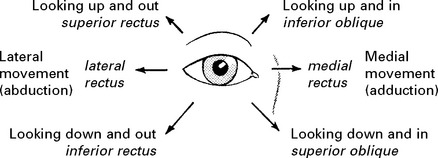
Eye movements are examined in the six different directions of gaze representing individual muscle action.
As a result of the angle of insertion into the globe, the inferior and superior recti and the oblique muscles also have a rotatory or torsion effect.

When the eye is turned out, the oblique muscles rotate the globe; when turned in, the inferior or superior recti rotate the globe.
OCULOMOTOR (III) nerve
The oculomotor nucleus lies in the ventral periaqueductal grey matter at the level of the superior colliculus. Nerve fibres pass through the red nucleus and substantia nigra and emerge medial to thecerebral peduncle.
The nucleus has a complex structure:
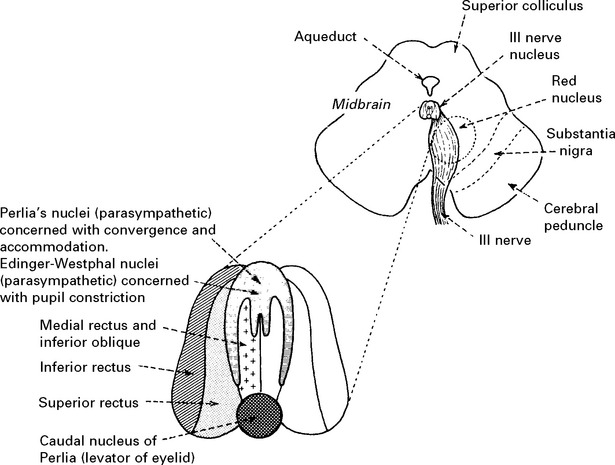
The nucleus is a paired structure which lies close to the midline, the portion representing the medial rectus abutting its neighbour.
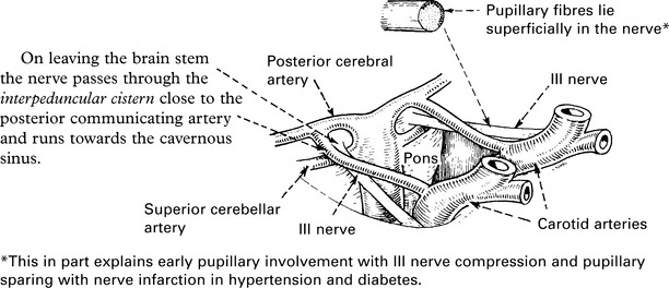

Here it divides into:
1. Superior branch to the levator of the eyelid and the superior rectus.
2. Inferior branch to the inferior oblique, medial and inferior recti.
TROCHLEAR (IV) nerve
This nerve supplies the superior oblique muscle of the eye.
The nucleus lies in the midbrain at the level of the inferior colliculus, near the ventral periaqueductal grey matter. The nerve passes laterally and dorsally around the central grey matter and decussates in the dorsal aspect of the brain stem in close proximity to the anterior medullary velum of the cerebellum.
Emerging from the brain stem the nerve passes laterally around the cerebral peduncle and pierces the dura to lie in the lateral wall of the cavernous sinus. Finally, it passes through the superior orbital fissure into the orbit.
ABDUCENS (VI) nerve
This nerve supplies the lateral rectus muscle of the eye.
The nucleus lies in the floor of the IV ventricle within the lower portion of the pons. The axons pass ventrally through the pons without decussating.
Note the close association of the VI and VII nuclei.

Emerging from the brain stem the nerve runs up anterior to the pons for approximately 15 mm before piercing the dura overlying the basilar portion of the occipital bone.

Under the dura the nerve runs up the petrous portion of the temporal bone and from its apex passes on to the lateral wall of the cavernous sinus and finally through the superior orbital fissure.
Note the long intracranial course and the proximity of the VI to the V cranial and greater superficial petrosal nerves at the apex of the petrous temporal bone.
DIPLOPIA
When the eyes fix on an image, impairment of movement of one eye results in projection of the image upon the macular area in the normal eye and to one side of the macula in the paretic eye; two images of the single object are thus perceived.

The image seen by the paretic eye is the false image; that seen by the normal eye is the true image. The false image is always outermost; this may lie in the vertical or the horizontal plane.
CLINICAL ASSESSMENT

III NERVE LESION

(Ptosis may be complete, unlike the partial ptosis of a Horner’s syndrome which disappears on looking up.)
IV NERVE LESION

VI NERVE LESION

Diplopia is horizontal (true and fake image side by side), is present only when looking to the paralysed side and is maximal at the extreme of binocular lateral vision.
NOTE: In partial oculomotor palsies, the patient may be aware of diplopia, although eye movements appear normal. When this occurs:
– check diplopia is ‘true’ by noting its disappearance on covering one eye.
– determine the direction of maximal image displacement and the eye responsible for the outermost image (see page 13).
This information is sufficient to differentiate a III, IV and VI nerve lesion.
OCULAR MUSCLES
If the limitation of eye movement is not restricted to one muscle, or group of muscles with a common innervation, and affects both eyes, look for:

CAUSES OF III NERVE LESION
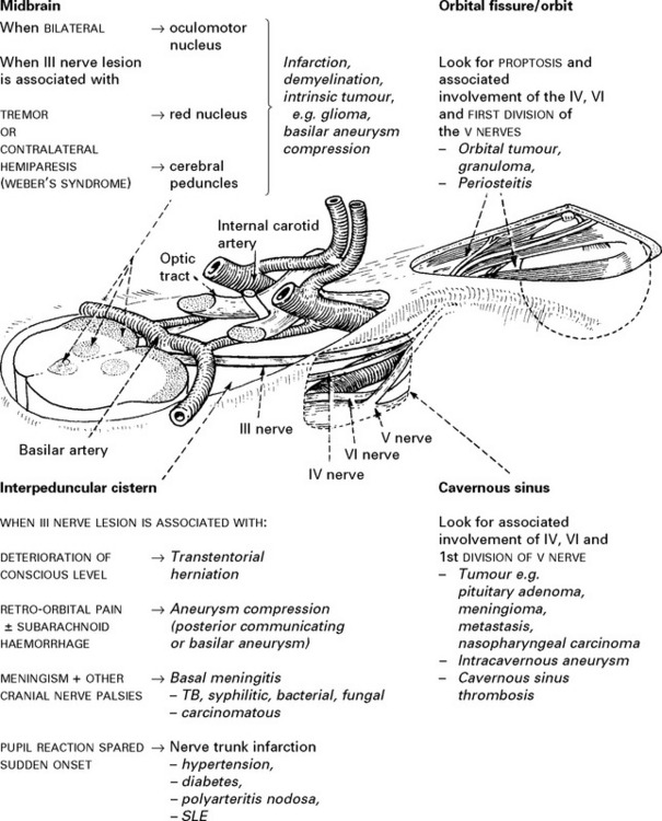
CAUSES OF IV AND VI NERVE LESIONS

NOTE: Infective or carcinomatous meningitis and nerve trunk infarction may also involve the IV and VI nerves, although less often than the III nerve.
Investigative approach
III, IV or VI nerve lesions require investigation with MRI (or CT); a III nerve lesion needs urgent investigation with CT/MR angiography to look for an enlarging aneurysm. Further investigation withinflammatory markers and CSF cytology, as directed by clinical circumstances. Elderly hypertensive or diabetic patients with complete pupillary sparing III nerve lesions will not need vascular imaging. Prognosis will depend on cause.
If myopathy or myasthenia gravis is suspected then acetyl choline receptor antibodies, EMG studies and perhaps muscle biopsy may be needed.
DISORDERS OF GAZE
ANATOMY AND PHYSIOLOGY
Two cortical centres of ocular control are recognised:
1. Middle gyrus of frontal lobe (frontal eye field).
2. Occipital cortex.

Note that the cortical descending pathways from one side activate the ipsilateral III nucleus and the contralateral VI nucleus thus swinging the direction of gaze to the opposite side.
It is important to distinguish between saccadic and pursuit movement. When following an object a slow pursuit movement maintains the image on the macular area of the retina. To fixate on a new object, rapid saccadic movement aligns the new target on the macular area. When locked into the new target, pursuit movement maintains fixation.
Eye movement occurs voluntarily in a conjugate (parallel) manner in any direction. Eye movements also occur reflexly to labyrinthine stimulation – the vestibular ocular reflex.
Gaze disorders usually follow vascular episodes (infarct or haemorrhage) but may also occur in traumatic, inflammatory or neoplastic disease. In gaze palsy eye movements are symmetrically limited in one direction.
CONJUGATE DEVIATION OF THE EYES

VERTICAL GAZE PALSY
Midbrain or pontine lesions may produce failure of upward or downward gaze. Disturbed downward gaze alone occurs with periaqueductal (Sylvian aqueduct) lesions. Impaired vertical eye movement is common in extrapyramidal disease (Progressive supranuclear palsy, page 366).
PARINAUD’S SYNDROME
This syndrome is characterised by impaired upward eye movements in association with a dorsal midbrain lesion (+).

– upward gaze and convergence are lost
– the pupils may dilate and the response to light and accommodation is impaired
Causes:
Third ventricular tumours
Pineal region tumours
Hydrocephalus
Multiple sclerosis
Wernicke’s encephalopathy
Encephalitis
INTERNUCLEAR OPHTHALMOPLEGIA (ataxic nystagmus)
This disorder, caused by damage to the medial longitudinal bundle, is dealt with on page 186. It is an internuclear disorder of eye movement and produces a disconjugate gaze palsy.

OCULAR APRAXIA
Bilateral prefrontal motor cortex damage will produce this unusual finding in which the patient does not move the eyes voluntarily to command, yet has a full range of random eye movement.
FACIAL PAIN AND SENSORY LOSS
The fifth cranial nerve subserves facial sensation and innervates the muscles of mastication.
Anatomy
The anatomical arrangement of the trigeminal central connections are complex.
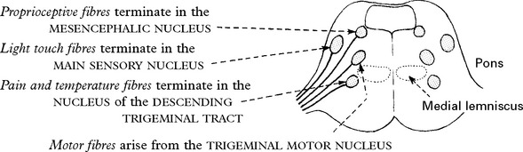
The separate location of the main sensory nucleus and nucleus of the descending trigeminal tract account for dissociated sensory loss, i.e. a low pontine or medullary lesion will result in loss of pain and temperature sensation with preservation of light touch.
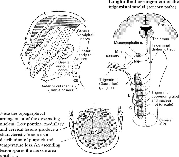
The peripheral course of the V nerve
The motor and sensory nerve roots emerge separately from the lateral aspect of the brain stem at the midpontine level. The Gasserian ganglion of the sensory root contains bipolar sensory nuclei and lies on the apex of the petrous bone in the middle fossa. Here the three divisions of the trigeminal nerve merge. Each passes through its own foramen and carries sensation from a specific area of the face.
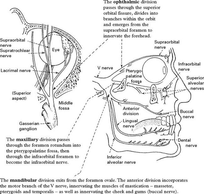
The lingual branch of the posterior trunk innervates the anterior two-thirds of the tongue (and is joined by the chordi tympani from the facial nerve carrying salivary secretomotor fibres and taste from the anterior two-thirds of the tongue).
EXAMINATION OF TRIGEMINAL NERVE FUNCTION
This should include examination of the corneal reflex and masticatory muscle function (page 14).

CAUSES OF V NERVE LESIONS

Sensory trigeminal neuropathy:
Progressive, painless loss of trigeminal sensation. Normally unilateral and without trigeminal motor weakness, the sensory loss may affect one or all trigeminal divisions. This condition is often associated with established connective tissue disease (scleroderma, Sjögren’s syndrome and mixed connective tissue disease (MCTD)). Diagnosis requires exclusion of intracranial granuloma and tumour compressing the trigeminal nerve – meningioma, schwannoma, epidermoid – by contrast enhanced MRI.
Mental neuropathy (numb chin syndrome):
Caused by a lesion of the mandibular nerve or inferior alveolar or mental branches, usually the result of metastatic compression of the nerve within the mandible. Bone scans or an enhanced CT/MRI combined with image-guided aspiration is diagnostic.
Infraorbital neuropathy (numb cheek syndrome) has similar etiology.
Gradenigo’s syndrome:
Lesions located at the petrous-temporal bone apex (osteitis or meningitis associated with otitis media) irritate the ophthalmic division of the trigeminal and abducens (VI) nerve. Forehead pain is accompanied by ipsilateral lateral rectus palsy and a Horner’s syndrome if sympathetic fibres are also involved. Tumours and trauma can also produce this syndrome.
Neuropathic keratitis
Corneal anaesthesia from a central or peripheral V nerve lesion may lead to a neuropathic keratitis. The corneal surface becomes hazy, ulcerated and infected and blindness may follow.
Patients with absent corneal sensation should wear a protective shield, attached to the side of spectacles, when out of doors.
FACIAL PAIN – DIAGNOSTIC APPROACH
Pain in the face may result from many different disorders and often presents as a diagnostic problem to the neurologist or neurosurgeon.
Consider:
1. Site of pain

2. Quality of pain
|
Trigeminal neuralgia Atypical facial pain Postherpetic neuralgia Dental Sinusitis Ocular Costen’s syndrome Cluster headache |
– sharp, stabbing, shooting, paroxysmal – dull, persisting – dull, burning, persisting, occasional paroxysm – dull – sharp, boring, worse in the morning – dull, throbbing – severe aching, aggravated by chewing – sharp, intermittent |
3. Associated symptoms/signs
|
Trigeminal neuralgia Atypical facial pain Postherpetic neuralgia Dental Sinusitis Ocular Costen’s syndrome Cluster headache |
– often no neurological deficit, but occasional blunting of pinprick over involved region – accompanying features of depressive illness – evidence of scarring associated with sensory loss – swelling of lips/face – puffy appearance around eyes, tenderness to percussion over involved sinus – glaucoma: associated visual symptoms – blurring/haloes/loss – tenderness over temporomandibular joint – associated lacrimation/rhinorrhoea |
Investigations
– guided by clinical suspicion
|
Blood tests: |
ESR, FBC, biochemistry. |
|
Imaging: |
CT/MRI, dental X-rays, isotope bone scan. |
FACIAL PAIN – TRIGEMINAL NEURALGIA
TRIGEMINAL NEURALGIA (tic douloureux)
Trigeminal neuralgia is characterised by paroxysmal attacks of severe, short, sharp, stabbing pain affecting one or more divisions of the trigeminal nerve. The pain involves the second or third divisions more often than the first; it rarely occurs bilaterally and never simultaneously on each side, occasionally more than one division is involved. Paroxysmal attacks last for several days or weeks; they are often superimposed on a more constant ache. When the attacks settle, the patient may remain pain free for many months.
Chewing, speaking, washing the face, tooth-brushing, cold winds, or touching a specific ‘trigger spot’, e.g. upper lip or gum, may all precipitate an attack of pain.

Trigeminal neuralgia more commonly affects females and patients over 50 years of age.
Aetiology
Trigeminal pain may be symptomatic of disorders which affect the nerve root or its entry zone.
Root or root entry zone compression
– arterial vessels often abut and sometimes clearly indent the trigeminal nerve root at the entry-zone into the pons, causing ephaptic transmission (short circuiting).
– tumours of the cerebellopontine angle lying against the V nerve roots, e.g. meningioma, epidermoid cyst, frequently present with trigeminal pain.
Demyelination – such a lesion in the pons should be considered in a ‘young’ person with trigeminal neuralgia. Trigger spots are rare. Remission occurs infrequently and the response to drug treatment is poor.
In some patients the cause remains unexplained, as do the long periods of remission.
Investigation
MR scan to exclude a cerebello-pontine angle lesion or demyelination.
Management
Drug therapy
CARBAMAZEPINE proves effective in most patients (and helps confirm the diagnosis). Provided toxicity does not become troublesome, i.e. drowsiness, ataxia, the dosage is increased until pain relief occurs (600–1600 mg/day). When remission is established, drug treatment can be discontinued.
If pain control is limited, other drugs – BACLOFEN, LAMOTRIGINE, GABAPENTIN, PHENYTOIN – may benefit.
Persistence of pain on full drug dosage or an intolerance of the drugs, indicates the need for more radical measures.
The choice lies between a range of lesional techniques, which all produce some damage to the trigeminal nerve with some consequent sensory loss, or microvascular decompression, which does not damage the nerve but has the risks associated with open neurosurgery.

Results and complications
Pain relief – no comparative trials have been done so accurate comparison of the wide variety of techniques used for trigeminal neuralgia is difficult. Microvascular decompression seems to be more likely to provide pain control with fewer relapses. Overall 80–85% of patients remain pain free for a 5-year period. Results of peripheral nerve avulsion are less satisfactory with pain recurring in 50% within 2 years.
Dysaesthesia/Anaesthesia dolorosa – this troublesome sensory disturbance follows any destructive technique to nerve or root in 5–30% of patients. Microvascular decompression avoids this.
Corneal anaesthesia – this occurs when root section or thermocoagulation involves the first division and keratitis may result.
Mortality – microvascular decompression and open root section carry a very low mortality (< 1%), but this must not be ignored when comparing results with safer methods.
Treatment selection: This depends on discussion of the differing risks with the patient. In younger patients the absence of sensory complications make microvascular decompression the procedure of first choice. Frail and elderly patients may tolerate glycerol injection, balloon compression and thermocoagulation more easily than other procedures.
FACIAL PAIN – OTHER CAUSES
Temporomandibular joint dysfunction (Costen’s syndrome)

Raeder’s syndrome (the paratrigeminal syndrome)
Pain and sensory loss in 1st and 2nd trigeminal divisions, maximal around the eye and associated with a sympathetic paresis (ptosis and small pupil). Sweating in the lower face is preserved. This may be associated with involvement of the other cranial nerves (IV & VI). This rare syndrome occurs with lesions of the middle fossa, e.g. nasopharyngeal carcinoma, granulomas and infection.
Tolosa Hunt syndrome

Atypical facial pain
The patient, often a young or middle-aged woman, experiences a dull, persistent pain, spreading diffusely over one or both sides of the face. These symptoms often result from an underlying depression and may respond well to antidepressant therapy.
Herpes zoster
Frequently affects the trigeminal territory, especially the ophthalmic division producing a painful ‘herpetic rash’ and often involving the cornea. The acute symptoms may resolve but lead to a chronic postherpetic neuralgia which slowly improves. Surgical procedures such as trigeminal root section do not help. The incidence of postherpetic neuralgia is not influenced by treatment with antiviral agents (acyclovir) in the acute phase.
Carotid artery dissection
This presents as acute retro-orbital pain with a Horner’s syndrome (page 145) and may be associated with ipsilateral amaurosis fugax or contralateral hemisphere symptoms.
‘Cluster’ headaches – see page 73.
FACIAL WEAKNESS
Related anatomy
The facial (VII) nerve contains mainly motor fibres supplying the muscles of facial expression, but also visceral efferent (parasympathetic) and visceral afferent (taste) fibres.
The motor nucleus lies in the lower pons medial to the descending nucleus and tract of the Vth cranial nerve. Axons from the motor nucleus wind around the nucleus of the VIth cranial nerve. The facial nerve and its visceral root (nervus intermedius) exit from the lateral aspect of the brain stem and cross the cerebellopontine angle immediately adjacent to the VIII cranial nerve. They enter the internal auditory meatus and, passing through the facial canal of the temporal bone, lie in close proximity to the inner ear and tympanic membrane. The facial nerve gives off several branches before exiting from the skull through the stylomastoid foramen.
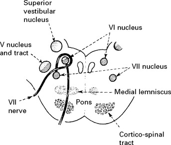

Visceral efferent and visceral afferent fibres arise and terminate in the superior salivary nucleus and nucleus/tractus solitarius respectively.
They run together as the nervus intermedius and accompany the facial nerve to the internal auditory meatus. The parasympathetic fibres (visceral efferent) pass in the greater petrosal nerve to the sphenopalatine ganglion and thence to the lacrimal gland to produce tears and in the chorda tympani nerve to the submandibular ganglion.

Supranuclear control of facial muscles
The muscles in the lower face are controlled by the contralateral hemisphere, whereas those in the upper face receive control from both hemispheres (bilateral representation). Hence a lower motor neuron lesion paralyses all facial muscles on that side, but an upper motor neuron (supranuclear) lesion paralyses only the muscles in the lower half of the face on the opposite side.
Clinical examination of the facial nerve (see page 15)
In addition to examining for facial weakness and taste impairment, also note whether the patient comments on reduced lacrimation or salivation on one side, or hyperacusis (exaggeration of sounds due to loss of the stapedius reflex).
LESION, LOCALISATION AND CAUSE
Note the distribution:
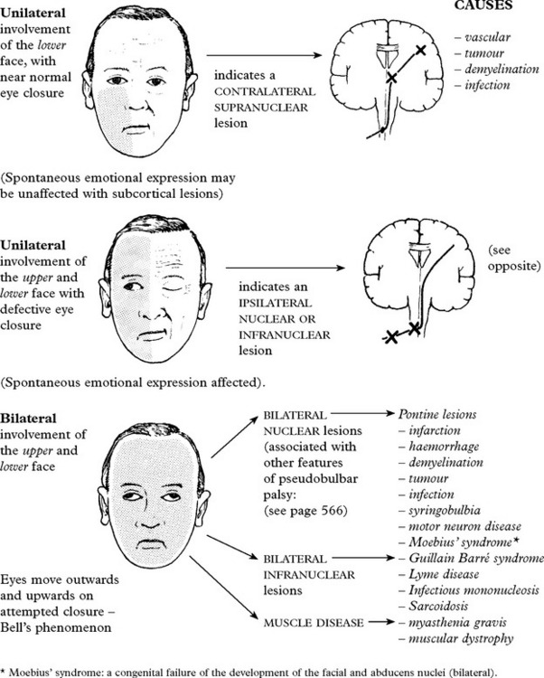
NUCLEAR/INFRANUCLEAR LESIONS
The following features (if present) help in lesion location:

BELL’S PALSY
Bell’s palsy is characterised by an acute paralysis of the face related to ‘inflammation’ and swelling of the facial nerve within the facial canal or at the stylomastoid foramen. It is usually unilateral, rarely bilateral, and may occur repetitively. In some, a family history of the condition is evident. Incidence 25/100,000/year.
Aetiology
Uncertain, but may be associated with viral infections, e.g. herpes simplex and varicella-zoster; epidemics of Bell’s palsy occur sporadically.
Symptoms
Pain of variable intensity over the ipsilateral mastoid precedes weakness, which develops over a 48-hour period.
Impairment of taste, hyperacusis and salivation depend on the extent of inflammation and will be lost in more severe cases. Lacrimation is seldom affected.
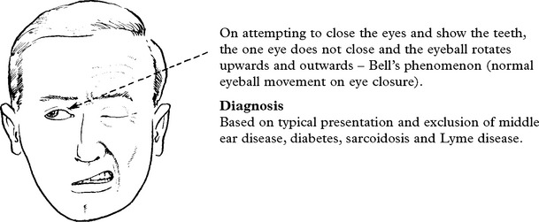
Treatment
During the acute stage protect the exposed eye during sleep.
There is good evidence prednisolone given in high dosage in the acute stage (50 mg per day for 10 days) improves recovery. The role of antiviral therapy is less clear as conflicting results have been found in recent large trials. Eye care (shielding and artificial tears) is important in preventing corneal abrasion.
Prognosis
Most patients (70%) recover in 4–8 weeks without treatment. In the remainder, residual facial asymmetry may require corrective surgery. Incomplete paralysis indicates a good prognosis. In patients with complete paralysis, electrical absence of denervation on electromyography is an optimistic sign.
Occasionally aberrant reinnervation occurs – movement of the angle of the mouth on closing the eyes (jaw winking) or lacrimation when facial muscles contract (crocodile tears).
OTHER FACIAL NERVE DISORDERS
RAMSAY HUNT SYNDROME
Herpes zoster infection of the geniculate (facial) ganglion causes sudden severe facial weakness with a typical zoster vesicular eruption within the external auditory meatus. Pain is a major feature and may precede the facial weakness. Serosanguinous fluid may discharge from the ear.
Deafness may result from VIII involvement. Occasionally, other cranial nerves from V–XII are affected.
Treatment
Antiviral agents (acyclovir) may help.
HEMIFACIAL SPASM
This condition is characterised by unilateral clonic spasms beginning in the orbicularis oculi and spreading to involve other facial muscles. The stapedius muscle can be affected producing a subjective ipsilateral clicking sound.
Contractions are irregular, intermittent and worsened by emotional stress and fatigue.
Onset usually occurs in middle to old age and women are preferentially affected.
Most cases arise from vascular compression of the facial nerve at the root entry zone (in the same way as trigeminal neuralgia). In some, compression is caused by a tumour. Occasionally hemifacial spasm follows a Bell’s palsy or traumatic facial injury.
The clinician must distinguish hemifacial spasm from milder habit spasms or tics which tend to be familial, and also from ‘focal’ seizures selectively affecting the face.
Investigations
MR scan of the posterior fossa excludes the presence of a cerebellar pontine angle lesion and may show an ectatic basilar artery.
Treatment
Drugs – Local infiltration with botulinum toxin of involved muscles is helpful. However, effect only lasts about 3 months and may produce temporary weakness.
Surgery – Posterior fossa exploration and microvascular decompression i.e. dissecting blood vessels off the facial nerve root entry zone, gives excellent results (cure rate 80%), but carries the risk of producing deafness and rarely brain stem damage.
TONIC FACIAL SPASM
Less common than hemifacial spasm. Occurs with cerebellar pontine angle lesions. It produces tonic elevation of the corner of the mouth with narrowing of the eye. The diagnosis is confirmed by CT/MR scanning and treatment is surgical.
FACIAL MYOKYMIA
A rare condition seen most often in multiple sclerosis. Flickering of facial muscles results from spontaneous discharge in the facial motor nucleus. Other brain stem signs are present. The facial movements respond to carbamazepine.
MYOCLONUS
Rhythmic facial movement associated with similar palatal movements and characteristic of dentate or olivary nucleus disease.
BLEPHAROSPASM
Spasmodic closing or screwing up of eyes (see page 371).
DEAFNESS, TINNITUS AND VERTIGO
Deafness, tinnitus and vertigo result from disorders affecting the auditory and vestibular apparatus or their central connections transmitted through the VIII cranial nerve.
MECHANISMS OF AUDITORY AND VESTIBULAR FUNCTION
|
Auditory function: the cochlea converts sound waves into action potentials in cochlear neurons. Sound waves are transmitted by the tympanic membrane and the ossicles to the oval window, setting up waves in the perilymph of the cochlea. The action of the waves on the spiral organ (of Corti) generates action potentials in the cochlear division of the VIII cranial nerve. |
Vestibular function: the vestibular system responds to rotational and linear acceleration (including gravity) and along with a visual and proprioceptive input maintains equilibrium and body orientation in space. Relative inertia of the endolymph within the semicircular canals during angular acceleration displaces hair cells imbedded in the cupula, activates the hair cells and transmits action potentials to the vestibular division of the VIII cranial nerve. Linear acceleration results in displacement of the otoliths within the utricle or saccule. This distorts the hair cells and increases or decreases the frequency of action potentials in the vestibular division of the VIII cranial nerve. |
CENTRAL CONNECTIONS

First order vestibular neurons lie in the vestibular division of the VIII nerve and relay information from the utricle, saccule and semicircular canals to the vestibular nuclei (superior, inferior, medial and lateral). Bipolar cell bodies lie in the vestibular ganglion.
The cochlear (acoustic) and vestibular divisions travel together through the petrous bone to the internal auditory meatus where they emerge to pass through the subarachnoid space in the cerebellopontine angle, each entering the brain stem separately at the pontomedullary junction.
Auditory: From the cochlear nucleus, second order neurons either pass upwards in the lateral lemniscus to the ipsilateral inferior colliculus or decussate in the trapezoid body and pass up in the lateral lemniscus to the contralateral inferior colliculus.
Third order neurons from the inferior colliculus on each side run to the medial geniculate body on both sides.
Fourth order neurons pass through the internal capsule and auditory radiation to the auditory cortex.
The bilateral nature of the connections ensures that a unilateral central lesion will not result in lateralised hearing loss.
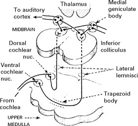
Vestibular

1. Directly to cerebellum.
2. Second order neurons arise in the vestibular nucleus and descend in the ipsilateral vestibulospinal tract.
3. Second order neurons project to the oculomotor nuclei (III, IV, VI) through the medial longitudinal fasciculus.
4. Second order neurons project to the cortex (temporal lobe). The pathway is unclear.
5. Second order neurons project to the cerebellum.
(There is a bilateral feedback loop to the vestibular nuclei from the cerebellum though the fastigial nucleus.)
DEAFNESS: Three types of hearing loss are recognised:
1. Conductive deafness: failure of sound conduction to the cochlea.
2. Sensorineural deafness: failure of action potential production or transmission due to disease of the cochlea, cochlear nerve or cochlear central connections.
Further subdivision into cochlear and retrocochlear deafness helps establish the causative lesion.
3. Pure word or cortical deafness: a bilateral or dominant posterior temporal lobe (auditory cortex) lesion produces a failure to understand spoken language despite preserved hearing.
TINNITUS: a sensation of noise of ringing, buzzing, pulsing, hissing or singing quality.
Tinnitus may be (i) continuous or intermittent, (ii) unilateral or bilateral, (iii) high or low pitch.
As a rule, when hearing loss is accompanied by tinnitus, conductive deafness is associated with low pitch tinnitus – sensorineural deafness is associated with high pitch tinnitus, except Menière’s disease where tinnitus is low pitch. Pulsing tinnitus may have a vascular cause. In most patients no cause is found.
VERTIGO: an illusion of rotatory movement due to disturbed orientation of the body in space. The sufferer may sense that the environment is moving. Vertigo may result from disease of the labyrinth, vestibular nerve or their central connections.
Clinical examination
Examination of the external auditory meatus, tympanic membrane and eye movements (for nystagmus) and Weber’s and Rinne’s tests (page 16) provide valuable information, but more detailed neuro-otological tests (pages 62, 63) are usually required to determine the exact nature of the auditory or vestibular dysfunction and to locate the lesion site. The results of these tests may indicate the need for further investigation (e.g. CT/MR scan).

Causes of tinnitus
Any lesion causing deafness may also cause tinnitus. Occasionally patients perceive a vibratory noise inside the head, transmitted from an arteriovenous malformation or carotid stenosis. A lesion is more likely with unilateral tinnitus. No cause is found in most patients with bilateral tinnitus.
Patients with non-specific disease, e.g. anaemia, fever, hypertension, occasionally complain of tinnitus.
DISORDERS OF THE LOWER CRANIAL NERVES
NINTH (GLOSSOPHARYNGEAL) CRANIAL NERVE
This is a mixed nerve with motor, sensory and parasympathetic functions.
1. Motor fibres to stylopharyngeus muscle arise in the nucleus ambiguus.
2. Preganglionic parasympathetic fibres arise in the inferior salivatory nucleus and pass to the otic ganglion. From there postganglionic fibres innervate the parotid gland.
3. General somatic sensory fibres innervate the area of skin behind the ear, pass to the superior ganglion and end in the nucleus and tract of the trigeminal nerve.
4. Sensory fibres innervate the posterior third of the tongue (taste), pharynx, eustachian tube and carotid body/sinus and terminate centrally in the nucleus solitarius. The cell bodies lie in the inferior ganglion.
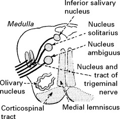

The IX nerve emerges as 5 or 6 rootlets from the medulla, dorsal to the olivary nucleus and passes with the vagus and accessory nerves through the jugular foramen in the neck.
Within the neck the nerve lies in close proximity to the internal carotid artery and internal jugular vein.
The superior and inferior ganglia lie in the jugular foramen, the otic ganglion in the neck below the foramen ovale.
Clinical examination (see page 17)
Disorders of the glossopharyngeal nerve
Glossopharyngeal palsy from either medullary or nerve root lesions does not occur in isolation. When associated with X and XI cranial nerve lesions, this constitutes the jugular foramen syndrome. Lesions producing this syndrome are listed on page 179.
GLOSSOPHARYNGEAL NEURALGIA
Short, sharp, lancinating attacks of pain, identical to trigeminal neuralgia in nature but affecting the posterior part of the pharynx or tonsillar area. The pain often radiates towards the ear and is triggered by swallowing. Reflex bradycardia and syncope occur due to stimulation of vagal nuclei by discharges from glossopharyngeal. As with trigeminal neuralgia, carbamazepine often provides effective relief – if not microvascular decompression or section of the IX nerve roots or nerve give good results.
TENTH (VAGUS) CRANIAL NERVE
This is a mixed nerve with motor, sensory and parasympathetic functions.
The central connections are complex though similar to those of the glossopharyngeal nerve.
1. Motor fibres supplying the pharynx, soft palate and larynx arise in the nucleus ambiguus.
2. Preganglionic parasympathetic fibres arise in the dorsal motor nucleus. Postganglionic fibres supply the thoracic and abdominal viscera.
3. Afferent fibres from the pharynx, larynx and external auditory meatus have cell bodies in the jugular ganglion and end in the nucleus and tract of the trigeminal nerve.
4. Afferent fibres from abdominal and thoracic viscera have cell bodies in the nodose ganglion and end in the nucleus solitarius. Taste perception in the pharynx ends similarly.
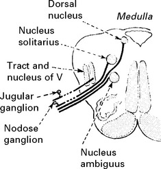
The nerve emerges from the brain stem as a series of converging rootlets. It exits from the cranial cavity by the jugular foramen where both ganglia lie.
Extracranial branches:
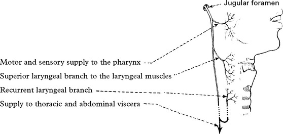
Disorders of the vagus nerve cause:
Palatal weakness
Unilateral – minimal symptoms.
Bilateral – nasal regurgitation of fluid, nasal quality of speech.

Clinical examination (see page 17)
Direct examination of the vocal cords helps identification of the lesion site.

Causes (see page 179)
ELEVENTH (ACCESSORY) CRANIAL NERVE
This is a purely motor nerve supplying the sternomastoid and trapezius muscles.
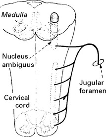
The cranial portion of the accessory nerve arises from the lowest part of the nucleus ambiguus in the medulla.
The spinal part arises in the ventral grey matter of the upper five cervical segments, ascends alongside the spinal cord and passes through the foramen magnum. After joining with the cranial portion it exits as the accessory nerve through the jugular foramen.
The supranuclear connections act on the ipsilateral sternomastoid (turning the head to the contralateral side) and on the contralateral trapezius. This results in:
– head turning away from the relevant hemisphere during the seizure
– head turning towards the relevant hemisphere with cerebral infarction.
Unilateral lower motor neuron weakness produces a lower shoulder on the affected side (trapezius) and weakness in turning the head to the opposite side (sternomastoid).
Clinical examination (see page 17) Causes (see page 179)
TWELFTH (HYPOGLOSSAL) CRANIAL NERVE
This is a purely motor nerve which supplies the intrinsic muscles of the tongue.
The nucleus lies in the floor of the IV ventricle and fibres pass ventrally to leave the brain stem lateral to the pyramidal tract.

Since each nucleus is bilaterally innervated, a unilateral supranuclear lesion will not produce signs or symptoms. A bilateral supranuclear lesion results in a thin pointed (spastic) tongue which cannot be protruded.
A lesion of the hypoglossal nerve results in atrophy and deviation of the tongue to the weak side
Clinical examination (see page 18) Causes (see page 179)
CAUSES OF LOWER CRANIAL NERVE PALSIES
Lower cranial nerve palsies seldom occur in isolation. Investigations include CT or MR imaging of the skull base. If negative, specific tests for systemic causes and EMG (for nerve and muscle disease) may be required.

CEREBELLAR DYSFUNCTION
Anatomy
The cerebellum lies in the posterior fossa, posterior to the brain stem, separated from the cerebrum above by the tentorium cerebelli.
The cerebellum consists of two laterally placed hemispheres and the midline structure – the vermis.
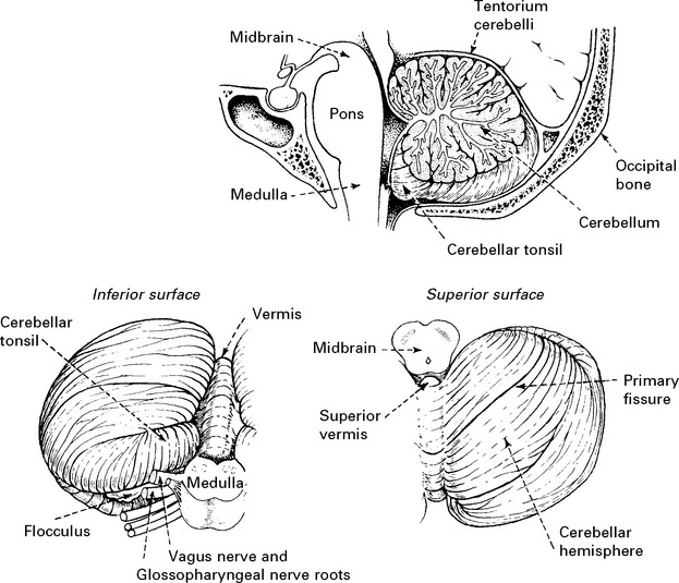
Three major phylogenetic subdivisions of the cerebellum are recognised.

The cerebellar cortex is made up of three cell layers. The middle or Purkinje layer contains Purkinje cells. These are the only neurons capable of transmitting efferent impulses. Deep within the cerebellar hemispheres in the roof of the 4th ventricle, lie four paired nuclei separated by white matter from the cortex.

The efferent system

The afferent system
Connections between the vestibular system and the cerebellum are described on page 173.
The spinocerebellar pathways form a major afferent input. These transmit ‘subconscious’ proprioception from muscles, joints and skin – especially of the lower limbs.
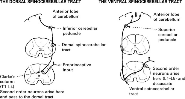
The cerebellar peduncles: Three peduncles connect the cerebellum to the brain stem:
Superior peduncle – afferent and efferent fibres.
Middle peduncle – afferent fibres only.
Inferior peduncle – afferent and efferent fibres.
SYMPTOMS AND SIGNS OF CEREBELLAR DYSFUNCTION
The close relationship of structures within the posterior fossa makes the identification of exclusively cerebellar symptoms and signs difficult. Disease of the brain stem and its connections may produce identical results.
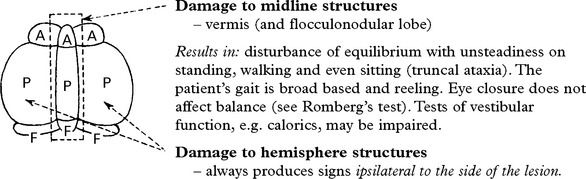
Results in: a loss of the normal capacity to modulate fine voluntary movements. Errors or inaccuracies cannot be corrected. The patient complains of impaired limb co-ordination and certain signs are recognised:
Ataxia of extremities with unsteadiness of gait towards the side of the lesion.
Dysmetria: a breakdown of movement with the patient ‘overshooting’ the target when performing a specific motor task, e.g. finger-to-nose test.
Dysdiadochokinesia: a failure to perform a rapid alternating movement.
Intention tremor: a tremor which increases as the limb approaches its target.

Eye movements
Nystagmus results from disease affecting cerebellar connections to the vestibular nuclei.
In unilateral disease, amplitude and rate increase when looking towards the diseased side.
Other ocular signs may occur, e.g. ocular dysmetria – an ‘overshoot’ when the eyes voluntarily fixate.
Disturbance of speech
Scanning dysarthria (where the same emphasis is put on each syllable like scanning a poem) may occur with speech occasionally delivered with sudden unexpected force – explosive speech. Whether dysarthria results from hemisphere or midline vermis disease remains debatable.
Dysarthria, like nystagmus, is an inconsistent finding in cerebellar disease.
Titubation
Titubation is a rhythmic ‘nodding’ tremor of the head from side to side or to and fro, usually associated with distal limb tremor. It appears to be of little localising value.
Head tilt
Abnormal head tilt suggests a lesion of the anterior vermis. Note that a IV (trochlear) cranial nerve palsy and tonsillar herniation also produce this abnormal posture.
Involuntary movements
Myoclonic jerks and choreiform involuntary movements occur with extensive cerebellar disease involving the deep nuclei.
NOTE: Cerebellar lesions may cause symptoms and signs relating to
– obstructive hydrocephalus
– cranial nerve involvement
– brain stem involvement.
(Extensor spasms from brain stem damage may be wrongly described as ‘cerebellar fits’.)
CLASSIFICATION OF CEREBELLAR DYSFUNCTION
The following disorders are dealt with in their specific sections.
|
Developmental – agenesis – Dandy-Walker malformation – Arnold-Chiari malformations – Von Hippel Lindau disease. |
|
Demyelinative – multiple sclerosis. – acute disseminated encephalomyelitis (ADEM) |
|
Degenerative/Hereditary – cerebellar degeneration – multi-system atrophy (MSA) – spino-cerebellar ataxias (SCA) |
|
Neoplastic – astrocytoma, medulloblastoma, haemangioblastoma, metastasis |
|
Paraneoplastic – subacute cerebellar degeneration |
|
Infectious – abscess formation – acute cerebellitis (viral) – Creutzfeldt–Jakob disease |
|
Metabolic – myxoedema – hypoxia, hypoglycaemia. – alcohol (vitamin B1 deficiency) – inborn disorders of metabolism. (lipid or amino acid metabolism) |
|
Vascular – cerebellar haemorrhage – cerebellar infarction. |
|
Drugs/toxins – alcohol – phenytoin. – carbamazepine. |
NYSTAGMUS
Nystagmus is defined as an involuntary ‘to and fro’ movement of the eyes in a horizontal, vertical, rotatory or mixed direction. The presence and characteristics of such movements help localise to the site of neurological disease.
Nystagmus may be pendular – equal velocity and amplitude in all directions, or jerk – with a fast phase (specifying the direction) and a slow phase.
The normal maintenance of ocular posture and alignment of the eyes with the environment depends upon:
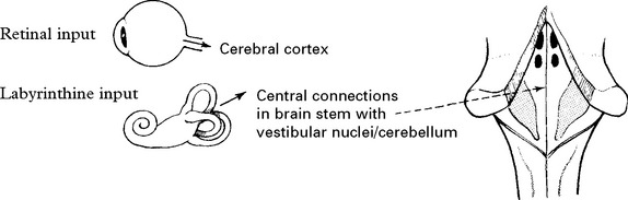
Nystagmus may result from:
– retinal disease
– labyrinthine disease, or
– disorders affecting the cerebellum or a substantial portion of the brain stem.
Examination for nystagmus
‘Nystagmoid’ movements of the eyes are present in many people at extremes of gaze.
Nystagmus present with the eyes deviated less than 30° from the midline is abnormal.

When nystagmus is present only with the eyes deviated to one side – 1st degree nystagmus.
With eyes deviated to one side and in the midline position also – 2nd degree nystagmus.
When present in all directions of gaze – 3rd degree nystagmus.
If nystagmus is detected, note the type (jerk or pendular), direction (of fast phase) and degree.
Nystagmus suppressed by visual fixation may appear in darkness, but this requires specialised techniques (electronystagmography – see page 65) to demonstrate.
RETINAL OR OCULAR nystagmus
Physiological: following moving objects beyond the limits of gaze – opticokinetic nystagmus. Pathological: occurs when vision is defective. Fixation is impaired and the eyes vainly search.
Nystagmus is:

Occurs in congenital cataract, congenital macula defect, albinism.
VESTIBULAR nystagmus
Nystagmus arises from:
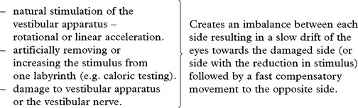
Physiological
(i) Rotational acceleration produces nystagmus in the plane of rotation.

(ii) Caloric testing sets up convection currents in the lateral semicircular canal producing a horizontal nystagmus (see page 65).
Pathological
Damage to labyrinth or vestibular nerve.

Often associated with tinnitus and hearing loss. Vertigo and nystagmus settle simultaneously.
Occurs in acute labyrinthine disease – Menière’s disease, vestibular neuronitis, vascular disease.
POSITIONAL nystagmus: this may occur in labyrinthine disease in association with vertigo when the patient assumes a certain posture.
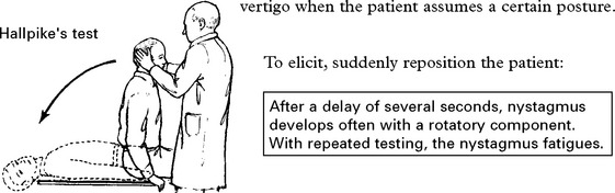
NYSTAGMUS
CENTRAL NERVOUS SYSTEM nystagmus
Central nystagmus arises from damage to the central vestibular connections in the vestibular nuclei and brain stem. The nystagmus may be horizontal, vertical, rotatory or dissociated (present in one eye only).
The direction (fast phase) is determined by direction of gaze (multidirectional). Vertigo is seldom present.
Signs of other nuclear or tract involvement in brain stem should be evident.
Central nystagmus occurs in vascular disease, demyelination, neoplasms, nutritional disease (Wernicke’s encephalopathy), alcohol intoxication and drug toxicity, e.g. phenytoin.
Posterior fossa lesions may produce positional nystagmus. This may be distinguished from labyrinthine disease by:
Absence of delay before onset, lack of fatiguing with repetitive testing, and a tendency to occur with any rather than one specific head movement.
Although nystagmus often occurs in cerebellar disease, the role of the cerebellum in its production remains unclear. The fast phase tends to occur to the side of the cerebellar damage (i.e. the opposite of labyrinthine disease).

INTERNUCLEAR OPHTHALMOPLEGIA (Ataxic nystagmus)
The median longitudinal fasciculus links, among other structures, the innervation of the lateral rectus with the contralateral medial rectus muscle in order to coordinate horizontal gaze. A lesion of this fasciculus will cause dissociate nystagmus.

In unilateral medial longitudinal fasciculus lesions the eye fails to adduct on the affected side.
N.B. Internuclear ophthalmoplegia differs from a bilateral III nerve or nuclear lesion in that the pupil is not affected and when testing eye movements individually, some adduction occurs.
The disorder characteristically occurs in multiple sclerosis but also in brain stem infarction, haemorrhage, trauma, syringobulbia and drug toxicity (phenytoin).
OTHER VARIETIES OF CENTRAL NERVOUS SYSTEM NYSTAGMUS

A group of confusing terms are used to describe abnormal, involuntary eye movements seen in cerebellar/brain stem disease:
Ocular bobbing – fast drift downwards, slow drift upwards; seen with large pontine lesions. (Horizontal eye movements are absent.)
Opsoclonus – rapid conjugate jerks of eyes; made worse by head movements. The eye movements are random.
Oscillopsia is a term used to describe the patient’s awareness of jumping of the environment as a consequence of rapid jerking eye movements.
TREMOR
Tremor is a rhythmic involuntary movement normally affecting the limbs. Diagnosis depends on examination of the character of the tremor as well as the presence of other specific features.

Observe:
– the rate (slow, 4–6 Hz), (rapid, 6–12 Hz)
– the amplitude (fine or coarse)
– the distribution: head, trunk or limbs (distal or proximal)
– associated features e.g. disorder of gait or balance
Most tremors disappear during sleep.
Physiological tremor is evident on maintaining a fixed posture, fast in rate (8–12 Hz), fine in character, distal in distribution and non-disabling. It is enhanced by fatigue, anxiety and drugs e.g. caffeine, steroids.
Pathological tremor occurs at rest or with movement, slow in rate, coarse in character, proximal or distal and often asymmetrical in distribution. This tremor is socially and physically disabling.
CHARACTERISTICS OF PATHOLOGICAL TREMOR
Tremor at rest

Tremor on maintaining posture and throughout range of movement

Specific types of postural tremor are recognised
|
FAMILIAL TREMOR ESSENTIAL TREMOR SENILE TREMOR |
– often Mendelian dominant. – no family history – develops in old age. |
The tremor may progress until handwriting becomes impossible and feeding difficult. Alcohol may temporarily abort the tremor; beta blockers may produce an improvement.
Tremor during and maximal at the end of movement

MYOCLONUS
Myoclonus is a shock-like contraction of muscles which occur irregularly and asymmetrically. Such jerks occur repetitively in the same muscle groups and range from a flicker in a single muscle to contraction in a group of muscles sufficient to displace the affected limb.
Pathophysiology
The precise nature of myoclonus remains unclear. Several forms exist, some clearly related to epilepsy; others may be associated with damage to inhibitory mechanisms in the brain stem reticular formation. Myoclonus may result from pathological changes affecting a variety of different sites including the motor cortex, cerebellum and spinal cord.
Clinical features
Myoclonic movements when repetitive vary in frequency between 5–60/minute. The muscles of the face, oral cavity and limbs are preferentially affected. The movements may be accentuated or precipitated by visual, auditory or tactile stimulation. Repetitive stimulation may result in a crescendo of myoclonus which resembles a seizure.
Physiological myoclonus occurs in sleep (hypnic jerks), with anxiety and in infants when feeding.
Causes
Myoclonus occurs in many rare conditions of the nervous system. Five groups of disorder are recognised:
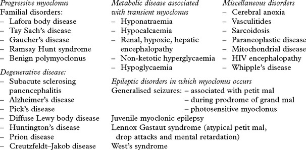
Palatal myoclonus – an unusual myoclonic disorder with rapid regular movements of the soft palate and occasionally of the pharyngeal and facial musculature. Palatal movements occur at a rate of 120–140/minute. This disorder is associated with degenerative changes in the olivary and dentate nuclei.
Treatment
Benzodiazepine drugs such as clonazepam may suppress myoclonic movements. Piracetam (G.A.B.A. analogue) and levetiracetam are also used.
An exaggerated startle response can be confused with myoclonus. This is often physiological but can be disabling – hyperekplexia (Startle disease).
DISORDERS OF STANCE AND GAIT
The normal gait is characterised by an erect posture, moderately sized steps and the medial malleoli of the tibia ‘tracing’ a straight line.
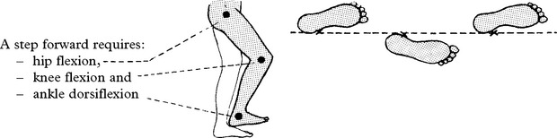
Co-ordination ensures fluidity of movement.
Antigravity reflexes maintain the erect posture. They depend upon spinal cord and brain stem connections to produce extension.
ASSESSMENT OF STANCE AND GAIT
In a patient complaining of disturbance of walking, careful assessment indicates the likely site of the causative lesion.
Watch the patient:
– walking
– performing tandem gait – heel to toe walking,
– standing with heels together with (a) eyes open, (b) eyes closed – this (Romberg’s test) distinguishes cerebellar from sensory ataxia. N.B. You cannot undertake Romberg’s test if the patient cannot stand with eyes open.
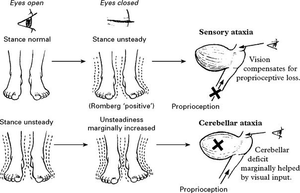
ATAXIC GAIT
1. Cerebellar The feet are separated widely when standing or walking. Steps are jerky and unsure, varying in size. The trunk sways forwards.
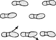
In mild cases: Tandem gait (heel-toe walking) is impaired; the patient falling to one or both sides.
2. Sensory
Disturbed conscious or unconscious proprioception due to interruption of afferents in peripheral nerves or spinal cord (posterior columns, spinocerebellar tracts).
The gait appears normal when the eyes are open although the feet usually ‘stamp’ on the ground. Examination reveals a positive Romberg’s test and impaired joint position sensation.
HEMIPLEGIC GAIT
The leg is extended and the toes forced downwards. When walking, abduction and circumduction at the hip prevent the toes from catching on the ground.
In paraplegia, strong adduction at the hips can produce a scissor-like posture of the lower limbs. In mild weakness, the gait may appear normal, but excessive wear occurs at the outer front aspect of the patient’s shoe sole.
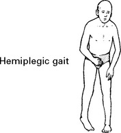
PARKINSONIAN (festinating) GAIT

The patient adopts a flexed, stooping posture. To initiate walking, he leans forwards and then hurries (festinates) to ‘catch up’ on himself. The steps are short and shuffling.

FRONTAL LOBE GAIT
Disturbance of connections between frontal cortex, basal ganglia and cerebellum produces this characteristic disturbance. The gait is wide based (feet wide apart). Initiation is difficult, the feet often seem ‘stuck’ to the floor. There is a tendency to fall backwards. Power and sensation are normal.
HYSTERICAL GAIT
Characterised by its bizarre nature.
Numerous variations are seen. The hallmark is inconsistency supported by the lack of neurological signs. Close observation is essential.
LIMB WEAKNESS
Limb weakness results from damage to the motor system at any level from the motor cortex to muscle.
UPPER MOTOR NEURON WEAKNESS

MUSCLE TONE
Hypertonicity develops after a period (a few days or weeks) of ‘neural shock’. Passive movements produce a ‘clasp knife’ quality, i.e. sudden ‘give’ towards the end of movement.
Clonus – present.
MUSCLE FASCICULATION
Absent.
MUSCLE WASTING
Absent – but, in the long term, disuse atrophy results.
REFLEXES
– Tendon – exaggerated.
– Superficial – depressed or absent (abdominal, cremasteric).
– Plantar response – extensor.
DISTRIBUTION
In general, whole limb or limbs are involved, e.g. monoplegia, hemiplegia, paraplegia.
Weakness shows a PREDILECTION for certain muscle group in a PYRAMIDAL DISTRIBUTION, i.e.

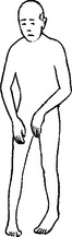
This results in the ‘spastic’ posture with the arm and the wrist flexed and the leg extended. In upper motor neuron lesions, SKILLED movements, e.g. fastening buttons, are always more affected than unskilled movements.
N.B. Dual innervation from each hemisphere results in sparing of the upper face, muscles of mastication, the palate and tongue with a unilateral upper motor neuron lesion.
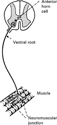
LOWER MOTOR NEURON WEAKNESS
MUSCLE TONE
Hypotonicity with diminished resistance to passive stretch.
Clonus – absent.
MUSCLE FASCICULATION
Present – irregular, non-rhythmical contractions of groups of motor units. More prevalent in anterior horn cell disease than in nerve root damage.
MUSCLE WASTING
Wasting becomes evident in the paretic muscle within 2–3 weeks of the onset.
REFLEXES
– Tendon – depressed or absent.
– Superficial – rarely affected (abdominal, cremasteric).
– Plantar response – flexor.
DISTRIBUTION
Either – muscle groups involved in distribution of a spinal segment/root, plexus or peripheral nerve,
or – generalised limb involvement affecting proximal or distal muscles.
NEUROMUSCULAR JUNCTION WEAKNESS
Muscle tone; muscle bulk; reflexes – all normal
Key feature – weakness fatigues with repetition; most commonly involves ocular muscles, bulbar muscles.
WEAKNESS FROM MUSCLE DISEASE
May be difficult to distinguish from lower motor neuron weakness.
Muscle tone – slightly reduced
Muscle bulk – slightly reduced; no fasciculation
Reflexes – depressed
Distribution – usually proximal weakness, though specific patterns can occur in particular myopathies (e.g. fascioscapulohumeral muscular dystrophy)
LESION LOCALISATION
The foregoing clinical features readily distinguish weakness of an upper motor neuron, lower motor neuron or mixed pattern. Combining these findings with other neurological signs enables localisation of the lesion site.
UPPER MOTOR NEURON LIMB WEAKNESS – UNILATERAL

UPPER MOTOR NEURON LIMB WEAKNESS – BILATERAL

MIXED UPPER AND LOWER MOTOR NEURON WEAKNESS – UNILATERAL OR BILATERAL
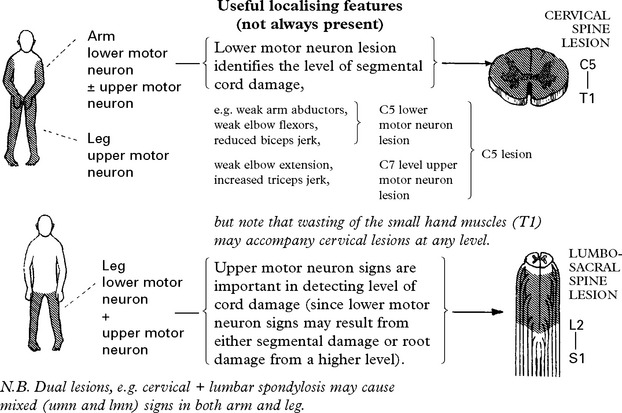
LOWER MOTOR NEURON LIMB WEAKNESS – BILATERAL


SENSORY IMPAIRMENT
ANATOMY AND PHYSIOLOGY
The sensory system relays information from both the external and the internal environment.

Cutaneous receptors are of several types and, while overlap does occur, each has some specific purpose.

Muscle and tendon receptors
These receptors along with those of pressure and touch provide information on body and limb position – proprioception.
Continual stimulation of most receptors results in a reduction in the action potential frequency – ADAPTATION

CENTRAL CONNECTIONS
Sensory neurons (bipolar cells) relay information to the spinal cord via the dorsal root to the dorsal root entry zone. The anatomical and physical characteristics of the neurons vary depending on the information they carry, as do the central pathways:

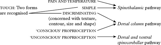
SPINOTHALAMIC PATHWAY
1. Fibres enter the root entry zone and pass up or down for several segments in Lissauer’s tract before terminating in the dorsal aspect of the dorsal horn.
2. Second order neurons synapse locally, cross the midline and run up the spinothalamic tract and lateral lemniscus to terminate in the posterolateral nucleus of the thalamus. Throughout its course, the fibres lie in a somatotopic arrangement with sacral fibres outermost. In the brain stem the lateral lemniscus gives off collateral branches to the reticular formation, which projects widely to the cerebral cortex and limbic system and is joined by fibres from the contralateral nucleus and tract of the trigeminal nerve.
3. From the thalamus, third order neurons project to the parietal cortex.


DORSAL COLUMN PATHWAY
1. Fibres enter in the root entry zone and run upwards in the dorsal columns to the lower medulla where they terminate in the nucleus gracilis and nucleus cuneatus.
2. Second order neurons decussate as the internal arcuate fibres and pass upwards in the medial lemniscus. Maintaining a somatotopic arrangement, they terminate in the ventral posterolateral thalamus.
3. Third order neurons arise in the thalamus and project to the parietal cortex.
DORSAL AND VENTRAL SPINOCEREBELLAR PATHWAYS: see Cerebellar dysfunction, page 181.
EXAMINATION OF THE SENSORY SYSTEM: see page 21
CLINICAL FEATURES
Sensory disturbance may result in:
|
NEGATIVE symptoms: |
‘a loss of feeling’ |
|
‘a deadness’. |
|
|
POSITIVE symptoms: |
‘a pins and needles sensation’ |
|
‘a burning feeling’. |
Lesions of the PERIPHERAL NERVES or NERVE ROOTS may produce ‘negative’ or ‘positive’ symptoms.
|
SPINOTHALAMIC TRACT lesions – seldom produce pain but usually a lack of awareness of pain and temperature. This may result in: – trophic changes: cold, blue extremities hair loss brittle nails – painless burns – joint deformation (Charcot’s joints). |
DORSAL COLUMN lesions – produce a discriminatory type of sensory loss. – impaired two point discrimination – astereognosis (failure to discriminate objects held in the hand). – sensory ataxia (disturbed proprioception). |
Lesions of the PARIETAL CORTEX also produce a discriminatory type of sensory loss. Minor lesions produce sensory inattention (perceptual rivalry) – with bilateral simultaneous limb stimulation, the stimulus is only perceived on the unaffected side.
LESION LOCALISATION
The pattern of the sensory deficit aids lesion localisation.

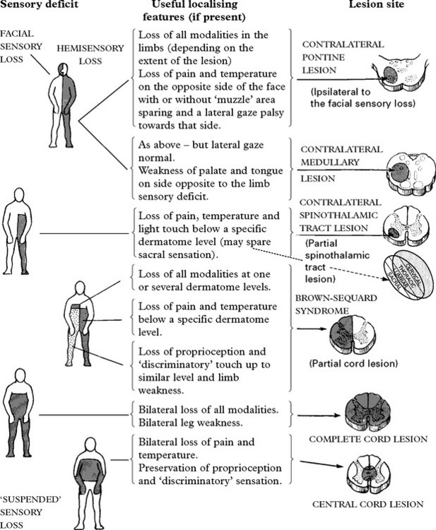

DIFFERENTIAL DIAGNOSIS –as for limb weakness – page 198
PAIN
Peripheral receptors of pain – free nerve endings lying in skin or other organs – are the distal axons of sensory neurons. Such unmyelinated or only thinly myelinated axons are of small diameter. The termination and central connections of these axons are described on page 200.
The type of stimulus required to activate free endings varies, e.g. in muscle – ischaemia, in abdominal viscera – distension.

CONTROL OF SENSORY (PAIN) INPUT
The gate control theory
A relay system in the posterior horn of the spinal cord modifies pain input. This involves interneuronal connections within the substantia gelatinosa (a layer of the posterior horn which extends throughout the whole length of the spinal cord on each side).
An afferent impulse arriving at the posterior horn in thick myelinated fibres has an inhibitory effect in the region of the substantia gelatinosa.
An afferent impulse arriving in thin myelinated or unmyelinated fibres (i.e. transmitting pain) has an excitatory effect in the region of the substantia gelatinosa.
The overall interaction of these inhibitory or excitatory effects determines the activity of second order neurons of the spinothalamic pathway.
A reduction in activity of large sensory fibres ‘opens’ the gate. Stimulation of large sensory fibres theoretically ‘closes’ the gate.
In addition to these segmental influences, higher centres also control the gate region and form part of a feed-back loop.
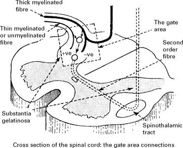
Pain perception
The awareness of pain is brought about by projection from the thalamus to cerebral cortex. Personality, mood and neuroticism all influence the intensity of pain perception. Diffuse projections through Lissauer’s tract and the reticular core of the spinal cord white matter to the reticular formation and limbic system probably contribute to the unpleasant, emotionally disturbing aspects of pain.
NEUROTRANSMITTER SUBSTANCES
Evidence based on both human and animal studies has shown that an endogenous system, lying within the central nervous system can induce a degree of analgesia. Electrical stimulation of certain sites, such as the periaqueductal grey matter, can inhibit pain perception.
Receptor sites for endogenous opiates have been found in the posterior horns and thalamus as well as at several other sites. The endogenous substances which bind to these sites are called encephalinsor endorphins.
Substance P, a polypeptide, found predominantly around free nerve ending receptors and in the spinal cord posterior horns, glutamate and calcitonin gene related peptide are the likely primary transmitters of pain.
DRUG TREATMENT
Sites of potential drug action:

|
Drug selection in pain treatment depends on the severity, cause and the expected duration the pain, i.e. acute pain – less than 2 weeks duration, e.g. postoperative, post-traumatic, renal colic. |
||
|
chronic pain |
– benign origin, e.g. |
postherpetic neuralgia |
|
phantom limb pain |
||
|
chronic back pain. |
||
|
– malignant origin. |
||
1. In acute pain, drug therapy ranges from mild analgesics – aspirin, paracetamol – to narcotic agents – morphine, heroin. Tranquillisers may also help.
2. In chronic pain of benign origin, narcotics and sedatives must be avoided. In these patients, depression usually plays a rôle and the clinician must not underestimate the value of tricyclic antidepressants.
Anticonvulsants – gabapentin and carbamazepine appear to benefit many patients, probably due to their membrane stabilizing effect.
Topical treatment – capsaicin blocks substance P and inhibits pain transmission in the skin. Used for postherpetic neuralgia.
3. In chronic pain from terminal malignancy, patients often require strong narcotics – morphine, heroin. Frequent administration of small doses provides the greatest effect.
PAIN – TREATMENT
PERIPHERAL TECHNIQUES
Generally used for more benign conditions and before resorting to central techniques.
NERVE BLOCKS: Injections of agents into peripheral nerves or roots abolishes pain in the appropriate dermatome; motor and sympathetic function are also lost. Local anaesthetics produce a temporary effect; neurolytic agents, e.g. phenol, alcohol, give permanent results.

CENTRAL TECHNIQUES
Used primarily in patients with intractable pain from malignancy

PAIN SYNDROMES
Pain is not primarily a pathological phenomenon, but serves a protective function. Conditions with loss of pain perception exemplify this, resulting in frequent injuries, burns and subsequent mutilations, e.g. syringomyelia, hereditary sensory neuropathy, congenital insensitivity to pain.
Pathological conditions do, however, cause pain – as a symptom of cancer, injury or other disease.
The following conditions produce characteristic pain syndrome.
CAUSALGIA (Complex Regional Pain Syndrome)
Causalgia is an intense, continuous, burning pain produced by an incomplete peripheral nerve injury. Touching the limb aggravates the pain, and the patient resents any interference or attempt at limb mobilisation. The skin becomes red, warm and swollen.
Theoretical mechanism

Causalgia only occurs with damage to peripheral nerves containing a large number of sympathetic fibres and responds in part to sympathetic blockade (pharmacological or surgical).
POSTHERPETIC NEURALGIA
Following activation of a latent infection with varicella zoster virus lying dormant in the dorsal root or gasserian ganglion, the patient develops a burning, constant pain with severe, sharp paroxysmal twinges over the area supplied by the affected sensory neurons. Touch exacerbates the pain. Thick myelinated fibres are preferentially damaged, possibly opening the ‘gate’.
Treatment of postherpetic neuralgia is particularly difficult. Carbamazepine and/or antidepressants may help. Ethylchloride spray over the affected area provides temporary relief. Capsaicin, a topical NSAID can be an effective treatment.
THALAMIC PAIN
Thalamic stimulation may produce or abolish pain depending upon the electrode site. A vascular accident which involves the inhibitory portion of the thalamus may result in pain – the thalamic syndrome.
Clinical features: Hemianaesthesia at onset contralateral to the lesion precedes the development of pain. This is burning and diffuse, and exacerbated by the touch of clothing.
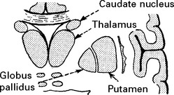
Treatment: Drug treatment gives poor results. A stereotactic procedure although increasing the sensory deficit may help.
Paradoxically the thalamic syndrome may occur following a thalamic stereotactic procedure for movement disorders.
PHANTOM LIMB PAIN
Following amputation of a limb, 10% of patients develop pain with a continuous persistent burning quality, caused by neuroma formation in the stump. The patient ‘feels’ the pain arising from some point on the missing limb (the pain input projects through pathways which retain the topographical image of the absent limb).

Treatment: Often responds to simple measures e.g. tricyclic antidepressants.
VISCERAL AND REFERRED PAIN
Deep visceral pain is dull and boring; it is the consequence of distension or traction on free nerve endings.
Referred pain of a dull quality relates to a specific area of the body surface – often hypersensitive to touch.
The basis of referred pain
The visceral afferents converge upon the same cells in the posterior horns as the somatic efferents. The patient ‘projects’ pain from the viscera to the area supplied by corresponding somatic afferent fibres.
A knowledge of the source of referred pain is important in diagnosis and treatment.

SITES OF REFERRED PAIN FROM SPECIFIC ORGANS

LIMB PAIN
Pain may arise from any anatomical structure within the limb. Each produces characteristic features:

CAUSES OF UPPER LIMB PAIN
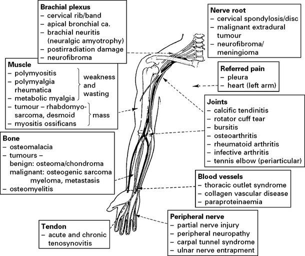
CAUSES OF LOWER LIMB PAIN

Meralgia paraesthetica: burning, tingling pain over the outer aspect of the thigh, increased when standing or by walking, due to a localised neuritis of the lateral cutaneous nerve of the thigh.
Restless legs syndrome (syn. Ekbom’s syndrome): occurs in about 2% of population. An intolerable tingling, burning sensation or pain in both legs, occurring only when sitting or lying down and relieved by walking; no associated neurological abnormality. Often responds to dopamine agonists (ropinerole and pramipexole), L-dopa and gabapentin.
Investigation of limb pain depends on the suspected cause and may include straight X-rays, CT scan, MRI, nerve conduction studies and EMG.
MUSCLE PAIN (MYALGIA)
Muscle pain is a common medical complaint. There are many causes and clinical evaluation and appropriate investigation is often difficult. The physiological mechanisms producing such a symptom are limited.
Mechanical pain results from excessive muscle tension or contraction and is ‘cramp like’.
Inflammatory pain results from disruption of muscle fibres, inflammatory exudate and fibre swelling.
Ischaemic pain results from metabolic change, usually in response to exercise and is deep and aching.
Muscle pain may be physiological – as a consequence of extreme exercise or pathological – as a consequence of muscle, soft tissue or systemic illness.
DIAGNOSTIC APPROACH TO MUSCLE PAIN
History
|
Is muscle pain – present at rest? |
– Polymyalgia rheumatica – Fibromyalgia – Parkinson’s disease – Collagen vascular disease |
|
present with exercise? |
– Physiological – Metabolic myopathies – Benign myalgic encephalomyelitis (ME) |
|
localised? |
– Muscle haematoma, abscess, tumour or fibromyalgia |
|
generalised? |
– Polymyalgia rheumatica – Parkinson’s disease – Metabolic myopathies – Inflammatory myopathies – Benign myalgic encephalomyelitis (ME) |
|
family history? |
– Metabolic myopathies |
|
exposure to toxins? |
– Drug induced myopathies – Alcoholic myopathy |
Examination
|
Is there |
– wasting/weakness? |
– Inflammatory myopathies – Metabolic myopathies – Drug induced myopathies – Alcoholic myopathy |
|
skin rash? |
– Inflammatory myopathy (dermatomyositis) – Collagen vascular disease |
|
|
stiffness or spasms? |
– Tetanus – Tetany – Spasticity – Neuroleptic malignant syndrome – Malignant hyperthermia |
|
|
muscle swelling? |
– Muscle abscess, tumour – Metabolic myopathy |
Investigations
|
Serum creatine kinase (muscle enzyme) – elevated in muscle necrosis, high levels result in myoglobinuria |
Muscle biopsy (needle or open) Essential in diagnosis of – inflammatory myopathies – metabolic myopathies Helpful in collagen vascular disease |
|
Imaging (occasionally used) Ultrasound, MR or CT in suspected muscle haematoma, abscess or tumour. Radionuclide (Gallium or in suspected muscle abscess, Technitium) |
Ischaemic lactate test Measurement of post exercise changes in serum lactate Reduced response in – metabolic myopathies (disorders of glycolytic pathway) |
|
Electromyography (EMG) Will confirm presence of myopathy (rarely more specific) |
Following extensive investigation, in a significant number of cases no cause of myalgia is found.
Most disorders are covered in relevant sections. Those that are not are briefly described.
|
Fibromyalgia |
Polymyalgia rheumatica |
|
A common condition of uncertain pathology in which generalised muscle pain with localised tender areas occurs without objective clinical or laboratory abnormalities. Psychiatric symptoms commonly co-exist. |
Proximal muscle pain encountered in the elderly and often associated with giant cell arteritis. The ESR is elevated and the EMG is normal. Muscle biopsy shows type 2 fibre loss. Steroids are effective. |
|
Muscle tumours |
|
|
Malignant hyperpyrexia |
These are rare. Mixed pathological and of varying degrees of malignancy |
|
Characterised by a sudden rise in body temperature whilst undergoing general anaesthesia, usually with halothane or succinylcholine. Certain hereditary myopathic disorders, e.g. myotonic dystrophy, central core disease – are unduly prone to this condition. |
|
|
Chronic fatigue syndrome/myalgic encephalomyelitis (CFS/ME) |
|
|
An idiopathic disorder that may follow viral illness, is often associated with exercise induced muscle pain and associated with fatigue. No clear underlying pathology has been found and diagnosis is based on symptoms and exclusion of other pathology. |
|
|
Muscle abscess |
|
|
Commonly Staphylococcal due to local trauma or blood-borne in debilitated persons. |
|
|
May respond to graded exercise, tricyclic antidepressants or cognitive behavioural therapy. |
OUTCOME AFTER BRAIN DAMAGE
Outcome after brain damage has major social and financial implications for both patients and their families. In a welfare state, society may carry most, if not all of the financial burden, particularly with more severe disability. The greater the disability, the greater the support required. Conditions causing brain damage do not respect age; survivors may need long-term care.
A variety of methods have been devised to categorise outcome. Such classifications provide end-points for audit and research, and a means of assessing therapeutic intervention. They permit prediction based on clinical and investigative findings early in the course of the disease. Most outcome scales have been developed with a particular disease in mind (e.g. Bartel/Rankin – stroke, Karnofsky – tumour). In 1975 Jennett and Bond developed the Glasgow Outcome Scale (GOS) for the assessment of head injured patients, and this is now widely applied in the assessment of patients with other causes of brain damage.
|
The Glasgow Outcome Scale |
Five categories exist – |
|
1. Death |
|
|
2. Persistent Vegetative State |
– see below. |
|
3. Severe Disability |
– dependent for some support in every 24 hour period. |
|
4. Moderate Disability |
– independent but disabled. May or may not be capable of return to work. |
|
5. Good recovery |
– good, but not necessarily complete recovery e.g. cranial nerve deficit. |
|
Could (although may not) return to work. |
|
The Vegetative State
Severe bilateral hemisphere damage may result in a state in which the patient has no awareness of themselves or of their environment. Although periods of eye opening and closure may occur suggesting sleep/wake cycles, along with spontaneous movements of the face, trunk and limbs, the patient does not communicate or interact with others in any way.
The vegetative state becomes ‘permanent’ when irreversibility can be established with a high degree of certainty, i.e. > 6 months after non-traumatic coma and > 12 months after traumatic coma. At one month after trauma, about 1/3 of patients in the vegetative state will show some improvement over the subsequent year. After non-traumatic coma, outcome is much worse; only about 7% show some improvement and have severe disability.
Outcome Prediction
Outcome from non-traumatic coma depends on a variety of factors including the patient’s age, the duration and depth of the coma, and the cause of the damage provided this is not drug induced.

Outcome from traumatic coma see page 238.
BRAIN DEATH
The advent of improved intensive care facilities and more aggressive resuscitation techniques has led to an increase in numbers of patients with irreversible brain damage in which tissue oxygenation is maintained by a persistent heart beat and artificial ventilation.
A government working party has published guidelines for the diagnosis of brain death which, when fulfilled, indicate that recovery is impossible. In these patients, organs may be removed for transplantation before discontinuing ventilation.
The tests are designed to detect failure of brain stem function, but certain preconditions must first be met.
Preconditions
Depressant drugs must not contribute towards the patient’s clinical state – if in doubt allow an adequate time interval to elapse to eliminate any possible persistent effect.
Hypothermia must not be a primary cause – ensure that temperature is not less than 35°C.
Severe metabolic or endocrine disturbance must be excluded as a possible cause of the patient’s condition.
The patient must be on a ventilator as a result of inadequate spontaneous respiration or respiratory arrest – if a neuromuscular blocking drug has been used, exclude a prolonged effect by observing a muscle twitch on nerve stimulation, e.g. electrical stimulation of the median nerve should cause a thumb twitch.
The cause of the patient’s condition must be established and this must be compatible with irreversible brain damage, e.g. severe head injury, spontaneous intracerebral haematoma. If in doubt, delay brain death testing.
BRAIN DEATH TESTS

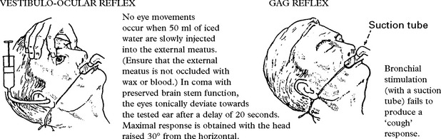
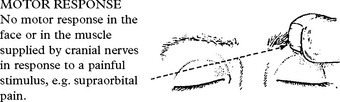
N.B. Limb responses are of no value in testing brain stem integrity. Movements can occur in response to limb or trunk stimulation (especially in the legs), and tendon reflexes may persist in a patient with brain stem death but intact cord function. Conversely, limb movements and reflexes may be absent in a patient with an intact brain stem and spinal cord damage.
RESPIRATORY MOVEMENTS
No respiratory movements are observed when the patient is disconnected from the ventilator. During this test, anoxia is prevented by passing 6 litres O2 per minute down the endotracheal tube. This should maintain adequate PO2 levels for up to 10 minutes. N.B. Ensure that apnoea is not a result of a low PCO2. This should be greater than 6.65 kPa (50 mmHg).
Clinician’s status
The British recommendations state that these tests should be carried out by two doctors, both with expertise in the field; one of consultant status, the other of consultant or senior trainee status. The doctors may carry out the tests individually or together.
Test repetition and timing
The test should be repeated but the interval should be left to the discretion of the clinician. The initial test may be performed within a few hours of the causal event, but in most instances is delayed for 12–24 hours, or longer if there is any doubt about the preconditions.
Timing of death
Certification of death occurs when brain death is established, i.e. at the time of the second test. Old concepts of death occurring at the time the heart ceases to beat are no longer applicable.
Supplementary investigations
Electroencephalography (EEG) is of no value in diagnosing brain death. Some patients with the potential to recover show a ‘flat’ trace; in others with irreversible brain stem damage, electrical activity can occasionally be recorded from the scalp electrodes.
Similarly, angiography or cerebral blood flow measurement are of no additional value to the clinical tests described above, provided the preconditions are fulfilled.