
Meredith A. Morgan, Randall K.Ten Haken, and Theodore S. Lawrence
INTRODUCTION
The beneficial use of radiation was launched by the experiments of Wilhelm Roentgen, who, in 1895, found that x-rays could pass through materials that were impenetrable to light. Emil Grubbe provided one of the early examples of the therapeutic use of radiation by treating an advanced ulcerated breast cancer with x-rays in January 1896. We have made great progress since these early days, which has been strongly influenced by research in radiation chemistry, biology, and physics.
BIOLOGIC ASPECTS OF RADIATION ONCOLOGY
Radiation-Induced DNA Damage
Radiation is administered to cells either in the form of photons (x-rays and gamma rays) or particles (protons, neutrons, and electrons). When photons or particles interact with biologic material, they cause ionizations that can either directly interact with subcellular structures or they can interact with water, the major constituent of cells, and generate free radicals that can then interact with subcellular structures (Fig. 13.1).
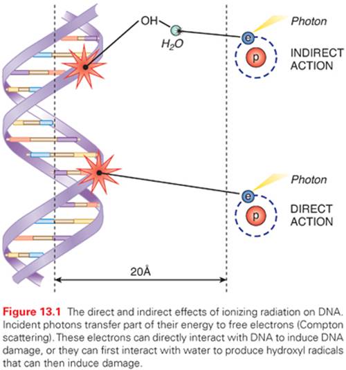
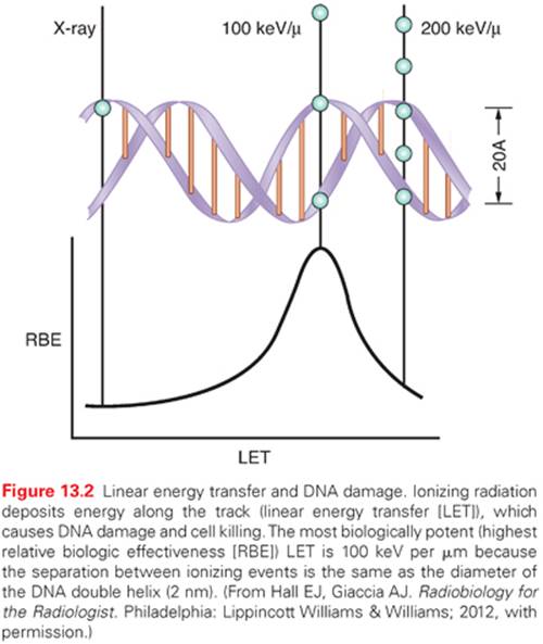
The direct effects of radiation are the consequence of the DNA in chromosomes absorbing energy that leads to ionizations. This is the major mechanism of DNA damage induced by charged nuclei (such as a carbon nucleus) and neutrons and is termed high linear energy transfer (Fig. 13.2). In contrast, the interaction of photons with other molecules, such as water, results in the production of free radicals, some of which possess a lifetime long enough to be able to diffuse to the nucleus and interact with DNA in the chromosomes. This is the major mechanism of DNA damage induced by x-rays and has been termed low linear energy transfer.1
A free radical generated through the interaction of photons with other molecules that possess an unpaired electron in their outermost shell (e.g., hydroxyl radicals) can abstract a hydrogen molecule from a macromolecule such as DNA to generate damage. Cells that have increased levels of free radical scavengers, such as glutathione, would have less DNA damage induced by x-rays, but would have similar levels of DNA damage induced by a carbon nucleus that is directly absorbed by chromosomal DNA. Furthermore, a low oxygen environment would also protect cells from x-ray–induced damage because there would be fewer radicals available to induce DNA damage in the absence of oxygen, but this environment would have little impact on DNA damage induced by carbon nuclei.2
Cellular Responses to Radiation-Induced DNA Damage
Checkpoint Pathways
The cell cycle must progress in a specific order; checkpoint genes ensure that the initiation of late events is delayed until earlier events are complete. There are three principal places in the cell cycle at which checkpoints induced by DNA damage function: the border between G1 phase and S phase, intra-S phase, and the border between G2 phase and mitosis (Fig. 13.3). Cells with an intact checkpoint function that have sustained DNA damage stop progressing through the cycle and become arrested at the next checkpoint in the cell cycle. For example, cells with damaged DNA in G1 phase avoid replicating that damage by arresting at the G1/S interface. If irradiated cells have already passed the restriction point, a position in G1 phase that is regulated by the phosphorylation of the retinoblastoma tumor suppressor gene (Rb) and its dissociation from the E2F family of transcription factors, they will transiently arrest in S phase. The G1/S and intra-S phase checkpoints inhibit the replication of damaged DNA and work in a coordinated manner with the DNA repair machinery to permit the restitution of DNA integrity, thereby increasing cell survival.
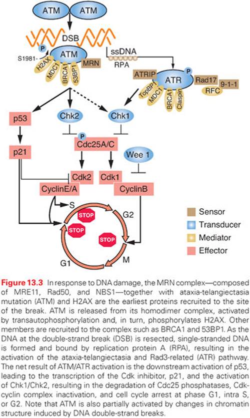
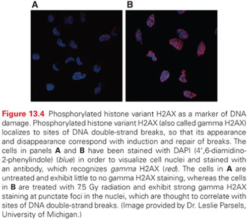
The earliest response to radiation is the activation of ataxia-telangiectasia mutated (ATM), which involves a conformational change that results in the activation of its kinase domain and phosphorylation of serine 1981 (see Fig. 13.3).3 This phosphorylation causes the ATM homodimer to dissociate into active monomers that phosphorylate a wide range of proteins such as 53BP1, the histone variant H2AX, Nbs1 (Nijmegen breakage syndrome; a member of the MRN complex, composed of Mre11, Rad50, and Nbs1), BRCA1, and SMC1 (structural maintenance of chromosomes), and these proteins coordinate repair with the cell cycle.4 In response to DNA damage, H2AX is rapidly phosphorylated by ATM and localizes to sites of DNA double-strand breaks in multiprotein complexes described as foci (Fig. 13.4). Phosphorylation of H2AX by ATM results in the direct recruitment of Mdc1 and forms a complex with H2AX to recruit additional ATM molecules, forming a positive feedback loop.
The G1/S phase checkpoint is the best understood. In response to DNA damage, activated ATM can directly phosphorylate p53 and mdm2, the ubiquitin ligase that targets p53 for degradation. These phosphorylations are important for increasing the stability of the p53 protein. In addition to ATM, checkpoint kinase 2 (Chk2) also phosphorylates p53 and can enhance p53 stability. Activated p53 transcriptionally increases the expression of the p21WAF1/CIPI gene, which results in a sustained inhibition of G1 cyclin/Cdk, and prevents phosphorylation of pRb and progression from G1 into S.5 Mutations in p53 that are commonly found in solid tumors result in loss of transcriptional activity and compromised checkpoint function.
Control of the S-phase checkpoint is mediated in part by the Cdc25A phosphatase inhibiting Cdk2 activity and the loading of Cdc45 onto chromatin. If Cdc45 fails to bind to chromatin, DNA polymerase α is not recruited to replication origins and replicon initiation fails to occur.6 A more prominent mechanism for S-phase arrest is signaled through the MRN complex and the cohesin protein SMC1 by ATM.7 Loss of ATM, MRN components, or SMC1 leads to the loss of the intra-S phase checkpoint function and increased radiosensitivity. Both the CDC45 and ATM pathways represent parallel, but seemingly independent, pathways to protect replication forks from trying to replicate through DNA strand breaks. Although ATM has received the lion’s share of attention in signaling checkpoint activation in response to ionizing radiation, its family member ATR (ataxia telangiectasia and rad3-related) also plays a role in S-phase checkpoint responses.8 ATM kinase activity is inducible by radiation, whereas ATR kinase activity is constitutive and does not significantly change with irradiation. (ATR is described in more detail in Chapter 19.) In contrast to Cdc45 and ATM, ATR is probably more important in monitoring perturbations in replication that are the result of stalled replication forks to prevent the formation of DNA double-strand breaks.
The arrest of cells in the G2 phase following DNA damage is one of the most conserved evolutionary responses to ionizing radiation. It makes sense to have a final checkpoint in the G2 phase to prevent cells from entering into mitosis with damaged DNA that could be transmitted to their progeny. It follows that cells lacking the G2 checkpoint are radiosensitive because they try to divide with damaged chromosomes that cannot be aligned at metaphase to be properly apportioned to daughter cells. At the biochemical level, the regulation of the mitosis-promoting factor cyclin B/Cdk1 is the critical step in the activation of this checkpoint. At the molecular level, ATM and Chk1/2 are activated by DNA damage in the G2 phase and inhibit the activation of Cdc25A and C phosphatases, which are essential for the activation of cyclin B/Cdk1.9,10 The pololike kinase family (Plk1 and Plk3) also responds to DNA damage and can inhibit Cdc25C activation.11 A great deal of effort has been focused on the development of small molecules to inhibit checkpoint response proteins, such as Chk1, with the idea that they would inhibit radiation-induced G2 arrest and perhaps repair and thus be used as radiation sensitizers.12
DNA Repair
Ionizing radiation causes base damage, single-strand breaks, double-strand breaks, and sugar damage, as well as DNA–DNA, and DNA–protein cross-links. The critical target for ionizing radiation-induced cell inactivation and cell killing is the DNA double-strand break.13,14 In eukaryotic cells, DNA double-strand breaks can be repaired by two processes: homologous recombination repair (HRR), which requires an undamaged DNA strand as a participant in the repair, and nonhomologous end joining (NHEJ), which mediates end-to-end joining.15 In lower eukaryotes, such as yeast, HRR is the predominant pathway used for repairing DNA double-strand breaks, whereas mammalian cells use both HHR and non-HHR to repair their DNA. In mammalian cells, the choice of repair is biased by the phase of the cell cycle and by the abundance of repetitive DNA. HRR is used primarily in the late S phase/G2 phases of the cell-cycle, and NHEJ predominates in the G1-phase of the cell cycle (Fig. 13.5). NHEJ and HRR are not mutually exclusive, and both have been found to be active in the late S/G2 phase of the cell cycle, indicating that factors in addition to the cell-cycle phase are important in determining which mechanism will be used to repair DNA strand breaks.
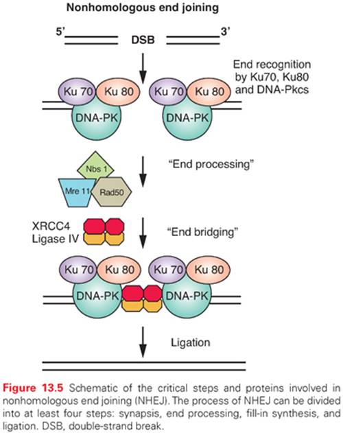
Nonhomologous End Joining. In the G1-phase of the cell cycle, the ligation of DNA double-strand breaks is primarily through NHEJ because a sister chromatid does not exist to provide a template for HRR. The damaged ends of DNA double-strand breaks must first be modified before rejoining. The process of NHEJ can be divided into at least four steps: synapsis, end processing, fill-in synthesis, and ligation (Fig. 13.6).16 Synapsis is the critical initial step where the Ku heterodimer and the DNA-dependent protein kinase catalytic subunit (DNA-PKcs) bind to the ends of the DNA double-strand break. Ku recruits not only DNA-PKcs to the DNA ends, but also artemis, a protein that possesses endonuclease activity for 5′ and 3′ overhangs as well as hairpins.17 DNA-PKcs that is bound to the broken DNA ends phosphorylates artemis and activates its endonuclease activity for end processing. This role of artemis’ endonuclease activity in NHEJ may not necessarily be required for the ligation of blunt ends or ends with compatible termini. DNA polymerase μ is associated with the Ku/DNA/XRCC4/DNA ligase IV complex, and is probably the polymerase that is used in the fill-in reaction. The actual rejoining of DNA ends is mediated by a XRCC4/DNA ligase IV complex, which is also probably recruited by the Ku heterodimer.18,19 Although NHEJ is effective at rejoining DNA double-strand breaks, it is highly error prone. In fact, the main physiologic role of NHEJ is to generate antibodies through V(D)J rejoining, and the error-prone nature of NHEJ is essential for generating antibody diversity.
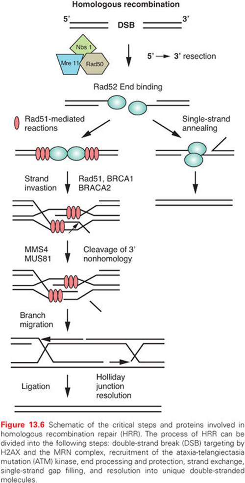
Homologous Recombination. HRR provides the mammalian genome a high-fidelity pathway of repairing DNA double-strand breaks. In contrast to NHEJ, HRR requires physical contact with an undamaged DNA template, such as a sister chromatid, for repair to occur. In response to a double-strand break, ATM as well as the complex of Mre11, Rad50, and Nbs1 proteins (MRN complex), are recruited to sites of DNA double-strand breaks (Fig. 13.6).20 The MRN complex is also involved in the recruitment of the breast cancer tumor suppressor gene, BRCA1, to the site of the break.21 In addition to recruiting BRCA1 to the site of the DNA strand break, Mre11 and as yet unidentified endonucleases resect the DNA, resulting in a 3′ single-strand DNA that serves as a binding site for Rad51. BRCA2, which is recruited to the double-strand break by BRCA1, facilitates the loading of the Rad51 protein onto replication protein A (RPA)-coated single-strand overhangs that are produced by endonuclease resection.22 The Rad51 protein is a homolog of the Escherichia coli recombinase RecA, and possesses the ability to form nucleofilaments and catalyze strand exchange with the complementary strand of the undamaged chromatid, an essential step in HRR. Five additional paralogs of Rad51 also bind to the RPA-coated single-stranded region and recruit Rad52, which binds DNA and protects against exonucleolytic degradation.23 To facilitate repair, the Rad54 protein uses its ATPase activity to unwind the double-stranded molecule. The two invading ends serve as primers for DNA synthesis, resulting in structures known as Holliday junctions. These Holliday junctions are resolved either by noncrossing over, in which case the Holliday junctions disengage and the DNA strands align followed by gap filling, or by crossing over of the Holliday junctions and gap filling. Because inactivation of most of the HRR genes discussed previously results in radiosensitivity and genomic instability, these genes provide a critical link between HRR and chromosome stability.
Chromosome Aberrations Result from Faulty DNA Double-Strand Break Repair
Unfaithful restitution of DNA strand breaks can lead to chromosome aberrations such as acentric fragments (no centromeres) or terminal deletions (uncapped chromosome ends). Radiation-induced DNA double-strand breaks also induce exchange-type aberrations that are the consequence of symmetric translocations between two DNA double-strand breaks in two different chromosomes (Fig. 13.7). Symmetrical chromosome translocations often do not lead to lethality, because genetic information is not lost in subsequent cell divisions. In contrast, when two DNA double-strand breaks in two different chromosomes recombine to form one chromosome with two centromeres and two fragments of chromosomes without centromeres or telomeres, cell death is inevitable. These types of chromosome aberrations are the consequence of asymmetrical chromosome translocations where the genetic material is recombined in what has been termed an illegitimate manner (e.g., a chromosome containing an extra centromere).
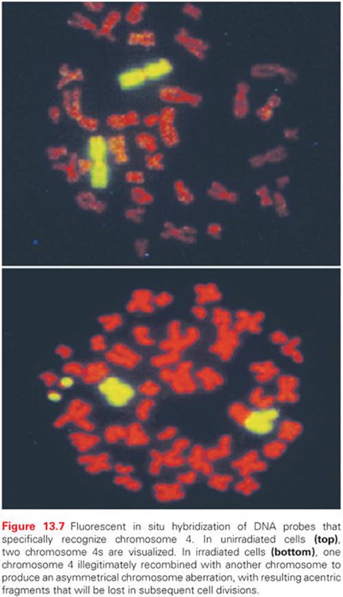
During mitosis, when a cell divides, aberrant chromosomes that have two centromeres, lack a centromere, or are in the shape of a ring have difficulty in separating, resulting in daughter cells with unequal or asymmetric distribution of the parental genetic material. The quantification of asymmetric chromosome aberrations induced by radiation is difficult and has to be performed by the first cell division because these aberrations will be lost during subsequent cell divisions. For this reason, symmetrical chromosome aberrations have been used to assess radiation-induced damage many generations after exposure because they are not lost from the population of exposed cells. In fact, symmetrical chromosome aberrations can be detected in the descendants of survivors of Hiroshima and Nagasaki, indicating that they are stable biomarkers of radiation exposure.24
Membrane Signaling
Apart from the direct of effects on DNA, radiation also affects cellular membranes. As part of the cellular stress response, radiation activates membrane receptor signaling pathways such those initiated via epidermal growth factor receptor (EGFR) and transforming growth factor β (TGF-β).25,26 Activation of these pathways promotes overall survival in response to radiation by promoting DNA damage repair and/or cellular proliferation. In addition, radiation also induces ceramide production at the membrane via activation of sphingomyelinases, which hydrolyze sphingomyelin to form ceramide. Ceramide production is linked to radiation-induced apoptosis.27
The Effect of Radiation on Cell Survival
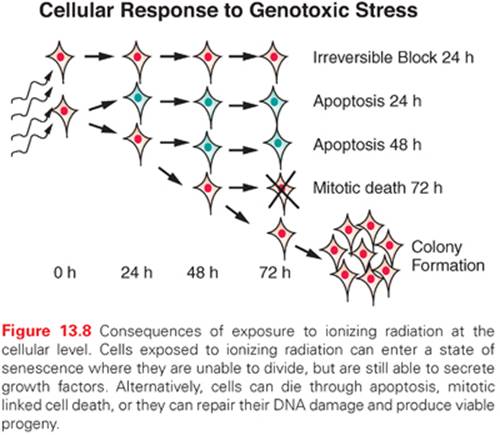
The major potential consequences of cells exposed to ionizing radiation are normal cell division, DNA damage–induced senescence (reproductively inactive but metabolically active), apoptosis, or mitotic-linked cell death (Fig. 13.8). These manifestations of DNA damage can occur within one or two cell divisions or can manifest at later times after many cell divisions.28 Effects that occur at later times have been termed delayed reproductive cell death and may also be influenced by secreted factors that are induced in response to radiation.29
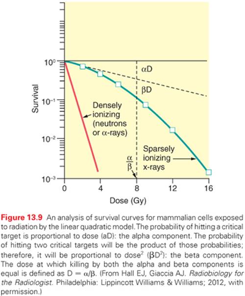
The ability to culture cells derived from both normal and tumor tissues has allowed us to gain insight into how radiosensitivity varies between tissues by analyzing the shape of survival curves. Survival curves of tumor cells often possess a shouldered region at low doses that becomes shallower as the dose increases and eventually becomes exponential. A shoulder on a survival means that these low doses of radiation are less efficient in cell killing, presumably because cells are efficient at repairing DNA strand breaks.13,14 Killing at low doses of radiation can be described in the form of a linear quadratic equation: S = e−αD−βD2 (Fig. 13.9).30 In this equation, S is the fraction of cells that survive a dose (D) of radiation, whereas α and β are constants. Cell killing by the linear and quadratic components are equal when αD = βD2 or D = α/β. Over a larger dose range, the relationship between cell killing and dose is more complex and is described by three different components: an initial slope (D1), a final slope (Do), and the width of the shoulder (n, the extrapolation number) or Dq, the quasi-threshold dose (Fig. 13.10). The extrapolation number, n, defines the place where the shoulder intersects the ordinate when the dose is extrapolated to zero, and the quasithreshold dose, Dq, defines the width of the shoulder by cutting the dose axis when there is a survival fraction of unity. In contrast to photons, the shoulder on the survival curve disappears when cells are exposed to densely ionizing radiation from particles, indicating that this form of radiation is highly effective at killing cells at both low and high doses.

In Vivo Survival Determination of Normal Tissue Response to Radiation
Although much of our knowledge on the effects of radiation on cell survival has come from cell culture studies, investigators have also devised experimental approaches to assess the clonogenic survival of normal tissues. The earliest example came from McCulloch and Till,31 who developed an assay to measure the clonogenic survival of bone marrow–derived cells in response to radiation by injecting them into a recipient mouse and quantifying the number of colonies that developed in the spleen. An analysis of these in vivo spleen assays indicated that bone marrow cells are highly radiosensitive (perhaps the most radiosensitive of all mammalian cells) in that their cell survival curve lacked a shoulder. These experiments represent two important firsts in the radiation sciences: They described the first development of an in vivo assay to assess normal tissue survival to radiation, and they demonstrated the first existence of normal tissue stem cells. Soon after, Withers and colleagues32 developed an assay to assess the survival of skin stem cells, and Withers and Elkind33 developed an assay to quantify the viability of small intestinal clonogens.
Because these ingenious approaches cannot be applied to all normal tissues, loss of tissue function instead of clonogenic survival has been used as an end point to assess radiation effects. Effects on tissue function can be grouped into the acute or late variety. Desquamation of skin by radiation is an example of an acute loss of function, whereas loss of spinal cord function is an example of a late functional effect. Acutely sensitive tissues such as skin, bone marrow, and intestinal mucosa possess a significant component of tissue cell division, whereas delayed sensitive tissues, such as spinal cord, breast, and bone, do not possess a significant amount of cell division or turnover and manifest radiation effects at later times.
In Vivo Determination of Tumor Response to Radiation
Assays have also been developed to assess the clonogenic survival of tumor cells in animals. Perhaps the most relevant of these assays is the tumor control dose 50% (TCD50) assay,34 in which the dose of radiation needed to control the growth of 50% of the tumors is determined in large cohorts of tumor-bearing animals. The TCD50 assay in animals most closely approximates the clinical situation because tumors are irradiated in animals and the ability to kill all viable tumor cells is assessed. Unlike assays in which tumor cells are irradiated ex vivo, the TCD50 assay takes into account the effects of the tumor microenvironment on tumor response. In contrast to the TCD50 assay, the tumor growth delay assay reflects the time after irradiation that a transplanted tumor reaches a fixed multiple of the pretreatment volume compared to an unirradiated control. This end point can be achieved by measuring tumor volume through the use of calipers or by a noninvasive measurement of tumor volume using bioluminescent molecules such as luciferase or fluorescent proteins. In the latter approach, all the tumor cells are stably transfected with a bioluminescent marker before implantation, and tumor growth is measured by bioluminescent activity.35 The advantage of this approach is that tumor cells can be assessed even if they are orthotopically transplanted into their tissue of origin. In another approach, tumors or cells are first irradiated in vivo, the tumor is excised and made into a single-cell suspension, and these cells are then injected into a non–tumor-bearing animal. If the cells are injected subcutaneously under the skin, the end point is tumor formation.36 If the tumor cells are injected in the tail vein of the mouse, the end point is colony formation in the lungs.37 The major advantage of these assays is that the actual number of viable cells can be determined.
FACTORS THAT AFFECT RADIATION RESPONSE
The Fundamental Principles of Radiobiology
Studies on split-dose repair (SDR) by Elkind et al.38 uncovered three of what we now recognize as the most fundamental principles of fractionated radiotherapy: repair, reassortment, and repopulation (Fig. 13.11). (Reoxygenation, described in the following paragraphs, is the fourth). SDR describes the increased survival or tumor growth delay found if a dose of radiation is split into two fractions compared to the same dose administered in one fraction. This repair is likely due to DNA double-strand break rejoining. Elkind et al. found that the survival of cells increased with an increase in time between doses for up to a maximum of about 6 hours. This finding is consistent with the clinical observation that a separation of radiation treatments by 6 hours produces similar normal tissue injury as a 24-hour separation. The shoulder of a survival curve is strongly influenced by SDR: The broader the shoulder, the more SDR and the smaller α/β ratio.
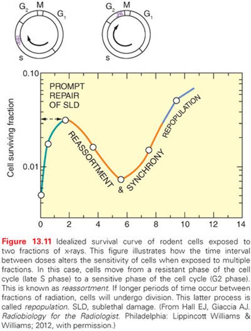
Similar to repair, reassortment and repopulation are also dependent on the interval of time between radiation fractions. If cells are given short time intervals between doses, they can progress from a resistant portion of the cell cycle (e.g., S phase) to a sensitive portion of the cell cycle (e.g., G2 phase). This transit between resistant and sensitive phases of the cell cycle is termed reassortment. If irradiated cells are provided even longer intervals of time between doses, the survival of the population of irradiated cells will increase. This increase in split-dose survival after longer periods of time is the result of cell division and has been termed repopulation. Reassortment and repopulation appear to have more protracted kinetics in normal tissues than rapidly proliferating tumor cells, and thereby enhance the tumor response to fractionated radiotherapy compared to normal tissues.
Dose-Rate Effects
For sparsely ionizing radiation, dose rate plays a critical factor in cell killing. Lowering the dose rate, and thereby increasing exposure time, reduces the effectiveness of killing by x-rays because of increased SDR. A further reduction in dose rate results in more SDR and reduces the shoulder of the survival curve. Thus, if one plots the survival for individual doses in a multifraction experiment so that there is sufficient time for SDR to occur, the resulting survival curve would have little shoulder and appear almost linear.39 In some cell types, there is a threshold to the lowering of dose rate, and in fact, one paradoxically finds an increase, instead of a decrease, in cell killing. This increase in cell killing under these conditions of protracted dose rate is due to the accumulation of cells in a radiosensitive portion of the cell cycle. In summary, the magnitude of the dose rate effect varies between cell types because of SDR, the redistribution of cells through the cell cycle, and the time for cell division to occur.
Cell Cycle
The phase of the cell cycle at the time of radiation influences the cell’s inherent sensitivity to radiation. Cells synchronized in late G1/early S and G2/M phases are most sensitive, whereas cells in G1 and mid to late S phase are more resistant to radiation.1 These differences in sensitivity during the cell cycle are exploited by the concept of reassortment during fractioned radiotherapy as well by the use of chemotherapeutic agents, which reassort cells into more sensitive phases of the cell cycle in combination with radiation.
Tumor Oxygenation
The major microenvironmental influence on tumor response to radiation is molecular oxygen.40 Decreased levels of oxygen (hypoxia) in tissue culture result in decreased killing after radiation, which can be expressed as an oxygen enhancement ratio (OER). Operationally, OER is defined as the ratio of doses to give the same killing under hypoxic and normoxic conditions. At high doses of radiation, the OER is approximately 3, whereas at low doses, it is closer to 2.41 Oxygen must be present within 10 μs of irradiation to achieve its radiosensitizing effect. Under hypoxic conditions, damage to DNA can be repaired more readily than under oxic conditions, where damage to DNA is “fixed” because of the interaction of oxygen with free radicals generated by radiation. These changes in radiation sensitivity are detectable at oxygen ranges below 30 mm Hg. Most tumor cells exhibit a survival difference halfway between fully aerobic and fully anoxic cells when exposed to a partial pressure of oxygen between 3 and 10 mm Hg.1The presence of hypoxia has greater significance for single-dose fractions used in the treatment of certain primary tumors and metastases and is less important for fractionated radiotherapy, where reoxygenation occurs between fractions. Furthermore, most hypoxic cells are not actively undergoing cell division, thus impeding the efficacy of conventional chemotherapeutic agents that are targeted to actively dividing cells.
Although normal tissue and tumors vary in their oxygen concentrations, only tumors possess levels of oxygen low enough to influence the effectiveness of radiation killing. Although the variations in normal tissue oxygenation are in large part due to physiology governing acute changes in oxygen consumption, the variations in tumor oxygen can be directly attributed to abnormal vasculature that results in a more chronic condition. Thomlinson and Gray42observed that variations in tumor oxygen occur because there is insufficient vasculature to provide oxygen to all tumor cells. They hypothesized that oxygen is unable to reach tumor cells beyond 10 to 12 cell diameters from the lumen of a tumor blood vessel because of metabolic consumption by respiring tumor cells. This form of hypoxia caused by metabolic consumption of oxygen has been termed chronic or diffusion-mediated hypoxia. In contrast, changes in blood flow due either to interstitial pressure changes in tumor blood vessels that lack a smooth muscle component or red blood cell fluxes can cause transient occlusion of blood vessels resulting in acute or transient hypoxia. Chronically, hypoxic cells will only become reoxygenated when their distance from the lumen of a blood vessel decreases, such as during fractionated radiotherapy when tumor cords shrink. In contrast, tumor cells that are acutely hypoxic because of changes in blood flow or interstitial pressure often cycle in an unpredictable manner between oxic and hypoxic states as blood flow changes.
Based on studies demonstrating that hypoxia can alter radiation sensitivity and decrease tumor control by radiotherapy, strategies have been developed to increase tumor oxygenation. Most importantly, it appears that tumor oxygen levels increase during a course of fractionated radiation. This may be one of the most important benefits of fractionated radiation and is termed reoxygenation (the fourth of the four Rs of radiobiology). Tumor reoxygenation during a course of fractionated radiation may also offer an explanation for the general lack of clinical efficacy of hypoxic cell sensitizers despite the clear evidence that hypoxia causes radioresistance.
Aside from using fractionated radiation, the most direct approach to increasing tumor oxygenation is to expose patients receiving radiotherapy to hyperbaric oxygen therapy. The underlying concept is that increasing the amount of oxygen in the bloodstream should result in more oxygen being available for diffusion to the hypoxic regions of tumors. Experimentally, hyperbaric oxygen therapy increases the sensitivity of transplanted tumors to radiation. The results of clinical studies with hyperbaric oxygen therapy, when combined with radiotherapy, showed improvement for two sites—head and neck cancers, and cervix cancers—but failed to show an improvement with other sites, thus calling into question its general usefulness in radiotherapy.43 In a related approach, erythropoietin (EPO), a hormone released by the kidney that increases red blood cell production, should also increase tumor oxygenation by increasing the delivery of hemoglobin-bound oxygen molecules. EPO has been effective at correcting anemia, but has not been successful in combination with radiation to control head and neck cancer and may, in fact, stimulate tumor growth.44,45
Another strategy to increase tumor oxygenation has been the combined use of nicotinamide, which increases tissue perfusion and carbogen (95% O2 and 5% CO2) breathing (accelerated radiotherapy with carbogen and nicotinamide [ARCON] therapy). Recently, a randomized phase III clinical trial demonstrated improved regional but not local tumor control in larynx cancer patients treated with nicotinamide, carbogen, and radiation versus radiation alone.46 Biologics such as antivascular endothelial growth factor (anti-VEGF) therapy have also been demonstrated to increase tumor oxygenation.47 Anti-VEGF therapy may increase tumor oxygenation by eliminating abnormal vessels that are inadequate in perfusing tumor cells—the so-called vascular normalization hypothesis. Although there is solid experimental evidence to support this hypothesis, there appears to be only a short window of time in which it could be effectively combined with radiotherapy.
Because the presence of hypoxia has both prognostic and potential therapeutic implications, a substantial effort has been invested in trying to image hypoxia.48 The goal of using imaging to “paint” radiation doses to different regions of tumors, although technically possible (as described in the next section, Radiation Physics), faces the problem that changes in oxygenation are dynamic.49 In the future, hypoxia-directed treatment may evolve from the use of hypoxic cell cytotoxins to targeted drugs that exploit cellular signaling changes induced by hypoxia such as hypoxia-inducible factor 1α (HIF-1α). However, despite the strong rationale supporting their use, at this time, there are no agents used in the clinic that target hypoxia.
Immune Response
The abscopal effects of radiation (i.e., tumor cell killing outside of the radiation field) have been attributed to the activation of antigen and cytokine release by radiation, which subsequently activates a systemic immune response against tumor cells.50,51 This response begins with the transfer of tumor cell antigens to dendritic cells and, subsequently, the activation of tumor-specific T cells and immunogenic tumor cell death. It is likely that radiation dose and fractionation influence the optimal immune response with higher doses and fewer fractions of radiation than those used in conventional fractionation schemes appearing superior in experimental models. Unfortunately, abscopal effects are uncommon because immune system evasion is an inherent characteristic of cancer cells that often dominates, even in the presence of a radiation-induced immune response. Strategies to amplify radiation-induced immune responses, and thus to overcome tumor cell evasion of the immune system, are under investigation. The combination of radiation with immune checkpoint modulators such as ipilimumab, an antibody against cytotoxic T-lymphocyte antigen 4 (CTLA-4), have shown promising, albeit anecdotal, clinical effects.
DRUGS THAT AFFECT RADIATION SENSITIVITY
For over 30 years now, chemotherapy and radiotherapy have been administered concurrently. In order to maximize the efficacy of radiochemotherapy, it is necessary to understand the biologic mechanisms underlying radiosensitization by chemotherapeutic agents. The several classes of standard chemotherapeutic agents as well as novel molecularly targeted agents that possess radiosensitizing properties will be discussed in this section.
Antimetabolites
5-fluorouracil is among the most commonly used chemotherapeutic radiation sensitizers. Given in combination with radiation, it has led to clinical improvements in a variety of cancers, including those of the head and neck, the esophagus, the stomach, the pancreas, the rectum, the anus, and the cervix. The combination of 5-fluorouracil with radiation is now a standard therapy for cancers of the stomach (adjuvant), the pancreas (unresectable), and the rectum. For other cancers such as head and neck, esophagus, or anal, 5-fluorouracil and radiation are combined with cisplatin or mitomycin C, respectively. Being an analog of uracil, 5-flourouracil is misincorporated into RNA and DNA. However, the ability of 5-fluorouracil to radiosensitize is related to its ability to inhibit thymidylate synthase, which leads to the depletion of thymidine triphosphate (dTTP) and the inhibition of DNA synthesis. This slowed, inappropriate progression through S phase in response to 5-fluorouracil is thought to be the mechanism underlying radiosensitization.52 Similar to 5-fluorouracil, the oral thymidylate synthase inhibitor, capecitabine, is also being increasingly used in combination with radiation.
Gemcitabine (2′, 2′-deoxyfluorocytdine [dFdCyd]) is another potent antimetabolite radiosensitizer. Preclinical studies have demonstrated that radiosensitization by gemcitabine involves the depletion of deoxyadenosine triphosphate (dATP) (related to the ability of gemcitabine diphosphate (dFdCDP) to inhibit ribonucleotide reductase) as well as the redistribution of cells into the early S phase of the cell cycle.53 The combination of gemcitabine with radiation in clinical trials has suggested improved clinical outcomes for patients with cancers of the lung, pancreas, and bladder. Gemcitabine-based chemoradiation has developed into a standard therapy for locally advanced pancreatic cancer. However, in some clinical trials, such as those in lung and head and neck cancers, the combination of gemcitabine with radiation has led to increased mucositis and esophagitis.54 Thus, it should be emphasized that in the presence of gemcitabine, radiation fields must be defined with great caution. Such is the case with pancreatic cancer, where the combination of full-dose gemcitabine with radiation to the gross tumor can be safely administered if clinically uninvolved lymph nodes are excluded.55 Conversely, the inclusion of the regional lymphatics in the treatment field in combination with full-dose gemcitabine produces unacceptable toxicities.56
Platinums and Temozolomide
Cisplatin is likely the most commonly used chemotherapeutic agent in combination with radiation. Although cisplatin was the prototype for several other platinum analogs, carboplatin is also frequently used in combination with radiation. Cisplatin, in combination with radiation, and sometimes in conjunction with a second chemotherapeutic agent, is indicated for cancers of the head and neck, esophagus (with 5-fluorouracil), the lung, the cervix, and the anus. Radiosensitization by cisplatin is related to its ability to cause inter- and intra-strand DNA cross-links. Removal of these cross-links during the repair process results in DNA strand breaks. Although there are multiple theories to explain the mechanism(s) of radiosensitization by cisplatin, two plausible explanations are that cisplatin inhibits the repair (both homologous and nonhomologous) of radiation-induced DNA double-strand breaks and/or increases the number of lethal radiation-induced double-strand breaks.57
Temozolomide in combination with radiation is standard therapy for glioblastoma. Temozolomide is an alkylating agent, which forms methyl adducts at the O6 position of guanine (as well as at N7 and N3-guanine) that are subsequently improperly repaired by the mismatch repair pathway. Radiosensitization by temozolomide involves the inhibition of DNA repair and/or an increase in radiation-induced DNA double-strand breaks due to radiation-induced single-strand breaks in proximity to O6 methyl adducts. Like cisplatin, temozolomide-mediated radiosensitization does not seem to require cell cycle redistribution.
Taxanes
The taxanes, paclitaxel and docetaxel, act to stabilize microtubules resulting in the accumulation of cells in G2/M, the most radiation-sensitive phase of the cell cycle. The radiosensitizing properties of the taxanes are thought to be attributable to the redistribution of cells into G2/M. Paclitaxel, in combination with radiation (and carboplatin), has demonstrated a clinical benefit in the treatment of resectable lung carcinoma.58
Molecularly Targeted Agents
Molecularly targeted agents are especially appealing in the context of radiosensitization because they are generally less toxic than standard chemotherapeutic agents and need to be given in multimodality regimens (given their often inadequate efficacy as single agents). The EGFR has been intensely pursued as a target; both antibody and small molecule EGFR inhibitors, such as cetuximab and erlotinib, respectively, have been developed. The head and neck seem to be the most promising tumor sites for the combination of EGFR inhibitors with radiation therapy. Preclinical data have demonstrated that the schedule of administration of EGFR inhibitors with radiation is important; EGFR inhibition before chemoradiation may produce antagonism.59 In a randomized phase III trial, cetuximab plus radiation produced a significant survival advantage over radiation alone in patients with locally advanced head and neck cancer.60 In a subsequent trial, however, cetuximab in combination with concurrent, cisplatin-based chemoradiation failed to produce a survival benefit in head and neck cancer patients.61 The combination of EGFR inhibitor with cisplatin-radiation requires further preclinical investigation.
Although EGFR inhibition, concurrent with radiation, is by far the best established combination of a molecularly targeted agent with radiation, other exciting molecularly targeted agents are being developed as radiation sensitizers. Targeting DNA damage response pathways is one approach to radiosensitization. Recently, agents that abrogate radiation-induced cell cycle checkpoints, such as Wee1 and Chk1 inhibitors, have been shown to radiosensitize tumor cells and are currently in clinical development in combination with chemotherapy, with clinical trials planned in combination with radiation.62,63 In addition, poly(ADP-ribose) polymerase (PARP) inhibitors have been demonstrated to preclinically induce radiosensitization, and several clinical trials combining PARP inhibitors with radiation therapy are underway.64
Other Agents
Although the most common clinically used agents in combination with radiation have been shown to produce significant clinical benefit, as described previously, other agents with different mechanisms of action have been used as radiation sensitizers as well as radiation protectors. The vinca alkaloids, such as vincristine, possess radiosensitizing properties due to their ability to block mitotic spindle assembly and, thus, arrest cells in M phase. Although vincristine is used in combination with radiation to treat medulloblastoma, rhabdomyosarcoma, and brain stem glioma, its use is principally based on its lack of myelosuppressive side effects, which are dose limiting for radiation in these types of tumors, rather than its potential radiosensitizing properties.
Also worth mention in a discussion of modulators of radiation sensitivity are agents designed to radioprotect normal tissues. One such type of drug, amifostine, is a free radical scavenger with some selectivity toward normal tissues that express more alkaline phosphatase than tumor cells, the enzyme of which converts amifostine to a free thiol metabolite. Clinical trials in head and neck as well as lung cancers have shown a reduction in radiation-related toxicities such as xerostomia, mucositis, esophagitis, and pneumonitis, respectively.65,66 However, further clinical investigations are necessary to conclusively demonstrate a lack of tumor protection and safety in combination with chemoradiotherapy regimens.
RADIATION PHYSICS
Physics of Photon Interactions
Tumors requiring radiation can be found at depths ranging from zero to 10s of centimeters below the skin. The goal of treatment is to deliver sufficient ionizing radiation to the tumor site, which can result in an absorbed dose. This involves both the availability of treatment beams and delivery techniques, and the methods to plan the treatments and ensure their safe delivery. This section will establish the general physical basis for the use of ionizing radiation in the treatment of tumors, briefly describe some of the treatment equipment, indicate physical qualities of the treatment beams themselves, and summarize the treatment planning process. Those who desire more in-depth details are referred to textbooks and other resources dedicated to medical physics and the technologic aspects of radiation oncology.67 Most patients who are treated with radiation receive high-energy, external-beam photon therapy. Here, external indicates that the treatment beam is generated and delivered from outside of the body. High-energy (6 to 20 MV) photon beams (electromagnetic radiation) penetrate tissue, enabling the treatment of deep-seated tumors. Modern equipment generates these beams with sufficient fluence to ensure delivery of therapeutic fractions of dose in short treatment sessions. Other types of particles and beams also exist for use in treating tumors both externally and internally. They are mentioned briefly later. However, as external photon beams dominate the practice (and as common basic physics principles related to delivered dose exist among the modalities), the focus here will be on photon beam generation and interactions in tissue.
As mentioned earlier, ionizing radiation kills cells via both direct and indirect mechanisms. Radiation therapy aims to instigate those ionizations and events in the tumor cells. Photons are massless, uncharged packets of energy that primarily interact with matter via electromagnetic processes. As a consequence of those interactions, an incident photon can become either entirely absorbed (giving up its energy to the ejection of an atomic electron [photoelectric effect]), or create an energetic electron-positron pair (pair production), or scatter off an electron with a reduction in energy and a change in direction and subsequent transfer of parts of its energy to the free electron (Compton scattering). The secondary electrons generated as a consequence of these interactions have residual energy, mass, and, most importantly, electric charge. They slow down in matter through multiple interactions with (primarily) the electrons of atoms, leading to excitation and ionization of those atoms. These ionizations (hence the term ionizing radiation) lead to a local absorption of energy (i.e., dose = energy absorbed per unit mass) and the direct and indirect cell killing effects necessary to treat tumors.
Thus, the use of external photon beams for cancer therapy involves a two-step process: interaction (scattering) of the photons, with subsequent dose deposition via the secondary electrons. The probability of photon interactions is energy dependent. Photoelectric interactions dominate at lower photon energies. Whereas these beams are ideal for diagnostic procedures (for their preferential absorption by tissues of differing atomic number, leading to good subject contrast), they are attenuated too quickly in tissues to supply enough interactions to be useful for therapy for any but the most superficial tumors. Pair production interactions dominate at higher photon energies; however, the probability of interacting in tissues for those high-energy photons is so low as to preclude them from general use as well. In the 10s to 100s of kiloelectron volt (keV) to the few megaelectron volt (MeV) photon energy range, Compton scattering dominates. As will be shown, these beams have sufficient penetration and can be generated with sufficient intensity to be useful for tumor treatments, especially when combined in treatment plans that comprise multiple beams entering the patient from different directions but overlapping at the tumor.
It is useful to point out physical scales of reference for external photon beam therapy. A typical megavoltage photon beam may have an average photon energy near 2 MeV. Those photons primarily undergo Compton scattering with a mean free path in tissue of approximately 20 cm. An average Compton interaction results in a secondary electron with a mean energy near 0.5 MeV (and a Compton scattered photon near 1.5 MeV, which likely escapes or scatters elsewhere in the patient). A typical secondary electron of approximately 0.5 MeV will cause excitations and ionizations of atoms as it dissipates its energy over a path length of approximately 2 mm. This could be expected to lead to approximately 10,000 ionizations, or about 5 ionizations per micron of tissue. As can be seen, therapeutic damage to the DNA of cancer cells (2 nm; see Fig. 13.2) will require very many Compton scatterings with statistical interaction among the ionizations resulting from the slowing down of the secondary electrons.
Photon Beam Generation and Treatment Delivery
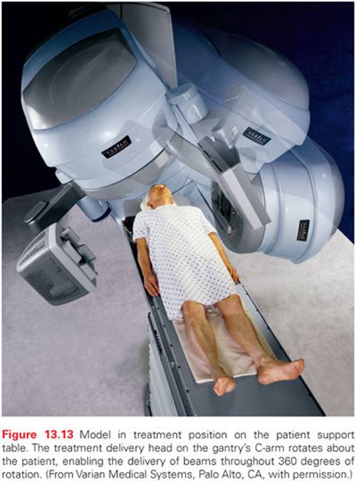
As previously mentioned, effective external-beam photon treatments require higher energy beams capable of reaching deep-seated tumors with sufficient fluence to make it likely that the dose deposition will kill the tumor cells. To spare normal tissues and maximize targeting, beams are arranged to enter the patient from several directions and to intersect at the center of the tumor (treatment isocenter). Although machines containing collimated beams from high-intensity radioactive sources (primarily cobalt 60 [60Co]) are still in use, today’s modern treatment machine accelerates electrons to high (MeV) energy and impinges them onto an x-ray production target, leading to the generation of intense beams of Bremsstrahlung x-rays. A typical photon beam treatment machine68,69 (Fig. 13.12) consists of a high-energy (6 to 20 MeV) linear electron accelerator, electromagnetic beam steering and monitoring systems, x-ray generation targets, high-density treatment field-shaping devices (collimators), and up to a ton of radiation shielding on a mechanical C-arm gantry that can rotate precisely around a treatment couch (Fig. 13.13). These treatment-delivery machines routinely maintain mechanical isocenters for patient treatments to within a sphere of 1 mm radius. The development of stereotactic radiotherapy, which will be described in the section titled Clinical Application of Types of Radiation, depends on this level of machine precision.

X-ray production by monoenergetic high-energy electrons results in an x-ray (photon) beam that contains a continuous spectrum of energies with maximum photon energy near that of the incident electron beam. Lower energy photons appear with a much greater probability than do the highest energy ones, but they also become preferentially filtered out of the beam through the absorption in the target and the attenuation in the flattening filter. This generally results in a treatment beam energy spectrum with a mean photon energy of approximately one-third of the initial electron beam energy. In this energy range, the resulting photon beam exits the production target with a narrow angular spread focused primarily in the forward direction. These forward-peaked intensity distributions generally need to be modulated (flattened) to produce a large (up to 40 cm diameter at the patient) photon beam with uniform intensity across the beam. All modern treatment units take advantage of extensive computer control, monitoring, and feedback to produce highly stable and reproducible treatment beams.
The resulting photon beam requires beam shaping for conformal dose delivery. Some combination of primary, high-density field blocks (collimators) together with additional edge blocks generally provide the required shaping and shielding. Modern machines use computer-controlled multileaf collimators (Fig. 13.14) for the edge sculpting subsequent to setting the primary collimators for maximal shielding. This computer control provides high precision and reproducibility in the definition of field edges. Additionally, automation allows for a precise reshaping of the treatment beam for each angle of incidence, allowing not only conformation of irradiation to target volumes, but also modulation of the beam intensity patterns across the field (intensity-modulated radiation therapy [IMRT]).

Variations on the standard linear accelerator (linac) plus C-arm scenario that are being used for external-beam radiation treatments throughout the body include helical tomotherapy and nonisocentric miniature linac robotic delivery systems.70 In helical tomotherapy, the accelerator, photon-production target, and collimation system are mounted on a ring gantry (similar to those found on diagnostic computed tomography [CT] scanners) (Fig. 13.15). It produces a fan beam of photons, and the intensity of each part of the fan being modulated by a binary collimator. As the gantry rotates, the patient simultaneously slides through the bore of the machine (again analogous to modern x-ray/CT imagers), which allows for the continuous delivery of intensity-modulated radiation in a helical pattern from all angles around a patient. Another delivery system uses an industrial robot to hold a miniature accelerator plus photon beam-production system (Fig. 13.16). The bulk of the system is reduced by keeping the field sizes small (spotlike). However, computer control of the robot provides flexibility in irradiating tumors from nearly any position external to the patient. The same control allows for the selection and use of many differing beam angles to build up the dose at the tumor location.


To take advantage of the precision of modern beam delivery, it is crucial to localize the patient’s tumor and normal tissue.71 This process can be divided into patient immobilization (i.e., limiting the motion of the patient) and localization (i.e., knowing the tumor and normal tissue location precisely in space). Although these concepts of immobilization and localization are related, they are not identical. Patients can be held reasonably comfortable in their treatment pose with the aid of foam molds and meshes (i.e., immobilization devices). Traditionally, localization has been achieved by indexing the immobilization device to the computer-controlled treatment couch and by using low-power laser beams aligned to skin marks. These techniques make it possible to reproducibly couple the surface of each patient with the treatment machine isocenter.
However, what is truly needed is to localize the tumor and normal tissues. The development of in-room, online x-ray, ultrasound, and infrared imaging equipment can now be used to ensure that the intended portions of each patient’s internal anatomy are correctly positioned at the time of treatment. In particular, the development of rugged, low-profile, active matrix, flat-panel imaging devices, either attached to the treatment gantry or placed in the vicinity of the treatment couch, together with diagnostic x-ray generators or the patient treatment beam (see Fig. 13.13), allows the digital capture of projection x-ray images of patient anatomy with respect to the isocenter and treatment field borders. These digitized electronic images are immediately available for analysis. Software tools allow for a comparison to reference images and the generation of correction coordinates, which are in turn available for downloading to the treatment couch for automated fine adjustment of the patient’s treatment position. Other precise localization systems rely on the identification of the positions of small, implanted radiopaque markers or other types of smart position-reporting devices. Careful use of these image-guided radiation therapy (IGRT) systems71,72 can result in the repeated reducibility of patient position to within a few millimeters over a 5- to 8-week course of treatment.
The final part of external-beam patient treatment is dose delivery. All modern treatment units have computer monitoring (and often control) of all mechanical and dose-delivery components. Treatment-planning information (treatment machine parameters, treatment field configurations, dose per treatment field segment) is downloaded to a work station at the treatment unit that first assists with and then records treatment. This information, together with the readbacks from the treatment machine, are used to reproducibly set up and then verify each patient’s treatment parameters, which prevents many of the variations that used to occur when all treatment was performed simply by following instructions written in a treatment chart.
Treatment Beam Characteristics and Dose-Calculation Algorithms
Beyond a basic understanding of the interactions of ionizing radiation with matter lies the requirement of being able to characterize the treatment beams for purposes of planning and verifying treatments. By virtue of a few underlying principles, this generally can be accomplished via a two-step process of absolute calibration of the dose at some reference point in a phantom (i.e., measurement media representative of a patient’s tissues), with relative scaling of dose values in other parts of the beam or phantom with respect to that point.
As mentioned earlier, the predominant mode of interaction for therapeutic energy photon beams in tissuelike materials is through Compton scattering. The probability of Compton scattering events is primarily proportional to the relative electron density of the media with which they interact. Because many body tissues are waterlike in composition, it has been possible to make photon beam dosimetric measurements in phantoms consisting mostly of water (water tanks) or tissue-equivalent plastic and to then scale the interactions via relative electron density values (for example, as can be derived from computed x-ray/CT) to other waterlike materials. Thus, the relative fluence of photons in a therapeutic treatment beam is attenuated as it passes through a phantom, primarily via Compton scattering.
It was stated earlier that the photon beam is generated at a small region in the head of the machine. That fluence of photons spreads out through the collimating system before reaching the patient. Thus, without any interactions (e.g., if the beam were in a vacuum), the number of photons crossing any plane perpendicular to the beam direction would remain constant. However, the cross-sectional area of the plane gets larger the farther it is located from the source point. In fact, both the width and length of the cross-sectional area increase in proportion to the distance from the source, and thus the area increases in proportion to the square of the distance. This means that the primary photon fluence per unit area in a plane perpendicular to the beam direction of a pointlike source also decreases as one over the square of the distance, the so-called 1/r2 reduction in fluence as a function of distance, r, from the source.
Thus, we have two processes, attenuation and 1/r2 reduction, which reduce the photon fluence from an external therapeutic beam as a function of depth in a patient. There is also a process that can increase the photon fluence at a point downstream. Recall that Compton scattering interactions lead not only to secondary electrons (which are responsible for deposition of dose), but also to Compton scattered photons. These photons are scattered from the interaction sites in multiple, predominantly forward-looking directions. Thus, Compton-scattered photons originating from many other places can add to the photon fluence at another point. As the irradiated area (field size) increases, the amount of scattered radiation also increases.
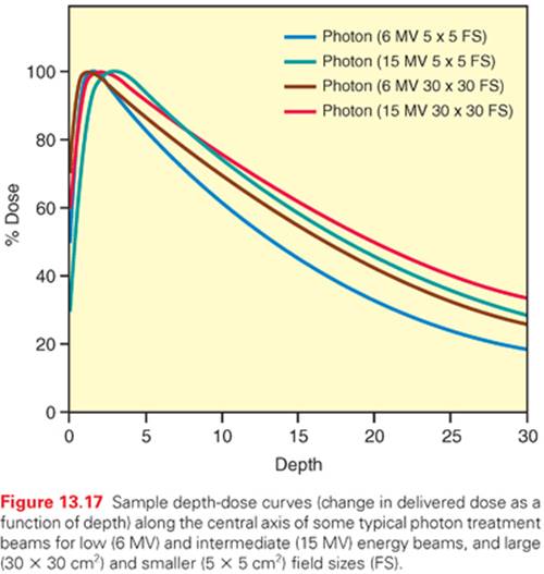
As mentioned earlier, dose deposition is a two-step process of photon interaction (proportional to the local fluence of photons) and energy transfer to the medium via the slowing down of secondary electrons. Thus, the point where a photon interacts is not the place where the dose is actually deposited, which happens over the track of the secondary electron. Dose has a very strict definition of energy absorbed per unit mass (i.e., due to the slowing down charged particles) and should be distinguished from the energy released at a point, defined as kerma (e.g., energy transfer from the scattering incident photon). Thus, although the photon beam fluence will always be greatest at the entrance to a patient or phantom, the actual absorbed dose for a megavoltage photon beam builds up over the first couple of centimeters, reaching a maximum (d-max) at a depth corresponding to the range of the higher energy Compton electrons set in motion. This turns out to be a second desirable characteristic of these beams (beyond their ability to treat deep-seated lesions), because the dose to the skin (a primary dose-limiting structure in earlier times) is greatly reduced.
The relative distributions of dose, normalized to an absolute dose measurement (using a small thimblelike air ionization chamber at a standard depth and for a standard field size according to nationally and internationally accepted protocols), are the major inputs into treatment-planning systems. The major features of these distributions are (1) the initial dose buildup up to a depth of d-max, with a more gradual drop off in dose as a function of depth into the phantom due to the attenuation and 1/r2 factors at deeper depths (relative depth dose), and (2) the shape of the dose in the plane perpendicular to the direction of the beams; both as a function of field size. Central axis depth dose curves for typical external photon beams are shown in Figure 13.17 for two beam energies and for both a large and smaller field size. Notice both the expected increase in penetration with increasing beam energy and the increase in dose at a particular depth with increasing field size; the latter effect due to increased numbers of secondary Compton-scattered photons for larger irradiated areas. The change in dose perpendicular to the central axis is less remarkable, because the beams are designed to be uniform across a field as a function of depth.
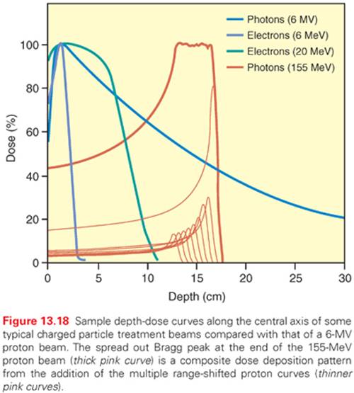
It is useful to also point out the depth dose characteristics of clinical external treatment beams produced using ionizing radiations other than photons, primarily through the direct use of charged particles. Those beams (Fig. 13.18) illustrate interesting characteristics, which, when added to the options available for treatment planning (or used by themselves), can produce advantageous results. Relative to the photon beam, the direct use of electron beams leads to deposition of dose over a more localized range, but at the expense of a relative lack of penetration. Thus, electron beams are most widely used for treating, or boosting the treatment of, more superficial tumors and regions (see the section titled Clinical Application of Types of Radiation). The heavier charged particle beams (protons and carbon ions) appear to exhibit even more interesting depth-dose characteristics, with the advantage of both (when necessary) being highly penetrating and also lacking a significant dose beyond a certain depth (a depth that can be controlled and purposefully placed, for example, at the distal edge of a target volume).
The results of measurements such as these have been modeled so as to develop dose-calculation algorithms used in treatment-planning systems. These models all use measured beam data to set or adjust parameters used by those algorithms in their dose-distribution computations. Because most of the input data used for beam fitting come from measurements in water phantoms (or waterlike plastic phantoms), patient-specific adjustments are needed for the water phantom data to account for both geometry and tissue properties. It is the task of the dose-calculation algorithms to take those changes into account. The accuracy and precision actually realized for all dose-calculation algorithms generally need to be traded off against the time required to complete the calculation. Although the availability of ever more powerful computers has made calculation time less of a concern for broad, open-beam treatment planning, issues still remain for more specialized planning exercises that use many small beams or parts of beams such as IMRT (discussed later). Typically, relative dose distributions can be computed within patients on the scale of a few millimeters with a precision of better than a few percentage points.
An important area of research is the development of treatment-planning systems that calculate dose based on the principles of how radiation interacts with tissues, rather than simply by fitting data. These approaches use Monte Carlo techniques,71,73 which build a dose distribution by summing the calculated paths of thousands of photons and scattered electrons. This approach is more accurate than beam-fitting algorithms in regions of differing tissue densities, such as the lung, and therefore, will ultimately replace the current generation of treatment-planning systems, particularly for complex conditions. However, the time to perform these calculations is still prohibitive for a clinic, and it is anticipated that Monte Carlo calculations will be introduced over a period of years by balancing the need for accuracy in a particular clinical situation with the need to initiate patient treatment.
TREATMENT PLANNING
As discussed in the previous section, single-treatment beams usually deposit more of the dose closer to where they enter the patient than they do at depths corresponding to where a deep-seated tumor might be located. The use of multiple beams entering the patient from different directions that overlap at the target produces more dose per unit volume throughout the tumor volume than is received by normal tissues. In fact, as noted earlier, the treatment-delivery machines are designed to make this easy to accomplish. Planning patient treatments under these circumstances should be a somewhat trivial matter of first selecting a sufficient number of beam angles to realize the desired buildup of the dose in the overlap region relative to the doses in the upstream parts of each beam, and then second, designing beam apertures that shape the edges of the beams to match the target. However, dose-limiting normal tissues often also lie in the paths of one or more of the beams. These normal tissues are often more sensitive to radiation damage than the tumor, and regardless, it is best practice to minimize the dose in any case as a general principle. Computerized treatment-planning systems function to develop patient-specific anatomic or geometric models and then use these models together with the beam-specific dose deposition properties (derived from phantom measurements, as previously described) to select beam angles, shapes, and intensities that meet an overall prescribed objective. That is, modern radiation oncology dose prescriptions contain both tumor and normal tissue objectives, and the modern computerized treatment-planning systems make it possible to design treatments that meet these objectives.
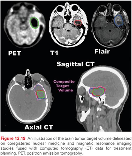
The development and use of three-dimensional (3D) models of each patient’s anatomy, treatment geometry, and dose distribution led to a paradigm shift in radiation therapy treatment planning. Computerized radiation treatment planning began in the 1980s as a mainly x-ray/CT–based reconstruction of 3D geometries from information manually contoured on multiple two-dimensional (2D) transverse CT images. Today, these models often incorporate imaging data from multiple sources. Geometrically accurate anatomic information from an x-ray/CT scan still anchors these studies (as well as provides tissue density information necessary for dose calculations). However, it is now quite common to also register the CT data set with other studies such as magnetic resonance imaging (MRI), which may add anatomic detail for soft tissues, or functional MRI or positron emission tomography (PET) studies,74,75which provide physiologic or molecular information about tumors and normal tissues. Once registered with each other, the unique or complementary information from each data set can be fused for inspection and incorporated into the design of each patient’s target and normal tissue volumes (Fig. 13.19). Beyond the ability to more fully define the extent of the primary target volume (for instance, as the encompassing envelope of disease appreciated on all the imaging studies) lies the ability to define subvolumes of the tumor volume that might be appropriate for simultaneous treatment to higher dose. For example, it should soon become possible to define different biologic components of the tumor that could potentially be targeted and then monitored for response using these same imaging techniques.76
Current treatment planning makes the tacit assumption that the planning image yields “the truth” about the location and condition of tumors and normal tissues throughout the course of treatment. However, this ignores the complexity inherent in attempting to build accurate 3D models from multimodality imaging for purposes of planning patient treatments. First, patients breathe and undergo other physiologic processes during a single treatment, changes that require dynamic modeling or other methods of accounting for the changes. Furthermore, the patient’s condition may change over time (and hence their model). Thus, a complete design and assessment of a patient undergoing high-precision treatment requires the construction of four-dimensional (4D) patient models. Indeed, the recent ready availability of multidetector CT scanners with subsecond gantry rotations, and even more recently, the availability of cone-beam CT capabilities on the radiation therapy treatment simulators and treatment machines themselves, now makes it possible to construct 4D patient models. A very active area of physics research72,75 deals with IGRT, including the formation of 4D patient models (including distortions and changes in anatomy) of the motion over time and the determination of the accumulated dose received by a moving tumor as well as the surrounding normal tissues such as uninvolved lung.
Complementary to the availability of these patient and dose models has come a much better understanding of the doses safely tolerated by normal tissues adjacent to a tumor volume (e.g., spinal cord) or surrounding it (e.g., brain, lung, liver).77 Indeed, not only has knowledge of whole organ tolerances to irradiation been obtained, but it has also become possible to characterize in some detail the complex dependence of the probability of incurring a complication with respect to the highly (intentionally) inhomogeneous dose distributions these normal tissues receive as part of the planning process designed to avoid treating them. Modeling partial organ tolerances to irradiation is of great use in planning patient treatments because it enables78 integration and manipulation of variable dose and volume distributions with respect to possible clinical outcomes.
Making the vast amount of tumor and normal tissue information useful for planning treatments requires equally sophisticated new ways of planning and delivering dose, potentially preferentially targeting subvolumes of the tumor regions or specifically avoiding selected portions of adjacent organs at risk. As mentioned earlier, modern treatment machines are capable of either varying the intensity of the radiation across each treatment port or projecting many small beams at a targeted region. This modulation of beam intensities (IMRT) from a given beam direction, together with the use of multiple beams (or parts of beams) from different directions, gives many degrees of freedom to create highly sculpted dose distributions, given that a system for designing the intensity modulation is available. Much computer programming and computational analysis has gone into the design of treatment-planning optimization systems to perform these functions.79,80
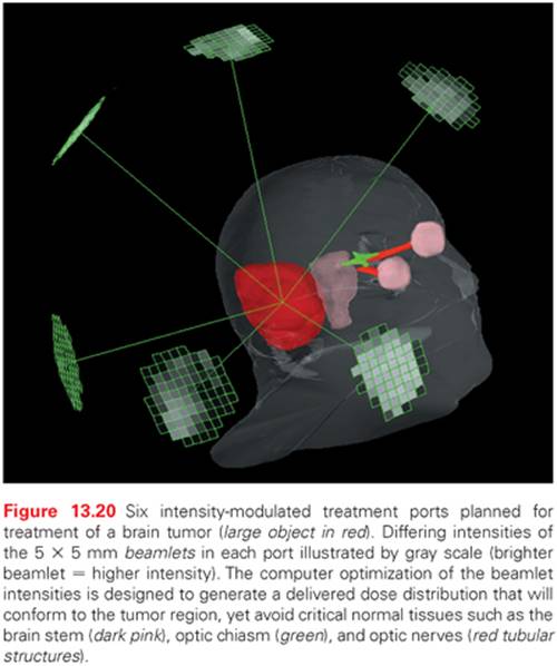
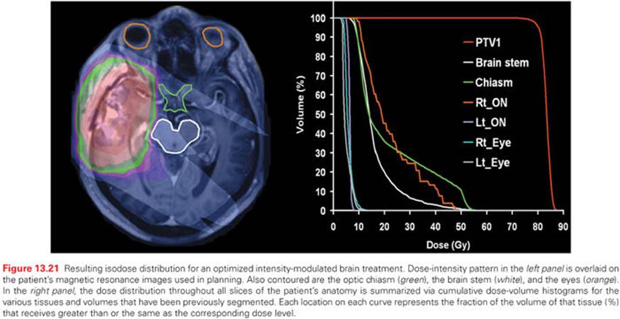
In IMRT, as most often applied, each treatment beam portal is broken down into simple basic components called beamlets, typically 0.5 to 1 cm × 1 cm in size, evenly distributed on a grid over the cross-section of each beam. Optimization begins with precomputation of the relative dose contribution that each of these beamlets gives to every subportion of tumor and normal tissue that the beamlet traverses as it goes through the patient model. Sophisticated optimization engines and search routines then iteratively alter the relative intensities of each beamlet in all the beams to minimize a cost function associated with target and normal tissue treatment goals. These, often hundreds of beamlets (each with its own intensity) (Fig. 13.20), provide the necessary flexibility and degrees of freedom to create dose distributions that can preferentially irradiate subportions of targets and also produce sharp dose gradients to avoid nearby organs at risk (Fig. 13.21). The cost-function approach also facilitates the ability to include factors such as the normal tissue and tumor-response models, mentioned previously in the optimization process, thus integrating the overall effects of the complex dose distributions across whole organ systems or target volumes within the planning process.
OTHER TREATMENT MODALITIES
Other types of external-beam radiation treatments use atomic or nuclear particles rather than photons. Beams of fast neutrons have been used for some cancers,81 primarily because of the dense ionization patterns they produce as they slow down in tissue (making cell killing less dependent on the indirect effect previously discussed). Being uncharged particles, neutron beams of therapeutic energy penetrate in tissue (have depth-dose characteristics) similar to photon beams, but with denser dose deposition in the cellular scale. Most other external-beam treatments use charged particles, primarily either electrons82(produced on the same machines used for photon beam treatments) or protons or heavier particles such as carbon ions.83,84 The latter beams have desirable dose-deposition properties (see Fig. 13.18), because they can spare tissues downstream from the target volume and generally give less overall dose to normal tissue. There can also be some radiobiologic advantage to the heavier charged particle beams, similar to neutrons. The generation and delivery of proton beams and heavier charged particle beams generally requires an accelerator (in its own vault) plus a beam transport system and some sort of treatment nozzle, often located on an isocentric gantry. The cost of the accelerator is generally leveraged by having it supply beams to multiple treatment rooms, but these units still cost many times that of a standard linear accelerator.
Brachytherapy85 is a form of treatment that uses direct placement of radioactive sources or materials within tumors (interstitial brachytherapy) or within body or surgical cavities (intracavitary brachytherapy), either permanently (allowing for full decay of short-lived radioactive materials) or temporarily (either in one extended application or over several shorter term applications). The ability to irradiate tumors from close range (even from the inside out) can lead to conformal treatments with low normal tissue doses. The radioactive isotopes most generally used for these treatments are contained within small tubelike or seedlike sealed source enclosures (which prevents direct contamination). They emit photons (gamma and x-rays) during their decay, which penetrate the source cover and interact with tissue via the same physical processes as described for external-beam treatments. The treatments have the advantage of providing a high fluence (and dose) very near each source that drops in intensity as 1 over the square of the distance from the source (1r2). Radioactive sources decay in an exponential fashion characterized by their individual half-lives. After each half-life (T1/2) the strength of each source decreases by half. Brachytherapy treatments are further generally classified into the two broad categories of low–dose-rate and high–dose-rate treatments. Low–dose-rate treatments attempt to deliver tumoricidal doses via continuous irradiation from implanted sources over a period of several days. High–dose-rate treatments use one or more higher activity sources (stored external to the patient) together with a remote applicator or source transfer system to give one or more higher dose treatments on time scales and schedules more like external-beam treatments.
Isotopes for brachytherapy treatments are selected on the basis of a combination of specific activity (i.e., how much activity can be achieved per unit mass [i.e., to keep the source sizes small]), the penetrating ability of the decay photons (together with the 1r2 fall off determines how many sources or source location will be required for treatment), and the half-life of the radioactive material (which must be accounted for in computation of dose, but also determines how often reusable sources will need to be replaced). Table 13.1 lists those isotopes most commonly used, along with some of their primary applications.

The dose-deposition patterns surrounding each type of source can be measured or computed. These data (or the parameterization of same) can be stored within a computerized treatment-planning system. Planning a brachytherapy treatment-delivery scheme (desirable source strengths and arrangements) proceeds within the planning system by distributing the sources throughout the treatment area and having the computer add up the contributions of each source to designated tumor and normal tissue locations (e.g., obtained from a CT scan). Source strengths or spacing can be adjusted until an acceptable result is obtained. Indeed, optimization systems are now routinely used to fine tune this process.
Other types of therapeutic treatments with internal sources of ionizing radiation, generally classified as systemic targeted radionuclide therapy (STaRT), use antibodies or other conjugates or carriers such as microspheres to selectively deliver radionuclides to cancer cells.86 Computing the effective dose to tumors and normal tissues via these techniques requires information on how much of the injected activity reaches the targets (biodistribution) as well as the energy and decay properties of the radionuclide being delivered. Imaging techniques and computer models are aiding in these computations.
CLINICAL APPLICATIONS OF RADIATION THERAPY
In contrast to surgical oncology and medical oncology, which focus on early- or late-stage disease, respectively, the field of radiation oncology encompasses the1p8.49 entire spectrum of oncology. Board certification requires 5 years of postdoctoral training, typically beginning with an internship in internal medicine or surgery, followed by 4 years of radiation oncology residency. Education, as defined by leaders in the field,87 begins with a thorough knowledge of the biology, physics, and clinical applications of radiation. It also includes training in the theoretical and practical aspects of the administration of radiation protectors and anticancer agents used as radiation sensitizers and the management of toxicities resulting from those treatments. In addition, residents receive education in palliative care, supportive care, and symptom and pain management. This training is in preparation for a practice that, in a given week, might include patients with a 2-mm vocal cord lesion or a 20-cm soft tissue sarcoma, both of whom can be treated with curative intent, as well as a patient with widely metastatic disease who needs palliative radiation, medical care for pain and depression, and discussion of end-of-life issues. More than 50% of (nonskin) cancer patients receive radiation therapy during the course of their illness.88
Clinical Application of Types of Radiation
Electrons are now the most widely used form of radiation for superficial treatments. Because the depth of penetration can be well controlled by the energy of the beam, it is possible to treat, for instance, skin cancer, a small part of the breast while sparing the underlying lung, or the cervical lymph nodes but not the spinal cord, which lies several centimeters more deeply. Superficial tumors, such as of skin cancers, can also be treated very effectively with low-energy (kilovoltage) photons, but their use has decreased because a separate machine is required for their production.
The main form of treatment for deep tumors is photons. As described in the Radiation Physics section, photons spare the skin and deposit dose along their entire path until the beam leaves the body. The use of multiple beams that intersect on the tumor permit high doses to be delivered to the tumor with a relative sparing of normal tissue. The pinnacle of this concept is IMRT, which uses hundreds of beams and can treat concave shapes with relative sparing of the central region (see Figs. 13.20 and 13.21). However, as each beam continues on its path beyond the tumor, this use of multiple beams means that a significant volume of normal tissue receives a low dose. There has been considerable debate concerning the magnitude of the risk of second cancers produced by radiating large volumes with low doses of radiation.89Charged particle beams (proton and carbon, in this discussion) differ from photons in that they interact only modestly with tissue until they reach the end of their path, where they then deposit the majority of their energy and stop (the Bragg peak; see Fig. 13.18). This ability to stop at a chosen depth decreases the region of low dose. The chief form of charged particle used today is the proton. In the decade from 1980 to 1990, proton therapy could deliver higher doses of radiation to the target than photon therapy because protons could produce a more rapid fall off of dose between the target and the critical normal tissue (e.g., tumor and brain stem). Therefore, initially, their main application was in the treatment uveal melanomas, base-of-skull chondrosarcomas, and chordomas. In contrast, today’s IMRT photons are more conformal in the high-dose region than protons due to the range uncertainty of the latter.90 Thus, it seems unlikely that protons will permit a higher target dose to be delivered than photons. In contrast, protons have the potential to decrease regions of low dose. This would be of particular advantage in the treatment of pediatric malignancies, where low doses of radiation would tend to increase the chance of second cancers and could affect neurocognitive function in the treatment of brain tumors.
A carbon ion beam has an additional potential biologic advantage over protons. As discussed in the section Biologic Aspects of Radiation Oncology, hypoxic cells, which are found in many tumors, are up to 3 times more resistant to photon or proton radiation than well-oxygenated cells. In contrast, hypoxia does not cause resistance to a carbon beam. Whether hypoxia is a cause of clinical resistance to fractionated radiation is still debated.91 A carbon beam is available at a few sites in Europe and Japan.
Two major issues have affected the widespread acceptance of protons. The most widely recognized is cost. Proton (approximately $120 million) and carbon beam facilities (in excess of $200 million) are substantially more expensive than a similar-sized photon facility (approximately $25 million). The operating costs appear to be significantly higher as well. Although the majority of patients who have received proton therapy have prostate cancer, there is no evidence that protons produce superior results to those obtained with IMRT planned photons.92,93 The lack of solid evidence that protons are superior to photons for any disease site and the magnitude of these costs are of societal importance.94 Although less expensive single gantry proton units are under construction, there are no functioning units at the time of this writing. A second, less well-appreciated issue concerns the need to develop full integration of charged particle beams with IGRT, as has already been accomplished with photons, although this feature is being incorporated into second-generation proton units.
Neutron therapy attracted significant interest in the 1980s, based on the principle that it would be more effective than photons against hypoxic cells that some have thought are responsible for radiation resistance of tumors. The effectiveness of neutron therapy has been limited by initial difficulties with collimation and targeting, although there is evidence that they have a role in the treatment of refractory parotid gland tumors.95
Brachytherapy refers to the placement of radioactive sources next to or inside the tumor. The chief sites where brachytherapy plays a role are in prostate and cervical cancer, although it has applications in head and neck cancers, soft tissue sarcomas, and other sites. In the case of prostate cancer, most experience is with low–dose-rate permanent implants using iodine-125 (125I) or, more recently, palladium-103 (103Pd). Over the last 5 years, there has been an increasing emphasis on improving the accuracy of seed placement, guided by ultrasound and confirmed by CT or MRI, and in skilled hands, outstanding results can be achieved.96 In the case of cervical cancer, high–dose-rate treatment, which can be performed in an outpatient setting, has essentially replaced low–dose-rate treatment, which typically requires general anesthesia and a 2-day hospital stay. The results from both techniques appear to be approximately equivalent.
Yttrium microspheres represent a distinct form of brachytherapy. These spheres carry yttrium-90 (90Y), a pure beta emitter with a range of about 1 cm. These have been used to treat both primary hepatocellular cancer and colorectal cancer metastatic to the liver (hepatic arterial or systemic chemotherapy) by administration through the hepatic artery.
TREATMENT INTENT
Radiation doses are chosen so as to maximize the chance of tumor control without producing unacceptable toxicity. The dose of radiation required depends on the tumor type, the volume of disease (number of tumor cells), and the use of radiation-modifying agents (such as chemotherapeutic drugs used as radiation sensitizers). Except for a subset of tumors that are exquisitely sensitive to radiation (e.g., seminoma, lymphoma), doses that are required are often close to the tolerance of the normal tissue. A key fact driving the choice of dose is that a 1-cm3 tumor contains approximately 1 billion cells. It follows that the reduction of a tumor that is 3 cm in diameter to 3 mm, which would be called a complete response by CT scan, would still leave 1 million tumor cells. Because each radiation fraction appears to kill a fixed fraction of the tumor, the dose to cure occult disease needs to be more similar to the dose for gross disease than one might otherwise expect. Thus, radiation doses (using the standard fractionation) of 45 to 54 Gy are typically used in the adjuvant setting when there is moderate suspicion for occult disease, 60 to 65 Gy for positive margins or when there is a high suspicion for occult disease, and 70 Gy or more for gross disease.
It is common during the course of radiation to give higher doses of radiation to regions that have a higher tumor burden. For example, regions that are suspected of harboring occult disease may be targeted to receive (in once daily 2-Gy fractions) 54 Gy, whereas, to control the gross tumor, the goal may be to administer a total dose of 70 Gy. Because the gross tumor will invariably reside within the region at risk for occult disease, it has become standard practice to deliver 50 Gy to the entire region, and then an additional boost dose of 20 Gy to the tumor. This sequence is called the shrinking field technique. With the development of IMRT, it has become possible to treat both regions with a different dose each day and achieve both goals simultaneously. For example, on each of the 35 days of treatment, the gross tumor might receive 2 Gy, and the region of occult disease 1.7 Gy, for a total dose of 59.5 Gy, which is of approximately equal biologic effectiveness to 54 Gy in 1.8-Gy fractions because of the lower dose per fraction (see the section Biologic Aspects of Radiation Oncology).
Radiation therapy alone is often used with curative intent for localized tumors. The decision to use surgery or radiation therapy involves factors determined by the tumor (e.g., is it resectable without a serious compromise in function?) and the patient (e.g., is the patient a good operative candidate?). The most common tumor in this group is prostate cancer, but patients with early-stage larynx cancer often receive radiation for voice preservation, and there are many patients with early-stage lung cancer who are not operative candidates. Control rates for these early-stage lesions are in excess of 70% (and as high as 90% for early-stage larynx cancer) and are usually a function of tumor size.
Stereotactic body radiation therapy (SBRT; sometimes called stereotactic ablative radiation) uses many (typically more than eight) cross-firing beams and provides an improved method of curing early-stage lung cancer97 and liver metastases.98 This approach uses precise localization and image guidance to deliver a small number (less than five) of high doses of radiation, with the concept of ablating the tumor, rather than using fractionation to achieve a therapeutic index (see the section title Fractionation). SBRT can provide long-term, local control rates of >90% for tumors less than 4 to 5 cm with minimal side effects.
Locally advanced or aggressive cancers can be cured with radiation alone or with a combination of radiation and chemotherapy or a molecularly targeted therapy. The most common examples here are locally advanced lung cancer, head and neck, esophageal, and cervix cancers, with cure rates in the 15% to 40% range, and are discussed in detail in their own chapters. A general principle that has emerged during the last decade is that combination chemoradiation has increased the cure rates of locally advanced cancers by 5% to 10% at the cost of increased toxicity.
An important consideration in the use of radiation (with or without chemotherapy) with curative intent is the concept of organ preservation. Perhaps the best example of achieving organ preservation in the face of gross disease involves the use of chemotherapy and radiation to replace laryngectomy in the treatment of advanced larynx cancer. Combined radiation and chemotherapy does not improve overall survival compared with radical surgery; however, the organ-conservation approach permits voice preservation in approximately two-thirds of patients with advanced larynx cancer.99 The treatment of anal cancer with chemoradiation can also be viewed in this light, with chemoradiotherapy producing organ conservation and cure rates superior to radical surgery used decades ago.100 Multiple randomized trials have demonstrated that lumpectomy plus radiation for breast cancer produces survival rates equal to that of modified radical mastectomy, while allowing for the preservation of the breast.
In the last decade, it has become clear that some patients with metastatic disease can be cured with radiation (with or without chemotherapy). The concept underlying this approach was established by the surgical practice of resecting a limited number of liver or lung metastases. A significant fraction of patients have a limited number of liver metastases that cannot be resected because of location, but are able to undergo high-dose radiation (often combined with chemotherapy). This radical approach to oligometastases101 can produce 5-year survivals in the range of 20% in selected patients.102 Patients with a limited number of lung metastases from colorectal cancer or soft tissue sarcomas are now being approached with stereotactic body radiation with a similar concept as has been used to justify surgical resection.102 In addition to the direct effect of radiation on metastatic tumor, there is now anecdotal but provocative evidence that radiation can stimulate the immune system so that tumors distant from the irradiated tumor can respond. Distant (abscopal) responses have been reported in patients who receive immune checkpoint inhibitors such as ipilimumab.103
Radiation therapy can also contribute to the cure of patients when used in an adjuvant setting. If the risk of recurrence after surgery is low or if a recurrence could be easily addressed by a second resection, adjuvant radiation therapy is not usually given. However, when a gross total resection of the tumor is still associated with a high risk of residual occult disease or if local recurrence is morbid, adjuvant treatment is often recommended. A general finding across many disease sites is that adjuvant radiation can reduce local failure rates to below 10%, even in high-risk patients, if a gross total resection is achieved. If gross disease or positive margins remain, higher doses and/or larger volumes may be required, which may be less well tolerated and are less successful in achieving tumor control.
Adjuvant therapy can be delivered before or after definitive surgery. There are some advantages to giving radiation therapy after surgery. The details of the tumor location are known and, with the surgeon’s cooperation, clips can be placed in the tumor bed, permitting increased treatment accuracy. In addition, compared with preoperative therapy, postoperative therapy is associated with fewer wound complications. However, in some cases, it is preferable to deliver preoperative radiation. Radiation can shrink the tumor, diminishing the extent of the resection, or making an unresectable tumor resectable. In the case of rectal cancer, the response to treatment may carry more prognostic information than the initial TNM staging.104 In patients who will undergo significant surgeries (particularly a Whipple procedure or an esophageal resection), preoperative (sometimes called neoadjuvant) therapy can be more reliably administered than postoperative therapy. Most importantly, after resection of abdominal or pelvic tumors (such as rectal cancers or retroperitoneal sarcomas), the small bowel may become fixed by adhesions in the region requiring treatment, thus increasing the morbidity of postoperative treatment. A randomized trial has shown that preoperative therapy produces fewer gastrointestinal side effects and has at least as good efficacy as postoperative adjuvant therapy for locally advanced rectal cancer.105 Taken together, there appears to be a trend toward preoperative or neoadjuvant therapy in cancers of the gastrointestinal track (esophagus, stomach, pancreas, rectum), postoperative radiation seems to be favored in head and neck, lung, and breast cancer, and soft tissue sarcoma seems equally split.
The effectiveness of adjuvant therapy in decreasing local recurrence has been demonstrated in randomized trials in lung, rectal, and breast cancers. More recently, randomized trials have shown that postmastectomy radiation improved the survival for women with breast cancer and four or more positive lymph nodes, all of whom also received adjuvant chemotherapy. A fascinating analysis has revealed that, across many treatment conditions, each 4% increase in 5-year local control is associated with a 1% increase in 5-year survival.106 It has been proposed that the long-term survival benefit of radiation in these more recent studies was revealed by the introduction of effective chemotherapy, which prevented such a high fraction of women from dying early with metastatic disease.107 This concept has been developed into a hypothesis that the effect of adjuvant radiation on survival will depend on the effectiveness of adjuvant chemotherapy. If chemotherapy is either ineffective or very effective, adjuvant radiation may have little influence on the survival in a disease in which systemic relapse dominates survival. Radiation will have its greatest impact on survival when chemotherapy is moderately effective.108
In addition to these curative roles, radiation plays an important part in palliative treatment. Perhaps most importantly, emergency irradiation can begin to reverse the devastating effects of spinal cord compression and of superior vena cava syndrome. A single 8-Gy fraction is highly effective for many patients with bone pain from a metastatic lesion. There is increasing evidence of the effectiveness of body stereotactic radiation to treat vertebral body metastases in patients who have a long projected survival or who need retreatment after previous radiation.109 Stereotactic treatment can relieve symptoms from a small number of brain metastasis, and fractionated whole-brain radiation can mitigate the effects of multiple metastases. Bronchial obstruction can often be relieved by a brief course of treatment as can duodenal obstruction from pancreatic cancer. Palliative treatment is usually delivered in a smaller number of larger radiation fractions (see the section titled Fractionation) because the desire to simplify the treatment for a patient with limited life expectancy outweighs the somewhat increased potential for late side effects.
FRACTIONATION
Two crucial features that influence the effectiveness of a physical dose of radiation are the dose given in each radiation treatment (i.e., the fraction) and the total amount of time required to complete the course of radiation. Standard fractionation for radiation therapy is defined as the delivery of one treatment of 1.8 to 2.25 Gy per day. This approach produces a fairly well-understood chance of tumor control and risk of normal tissue damage (as a function of volume). By altering the fractionation schemes, one may be able to improve the outcome for patients undergoing curative treatment or to simplify the treatment for patients receiving palliative therapy.
Two forms of altered fractionation have been tested for patients undergoing curative treatment: accelerated fractionation and hyperfractionation. Accelerated fractionation emerged from analyses of the control of head and neck cancer as a function of dose administered and total treatment time. It was found that with an increasing dose there was increasing local control, but that protraction of treatment was associated with a loss of local control that was equivalent to about 0.75 Gy per day.110 The data were best modeled by assuming that, approximately 2 weeks into treatment, tumor cells began to proliferate more rapidly than they were proliferating early in treatment (called accelerated repopulation).111 In accelerated fractionation, the goal is to complete radiation before the accelerated tumor cell proliferation occurs. The most common method of achieving accelerated fractionation is to give a standard fraction to the entire field in the morning and to give a second treatment to the boost field in the afternoon (called concomitant boost). As in standard radiation, the boost would be given by extending the length of the treatment course; this concomitant boost approach can shorten treatment from 7 weeks to 5 weeks in head and neck cancer.
The second approach to altering fractionation is called hyperfractionation. Hyperfractionation is defined as the use of more than one fraction per day separated by more than 6 hours (see the section titledBiologic Aspects of Radiation Oncology), with a dose per fraction that is less than standard. Hyperfractionation is expected to produce fewer late complications for the same acute effects against both rapidly dividing normal tissues and tumors. Pure hyperfractionation might give 1 Gy twice a day, so that the total dose per day would be 2 Gy, and thus be equal to standard fractionation. In practice, hyperfractionated treatments are usually in the range of 1.2 Gy, which means that, compared with a standard fractionation, a somewhat higher dose is administered during the same period of time (so that most hyperfractionation also includes modest acceleration). The overall effect is to increase the acute toxicity (which resolves) and tumor response, while not increasing the (dose-limiting) late toxicity, which can improve cure rate. Both accelerated fractionation and hyperfractionation have been demonstrated in a meta-analysis to be superior to standard fractionation in the treatment of head and neck cancer with radiation alone.112 However, a recent randomized trial has shown that there is no increase in control or survival, but there is an increase toxicity using chemotherapy with hyperfractionation compared to standard chemoradiation; therefore, the use of altered fractionation schemes has decreased dramatically during the last few years.113
Hypofractionation refers to the administration of a smaller number of larger fractions than is standard. Hypofractionation might be expected to cause more late toxicity for the same antitumor effect than standard or hyperfractionation. In the past, this approach was reserved for palliative cases, with the sense that a modest potential for increased late toxicity was not a major concern in patients with limited life expectancy. However, more recently, it has been proposed that the ability to better exclude normal tissue by using IGRT may permit hypofractionation to be used safely and that, in the specific case of prostate cancer, hypofractionation may have beneficial effects.114
ADVERSE EFFECTS
Radiation produces adverse effects in normal tissues. Although these are discussed in detail in later chapters as part of comprehensive discussions of organ toxicity, it is worth making some general comments here from the perspective of how radiation biology relates to the clinical toxicities. The term radiation toxicity is used to describe the adverse effects caused by radiation alone and radiation plus chemotherapy. Although this latter toxicity would be better labeled as combined modality toxicity, the pattern typically resembles a more severe form of the toxicity produced by radiation alone. Adverse effects from radiation can be divided into acute, subacute, and chronic (or late) effects. Acute effects are common, rarely serious, and usually self-limiting. Acute effects tend to occur in organs that depend on rapid self-renewal, most commonly the skin or mucosal surfaces (oropharynx, esophagus, small intestine, rectum, and bladder). This is due to radiation-induced cell death that occurs during mitosis, so that cells that divide rapidly show the most rapid cell loss. In the treatment of head and neck cancer, mucositis becomes worse during the first 3 to 4 weeks of therapy, but then will often stabilize as the normal mucosa cell proliferation increases in response to mucosal cell loss. It seems likely that normal tissue stem cells are relatively resistant to radiation compared with the more differentiated cells, because these stem cells survive to permit the normal mucosa to reepithelialize. Acute side effects typically resolve within 1 to 2 weeks of treatment completion, although occasionally these effects are so severe that they lead to consequential late effects, as described later.
Because lymphocytes are exquisitely sensitive to radiation, there has been considerable investigation into the effects of radiation on immune function. In contrast to mucosal cell killing, which requires mitosis, radiation kills lymphocytes in all phases of the cell cycle by apoptosis, so that lymphocyte counts decrease within days of initiating treatment. These effects do not tend to put patients at risk for infection, because granulocytes, which are chiefly responsible for combating infections, are relatively unaffected.
Two acute side effects of radiation do not fit neatly into these models relating to cell kill: nausea115,116 and fatigue.117,118 The origin of radiation-induced nausea is not related to acute cell loss, because it can occur within hours of the first treatment. Nausea is usually associated with radiation of the stomach, but it can sometimes occur during brain irradiation or from large-volume irradiation that involves neither the brain nor the stomach. Irradiation typically produces fatigue, even if relatively small volumes are irradiated. It seems likely that the origins of both of these abscopal effects of radiation (i.e., effects that occur systemically or at a distance for the site of irradiation) are related to the release of cytokines, but little is known.
Radiation can also produce subacute toxicities in the form of radiation pneumonitis and radiation-induced liver disease. These typically occur 2 weeks to 3 months after radiation is completed. The risk of radiation pneumonitis and radiation-induced liver disease is proportional to the mean dose delivered.119,120 Thus, the 3D tools that permit the calculation of dose-volume histograms (described in the physics section) are currently used to determine the maximum safe treatment that can be delivered in terms of dose and volume. These toxicities appear to be initiated subclinically during the course of radiation as a cascade of cytokines in which TGF-β, tumor necrosis factor α, interleukin 6, and other cytokines play a role.121 High TGF-β plasma levels during a course of treatment have been found to be associated with a greater risk of radiation pneumonitis.122 Thus, in the future, we might look toward a combination of physical dose delivery, measured by the dose-volume histogram, the functional imaging of normal tissue damage, and the detection of biomarkers of toxicity, such as TGF-β, to improve the ability to individualize therapy. Attempts to determine the genomic basis of radiation sensitivity, beyond the known rare genetic defects such as ataxia telangiectasia, have not yet been successful.123
Late effects, which are typically seen 6 or more months after a course of radiation, include fibrosis, fistula formation, or long-term organ damage. Two theories for the origin of late effects have been put forth: late damage to the microvasculature and direct damage to the parenchyma. Although the vascular damage theory is attractive, it does not account for the differing sensitivities of organs to radiation. Perhaps the microvasculature is unique in each organ.124 Regardless of the mechanism of toxicity, the tolerance of whole-organ radiation is now fairly well established (Table 13.2). Late complications can also be divided into two categories: consequential and true late effects. The best example of a consequential late effect is fibrosis and dysphagia after high-dose chemoradiation for head and neck cancer. Here, late fibrosis or ulceration appears to be the result of the mucosa becoming denuded for a prolonged time period. Late consequential effects are distinct from true late effects, which can follow a normal treatment course of self-limited toxicity and a 6-month or more symptom-free period. Examples of true late effects are radiation myelitis, radiation brain necrosis, and radiation-induced bowel obstruction. In the past, radiation fibrosis was thought to be an irreversible condition. Therefore, an exciting recent development is that severe radiation-induced breast fibrosis is an active process that can be reversed by drug therapy (pentoxifylline and vitamin E).125
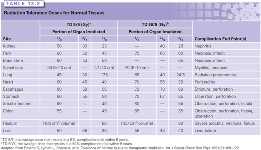
PRINCIPLES OF COMBINING ANTICANCER AGENTS WITH RADIATION THERAPY
Combining chemotherapy with radiation therapy has produced important improvements in treatment outcome. Randomized clinical trials show improved local control and survival through the use of concurrent chemotherapy and radiation therapy for patients with high-grade gliomas and locally advanced cancers of the head and neck, lung, esophagus, stomach, rectum, prostate, and anus. There are least two proposed reasons why chemoradiotherapy might be successful. The first is radiosensitization. In the laboratory, radiosensitization is defined as a synergistic relationship, using mathematical approaches such as isobologram or median effect analysis.126,127 The underlying concept is that the observed effect of using chemotherapy and radiation concurrently is greater than simply adding the two together. A second proposed reason to combine radiation and chemotherapy is to realize the benefit of improved local control radiation along with the systemic effect of chemotherapy, a concept called spatial additivity.128
Clinical results show that both radiosensitization and spatial additivity contribute to varying extents in different clinical settings. In the case of head and neck cancer, radiosensitization predominates. This conclusion is supported by the meta-analysis of head and neck cancer: sequential chemotherapy and radiotherapy produces little if any improvement in survival, whereas concurrent chemoradiation produces a significant increase in survival.129 Furthermore, in the early positive studies using concurrent chemoradiation, systemic metastases were unaffected even though survival was improved. Radiosensitization may also predominate in the success of chemoradiotherapy for locally advanced lung cancer. For instance, although initial studies indicated that sequential chemotherapy and radiation had some benefit for lung cancer,130 more recent work indicates that concurrent therapy is superior, and it is now the standard treatment.131 However, there are also examples of spatial additivity. For example, both radiosensitization and spatial additivity is provided by the use of chemoradiation for locally advanced cervical cancer in that both local and systemic relapses are decreased by combined therapy.132
By targeting the aberrant growth factor or proangiogenic pathways that are specific to cancer cells rather than all rapidly proliferating cells, molecularly targeted therapies offer the potential to improve outcome without increasing toxicity. Even a selective cytostatic effect against the tumor would be predicted to act synergistically with radiation (Fig. 13.22). Although preclinical studies (summarized in the previous biology section) have highlighted the potential therapeutic gains that could be achieved by adding EGFR inhibitors to radiation, the best validation of this combination has been from the results of clinical trials in head and neck cancer. A phase III clinical trial demonstrated that, in a cohort of 424 patients with local–regionally advanced squamous cell carcinoma of the head and neck, the addition of cetuximab nearly doubled the median survival of patients (compared to radiotherapy alone), from 28 to 54 months. This study represents the first major success achieved by the addition of an EGFR antagonist to radiotherapy. This improvement was achieved without enhanced toxicity. Notably, the rates of pharyngitis and weight loss were identical in the two arms.60 Local control was improved rather than the development of metastases, suggesting synergy rather than spatial additivity. Thus, the principle that can be derived from this study is that in tumors expressing high EGFR levels and that are likely to depend on aberrant EGF signaling, combining a true cytotoxic agent such as radiation with a cytostatic agent such as cetuximab has considerable promise.
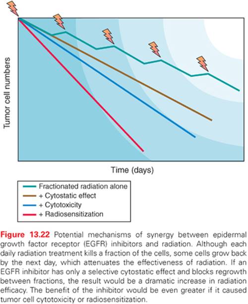
Because of the success of chemoradiotherapy, the natural tendency has not been to substitute molecularly targeted agents such as cetuximab for chemotherapy, but to add cetuximab to chemoradiotherapy. Thus, the combination of cisplatin, cetuximab, and radiation was recently found to have the same control rate as cisplatin and radiation for patients with locally advanced head and neck cancer, but the cetuximab arm had greater toxicity. Unfortunately, the triple therapy was never evaluated preclinically, and it has been shown preclinically that when EGFR inhibitors are given prior to chemotherapy, they can produce antagonism.133 The principles of adding molecularly targeted therapy to chemoradiation are still evolving.63
REFERENCES
1. Hall EJ, Giaccia AJ. Radiobiology for the Radiologist. Philadelphia: Lippincott Williams & Williams; 2012.
2. Fowler JF. Developing aspects of radiation oncology. Med Phys 1981;8:427–434.
3. Lavin MF. Ataxia-telangiectasia: from a rare disorder to a paradigm for cell signalling and cancer. Nat Rev Mol Cell Biol 2008;9:759–769.
4. Thompson LH. Recognition, signaling, and repair of DNA double-strand breaks produced by ionizing radiation in mammalian cells: the molecular choreography. Mutat Res 2012;751:158–246.
5. Sherr CJ, McCormick F. The RB and p53 pathways in cancer. Cancer Cell 2002;2:103–112.
6. Bartek J, Lukas J. Chk1 and Chk2 kinases in checkpoint control and cancer. Cancer Cell 2003;3:421–429.
7. Kitagawa R, Bakkenist CJ, McKinnon PJ, et al. Phosphorylation of SMC1 is a critical downstream event in the ATM-NBS1-BRCA1 pathway. Genes Dev 2004;18:1423–1438.
8. Abraham RT. Cell cycle checkpoint signaling through the ATM and ATR kinases. Genes Dev 2001;15:2177–2196.
9. Mailand N, Podtelejnikov AV, Groth A, et al. Regulation of G(2)/M events by Cdc25A through phosphorylation-dependent modulation of its stability. EMBO J 2002;21:5911–5920.
10. Donzelli M, Draetta GF. Regulating mammalian checkpoints through Cdc25 inactivation. EMBO Rep 2003;4:671–677.
11. Tsvetkov L. Polo-like kinases and Chk2 at the interface of DNA damage checkpoint pathways and mitotic regulation. IUBMB Life 2004;56:449–456.
12. Dai Y, Grant S. New insights into checkpoint kinase 1 in the DNA damage response signaling network. Clin Cancer Res 2010;16:376–383.
13. Giaccia A, Weinstein R, Hu J, et al. Cell cycle-dependent repair of double-strand DNA breaks in a gamma-ray-sensitive Chinese hamster cell. Somat Cell Mol Genet 1985;11:485–491.
14. Kemp LM, Sedgwick SG, Jeggo PA. X-ray sensitive mutants of Chinese hamster ovary cells defective in double-strand break rejoining. Mutat Res 1984;132:189–196.
15. Helleday T, Lo J, van Gent DC, et al. DNA double-strand break repair: from mechanistic understanding to cancer treatment. DNA Repair (Amst) 2007;6:923–935.
16. Hefferin ML, Tomkinson AE. Mechanism of DNA double-strand break repair by non-homologous end joining. DNA Repair (Amst) 2005;4:639–648.
17. Ma Y, Pannicke U, Schwarz K, et al. Hairpin opening and overhang processing by an Artemis/DNA-dependent protein kinase complex in nonhomologous end joining and V(D)J recombination. Cell 2002;108:781–794.
18. Grawunder U, Wilm M, Wu X, et al. Activity of DNA ligase IV stimulated by complex formation with XRCC4 protein in mammalian cells. Nature 1997;388:492–495.
19. Chen L, Trujillo K, Sung P, et al. Interactions of the DNA ligase IV-XRCC4 complex with DNA ends and the DNA-dependent protein kinase. J Biol Chem 2000;275:26196–26205.
20. Tsukuda T, Fleming AB, Nickoloff JA, et al. Chromatin remodelling at a DNA double-strand break site in Saccharomyces cerevisiae. Nature 2005;438:379–383.
21. Yang YG, Saidi A, Frappart PO, et al. Conditional deletion of Nbs1 in murine cells reveals its role in branching repair pathways of DNA double-strand breaks. EMBO J 2006;25:5527–5538.
22. Esashi F, Galkin VE, Yu X, et al. Stabilization of RAD51 nucleoprotein filaments by the C-terminal region of BRCA2. Nat Struct Mol Biol 2007;14:468–474.
23. Sleeth KM, Sorensen CS, Issaeva N, et al. RPA mediates recombination repair during replication stress and is displaced from DNA by checkpoint signalling in human cells. J Mol Biol 2007;373:38–47.
24. Littlefield LG, Kleinerman RA, Sayer AM, et al. Chromosome aberrations in lymphocytes—biomonitors of radiation exposure. Prog Clin Biol Res 1991;372:387–397.
25. Toulany M, Rodemann HP. Membrane receptor signaling and control of DNA repair after exposure to ionizing radiation. Nuklearmedizin 2010;49:S26–S30.
26. Barcellos-Hoff MH, Akhurst RJ. Transforming growth factor-beta in breast cancer: too much, too late. Breast Cancer Res 2009;11:202.
27. Deng X, Yin X, Allan R, et al. Ceramide biogenesis is required for radiation-induced apoptosis in the germ line of C. elegans. Science 2008;322:110–115.
28. Thompson LH, Suit HD. Proliferation kinetics of x-irradiated mouse L cells studied WITH TIME-lapse photography. II. Int J Radiat Biol Relat Stud Phys Chem Med 1969;15:347–362.
29. Sowa Resat MB, Morgan WF. Radiation-induced genomic instability: a role for secreted soluble factors in communicating the radiation response to non-irradiated cells. J Cell Biochem 2004;92:1013–1019.
30. Elkind MM. The initial part of the survival curve: does it predict the outcome of fractionated radiotherapy? Radiat Res 1988;114:425–436.
31. McCulloch EA, Till JE. The sensitivity of cells from normal mouse bone marrow to gamma radiation in vitro and in vivo. Radiat Res 1962;16:822–832.
32. Withers HR. Recovery and repopulation in vivo by mouse skin epithelial cells during fractionated irradiation. Radiat Res 1967;32:227–239.
33. Withers HR, Elkind MM. Microcolony survival assay for cells of mouse intestinal mucosa exposed to radiation. Int J Radiat Biol Relat Stud Phys Chem Med 1970;17:261–267.
34. Suit H, Wette R. Radiation dose fractionation and tumor control probability. Radiat Res 1966;29:267–281.
35. O’Neill K, Lyons SK, Gallagher WM, et al. Bioluminescent imaging: a critical tool in pre-clinical oncology research. J Pathol 2010;220:317–327.
36. Hewitt HB, Wilson CW. Survival curves for tumor cells irradiated in vivo. Ann N Y Acad Sci 1961;95:818–827.
37. Hill RP, Bush RS. A lung-colony assay to determine the radiosensitivity of cells of a solid tumour. Int J Radiat Biol Relat Stud Phys Chem Med 1969;15:435–444.
38. Elkind MM, Sutton-Gilbert H, Moses WB, et al. Radiation response of mammalian cells grown in culture. V. Temperature dependence of the repair of x-ray damage in surviving cells (aerobic and hypoxic). Radiat Res 1965;25:359–376.
39. Elkind MM, Whitmore GF. Radiobiology of Cultured Mammalian Cells. New York: Gordon and Breach; 1967.
40. Mottram JC. Factors of importance in radiosensitivity of tumors. Br J Radiol 1936;9:606.
41. Palcic B, Skarsgard LD. Reduced oxygen enhancement ratio at low doses of ionizing radiation. Radiat Res 1984;100:328–339.
42. Thomlinson RH, Gray LH. The histological structure of some human lung cancers and the possible implications for radiotherapy. Br J Cancer 1955;9:539–549.
43. Overgaard J, Horsman MR. Modification of hypoxia-induced radioresistance in tumors by the use of oxygen and sensitizers. Semin Radiat Oncol 1996;6:10–21.
44. Machtay M, Pajak TF, Suntharalingam M, et al. Radiotherapy with or without erythropoietin for anemic patients with head and neck cancer: a randomized trial of the Radiation Therapy Oncology Group (RTOG 99-03). Int J Radiat Oncol Biol Phys 2007;69:1008–1017.
45. Henke M, Laszig R, Rube C, et al. Erythropoietin to treat head and neck cancer patients with anaemia undergoing radiotherapy: randomised, double-blind, placebo-controlled trial. Lancet 2003;362:1255–1260.
46. Janssens GO, Rademakers SE, Terhaard CH, et al. Accelerated radiotherapy with carbogen and nicotinamide for laryngeal cancer: results of a phase III randomized trial. J Clin Oncol 2012;30:1777–1783.
47. Willett CG, Boucher Y, di Tomaso E, et al. Direct evidence that the VEGF-specific antibody bevacizumab has antivascular effects in human rectal cancer. Nat Med 2004;10:145–147.
48. Lapi SE, Voller TF, Welch MJ. Positron emission tomography imaging of hypoxia. PET Clin 2009;4:39–47.
49. Lee NY, Mechalakos JG, Nehmeh S, et al. Fluorine-18-labeled fluoromisonidazole positron emission and computed tomography-guided intensity-modulated radiotherapy for head and neck cancer: a feasibility study. Int J Radiat Oncol Biol Phys 2008;70:2–13.
50. Schaue D, Xie MW, Ratikan JA, et al. Regulatory T cells in radiotherapeutic responses. Front Oncol 2012;2:90.
51. Formenti SC, Demaria S. Combining radiotherapy and cancer immunotherapy: a paradigm shift. J Natl Cancer Inst 2013;105:256–265.
52. Lawrence TS, Davis MA, Tang HY, et al. Fluorodeoxyuridine-mediated cytotoxicity and radiosensitization require S phase progression. Int J Radiat Biol 1996;70:273–280.
53. Lawrence TS, Chang EY, Hahn TM, et al. Radiosensitization of pancreatic cancer cells by 2′,2′-difluoro-2′-deoxycytidine. Int J Radiat Oncol Biol Phys 1996;34:867–872.
54. Eisbruch A, Shewach DS, Bradford CR, et al. Radiation concurrent with gemcitabine for locally advanced head and neck cancer: a phase I trial and intracellular drug incorporation study. J Clin Oncol 2001;19:792–799.
55. Ben-Josef E, Schipper M, Francis IR, et al. A phase I/II trial of intensity modulated radiation (IMRT) dose escalation with concurrent fixed-dose rate gemcitabine (FDR-G) in patients with unresectable pancreatic cancer. Int J Radiat Oncol Biol Phys 2012;84:1166–1171.
56. Wolff RA, Evans DB, Gravel DM, et al. Phase I trial of gemcitabine combined with radiation for the treatment of locally advanced pancreatic adenocarcinoma. Clin Cancer Res 2001;7:2246–2253.
57. Wilson GD, Bentzen SM, Harari PM. Biologic basis for combining drugs with radiation. Semin Radiat Oncol 2006;16:2–9.
58. Bradley JD, Paulus R, Graham MV, et al. Phase II trial of postoperative adjuvant paclitaxel/carboplatin and thoracic radiotherapy in resected stage II and IIIA non-small-cell lung cancer: promising long-term results of the Radiation Therapy Oncology Group—RTOG 9705. J Clin Oncol 2005;23:3480–3487.
59. Nyati MK, Morgan MA, Feng FY, et al. Integration of EGFR inhibitors with radiochemotherapy. Nat Rev Cancer 2006;6:876–885.
60. Bonner JA, Harari PM, Giralt J, et al. Radiotherapy plus cetuximab for locoregionally advanced head and neck cancer: 5-year survival data from a phase 3 randomised trial, and relation between cetuximab-induced rash and survival. Lancet Oncol 2010;11:21–28.
61. Ang KK, Zhang QE, Rosenthal DI, et al. A randomized phase III trial (RTOG 0522) of concurrent accelerated radiation plus cisplatin with or without cetuximab for stage III-IV head and neck squamous cell carcinomas (HNC). J Clin Oncol 2011;29.
62. Engelke CG, Parsels LA, Qian Y, et al. Sensitization of pancreatic cancer to chemoradiation by the Chk1 inhibitor MK8776. Clin Cancer Res 2013;19:4412–4421.
63. Morgan MA, Parsels LA, Maybaum J, et al. Improving the efficacy of chemoradiation with targeted agents. Cancer Discov 2014;4:280–291.
64. Chalmers AJ, Lakshman M, Chan N, et al. Poly(ADP-ribose) polymerase inhibition as a model for synthetic lethality in developing radiation oncology targets. Semin Radiat Oncol 2010;20:274–281.
65. Brizel DM, Wasserman TH, Henke M, et al. Phase III randomized trial of amifostine as a radioprotector in head and neck cancer. J Clin Oncol 2000;18:3339–3345.
66. Winczura P, Jassem J. Combined treatment with cytoprotective agents and radiotherapy. Cancer Treat Rev 2010;36:268–275.
67. Van Dyk J, ed. Radiation oncology medical physics resources for working, teaching, and learning. In: The Modern Technology of Radiation Oncology. Volume 3. Medical Physics Publishing Web site. http://www.medicalphysics.org/vandykch16.pdf. Madison, WI: Medical Physics Publishing; 2013.
68. Karzmark C, Nunan C, Tanabe E. Medical Electron Accelerators. New York: McGraw-Hill Ryerson; 1993.
69. Greene D, Williams P. Linear Accelerators for Radiation Therapy. 2nd ed. New York: Taylor and Francis Group; 1997.
70. Fenwick JD, Tome WA, Soisson ET, et al. Tomotherapy and other innovative IMRT delivery systems. Semin Radiat Oncol 2006;16:199–208.
71. Curran B, Balter J, Chetty I, eds. Integrating New Technologies into the Clinic: Monte Carlo and Image-Guided Radiation Therapy. Madison, WI: Medical Physics Publishing; 2006.
72. Bourland J, ed. Image-Guided Radiation Therapy. Boca Raton, FL: Taylor & Francis; 2012.
73. Seco J, Verhaegen F eds. Monte Carlo Techniques in Radiation Therapy. Boca Raton, FL: Taylor & Francis; 2013.
74. Kessler ML. Image registration and data fusion in radiation therapy. Br J Radiol 2006;79:S99–S108.
75. Brock K, ed. Image Processing in Radiation Therapy. Boca Raton, FL: Taylor & Francis Group; 2014.
76. Sovik A, Malinen E, Olsen DR. Strategies for biologic image-guided dose escalation: a review. Int J Radiat Oncol Biol Phys 2009;73:650–658.
77. Marks LB, Ten Haken RK, Martel MK. Guest editor’s introduction to QUANTEC: a users guide. Int J Radiat Oncol Biol Phys 2010;76:S1–S2.
78. Li A, Alber M, Deasy JO, et al. The use and QA of biologically related models for treatment planning: short report of the TG-166 of the therapy physics committee of the AAPM. Med Phys 2012;39:1386–1409.
79. Bortfeld T. IMRT: a review and preview. Phys Med Biol 2006;51:R363–R379.
80. Webb S. Contemporary IMRT Developing Physics and Clinical Implementation. London: IOP Publishing; 2005.
81. Maughan R, Yudelev M. Neutron therapy. In: Van Dyk J, ed. The Modern Technology of Radiation Oncology. Madison, WI: Medical Physics Publishing; 1999.
82. Hogstrom KR, Almond PR. Review of electron beam therapy physics. Phys Med Biol 2006;51:R455–R489.
83. Schlegel W, Bortfeld T, Grosu A, eds. New Technologies in Radiation Oncology. Heidelberg: Springer-Verlag; 2006.
84. Ma C-MC, Lomax T, eds. Proton and Carbon Ion Therapy. Boca Raton, FL: Taylor & Francis Group; 2012.
85. Venselaar J, Meigooni A, Baltas D, et al., eds. Comprehensive Brachytherapy: Physical and Clinical Aspects. Boca Raton, FL: Taylor & Francis Group; 2012.
86. Meredith RF. Systemic targeted radionuclide therapy symposium introduction. Int J Radiat Oncol Biol Phys 2006;66:S7.
87. Tripuraneni P, Watson RL, Ang KK, et al. Intersociety Radiation Oncology Summit-SCOPE II. Int J Radiat Oncol Biol Phys 2008;72:323–326.
88. Delaney G, Jacob S, Featherstone C, et al. The role of radiotherapy in cancer treatment: estimating optimal utilization from a review of evidence-based clinical guidelines. Cancer 2005;104:1129–1137.
89. Zelefsky MJ, Housman DM, Pei X, et al. Incidence of secondary cancer development after high-dose intensity-modulated radiotherapy and image-guided brachytherapy for the treatment of localized prostate cancer. Int J Radiat Oncol Biol Phys 2012;83:953–959.
90. Combs SE, Laperriere N, Brada M. Clinical controversies: proton radiation therapy for brain and skull base tumors. Semin Radiat Oncol 2013;23:120–126.
91. Overgaard J. Hypoxic radiosensitization: adored and ignored. J Clin Oncol 2007;25:4066–4074.
92. Mouw KW, Trofimov A, Zietman AL, et al. Clinical controversies: proton therapy for prostate cancer. Semin Radiat Oncol 2013;23:109–114.
93. Yu JB, Soulos PR, Herrin J, et al. Proton versus intensity-modulated radiotherapy for prostate cancer: patterns of care and early toxicity. J Natl Cancer Inst 2013;105:25–32.
94. Brada M, Pijls-Johannesma M, De Ruysscher D. Proton therapy in clinical practice: current clinical evidence. J Clin Oncol 2007;25:965–970.
95. Douglas JG, Koh WJ, Austin-Seymour M, et al. Treatment of salivary gland neoplasms with fast neutron radiotherapy. Arch Otolaryngol Head Neck Surg 2003;129:944–948.
96. Shilkrut M, Merrick GS, McLaughlin PW, et al. The addition of low-dose-rate brachytherapy and androgen-deprivation therapy decreases biochemical failure and prostate cancer death compared with dose-escalated external-beam radiation therapy for high-risk prostate cancer. Cancer 2013;119:681–690.
97. Iyengar P, Timmerman R. Stereotactic ablative radiotherapy for non-small cell lung cancer: rationale and outcomes. J Natl Compr Canc Netw 2012;10:1514–1520.
98. Lo SS, Moffatt-Bruce SD, Dawson LA, et al. The role of local therapy in the management of lung and liver oligometastases. Nat Rev Clin Oncol 2011;8:405–416.
99. Forastiere AA, Zhang Q, Weber RS, et al. Long-term results of RTOG 91-11: a comparison of three nonsurgical treatment strategies to preserve the larynx in patients with locally advanced larynx cancer. J Clin Oncol 2013;31:845–852.
100. Gunderson LL, Winter KA, Ajani JA, et al. Long-term update of US GI intergroup RTOG 98-11 phase III trial for anal carcinoma: survival, relapse, and colostomy failure with concurrent chemoradiation involving fluorouracil/mitomycin versus fluorouracil/cisplatin. J Clin Oncol 2012;30:4344–4351.
101. Hellman S, Weichselbaum RR. Oligometastases. J Clin Oncol 1995;13:8–10.
102. Hortobagyi GN. Can we cure limited metastatic breast cancer? J Clin Oncol 2002;20:620–623.
103. Stamell EF, Wolchok JD, Gnjatic S, et al. The abscopal effect associated with a systemic anti-melanoma immune response. Int J Radiat Oncol Biol Phys 2013;85:293–295.
104. Nagtegaal ID, Gosens MJ, Marijnen CA, et al. Combinations of tumor and treatment parameters are more discriminative for prognosis than the present TNM system in rectal cancer. J Clin Oncol 2007;25:1647–1650.
105. Sauer R, Becker H, Hohenberger W, et al. Preoperative versus postoperative chemoradiotherapy for rectal cancer. N Engl J Med 2004;351:1731–1740.
106. Clarke M, Collins R, Darby S, et al. Effects of radiotherapy and of differences in the extent of surgery for early breast cancer on local recurrence and 15-year survival: an overview of the randomised trials. Lancet 2005;366:2087–2106.
107. Hellman S. Stopping metastases at their source. N Engl J Med 1997;337:996–997.
108. Marks LB, Prosnitz LR. Postoperative radiotherapy for lung cancer: the breast cancer story all over again? Int J Radiat Oncol Biol Phys 2000;48:625–627.
109. Wang XS, Rhines LD, Shiu AS, et al. Stereotactic body radiation therapy for management of spinal metastases in patients without spinal cord compression: a phase 1-2 trial. Lancet Oncol 2012;13:395–402.
110. Tarnawski R, Fowler J, Skladowski K, et al. How fast is repopulation of tumor cells during the treatment gap? Int J Radiat Oncol Biol Phys 2002;54:229–236.
111. Peters LJ, Withers HR. Applying radiobiological principles to combined modality treatment of head and neck cancer—the time factor. Int J Radiat Oncol Biol Phys 1997;39:831–836.
112. Bourhis J, Overgaard J, Audry H, et al. Hyperfractionated or accelerated radiotherapy in head and neck cancer: a meta-analysis. Lancet 2006;368:843–854.
113. Bourhis J, Sire C, Graff P, et al. Concomitant chemoradiotherapy versus acceleration of radiotherapy with or without concomitant chemotherapy in locally advanced head and neck carcinoma (GORTEC 99-02): an open-label phase 3 randomised trial. Lancet Oncol2012;13:145–153.
114. Adkison JB, McHaffie DR, Bentzen SM, et al. Phase I trial of pelvic nodal dose escalation with hypofractionated IMRT for high-risk prostate cancer. Int J Radiat Oncol Biol Phys 2012;82:184–190.
115. Feyer P, Maranzano E, Molassiotis A, et al. Radiotherapy-induced nausea and vomiting (RINV): antiemetic guidelines. Support Care Cancer 2005;13:122–128.
116. Horiot JC. Prophylaxis versus treatment: is there a better way to manage radiotherapy-induced nausea and vomiting? Int J Radiat Oncol Biol Phys 2004;60:1018–1025.
117. Hickok JT, Roscoe JA, Morrow GR, et al. Frequency, severity, clinical course, and correlates of fatigue in 372 patients during 5 weeks of radiotherapy for cancer. Cancer 2005;104:1772–1778.
118. Schwartz AL, Nail LM, Chen S, et al. Fatigue patterns observed in patients receiving chemotherapy and radiotherapy. Cancer Invest 2000;18:11–19.
119. Dawson LA, Ten Haken RK. Partial volume tolerance of the liver to radiation. Semin Radiat Oncol 2005;15:279–283.
120. Kong FM, Hayman JA, Griffith KA, et al. Final toxicity results of a radiation-dose escalation study in patients with non-small-cell lung cancer (NSCLC): predictors for radiation pneumonitis and fibrosis. Int J Radiat Oncol Biol Phys 2006;65:1075–1086.
121. Fleckenstein K, Gauter-Fleckenstein B, Jackson IL, et al. Using biological markers to predict risk of radiation injury. Semin Radiat Oncol 2007;17:89–98.
122. Hart JP, Broadwater G, Rabbani Z, et al. Cytokine profiling for prediction of symptomatic radiation-induced lung injury. Int J Radiat Oncol Biol Phys 2005;63:1448–1454.
123. Barnett GC, Coles CE, Elliott RM, et al. Independent validation of genes and polymorphisms reported to be associated with radiation toxicity: a prospective analysis study. Lancet Oncol 2012;13:65–77.
124. Fajardo LF. Is the pathology of radiation injury different in small vs large blood vessels? Cardiovasc Radiat Med 1999;1:108–110.
125. Delanian S, Porcher R, Rudant J, et al. Kinetics of response to long-term treatment combining pentoxifylline and tocopherol in patients with superficial radiation-induced fibrosis. J Clin Oncol 2005;23:8570–8579.
126. Chou TC, Talalay P. Quantitative analysis of dose-effect relationships: the combined effects of multiple drugs or enzyme inhibitors. Adv Enzyme Regul 1984;22:27–55.
127. Steel GG, Peckham MJ. Exploitable mechanisms in combined radiotherapy-chemotherapy: the concept of additivity. Int J Radiat Oncol Biol Phys 1979;5:85–91.
128. Tannock IF. Treatment of cancer with radiation and drugs. J Clin Oncol 1996;14:3156–3174.
129. Blanchard P, Baujat B, Holostenco V, et al. Meta-analysis of chemotherapy in head and neck cancer (MACH-NC): a comprehensive analysis by tumour site. Radiother Oncol 2011;100:33–40.
130. Dillman RO, Herndon J, Seagren SL, et al. Improved survival in stage III non-small-cell lung cancer: seven-year follow-up of cancer and leukemia group B (CALGB) 8433 trial. J Natl Cancer Inst 1996;88:1210–1215.
131. De Ruysscher D, Belderbos J, Reymen B, et al. State of the art radiation therapy for lung cancer 2012: a glimpse of the future. Clin Lung Cancer 2013;14:89–95.
132. Klopp AH, Eifel PJ. Chemoradiotherapy for cervical cancer in 2010. Curr Oncol Rep 2011;13:77–85.
133. Chun PY, Feng FY, Scheurer AM, et al. Synergistic effects of gemcitabine and gefitinib in the treatment of head and neck carcinoma. Cancer Res 2006;66:981–988.