
Steven A. Rosenberg, Paul F. Robbins, Giao Q. Phan, Steven A. Feldman, and James N. Kochenderfer
INTRODUCTION
Progress in understanding basic aspects of cellular immunology and tumor–host immune interactions have led to the development of immune-based therapies capable of mediating the rejection of metastatic cancer in humans. Early studies of allografts and transplanted syngeneic tumors in mice demonstrated that it was the cellular arm of the immune response rather than the action of antibodies (humoral immunity) that was responsible for tissue rejection. Thus, studies of immunotherapy have focused on enhancing antitumor immune responses of T cells that recognize cancer antigens. Antibodies that recognize growth factors on the surface of tumors can contribute to tumor regression, primarily by interfering with growth signals rather than by the direct destruction of tumor cells. The use of monoclonal antibodies in cancer treatment will be considered in Chapter 29.
Evidence for specific tumor recognition by cells of the immune system was obtained in experiments first conducted in the 1940s using murine tumors generated or induced by the mutagen methylcholanthrene (MCA). Mice that received a surgical resection of previously inoculated tumors could be protected against a subsequent tumor challenge with the immunizing tumor but not generally protected against challenge with additional MCA tumors. The observation that CD8+ cytotoxic T cells were primarily responsible for mediating the rejection of MCA-induced tumors in mice led to the identification of genes that encoded tumor rejection antigens expressed on murine tumors as well as the subsequent identification of antigens recognized by human tumor-reactive T cells. The identification of widely shared nonmutated tumor antigens led to the expectation that effective vaccine therapies could be developed for the treatment of cancer patients; however, the response rates in clinical cancer vaccine trials targeting these antigens have, to this point, been disappointingly low. Vaccination with viruslike particles expressing human papilloma virus (HPV) proteins are successful in preventing the establishment of cervical cancer and immunization with peptides derived from the oncogenic HPV E6 and E7 proteins can mediate tumor regression in woman with high vulvar neoplasia.1 Immune-based therapies have, however, been identified that mediate the regression of large, established tumor metastases. Nonspecific immune stimulation with interleukin-2 (IL-2) administration can lead to objective clinical responses in patients with melanoma and renal cancer,2 and inhibition of regulatory pathways mediated by CTLA-43 or PD-14 can lead to tumor regression in patients with metastatic melanoma and lung cancer. The adoptive transfer of melanoma reactive T cells can mediate objective clinical responses in 50% to 70% of patients with melanoma,5 and the ability to genetically modify antitumor lymphocytes is expanding this cell transfer therapy approach to the treatment of patients with other cancer histologies.6 Studies aimed at identifying potent tumor rejection antigens, as well as mechanisms that regulate immune responses to cancer, are being actively pursued.
HUMAN TUMOR ANTIGENS
To be recognized by immune lymphocytes, intracellular proteins must be digested and the resulting peptides transported to the cell surface and bound to Class I or II main histocompatibility molecules (Fig. 14.1). A variety of approaches have been used to identify the antigens that are naturally processed and presented on tumor cells. These include evaluating the ability of cells transfected with tumor cDNA library pools along with genes encoding autologous major histocompatibility complex (MHC) molecules, as well as the ability of target cells pulsed with peptides eluted from tumor cell surface MHC molecules for their ability to stimulate tumor reactive T cells. Reverse immunology approaches that involve either repeated in vitro T cell sensitization or in vivo immunization with candidate peptides or proteins have also lead to the identification of tumor antigens. Candidate epitopes identified on the basis of their ability to bind to a particular MHC molecule, however, may not necessarily be naturally processed and presented on the tumor cell surface, and there are conflicting reports on the ability of T cells generated using some candidate epitopes to recognize unmanipulated tumor targets, as discussed further.

Additional tumor antigens have been identified using antisera from cancer patients to screen tumor cell cDNA libraries, a method that has been termed serological analysis of recombinant cDNA expression (SEREX).7 Although some of the proteins identified using this technique are expressed in a tumor-specific manner, many of these antigens are simply expressed at higher levels in tumor cells than in normal cells. This may occur due to the release of normal self-proteins from necrotic and apoptotic tumor cells leading to the generation of antibodies against intracellular proteins that are normally sequestered from the immune system.
Finally, the use of recently described approaches involving whole exomic sequencing of tumor cells has led to the identification of mutated tumor antigens. These studies will be discussed further in the section devoted to mutated tumor antigens
Cancer/Germ-Line Antigens
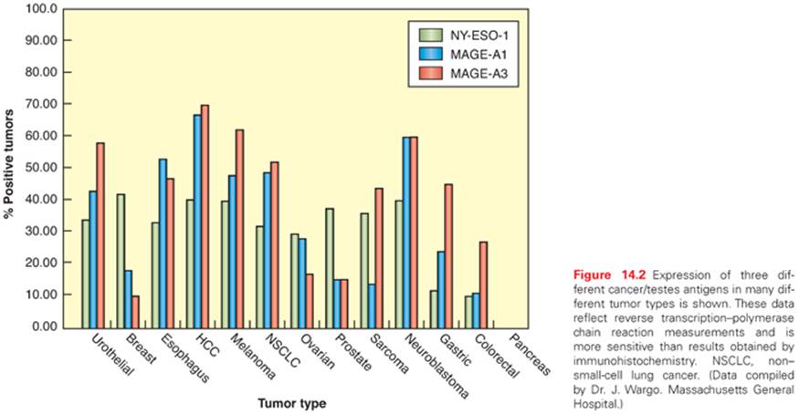
The first antigen identified as a target of human tumor reactive T cells was isolated by screening a melanoma genomic DNA library with an autologous cytotoxic T lymphocyte (CTL) clone.8 The gene that was isolated, termed MAGE-1, was found to be a nonmutated gene that was a member of a large, previously unidentified gene family, many of whose members encode antigens recognized by tumor reactive T cells.9 Members of this family of antigens are expressed in the testes and placenta, both of which lack an expression of MHC molecules, but often not in other normal tissues, which has led to their designation as cancer germ-line (CG) antigens. Members of the MAGE gene family are expressed in a variety of tumor types, including melanoma, breast, prostate, and esophageal cancers. The expression patterns of three different cancer/testes antigens in multiple tumor types is shown in Figure 14.2. The NY-ESO-1 antigen—a CG antigen that is unrelated to the MAGE family of genes—is expressed in approximately 30% of breast, prostate, and melanoma tumors, as well as between 70% and 80% of synovial cell sarcomas.10
Clinical adoptive immunotherapy trials targeting CG antigens have now been conducted in patients with melanoma as well as other tumor types. In a recent trial, objective clinical responses were seen in approximately 50% of patients with melanoma and 80% of patients with synovial cell sarcoma receiving autologous peripheral blood mononuclear cell (PBMC) transduced with a T-cell receptor directed against an HLA-A*02:01 restricted NY-ESO-1 epitope.6 A trial targeting a MAGEA3 epitopes was recently carried out using a T-cell receptor (TCR) isolated from an HLA-A*02:01+ transgenic mouse immunized with the MAGEA3:112–120 peptide.11 Objective clinical responses were observed in five of nine melanoma patients receiving the adoptively transferred PBMC that were transduced with the MAGEA3-reactive TCR.12 Unexpectedly, neural toxicity was observed in three of the patients treated in this trial, two of whom lapsed into a coma and subsequently died. Autopsy samples of patients’ brains revealed that MAGEA12, which encodes a cross-reactive epitope recognized by the MAGEA3 TCR, was expressed at low levels in patients’ brains, which may have been responsible for the observed neurologic toxicities. In a recent trial carried out using an affinity-enhanced human TCR directed against the HLA-A*01:01-restricted, MAGEA3:168-176 epitope, the first two patients receiving TCR-transduced autologous PBMC died of cardiac arrest 4 to 5 days following infusion, which was attributed to cross-reactivity with titin, a protein expressed at high levels in cardimyocytes.13 Taken together, these findings demonstrate the need for caution in evaluating cross-reactivity of high affinity TCRs recognizing tumor antigens.
Melanocyte Differentiation Antigens
Melanoma-reactive T cells have been frequently found to recognize gene products, termed melanocyte differentiation antigens (MDA), that are expressed in melanomas as well as in normal melanocytes present in the skin, eye, and ear but not in other normal tissues or tumor types. These include epitopes derived from gp100,14,15 tyrosinase,16 TRP-1,17 and TRP-2,18 proteins that had previously been found to play important roles in melanin synthesis. The screening of melanoma cDNA libraries with an HLA-A2–restricted tumor reactive T cells lead to the isolation of a previously unidentified gene, termed MART-119 or Melan-A.20 The MART-1 antigen, which is expressed in 80% to 90% of fresh melanomas and cultured melanoma cell lines as well as normal melanocytes, represents an MDA of unknown function. The majority of melanoma reactive, HLA-A2–restricted tumor-infiltrating lymphocytes (TIL) recognize a single MART-1 epitope.21 Studies carried out using a variety of approaches have also resulted in the identification of human leukocyte antigen (HLA) class II restricted epitopes of tyrosinase, TRP-1, TRP-2, and gp100.9
Overexpressed Gene Products
Gene products that are expressed at low levels in a variety of normal tissues but are overexpressed in a variety of tumor types have also been shown to be recognized by T cells. Screening of an autologous renal carcinoma cDNA library with a tumor reactive, HLA-A3–restricted T-cell clone resulted in the isolation of FGF5,22 a protein that was expressed only at low levels in normal tissues but upregulated in multiple renal carcinomas as well as prostate and breast carcinomas. The peptide epitope recognized by FGF5-reactive T cells was generated by protein splicing, a process in which distant protein regions are joined together in the proteasome that had previously only been described in plants23 and unicellular organisms.24 Subsequent studies have led to the identification of multiple epitopes that result from protein splicing, suggesting that this represents a general mechanism for generating T-cell epitopes.25–28Screening of an autologous cDNA library led to the identification of a previously unknown gene that was termed PRAME.29 This gene product was expressed in relatively high levels in melanomas as well as in additional tumor types but was also expressed at lower levels in a variety of normal tissues that included the testis, endometrium, ovary, and adrenals. The HLA-A24–restricted PRAME reactive T-cell clone, however, expressed the natural killer (NK) inhibitory receptor p58.2, and tumor cell recognition was dependent on the loss of expression of the HLA C*07 allele that represented the ligand for the inhibitory receptor, which may explain the lack of recognition of normal tissues that express relatively high levels of this HLA gene product.
Attempts have also been made to generate T cells directed against overexpressed candidate antigens by repeatedly stimulating PBMC in vitro with peptides that were identified as high binders for particular MHC molecules either using direct binding assays or in silico analysis carried out using peptide/MHC binding algorithms.30,31 Using this approach, candidate epitopes have been identified from a variety of proteins that include prostate-specific antigen (PSA)32 and prostate-specific membrane antigen (PSMA),33 as well as Her-2/neu, a protein that is frequently overexpressed in a variety of tumor types, including breast carcinomas. Initial studies indicated that T cells derived by in vitro stimulation with a peptide that was predicted to bind with high affinity to HLA-A*02:01, Her-2/neu:369–377, recognized the appropriate natural tumor targets.34 In one study, T cells generated following two in vitro stimulations of postvaccination PBMC from three of the four patients who were tested efficiently recognized peptide-pulsed targets but failed to recognize appropriate tumor targets.35 Similarly, although stimulation with a peptide corresponding to amino acids 540 through 548 of the human telomerase reverse transcriptase (hTERT) catalytic subunit was initially reported to generate tumor-reactive T cells,36 additional observations indicated that T cells generated using this peptide failed to recognize tumor targets.37 These factors responsible for these discrepancies remain unresolved, although the in vitro stimulation of T cells with target cells pulsed with relatively high peptide concentrations could have led to the generation of low-avidity T cells that were incapable of recognizing naturally processed antigens.
Alternative screening approaches employed for tumor antigen discovery that may help to address these issues include the use of tandem mass spectrometry to sequence peptides that have been eluted from tumor cell surface MHC molecules. Use of this technique, coupled with microarray gene expression profiling, resulted in the identification of peptides derived from proteins that appeared to be overexpressed in tumor cells.38 Peptides identified using this approach may, in many cases, not be immunogenic due to the fact that their expression in normal tissues, although lower than in tumor cells, may be high enough to lead to central or peripheral tolerance. Nevertheless, one of the peptides that were identified in this study also appeared to be recognized by human tumor reactive T cells. Recently, a similar approach was used to identify candidate peptides presented on cell surface MHC molecules that appeared to be derived from proteins that were overexpressed on glioblastomas.39 In a clinical trial involving vaccination of patients with pools of the identified peptides, overall survival was associated with the number of peptides in the vaccine pool that elicited immune response40; however, this may simply reflect the fact that T cells from healthier patients can more readily generate peptide-specific responses.
Transgenic mice that express human HLA molecules have also been immunized with candidate antigens in an attempt to identify high avidity tumor-reactive T cells. Immunization of transgenic mice expressing HLA-A*0201 with the native human p53:264–272 peptide that differed from the corresponding murine p53 sequence at a single position lead to the generation of T cells that recognized tumor cells expressing high levels of p53.41 Human T cells transduced with a murine p53 TCR isolated from an immunized mouse recognized a variety of human tumor cells; however, transduced T cells also recognized normal cells expressing lower p53 levels, indicating the dangers of targeting a normal self-protein whose expression is not strictly limited to tumor cells.42 Similarly, a TCR that was highly reactive with HLA-A*02:01+ tumor cells expressing the human carcinoembryonic antigen (CEA), a protein that is overexpressed in colon and breast carcinomas, was isolated by immunizing HLA-A*02:01+ transgenic mice with the CEA:691–699 peptide.43 The adoptive transfer of human PBMC transduced with the CEA-reactive TCR lead to an objective clinical response in one of the three treated patients; however, severe colitis was observed in all three of the treated patients.44 In general, immunotherapies that target antigens present even in small amounts on normal tissues have led to normal tissue destruction and must be applied with caution.
Mutated Gene Products Recognized by CD8+ and CD4+ T Cells
A variety of mutated antigens have also been identified as targets of tumor reactive T cells. The majority of mutated antigens identified using these approaches appear to be unique or only expressed in a relatively small percentage of cancers, and so do not represent targets that are broadly applicable to the treatment of multiple patients. Nevertheless, these studies have in some cases provided insights into mechanisms involved with tumor development, as the mutations may represent drivers of the transformed phenotype. The CDK4 gene product that was cloned using a CTL clone contained a point mutation that enhanced the binding to the HLA-A2 restriction element.45 This mutation, which was identified in 1 of an additional 28 melanomas that were analyzed, led to the inhibition of binding to the cell cycle inhibitory protein p16INK4a and may have played a role in the loss of growth control in this tumor cell. A point-mutated product of the β-catenin gene, containing a substitution of phenylalanine for serine at position 37, was isolated by screening a cDNA library with an HLA-24–restricted, melanoma reactive TIL.46 This mutation was found to stabilize the β-catenin gene product by altering a critical serine phosphorylation site, and 2 of 24 additional melanoma cell lines were found to express transcripts with identical mutations.47
The observation that immunization against individual murine tumors did not generally cross-protect against challenge with additional syngeneic murine tumors has provided support for the hypothesis that mutant T-cell epitopes represent the predominant antigens responsible for tumor rejection.48 Mutated epitopes also represent a foreign antigen, which may render them more immunogenic than the majority of normal self-antigens. Although many of the mutations are specific for individual tumors, T cells have been generated by carrying out in vitro sensitization with peptides encoded at mutational hot spots present in driver genes.49
Recently, novel approaches have been developed that involve the sequencing of tumor cell DNA to identify potential mutated epitopes. In one study, whole exome sequencing of the murine B16 melanoma led to the identification of mutated epitopes that elicited a T cell that appeared to specifically recognize the mutated but not the corresponding wild-type peptides.50 In a second study, a mutated antigen was identified by screening candidate epitopes that were expressed by tumors derived from immunodeficient mice that regressed in immune-competent mice.51 More recently, melanomas from three patients who responded to adoptive immunotherapy were subjected to whole exome sequencing, followed by in silico analysis using peptide/MHC binding algorithms to identify candidate epitopes that were predicted to bind to the patients’ MHC molecules.52 Using this approach, a total of seven peptides were identified as targets of the TIL that were administered to these patients. Two mutated epitopes were recently identified by whole exome sequencing of a melanoma from a patient who demonstrated a partial response to treatment with the anti–CTLA-4 antibody ipilimumab, followed by a screening of a panel of mutated candidate peptide/MHC tetramers that were predicted to bind to the patient’s HLA-A and B alleles.53 In addition, a mutated epitope expressed by a bile duct cancer was identified by screening tandem minigenes encoding all mutated epitopes that were identified by whole exome sequencing.54 The adoptive transfer of T cells directed against this mutation-mediated regression of the patient’s cancer. Mutations unique to each cancer represent ideal targets for immunotherapy and can potentially lead to the development of personalized therapies directed against these unique targets.
Antigens Identified in Viral-Associated Cancers
Viruses do not appear to play a role in the development of the majority of human cancers; however, an infection with HPV, a group of double-stranded DNA viruses that infect squamous epithelium, is highly associated with the development of a variety of genital lesions that range from warts to carcinomas, as well as the majority of oropharyngeal carcinomas. Recombinant vaccines have been produced by the generation of viruslike particles (VLP), self-assembling particles that form following the expression of the HPV L1 protein in recombinant viral and yeast systems that were initially found to be protective in animal models. The results of a phase II trial in which 2,392 women between 16 and 23 years of age were immunized with HPV-16 VLPs indicated that 100% of those who were vaccinated were protected against infection with HPV-16.55,56 Although vaccination with VLP does not lead to the regression of established disease, some success has been seen in therapeutic vaccination trials that target the oncogenic viral proteins E6 and E7. In a trial involving the vaccination of women with HPV-16–positive high-grade vulvar intraepithelial neoplasia with synthetic long peptides that encompass both HLA class I and class II restricted epitopes from the oncogenic HPV proteins E6 and E, clinical responses were observed in 15 of the 19 vaccinated patients, and complete regression of all lesions were seen in 9 of the 19 patients in this trial.1
Targeting foreign antigens thus may represent a strategy that can lead to more effective immunotherapies. These include viral epitopes as well as mutated epitopes that are also foreign to the host and therefore may represent more effective targets for these therapies than normal self-antigen.
HUMAN CANCER IMMUNOTHERAPIES
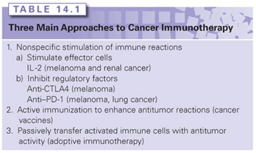
A wide variety of therapies have been evaluated in model systems and are now being developed for the treatment of patients with cancer. These include nonspecific approaches, those that involve direct immunization of patients with a variety of immunogens and approaches that involve the adoptive transfer of activated effector cells (Table 14.1). Much confusion related to the effectiveness of cancer immunotherapy has resulted from the lack of proper evaluation of the results of therapy using standard, accepted oncologic criteria such as the World Health Organization or the Response Evaluation Criteria in Solid Tumors (RECIST). Many clinical trials reported a positive use of soft criteria such as lymphoid infiltration or tumor necrosis that can occur in the natural course of cancer growth. Because of the delayed responses seen with some immunotherapy approaches, including tumor regression after initial tumor growth, guidelines have been published suggesting the use of an alternate set of immune-related response criteria for the evaluation of immune-based cancer treatments.57,58 Other confusion has arisen from the use of inappropriate animal models. Although animal model systems have provided important clues that may lead to improved therapies, model systems that employ artificially introduced foreign antigens or that evaluate protection from tumor challenge do not appear to be relevant to the treatment of patients with bulky metastases. Short-term lung metastasis models involve the treatment of relatively small, nonvascularized tumors and also may not be directly relevant to the majority of tumors that are the targets of current clinical trials.
Nonspecific Approaches to Cancer Immunotherapy
Progress has surged in the past 10 years in the understanding and utilization of nonspecific immune stimulation for the treatment of metastatic cancers. These agents aim to activate quiescent tumor-reactive immune cells or to remove inhibitory mechanisms to allow immunosuppressed cells to function to their full capacity. Although IL-2 and ipilimumab are currently the only immune stimulants approved by the U.S. Food and Drug Administration (FDA) for the treatment of metastatic renal cell carcinoma (IL-2) and melanoma (IL-2 and ipilimumab), new immune checkpoint inhibitors such as anti–programmed cell death 1 (anti–PD-1) have shown impressive results in recent clinical trials for patients with melanoma, renal cell cancer, and also non–small-cell lung cancer (NSCLC), and will likely be approved in the near future. As expected with nonspecific immunostimulation, systemic and bystander immune-related adverse events such as colitis has been reported with all agents in varying degrees, although most side effects are controllable and reversible if addressed aggressively and promptly by experienced clinicians. Importantly, antitumor responses seen with these immune-based modalities appear to be durable for some patients and may even be potentially curative. As with many therapies for metastatic solid tumors, preliminary trials using combination therapies have suggested better than expected response rates and survival, and confirmatory trials are in process to validate and ensure that toxicities from combining agents would not be prohibitive. Overall, patients with metastatic solid tumors may soon have wider armamentarium of off-the-shelf immunotherapy options.
Interleukin-2
Morgan et al.59 showed that a factor produced in the medium from stimulated normal human blood lymphocytes can allow ex vivo growth and expansion of human T lymphocytes. The identification of this soluble T-cell growth factor (IL-2)60,61 allowed the ability to culture T cells in vitro. IL-2 is a 15-kd glycoprotein produced in minute amounts by activated peripheral blood lymphocytes, and even with using T-cell hybridomas, minimal quantities could be purified; thus, research using IL-2 was impeded by the limited amounts of purified IL-2 available. The isolation of the cDNA clone in 198362 enabled the development in 1984 of recombinant IL-2,63 which permitted the ability to mass manufacture IL-2. Although murine studies demonstrated the ability of IL-2 to mediate tumor regression,64 early phase I clinical trials did not show any antitumor response,65 but was instructive in showing pharmacokinetics and toxicities, which led to more effective regimens. Subsequently, IL-2 was given in higher doses (up to 720,000 IU/kg intravenously every 8 hours) in a landmark trial involving 25 patients, along with nonspecific lymphokine-activated natural killer (LAK) cells, which are non-T and non-B lymphocytes.66 This report was the first to document the regression of advanced solid cancers (melanoma, renal cell, lung, and colon) using immunotherapy in humans.66 A follow-up trial randomizing 181 patients to either high-dose IL-2 alone (720,000 IU/kg intravenously every 8 hours) or high-dose IL-2 and LAK cells showed that the tumor response was due to IL-2 alone and not to the nonspecific LAK cells.67 This study also narrowed the IL-2–sensitive histologies to melanoma and renal cell cancer, which had more consistent responses.
IL-2 Therapy for Metastatic Renal Cell Cancer
Subsequent to the studies discussed previously, high-dose IL-2 was tested by additional centers and in combination with other agents for renal cell cancer. A randomized phase II trial involving 99 kidney cancer patients showed no increase in antitumor responses with the addition of interferon alfa-2b (IFNα-2b). Responses were seen for 12 (17%) of 71 patients who received high-dose IL-2 alone, with 4 complete regressions.68 A summary report of 227 patients with metastatic renal cell cancer treated with high-dose IL-2 (defined as 600,000 IU/kg or 720,000 IU/kg given intravenously every 8 hours as tolerated up to 15 doses) from 1985 to 1996 at the Surgery Branch of the National Cancer Institute (NCI) documented a total response rate of 19%, with 10% partial and 9% complete; the longest duration of a complete response was over 10 years ongoing (134+ months).69 Another summary report from seven phase II clinical trials from multiple institutions involving 255 patients with metastatic renal cell cancer receiving high-dose IL-2 showed the overall response rate was 14%, with 9% partial and 5% complete, and responses occurred in all sites of disease, including primary kidney tumors, bone metastases, and bulking visceral tumor burdens.70 Although the response rates were modest, the durability of the responses was remarkable, with many responses lasting over 5 years ongoing (see Fig. 14.2). Because of the striking durability of the antitumor responses, IL-2 received FDA approval for the treatment of metastatic renal cell cancer in 1992. A follow-up report in 2000 showing the response rates of the 255 renal cell patients in the seven phase II studies to be the same, with complete responses lasting over 10 years ongoing (131+ months for the longest responder), suggesting a potential cure.71
To ascertain whether lower doses and/or different administration routes, which would decrease toxicity and obviate the need for inpatient hospitalization for IL-2 therapy, a trial randomizing 400 patients with metastatic renal cell cancer to either standard high-dose intravenous IL-2, low-dose intravenous IL-2 (at 72,000 IU/kg), or low-dose subcutaneous IL-2 (250,000 U/kg per dose daily Monday through Friday in the first week and then 125,000 U/kg per dose daily during the next 5 weeks).72 Although responses were seen with all three regimens, including complete responses in the low-dose subcutaneous regimen, standard high-dose IL-2 had higher overall response rates (21%) versus low-dose intravenous IL-2 (13%; p = 0.048) and low-dose subcutaneous IL-2 (10%; p = 0.033), suggesting the superiority of the high-dose intravenous regimen.72
The administration of IL-2 represents the only known curative treatment for patients with metastatic renal cell cancer and should be considered as front-line therapy for suitable patients.
IL-2 Therapy for Metastatic Melanoma
Between 1985 and 1993, 270 patients with metastatic melanoma enrolled into eight clinical trials in multiple centers using high-dose IL-2 (defined as 600,000 IU/kg or 720,000 IU/kg given intravenously every 8 hours as tolerated up to 15 doses). Atkins et al.73 reported overall response rates of 16% (43 patients), with 10% partial and 6% complete; responses occurred at all tumor sites and regardless of initial tumor burden. With median follow-up at that time of 62 months, 20 responders (47%) were still alive, with 15 surviving over 5 years.73 A follow-up report on those patients in 2000 showed that the response rates were unchanged; with the longest response duration of >12 years ongoing, disease progression was not observed in any patient responding greater than 30 months.74 As with renal cell cancer, the flat tailof the Kaplan-Meier response duration and overall survival curves (Fig. 14.3), showing the potential curative nature of the antitumor responses, was the main compelling reason the FDA approved IL-2 for the treatment of metastatic melanoma in 1998.

Research in subsequent years aimed to increase the response rates of IL-2, led by increasing interests in tumor vaccinations as melanoma-associated antigens were being characterized.75 Pilot studies suggested that vaccinations using modified melanoma differentiation antigens such as gp100:209–217(210M) could elicit immunologic responses in nearly all patients, and when combined with high-dose IL-2, could elicit potentially higher than expected clinical antitumor responses.75 A follow-up phase III study76 randomized 185 patients with HLA*A0201 from 21 centers to either high-dose IL-2 or high-dose IL-2 plus gp100:209–217(210M) concurrent immunization. Although the response rates for the IL-2 plus vaccine arm was statistically improved compared to IL-2 alone (16% versus 6%; p = 0.03), the IL-2 alone arm was notable for being much lower than in all prior studies.76 In addition, a pilot trial of 36 melanoma patients treated high-dose IL-2 concurrently with ipilimumab (an antibody against cytotoxic T lymphocyte–associated antigen 4 discussed in the following section) gave a 25% OR rate, with 17% achieving complete response77; however, these data have not been further tested.
Correlative studies suggest that the total doses of IL-2 received during the first treatment course was significantly higher in patients achieving a complete response69; however, when limited to patients who were able to complete both cycles of the course, there was no statistical significance, suggesting that patients whose tumors progressed significantly after one cycle (and was not able to complete the second cycle of the course) accounted for some of the difference seen.78 Responders did have a higher maximal lymphocyte count69,78 immediately posttherapy and were more likely to develop vitiligo and thyroid dysfunction.78 There has not been a consistent pretherapy factor that is predictive of response, although one retrospective correlative study involving 374 patients showed that patients with M1a (subcutaneous- and/or cutaneous-only disease) have a response rate of 54% compared with 12% for those with visceral M1b/c (P2 <0.0001).78
Toxicities and Safe Administration of IL-2
High-dose IL-2 has been shown to be associated with adverse events that impact multiple organ systems.73,79,80 The main component of the toxicities is due to an inflammatory response mediated by the release of cytokines such as IFNγ and tumor necrosis factor alpha (TNF-α)81 resulting in a capillary-leak syndrome82 and decreased systemic vascular resistance, which can lead to fever, hypotension, cardiac arrhythmia, lethargy, renal insufficiency, hepatic dysfunction, body edema, pulmonary edema, and confusion; other side effects can also include nausea, diarrhea, rash, anemia, thrombocytopenia, lymphocytosis, and neutrophil chemotactic defect83 that predispose patients to gram-positive line infections. Since the first clinical trials with IL-2 in 1984, however, much has been learned to permit its safe dosing for appropriately screened patients82,84,85; importantly, if patients are appropriately supported, side effects are quickly reversible once IL-2 dosing ceases.85 Kammula et al.86 compared the incidences of grade 3/4 toxicities between the 155 patients treated from 1985 to 1986 to 156 patients treated from 1993 through 1997 at the NCI Surgery Branch: grade 3/4 hypotension decreased from 81% to 31%, intubations from 12% to 3%, neuropsychiatric toxicities from 19% to 8%, diarrhea from 92% to 12%, line sepsis from 18% to 4%, cardiac ischemia from 3% to 0%, and mortality from 3% to 0%. In fact, no fatality occurred strictly due to IL-2 therapy since 1989.86 Overall strategies for the safe administration of high-dose IL-2 include careful screening for appropriately selected patients with adequate cardiopulmonary reserve, having an experienced team of physicians and nurses who are cognizant of the expected toxicities of IL-2, having routine preemptive measures such as prophylactic antibiotics to prevent line infections, and aggressive and prompt management of toxicities.
Checkpoint Modulators
Anti–Cytotoxic T Lymphocyte Antigen 4
CTLA-4 is an immunosuppressive costimulatory receptor found on newly activated T cells (and on regulatory T cells) that binds with costimulatory ligands B7-1 and B7-2 on antigen-presenting cells.87,88When CTLA-4 is engaged by B7-1 or B7-2, the T cells becomes inhibited,89,90 suggesting that CTLA-4 likely evolved as a self-protective mechanism to prevent autoimmunity (Fig. 14.4). Thus, overcoming this checkpoint molecule was an aim of cancer immunotherapy. After CTLA-4 blockade in murine models led to antitumor immunity,91,92 anti–CTLA-4 antibodies were tested in clinical trials starting in 2002.

The combination of anti–CTLA-4 blocking antibodies and vaccination worked well in murine models and led to one of the early phase II studies using ipilimumab (a fully human immunoglobulin [IgG1] monoclonal antibody previously called MDX-010) with two gp100 vaccines, gp100:209–217(210M) and gp100:280–288(288V), in patients with metastatic melanoma.93 Antitumor regressions were seen (from 11% to 22% overall response rates, with up to 8% complete response rates), along with severe autoimmune toxicities such as colitis, dermatitis, and even hypophysitis,93–95 as would be expected based on the mechanism of CTLA-4 blockade. In fact, autoimmunity adverse events appeared to correlate with response to ipilimumab.3 The experience with these early studies led to management strategies to screen aggressively for immune-related adverse events (IRAE), such as routine screening of endocrinopathies, and to treat IRAEs promptly, including high-dose steroids if needed for severe colitis.96,97 Overall, ipilimumab was in some ways easier to manage for the patients than IL-2 because it was an outpatient infusion given every 3 weeks; IRAEs were unpredictable, however, and can appear suddenly many weeks after receiving a dose.
In 2010, results from a landmark phase III randomized trial comparing three treatment strategies (ipilimumab alone, gp100 peptide vaccine alone, or ipilimumab plus gp100 peptide vaccine) in 676 patients with metastatic melanoma were published showing improvement in median survival in the two arms that received ipilimumab (10 months) compared to the gp100 alone arm (6 months, p <0.001), despite showing a low response rate of 7% (among 540 patients who received ipilimumab).98 Another phase III randomized trial comparing dacarbazine plus ipilimumab versus dacarbazine alone again showed improved survival in that arm containing dacarbazine (11.2 months versus 9.1 months; p <0.001).99 These studies showing survival benefit led to FDA approval of ipilimumab for advanced melanoma in 2011.
The responses seen with ipilimumab appear to be durable.100 A follow-up study of 177 patients with metastatic melanoma treated on the earliest trials at the NCI Surgery Branch using ipilimumab showed that response duration could last 99+ months ongoing.77 In fact, 14 out of the 15 complete responders remain disease free 54+ to 99+ months ongoing, suggesting a potential cure for some patients. Interestingly, several patients who were deemed partial responders converted to complete responders several years later, because it look an average of 30 months to have all visible tumor marks on imaging scans to disappear.77
Ipilimumab was also tested on other solid tumors, and renal cell cancer again appears to be the only other type beside melanoma that had significant responses. Sixty-one patients with metastatic renal cell cancer were treated, and six developed a response (10%); however, 33% developed grade 3/4 IRAEs.101 Subsequently, the availability of agents with lower toxicity profiles such as sunitinib and sorafenib prevented further enthusiasm to pursue this drug for renal cell cancer.
Another anti–CTLA-4 antibody, tremelimumab (previously called CP-675,206), has also demonstrated durable responses in melanoma patients.102,103 A phase III randomized trial randomizing 655 patients with metastatic melanoma to either tremelimumab or physician’s choice chemotherapy, however, failed to show a survival difference (despite a significantly different response duration favoring tremelimumab, 35.8 months versus 13.7 months; p = 0.0011), possibly due to crossover of chemotherapy patients enrolling into ipilimumab trials and expanded access programs.103
Anti–Programmed Death 1 and Anti–Programmed Death Ligand 1
PD-1 is another checkpoint modulator expressed on activated T cells. Although CTLA-4 appears to be involved in the early activation of T cells, PD-1 is involved in the later effector phase of T-cell activation and can function to prevent excessive damage to self by activated T cells in the periphery.104,105 Interaction with its corresponding ligand, PD-L1 (B7-H1) and PD-L2 (B7-H2) leads to suppressed T-effector function. PD-L1 is expressed on hematopoietic and epithelial cells and is upregulated by cytokines such as IFNγ,106 whereas PD-L1 is mainly on antigen-presenting cells. Given the clinical results with inhibiting the CTLA-4 checkpoint, recent efforts have focused on inhibiting the PD-1/PD-L1 and PD-1/PD-L2 interactions.
Nivolumab (previously known as BMS-936558, MDX-1106, and ONO-4538) is a fully human anti–PD-1 IgG4 monoclonal antibody that was initially tested in a phase I trial published in 2010 in which 39 patients with advanced solid cancers were treated in escalating doses.107 Responses were seen in one patient with colon cancer, one with melanoma, and one with renal cell cancer; one patient developed colitis.108 These hopeful results lead to a larger study in which 236 patients with either NSCLC (74 patients), melanoma (94 patients), or renal cell cancer (33 patients).4 Objective responses were seen in 18% of patients with NSCLC, 28% with melanoma, and 27% with renal cell cancer.4 Grade 3/4 adverse events occurred in 14% of patients, including those previously seen with ipilimumab (dermatitis, colitis, hepatitis, thyroiditis, hypophysitis, and pneumonitis). Nine patients developed pneumonitis, six of whom was reversible, and three (1%) with grade 3/4 died despite steroids and infliximab therapy.4 An update on the status of 107 melanoma patients treated from 2008 to 2012 shows a 31% tumor response rate, with a median response duration of 2 years and a median overall survival of 16.8 months.109
Nivolumab was also tested in combination with ipilimumab in melanoma in either concurrent (53 patients) or sequenced (33 patients) regimens. The concurrent group experienced an overall response rate of 40%, whereas the sequenced group had a 20% response rate.110 The concurrent group also experienced a higher rate of grade 3/4 adverse events (53%), compared to 18% in the sequenced group. Interestingly, 16 of 21 responders in the concurrent group experienced tumor reduction of 80% or greater by 12 weeks,110 a tempo that is faster than was seen with ipilimumab.
Another anti–PD-1 developed independently, lambrolizumab (previously known as MK-3475, a humanized IgG4κ monoclonal antibody), was tested on 135 patients with metastatic melanoma.111 The response rate was found to be 38% and was similar between those who had received ipilimumab and those who were ipilimumab naïve,111 confirming that the antitumor response from lambrolizumab occurs via a different mechanism. Similar to nivolumab, 13% of patients developed grade 3/4 adverse events, with 4% developing pneumonitis, although none developed grade 3/4 pneumonitis.111
BMS-936559 is a fully human IgG4 monoclonal antibody that blocks PD-L1 ligation to both PD-1 and CD80. A phase I study was tested in 207 patients (75 with NSCLC, 55 with melanoma, 18 with colon cancer, and 17 with renal cell cancer, 17 with ovarian cancer, 14 with pancreatic cancer, 7 with gastric cancer, and 4 with breast cancer).112 Among patients who were evaluated for response, objective responses were seen in 16% of melanoma patients, 17% of renal cell cancer patients, 10% of NSCLC patients, and 1 out of 17 ovarian cancer patients. Grade 3/4 toxicities were seen in 9% of patients.112
The advent of these checkpoint inhibitors brings additional treatment options to patients with selected advanced cancers, particularly those with histologies deemed previously to be outside the realm of immunotherapy such as NSCLC.108,113 In addition, a new anti–PD-L1 (MPDL3280A) in clinical trials has also shown some efficacy in melanoma, renal cell cancer, and NSCLC in early reports.
Active Immunization Approaches to Cancer Therapy (Cancer Vaccines)
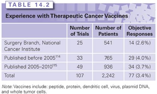
The molecular characterization of multiple cancer antigens led to a large number of clinical trials that attempted to actively immunize against these antigens with the expectation that cellular immune reactions would be generated capable of inhibiting the growth of established cancers. The results of these efforts have yet to produce significant vaccine efforts of value in the treatment of human cancer. There is a paucity of murine tumor models that suggests that active vaccine approaches can mediate the regression of established vascularized tumors; therefore, it is not surprising that these approaches have, with a few exceptions, shown little efficacy in humans. Enthusiasm about the effectiveness of cancer vaccines has often been grounded in surrogate and subjective end points, rather than reliable objective cancer regressions using standard oncologic criteria. In a review of the world literature, including 107 published cancer vaccine trials involving 2,242 patients, a 3.4% overall objective response rate was observed (Table 14.2).114,115 In many cases, relatively soft criteria such as stable disease or the regression of individual metastases in the presence of progressive disease at other sites have been reported. A variety of immunizing vectors have been used, including tumor-derived peptides, proteins, whole tumor cells, recombinant viruses, dendritic cells, and heat-shock proteins.116–122 Although many of these approaches can lead to the development of circulating T cells that can recognize the immunizing tumor antigen, these T cells rarely cause the inhibition of established tumors, a point that has led to much confusion in the field of tumor immunology. The generation of antitumor T cells in vivo is likely a necessary, but certainly not a sufficient criteria for the development of a clinically active immunotherapy. Often, T cells with weak avidity for tumor recognition are generated, and the tolerizing and inhibitory influences that exist in vivo must be overcome for an effective immune response to cause tumor destruction.
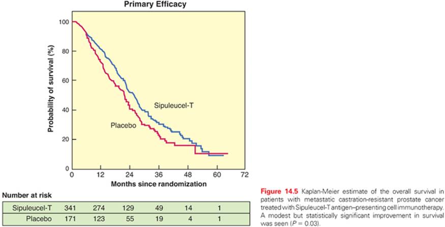
A prospective randomized trial of immunization with antigen-presenting cells was carried out by the Dendreon Corporation (Seattle, Washington). This trial used an antigen-presenting cell vaccine loaded with prostatic acid phosphatase linked to GM-CSF compared to placebo in men with hormone-refractory prostate cancer.123 Of 330 patients who received the vaccine treatment, 1 objective partial response was seen. Only 8 patients experienced a PSA drop of at least 50%. There was no difference in the time to disease progression; however, the vaccine group had a median survival of 25.8 months compared to 21.7 months in the placebo group, and based on this statistically significant survival improvement, this treatment was approved by the FDA (Fig. 14.5).
Adoptive Cell Transfer Immunotherapy
Adoptive cellular immunotherapy refers to the transfer to the tumor-bearing host of immune lymphocytes with anticancer activity. The first successful administration of adoptive cell therapy (ACT) involving TIL, in combination with high-dose IL-2 was carried out at the National Cancer Institute Surgery Branch in 1988.124 Studies that used cell transfer therapy in patients with metastatic melanoma have provided the clearest evidence of the power of the immune system to mediate the regression of advanced metastatic cancers in humans. Adoptive cell therapy has several theoretical as well as practical advantages.125Lymphocytes with antitumor activity can be expanded to very large numbers ex vivo for infusion into cancer patients. These cells can be tested in vitro for antitumor activity, and cells with appropriate properties such as high avidity for tumor recognition and a high proliferative potential can be identified and selectively expanded for treatment. These cells can be activated in vitro and thus are not subjected to the tolerizing influences that exist in vivo. Perhaps, most important, the host can be manipulated prior to the transfer of the anticancer cells to provide an optimal tumor microenvironment free of in vivo suppressive factors.125 Studies have shown that the transfer of cultured lymphocytes with antiviral activity can prevent Epstein-Barr virus (EBV) infections as well as the subsequent development of posttransplant lymphoproliferative diseases. Cultured lymphocytes have been used for the treatment of patients with established EBV-induced lymphomas.126
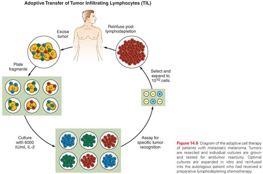
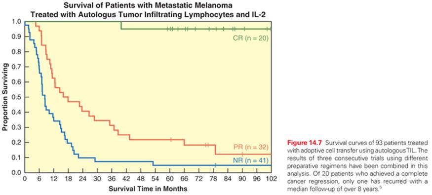
The best evidence for the ability of adoptive cell transfer to successfully treat patients with solid tumors comes from the treatment of patients with metastatic melanoma. A diagram that describes the nature of this treatment is shown in Figure 14.6. In patients with metastatic melanoma, TILs can be obtained from resected tumor deposits and individual cultures tested to identify those with optimal anticancer activity.124,127 These cells are then expanded ex vivo and reinfused along with IL-2, which is the requisite growth factor required for the survival and persistence of these cells. The administration of a preparative lymphodepleting chemotherapy regimen, consisting of cyclophosphamide and fludarabine with or without 2 or 12 Gy total body irradiation, could substantially enhance the survival and persistence of the transferred cells and increase their in vivo antitumor effectiveness.128,129 In a series of three pilot trials with 93 patients, objective responses were seen in 49% to 72% of patients.5,130 Of the 93 patients, 20 (22%) experienced a complete regression of all metastatic melanoma. Only 1 of these 20 patients has recurred, and the remaining patients have ongoing complete regressions from 80 to over 104 months (Table 14.3, Fig. 14.7). The 5-year survival of these 93 patients was 29% and was similar regardless of the prior treatments that these patients had received.

Extensive genomic studies have shown that TILs that mediate complete cancer regressions recognize mutated epitopes presented by the cancer.52 The use of exomic sequencing combined with in vitro tests of antitumor activity can be used to select for T-cell populations reactive against the cancer. This approach has now been utilized to identify T cells used to successfully treat a patient with chemotherapy-refractory cholangiocarcinoma and provides a blueprint for the application of cell transfer therapy for a variety of common epithelial cancers.54
The difficulty in obtaining TILs with antitumor activity from cancers other than melanoma has also led to the development of approaches using lymphocytes genetically modified using retroviral transduction to insert antitumor T-cell receptors into the normal lymphocytes of patients.131
Genetic Modification of Lymphocytes for Use in Adoptive Cell Therapy: Basic Principles and Applications to Solid Tumors
Efforts are in progress to genetically engineer autologous PBMCs through the introduction of exogenous high avidity receptors that specifically recognize tumor antigens (Fig. 14.8). These cells can then be expanded to large numbers in vitro and be readministered back to the patient similar to TILs in order to mediate tumor regression. The use of gene-modified cells for ACT has resulted in objective clinical responses for a variety of cancer histologies including melanoma, synovial sarcoma, and CD19-positive B-cell malignancies.6,131–133
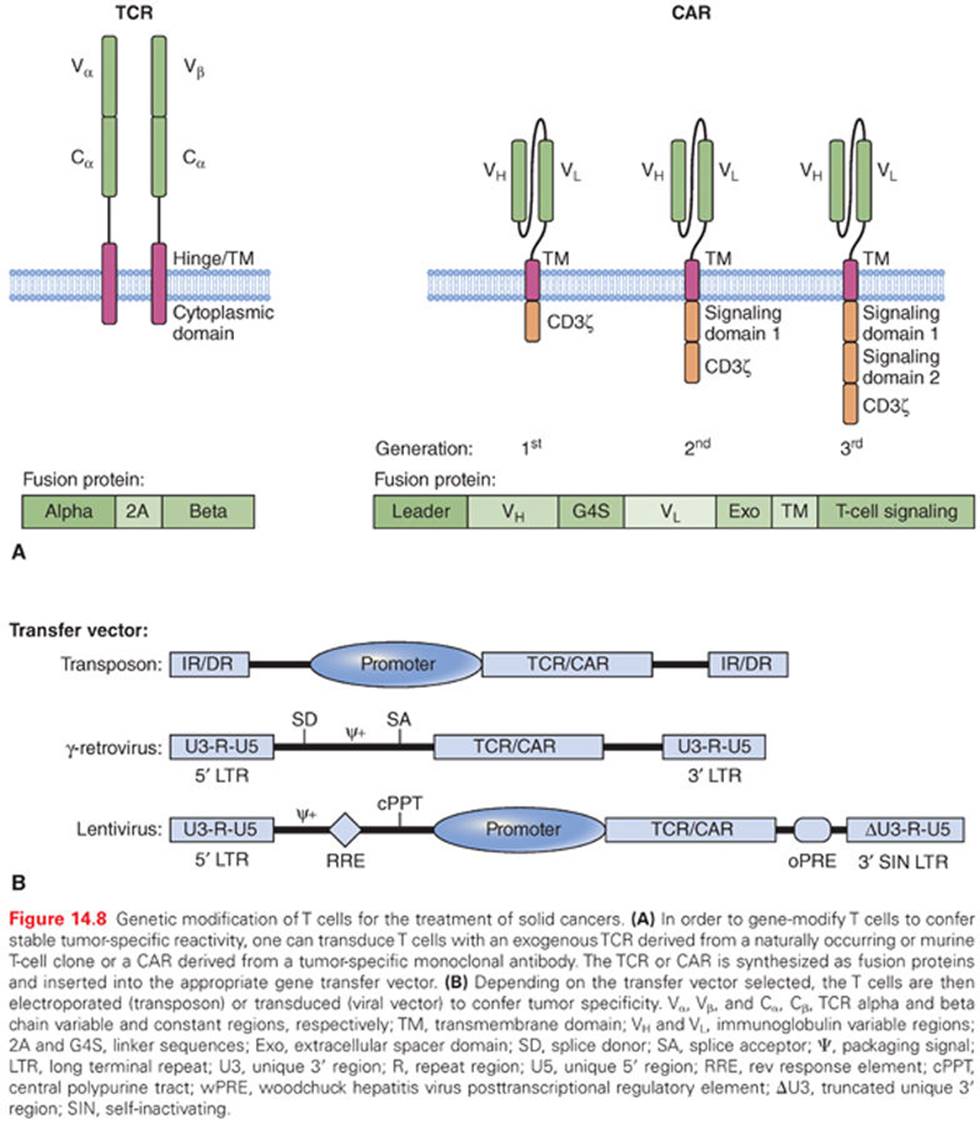
There are two key requirements necessary for the use of gene-modified cells for the treatment of solid cancer. The first is the selection of an appropriate gene transfer method in order to achieve high receptor expression levels in the transferred T cells. For this discussion, we will consider both nonviral and viral-based gene delivery platforms. Generally speaking, there are two categories of nonviral gene transfer, chemical and physical. Chemical gene transfer involves the use of positively charged delivery vehicle such as calcium phosphate, cationic lipids, or polymers to form DNA complexes capable of entering a cell through endocytosis.134 These reagents benefit from their ease of manufacture and ability to form complexes with large DNA sequences; however, low transfection efficiency of human T cells continues to be an issue. Physical methods for gene delivery may involve direct delivery of DNA into a cell via microinjection or indirect DNA uptake via electroporation.135 Electroporation of messenger RNA (mRNA) can achieve high levels of protein expression in cells, comparable to many of the viral-mediated gene delivery systems (gammaretroviral or lentiviral).135,136 High-throughput electroporators should allow one to gene modify large numbers of T cells ex vivo.137mRNA electroporation appears to be most suited for this application, because there is significant loss of cell viability following electroporation of large amounts of DNA.136 The electroporation of mRNA, although gaining traction as a means of redirecting T cell specificity,137 provides for transient receptor expression because the mRNA will degrade over time. Currently, it is not clear if stable long-term receptor expression is required to mediate tumor regression. However, the main criticism of the non-viral methods described is the lack of stable gene transfer. To overcome this problem, many investigators are now using transposons such as sleeping beauty or piggybac.138 Transposons are mobile DNA gene delivery elements encoding a gene of interest (i.e., TCR or chimeric antigen receptors [CAR]) that can randomly integrate into the genome in the presence of the transposase enzyme, thereby allowing for stable gene expression. This technology is currently being used for the ACT of CAR-modified cells targeting B-cell malignancies (see Fig. 14.8B).139
Viral-mediated gene delivery is currently the most common method for the genetic modification of immune cells for cancer ACT. Retroviridae is a family of RNA viruses that, upon entry into cells, undergo a process called reverse transcription whereby the viral RNA is converted into DNA as it stably integrates into the host genome. The two most common retroviral vector systems are based on the gammaretrovirus, Moloney murine leukemia virus (MLV), and the lentivirus, HIV type 1 (see Fig. 14.8B). Gammaretroviral vectors have been used in human clinical applications for over 20 years. The only reported toxicity associated with gammaretroviral engineering of human cells involved the retroviral transduction of hematopoietic stem cells for the treatment of children with severe combined immunodeficiency syndrome (X-SCID).140 There have been no reports of clonal outgrowth following the retroviral transduction of mature T lymphocytes in adults. Highly active vectors have been generated from a variety of murine retroviruses including spleen focus forming virus (SFFV), myeloproliferative sarcoma virus (MPSV), and the murine stem cell virus (MSCV).141–147 In most cases, these vectors are replication incompetent, but non–self-inactivating in that the promoter for transgene expression is derived from the viral long terminal repeat (LTR). Self-inactivating (SIN) gammaretroviral vectors have been developed that require an internal promoter to drive transgene expression. The advantage of non-SIN vectors is the ability to use a variety of retroviral packaging cell lines (PG13, Phoenix) engineered to constitutively express gag (capsid protein), pol (reverse transcriptase, integrase, and RNase H enzymes) and env (envelope protein). Transduction of these packaging lines with a non-SIN retroviral vector encoding a transgene allows for the generation of a stable packaging cell line that constitutively releases vector into the medium. This platform is easily scaled up to support large-scale vector production efforts. An alternative to the gammaretroviral vector platform is the lentiviral vector platform. There are some advantages to selecting a lentiviral vector for T-cell engineering in that one can transduce large numbers of minimally stimulated T cells,148transfer more complex and larger gene expression cassettes, and yield a potentially safer chromosomal integration profile as compared to gammaretroviruses. However, there has been at least one instance of clonal outgrowth following lentiviral vector transduction of CD34+ stem cells.149 Therefore, more data will be needed to better understand the risk of insertional mutagenesis associated with the use of lentiviral vectors. The major disadvantage with using lentiviral vectors for ACT is the lack of a robust packaging cell line, which requires transient vector production and is difficult to scale up.
The first successful application ACT involved the use of autologous T cells genetically modified with a conventional αβ TCR targeting MART-1 for the treatment of patients with melanoma.131 The success of this approach relies on the ability to identify naturally occurring TCRs with sufficiently high avidity for the tumor antigen. For this clinical trial, a tumor-specific TCR was cloned directly from melanoma TIL. Exogenous TCR can also be generated from human PBMC following a variety of in vitro sensitization techniques or immunization of transgenic mice expressing HLA molecules. A T-cell clone expressing a low avidity TCR recognizing MART-1 was isolated and the α and β chains cloned into a gammaretroviral vector. The objective response rate from this trial was 13% (2/15).131 In a follow-up trial with a higher avidity TCR that was cloned from the same melanoma TIL, the objective response rate increased to 30% (6/20).150 However, patients in this trial experienced significant on-target, off-tumor toxicity with the destruction of normal melanocytes in the skin, eye, and ear. These trials showed the potential to use ACT for the treatment of solid cancers, but also highlight the importance of selecting appropriate tumor antigens to target in order to minimize normal tissue toxicities. Perhaps a better class of antigen to target for ACT would be the cancer testes antigens (CTA) that are expressed only on germ cells during fetal development and then reexpressed on cancers but not other normal tissues with the exception of the testes (see Table 14.1). Because the testes do not express class I MHC molecules, they are protected from any adverse immune response.151 NY-ESO-1 is a CTA overexpressed on melanoma, as well as a variety of solid epithelial cancers.152–154 A high-avidity TCR was developed targeting NY-ESO-1 and patients with metastatic melanoma or synovial cell sarcoma were treated following adoptive cell transfer using autologous lymphocytes transduced with a gammaretrovirus encoding this receptor.6 In updated results from this trial, 8 of 17 patients (47%) with melanoma showed objective tumor responses, two of which were complete responses and ongoing at 51 and 48 months after treatment. Nine of 19 patients (47%) with synovial cell sarcoma showed objective tumor response, only one of which is complete and ongoing at 12 months. Of note, no toxicities were observed in any of these trials. Thus, targeting NY-ESO-1 and other CTAs is an attractive strategy for the application of ACT for the treatment of solid cancers (see Table 14.4 for other trials conducted at the National Cancer Institute, Surgery Branch).
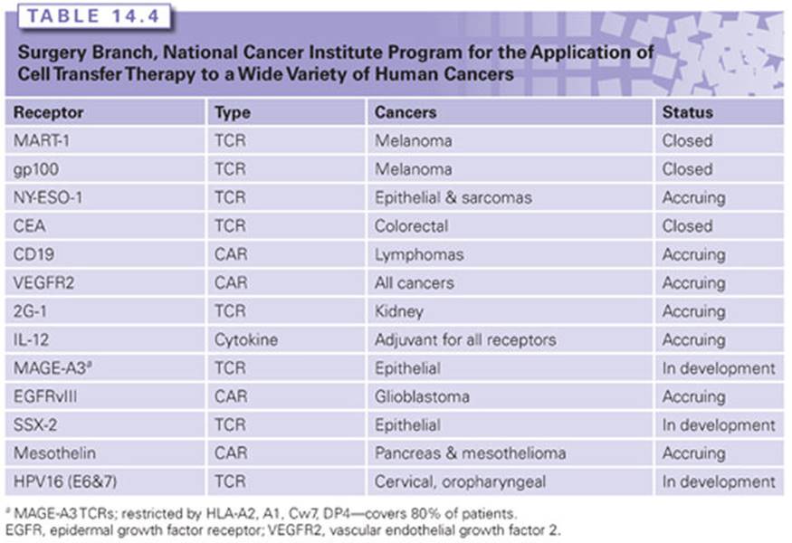
Redirection of T-cell specificity using conventional TCR is constrained by HLA restriction, which limits treatment only to patients expressing a particular MHC haplotype. An alternate approach is to use CAR comprised of an monoclonal antibody single chain variable fragment (scFv) fused in frame to T-cell intracellular signaling domains capable of T-cell activation following antigen-specific binding (see Fig. 14.8A).155 CARs, unlike conventional TCRs, are not MHC restricted but are limited by the requirement for the tumor antigen to be expressed on the cell surface. CARs can also recognize carbohydrate and lipid moieties further expanding their application. To date, there has been limited success using CAR-based ACT for the treatment of solid cancers. In 2008, the first successful CAR trial targeting the disialoganglioside, GD2, for the treatment of neuroblastoma was reported.156 In this trial, 4 out of 8 patients (50%) with evaluable tumor experienced tumor regression or necrosis with one complete responder. In that same year, a second CAR trial targeting CD20 on non-Hodgkin and mantle cell lymphomas was reported.157 Of the 7 patients treated, one achieved a partial response. Much greater success has now been achieved using a CAR targeting CD19, a molecule expressed on normal B cells and virtually all B-cell lymphomas. In a trial conducted at the National Cancer Institute, Surgery Branch, Kochenderfer et al.132 first reported that autologous T cells expressing a CAR targeting CD19 was able to mediate tumor regression in a patient with B-cell lymphoma (hematologic malignancies will be discussed in more detail elsewhere). Successfully expanding CAR-based ACT to other cancer histologies has been limited by the inability to identify suitable tumor antigens to target. At the National Cancer Institute, Surgery Branch, there are active clinical programs with CAR targeting the mutated epidermal growth factor receptor, EGFRvIII, expressed on approximately 40% of glioblastomas as well as head and neck cancers158; the vascular endothelial growth factor-2 receptor, VEGFR-2, expressed on tumor vasculature159; and mesothelin, expressed on the mesothelial lining of the pleura, peritoneum, and pericardium, but overexpressed on mesothelioma, pancreatic, and ovarian cancers.160 These trials are currently accruing patients; however, no objective clinical responses have been observed to date. A summary of clinical trials at the National Cancer Institute, Surgery Branch using gene-modified autologous T cells for ACT are shown in Table 14.4. ACT can mediate the regression of large, established tumors in humans. Efforts to identify and specifically target novel tumor antigens are currently underway with the hope that ACT using gene-modified T cells will develop into an effective treatment for patients with a variety of solid cancers.
Genetic Modification of Lymphocytes to Treat Hematologic Malignancies
Immunologic therapies can be useful treatments for some hematologic malignancies as demonstrated by the effectiveness of monoclonal antibodies in treating B-cell malignancies and the fact that allogeneic hematopoietic stem cell transplantation (alloHSCT) can cure a variety of hematologic malignancies.161–167 The results with monoclonal antibodies and alloHSCT clearly prove that immunologic therapies have significant activity against hematologic malignancies, but monoclonal antibodies are not curative as single agents,162,166 and alloHSCT has a substantial transplant-related mortality rate due to infections and an immunologic attack against normal tissues known as graft versus host disease (GVHD).163,165 The proven curative potential of alloHSCT and the effectiveness of autologous T-cell transfer therapies for melanoma have encouraged the development of autologous T-cell therapies for hematologic malignancies.125,129,163,165 Genetically engineering T cells to specifically recognize antigens expressed by malignant cells has emerged as a very promising strategy for cancer immunotherapy.125,129,168
T cells can be genetically engineered to express either of two types of receptors, CARs168–171 or natural TCRs.6,131,172 T cells expressing either a CAR or TCR gain the ability to specifically recognize an antigen.171,172 CARs are artificial fusion proteins that incorporate antigen recognition domains and T-cell activation domains.168,170,172 The antigen recognition domains are most often derived from monoclonal antibodies.168,170,172 Antigen recognition by TCRs is major histocompatibility complex restricted.125,128 In contrast to TCRs, recognition of antigens by CARs is not dependent on MHC molecules. An advantage of TCRs over CARs is that TCRs can recognize intracellular antigens, whereas CARs can only recognize cell-surface antigens.
Chimeric Antigen Receptors
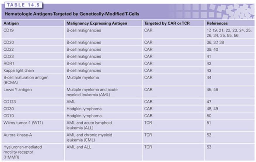
CARs targeting hematopoietic antigens have been extensively studied in preclinical experiments and early-stage clinical trials.168,170,172,173 For a protein to be a promising target for CAR-expressing T cells, it should be uniformly expressed on the malignant cells being targeted but not expressed on essential normal cells. Many cell-surface proteins with restricted normal tissue expression patterns have been identified on malignant hematologic cells, and CARs targeting many of these proteins are under development (Table 14.5).
Many factors can affect CAR T-cell therapies. The types of gene-therapy vectors encoding the DNA of the CAR could be an important factor. The types of vectors currently being used in clinical trials of CAR T cells are gammaretroviruses, lentiviruses, and transposon-based systems.132,133,174–181 The design of the CAR fusion protein is another important factor. CAR fusion proteins include an antigen-recognition domain that is most often derived from an antibody, costimulatory domains such as CD28 and 4-1BB, and T-cell activation domains that are usually derived from the CD3z molecule.168,170,171,182 Other factors that could impact the effectiveness of CAR T-cell therapies include the cell culture method used to prepare the cells and administration of chemotherapy or radiation therapy prior to the CAR T-cell infusions.170,178,179 In mouse models, a profound enhancement of the antimalignancy activity of infused T cells occurs when the T-cell infusions are preceded by lymphocyte-depleting chemotherapy or radiation therapy.183–185 Because chemotherapy can have a direct antimalignancy effect against hematologic malignancies, the administration of chemotherapy prior to infusions of T cells is a confounding factor that must always be kept in mind when interpreting the results of clinical trials of T-cell therapies.
Anti-CD19 Chimeric Antigen Receptors
CD19 is an appealing target antigen for CARs because CD19 is expressed on almost all malignant B cells, but CD19 is not expressed on normal cells except B cells.186 The first preclinical studies of anti-CD19 CARs utilized either gammaretrovirus vectors174 or plasmid electroporation176 to insert genes encoding anti-CD19 CARs into human T cells. These studies and subsequent preclinical work by other groups showed that T cells expressing anti-CD19 CARs could specifically recognize and kill CD19-expressing malignant B cells in vitro and in vivo.174–176,187 These preclinical studies compared many different CAR signaling moieties, which led most groups to utilize CARs with T-cell activation domains from the CD3z molecule and costimulatory molecules from either CD28 or 4-1BB (CD137).165,180,182,187,188 Preclinical studies showed that lymphocyte-depleting radiation therapy administered before anti-CD19 CAR T-cell infusions was critical to the antimalignancy activity of CAR T cells.183 The addition of lymphocyte-depleting radiation therapy prior to infusions of anti-CD19 CAR T cells increased the percentage of mice cured of lymphoma by the CAR T cells from 0% to 100%.183 Preclinical experiments with anti-CD19 CARs have led to several early-phase clinical trials.
The first clinical trial to demonstrate in vivo activity of anti-CD19 CAR T cells in humans was conducted in the Surgery Branch of the National Cancer Institute.132 The gammaretroviral vector used in this trial encoded a CAR with a CD28 costimulatory domain. Patients treated on this clinical trial received cyclophosphamide and fludarabine chemotherapy followed by an infusion of anti-CD19 CAR T cells and a short course of intravenous IL-2.132,181Clear antigen-specific activity of the anti-CD19 CAR T cells was demonstrated because blood B cells were selectively eliminated from four of the seven evaluable patients for several months.181 The duration of B-cell depletion in these patients was much longer than the duration of B-cell depletion caused by the chemotherapy that the patients received.132,181 This study also generated evidence of an antimalignancy effect by the anti-CD19 CAR T cells because six of seven evaluable patients with advanced B-cell malignancies obtained either complete remissions or partial remissions (Fig. 14.9).181 One of these remissions is ongoing 45 months after treatment, and another remission is ongoing 31 months after treatment. Significant toxicity, including hypotension and neurologic toxicity, occurred during this clinical trial.181 The severity of these toxicities correlated with the levels of serum inflammatory cytokines.181Except for one patient who died with influenza pneumonia, the toxicities were transient, with all toxicities resolving within 3 weeks of the anti-CD19 CAR T-cell infusions.181

Investigators at the Memorial Sloan Kettering Cancer Center treated nine patients with chronic lymphocytic leukemia (CLL) or acute lymphocytic leukemia (ALL) by infusing T cells that expressed a CAR with a CD28 costimulatory domain.179 The gene therapy vector used in this work was a gammaretrovirus.179 None of three patients treated with CAR T cells alone experienced a regression of leukemia, and CLL regressed in one of four evaluable patients treated with cyclophosphamide followed by an infusion of CAR T cells. Using the same CAR, the same group went on to treat five patients with ALL.173Patients received chemotherapy followed by an infusion of anti-CD19 CAR T cells. Four patients had detectable leukemia prior to their CAR T-cell infusions, and all of these patients became minimal residual disease negative after infusion of CAR T cells. Four of five patients on this trial rapidly underwent allogeneic stem cell transplantation after their CAR T-cell infusions.173
Investigators at the Baylor College of Medicine conducted clinical trials of anti-CD19 CAR T cells in which each patient simultaneously received infusions of two types of anti-CD19 CAR T cells.189 One type of T cell expressed a CAR expressing a CD28 costimulatory domain. The other type of T cell was identical except that the CAR it expressed lacked a CD28 domain. Compared to the T cells lacking a CD28 moiety, the T cells expressing a CAR with a CD28 moiety had higher peak blood levels and longer in vivo persistence.189 Patients on this trial did not receive chemotherapy, and there were no remissions of malignancy or long-term B-cell depletion.189
Investigators at the University of Pennsylvania reported results from three patients with CLL who were treated with chemotherapy followed by infusions of anti-CD19 CAR-expressing T cells.133,180 The CAR used in this study was encoded by a lentiviral vector and contained a costimulatory domain from the 4-1BB molecule. Two of the three reported patients obtained prolonged complete remissions.180 This same CAR design was subsequently evaluated in a clinical trial enrolling patients with ALL.190 One ALL patient obtained a prolonged complete remission but also experienced significant toxicity that was associated with elevated levels of serum cytokines.190
Overall, the early results with anti-CD19 CAR T cells show that this strategy holds great promise to improve the treatment of B-cell malignancies, but anti-CD19 CAR T-cell infusions are also associated with significant toxicity that is usually of short duration. Future progress will require decreasing the toxicity of anti-CD19 CAR T cells while maintaining or enhancing their antimalignancy activity. Parameters that are being studied in an effort to improve anti-CD19 CAR therapy include vector selection, CAR design, cell culture methods, and clinical application.
Chimeric Antigen Receptors and T-Cell Receptors Targeting Hematologic Antigens Other than CD19
CARs and TCRs targeting several hematologic antigens other than CD19 have been evaluated in preclinical or clinical studies. Except for CD19, the B-cell antigen CD20 has been the hematologic antigen most extensively studied as a target of CAR T cells.191–193 Plasmid electroporation, which is not an optimal method of T-cell genetic modification, was used to transfer the anti-CD20 CAR gene to T cells in these studies. In one trial of anti-CD20 CAR T cells, patients received chemotherapy followed by infusions of T cells expressing a CAR without costimulatory domains.192 One of seven patients obtained a partial remission that lasted 3 months. In a second trial, patients received chemotherapy followed by anti-CD20 CAR T cells expressing a CAR with both CD28 and 4-1BB costimulatory domains; in this trial, the only evaluable patient obtained a partial remission.193
CARs targeting other B-cell antigens including CD22,157,194 CD23,195 receptor tyrosine kinase–like orphan receptor-1 (ROR1),196 and the immunoglobulin kappa light chain197 have been evaluated in preclinical studies. CARs for treating multiple myeloma are currently being developed. B-cell maturation antigen (BCMA) is expressed on normal and malignant plasma cells, but it is not known to be expressed on other normal cells except for a small subset of mature B cells.198 CARs targeting BCMA have undergone preclinical testing, and a clinical trial of an anti-BCMA CAR will open soon.198 Preclinical studies have been performed on CARs targeting the Lewis Y antigen as a treatment for multiple myeloma and acute myeloid leukemia (AML),199 and activity against AML was recently demonstrated in a phase I clinical trial of a CAR targeting the Lewis Y antigen.200 CARs targeting the CD123 protein are undergoing preclinical testing for potential use against AML.201 For Hodgkin lymphoma, CARs have been developed that target the CD30 protein and the CD70 protein, and anti-CD30 CARs are entering early-phase clinical trials.202–204
MHC-restricted TCRs targeting some antigens expressed on hematologic malignancies have undergone preclinical testing, but TCRs for treating hematologic malignancies are at a much earlier stage of development than CARs (see Table 14.1). TCRs targeting the Wilms tumor antigen-1 (WT1) are under development to treat ALL and AML.205 Aurora kinase-A–specific TCRs and hyaluronan-mediated motility receptor (HMMR)-specific TCRs are under preclinical development as leukemia treatments.206,207
T-Cell Gene Therapy in the Setting of Allogeneic Hematopoietic Stem Cell Transplantation
A leading cause of death among patients undergoing alloHSCT is relapse of malignancy, and alloHSCT is often complicated by GVHD.164,165,208 Therefore, a central goal in the field of alloHSCT is to increase the antimalignancy activity of allogeneic T cells without worsening GVHD. One way to accomplish this goal might be to genetically modify T cells to give them the ability to specifically recognize antigens expressed by malignant cells. CARs are well-suited for this task.
Two groups have recently reported promising early results treating B-cell malignancies after alloHSCT with allogeneic donor-derived T cells expressing anti-CD19 CARs.209,210 Investigators at the National Cancer Institute treated 10 patients with B-cell malignancies that persisted despite alloHSCT and standard donor lymphocyte infusions.209 Although patients on this trial did not receive chemotherapy before their T-cell infusions, 3 of 10 patients had objective regressions of their malignancies, and 1 patient with CLL remains in CR more than 1 year after treatment.209 No patient developed GVHD after receiving allogeneic anti-CD19 CAR T cells on this trial.209 Investigators at the Baylor College of Medicine reported objective antimalignancy responses in two of six patients with relapsed malignancy after infusion of donor-derived allogeneic anti-CD19 CAR T cells that were also specific for viral antigens.210
In an effort to improve the safety of infusions of allogeneic lymphocytes by limiting GVHD, investigators have genetically modified T cells to express suicide genes that cause death of the T cells containing the suicide gene when certain drugs are administered.211–214 Suicide gene–expressing T cells are infused to treat malignancy after alloHSCT. This approach has been tested in clinical trials, and rapid abrogation of GVHD has been demonstrated.211,213,214
REFERENCES
1. Kenter GG, Welters MJ, Valentijn AR, et al. Vaccination against HPV-16 oncoproteins for vulvar intraepithelial neoplasia. N Engl J Med 2009;361:1838–1847.
2. Gaffen SL, Liu KD. Overview of interleukin-2 function, production and clinical applications. Cytokine 2004;28:109–123.
3. Attia P, Phan GQ, Maker AV, et al. Autoimmunity correlates with tumor regression in patients with metastatic melanoma treated with anti-cytotoxic T-lymphocyte antigen-4. J Clin Oncol 2005;23:6043–6053.
4. Topalian SL, Hodi FS, Brahmer JR, et al. Safety, activity, and immune correlates of anti-PD-1 antibody in cancer. N Engl J Med 2012;366:2443–2454.
5. Rosenberg SA, Yang JC, Sherry RM, et al. Durable complete responses in heavily pretreated patients with metastatic melanoma using T-cell transfer immunotherapy. Clin Cancer Res 2011;17:4550–4557.
6. Robbins PF, Morgan RA, Feldman SA, et al. Tumor regression in patients with metastatic synovial sarcoma and melanoma using genetically engineered lymphocytes reactive with NY-ESO-1. J Clin Oncol 2011;29:917–924.
7. Chen YT, Scanlan MJ, Sahin U, et al. A testicular antigen aberrantly expressed in human cancers detected by autologous antibody screening. Proc Natl Acad Sci U S A 1997;94:1914–1918.
8. Van der Bruggen P, Traversari C, Chomez P, et al. A gene encoding an antigen recognized by cytolytic T lymphocytes on a human melanoma. Science 1991;254:1643–1647.
9. van der Bruggen P, Stroobant V, Vigneron N, et al. Peptide database: T cell-defined tumor antigens. Cancer Immun 2013. https://www.cancerimmunity.org/peptide/
10. Gnjatic S, Nishikawa H, Jungbluth AA, et al. NY-ESO-1: review of an immunogenic tumor antigen. Adv Cancer Res 2006;95:1–30.
11. Chinnasamy N, Wargo JA, Yu Z, et al. A TCR targeting the HLA-A*0201-restricted epitope of MAGE-A3 recognizes multiple epitopes of the MAGE-A antigen superfamily in several types of cancer. J Immunol 2011;186:685–696.
12. Morgan RA, Chinnasamy N, Abate-Daga D, et al. Cancer regression and neurological toxicity following anti-MAGE-A3 TCR gene therapy. J Immunother 2013;36:133–151.
13. Linette GP, Stadtmauer EA, Maus MV, et al. Cardiovascular toxicity and titin cross-reactivity of affinity-enhanced T cells in myeloma and melanoma. Blood 2013;122:863–871.
14. Cox AL, Skipper J, Chen Y, et al. Identification of a peptide recognized by five melanoma-specific human cytotoxic T cell lines. Science 1994;264:716–719.
15. Kawakami Y, Eliyahu S, Jennings C, et al. Recognition of multiple epitopes in the human melanoma antigen gp100 by tumor infiltrating T-lymphocytes associated with in vivo tumor regression. J Immunol 1995;154:3961–3968.
16. Brichard V, Van Pel A, Wolfel T, et al. The tyrosinase gene codes for an antigen recognized by autologous cytolytic T lymphocytes on HLA-A2 melanomas. J Exp Med 1993;178:489–495.
17. Wang RF, Robbins PF, Kawakami Y, et al. Identification of a gene encoding a melanoma tumor antigen recognized by HLA-A31-restricted tumor-infiltrating lymphocytes. J Exp Med 1995;181:799–804.
18. Wang R-F, Appella E, Kawakami Y, et al. Identification of TRP-2 as a human tumor antigen recognized by cytotoxic T lymphocytes. J Exp Med 1996;184:2207–2216.
19. Kawakami Y, Eliyahu S, Delgado CH, et al. Cloning of the gene coding for a shared human melanoma antigen recognized by autologous T cells infiltrating into tumor. Proc Natl Acad Sci U S A 1994;91:3515–3519.
20. Coulie PG, Brichard V, Van Pel A, et al. A new gene coding for a differentiation antigen recognized by autologous cytolytic T lymphocytes on HLA-A2 melanomas. J Exp Med 1992;180:35–42.
21. Kawakami Y, Eliyahu S, Sakaguchi K, et al. Identification of the immunodominant peptides of the MART-1 human melanoma antigen recognized by the majority of HLA-A2-restricted tumor infiltrating lymphocytes. J Exp Med 1994;180:347–352.
22. Hanada K, Yewdell JW, Yang JC. Immune recognition of a human renal cancer antigen through post-translational protein splicing. Nature 2004;427:252–256.
23. Carrington DM, Auffret A, Hanke DE. Polypeptide ligation occurs during post-translational modification of concanavalin A. Nature 1985;313:64–67.
24. Paulus H. Protein splicing and related forms of protein autoprocessing. Annu Rev Biochem 2000;69:447–496.
25. Vigneron N, Stroobant V, Chapiro J, et al. An antigenic peptide produced by peptide splicing in the proteasome. Science 2004;304:587–590.
26. Warren EH, Vigneron NJ, Gavin MA, et al. An antigen produced by splicing of noncontiguous peptides in the reverse order. Science 2006;313:1444–1447.
27. Dalet A, Robbins PF, Stroobant V, et al. An antigenic peptide produced by reverse splicing and double asparagine deamidation. Proc Natl Acad Sci U S A 2011;108:E323–E331.
28. Michaux A, Larrieu P, Stroobant V, et al. A spliced antigenic peptide comprising a single spliced amino acid is produced in the proteasome by reverse splicing of a longer peptide fragment followed by trimming. J Immunol 2014;192:1962–1971.
29. Ikeda H, Lethe B, Lehmann F. Characterization of an antigen that is recognized on a melanoma showing partial HLA loss by CTL expressing an NK inhibitory receptor. Immunity 1999;6:199–208.
30. Lundegaard C, Lamberth K, Harndahl M, et al. NetMHC-3.0: accurate web accessible predictions of human, mouse and monkey MHC class I affinities for peptides of length 8-11. Nucleic Acids Res 2008;36:W509–W512.
31. Peters B, Sette A. Generating quantitative models describing the sequence specificity of biological processes with the stabilized matrix method. BMC Bioinformatics 2005;6:132.
32. Xue BH, Zhang Y, Sosman JA, et al. Induction of human cytotoxic T lymphocytes specific for prostate-specific antigen. Prostate 1997;30:73–78.
33. Horiguchi Y, Nukaya I, Okazawa K, et al. Screening of HLA-A24-restricted epitope peptides from prostate-specific membrane antigen that induce specific antitumor cytotoxic T lymphocytes. Clin Cancer Res 2002;8:3885–3892.
34. Peoples GE, Goedegeburre PS, Smith R. Breast and ovarian cancer-specific cytotoxic T lymphocytes recognize the same HER2/neu-derived peptide. Proc Natl Acad U S A 1995;92:432–436.
35. Zaks TZ, Rosenberg SA. Immunization with a peptide epitope (p369-377) from HER-2/neu leads to peptide-specific cytotoxic T lymphocytes that fail to recognize HER-2/neu+ tumors. Cancer Res 1998;58:4902–4908.
36. Vonderheide RH, Hahn WC, Schultze JL, et al. The telomerase catalytic subunit is a widely expressed tumor-associated antigen recognized by cytotoxic T lymphocytes. Immunity 1999;10:673–679.
37. Parkhurst MR, Riley JP, Igarashi T, et al. Immunization of patients with the hTERT:540-548 peptide induces peptide-reactive T lymphocytes that do not recognize tumors endogenously expressing telomerase. Clin Cancer Res 2004;10:4688–4698.
38. Weinschenk T, Gouttefangeas C, Schirle M, et al. Integrated functional genomics approach for the design of patient-individual antitumor vaccines. Cancer Res 2002;62: 5818–5827.
39. Dutoit V, Herold-Mende C, Hilf N, et al. Exploiting the glioblastoma peptidome to discover novel tumour-associated antigens for immunotherapy. Brain 2012;135:1042–1054.
40. Walter S, Weinschenk T, Stenzl A, et al. Multipeptide immune response to cancer vaccine IMA901 after single-dose cyclophosphamide associates with longer patient survival. Nat Med 2012;18:1254–1261.
41. Theobald M, Biggs J, Dittmer D, et al. Targeting p53 as a general tumor antigen. Proc Natl Acad Sci U S A 1995;92:11993–11997.
42. Theoret MR, Cohen CJ, Nahvi AV, et al. Relationship of p53 overexpression on cancers and recognition by anti-p53 T cell receptor-transduced T cells. Hum Gene Ther 2008;19:1219–1231.
43. Parkhurst M, Joo J, Riley JP, et al. Characterization of genetically modified T cell receptors that recognize the CEA:691-699 peptide in the context of HLA-A2.1 on human colorectal cancer cells. Clin Cancer Res 2009;15:169–180.
44. Parkhurst MR, Yang JC, Langan RC, et al. T cells targeting carcinoembryonic antigen can mediate regression of metastatic colorectal cancer but induce severe transient colitis. Mol Ther 2011;19:620–626.
45. Wolfel T, Hauer M, Schneider J, et al. A p16INK4A-insensitive CDK4 mutant targeted by cytolytic T lymphocytes in a human melanoma. Science 1995;269:1281–1284.
46. Robbins PF, El-Gamil M, Li YF, et al. A mutated B-catenin gene encodes a melanoma-specific antigen recognized by tumor infiltrating lymphocytes. J Exp Med 1996;183:1185–1192.
47. Rubinfeld B, Robbins P, El-Gamil M. Stabilization of beta-catenin by genetic defects in melanoma cell lines. Science 1997;275:1790–1792.
48. Mumberg D, Wick M, Schreiber H. Unique tumor antigens redefined as mutant tumor-specific antigens. Semin Immunol 1996;8:289–293.
49. Cheever MA, Chen W, Disis ML, et al. T-cell immunity to oncogenic proteins including mutated ras and chimeric bcr-abl. Ann N Y Acad Sci 1993;690:101–112.
50. Castle JC, Kreiter S, Diekmann J, et al. Exploiting the mutanome for tumor vaccination. Cancer Res 2012;72:1081–1091.
51. Matsushita H, Vesely MD, Koboldt DC, et al. Cancer exome analysis reveals a T-cell-dependent mechanism of cancer immunoediting. Nature 2012;482:400–404.
52. Robbins PF, Lu YC, El-Gamil M, et al. Mining exomic sequencing data to identify mutated antigens recognized by adoptively transferred tumor-reactive T cells. Nat Med 2013;19:747–752.
53. van Rooij N, van Buuren MM, Philips D, et al. Tumor exome analysis reveals neoantigen-specific T-cell reactivity in an ipilimumab-responsive melanoma. J Clin Oncol 2013;31:e439–e442.
54. Tran E, Turcotte S, Gros A, et al. Cancer immunotherapy based on mutation-specific CD4+ T cells in a patient with epithelial cancer. Science 2014;344:641–645.
55. Koutsky LA, Ault KA, Wheeler CM, et al. A controlled trial of a human papillomavirus type 16 vaccine. N Engl J Med 2002;347:1645–1651.
56. Villa LL, Costa RL, Petta CA, et al. Prophylactic quadrivalent human papillomavirus (types 6, 11, 16, and 18) L1 virus-like particle vaccine in young women: a randomised double-blind placebo-controlled multicentre phase II efficacy trial. Lancet Oncol 2005;6:271–278.
57. Wolchok JD, Hoos A, O’Day S, et al. Guidelines for the evaluation of immune therapy activity in solid tumors: immune-related response criteria. Clin Cancer Res 2009;15:7412–7420.
58. Hoos A, Eggermont AM, Janetzki S, et al. Improved endpoints for cancer immunotherapy trials. J Natl Cancer Inst 2010;102:1388–1397.
59. Morgan DA, Ruscetti FW, Gallo R. Selective in vitro growth of T lymphocytes from normal human bone marrows. Science 1976;193:1007–1008.
60. Smith KA, Gilbride KJ, Favata MF. Lymphocyte activating factor promotes T-cell growth factor production by cloned murine lymphoma cells. Nature 1980;287:853–855.
61. Smith KA, Lachman LB, Oppenheim JJ, et al. The functional relationship of the interleukins. J Exp Med 1980;151:1551–1556.
62. Taniguchi T, Matsui H, Fujita T. Structure and expression of a cloned cDNA for human interleukin-2. Nature 1983;302:305–307.
63. Rosenberg SA, Grimm EA, McGrogan M, et al. Biological activity of recombinant human interleukin-2 produced in Escherichia coli. Science 1984;223:1412–1414.
64. Rosenberg SA, Mule JJ, Spiess PJ, et al. Regression of established pulmonary metastases and subcutaneous tumor mediated by the systemic administration of high dose recombinant IL-2. J Exp Med 1985;161:1169–1188.
65. Lotze MT, Matory YL, Ettinghausen SE, et al. In vivo administration of purified human interleukin-2. II. Half life, immunologic effects and expansion of peripheral lymphoid cells in vivo with recombinant IL-2. J Immunol 1985;135:2865–2875.
66. Rosenberg SA, Lotze MT, Muul LM, et al. Observations on the systemic administration of autologous lymphokine-activated killer cells and recombinant interleukin-2 to patients with metastatic cancer. N Engl J Med 1985;313:1485–1492.
67. Rosenberg SA, Lotze MT, Yang JC, et al. Prospective randomized trial of high-dose interleukin-2 alone or in conjunction with lymphokine-activated killer cells for the treatment of patients with advanced cancer. J Natl Cancer Inst 1993;85:622–632.
68. Atkins MB, Sparano J, Fisher RI, et al. Randomized phase II trial of high-dose interleukin-2 either alone or in combination with interferon alfa-2b in advanced renal cell carcinoma. J Clin Oncol 1993;11:661–670.
69. Rosenberg SA, Yang JC, White DE, et al. Durability of complete responses in patients with metastatic cancer treated with high-dose interleukin-2: identification of the antigens mediating response. Ann Surg 1998;228:307–319.
70. Fyfe G, Fisher R, Sznol M, et al. Results of treatment of 255 patients with metastatic renal cell carcinoma who received high dose proleukin interleukin-2 therapy. J Clin Oncol 1995;13:688–696.
71. Fisher RI, Rosenberg SA, Fyfe G. Long-term survival update for high-dose recombinant interleukin-2 in patients with renal cell carcinoma. Cancer J Sci Am 2000;6:S55–S57.
72. Yang JC, Sherry RM, Steinberg SM, et al. Randomized study of high-dose and low-dose interleukin-2 in patients with metastatic renal cancer. J Clin Oncol 2003;21:3127–3132.
73. Atkins MB, Lotze MT, Dutcher JP, et al. High-dose recombinant interleukin 2 therapy for patients with metastatic melanoma: analysis of 270 patients treated between 1985 and 1993. J Clin Oncol 1999l;17:2105–2116.
74. Atkins MB, Kunkel L, Sznol M, et al. High-dose recombinant interleukin-2 therapy in patients with metastatic melanoma: long-term survival update. Cancer J Sci Am 2000;6:S11–S14.
75. Rosenberg SA, Yang JC, Schwartzentruber DJ, et al. Immunologic and therapeutic evaluation of a synthetic peptide vaccine for the treatment of patients with metastatic melanoma. Nat Med 1998;4:321–327.
76. Schwartzentruber DJ, Lawson DH, Richards JM, et al. gp100 peptide vaccine and interleukin-2 in patients with advanced melanoma. N Engl J Med 2011;364:2119–2127.
77. Prieto PA, Yang JC, Sherry RM, et al. CTLA-4 blockade with ipilimumab: long-term follow-up of 177 patients with metastatic melanoma. Clin Cancer Res 2012;18:2039–2047.
78. Phan GQ, Attia P, Steinberg SM, et al. Factors associated with response to high-dose interleukin-2 in patients with metastatic melanoma. J Clin Oncol 2001;19:3477–3482.
79. Rosenberg SA, Yang JC, Topalian SL, et al. Treatment of 283 consecutive patients with metastatic melanoma or renal cell cancer using high-dose bolus interleukin 2. JAMA 1994;271:907–913.
80. Rosenberg SA, Lotze MT, Yang JC, et al. Experience with the use of high-dose interleukin-2 in the treatment of 652 cancer patients. Ann Surg 1989;210:474–484.
81. Gemlo BT, Palladino Jr MA, Jaffe HS, et al. Circulating cytokines in patients with metastatic cancer treated with recombinant interleukin 2 and lymphokine-activated killer cells. Cancer Res 1988;48:5864–5867.
82. Pockaj BA, Yang JC, Lotze MT, et al. A prospective randomized trial evaluating colloid versus crystalloid resuscitation in the treatment of the vascular leak syndrome associated with interleukin-2 therapy. J Immunother Emphasis Tumor Immunol 1994;15:22–28.
83. Klempner MS, Noring R, Mier JW, et al. An acquired chemotactic defect in neutrophils from patients receiving interleukin-2 immunotherapy. N Engl J Med 1990;322:959–965.
84. Lee RE, Lotze MT, Skibber JM, et al. Cardiorespiratory effects of immunotherapy with interleukin-2. J Clin Oncol 1989;7:7–20.
85. Schwartzentruber DJ. Guidelines for the safe administration of high-dose interleukin-2. J Immunother 2001;24:287–293.
86. Kammula US, White DE, Rosenberg SA. Trends in the safety of high dose bolus interleukin-2 administration in patients with metastatic cancer. Cancer 1998;83:797–805.
87. Brunet JF, Denizot F, Luciani MF, et al. A new member of the immunoglobulin superfamily—CTLA-4. Nature 1987;328:267–270.
88. Linsley PS, Brady W, Urnes M, et al. CTLA-4 is a second receptor for the B cell activation antigen B7. J Exp Med 1991;174:561–569.
89. Walunas TL, Lenschow DJ, Bakker CY, et al. CTLA-4 can function as a negative regulator of T cell activation. Immunity 1994;1:405–413.
90. Walunas TL, Bakker CY, Bluestone JA. CTLA-4 ligation blocks CD28-dependent T cell activation. J Exp Med 1996;183:2541–2550.
91. van Elsas A, Hurwitz AA, Allison JP. Combination immunotherapy of B16 melanoma using anti-cytotoxic T lymphocyte-associated antigen 4 (CTLA-4) and granulocyte/macrophage colony-stimulating factor (GM-CSF)-producing vaccines induces rejection of subcutaneous and metastatic tumors accompanied by autoimmune depigmentation. J Exp Med 1999;190:355–366.
92. Hurwitz AA, Yu TF, Leach DR, et al. CTLA-4 blockade synergizes with tumor-derived granulocyte-macrophage colony-stimulating factor for treatment of an experimental mammary carcinoma. Proc Natl Acad Sci U S A 1998;95:10067–10071.
93. Phan GQ, Yang JC, Sherry RM, et al. Cancer regression and autoimmunity induced by cytotoxic T lymphocyte-associated antigen 4 blockade in patients with metastatic melanoma. Proc Natl Acad Sci U S A 2003;100:8372–8377.
94. Maker AV, Phan GQ, Attia P, et al. Tumor regression and autoimmunity in patients treated with cytotoxic T lymphocyte-associated antigen 4 blockade and interleukin 2: a phase I/II study. Ann Surg Oncol 2005;12:1005–1016.
95. Maker AV, Yang JC, Sherry RM, et al. Intrapatient dose escalation of anti-CTLA-4 antibody in patients with metastatic melanoma. J Immunother 2006;29:455–463.
96. Beck KE, Blansfield JA, Tran KQ, et al. Enterocolitis in patients with cancer after antibody blockade of cytotoxic T-lymphocyte-associated antigen 4. J Clin Oncol 2006;24:2283–2289.
97. Robinson MR, Chan CC, Yang JC, et al. Cytotoxic T lymphocyte-associated antigen 4 blockade in patients with metastatic melanoma: a new cause of uveitis. J Immunother 2004;27:478–479.
98. Hodi FS, O’Day SJ, McDermott DF, et al. Improved survival with ipilimumab in patients with metastatic melanoma. N Engl J Med 2010;363:711–723.
99. Robert C, Thomas L, Bondarenko I, et al. Ipilimumab plus dacarbazine for previously untreated metastatic melanoma. N Engl J Med 2011;364:2517–2526.
100. Wolchok JD, Weber JS, Maio M, et al. Four-year survival rates for patients with metastatic melanoma who received ipilimumab in phase II clinical trials. Ann Oncol 2013;24:2174–2180.
101. Yang JC, Hughes M, Kammula U, et al. Ipilimumab (anti-CTLA4 antibody) causes regression of metastatic renal cell cancer associated with enteritis and hypophysitis. J Immunother 2007;30:825–830.
102. Kirkwood JM, Lorigan P, Hersey P, et al. Phase II trial of tremelimumab (CP-675,206) in patients with advanced refractory or relapsed melanoma. Clin Cancer Res 2010;16:1042–1048.
103. Ribas A, Kefford R, Marshall MA, et al. Phase III randomized clinical trial comparing tremelimumab with standard-of-care chemotherapy in patients with advanced melanoma. J Clin Oncol 2013;31:616–622.
104. Ott PA, Hodi FS, Robert C. CTLA-4 and PD-1/PD-L1 blockade: new immunotherapeutic modalities with durable clinical benefit in melanoma patients. Clin Cancer Res 2013;19:5300–5309.
105. Keir ME, Butte MJ, Freeman GJ, et al. PD-1 and its ligands in tolerance and immunity. Annu Rev Immunol 2008;26:677–704.
106. Dong H, Strome SE, Salomao DR, et al. Tumor-associated B7-H1 promotes T-cell apoptosis: a potential mechanism of immune evasion. Nat Med 2002;8:793–800.
107. Brahmer JR, Drake CG, Wollner I, et al. Phase I study of single-agent anti-programmed death-1 (MDX-1106) in refractory solid tumors: safety, clinical activity, pharmacodynamics, and immunologic correlates. J Clin Oncol 2010;28:3167–3175.
108. Brahmer JR. Immune checkpoint blockade: the hope for immunotherapy as a treatment of lung cancer? Semin Oncol 2014;41:126–132.
109. Topalian SL, Sznol M, McDermott DF, et al. Survival, durable tumor remission, and long-term safety in patients with advanced melanoma receiving nivolumab. J Clin Oncol 2014;32:1020–1030.
110. Wolchok JD, Kluger H, Callahan MK, et al. Nivolumab plus ipilimumab in advanced melanoma. N Engl J Med 2013;369:122–133.
111. Hamid O, Robert C, Daud A, et al. Safety and tumor responses with lambrolizumab (anti-PD-1) in melanoma. N Engl J Med 2013;369:134–144.
112. Brahmer JR, Tykodi SS, Chow LQ, et al. Safety and activity of anti-PD-L1 antibody in patients with advanced cancer. N Engl J Med 2012;366:2455–2465.
113. Drake CG, Lipson EJ, Brahmer JR. Breathing new life into immunotherapy: review of melanoma, lung and kidney cancer. Nat Rev Clin Oncol 2014;11:24–37.
114. Rosenberg SA, Yang JC, Restifo NP. Cancer immunotherapy: moving beyond current vaccines. Nat Med 2004;10:909–915.
115. Klebanoff CA, Acquavella N, Yu Z, et al. Therapeutic cancer vaccines: are we there yet? Immunol Rev 2011;239:27–44.
116. Slingluff CL, Yamshchikov G, Neese P, et al. Phase I trial of a melanoma vaccine with gp100 280-288 peptide and tetanus helper peptide in adjuvant: immunologic and clinical outcomes. Clin Cancer Res 2001;7:3012–3024.
117. Schaed SG, Klimek VM, Panageas KS, et al. T-cell responses against tyrosinase 368-376(370D) peptide in HLA*A0201+ melanoma patients: randomized trial comparing incomplete Freund’s adjuvant, granulocyte macrophage colony-stimulating factor, and QS-21 as immunological adjuvants. Clin Cancer Res 2004;8:967–972.
118. Marshall JL, Hoyer RJ, Toomey MA, et al. Phase I study in advanced cancer patients of a diversified prime-and-boost vaccination protocol using recombinant vaccinia virus and recombinant nonreplicating avipox virus to elicit anti-carcinoembryonic antigen immune responses. J Clin Oncol 2000;18:3964–3973.
119. Eder JP, Kantoff PW, Roper K, et al. A phase I trial of a recombinant vaccinia virus expressing prostate-specific antigen in advanced prostate cancer. Clin Cancer Res 2003;6:1632–1638.
120. Lurquin C, Lethe B, De Plaen E, et al. Contrasting frequencies of antitumor and anti-vaccine T cells in metastases of a melanoma patient vaccinated with a MAGE tumor antigen. J Exp Med 2005;201:249–257.
121. Marincola FM, Rivoltini L, Salgaller ML, et al. Differential anti-MART-1/MelanA CTL activity in peripheral blood of HLA-A2 melanoma patients in comparison to healthy donors: evidence of in vivo priming by tumor cells. J Immunother Emphasis Tumor Immunol1996;19:266–277.
122. Cormier JN, Salgaller ML, Prevette T, et al. Enhancement of cellular immunity in melanoma patients immunized with a peptide from MART-1/Melan A. Cancer J Sci Am 1997;3:37–44.
123. Kantoff PW, Higano CS, Shore ND, et al. Sipuleucel-T immunotherapy for castration-resistant prostate cancer. N Engl J Med 2010;363:422.
124. Rosenberg SA, Packard BS, Aebersold PM, et al. Use of tumor infiltrating lymphocytes and interleukin-2 in the immunotherapy of patients with metastatic melanoma. Preliminary report. N Engl J Med 1988;319:1676–1680.
125. Restifo NP, Dudley ME, Rosenberg SA. Adoptive immunotherapy for cancer: harnessing the T cell response. Nat Rev Immunol 2012;12:269–281.
126. Rooney CM, Smith CA, Ng CY, et al. Infusion of cytotoxic T cells for the prevention and treatment of Epstein-Barr virus-induced lymphoma in allogeneic transplant recipients. Blood 1998;92:1549–1555.
127. Rosenberg SA, Yannelli JR, Yang JC, et al. Treatment of patients with metastatic melanoma using autologous tumor-infiltrating lymphocytes and interleukin-2. J Natl Cancer Inst 1994;86:1159–1166.
128. Dudley ME, Wunderlich JR, Robbins PF, et al. Cancer regression and autoimmunity in patients after clonal repopulation with anti-tumor lymphocytes. Science 2002;298:850–854.
129. Dudley ME, Yang JC, Sherry R, et al. Adoptive cell therapy for patients with metastatic melanoma: Evaluation of intensive myeloablative chemoradiation preparative regimens. J Clin Oncol 2008;26:5233–5239.
130. Rosenberg SA. Cell transfer immunotherapy for metastatic solid cancer—what clinicians need to know. Nat Rev Clin Oncol 2011;8:577–585.
131. Morgan RA, Dudley ME, Wunderlich JR, et al. Cancer regression in patients after transfer of genetically engineered lymphocytes. Science 2006;314:126–129.
132. Kochenderfer JN, Wilson WH, Janik E, et al. Eradication of B-lineage cells and regression of lymphoma in a patient treated with autologous T cells genetically engineered to recognize CD19. Blood 2010;116:4099–4102.
133. Porter DL, Levine BL, Kalos M, et al. Chimeric antigen receptor-modified T cells in chronic lymphoid leukemia. N Engl J Med 2011;365:725–733.
134. Tiera MJ, Winnik FO, Fernandes JC. Synthetic and natural polycations for gene therapy: state of the art and new perspectives. Curr Gene Ther 2006;6:59–71.
135. Birkholz K, Hombach A, Krug C, et al. Transfer of mRNA encoding recombinant immunoreceptors reprograms CD4+ and CD8+ T cells for use in the adoptive immunotherapy of cancer. Gene Ther 2009;16:596–604.
136. Zhao Y, Zheng Z, Cohen CJ, et al. High-efficiency transfection of primary human and mouse T lymphocytes using RNA electroporation. Mol Ther 2006;13:151–159.
137. Li L, Liu LC, Feller S, et al. Expression of chimeric antigen receptors in natural killer cells with a regulatory-compliant non-viral method. Cancer Gene Ther 2010;17:147–154.
138. Zhao Y, Moon E, Carpenito C, et al. Multiple injections of electroporated autologous T cells expressing a chimeric antigen receptor mediate regression of human disseminated tumor. Cancer Res 2010;70:9053–9061.
139. Ivics Z, Izsvak Z. Transposons for gene therapy! Curr Gene Ther 2006;6:593–607.
140. Singh H, Huls H, Kebriaei P, et al. A new approach to gene therapy using Sleeping Beauty to genetically modify clinical-grade T cells to target CD19. Immunol Rev 2014;257:181–190.
141. Hacein-Bey-Abina S, von Kalle C, Schmidt M, et al. A serious adverse event after successful gene therapy for X-linked severe combined immunodeficiency. N Engl J Med 2003;348:255–256.
142. Riviere I, Brose K, Mulligan RC. Effects of retroviral vector design on expression of human adenosine deaminase in murine bone marrow transplant recipients engrafted with genetically modified cells. Proc Natl Acad Sci U S A 1995;92:6733–6737.
143. Maetzig T, Galla M, Baum C, et al. Gammaretroviral vectors: biology, technology and application. Viruses 2011;3:677–713.
144. Schambach A, Swaney WP, van der Loo JC. Design and production of retro- and lentiviral vectors for gene expression in hematopoietic cells. Methods Mol Biol 2009;506:191–205.
145. Hughes MS, Yu YYL, Dudley ME, et al. Transfer of a TCR gene derived from a patient with a marked antitumor response conveys highly active T-cell effector functions. Hum Gene Ther 2005;16:457–472.
146. Zhang X, Godbey WT. Viral vectors for gene delivery in tissue engineering. Adv Drug Deliv Rev 2006;58:515–534.
147. Yu SS, Han E, Hong Y, et al. Construction of a retroviral vector production system with the minimum possibility of a homologous recombination. Gene Ther 2003;10:706–711.
148. Cavalieri S, Cazzaniga S, Geuna M, et al. Human T lymphocytes transduced by lentiviral vectors in the absence of TCR activation maintain an intact immune competence. Blood 2003;102:497–505.
149. Cavazzana-Calvo M, Payen E, Negre O, et al. Transfusion independence and HMGA2 activation after gene therapy of human beta-thalassaemia. Nature 2010;467:318–322.
150. Johnson LA, Morgan RA, Dudley ME, et al. Gene therapy with human and mouse T-cell receptors mediates cancer regression and targets normal tissues expressing cognate antigen. Blood 2009;114:535–546.
151. Simpson AJ, Caballero OL, Jungbluth A, et al. Cancer/testis antigens, gametogenesis and cancer. Nat Rev Cancer 2005;5:625.
152. Hofmann O, Caballero OL, Stevenson BJ, et al. Genome-wide analysis of cancer/testis gene expression.Proc Natl Acad Sci U S A 2008;105: 20422–20427.
153. Zhang Y, Wang Z, Liu H, et al. Pattern of gene expression and immune responses to Semenogelin 1 in chronic hematologic malignancies. J Immunother 2003;26:461–467.
154. Jungbluth AA, Antonescu CR, Busam KJ, et al. Monophasic and biphasic synovial sarcomas abundantly express cancer/testis antigen NY-ESO-1 but not MAGE-A1 or CT7. Int J Cancer 2001;94:252–256.
155. Gross G, Waks T, Eshhar Z. Expression of immunoglobulin-T-cell receptor chimeric molecules as functional receptors with antibody-type specificity. Proc Natl Acad Sci U S A 1989;86:10024–10028.
156. Pule MA, Savoldo B, Myers GD, et al. Virus-specific T cells engineered to coexpress tumor-specific receptors: persistence and antitumor activity in individuals with neuroblastoma. Nat Med 2008;14:1264–1270.
157. James SE, Greenberg PD, Jensen MC, et al. Antigen sensitivity of CD22-specific chimeric TCR is modulated by target epitope distance from the cell membrane. J Immunol 2008;180:7028–7038.
158. Morgan RA, Johnson LA, Davis JL, et al. Recognition of glioma stem cells by genetically modified T cells targeting EGFRvIII and development of adoptive cell therapy for glioma. Hum Gene Ther 2012;23:1043–1053.
159. Chinnasamy D, Yu Z, Theoret MR, et al. Gene therapy using genetically modified lymphocytes targeting VEGFR-2 inhibits the growth of vascularized syngenic tumors in mice. J Clin Invest 2010;120:3953–3968.
160. Carpenito C, Milone MC, Hassan R, et al. Control of large, established tumor xenografts with genetically retargeted human T cells containing CD28 and CD137 domains. Proc Natl Acad Sci U S A 2009;106:3360–3365.
161. Feugier P, Van HA, Sebban C, et al. Long-term results of the R-CHOP study in the treatment of elderly patients with diffuse large B-cell lymphoma: a study by the Groupe d’Etude des Lymphomes de l’Adulte. J Clin Oncol 2005;23:4117–4126.
162. Gribben JG, O’Brien S. Update on therapy of chronic lymphocytic leukemia. J Clin Oncol 2011;29:544–550.
163. Sorror ML, Sandmaier BM, Storer BE, et al. Long-term outcomes among older patients following nonmyeloablative conditioning and allogeneic hematopoietic cell transplantation for advanced hematologic malignancies. JAMA 2011;306:1874–1883.
164. Van BK. Current status of allogeneic transplantation for aggressive non-Hodgkin lymphoma. Curr Opin Oncol 2011;23:681–691.
165. Van BK. Stem cell transplantation for indolent lymphoma: a reappraisal. Blood Rev 2011;25:223–228.
166. McLaughlin P, Grillo-Lopez AJ, Link BK, et al. Rituximab chimeric anti-CD20 monoclonal antibody therapy for relapsed indolent lymphoma: half of patients respond to a four-dose treatment program. J Clin Oncol 1998;16:2825–2833.
167. Bacher U, Klyuchnikov E, Le-Rademacher J, et al. Conditioning regimens for allotransplants for diffuse large B-cell lymphoma: myeloablative or reduced intensity? Blood 2012;120:4256–4262.
168. Dotti G, Gottschalk S, Savoldo B, et al. Design and development of therapies using chimeric antigen receptor-expressing T cells. Immunol Rev 2014;257:107–126.
169. Eshhar Z, Waks T, Gross G, et al. Specific activation and targeting of cytotoxic lymphocytes through chimeric single chains consisting of antibody-binding domains and the gamma or zeta subunits of the immunoglobulin and T-cell receptors. Proc Natl Acad Sci U S A1993;90:720–724.
170. Kochenderfer JN, Rosenberg SA. Treating B-cell cancer with T cells expressing anti-CD19 chimeric antigen receptors. Nat Rev Clin Oncol 2013;10:267–276.
171. Sadelain M, Brentjens R, Riviere I. The basic principles of chimeric antigen receptor design. Cancer Discov 2013;3:388–398.
172. Kershaw MH, Westwood JA, Darcy PK. Gene-engineered T cells for cancer therapy. Nat Rev Cancer 2013;13:525–541.
173. Brentjens RJ, Davila ML, Riviere I, et al. CD19-targeted T cells rapidly induce molecular remissions in adults with chemotherapy-refractory acute lymphoblastic leukemia. Sci Transl Med 2013;5:177ra38.
174. Brentjens RJ, Latouche JB, Santos E, et al. Eradication of systemic B-cell tumors by genetically targeted human T lymphocytes co-stimulated by CD80 and interleukin-15. Nat Med 2003;9:279–286.
175. Milone MC, Fish JD, Carpenito C, et al. Chimeric receptors containing CD137 signal transduction domains mediate enhanced survival of T cells and increased antileukemic efficacy in vivo. Mol Ther 2009;17:1453–1464.
176. Cooper LJ, Topp MS, Serrano LM, et al. T-cell clones can be rendered specific for CD19: toward the selective augmentation of the graft-versus-B lineage leukemia effect. Blood 2003;101:1637–1644.
177. Kebriaei P, Huls H, Jena B, et al. Infusing CD19-directed T cells to augment disease control in patients undergoing autologous hematopoietic stem-cell transplantation for advanced B-lymphoid malignancies. Hum Gene Ther 2012;23:444–450.
178. Wang X, Naranjo A, Brown CE, et al. Phenotypic and functional attributes of lentivirus-modified CD19-specific human CD8+ central memory T cells manufactured at clinical scale. J Immunother 2012;35:689–701.
179. Brentjens RJ, Rivière I, Park JH, et al. Safety and persistence of adoptively transferred autologous CD19-targeted T cells in patients with relapsed or chemotherapy refractory B-cell leukemias. Blood 2011;118:4817–4828.
180. Kalos M, Levine BL, Porter DL, et al. T cells with chimeric antigen receptors have potent antitumor effects and can establish memory in patients with advanced leukemia. Sci Transl Med 2011;3:95ra73.
181. Kochenderfer JN, Dudley ME, Feldman SA, et al. B-cell depletion and remissions of malignancy along with cytokine-associated toxicity in a clinical trial of anti-CD19 chimeric-antigen-receptor-transduced T cells. Blood 2011;119:2709–2720.
182. Imai C, Mihara K, Andreansky M, et al. Chimeric receptors with 4-1BB signaling capacity provoke potent cytotoxicity against acute lymphoblastic leukemia. Leukemia 2004;18:678–684.
183. Kochenderfer JN, Yu Z, Frasheri D, et al. Adoptive transfer of syngeneic T cells transduced with a chimeric antigen receptor that recognizes murine CD19 can eradicate lymphoma and normal B cells. Blood 2010;116:3875–3886.
184. North RJ. Cyclophosphamide-facilitated adoptive immunotherapy of an established tumor depends on elimination of tumor-induced suppressor T cells. J Exp Med 1982;155:1063–1074.
185. Gattinoni L, Finkelstein SE, Klebanoff CA, et al. Removal of homeostatic cytokine sinks by lymphodepletion enhances the efficacy of adoptively transferred tumor-specific CD8+ T cells. J Exp Med 2005;202:907–912.
186. Uckun FM, Jaszcz W, Ambrus JL, et al. Detailed studies on expression and function of CD19 surface determinant by using B43 monoclonal antibody and the clinical potential of anti-CD19 immunotoxins. Blood 1988;71:13–29.
187. Kochenderfer JN, Feldman SA, Zhao Y, et al. Construction and preclinical evaluation of an anti-CD19 chimeric antigen receptor. J Immunother 2009;32:689–702.
188. Brentjens RJ, Santos E, Nikhamin Y, et al. Genetically targeted T cells eradicate systemic acute lymphoblastic leukemia xeongrafts. Clin Cancer Res 2007;13:5426–5435.
189. Savoldo B, Ramos CA, Liu E, et al. CD28 costimulation improves expansion and persistence of chimeric antigen receptor-modified T cells in lymphoma patients. J Clin Invest 2011;121:1822–1826.
190. Grupp SA, Kalos M, Barrett D, et al. Chimeric antigen receptor-modified T cells for acute lymphoid leukemia. N Engl J Med 2013;368:1509–1518.
191. Jensen MC, Popplewell L, Cooper LJ, et al. Antitransgene rejection responses contribute to attenuated persistence of adoptively transferred CD20/CD19-specific chimeric antigen receptor redirected T cells in humans. Biol Blood Marrow Transplant 2010;16:1245–1356.
192. Till BG, Jensen MC, Wang J, et al. Adoptive immunotherapy for indolent non-Hodgkin lymphoma and mantle cell lymphoma using genetically modified autologous CD20-specific T cells. Blood 2008;112:2261–2271.
193. Till BG, Jensen MC, Wang J, et al. CD20-specific adoptive immunotherapy for lymphoma using a chimeric antigen receptor with both CD28 and 4-1BB domains: pilot clinical trial results. Blood 2012;119:3940–3950.
194. Haso W, Lee DW, Shah NN, et al. Anti-CD22-chimeric antigen receptors targeting B-cell precursor acute lymphoblastic leukemia. Blood 2013;121:1165–1174.
195. Giordano Attianese GM, Marin V, Hoyos V, et al. In vitro and in vivo model of a novel immunotherapy approach for chronic lymphocytic leukemia by anti-CD23 chimeric antigen receptor. Blood 2011;117:4736–4745.
196. Hudecek M, Schmitt TM, Baskar S, et al. The B-cell tumor-associated antigen ROR1 can be targeted with T cells modified to express a ROR1-specific chimeric antigen receptor. Blood 2010;116: 4532–4541.
197. Vera J, Savoldo B, Vigouroux S, et al. T lymphocytes redirected against the kappa light chain of human immunoglobulin efficiently kill mature B lymphocyte-derived malignant cells. Blood 2006;108:3890–3897.
198. Carpenter RO, Evbuomwan MO, Pittaluga S, et al. B-cell maturation antigen is a promising target for adoptive T-cell therapy of multiple myeloma. Clin Cancer Res 2013;19:2048–2060.
199. Peinert S, Prince HM, Guru PM, et al. Gene-modified T cells as immunotherapy for multiple myeloma and acute myeloid leukemia expressing the Lewis Y antigen. Gene Ther 2010;17:678–686.
200. Ritchie DS, Neeson PJ, Khot A, et al. Persistence and efficacy of second generation CAR T cell against the LeY antigen in acute myeloid leukemia. Mol Ther 2013;21:2122–2129.
201. Mardiros A, Dos SC, McDonald T, et al. T cells expressing CD123-specific chimeric antigen receptors exhibit specific cytolytic effector functions and antitumor effects against human acute myeloid leukemia. Blood 2013;122:3138–3148.
202. Savoldo B, Rooney CM, Di SA, et al. Epstein Barr virus specific cytotoxic T lymphocytes expressing the anti-CD30zeta artificial chimeric T-cell receptor for immunotherapy of Hodgkin disease. Blood 2007;110:2620–2630.
203. Hombach A, Heuser C, Sircar R, et al. An anti-CD30 chimeric receptor that mediates CD3-zeta-independent T-cell activation against Hodgkin’s lymphoma cells in the presence of soluble CD30. Cancer Res 1998;58:1116–1119.
204. Shaffer DR, Savoldo B, Yi Z, et al. T cells redirected against CD70 for the immunotherapy of CD70-positive malignancies. Blood 2011;117:4304–4314.
205. Xue SA, Gao L, Hart D, et al. Elimination of human leukemia cells in NOD/SCID mice by WT1-TCR gene-transduced human T cells. Blood 2005;106:3062–3067.
206. Nagai K, Ochi T, Fujiwara H, et al. Aurora kinase A-specific T-cell receptor gene transfer redirects T lymphocytes to display effective antileukemia reactivity. Blood 2012;119:368–376.
207. Spranger S, Jeremias I, Wilde S, et al. TCR-transgenic lymphocytes specific for HMMR/Rhamm limit tumor outgrowth in vivo. Blood 2012;119:3440–3449.
208. Hale GA, Shrestha S, Le-Rademacher J, et al. Alternate donor hematopoietic cell transplantation (HCT) in non-Hodgkin lymphoma using lower intensity conditioning: a report from the CIBMTR. Biol Blood Marrow Transplant 2012;18:1036–1043.
209. Kochenderfer JN, Dudley ME, Carpenter RO, et al. Donor-derived CD19-targeted T cells cause regression of malignancy persisting after allogeneic hematopoietic stem cell transplantation. Blood 2013;122:4129–4139.
210. Cruz CR, Micklethwaite KP, Savoldo B, et al. Infusion of donor-derived CD19-redirected virus-specific T cells for B-cell malignancies relapsed after allogeneic stem cell transplant: a phase 1 study. Blood 2013;122:2965–2973.
211. Traversari C, Marktel S, Magnani Z, et al. The potential immunogenicity of the TK suicide gene does not prevent full clinical benefit associated with the use of TK-transduced donor lymphocytes in HSCT for hematologic malignancies. Blood 2007;109:4708–4715.
212. Tey S, Dotti G, Rooney CM, et al. Inducible caspase 9 suicide gene to improve the safety of allodepleted T cells after haploidentical stem cell transplantation. Biol Blood Marrow Transplant 2007;13:924.
213. Ciceri F, Bonini C, Stanghellini MT, et al. Infusion of suicide-gene-engineered donor lymphocytes after family haploidentical haemopoietic stem-cell transplantation for leukaemia (the TK007 trial): a non-randomised phase I-II study. Lancet Oncol 2009;10:489–500.
214. Di Stasi A, Tey SK, Dotti G, et al. Inducible apoptosis as a safety switch for adoptive cell therapy. N Engl J Med 2011;365:1673–1683.