
Steven D. Gore, Stephen B. Baylin, and James G. Herman
INTRODUCTION
The past decade has seen an explosive growth, especially at a genome-wide level, in our understanding of the role of chromatin in the normal regulation of gene expression and in the concept of the epigenome.1–3 Concomitant with these advances has been the increasing appreciation of the role of epigenetic abnormalities in the progression of cancer4–7 and the concept of the cancer epigenome. The translational consequences of this research include the possibilities for developing therapies in cancer that target epigenetic abnormalities. These are being explored in clinical trials and several have entered clinical practice.4,5,7,8 Of these epigenetic abnormalities, the most thoroughly examined is the occurrence of abnormal cytosine guanine (CpG) promoter region DNA methylation and associated altered chromatin involving histone modifications, in the transcriptional silencing of genes, including a group of well-defined tumor suppressor genes.4,5,7,8 However, targeting epigenetic processes to downregulate the action of overexpressed genes is also an emerging area of research.9,10 This chapter describes the basis of epigenetic changes in cancer and discusses some of the latest approaches that target epigenetic abnormalities in cancer,11 including those designed to induce the reexpression of silenced genes, for cancer therapy. The two approaches most mature in development are the inhibition of DNA methyltransferases, which mediate the abnormal promoter DNA methylation, and the inhibition of histone deacetylases, which remove histone modifications associated with active chromatin that alone, or in association with DNA methylation, are associated with transcriptional repression.4,5,7,8 However, several exciting newer approaches are now in clinical trials and these will be mentioned.
Aberrant gene function and altered patterns of gene expression are key features of cancer.4 Although genetic alterations remain the best characterized in the development and progression of cancer, increasingly it is appreciated that epigenetic abnormalities cooperate with genetic alterations in multiple ways to cause dysfunction of key regulatory pathways. Through genomic approaches to mutation discovery, there is growing recognition of the frequency of mutations in genes encoding for proteins that regulate the epigenome.12 This chapter will outline the understanding of how each of these epigenetic alterations contribute to cancer and how derivation of therapeutic approaches may depend on understanding the biology of these changes.
EPIGENETIC ABNORMALITIES AND GENE EXPRESSION CHANGES IN CANCER
Epigenetic changes are defined as heritable alterations of gene expression patterns and cell phenotypes, which are not accompanied by changes in DNA sequence.13 This definition clearly delineates the two key features of epigenetic regulation important for an understanding of therapies described in this chapter. Specifically, in contrast to genetic alterations (point mutations, deletions, or translocations), epigenetic changes do not alter the coding sequence of targeted genes. Thus, reversal of epigenetic changes can potentially restore the normal function of affected genes and their encoded proteins. Second, the heritable nature of epigenetic changes—that is, the ability of a cell to pass on regulation of gene expression through DNA replication—suggests that such changes, while relatively stable, can be reversed. Thus, therapeutic reprogramming of patterns of gene expression could theoretically result in a long-term change in the cancer cell phenotype, even after the inducing drugs are removed, although to date, this has not been accomplished.
The fundamental unit that determines epigenetic states is the nucleosome that contains an octamer of histone proteins around which approximately 160 base pairs of DNA are wrapped.13 It is the positioning of these structures, and the three-dimensional aspects of their spacing, and the regulation of this process by posttranslational modifications of the constituent histones that underpins the functions of the epigenome.13,14
Abnormal Gene Silencing
One key alteration in cancer, which can be associated with altered epigenetic control, is abnormal gene silencing. Normally, such silencing is fundamental and required at the level of chromatin and DNA methylation regulation for the life of multicellular eukaryotic organisms. The silencing is critical for regulating important biologic processes, including all aspects of development, differentiation, imprinting, and silencing of large chromosomal domains, including the X chromosome of female mammals.13 For example, the diversity of structure and function of cells derived from epithelial or mesenchymal origin, ultimately differentiating into cells lining the intestine or lung or forming mature granulocytes and myocytes, result from heritable changes in gene expression that are not the result of a change in DNA sequence. Although in many species, silencing can be initiated and maintained solely by processes involving the covalent modifications of histones and other chromatin components, vertebrates utilize an additional layer of gene regulation. This process involves the only natural covalent modification of DNA in humans and is characterized by DNA cytosine methylation that occurs nearly exclusively at the fifth position of the cytosine ring in cytosines preceding guanine, the so-called CpG dinucleotide (Fig. 23.1).13,15
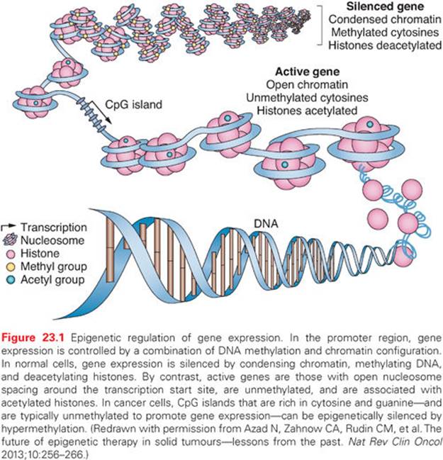
Like most biologic processes, the normal patterns of silencing can be altered, resulting in the development of disease states. Thus, activation of genes normally not expressed, or silencing of a gene that should be expressed, can contribute to the dysregulation of gene function that characterizes cancer and, when stably present, represent epigenetic alterations.4–7 Most studies have focused on the silencing of normally expressed genes. For the purposes of understanding the rationale behind epigenetic therapy, it is important to understand the mechanisms through which such silencing occurs. Alterations in gene expression associated with epigenetic changes that give rise to a growth advantage would be expected to be selected for in the host tissue, leading to progressive dysregulated growth of the tumor. Such dysregulation is commonly associated with increases in promoter region DNA methylation and is associated with repressive chromatin changes.
Changes in DNA Methylation
The importance of abnormal cytosine methylation and gene silencing has been clearly established in the past 2 decades and been shown convincingly to be involved in cancer development.4–7 The CpG dinucleotide, usually underrepresented in the genome, is clustered in the promoter regions of approximately 50% of human genes in regions termed CpG islands. These regions are largely protected from DNA methylation in normal cells, with the exception of genes on the inactive X chromosome and imprinted genes.16 This protection is critical, because the methylation of promoter region CpG islands is associated with a loss of gene expression.4–7 Abnormal de novo DNA methylation of gene promoter CpG islands is a very frequent abnormality in virtually all cancer types and is associated with a process that can serve as an alternative mechanism for loss of tumor suppressor gene function.4–7 Although a limited number of classic tumor suppressor genes can be affected by this process, a patient’s individual cancer may harbor hundreds of such genes.4–7 Which of these latter genes are drivers of cancer, individually or in groups, versus those which are passengers reflecting only the widespread effects of a global epigenetic abnormality is a leading question in the field and the target of much research.5,6 A clue to the importance of at least groups of the previous DNA hypermethylated genes may come from the fact that an inordinate number of them are involved in holding normal embryonic and adult stem cells in the self-renewal state and/or rendering such cells refractory to differentiation cues.17,18 Normally, these genes are then in a poised expression state and can be induced to be activated or repressed as needed for changes in cell state.18 Abnormal promoter DNA methylation of such genes renders them more repressed and could be a factor in the fact that cancers inevitably exhibit cell populations with enhanced self-renewal or refractoriness to full differentiation.18
Recent studies have also suggested that DNA regions other than promoter CpG islands may undergo changes of DNA methylation in cancer. For example, non–CpG-rich sequences surrounding promoter CpG islands, termed CpG island shores, are abnormally methylated in cancers19 and may be altered in stem cell populations.20 Thus, the relative cancer specificity of changes of DNA methylation in multiple CpG regions makes reversal of these changes by targeting DNA methyltransferases, the enzymes that catalyze DNA methylation, logical for cancer therapeutics.
As a key example of the previous points, perhaps the most studied tumor suppressor gene for promoter hypermethylation is the p16 gene, currently designated CDKN2A, a cyclin-dependent kinase inhibitor that functions in the regulation of the phosphorylation of the Rb protein. Hypermethylation associated with loss of expression of the CDKN2A gene has been found to be one of the most frequent alterations in neoplasia being common in the lung, head and neck, gliomas, colorectal, and breast carcinomas21,22 and other cancer types. A member of the same gene family, p15 or CDKN2B, also regulates Rb and is silenced in association with promoter methylation in many forms of leukemia and in the chronic myeloid neoplasm myelodysplastic syndrome (MDS).23 These two previous changes are of much relevance for the clinical uses of epigenetic therapies discussed later.
As mentioned, many hundreds of genes may be inactivated in a single cancer by promoter methylation,5,6,18,24 providing potential targets for gene reactivation using epigenetic therapies.25–27 The latter represents one of the potential ways in which epigenetic therapy may be effective: Multiple genes and gene pathways, all repressed by changes in DNA methylation and chromatin modification, can be reactivated by DNA methyltransferase inhibitors and histone deacetylase (HDAC) inhibitors (HDACi), thereby restoring normal cell cycle control, differentiation, and apoptotic signaling (Fig. 23.2).8,26,28 In general, methylated CpG islands are not capable of the initiation of transcription unless the methylation signal can be overridden by alterations in factors that modulate chromatin, such as the removal of methylated cytosine-binding proteins. However, reversal of DNA methylation with secondary changes in histone modification or directed reversal of repressive histone modifications represent a target for epigenetic therapies.8,26,28
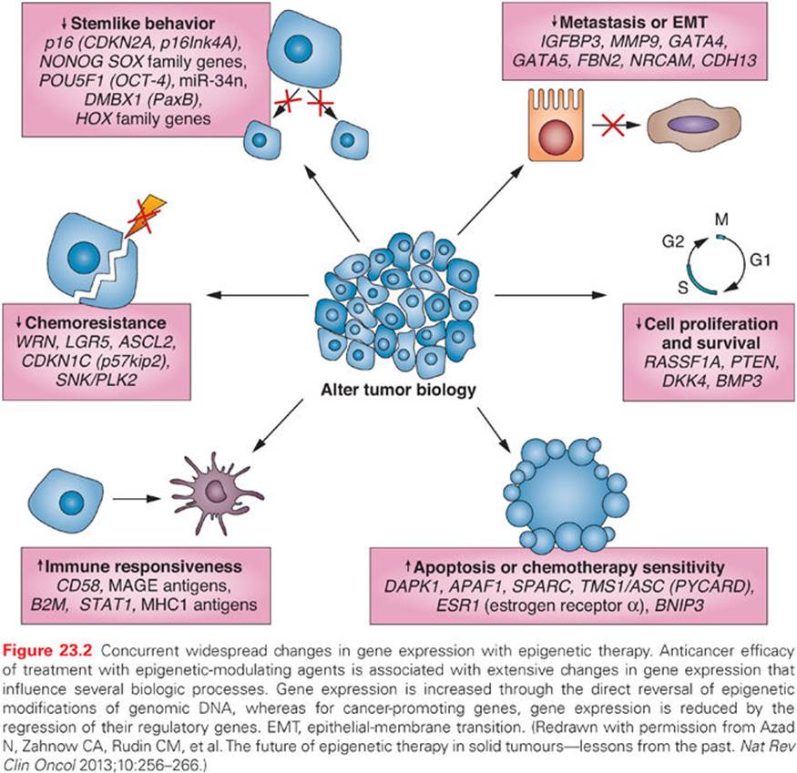
Most studies of DNA methylation, particularly in the study of cancer, have focused on CpG island promoter methylation. However, about 40% of human genes do not contain bona fide CpG islands in their promoters.29 The primary focus on CpG islands has resulted from the clear demonstration that CpG-island promoter methylation permanently silences genes both physiologically and pathologically in mammalian cells. However, recent work has shown correlations between tissue-specific expression and methylation of non-CpG islands, including, for example, the maspin gene,30 and as mentioned previously, regions near CpG islands,19,20 suggesting that many additional genes could be regulated, either normally or abnormally, by changes in DNA methylation.
An exciting new area of DNA methylation research involves the role of this change in regulating gene enhancers: small DNA regions that regulate the expression of multiple target genes.31–33 The presence of DNA methylation in these areas, which can reside considerable distances from the genes that are being regulated, generally works together with histone modifications to mediate a repressive state for that enhancer.31–33 The status of enhancers is also emerging as important for cancer risk states.34
Chromatin in Gene Regulation
Heritable gene silencing involves the interplay between DNA methylation and histone covalent modifications. Complexes of proteins that can regulate how nucleosomes are positioned perform nucleosomal remodeling.35–37 What was initially termed the histone code, with reference to how histones are modified, has emerged to be much more complex than originally envisioned. An explosion of research findings during the last several years now allows for an appreciation of how the epigenome is controlled by a complex interplay between a myriad of posttranslational histone modifications that occur on key amino acid residues of these proteins.37 Acetylation, deacetylation, methylation, phosphorylation, and other modifications all modify chromatin structure and thereby alter gene expression.38 Some of the enzymes that catalyze these modifications include HDACs, histone methyltransferases (HMT), and most recently, histone demethylases.13,14,39,40 These modifications help establish heritable states at the start site of genes, but also at enhancers and other transcribed DNA regions not encoding for canonical genes. The latter areas contain noncoding RNAs (ncRNAs) and micro-RNAs (miRNAs), which play key modulatory roles for overall gene expression and protein patterns that can be altered in cancer.41–43 Again, much research is being focused on epigenetic changes in these DNA regions, which may be important to cancer development and, potentially, to cancer management.
A link between covalent histone modifications and DNA methylation has been clearly established.44–46 In this interaction, cytosine methylation attracts methylated DNA-binding proteins and HDACs to methylated CpG sites during chromatin compaction and gene silencing.46,47 In addition, the DNA methylation binding protein (MBD2) interacts with the nucleosomal remodeling complex (NuRD) and directs the complex to methylated DNA.48 This complex also binds HDACs and has recently been identified as a central player for the abnormal silencing of genes associated with promoter DNA hypermethylation in cancer.47 Thus, the three processes of DNA cytosine methylation, histone modification, and nucleosomal remodeling are intimately linked, and alterations in these processes can result in abnormalities of gene expression in cancer-relevant genes.
Enzymes Regulating DNA Methylation and Histone Acetylation
DNA methylation involves the covalent addition of a methyl group to the 5′ position of cytosine. In mammals, three enzymes have been shown to catalyze this transfer of a methyl group from the methyl donor S-adenosylmethionine. Most of the methyltransferase activity present in differentiated cells is derived from the expression of DNMT1.49 This enzyme is thought to be most important in maintaining DNA methylation patterns following DNA replication and thus is referred to as a maintenance methyltransferase. However, the enzyme does possess the ability to methylate previously unmethylated DNA sequences (de novo activity).50 In contrast, the other enzymes, DNMT3a and DNMT3b, are efficient at methylating previously unmethylated DNA and thus are considered de novo methyltransferases. Each of these enzymes possesses a similar catalytic site,51 a fact important for the inhibition of DNMT enzymes by nucleoside analogs, discussed later in this chapter.
DNA methylation is closely associated with changes in the histone modifications. As previously discussed, histone proteins are the central components of the nucleosome, and modifications of the histone tails of core histones are associated with active or repressed chromatin.52 Although it is beyond the scope of this chapter to fully discuss the complex series of modifications to the histone tails of histone H3 and H4, a few well-characterized modifications should be mentioned that are relevant to therapies designed to target epigenetic abnormalities in cancer. In reference to currently investigated epigenetic therapies, changes in histone acetylation are of importance. Acetylation of histones H3 and H4 at key amino acids is associated with the active chromatin present at the promoters of transcribed genes, whereas the absence of histone acetylation is associated with repressed, silenced genes.13,14,53 Histone acetyltransferases (HAT) HDACs have opposing functions to maintain the proper level of histone acetylation for gene expression.13,14,53 HDACs specifically deacetylate the lysine residues of the histone tails, and this deacetylation is associated with condensation of nucleosome positions in what is termed a closed chromatin formation. This scenario is key to transcriptional repression. There are four classes of HDACs.53 Class I HDACs are characterized by their similarity to the yeast Rpd3 HDAC. In humans, this class of enzymes includes HDAC1, 2, 3, and 8. These HDACs are thought to be ubiquitously expressed in tissue throughout the body. In contrast, class II HDACs are similar to yeast Hda1 and include HDAC4, 5, 6, 7, 9, and 10, and they have a greater degree of tissue specificity. Class III HDACs are similar to yeast Sir2 and are set apart from the other classes by their dependence on nicotinamide adenine dinucleotide (NAD+) as a cofactor. Finally, class IV includes HDAC11.53
Of the previously listed HDACs, class I and 2 HDACs have been most closely tied to gene silencing associated with abnormal promoter DNA hypermethylation.48 These are bound to the nucleosome remodeling complex, NuRD.48,49 Experimental decreases in NURD, after use of a DNA demethylating agent, can augment reactivation of many abnormally silenced and DNA hypermethylated genes in colon cancer cells.48 Manipulation of these HDACs is under study in clinical trials, with and without the use of DNA methyltransferase inhibitors, and is discussed later. Another HDAC, SIRT1 in the class III of these proteins, is also involved with gene silencing.54,55 This deacetylase has been linked to silencing of DNA hypermethylated genes, and blocking its activity can be associated with reactivation of such genes.55
Reversal of Layers of Gene Silencing
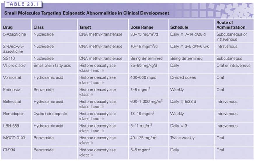
The interaction between DNA methylation and HDAC activity and repressive chromatin marks in maintaining aberrant silencing of hypermethylated genes in cancer has therapeutic implications for epigenetic therapies. Experimental evidence suggests that DNA methylation functions as a dominant event that stably establishes transcriptional repression. Inhibition of HDAC activity alone, by potent and specific HDACis, does not generally result in the reactivation of aberrantly silenced and densely hypermethylated genes in tumor cells.56 In contrast, treatment with HDACis can reactivate densely silenced genes if the cells are first treated with demethylating drugs, such as 5-azacitidine.56 The clinical implications of this observation are discussed in more detail in the following section (Table 23.1).
DNA Methyltransferase Inhibitors
Originally synthesized as cytotoxic antimetabolite drugs in the 1960s,57 azacytosine nucleosides were recognized as inhibitors of DNA methylation in the early 1980s. The inhibitors 5-azacitidine (5AC) and 2′-deoxy-5-azacytidine induced muscle, fat, and chondrocyte differentiation in mouse embryo cells, in association with a reversal of DNA methylation.58,59 The incorporation of azacytosine nucleosides into DNA in lieu of cytosine residues was shown to be associated with inhibition of DNMT activity.59,60 DNMT inhibition requires the incorporation of decitabine triphosphate into DNA. The incorporated azacytosine nucleoside forms an irreversible inactive adduct with DNMT. The sequential reversal of DNA methylation then results when DNA replication proceeds in the absence of active DNMT.61 The inhibitor 5AC must be phosphorylated and converted to decitabine diphosphate by ribonucleotide reductase before it can be activated through triphosphorylation, whereas decitabine does not require ribonucleotide reductase. The inhibitor 5AC can also be incorporated into RNA. DNMT2, a misnamed protein that is actually an RNA-specific methyltransferase,62 becomes inhibited, leading to the depletion of methylated tRNA.60 This may contribute to the inhibition of protein synthesis and is a potential difference between azacitidine and decitabine.63 The previous DNA methyltransferase inhibitors not only block the catalytic activities of DNMTs, but also trigger degradation of these proteins, especially DNMTs 1 and 3B.64–68 This latter activity is potentially important for their activities for gene reexpression because each of these two proteins, experimentally, possess transcriptional repression properties independent of their DNA methylation catalytic sites.69,70
The azacytosine nucleosides exhibit complex dose–response characteristics. At low concentrations (0.2 to 1 μM), the epigenetic activities of these drugs predominate, with dose-dependent reversal of DNA methylation71,72 and induction of terminal differentiation in some systems.28,71 As concentrations are increased, DNA damage and apoptosis become more prominent.28,72 Cell lines with 30-fold resistance to the cytotoxic effects of doxifluridine, adriamycin, cyclophosphamide (DAC) continue to reverse methylation in response to this nucleoside, suggesting that the methylation reversing and cytotoxic activities of this compound can be separated.73 The ability of these drugs to inhibit the cell cycle, at least in part through induction of p21WAF1/CIP1 expression, complicates the goal of reversing DNA methylation, because the latter requires DNA replication with the azacytosine nucleoside incorporated into the DNA.
The importance of low doses of the two azacytosine nucleosides to achieve a targeted therapeutic effect has been recently explored in a series of laboratory observations. Transient exposure of both leukemia and solid tumor cells to submicromolar doses induce such cells to undergo cellular reprogramming, accompanied by decreases in ability to clone in long-term self-renewal assays and to grow as explants in immune-incompetent mice.28 These effects occur with partial genome-wide DNA demethylation and changes in gene expression in multiple pathways potentially key for driving tumorigenesis.
The pharmacokinetic properties of the two azacytosine nucleosides are also very important to consider for their clinical use. In this regard, a major potential challenge for their usage is the fact that these drugs are highly unstable in an aqueous solution, resulting in their rapid hydrolysis and resultant inactivation.74 In clinical practice, the drugs must be administered shortly after reconstitution. The drugs are also metabolized by cytidine deaminase,74 leading to a short half-life in plasma. When injected subcutaneously, 5AC reaches a maximal plasma concentration at 30 minutes, with a terminal half-life of 1.5 to 2.3 hours.75,76 At the U.S. Food and Drug Administration (FDA) approved dose of 5AC (75 mg/m2 administered subcutaneously daily for 7 days), peak plasma concentrations were 3 to 5 μM, which is well within the range of DNMT inhibitory concentrations.75,76 Intravenous (IV) administration of the same dose has led to higher peak plasma concentrations (11 μM) with a shorter half-life (approximately 22 minutes).75DAC given over 1 hour IV at 15 to 20 mg/m2 produced plasma concentrations of 1.1 to 1.6 μM during the infusion,77whereas in a phase 1 study in patients with thoracic malignancies, patients were treated with escalating doses of decitabine for 72-hour IV infusions for two 35-day cycles. The maximum tolerated total dose was 60 to 75 mg/m2 with neutropenia as the dose-limiting toxicity. Steady-state plasma concentrations ranged from 25 to 40 nM, which is less than those usually used to induce expression of methylated genes in tissue culture models.78 An oral formulation of 5AC has also been studied. The oral bioavailability of oral azacitidine ranged from 6% to 20%. Nonetheless, MDS and acute myelogenous leukemia (AML) patients receiving oral azacitidine developed clinical responses similar to patients receiving parenteral azacitidine. Oral azacitidine has also been safely administered on 14-daily and 21-daily schedules repeated monthly. The extended administration of lower daily doses may provide favorable pharmacodynamics of DNA methylation reversal given the need for ongoing cell cycling to effect methylation reversal.79
SGI-110 is a dinucleoside that acts as a prodrug for decitabine. This drug is being studied in myelodysplasia and AML.80
HISTONE DEACETYLASE INHIBITORS
The increasing recognition of the critical importance of histone modifications in regulating the transcriptional permissively of chromatin has led to intense interest in compounds that can inhibit the activity of HDAC proteins, facilitating the acetylation of lysines associated with transcriptional activation of genes. As with the DNMT inhibitors discussed previously, there are multiple, sometimes dose-dependent, effects of HDACis in preclinical studies. Some of these may truly be epigenetic, others strictly cytotoxic, and others a combination of both.9,81–84 Some actions of HDACis may relate to altering how chromatin is central to the repair of DNA. Thus, at especially high doses, these compounds can blunt efficient repair and even induce DNA breaks.84,85 These effects may underlie cell cycle arrest and induction of cell death as is often observed in preclinical studies of HDACis.81–84
Perhaps novel uses of these drugs may be inferred by results from recent studies suggesting they could be extremely powerful epigenetic therapy agents when used in proper doses, for targeted purposes, and at key time intervals. Recent studies by Settleman and colleagues86 suggest that histone acetylation changes, and thus epigenetic mechanisms, could be a key factor for cancer therapy resistance to both targeted therapy agents and conventional chemotherapy. The mechanisms involved may involve the emergence of drug-tolerant stem-like cells.86 In such cells, gene expression studies suggest that a protein upregulated in resistance is a histone demethylase, which diminishes a key histone modification for active transcription, H3K4methyl.86 A very similar enzyme has been shown in other studies to be central to self-renewal of stem-like melanoma cells.87 Key to the therapies under discussion is that, in the previous drug-resistance studies, low doses of HDACis, could reversibly reduce drug-resistant cells induced by the various anticancer drugs.86 It is essential going forward to sort out which of these effects are dose-related off-target effects and which are desired on-target effects that can be optimized for efficacious therapy strategies.
Types of Histone Deacetylase Inhibitors
Small Chain Fatty Acids
The earliest report of the use of an HDACi to treat leukemia described the treatment of a child with refractory AML with intravenous sodium butyrate, with a concomitant clearance of peripheral blood blast cells and a decrement in bone marrow blasts.88 No responses developed in a subsequent study of nine AML patients who were treated with intravenous butyrate.89 Phase 1 studies of sodium phenylbutyrate (NaPB) in MDS and AML explored 7-day continuous infusions administered monthly or biweekly, and 21-day continuous infusions administered monthly.90,91 At the maximum tolerated dose (375 mg per kilogram per day), the mean steady-state plasma concentration was 0.3 mM, within the range of HDAC inhibition.90–92 Isolated patients developed hematologic improvement in response to NaPB.
Similar to NaPB, valproic acid (VPA) requires near millimolar concentrations to effectively inhibit HDACs. Of 18 patients with MDS or AML with trilineage dysplasia treated with VPA to target plasma concentrations of 0.3 to 0.7 mM, 6 patients developed hematologic improvement.93 Of 20 elderly patients with AML treated with VPA, only 11 could remain in control long enough to be considered evaluable for response. Five had improvement in platelet counts.94 VPA induced hematologic improvement in combination with all-transretinoic acid in two of eight patients treated with AML; a fluorescence in situ hybridization analysis showed definitive evidence of terminal differentiation of the malignant cells.95 A larger study of this combination induced hematologic response in only 2 of 26 elderly patients with AML.96 It appears unlikely that the small chain fatty acids will develop an important role in the treatment of malignancy given the availability of HDACis with vastly greater potency.
Hydroxamic Acids
The FDA approved vorinostat as the first commercially available HDACi. The approval was based on activity of this agent in cutaneous T-cell lymphoma (CTCL). Thirty-three patients with a median number of five prior systemic therapy regimens received one of three dose schedules of vorinostat in a single institution study.97 Eight patients achieved a partial response, with a median time to response of 12 weeks and a median duration of response of 15 weeks. Overall, 45% of patients had relief of pruritus. Fatigue, diarrhea, nausea, and thrombocytopenia were common toxicities. In a multicenter phase 2 trial, 74 patients with relapsed or refractory CTCL were treated with 400 mg daily.98 Similar to the prior study, 29% of patients responded, consisting almost entirely of partial responses. Median time to response was 56 days, and median duration of response was greater than 6 months. In phase 1 trials, responses to vorinostat have developed in other non-Hodgkin’s and Hodgkin’s lymphoma cases.99 More recently, in a trial combining vorinostat with carboplatin and paclitaxel in patients with untreated, advanced, non–small-cell lung cancer (NSCLC), response rates increased significantly from 12.5% to 34%, and a trend to improved progression-free survival and overall survival was observed.100
Panobinostat (LBH589), a cinnamic hydroxamic acid HDACi, reduced peripheral blood blast percentage but did not induce remissions in a phase 1 trial of daily times 7 oral dosing in patients with a variety of relapsed hematologic malignancies.101 Asymptomatic changes in electrocardiographic T waves developed in 80% of treated patients. Gastrointestinal symptoms and thrombocytopenia were common. Panobinostat has recently been approved by the FDA for the treatment of multiple myeloma.102
Cyclic Tetrapeptides
Romidepsin is FDA approved for the treatment of CTCL103 and peripheral T-cell lymphoma.104,105 Antitumor activity, including tumor lysis syndrome, was demonstrated in a phase 1 study that enrolled patients with chronic lymphocytic leukemia and AML, but no complete or partial remissions were seen.75 The administration of romidepsin induces electrocardiographic changes, including T-wave flattening and ST-T wave depression in greater than half of the posttreatment tracings; however, no changes in serum cardiac troponin levels or left ventricular ejection fraction have been reported.106
Benzamides
Entinostat, formerly known as MS-275, was administered weekly times four to patients with relapsed and refractory AML in a phase 1 study. Infections, unsteady gate, and somnolence were dose-limiting toxicities. No clinical responses developed, although improvements in neutrophil counts were observed.107 Entinostat did not increase the response rate in patients with higher risk MDS and AML with MDS-related changes when combined with azacitidine compared to azacitidine alone.108 Most recently, however, studies NSCLC suggest that entinostat could be a valuable therapeutic agent in solid tumors when used with established therapies. When combined with the epidermal growth factor inhibitor erlotinib, in a randomized phase 2 trial for patients with recurrent advanced NSCLC, entinostat was not efficacious alone but appeared to combine with erlotinib to benefit a group of patients whose tumors contained baseline high E-cadherin levels. Overall survival in these latter patients yielded an increased survival benefit of 9.4 versus 5.4 months.109 Finally, entinostat significantly increased survival when combined with an aromatase inhibitor in a phase 2 trial for patients with breast cancer.110
Pharmacodynamic Properties
The administration of oral vorinostat was associated with a transient increase in acetylation of histone H3 in peripheral blood lymphocytes, which peaked at 2 hours post dosing and reverted to baseline by 8 hours; similar changes were observed in the lymph node of a treated patient with lymphoma.99 Treatment with vorinostat was associated with translocation of phosphorylated signal transducer and activator of transcription 3 (STAT-3) from nucleus to cytoplasm in responding patients and with reduced microvessel density.97
Similar changes in the acetylation of histones 2B and 3 were observed in peripheral blood cells from patients treated with LBH589.101 Romidepsin induced acetylation of H3 and H4 in peripheral blood tumor cells within 4 hours of dosing111; of interest, p21WAF1/CIP1 protein levels also increased, associated with an increase in acetylation of H4 at the p21 promoter (using chromatin immunoprecipitation). Treatment with entinostat led to increased acetylation of H3 and H4 in both peripheral blood and bone marrow. This increase was detectable within 8 hours and remained above baseline throughout the treatment cycle. Thus, this compound may provide the most prolonged inhibition of protein deacetylation of HDACis and is under current investigation.107 Increases in p21WAF1/CIP1 and activation of caspase 3 were also demonstrated in these samples.
EPIGENETIC THERAPY FOR HEMATOLOGIC MALIGNANCIES
DNA Methyltransferase Inhibitors
Epigenetic therapy has seen the most widespread use to date and achieved the greatest efficacy in hematologic malignancies. The therapeutic efficacy of 5AC and DAC for patients with the chronic myeloid neoplasm myelodysplasia (MDS) and AML has been well reviewed.26,27 Their FDA approval for MDS/AML emerged only after doses were reduced, with resultant diminishing toxicities for patients. The successful development of 5AC for the treatment of MDS can be credited largely to Silverman et al.25,112,114 in the Cancer and Leukemia Group B (CALGB). The inhibitor 5AC had successfully induced the expression of hemoglobin F in patients with sickle cell anemia.25,112 Viewing this compound as a potential inducer of terminal differentiation, Silverman et al. conducted a series of phase 2 trials of 5AC administered as a continuous intravenous infusion or as subcutaneous injections for the treatment of MDS.113,114 Based on significant hematologic responses, the group performed a phase 2 trial (CALGB 9221) in which patients with low- and high-risk MDS with significant hematopoietic compromise were randomly assigned to receive subcutaneous 5AC (75 mg/m2 per day daily for 7 days, repeated on a 28-day cycle) or observation. Patients on the observation arm with progressive disease could cross over to receive 5AC. This study firmly established the ability of 5AC to induce hematologic improvement, and, less frequently, complete and partial responses.113,115 The median time to development of AML (defined by 30% bone marrow blast cells) or death was greater in the 5AC arm by 9 months (21 versus 12 months); of note, the observation arm included patients who subsequently crossed over to 5AC treatment.
In a subsequent phase 3 trial (AZA001),116 patients with higher risk myelodysplastic syndromes were randomly assigned one-to-one to receive 5AC (75 mg/m2 per day for 7 days every 28 days) or conventional care (best supportive care, low-dose cytarabine, or intensive chemotherapy as selected by investigators before randomization). Three hundred fifty-eight patients were randomly assigned to receive 5AC (n = 179) or conventional care regimens (n = 179). After a median follow-up of 21.1 months (interquartile range [IQR] 15.1 to 26.9), median overall survival was 24.5 months (9.9 not reached) for the azacitidine group versus 15.0 months (5.6 to 24.1) for the conventional care group (hazard ratio [HR] 0.58; 95% confidence interval [CI], 0.43 to 0.77; p = 0.0001). At 2 years, on the basis of Kaplan-Meier estimates, 50.8% (95% CI, 42.1 to 58.8) of patients in the 5AC group were alive compared with 26.2% (95% CI, 18.7 to 34.3) in the conventional care group (p < 0.0001). Median time to AML transformation was 17.8 months (IQR 8.6 to 36.8; 95% CI, 13.6 to 23.6) in the 5AC group compared with 11.5 months (4.9 not reached; 8.3 to 14.5) in the conventional care group (HR 0.50; 95% CI, 0.35 to 0.70; p < 0.0001). Subsequent unplanned analyses of AZA001 included an examination of elderly patients with what would now be classified as AML (blast count 20% to 30%). In these 113 patients, there remains a statistically significant improvement in survival of 24.5 months versus 16.0 months (HR 0.47; 95% CI; p = 0.0001).117
The early development of decitabine in MDS took place primarily in Europe under the leadership of Wijermans et al.118,119 These investigators pursued intravenous scheduling of decitabine administered three times daily for 3 days (45 mg/m2 per day total dose). This cycle was repeated every 6 weeks. Phase 2 studies suggested a response rate of approximately 50% in MDS patients. In a randomized trial of DAC versus observation, patients with International Prognostic Score risk categories intermediate 1 to high received the previously listed schedule of decitabine or observation. No crossover was allowed in this trial. Response rates reported were: complete response: 9%, partial response: 8%, and hematologic improvement: 13%.120 A 10% induction death rate occurred, suggesting that this schedule of DAC may be more toxic than the CALGB schedule of 5AC (1% induction mortality). DAC has also been investigated in low-dose daily intravenous dosing121 and in daily-times-five schedules. The latter appears convenient and well tolerated. A daily-times-five schedule (20 mg/m2 per day) has been FDA approved121; 99 patients with MDS (de novo or secondary) of any French-American-British (FAB) subtype and an International Prognostic Scoring System (IPSS) score equal to or greater than 0.5 were treated, with an overall response rate of 32% (17 complete responses [CR] plus 15 marrow CRs [mCR]).122 Among patients who improved, 82% demonstrated responses by the end of cycle two. This well-tolerated regimen allows outpatient administration and, as noted previously, provides plasma levels of decitabine that inhibit DNMTs.
The 3-day intravenous schedule of DAC has been studied in two randomized trials compared to supportive care in patients with higher risk MDS. The first trial confirmed the hematologic activity of decitabine in this patient population but failed to show an improvement in survival in the DAC-treated patients.123 Survival was also not increased in the subsequent trial, performed by the European Organization for Research and Treatment of Cancer (EORTC).124 The failure of the randomized decitabine trials to show a survival benefit may be partially due to study design. Both randomized trials of 5AC continued treatment until disease progression for patients who did not achieve complete remission; in fact, this meant that most patients received maintenance therapy. In contrast, both randomized trials of decitabine allowed a maximum of eight cycles of treatment. The need for maintenance therapy in patients treated with DNMT inhibitors has not been tested in prospective randomized trials. An additional difference in the conduct of the two sets of DNMT inhibitor trials involves the duration of therapy administered. The median number of cycles of treatment administered in the two randomized trials of decitabine was three, compared to nine in the azacytidine trials. This may reflect greater toxicity of the originally 3-day schedule of decitabine compared to that of the approved schedule of 5AC. Although the differences in survival may reflect differences in trial design and trial conduct, emerging data suggests that despite similarities in methylation reversal, the two drugs differ in other potentially important biologic parameters, which may contribute to clinical outcomes.62,63,125
Two randomized phase 3 trials have been published treating elderly AML patients (greater than 20% blasts) with decitabine, both demonstrating improvement in survival that was not statistically significant. In the European study, 233 patients received either DAC at 15 mg/m2 × 9 doses over 3 days on 42-day cycles or best supportive care. The patients received a median of 4 cycles (0 to 9), and the overall survival was improved in the decitabine-treated patients, but did not reach statistical significance (median overall survival [OS], 10.1 versus 8.5 months, respectively; HR, 0.88; 95% CI, 0.66 to 1.17; two-sided, log-rank p = 0.38).126 In the M.D. Anderson Cancer Center–led multicenter trial,127 485 patients 65 years or older were randomly assigned to receive decitabine 20 mg/m2 per day as a 1-hour intravenous infusion for 5 consecutive days every 4 weeks or best supportive care or low-dose cytarabine (20 mg/m2 per day for 10 days every 4 weeks). There was a similar improvement in OS with decitabine (7.7 months; 95% CI, 6.2 to 9.2) versus the control group (5.0 months; 95% CI, 4.3 to 6.3; p = 0.108; HR, 0.85; 95% CI, 0.69 to 1.04).127
The azacytosine nucleosides require prolonged administration to demonstrate hematologic improvement in MDS. Median time to development of first clinical response in the CALGB studies of 5AC was three cycles; 90% of responses developed by cycle six.114 In the phase 3 trial of decitabine, the median time to response was two cycles,123 as also seen in the alternative regimen of decitabine.122 It is, therefore, extremely important when treating patients with azacytosine nucleosides to commit to administering between four and six cycles of therapy before determining whether a patient is responding to treatment. Furthermore, survival benefit is seen even in patients not showing bone marrow improvement for 5AC, perhaps related to decreased transfusion requirements or delayed progression to AML.116
Because AML in the context of MDS is arbitrarily defined based on marrow blast count, activity of the azanucleoside analogs in AML should not be surprising. In CALGB 9221, 20 patients were reclassified upon central pathology review as meeting criteria for AML (greater than 30% blasts). Their outcomes were comparable to the overall population in the study.115 In all three CALGB studies among patients meeting current World Health Organization (WHO) criteria for AML (greater than 20% blasts), a complete response was achieved in 9% and hematologic improvement in 26%.114 A retrospective review of 20 patients with AML, including 8 patients with bone marrow blasts greater than 29% treated with 5AC, reported a complete remission in 4 patients, a partial response in 5, and a hematologic improvement in 3. The median duration of response was 8 months (range: 3 to 33 months).128DAC induced a complete hematologic response in 2 of 20 patients treated who had the blastic phase of chronic myeloid leukemia.129These studies suggest activity of the azacytosine nucleosides in the treatment of a subset of AML patients. Current studies do not allow for the determination of whether this subset is limited to MDS-associated AML (AML with MDS-related changes), which tends to have low white blood cell counts and have a low proliferative rate, or whether these compounds are also active for those with AML without a history of antecedent hematologic disorder. Several reports describe the sensitivity MDS and AML, characterized by abnormalities of chromosome 7 and associated with poor outcomes in response to cytarabine-based therapy to azanucleosides. In one nonrandomized retrospective study, survival of such patients following the administration of DNMT inhibitors surpassed survival in response to conventional cytotoxic chemotherapy, similar to the outcomes of AZA001.130–132
Although the mechanisms underlying the clinical activity of azacytosine analogs may involve reversal of gene methylation, other actions need to be considered. The administration of DAC has been shown to induce transient decrements of methylation in noncoding regions, including long interspersed nuclear element (LINE) and ALU elements.133 Early studies that examined methylation reversal of the target gene p15INK4B in response to DAC showed no correlation between methylation reversal and clinical response.134,135 Clinical responders to DAC developed significantly higher expression of this gene following treatment, and certainly key biologic roles for this gene and its low basal expression are probable. Moreover, in one study, the clinical response was closely associated with the reversal of methylation of p15 or CDH-1 during the first cycle of treatment with 5AC followed by the HDACi NaPB.25 In that study, it was noteworthy that the administration of 5AC prior to the addition of an HDACi was associated with the induction of histone acetylation. Although the mechanism underlying this activity is unknown, histone acetylation has been observed following DNA damage due to gamma irradiation.136 Subsequent studies have found demethylation following treatment with either DAC or 5AC137–140 but not consistently associated with response.137,138,140 More work will be required to answer the important mechanistic question underpinning the clinical activity of azacytosine analogs.
Combining Inhibitors in the Treatment of Hematologic Malignancies
It is almost certain that the biggest promise of epigenetic therapy lies in strategies to combine existing and newer drugs with each other and with current chemotherapies and targeted therapies. To date, the example for existing agents is the combination of DNMT inhibitors and HDAC inhibitors based on the hypothesis from the laboratory that this paradigm leads to optimal reexpression of transcriptionally silenced genes with promoter methylation.56,141 This in vitro treatment paradigm has led to a variety of clinical studies that have attempted to apply this concept to the treatment of hematologic malignancies. Much remains to be determined with regard to its efficacy and precisely what determines this. The first study of sequential DNMT/HDAC inhibitors administered a variety of doses of 5AC for 5 to 14 days followed by 7 days of NaPB by continuous infusion at its maximum tolerated dose to patients with MDS and AML.25 The combination was well tolerated, and clinical responses were frequent in patients receiving 5AC at 50 mg/m2 per day daily for 10 days and 25 mg/m2 per day daily for 14 days, with 5 of 14 patients at those dose schedules achieving complete or partial response.
In a pilot study, 10 patients with MDS or AML were treated with 5AC at 75 mg/m2 per day daily times seven followed by 5 days of NaPB given at 200 mg per kilogram per day as a 1- to 2-hour infusion. Three patients developed a partial response.142
In a similar study, investigators at the M.D. Anderson Cancer Center treated leukemic patients with decitabine (15 mg/m2 per day IV daily times 10) and concomitant VPA at a variety of doses. Of 54 patients, 12 achieved complete remission or complete remission with incomplete platelet recovery.143 The inhibitors 5AC, VPA, and all-transretinoic acid have been administered to patients with AML and MDS. Of 33 previously untreated patients, 14 over the age of 60 years developed a complete remission or a complete remission with inadequate platelet recovery.144 A subsequent study of 5AC and VPA suggests increased efficacy of this combination in high-risk MDS.145
Entinostat has been successfully combined with azacytidine in patients with myeloid malignancies.140 The US Leukemia Intergroup recently completed a randomized phase 2 trial of this combination compared with 5AC alone. In this study, of 149 patients, the primary endpoint of hematologic normalization was statistically similar, with 32% (95% CI, 22% to 44%) of the 5AC group reaching hematologic normalization (HN) versus 27% (95% CI, 17% to 39%) in the AZA + entinostat group. Median overall survivals were 18 months for the AZA group and 13 months for the AZA + entinostat group, but were also not statistically significant.108 In the latter study, the administration of the combination was associated with less DNA methylation reversal compared to azacitidine monotherapy, likely due to cell cycle inhibitory effects of the HDACi. This highlights the complexity of effectively targeting epigenetic gene regulation. It remains to be established whether combination therapies are more effective than single-agent demethylating therapies.
Epigenetically Targeted Therapy in Nonhematologic Malignancies
The efficacies that have emerged in the application of epigenetically targeted drugs to hematologic malignancies has spurred interest in using epigenetic therapy for other types of cancer. As outlined as follows, laboratory studies and clinical trials support this approach. Studies in the lab have been directed by lessons learned from therapy in hematologic malignancies, suggesting that low doses of drugs like DAC and 5AC, in the nanomolar range, may avoid excess toxicities due to off-target effects of the drugs and may maximize epigenetic effects of the agents.28 The desired effects may require minimizing initial cellular cytotoxicity, giving tumor cells time to accrue maximal cellular reprogramming responses to the inhibition of DNMTs.28 DAC and 5AC are effective only when they have been incorporated into DNA, after which they irreversibly inhibit DNMT catalytic activity and target these proteins for degradation.64–68 In cell culture and mouse explants, low nanomolar doses appear to induce both human leukemic and solid tumor cells to exhibit blunting of self-renewal and tumorigenic activity of tumor stem-like cells.28 These preclinical results suggest a key possibility that use of epigenetic therapies might inhibit these latter cell populations, which often are difficult to eradicate and are a factor in resistance to many standard cancer therapies.146 Exhaustion of such cells over time during therapy with DAC or 5AC might explain the observation that most patients with MDS/AML take several months to reach best response.147 Leukemic stem cells were not eliminated in one study in MDS and AML patients treated with 5AC in combination with VPA, although their frequency decreased in clinical responders.148
Clinical trials for common solid tumors, informed through the previous laboratory studies, have been initiated including phase 2 designs using low-dose strategies with 5AC often combined with use of histone deacetylase inhibitors. Sixty-five patients with advanced, multiply treated NSCLCs were treated with 5AC plus entinostat.149 Only 3% of patients developed Response Evaluation Criteria (RECIST)-measureable responses; however, these two patients had durable responses, with survival of 3 to 4 years.149 Upregulation of immunogenic pathways in NSCLC and other solid tumor cells, observed in laboratory studies, suggest a potential for sequencing DNMT inhibitors with immune checkpoint inhibitors.150 This drug is also reported to induce antitumor responses and immune recognition in a model of pancreatic cancer.151 Other laboratory results and emerging clinical trials also suggest the promise of combining epigenetic therapy approaches to sensitize cancers other than NSCLC to subsequent therapies. Low-dose DAC appears able to upregulate a key mediator of 5-fluorouracil (5FU) action, uridine monophosphate (UMP) kinase, in colorectal cancer cell lines.152 These increases correlated with a reversal of 5FU resistance. Similar to studies discussed previously, DAC plus the HDACi, trichostatin A, decreased marker identified self-renewal populations in ovarian cancer while simultaneously inducing increased sensitivity to cisplatin.153 In advanced ovarian cancer, 5AC or DAC plus carboplatin have yielded durable responses and induced stable disease in ovarian cancer patients.154,155 These early results are being extrapolated for verification in larger, ongoing clinical trials.
NEW APROACHES TO EPIGENETIC THERAPY
As we have outlined previously, the emerging promise for epigenetic therapy and the future of the approaches may lie in combinatorial drug strategies. Although this is already being explored with older agents, new drugs for new targets are now entering the picture.9,11,156–158 In these efforts, several themes we have introduced in this chapter will likely dominate.
Most epigenetic therapies will not induce, when used at truly targeting doses, immediate cytotoxic effects. Therapeutic efficacy based on cellular reprogramming may require significant time to manifest. Clinical trial designs may need adaptation so that effective therapies are not discarded due to premature response evaluations. Finally, the ultimate promise for epigenetic therapy may lie with newer drugs now entering clinical trials. Outcomes with DNMT inhibitors may be improved with alternative scheduling of oral azacitidine or through prolonged pharmacokinetics of the decitabine prodrug SGI110.79 Also, drugs targeting other proteins including BET family bromodomain proteins are generating much excitement.9,82,156–159 BET inhibitors may interfere with localization of the oncogene C-MYC to acetylated lysines in regulatory regions of target genes.9,82,156–159 These inhibitors are now entering clinical trials. Other promising approaches include the use of inhibitors of EZH2, the enzyme in the PcG system, which catalyzes the repressive histone mark H3K27me3.9,82,156–159 Another clinical trial underway employs targeting of the translocation in which the protein mixed lineage leukemia (MLL) is fused with several targets, such as in infant leukemias. These translocations result in abnormal recruitment of the histone methyltransferase, DOT1L, to target genes like HOXA9.158 This fusion induces hypermethylation of H3K79 and abnormal activation of MLL target genes.158,160 Very selective inhibitors of DOT1L are now in clinical trials.
Epigenetically targeted therapies continue to hold great promise that reprogramming of malignant cells could alter approaches to cancer management. Strategies to merge older drugs, which we have focused on in this chapter, with the newer agents briefly discussed in this section, will underpin future trials to test this approach.
REFERENCES
1. Bernstein BE, Meissner A, Lander ES. The mammalian epigenome. Cell 2007;128:669–681.
2. Young RA. Control of the embryonic stem cell state. Cell 2011;144:940–954.
3. Suva ML, Riggi N, Bernstein BE. Epigenetic reprogramming in cancer. Science 2013;339:1567–1570.
4. Herman JG, Baylin SB. Gene silencing in cancer in association with promoter hypermethylation. N Engl J Med 2003;349:2042–2054.
5. Jones PA, Baylin SB. The epigenomics of cancer. Cell 2007;128:683–692.
6. Baylin SB, Jones PA. A decade of exploring the cancer epigenome — biological and translational implications. Nat Rev Cancer 2011;11:726–734.
7. Esteller M. Cancer epigenomics: DNA methylomes and histone-modification maps. Nat Rev Genet 2007;8:286–298.
8. Yoo CB, Jones PA. Epigenetic therapy of cancer: past, present and future. Nat Rev Drug Discov 2006;5:37–50.
9. Dawson MA, Kouzarides T. Cancer epigenetics: from mechanism to therapy. Cell 2012;150:12–27.
10. Dawson MA, Kouzarides T, Huntly BJ. Targeting epigenetic readers in cancer. N Engl J Med 2012;367:647–657.
11. Bradner J. New targets for hematologic malignancies. Clin Adv Hematol Oncol 2013;11:375–376.
12. You JS, Jones PA. Cancer genetics and epigenetics: two sides of the same coin? Cancer Cell 2012;22:9–20.
13. Allis C, Jenuwein T, Reinberg D. Epigenetics, Vol. 1. Cold Spring Harbor, NY: Cold Spring Harbor Laboratory Press; 2007.
14. Kouzarides T. Chromatin modifications and their function. Cell 2007;128:693–705.
15. Baylin SB, Jones PA. Epigenetic determinants of cancer. In: Allis CD, Jenuwein T, Reinberg D, eds. Epigenetics. Cold Spring Harbor, NY: Cold Spring Harbor Laboratory Press; 2006: 457–476.
16. Bird AP. CpG-rich islands and the function of DNA methylation. Nature 1986;321:209–213.
17. Morey L, Pascual G, Cozzuto L, et al. Nonoverlapping functions of the Polycomb group Cbx family of proteins in embryonic stem cells. Cell Stem Cell 2012;10:47–62.
18. Easwaran H, Johnstone SE, Van Neste L, et al. A DNA hypermethylation module for the stem/progenitor cell signature of cancer. Genome Res 2012;22:837–849.
19. Irizarry RA, Ladd-Acosta C, Wen B, et al. The human colon cancer methylome shows similar hypo- and hypermethylation at conserved tissue-specific CpG island shores. Nat Genet 2009;41:178–186.
20. Doi A, Park IH, Wen B, et al. Differential methylation of tissue- and cancer-specific CpG island shores distinguishes human induced pluripotent stem cells, embryonic stem cells and fibroblasts. Nat Genet 2009;41:1350–1353.
21. Herman JG, Merlo A, Mao L, et al. Inactivation of the CDKN2/p16/MTS1 gene is frequently associated with aberrant DNA methylation in all common human cancers. Cancer Res 1995;55:4525–4530.
22. Merlo A, Herman JG, Mao L, et al. 5′ CpG island methylation is associated with transcriptional silencing of the tumour suppressor p16/CDKN2/MTS1 in human cancers. Nat Med 1995;1:686–692.
23. Herman JG, Civin CI, Issa JP, et al. Distinct patterns of inactivation of p15INK4B and p16INK4A characterize the major types of hematological malignancies. Cancer Res 1997;57:837–841.
24. Costello JF, Fruhwald MC, Smiraglia DJ, et al. Aberrant CpG-island methylation has non-random and tumour-type-specific patterns. Nat Genet 2000;24:132–138.
25. Gore SD, Baylin S, Sugar E, et al. Combined DNA methyltransferase and histone deacetylase inhibition in the treatment of myeloid neoplasms. Cancer Res 2006;66:6361–6369.
26. Azad N, Zahnow CA, Rudin CM, et al. The future of epigenetic therapy in solid tumours—lessons from the past. Nat Rev Clin Oncol 2013;10:256–266.
27. Issa JP, Kantarjian HM. Targeting DNA methylation. Clin Cancer Res 2009;15:3938–3946.
28. Tsai HC, Li H, Van Neste L, et al. Transient low doses of DNA-demethylating agents exert durable antitumor effects on hematological and epithelial tumor cells. Cancer Cell 2012;21:430–446.
29. Takai D, Jones PA. Comprehensive analysis of CpG islands in human chromosomes 21 and 22. Proc Natl Acad Sci U S A 2002;99:3740–3745.
30. Futscher BW, Oshiro MM, Wozniak RJ, et al. Role for DNA methylation in the control of cell type specific maspin expression. Nat Genet 2002;31:175–179.
31. Feldmann A, Ivanek R, Murr R, et al. Transcription factor occupancy can mediate active turnover of DNA methylation at regulatory regions. PLoS Genet 2013;9:e1003994.
32. Aran D, Hellman A. Unmasking risk loci: DNA methylation illuminates the biology of cancer predisposition: analyzing DNA methylation of transcriptional enhancers reveals missed regulatory links between cancer risk loci and genes. Bioessays 2014;36:184–190.
33. Ziller MJ, Gu H, Muller F, et al. Charting a dynamic DNA methylation landscape of the human genome. Nature 2013;500:477–481.
34. Akhtar-Zaidi B, Cowper-Sal-lari R, Corradin O, et al. Epigenomic enhancer profiling defines a signature of colon cancer. Science 2012;336:736–739.
35. Kingston R, Tamkun JW. Transcriptional regulation by trithorax group. In: Allis CD, Jenuwein T, Reinberd D, eds. Epigenetics. Cold Spring Harbor, NY: Cold Spring Harbor Laboratory Press; 2006: 231–248.
36. Becker PB, Workman JL. Nucleosome remodeling and epigenetics. Cold Spring Harb Perspect Biol 2013;5.
37. Petty E, Pillus L. Balancing chromatin remodeling and histone modifications in transcription. Trends Genet 2013;29:621–629.
38. Jones PA. Functions of DNA methylation: islands, start sites, gene bodies and beyond. Nat Rev Genet 2012;13:484–492.
39. Bannister AJ, Kouzarides T. Reversing histone methylation. Nature 2005;436:1103–1106.
40. Bannister AJ, Kouzarides T. Regulation of chromatin by histone modifications. Cell Res 2011;21:381–395.
41. Di Leva G, Garofalo M, Croce CM. MicroRNAs in cancer. Annu Rev Pathol 2014;9:287–314.
42. Han BW, Chen YQ. Potential pathological and functional links between long noncoding RNAs and hematopoiesis. Sci Signal 2013;6:re5.
43. Xi JJ. MicroRNAs in Cancer. Cancer Treat Res 2013;158:119–137.
44. Nan X, Ng HH, Johnson CA, et al. Transcriptional repression by the methyl-CpG-binding protein MeCP2 involves a histone deacetylase complex. Nature 1998;393:386–389.
45. Jones PL, Veenstra GJ, Wade PA, et al. Methylated DNA and MeCP2 recruit histone deacetylase to repress transcription. Nat Genet 1998;19:187–191.
46. Parry L, Clarke AR. The Roles of the Methyl-CpG Binding Proteins in Cancer. Genes Cancer 2011;2:618–630.
47. Lopez-Serra L, Esteller M. Proteins that bind methylated DNA and human cancer: reading the wrong words. Br J Cancer 2008;98:1881–1885.
48. Cai Y, Geutjes EJ, de Lint K, et al. The NuRD complex cooperates with DNMTs to maintain silencing of key colorectal tumor suppressor genes. Oncogene 2014;33:2157–2168.
49. Bestor TH. Cloning of a mammalian DNA methyltransferase. Gene 1998;74:9–12.
50. Jair KW, Bachman KE, Suzuki H, et al. De novo CpG island methylation in human cancer cells. Cancer Res 2006;66:682–692.
51. Rius M, Lyko F. Epigenetic cancer therapy: rationales, targets and drugs. Oncogene 2012;31:4257–4265.
52. Jenuwein T, Allis CD. Translating the histone code. Science 2001;293:1074–1080.
53. Bolden JE, Peart MJ, Johnstone RW. Anticancer activities of histone deacetylase inhibitors. Nat Rev Drug Discov 2006;5:769–784.
54. Vaquero A, Scher M, Erdjument-Bromage H, et al. SIRT1 regulates the histone methyl-transferase SUV39H1 during heterochromatin formation. Nature 2007;450:440–444.
55. Pruitt K, Zinn RL, Ohm JE, et al. Inhibition of SIRT1 reactivates silenced cancer genes without loss of promoter DNA hypermethylation. PLoS Genet 2006;2:344–352.
56. Cameron EE, Bachman KE, Myohanen S, et al. Synergy of demethylation and histone deacetylase inhibition in the re-expression of genes silenced in cancer. Nat Genet 1999;21:103–107.
57. Sorm F, Piskala A, Cihak A, et al. 5-Azacytidine, a new, highly effective cancerostatic. Experientia 1964;20:202–203.
58. Taylor SM, Jones PA. Multiple new phenotypes induced in 10T1/2 and 3T3 cells treated with 5-azacytidine. Cell 1979;17:771–779.
59. Jones PA, Taylor SM. Hemimethylated duplex DNAs prepared from 5-azacytidine-treated cells. Nucleic Acids Res 1981;9:2933–2947.
60. Lu SH, Ohshima H, Bartsch H. Recent studies on N-nitroso compounds as possible etiological factors in oesophageal cancer. IARC Sci Publ 1984;947–953.
61. Taylor SM, Jones PA. Mechanism of action of eukaryotic DNA methyltransferase. Use of 5-azacytosine-containing DNA. J Mol Biol 1982;162:679–692.
62. Schaefer M, Hagemann S, Hanna K, et al. Azacytidine inhibits RNA methylation at DNMT2 target sites in human cancer cell lines. Cancer Res 2009;69:8127–8132.
63. Hollenbach PW, Nguyen AN, Brady H, et al. A comparison of azacitidine and decitabine activities in acute myeloid leukemia cell lines. PLoS One 2010;5:e9001.
64. Kelly TK, De Carvalho DD, Jones PA. Epigenetic modifications as therapeutic targets. Nat Biotechnol 2010;28:1069–1078.
65. Ferguson AT, Vertino PM, Spitzner JR, et al. Role of estrogen receptor gene demethylation and DNA methyltransferase. DNA adduct formation in 5-aza-2′deoxycytidine-induced cytotoxicity in human breast cancer cells. J Biol Chem 1997;272:32260–32266.
66. Gabbara S, Bhagwat AS. The mechanism of inhibition of DNA (cytosine-5-)-methyltransferases by 5-azacytosine is likely to involve methyl transfer to the inhibitor. Biochem J 1995;307:87–92.
67. Santi DV, Norment A, Garrett CE. Covalent bond formation between a DNA-cytosine methyltransferase and DNA containing 5-azacytosine. Proc Natl Acad Sci U S A 1984;81:6993–6997.
68. Ghoshal K, Datta J, Majumder S, et al. 5-Aza-deoxycytidine induces selective degradation of DNA methyltransferase 1 by a proteasomal pathway that requires the KEN box, bromo-adjacent homology domain, and nuclear localization signal. Mol Cell Biol 2005;25:4727–4741.
69. Rountree MR, Bachman KE, Baylin SB. DNMT1 binds HDAC2 and a new co-repressor, DMAP1, to form a complex at replication foci. Nat Genet 2000;25:269–277.
70. Bachman KE, Rountree MR, Baylin SB. Dnmt3a and Dnmt3b are transcriptional repressors that exhibit unique localization properties to heterochromatin. J Biol Chem 2001;276:32282–32287.
71. Jones PA, Taylor SM. Cellular differentiation, cytidine analogs and DNA methylation. Cell 1980;20:85–93.
72. Berg T, Guo Y, Abdelkarim M, et al. Reversal of p15/INK4b hypermethylation in AML1/ETO-positive and negative myeloid leukemia cell lines. Leuk Res 2007;31:497–506.
73. Flatau E, Gonzales FA, Michalowsky LA, et al. DNA methylation in 5-aza-2′-deoxycytidine-resistant variants of C3H 10T1/2 C18 cells. Mol Cell Biol 1984;4:2098–2102.
74. Chan KK, Giannini DD, Staroscik JA, et al. 5-Azacytidine hydrolysis kinetics measured by high-pressure liquid chromatography and 13C-NMR spectroscopy. J Pharm Sci 1979;68:807–812.
75. Gore SD, Weng LJ, Figg WD, et al. Impact of prolonged infusions of the putative differentiating agent sodium phenylbutyrate on myelodysplastic syndromes and acute myeloid leukemia. Clin Cancer Res 2002;8:963–970.
76. Yu L, Liu C, Vandeusen J, et al. Global assessment of promoter methylation in a mouse model of cancer identifies ID4 as a putative tumor-suppressor gene in human leukemia. Nat Genet 2005;37:265–274.
77. Blum W, Klisovic RB, Hackanson B, et al. Phase I study of decitabine alone or in combination with valproic acid in acute myeloid leukemia. J Clin Oncol 2007;25:3884–3891.
78. Schrump DS, Fischette MR, Nguyen DM, et al. Phase I study of decitabine-mediated gene expression in patients with cancers involving the lungs, esophagus, or pleura. Clin Cancer Res 2006;12:5777–5785.
79. Garcia-Manero G, Gore SD, Cogle C, et al. Phase I study of oral azacitidine in myelodysplastic syndromes, chronic myelomonocytic leukemia, and acute myeloid leukemia. J Clin Oncol 2011;29:2521–2527.
80. Issa JP, Roboz G, Rizzieri D, et al. Abstract LB-214: Interim results from a randomized Phase 1-2 first-in-human (FIH) study of PK/PD guided escalating doses of SGI-110, a novel subcutaneous (SQ) second generation hypomethylating agent (HMA) in relapsed/refractory MDS and AML. Cancer Res2012;72:LB–214.
81. Mund C, Lyko F. Epigenetic cancer therapy: Proof of concept and remaining challenges. Bioessays 2010;32:949–957.
82. Popovic R, Licht JD. Emerging epigenetic targets and therapies in cancer medicine. Cancer Discov 2012;2:405–413.
83. Verbrugge I, Johnstone RW, Bots M. Promises and challenges of anticancer drugs that target the epigenome. Epigenomics 2011;3:547–565.
84. Robert C, Rassool FV. HDAC inhibitors: roles of DNA damage and repair. Adv Cancer Res 2012;116:87–129.
85. Kachhap SK, Rosmus N, Collis SJ, et al. Downregulation of homologous recombination DNA repair genes by HDAC inhibition in prostate cancer is mediated through the E2F1 transcription factor. PLoS One 2010;5:e11208.
86. Sharma SV, Lee DY, Li B, et al. A chromatin-mediated reversible drug-tolerant state in cancer cell subpopulations. Cell 2010;141:69–80.
87. Villanueva J, Vultur A, Lee JT, et al. Acquired resistance to BRAF inhibitors mediated by a RAF kinase switch in melanoma can be overcome by cotargeting MEK and IGF-1R/PI3K. Cancer Cell 2010;18:683–695.
88. Novogrodsky A, Dvir A, Ravid A, et al. Effect of polar organic compounds on leukemic cells. Butyrate-induced partial remission of acute myelogenous leukemia in a child. Cancer 1983;51:9–14.
89. Miller AA, Kurschel E, Osieka R, et al. Clinical pharmacology of sodium butyrate in patients with acute leukemia. Eur J Cancer Clin Oncol 1987;23:1283–1287.
90. Gore SD, Weng LJ, Zhai S, et al. Impact of the putative differentiating agent sodium phenylbutyrate on myelodysplastic syndromes and acute myeloid leukemia. Clin Cancer Res 2001;7:2330–2339.
91. Gore SD, Weng LJ, Figg WD, et al. Impact of prolonged infusions of the putative differentiating agent sodium phenylbutyrate on myelodysplastic syndromes and acute myeloid leukemia. Clin Cancer Res 2002;8:963–970.
92. DiGiuseppe JA, Weng LJ, Yu KH, et al. Phenylbutyrate-induced G1 arrest and apoptosis in myeloid leukemia cells: structure-function analysis. Leukemia 1999;13:1243–1253.
93. Kuendgen A, Strupp C, Aivado M, et al. Treatment of myelodysplastic syndromes with valproic acid alone or in combination with all-trans retinoic acid. Blood 2004;104:1266–1269.
94. Pilatrino C, Cilloni D, Messa E, et al. Increase in platelet count in older, poor-risk patients with acute myeloid leukemia or myelodysplastic syndrome treated with valproic acid and all-trans retinoic acid. Cancer 2005;104:101–109.
95. Fizzotti M, Cimino G, Pisegna S, et al. Detection of homozygous deletions of the cyclin-dependent kinase 4 inhibitor (p16) gene in acute lymphoblastic leukemia and association with adverse prognostic features. Blood 1995;85:2685–2690.
96. Xu GL, Bestor TH, Bourc’his D, et al. Chromosome instability and immunodeficiency syndrome caused by mutations in a DNA methyltransferase gene. Nature 1999;402:187–191.
97. Duvic M, Talpur R, Ni X, et al. Phase 2 trial of oral vorinostat (suberoylanilide hydroxamic acid, SAHA) for refractory cutaneous T-cell lymphoma (CTCL). Blood 2007;109:31–39.
98. Olsen EA, Kim YH, Kuzel TM, et al. Phase IIb multicenter trial of vorinostat in patients with persistent, progressive, or treatment refractory cutaneous T-cell lymphoma. J Clin Oncol 2007;25:3109–3115.
99. O’Connor OA, Heaney ML, Schwartz L, et al. Clinical experience with intravenous and oral formulations of the novel histone deacetylase inhibitor suberoylanilide hydroxamic acid in patients with advanced hematologic malignancies. J Clin Oncol 2006;24:166–173.
100. Ramalingam SS, Maitland ML, Frankel P, et al. Carboplatin and Paclitaxel in combination with either vorinostat or placebo for first-line therapy of advanced non-small-cell lung cancer. J Clin Oncol 2010;28:56–62.
101. Giles F, Fischer T, Cortes J, et al. A phase I study of intravenous LBH589, a novel cinnamic hydroxamic acid analogue histone deacetylase inhibitor, in patients with refractory hematologic malignancies. Clin Cancer Res 2006;12:4628–4635.
102. Richardson PG, Hungria VTM, Yoon S-S, et al. Panorama 1: A randomized, double-blind, phase 3 study of panobinostat or placebo plus bortezomib and dexamethasone in relapsed or relapsed and refractory multiple myeloma. ASCO Meeting Abstracts 2014;32:8510.
103. Piekarz RL, Robey R, Sandor V, et al. Inhibitor of histone deacetylation, depsipeptide (FR901228), in the treatment of peripheral and cutaneous T-cell lymphoma: a case report. Blood 2001;98:2865–2868.
104. Coiffier B, Pro B, Prince HM, et al. Romidepsin for the treatment of relapsed/refractory peripheral T-cell lymphoma: pivotal study update demonstrates durable responses. J Hematol Oncol 2014;7:11.
105. Coiffier B, Pro B, Prince HM, et al. Results from a pivotal, open-label, phase II study of romidepsin in relapsed or refractory peripheral T-cell lymphoma after prior systemic therapy. J Clin Oncol 2012;30:631–636.
106. Piekarz RL, Frye AR, Wright JJ, et al. Cardiac studies in patients treated with depsipeptide, FK228, in a phase II trial for T-cell lymphoma. Clin Cancer Res 2006;12:3762–3773.
107. Gojo I, Jiemjit A, Trepel JB, et al. Phase 1 and pharmacologic study of MS-275, a histone deacetylase inhibitor, in adults with refractory and relapsed acute leukemias. Blood 2007;109:2781–2790.
108. Prebet T, Sun Z, Figueroa ME, et al. Prolonged administration of azacitidine with or without entinostat for myelodysplastic syndrome and acute myeloid leukemia with myelodysplasia-related changes: results of the US Leukemia Intergroup Trial E1905. J Clin Oncol2014;32:1242–1248.
109. Witta SE, Jotte RM, Konduri K, et al. Randomized phase II trial of erlotinib with and without entinostat in patients with advanced non-small-cell lung cancer who progressed on prior chemotherapy. J Clin Oncol 2012;30:2248–2255.
110. Yardley DA, Ismail-Khan RR, Melichar B, et al. Randomized phase II, double-blind, placebo-controlled study of exemestane with or without entinostat in postmenopausal women with locally recurrent or metastatic estrogen receptor-positive breast cancer progressing on treatment with a nonsteroidal aromatase inhibitor. J Clin Oncol 2013;31:2128–2135.
111. Byrd JC, Marcucci G, Parthun MR, et al. A phase 1 and pharmacodynamic study of depsipeptide (FK228) in chronic lymphocytic leukemia and acute myeloid leukemia. Blood 2005;105:959–967.
112. Charache S, Dover G, Smith K, et al. Treatment of sickle cell anemia with 5-azacytidine results in increased fetal hemoglobin production and is associated with nonrandom hypomethylation of DNA around the gamma-delta-beta-globin gene complex. Proc Natl Acad Sci U S A 1983;80:4842–4846.
113. Silverman LR, Holland JF, Weinberg RS, et al. Effects of treatment with 5-azacytidine on the in vivo and in vitro hematopoiesis in patients with myelodysplastic syndromes. Leukemia 1993;7:21–29.
114. Silverman LR, McKenzie DR, Peterson BL, et al. Further analysis of trials with azacitidine in patients with myelodysplastic syndrome: studies 8421, 8921, and 9221 by the Cancer and Leukemia Group B. J Clin Oncol 2006;24:3895–3903.
115. Silverman LR, Demakos EP, Peterson BL, et al. Randomized controlled trial of azacitidine in patients with the myelodysplastic syndrome: a study of the Cancer and Leukemia Group B. J Clin Oncol 2002;20:2429–2440.
116. Fenaux P, Mufti GJ, Hellstrom-Lindberg E, et al. Efficacy of azacitidine compared with that of conventional care regimens in the treatment of higher-risk myelodysplastic syndromes: a randomised, open-label, phase III study. Lancet Oncol 2009;10:223–232.
117. Fenaux P, Mufti GJ, Hellstrom-Lindberg E, et al. Azacitidine prolongs overall survival compared with conventional care regimens in elderly patients with low bone marrow blast count acute myeloid leukemia. J Clin Oncol 2010;28:562–569.
118. Wijermans P, Lubbert M, Verhoef G, et al. Low-dose 5-aza-2′-deoxycytidine, a DNA hypomethylating agent, for the treatment of high-risk myelodysplastic syndrome: a multicenter phase II study in elderly patients. J Clin Oncol 2000;18:956–962.
119. Wijermans PW, Krulder JW, Huijgens PC, et al. Continuous infusion of low-dose 5-Aza-2′-deoxycytidine in elderly patients with high-risk myelodysplastic syndrome. Leukemia 1997;11:1–5.
120. Kantarjian HM, O’Brien S, Cortes J, et al. Results of decitabine (5-aza-2′deoxycytidine) therapy in 130 patients with chronic myelogenous leukemia. Cancer 2003;98:522–528.
121. Kantarjian H, Oki Y, Garcia-Manero G, et al. Results of a randomized study of 3 schedules of low-dose decitabine in higher-risk myelodysplastic syndrome and chronic myelomonocytic leukemia. Blood 2007;109:52–57.
122. Steensma DP, Baer MR, Slack JL, et al. Multicenter study of decitabine administered daily for 5 days every 4 weeks to adults with myelodysplastic syndromes: the alternative dosing for outpatient treatment (ADOPT) trial. J Clin Oncol 2009;27:3842–3848.
123. Kantarjian H, Issa JP, Rosenfeld CS, et al. Decitabine improves patient outcomes in myelodysplastic syndromes: results of a phase III randomized study. Cancer 2006;106:1794–1803.
124. WijerMans P, Suciu S, Baila L, et al. Low-dose decitabine versus best supportive care in elderly patients with intermediate or high risk MDS not eligible for intensive chemotherapy: final results of the randomized phase III study (06011) of the EORTC Leukemia and German MDS Study Groups. ASH Annual Meeting Abstracts 2008;112:226.
125. Flotho C, Claus R, Batz C, et al. The DNA methyltransferase inhibitors azacitidine, decitabine and zebularine exert differential effects on cancer gene expression in acute myeloid leukemia cells. Leukemia 2009;23:1019–1028.
126. Lübbert M, Suciu S, Baila L, et al. Low-dose decitabine versus best supportive care in elderly patients with intermediate- or high-risk myelodysplastic syndrome (MDS) ineligible for intensive chemotherapy: final results of the randomized phase III study (06011) of the European Organisation for Research and Treatment of Cancer Leukemia Group and the German MDS Study Group. J Clin Oncol 2011;29:1987–1996.
127. Kantarjian HM, Thomas XG, Dmoszynska A, et al. Multicenter, randomized, open-label, phase III trial of decitabine versus patient choice, with physician advice, of either supportive care or low-dose cytarabine for the treatment of older patients with newly diagnosed acute myeloid leukemia. J Clin Oncol2012;30:2670–2677.
128. Sudan N, Rossetti JM, Shadduck RK, et al. Treatment of acute myelogenous leukemia with outpatient azacitidine. Cancer 2006;107:1839–1843.
129. Kantarjian HM, O’Brien SM, Keating M, et al. Results of decitabine therapy in the accelerated and blastic phases of chronic myelogenous leukemia. Leukemia 1997;11:1617–1620.
130. Ruter B, Wijermans P, Claus R, et al. Preferential cytogenetic response to continuous intravenous low-dose decitabine (DAC) administration in myelodysplastic syndrome with monosomy 7. Blood 2007;110:1080–1082.
131. Raj K, John A, Ho A, et al. CDKN2B methylation status and isolated chromosome 7 abnormalities predict responses to treatment with 5-azacytidine. Leukemia 2007;21:1937–1944.
132. Ravandi F, Issa JP, Garcia-Manero G, et al. Superior outcome with hypomethylating therapy in patients with acute myeloid leukemia and high-risk myelodysplastic syndrome and chromosome 5 and 7 abnormalities. Cancer 2009;115:5746–5751.
133. Yang AS, Doshi KD, Choi SW, et al. DNA methylation changes after 5-aza-2′-deoxycytidine therapy in patients with leukemia. Cancer Res 2006;66:5495–5503.
134. Yang AS, Estecio MR, Doshi K, et al. A simple method for estimating global DNA methylation using bisulfite PCR of repetitive DNA elements. Nucleic Acids Res 2004;32:e38.
135. Daskalakis M, Nguyen TT, Nguyen C, et al. Demethylation of a hypermethylated P15/INK4B gene in patients with myelodysplastic syndrome by 5-Aza-2′-deoxycytidine (decitabine) treatment. Blood 2002;100:2957–2964.
136. Bakkenist CJ, Kastan MB. DNA damage activates ATM through intermolecular autophosphorylation and dimer dissociation. Nature 2003;421:499–506.
137. Borthakur G, Ahdab SE, Ravandi F, et al. Activity of decitabine in patients with myelodysplastic syndrome previously treated with azacitidine. Leuk Lymphoma 2008;49:690–695.
138. Shen L, Kantarjian H, Guo Y, et al. DNA methylation predicts survival and response to therapy in patients with myelodysplastic syndromes. J Clin Oncol 2010;28:605–613.
139. Figueroa ME, Skrabanek L, Li Y, et al. MDS and secondary AML display unique patterns and abundance of aberrant DNA methylation. Blood 2009;114:3448–3458.
140. Fandy TE, Herman JG, Kerns P, et al. Early epigenetic changes and DNA damage do not predict clinical response in an overlapping schedule of 5-azacytidine and entinostat in patients with myeloid malignancies. Blood 2009;114:2764–2773.
141. Schuebel KE, Chen W, Cope L, et al. Comparing the DNA hypermethylome with gene mutations in human colorectal cancer. PLoS Genet 2007;3:1709–1723.
142. Maslak P, Chanel S, Camacho LH, et al. Pilot study of combination transcriptional modulation therapy with sodium phenylbutyrate and 5-azacytidine in patients with acute myeloid leukemia or myelodysplastic syndrome. Leukemia 2006;20:212–217.
143. Garcia-Manero G, Kantarjian HM, Sanchez-Gonzalez B, et al. Phase I/II study of the combination of 5-aza-2′-deoxycytidine with valproic acid in patients with leukemia. Blood 2006;108:3271–3279.
144. Soriano AO, Yang H, Faderl S, et al. Safety and clinical activity of the combination of 5-azacytidine, valproic acid, and all-trans retinoic acid in acute myeloid leukemia and myelodysplastic syndrome. Blood 2007;110:2302–2308.
145. Voso MT, Santini V, Finelli C, et al. Valproic acid at therapeutic plasma levels may increase 5-azacytidine efficacy in higher risk myelodysplastic syndromes. Clin Cancer Res 2009;15:5002–5007.
146. Sharma S, Kelly TK, Jones PA. Epigenetics in cancer. Carcinogenesis 2010;31:27–36.
147. Silverman LR, Fenaux P, Mufti GJ, et al. Continued azacitidine therapy beyond time of first response improves quality of response in patients with higher-risk myelodysplastic syndromes. Cancer 2011;117:2697–2702.
148. Craddock C, Quek L, Goardon N, et al. Azacitidine fails to eradicate leukemic stem/progenitor cell populations in patients with acute myeloid leukemia and myelodysplasia. Leukemia 2013;27:1028–1036.
149. Juergens RA, Wrangle J, Vendetti FP, et al. Combination epigenetic therapy has efficacy in patients with refractory advanced non-small cell lung cancer. Cancer Discov 2011;1: 598–607.
150. Wrangle J, Wang W, Koch A, et al. Alterations of immune response of Non-Small Cell Lung Cancer with Azacytidine. Oncotarget 2013;4:2067–2079.
151. Shakya R, Gonda TA, Quante M, et al. Hypomethylating therapy in an aggressive stroma-rich model of pancreatic carcinoma. Cancer Res 2013;73:885–896.
152. Humeniuk R, Menon LG, Mishra PJ, et al. Decreased levels of UMP kinase as a mechanism of fluoropyrimidine resistance. Mol Cancer Ther 2009;8:1037–1044.
153. Meng F, Sun G, Zhong M, et al. Anticancer efficacy of the combination of low-dose cisplatin and trichostatin A or 5-aza-2′-deoxycytidine in ovarian cancer cells. Paper presented at: 2012 ASCO Annual Meeting; 2012; Chicago, IL.
154. Matei D, Fang F, Shen C, et al. Epigenetic resensitization to platinum in ovarian cancer. Cancer Res 2012;72:2197–2205.
155. Fu S, Hu W, Iyer R, et al. Phase 1b-2a study to reverse platinum resistance through use of a hypomethylating agent, azacitidine, in patients with platinum-resistant or platinum-refractory epithelial ovarian cancer. Cancer 2011;117:1661–1669.
156. Chung CW, Coste H, White JH, et al. Discovery and characterization of small molecule inhibitors of the BET family bromodomains. J Med Chem 2011;54:3827–3838.
157. Bernt KM, Zhu N, Sinha AU, et al. MLL-rearranged leukemia is dependent on aberrant H3K79 methylation by DOT1L. Cancer Cell 2011;20:66–78.
158. Daigle SR, Olhava EJ, Therkelsen CA, et al. Selective killing of mixed lineage leukemia cells by a potent small-molecule DOT1L inhibitor. Cancer Cell 2011;20:53–65.
159. Spannhoff A, Hauser AT, Heinke R, et al. The emerging therapeutic potential of histone methyltransferase and demethylase inhibitors. ChemMedChem 2009;4:1568–1582.
160. Okada Y, Feng Q, Lin Y, et al. hDOT1L links histone methylation to leukemogenesis. Cell 2005;121:167–178.