
Benjamin Izar, Jeffrey W. Clark, Bruce A. Chabner
INTRODUCTION
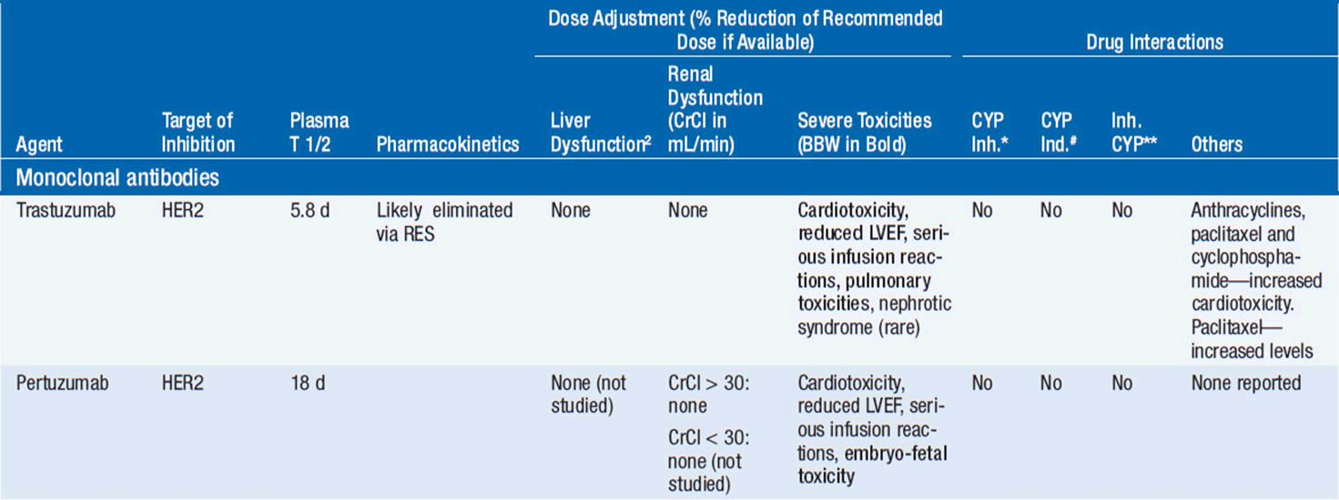
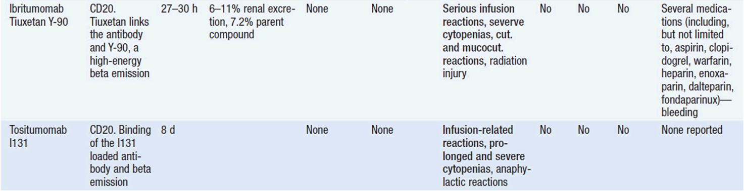
Important discoveries have revealed the molecular basis for the transformation, proliferation, and survival of cancer cells. These advances have revealed new targets for cancer drug design, and have produced agents that inhibit the signaling molecules and pathways responsible for cancer (Figure 10-1) (1). These inhibitors of cancer-associated targets include monoclonal antibodies (mAbs) either alone or coupled with cytotoxic agents or radioisotopes; modified proteins and peptidomimetic molecules; and small-molecular-weight drugs. Still in the development stage are small interfering RNAs (siRNA), antisense oligonucleotides, gene therapy approaches, and ribozymes or DNAzymes. In this chapter we will consider the small molecules that have been approved for clinical use. Monoclonal antibodies and their conjugates will be considered elsewhere (Chapter 15) (see Table 10-1) (2–9).

FIGURE 10-1 Schematic of growth factor receptor signaling in tumor cells.
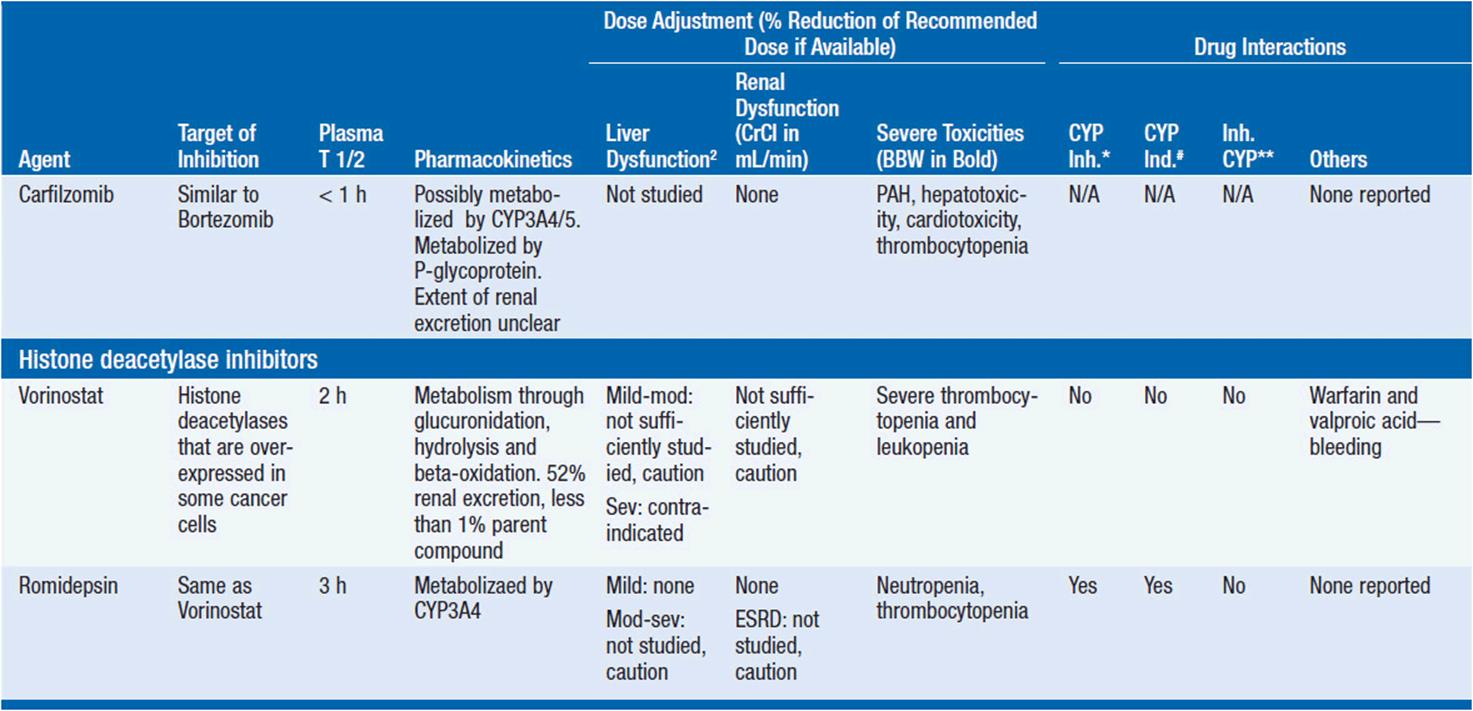
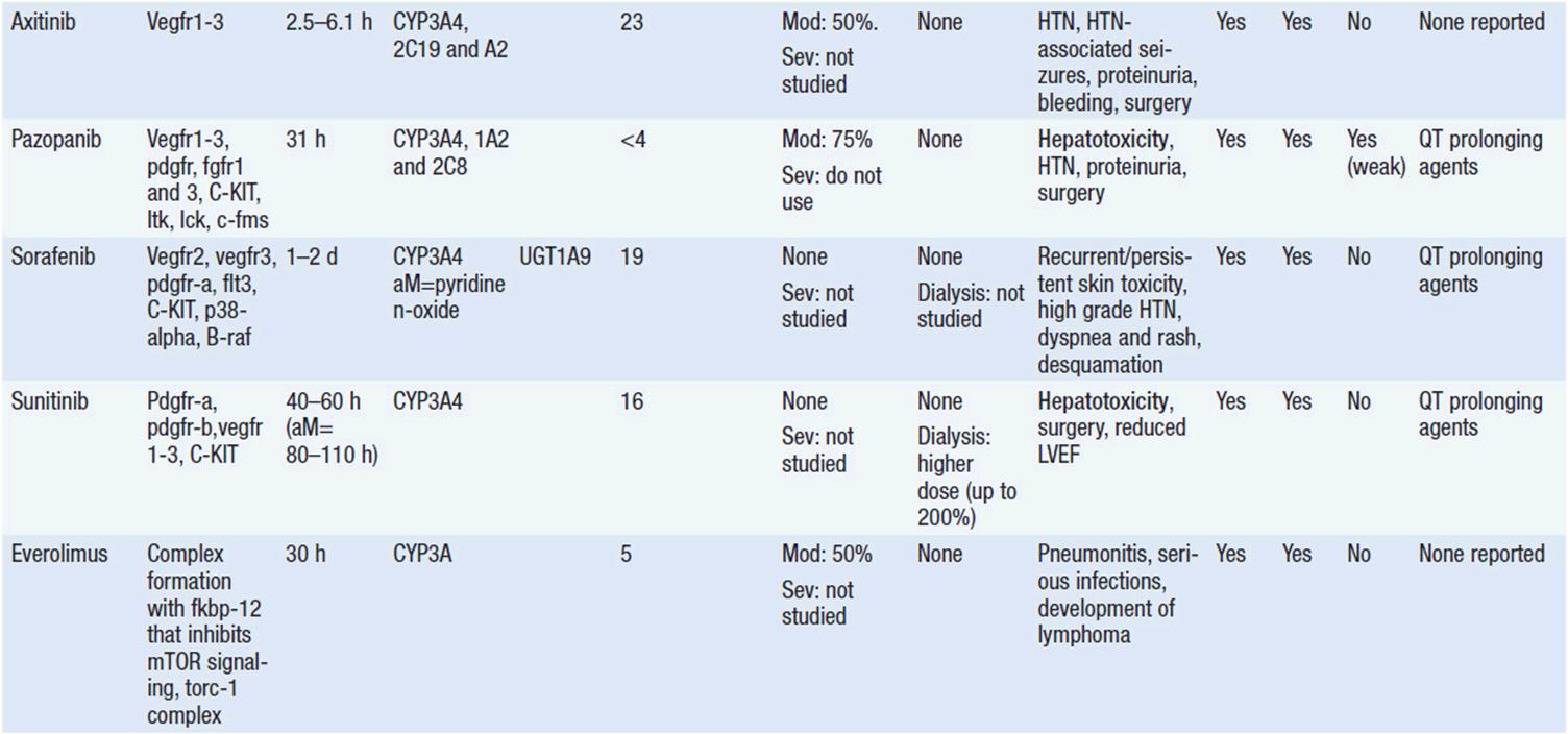
TABLE 10-1








MOLECULAR TARGETS IN CANCER
A rational approach to therapeutic discovery is based on our rapidly growing knowledge of pathways and proteins essential for cancer cell survival, growth, and metastasis. These pathways may be qualitatively unique to cancer (Figure 10-1), or may be simply overexpressed or amplified wild-type proteins. Mutant genes, unique to cancer cells, are particularly attractive in that they alter critical cellular functions and lead to uncontrolled growth, inhibition of apoptosis, escape from growth suppression, invasion of surrounding normal tissues, modifications of the tumor microenvironment including angiogenesis, and metastasis. Inhibition of these mutant functions leads to cancer cell death.
Alterations in a number of fundamental cellular processes may lead to malignant transformation and uncontrolled growth. Malignancy may result from overexpression or amplification of growth factors or their receptors, such as activating mutations or amplifications of the epidermal growth factor receptor (EGFR) family; activation of critical intracellular phosphorylating enzymes such as B-RAF (e.g., by mutation); modulation of the tumor microenvironment such as by activation of angiogenic pathways (e.g., vascular endothelial growth factors [VEGFs] and their receptors); changes in metabolism such as occur with mutations in glucose utilization (such as mutations in the IDH1 or IDH2 genes); epigenetic changes; or activation of anti-apoptotic pathways such as overexpression of Bcl-2 or decreased BAX expression.
 MUTATIONS AND IMPLICATIONS OF “DRIVER GENES” IN CANCER CELLS
Certain mutations in cancer cells, particularly those that activate cell receptor tyrosine kinases or downstream proteins involved in signaling within cells, can provide the primary stimulus for cell proliferation and survival. Examples of mutations or translocations in receptors leading to uncontrolled cancer cell proliferation include C-KIT mutations in gastrointestinal stromal tumors (GIST), and EGFR mutations or EML-4/ALK translocations in subsets of non-small cell lung cancer (NSCLC), primarily with adenocarcinoma histology. Mutations of genes in intracellular signal transduction pathways can also become drivers for malignant cell growth or survival. The prototypic example of this is the BCR-ABL translocation in chronic myelogenous leukemia (CML). Another cogent example is the constitutive activation of B-RAF, a protein in the RAS-RAF-MEK pathway, by mutation in melanoma (3, 4). These mutations create “addiction” to the continuous signaling, so that when signaling is blocked, the cancer cells die. In experimental settings, siRNAs directed against these mutant genes turn off the survival signals and lead to cell death, while small molecular inhibitors of the offending kinase cause tumor cell death (3, 4, 8, 9) in human subjects. The first agent to target the RAS-RAF-MEK pathway in human cancer is vemurafenib, which inhibits the V600E mutant form of the B-RAF kinase in melanoma (3, 8, 9). Interestingly, activating mutations of B-RAF are also found in colon, lung, and a number of other cancers, although these mutations have not been as responsive to vemurafenib as melanomas, indicating that the cellular context matters. Inhibitors of PI-3 kinase and its downstream signaling partner mTOR have led to beneficial treatment of renal cancers and neuroendocrine tumors (4). Multiple agents specific for isoenzymes of PI-3 kinase itself are undergoing evaluation in breast cancer, lymphoma, and endometrial cancer.
Mutations in tumor suppressor genes such as p53, retinoblastoma (RB), or the phosphate and tensin homolog (PTEN) that regulates the PI-3 kinase pathway can produce loss of important brakes on proliferation and enhance cell survival, leading to transformation of normal cells as well as contributing to the prolonged survival of cancer cells. These changes have proved harder to target because it is more difficult to return normal function to a protein than to inhibit aberrant function. However, new approaches are being explored aimed at indirectly reversing adverse effects of suppressor gene mutations such as by modulating downstream effectors of the mutant proteins or by targeting epigenetic factors or miRNAs important in control of functions of the tumor suppressor gene (10).
In the following, we discuss several specific examples of clinically effective targeted therapies.
1. Inhibitors of growth factor receptors or their ligands. Growth factor receptors and their ligands are overexpressed or amplified in many epithelial malignancies and are mutated in others (2, 3, 6-8). They are essential for promoting proliferation, survival, and metastasis of various kinds of cancer. The expression of mutated receptors on the cell surface and the presence of their ligands in the circulation make these altered pathways accessible to monoclonal antibodies. Examples of growth factors and receptors currently being effectively targeted include the following:
• EGFR family, including HER1 (EGFR), HER2, and HER3. EGFRs are present on normal epithelium and overexpressed in many cancers and mutated in a subset of NSCLC. The majority of NSCLC with mutated EGFR respond to anti-EGFR therapy, either drugs or antibodies. Anti-EGFR therapy with antibodies has also proved useful in colorectal and head and neck cancers in which the EGFR is not mutated. Amplified and overexpressed HER2 is a major target for a subset of breast and gastric cancers.
• ALK: Activating translocations were originally identified in anaplastic large cell lymphoma. Subsequently the mutated and translocated receptor was shown to respond to crizotinib in a subset of NSCLC adenocarcinomas as well as in patients with inflammatory myofibroblastic sarcoma. Activating mutations of ALK are also present in a subset of patients with neuroblastoma. A closely related receptor, ROS1 kinase, is translocated in a small subset of NSCLC and is susceptible to inhibition by crizotinib.
C-KIT mutations are frequently found in GIST tumors and uncommonly in several other neoplasms, including mucosal melanomas and mast cell disease.
• VEGF/VEGFR: These play an important role in tumor-associated angiogenesis for many epithelial and mesenchymal tumors, and for primary brain tumors.
Various strategies have been employed to inhibit function of these receptor pathways. Responses are well documented in patients treated with mAbs to HER2 (e.g., trastuzumab) or EGFR (e.g., cetuximab), and small-molecular-weight inhibitors (e.g., erlotinib for EGFR mutated NSCLC) (2, 3, 6). Bevacizimab, a monoclonal antibody directed against VEGF, is effective either alone for the treatment of renal cell cancer (RCC) or glioblastomas or combined with chemotherapy for colorectal or lung cancer (7). Small molecular inhibitors (such as axitinib or pazopanib) of VEGFRs have also proved effective in RCC (7) and in treating soft tissue sarcomas, while numerous small-molecular-weight drugs effectively block receptor tyrosine kinases.
2. Inhibitors of signal transduction. A number of signaling pathways downstream of growth factor receptors transmit aberrant growth signals and play essential roles in malignancies. These include the RAS-RAF-MEK pathway, the PI-3 kinase pathway, and the NF-kB pathway. Targeting of mTOR (in the PI-3 kinase pathway) is useful in treating renal cancer, breast cancer, and neuroendocrine tumors of gastrointestinal origin. PI-3 kinase inhibitors, some with broad activity against multiple PI3K isoenzymes while others that are specific for specific isoenzyme, are in active clinical development. Inhibitors of the mutated JAK2 oncoprotein, an important signal mediator in myeloid malignancies, are effective in myeloid metaplasia and other myeloproliferative diseases.
3. Inhibitors of cell cycle control. Many of the currently available cytotoxic agents inhibit DNA synthesis, and cell division, but display limited specificity for cancer. Approaches targeting specific overexpressed or otherwise aberrant cyclin-dependent kinases and other cell cycle regulatory proteins are currently being evaluated based on better understanding of the roles that these play in specific malignant cells.
4. Promoters of apoptosis. Apoptosis, or cell death, is dependent on the balance of activity of pro- and anti-apoptotic proteins. This balance is shifted in favor of anti-apoptotic proteins in many neoplastic cells, as for example the BCL-2 protein activated in follicular lymphoma. Inhibitors of the BH3 family of anti-apoptotic proteins have demonstrated activity against chronic lymphocytic leukemia, and continue in clinical evaluation, alone, combined with other targeted agents, and in combinations with chemotherapy.
5. Restoration of the function of tumor suppressor proteins. Research continues on approaches aimed at restoring normal functions of these critical proteins (p53, p21) to cancer cells (5, 11). Since restoration of the function of the proteins themselves has so far proved intractable, most of the current emphasis is aimed at indirectly modulating this function (see above for further discussion).
7. Inhibition of telomerase. Although much has been learned about the role of telomerase in maintaining telomeres in neoplastic cells and thus allowing continued survival and proliferation, significant preclinical work is needed to translate this into a useful anticancer approach. The furthest along of the approaches directed at telomerase is a vaccine targeting a portion of the telomerase protein. This vaccine is currently being evaluated in a phase III trial in combination with chemotherapy for metastatic pancreatic cancer (12).
8. Inhibitors of chromatin modifiers and epigenetic factors. Methylation of DNA or histones, acetylation of histones, and production of micro-RNAs, and long noncoding RNAs all modify gene expression and differentiation in normal and malignant cells (5, 11). Examples of approved agents targeting epigenetic factors include the histone deacetylase inhibitors (vorinostat, romidepsin), which have activity against cutaneous T-cell lymphoma (CTCL), and inhibitors of DNA methylation (azacytidine) in myelodysplasia.
9. Inhibitors of metabolism. Abnormal dependence on glycolysis has long been known to be an important aspect of malignant cells. The past decade has revealed other dramatic metabolic differences in tumors, including enhanced utilization of glycine or glutamine and activation of a number of enzymes (e.g., IDH-1 and 2, DNA methyltransferase, altered pyruvate kinase). These and other metabolic processes have become targets for drug development (13).
While single specific targets may initiate malignant transformation, whole genome sequencing of tumors has revealed that most cancers contain multiple mutations. In most cases, the role of these additional mutations in drug resistance and survival is unknown. In addition there are important influences on tumor biology coming from the tumor microenvironment. Thus, targeting multiple genes and their protein products may be necessary in order to kill the heterogeneous clones of cells within any given cancer. Recent studies have confirmed the marked heterogeneity of mutations and other changes in different cells within the same malignancy (14). Thus, targeting multiple proteins or pathways, either through multitargeted single inhibitors, such as sunitinib, sorafenib, or regorafenib, or through combination strategies (the combination of B-RAF and MEK inhibitors in melanoma) (15), will likely be required to maximize antitumor efficacy. In addition, targeting the environment is also important, as demonstrated by activity of antiangiogenic agents (5, 7).
CURRENTLY APPROVED TARGETED AGENTS IN CANCER THERAPY (USA)
SMALL MOLECULES
Because they can easily be subjected to high throughput screening and readily modified to incorporate favorable pharmacologic properties, small molecules remain the most attractive and straightforward class of agents for targeted therapy (3, 4, 6-9). High-throughput screening against recombinant proteins allows identification of lead compounds with affinity for the specific target of interest. Subsequent preclinical evaluation, using crystallography and in vitro testing, and analogue chemistry, yields compounds of high target affinity and specificity, with favorable drug properties (e.g., oral bioavailability, extended plasma half-life, decreased toxicity). The major pharmacological properties of representative approved targeted small molecules (grouped by target) are discussed below and are summarized in Table 10-1, which provides information of targets, pharmacokinetics, toxicity, and drug interactions.
TARGET: BCR-ABL
Mechanism of action: The 9:22 translocation in CML places the ABL tyrosine kinase gene on chromosome 9 in juxtaposition to the breakpoint cluster region (BCR) of chromosome 22 with a resultant protein that has constitutive phosphorylating activity, activating multiple downstream signaling pathways and leading to enhanced cell proliferation and survival. Like most other kinase inhibitors, imatinib competes for the ATP binding site of its target protein leading to potent inhibition of the tyrosine kinase activity (8). It binds to the enzyme in the protein’s inactive conformation and prevents its catalytic activity. Imatinib also potently inhibits C-KIT kinase, which is frequently mutated in GIST, and PDGFR-alpha, which is mutated in a smaller percentage of GIST tumors, but is also overexpressed in eosinophilic leukemia and hypereosinophilia (8). Resistance arises most commonly through one of several different mutations in the BCR-ABL protein (especially mutations that affect access to the ATP catalytic binding domain [e.g. T315I mutations, or so-called gatekeeper mutations] and those that hold the enzyme in an active configuration), leading to decreased drug binding.
Toxicity: Usually well tolerated. Potential toxicities include neutropenia, thrombocytopenia, anemia; hepatotoxicity (usually manifested by elevated liver enzymes but rarely severe); fluid retention/edema; musculoskeletal pains/cramps; rash; diarrhea; GI irritation; bleeding (GI tract or intratumoral); hypophosphatemia; and, rarely, congestive heart failure.
Pharmacokinetics: As is true for most tyrosine kinase inhibitors, imatinib is metabolized by hepatic CYP3A4. It is therefore important to monitor the dose when given with CYP3A4 inhibitors (e.g., itraconazole, erythromycin) or inducers (phenytoin, barbiturates) and alter dose as necessary. Its plasma t1/2 is approximately 18 h. Doses should be reduced in patients with hepatic and renal dysfunction (see package insert).
Clinical indications:
1. Newly diagnosed PH+ chronic phase CML (cCML)
2. Myelodysplastic/myeloproliferative diseases (MDS/MPD) with PDGFR gene re-arrangements
3. Aggressive systemic mastocytosis (ASM) without the D816V C-KIT mutation or with unknown C-KIT mutational status
4. Hypereosinophilic syndrome (HES) and/or chronic eosinophilic leukemia (CEL)
5. Unresectable, recurrent and/or metastatic dermatofibrosarcoma protuberans (DFSP)
6. C-KIT+ unresectable and/or metastatic GIST
DASATINIB
Mechanism of action: In comparison to imatinib, dasatinib is a more potent inhibitor of the tyrosine kinase activity of BCR-ABL (8). It inhibits the active, or open, conformation of the enzyme. It also inhibits a number of other kinases, including src family members, C-KIT, EPHA 2, and PDGFR. In CML, resistance arises through the selection of cells with resistance point mutations in the BCR-ABL protein catalytic site (e.g., the T315I mutation), but is unaffected by the numerous possible mutations that hold the enzyme in an active configuration.
Toxicity: Similar to imatinib, including neutropenia, thrombocytopenia, anemia; hepatotoxicity (usually manifested by elevated liver enzymes); fluid retention/edema; musculoskeletal pains/cramps; headaches; fatigue; rash; diarrhea; GI bleeding; hypophosphatemia; and, rarely, congestive heart failure. It also can cause prolongation of the QT interval and should thus not be used in patients with hypokalemia, hypomagnesemia, or prolonged QTc syndrome.
Pharmacokinetics: Similar to imatinib, it is metabolized by CYP3A4. It is therefore important to monitor the dose when given with CYP3A4 inhibitors or inducers. Doses should be reduced in the presence of hepatic dysfunction.
Clinical effectiveness (CML): Dasatinib has activity against untreated CML but also produces clinical hematologic responses in many of the CML or PH+ ALL patients who have become resistant to imatinib therapy through mutation of binding to the ATP-catalytic site on the enzyme (8). The BCR-ABL kinase gene containing T315I mutations is resistant. FDA-approved indications are:
1. Newly diagnosed PH+ chronic phase, accelerated phase, or blastic crisis phase CML
2. PH+ ALL
NILOTINIB
Mechanism of action: Similar to dasatinib, nilotinib is a potent inhibitor of the active conformation of the tyrosine kinase activity of BCR-ABL (8). It is approximately 30 times more potent than imatinib in vitro and retains activity against most mutations except T315I.
Toxicity: Similar to imatinib and dasatinib, including neutropenia, thrombocytopenia, anemia; hepatotoxicity (elevated liver enzymes); fluid retention/edema; musculoskeletal pains/cramps; headaches; fatigue; rash; diarrhea; GI irritation; bleeding; hypophosphatemia; and, rarely, congestive heart failure. It also can cause prolongation of the QT interval, predisposing to ventricular arrhythmias, and should not be used in patients with hypokalemia, hypomagnesemia, or prolonged QTc syndrome.
Pharmacokinetics: It is metabolized by CYP3A4, with a plasma half-life of 17 h. It is therefore important to monitor the dose when given with CYP3A4 inhibitors and inducers. Dose modifications are recommended in patients with liver dysfunction.
Clinical effectiveness (CML): Nilotinib produces clinical hematologic responses in both untreated CML or PH+ ALL patients as well as many of those who have become resistant to imatinib therapy through mutations that affect imatinib binding to the ATP-catalytic site on the enzyme (8). Nilotinib is FDA approved for following indications:
1. PH+ cCML, AP-CML, BC-CML
BOSUTINIB
Mechanism of action: Similar to imatinib, dasatinib, and nilotinib, it is an effective inhibitor of BCR-ALB kinase. Bosutinib also exhibits strong activity against kinases of the WIDTH="14" HEIGHT="15" SRC family, including WIDTH="14" HEIGHT="15" SRC, Lyn, and HCK.
Pharmacokinetics: It is metabolized by CYP3A4. Use with CYP inducers or inhibitors should be avoided if possible and may require dose modification when their use is necessary. Its plasma t1/2 is approximately 22.5 h.
Toxicity: Bosutinib can cause thrombocytopenia, anemia, neutropenia; hepatotoxicity; fluid retention and edema; gastrointestinal toxicities, including diarrhea, abdominal pain, nausea, vomiting; rash; and QT prolongation.
Clinical effectiveness: Bosutinib is indicated for the treatment of chronic, accelerated, or blast phase PH+ CML with resistance or intolerance to one or more TKIs, including imatinib, dasatinib, or nilotinib. It lacks activity against the T315I and V299L mutations in BCR-ABL that are among the mutations that confer resistance to imatinib.
PONATINIB
Mechanism of action: Similar to imatinib, dasatinib, nilotinib, and bosutinib, it is an effective inhibitor of the tyrosine kinase activity of BCR-ABL. In contrast, it contains a linker that allows it to retain activity against the T315I mutation, and against other mutations. Ponatinib also inhibits a number of other tyrosine kinases including fibroblast growth factor receptors 1-4, FLT-3, PDGFRA, and C-KIT.
Pharmacokinetics: Its plasma t1/2 is approximately 24 h. It interacts with CYP3A4 and the dose needs to be modified when used with strong CYP3A4 inhibitors. It is also an inhibitor of the multidrug resistance transporter.
Toxicity: Ponatinib can cause thrombocytopenia, anemia, neutropenia; gastrointestinal toxicities, including pancreatitis, constipation, abdominal pain, nausea; rash, dry skin; fever; arthralgia; hypertension; fluid retention; and headache. The most common potentially serious toxicities include hepatotoxicity and arterial thrombosis. Less common potentially serious toxicities include congestive heart failure, cardiac arrhythmias, venous thrombosis, and hemorrhage.
Clinical effectiveness: Ponatinib is indicated for the treatment of chronic, accelerated, or blast phase PH+ CML with resistance or intolerance to one or more TKIs, including imatinib, dasatinib, or nilotinib.
TARGET: EGFR FAMILY
ERLOTINIB
Mechanism of action: Erlotinib (Tarceva) is a potent specific inhibitor of the ATP-binding pocket of the EGFR (HER1) tyrosine kinase (6, 8). It targets the ATP binding site of the protein. Resistance arises by a number of mechanisms, including through selection of resistance mutations of the target protein (e.g., T790M, a gatekeeper mutation), by activation of alternate growth factor signaling pathways, especially the C-MET pathway, or by tumor conversion to a small cell cancer histology.
Pharmacokinetics: It has a plasma t1/2 of approximately 17 h, is metabolized by CYP3A4 and other CYP enzymes, and requires dose modification when used with CYP inducers or inhibitors.
Toxicity: Rash, dermatitis, and pruritus in the majority of patients, diarrhea (although uncommonly severe), nausea, fatigue, uncommon bleeding or clotting, and, uncommonly, interstitial pneumonitis.
Clinical effectiveness: Erlotinib has its most potent single agent activity against NSCLC with activating mutations in the EGFR kinase domain. Specifically, erlotinib is indicated for:
1. EGFR mutant NSCLC
2. Maintenance treatment in locally advanced or metastatic NSCLC without progression after 4 cycles of platinum-based first-line chemotherapy
3. Locally advanced or metastatic NSCLC after failure of at least one prior chemotherapy regimen
4. In combination with gemcitabine in locally advanced, unresectable or metastatic pancreatic cancer
LAPATINIB
Mechanism of action: Lapatinib is a potent specific inhibitor of both the EGFR (ErbB1) and the HER2 (ErbB2) tyrosine kinases (6, 8). It targets the ATP binding sites of the proteins. Resistance arises by a number of mechanisms, including (but not limited to) selection of resistance mutations of the target protein or by activation of alternate signalling pathways (the PI-3 kinase pathway).
Pharmacokinetics: It has a plasma t1/2 of approximately 24 h, is metabolized by CYP3A4 and other CYP enzymes, and requires dose modification when used with CYP inducers or inhibitors.
Toxicity: Common toxicities include rash, nausea/vomiting, diarrhea, fatigue, mucosal irritation, palmar-plantar erythrodysesthesia, and elevated liver function tests. Serious but uncommon toxicities include decreased left ventricular ejection fraction, hepatic toxicity, interstitial pneumonitis, severe diarrhea, and QTc prolongation.
Clinical effectiveness: Lapatinib is approved in combination with capecitabine for patients with HER2-amplified metastatic breast cancers that have progressed on prior therapies, or in combination with letrozole for postmenopausal women with hormone receptor positive breast cancers.
TARGET: ALK
CRIZOTINIB
Mechanism of action: Crizotinib (Xalkori) is a specific inhibitor of the ALK tyrosine kinase by targeting the ATP binding site (8). It also has significant activity against ROS1 and MET kinases. Similar to erlotinib (see above), resistance arises through a number of mechanisms including target gene amplification, induction of other growth factor receptor pathways, or selection of cells with resistance mutations in the gatekeeper mutations in the ATP-binding domain of the enzyme.
Pharmacokinetics: It is slowly metabolized by CYP3A4, and has a t1/2 of approximately 50 h in plasma.
Toxicity: Nausea, vomiting, diarrhea, and visual changes (temporary changes in visual acuity: trailing lights seen in transitions between light and dark) are the most common and usually manageable side effects. Fatigue, edema, elevated liver function tests, neuropathy, dysgeusia, rash, development of renal cysts, and asthenia can be seen. Uncommon but potentially serious toxicities include liver function test elevations with hyperbilirubinemia, and rarely liver failure, and interstitial pneumonitis that can be life-threatening or fatal.
Clinical effectiveness: It has activity against approximately 3%-5% of NSCLC that have translocations of the ALK gene, most commonly with EML4 as a partner (8, 9, 16). It also has potent clinical activity against NSCLC with ROS1 translocations.
TARGET: JAK 2 KINASE
RUXOLITINIB
Mechanism of action: Ruxolitinib is a specific inhibitor of the JAK 1 and 2 tyrosine kinases by targeting the ATP binding site.
Pharmacokinetics: It has a short t1/2 of approximately 2-3 h.
Toxicity: Bruising, dizziness, headache, elevated LFTs, anemia, thrombocytopenia, and leukopenia. All of these toxicities tended to be mild and controllable.
Clinical effectiveness: It is approved for the treatment of myelofibrosis, in which it decreases spleen size and relieves symptoms, and is under investigation for other myeloproliferative syndromes.
TARGET: HEDGEHOG PATHWAY
VISMODEGIB
Mechanism of action: Vismodegib is an inhibitor of smoothened, a transmembrane protein in the sonic hedgehog (SHH) pathway. Basal cell carcinoma is associated with activation of the SHH pathway through smoothed activation.
Pharmacokinetics: It has a plasma t1/2 of approximately 4 days. Although it interacts with CYP enzymes, it doses do not need to be altered in presence of CYP3A4 inhibitors. It is an inhibitor of the multidrug resistance exporter.
Toxicity: Common toxicities include diarrhea, constipation, nausea/vomiting, mucosal irritation, altered taste, decreased appetite, weight loss, fatigue, muscle spasms, arthralgias, alopecia, amenorrhea, lower levels of potassium/sodium, and elevated creatinine.
Clinical effectiveness: It is approved for metastatic or surgically unresectable basal cell carcinomas that are not candidates for radiation therapy.
TARGET: VEGFR AND OTHER KINASES
SORAFENIB
Mechanism of action: Sorafenib is an orally available multitargeted kinase inhibitor, with activity against RAF (C-RAF and B-RAF) kinases, VEGFR-2, VEGFR-3, PDGFR-beta, FLT3, and C-KIT (8). Its activity against the VEGF receptors is believed to be primarily responsible for its clinical activity against renal and hepatic cancers. Determinants of resistance are not known.
Pharmacokinetics: It is metabolized by CYP3A4 and by UGT 1A9 and has a plasma t1/2 of approximately 24-48 h. Doses should be modified in the presence of inducers or inhibitors of CYP enzymes.
Toxicity: Rash, hand-foot syndrome, hypertension, diarrhea, elevated amylase/lipase (usually without clinical pancreatitis), alopecia, myalgias, arthralgias, mild bone marrow suppression, and uncommonly bleeding or clotting.
Clinical effectiveness: Sorafenib is indicated for patients with unresectable hepatocellular carcinoma and advanced renal cell cancers.
REGORAFENIB
Mechanism of action: Regorafinib is an inhibitor of multiple kinases (similar to sorafenib to which it is closely related structurally), including VEGFR-1-3, KIT, PDGFR-alpha and beta (B-RAF and C-RAF), FGFR1/2, and others. As is the case for sorafenib, its most relevant antitumor activity is believed to be related to its antiangiogenic effects.
Pharmacokinetics: It is metabolized by CYP3A4 and has a t1/2 of approximately 28 h. It is subject to drug interactions with inducers/inhibitors of the CYP system and requires dose modification in their presence.
Toxicity: Hepatotoxicity (rarely fatal, LFTs should be monitored frequently), hemorrhage, gastrointestinal perforation, asthenia, fatigue, pain, fever, anorexia, rash, diarrhea, mucositis, dysphonia, headache, infection, weight loss, hypertension.
Clinical effectiveness: Regorafenib is indicated for the treatment of metastatic colorectal cancer in patient who failed all other standard treatment with fluoropyrimidine-, oxaliplatin-, and irinotecan-based chemotherapy, and targeted therapy, including an anti-VEGF/VEGFR therapy and, in KRAS wild-type disease, after failure of anti-EGFR therapy (8).
SUNITINIB
Mechanism of action: Sunitinib is an orally available small-molecular-weight inhibitor of multiple kinases including the tyrosine kinase activity of the VEGFR-2, PDGFR, and C-KIT receptors (8). In the treatment of GIST, resistance is related to emergence of mutations in the C-KIT gene.
Pharmacokinetics: It is metabolized by CYP3A4, with a plasma t1/2 of approximately 40-60 h, and doses must be modified in the presence of inducers or inhibitors of CYP3A4.
Toxicity: Cytopenias, bleeding, skin discoloration, diarrhea, mucocutaneous inflammation, altered taste, asthenia, left ventricular dysfunction (uncommon), GI perforation (rare), and pancreatitis (rare).
Clinical effectiveness: It has shown sufficient activity to be approved for treatment of GIST (either intolerant of or after progression on imatinib), advanced RCC, and unresectable or metastatic peripheral neuroendocrine tumors (PNET).
CABOZANTINIB
Mechanism of action: Cabozantinib is an inhibitor of multiple kinases including the tyrosine kinase activity of the VEGFR-2, MET, and RET receptors. These receptors are involved in a number of cellular processes critical for tumor growth or maintenance, including tumor cell proliferation, invasion, angiogenesis, and maintenance of the tumor microenvironment.
Pharmacokinetics: It is metabolized by CYP3A4, with a plasma t1/2 of approximately 91 h, and doses must be modified in the presence of inducers or inhibitors of CYP3A4.
Toxicity: Common toxicities include diarrhea, constipation, mucocutaneous inflammation, nausea/vomiting, altered taste, decreased appetite, abdominal pain, fatigue, asthenia, hypertension, elevated transaminases and bilirubin, lower levels of calcium/phosphorous/magnesium /potassium/sodium, palmar-plantar erythrodysesthesia syndrome, changes in hair color or skin pigmentation, and rash. Rare but potentially serious toxicities include visceral perforation or fistula formation, altered wound healing, hemorrhage, arterial thrombosis, nephritic syndrome, osteonecrosis of the jaw, and reversible posterior leukoencephalopathy syndrome.
Clinical effectiveness: It is approved for treatment of medullary thyroid cancer (MCT). It is actively being evaluated for efficacy against a number of other malignancies including metastatic prostate cancer.
PAZOPANIB
Mechanism of action: Pazobanib is an inhibitor of multiple kinases including the tyrosine kinase activity of the VEGFR-1-3, PDGFRs, C-KIT, and cFMS receptors as well as downstream signaling molecules LCK and ITK kinases. Its antitumor activity is likely related to its antiangiogenic effect.
Pharmacokinetics: It is metabolized by CYP3A4, with a plasma t1/2 of approximately 31 h, and doses must be modified in the presence of inducers or inhibitors of CYP3A4.
Toxicity: Common toxicities include diarrhea, nausea/vomiting, altered taste, decreased appetite, abdominal pain, fatigue, asthenia, hypertension, elevated transaminases and bilirubin, lower levels of phosphorous/magnesium/potassium/sodium/glucose, palmar-plantar erythrodysesthesia syndrome, hypothyroidism, and changes in hair color. Less common but potentially serious toxicities include hemorrhage, arterial thrombosis, hepatotoxicity, and prolonged QTc.
Clinical effectiveness: It has shown sufficient activity to be approved for treatment of metastatic renal cancer. It is actively being evaluated against a number of other malignancies, including neuroendocrine cancers.
AXITINIB
Mechanism of action: Axitinib is an inhibitor of multiple kinases including VEGFR-1-3, PDGFRs, and C-KIT. Its major antitumor activity is believed to be mediated by inhibition of VEGF receptors.
Pharmacokinetics: It is metabolized by CYP3A4, with a plasma t1/2 of approximately 2.5-6.1 h, and doses must be modified in the presence of inducers or inhibitors of CYP3A4.
Toxicity: Common toxicities include diarrhea, nausea/vomiting, mucosal irritation, rash, altered taste, decreased appetite, abdominal pain, fatigue, asthenia, hypertension, elevated transaminases, elevated amylase/lipase, lower levels of calcium/phosphorous/potassium/sodium, elevated or decreased glucose levels, palmar-plantar erythrodysesthesia syndrome, hypothyroidism, and changes in hair color. Less common but potentially serious toxicities include hemorrhage, arterial/venous thrombosis, hepatotoxicity, hypertensive crisis, prolonged QTc, gastrointestinal perforation or fistula formation, decreased wound healing, and reversible posterior leukoencephalopathy syndrome.
Clinical effectiveness: It is approved for treatment of metastatic renal cancer after failure of one systemic therapy.
TARGET: MTOR
TEMSIROLIMUS
Mechanism of action: An intravenous inhibitor of the mTORC1 complex, a critical enzyme in the PI-3 kinase-AKT pathway, important in modifying tumor metabolism, inducing glycolysis, and promoting cell survival and proliferation (4, 8).
Pharmacokinetics. The drug has a plasma t1/2 of approximately 17.3 h. Its active metabolite has a t1/2 of 54 h. It is eliminated by CYP3A4, and is subject to drug interactions with inducers or inhibitors of the CYP system.
Toxicity: Rash, edema, anorexia, nausea, asthenia, fatigue, mucositis, cough, pneumonitis, diarrhea, hyperglycemia, hyperlipidemia, elevated liver function tests, bone marrow suppression, increased risk of infections, and renal dysfunction.
Clinical effectiveness: It is approved for the treatment of advanced renal cell carcinoma.
EVEROLIMUS
Mechanism of action: An oral inhibitor of mTOR, a critical enzyme in the PI-3 kinase-AKT pathway (see temsirolimus, above).
Pharmacokinetics: It is metabolized by CYP3A4 and is subject to drug interaction with inhibitors or inducers of this enzyme. It has a plasma t1/2 of approximately 30 h.
Toxicity: Rash, edema, anorexia, nausea, asthenia, fatigue, mucositis, cough, pneumonitis, diarrhea, hyperglycemia, hyperlipidemia, elevated liver function tests, bone marrow suppression, increased risk of infections, and increased serum creatinine.
Clinical effectiveness: It has activity against advanced RCC and is approved for use after progression on either sunitinib or sorafenib. It is also approved for the treatment of peripheral neuroectodermal tumors, hormone positive breast cancer (combined with exemestane), and subependymal giant cell astrocytoma.
TARGET: B-RAF
VEMURAFENIB
Mechanism of action: An oral inhibitor of activated B-RAF kinase carrying a V600E mutation. B-RAF is a serine/threonine kinase in the RAS-RAFMEK signaling pathway. Mutations in this pathway are frequently found in melanoma, colon cancer, lung cancer, and thyroid cancer.
Pharmacokinetics: Vemurafenib is metabolized by CYP3A4 and is subject to drug interaction with inhibitors or inducers of this enzyme as well as several CYP2 substrates such as warfarin. It has a plasma t1/2of approximately 57 h.
Toxicity: Rash, photosensitivity, pruritis, dry skin, hyperkeratosis, alopecia, joint pain, nausea, diarrhea, fatigue, elevated liver function tests, prolonged QTc interval, skin papillomas, and cutaneous squamous cell carcinomas.
Clinical effectiveness: It is approved for treatment of melanomas carrying a B-RAF V600E mutation.
FUTURE OF TARGETED THERAPY FOR TREATING CANCER
Many additional agents targeting proteins (primarily kinases) of interest as drivers of malignancy are currently undergoing clinical investigation. In addition to development of agents targeted against cell surface receptors, much current interest involves inhibitors of steps in signal transduction pathways from cell surface to nucleus, including steps in the PI3K-mTOR and RAS-RAF-MEK pathways (2-4, 6-9). Recent studies have shown significant activity of MEK inhibitors, either alone or in combination with B-RAF inhibitors, against melanoma (11, 15). Clearly, there are many other potential targets within cells, including proteins involved in other signaling pathways, proteins involved in survival, regulatory proteins such as transcription factors, enzymes involved in intermediary metabolic processes, epigenetic modifiers, and proteins that enhance antitumor immune function by blocking inhibitory proteins (e.g., anti-PD1, anti-PDL1, anti-CTLA4) (5, 10, 12, 13). Certain agents under development have greater specificity for one protein or gene, whereas others have activity against a number of proteins. It is not known whether having agents with highly specific activity (and potentially combining different agents each with specific activity) or having broader activity within one agent will be more clinically effective against any specific cancer. This will likely vary depending on disease indications, targets, and agents.
Given the complexity of genetic, epigenetic, and tumor microenvironment changes in most cancers, it is unlikely that modulation of single targets will have long-term antitumor efficacy against most cancers. Thus, combinations of target approaches are being explored. Strategies to inhibit multiple sequential steps in a given pathway (such as a signal transduction pathway) or multiple receptors or pathways in parallel are being evaluated. Strategies for combining different classes of targeted agents (e.g., mAbs and small molecules, which tend to have fewer overlapping toxicities than two agents of the same class) are also being pursued. In some instances, a combination of monoclonal antibodies targeting different sites on the same molecule may be more effective than single agents. Other approaches, such as specifically delivering cytotoxic compounds to malignant cells by coupling them to mAbs (e.g., TDM1, which contains the mAB trastuzumab coupled with the antimitotic agent emtansine, targeted against HER2) are attracting increasing attention because of the success of the antibody-drug conjugate brentuximab vedotin against Hodgkin disease and anaplastic large cell lymphoma.
To date, clinically useful targeted compounds have come from one of three classes of agents (mABs, small molecules, or modified proteins or peptides). However, other classes of compounds are likely to have utility as anticancer agents, such as RNA interference with small inhibitory RNA (siRNA) (5, 10). siRNAs bind to complementary RNA molecules leading to their cleavage and produce post-transcriptional gene silencing (PTGS), a powerful tool for studying the effects of silencing specific genes, and in fact represents a potential therapy modality if barriers in the delivery of the molecule can be solved.
Biomarkers are essential to define the population of patients who represent appropriate candidates for specific targeted therapy. Biomarkers are also needed for monitoring the effectiveness of that therapy. Therefore, a significant effort is being devoted to identifying the most useful biomarkers for different agents and malignancies. Given the critical need to have a uniform, standard, and widely available test to select patients that will benefit from the agent, the FDA is now mandating that, when it is feasible, the appropriate diagnostic test must be developed at the same time that the drug is being evaluated and the test must be validated at the time of approval. Both vemurafenib (with a companion pcr test for the V600E B-RAF mutation) for melanoma and crizotinib (with a companion FISH analysis for presence of EML4-ALK translocation for NSCLC) were codeveloped with biomarker tests that were available at the time of approval (3, 9). There remain certain targeted agents (e.g., angiogenesis inhibitors including VEGF and VEGFR inhibitors, histone deacetylase inhibitors, proteosome inhibitors) for which it has not yet been possible to define clinically meaningful biomarkers beyond the specific diseases for which they have been approved. Identification and validation of appropriate biomarkers for specific agents continue to be essential areas of study.
Modeling approaches, including computer simulations, can be helpful in both improving drug design for enhanced efficacy and identifying potential toxicities of agents prior to clinical testing (17). As knowledge of the important factors that determine both efficacy and toxicity improves, models can be more precise in helping to decide which agents to carry forward into clinical trials.
Another major area of research aims to prevent or overcome development of resistance by cancers to targeted agents. Many mechanisms leading to either primary or secondary development of resistance to targeted agents have been defined, as discussed above. Pharmacokinetic sanctuary sites (such as the brain) continue to be a problem for most new drugs (16, 18). Acquired genetic mutations of a target are detectable through tumor biopsies taken at the time of disease progression, and provide guidance for efforts to develop better drugs and combinations of drugs (19). Mechanisms of drug resistance to antiangiogenic agents, such as bevacizumab resistance in RCC, are poorly understood and thus remain a major challenge. Exploration of strategies to overcome the various mechanisms of resistance is critical for development of new targeted therapies that will be effective in controlling disease for prolonged periods.
Continued improvement in understanding critical processes in cancer development, growth, survival, and metastasis will provide new targets and better drugs, as well as better biomarkers for defining the appropriate patients for specific agents, and a more complete understanding of resistance to targeted agents. Given the complexity and heterogeneity of most cancers, better ways of integrating targeted agents with other anticancer treatment approaches will have to be developed in order to achieve successful long-term control of various cancers.
REFERENCES
1. Hanahan D, Weinberg RA. Hallmarks of cancer: the next generation. Cell. 2011; 144: 646-74.
2. Scott AM, Wolchok JD, Old LJ. Antibody therapy of cancer. Nat Rev Cancer. 2012; 12: 278-287.
3. Yauch RL, Settleman J. Recent advances in pathway-targeted cancer drug therapies emerging from cancer genome analysis. Curr Opin Genet Dev. 2012; 22: 45-49.
4. Sheppard K, Kinross KM, Solomon B, et al. Targeting PI3 inase/AKT/mTOR signalling in cancer. Crit Rev Oncog. 2012; 17: 69-95.
5. Lujambio A, Lowe SW. The microcosmos of cancer. Nature. 2012; 482: 347-355.
6. Dhomen NS, Mariadason J, Tebbutt N, et al. Therapeutic targeting of the epidermal growth factor receptor in human cancer. Crit Rev Oncog. 2012; 17: 31-50.
7. Waldner MJ, Neurath MF. Targeting the VEGF signalling pathway in cancer therapy. Expert Opin Ther Targets. 2012; 16: 5-13.
8. Copyright © 2012 PDR Network, LLC, Montvale, NJ 07645.
9. Chabner BA. Early accelerated approval for highly targeted cancer drugs. N Engl J Med. 2011; 364: 1087-1089.
10. Wang Z, Rao DD, Senzer N, Nemunaitis J. RNA interference and cancer therapy. Pharm Res. 2011: 2983-2995.
11. Flaherty KT, Robert C, Hersey P, et al. Improved survival with MEK inhibition in BRAF-mutated melanoma. N Engl J Med. 2012; 367: 107-114.
12. Xu Y, He K, Goldkorn A. Telomerase targeted therapy in cancer and cancer stem cells. Clin Adv Hematol Oncol. 2011; 9: 442-455.
13. Muñoz-Pinedo C, El Mjiyad N, Ricci JE. Cancer metabolism: current perspectives and future directions. Cell Death Dis. 2012; 3: e248.
14. Gerlinger M, Rowan AJ, Horswell S, et al. Intratumor heterogeneity and branched evolution revealed by multiregion sequencing. N Engl J Med. 2012; 366: 883-892.
15. Flaherty K, Infante JR, Falchook GS, et al. Phase I/II study of BRAFi GSK2118436 + MEKi GSK1120212 in patients with BRAF mutant metastatic melanoma who progressed on a prior BRAFi. Pigment Cell Melanoma Res. 2011; 25: E1-E11.
16. Katayama R, Shaw AT, Khan TM, et al. Mechanisms of acquired crizotinib resistance in ALK-rearranged lung cancers. Sci Transl Med. 2012 4: 120ra17.
17. Lounkine E, Keiser MJ, Whitebread S, et al. Large-scale prediction and testing of drug activity on side-effect targets. Nature. 2012; 486: 361-367.
18. Turner NC, Reis-Filho JS. Genetic heterogeneity and cancer drug resistance. Lancet Oncol. 2012; 13: e178-e185.
19. Kobayashi S, Boggon T, Dayaram T, et al. Mutation and resistance of non–small-cell lung cancer to gefitinib. N Engl J Med. 2005; 352: 786-792.