
Bruce A. Chabner
The antifolates were introduced as antileukemic drugs in 1948; in landmark experiments treating children with acute lymphocytic leukemia (ALL), Sidney Farber produced the first evidence that chemotherapy with a folate analogue, aminopterin, could lead to complete remissions (1). Methotrexate subsequently became the standard antifolate in treatment of ALL. It has since gained an important role in regimens for lymphomas, and choriocarcinoma, and as an immunosuppressive following allogeneic bone marrow transplantation. It is also a standard agent for treating rheumatoid arthritis, Wegener’s granulomatosis, and other inflammatory/autoimmune diseases. Pemetrexed (Alimta), a closely related structure but with a different site of action, is widely used for non-small cell lung cancer, mesothelioma, and ovarian cancer. Pralatrexate, the newest antifolate similar in action to methotrexate, is highly active against peripheral T-cell lymphoma and cutaneous T-cell lymphoma.
The structures of antifolates are shown in Figure 2-1. The analogues closely resemble naturally occurring folates, but contain substitutions in the basic pteridine ring system, as in pralatrexate and pemetrexed, which enhance binding and transport. The key addition of the amino group on the C-4 position of the pteridine ring, as found in methotrexate, enhances inhibition of dihydrofolate reductase. Changes in the bridge system connecting the unsaturated rings to para-aminobenzoyl glutamate (PABG) enhance the active uptake and polyglutamation of pemetrexed and pralatrexate. Because of their strong electronegative charge at physiologic pH, the parent antifolates, like physiologic folates, require active transport into cells via the reduced folate carrier (2). In selected cells, such as choriocarcinomas, a second carrier, the folate binding protein, mediates folate and methotrexate transport, and becomes the preferred transporter. Pemetrexed is also transported by a third carrier, the proton-coupled folate carrier, which may be responsible for its unique activity against epithelial cancer and mesothelioma (3). Inside the cell, the analogues, like the physiologic folates, are converted at their PABG terminus to highly charged, long-chain polyglutamates. These polyglutamate metabolites are retained preferentially within cells and inhibit, with increased affinity, a number of folate-dependent enzymes critical for both thymidine and purine biosynthesis (Figure 2-2). Through their inhibition of dihydrofolate reductase, methotrexate and pralatrexate deplete intracellular folates, leading to a block in both purine and pyrimidine biosynthesis. The primary action of pemetrexed is its inhibition of another folate-dependent enzyme, thymidylate synthase.


FIGURE 2-1 Molecular structure of folic acid, methotrexate, pemetrexed, and pralatrexate.
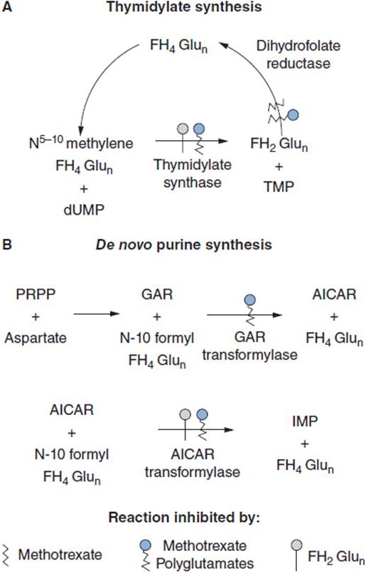
FIGURE 2-2 Multiple sites of inhibitory action of methotrexate, its polyglutamate metabolites, and dihydrofolate polyglutamates, the substrate that accumulates when dihydrofolate reductase is inhibited. AICAR: aminoimidazole carboxamide; TMP: thymidine monophosphate; dUMP: deoxyuridine monophosphate; FH2Glun: dihydrofolate polyglutamate; FH4Glun: tetrahydrofolate polyglutamate; GAR: glycinamide ribonucleotide; IMP: inosine monophosphate; PRPP: 5-phosphoribosyl-1-pyrophosphate. Pralatrexate acts in a very similar fashion, while the polyglutamates of pemetrexed primarily act as direct inhibitors of TS.
PHARMACOLOGIC CONSIDERATIONS
Methotrexate and Pralatrexate antifolates kill cells through depletion of thymidylate and purine precursors of DNA, and are thus most effective against rapidly growing tumors, such as leukemias and lymphomas. Cell kill by methotrexate depends on both drug concentration and duration of exposure. Its threshold concentration for drug toxicity for normal cells lies in the range of 10 nM. Methotrexate-resistant cells arise through loss of the reduced folate transporter, loss of the ability to polyglutamate antifolates, or amplification of the gene coding for dihydrofolate reductase, all of which have been demonstrated in ALL cells in association with relapse (4, 5). High levels of TS expression are associated with poor response to pemetrexed (4), while resistance to pralatrexate likely parallels that of methotrexate but is incompletely understood.
Inherited genetic variants modify folate metabolism (6, 7). The methylene tetrahydrofolate reductase variant C677T increases the intracellular level of 5–10 methylene tetrahydrofolic acid, the substrate for TS, and is associated with an increased rate of relapse in childhood ALL (7). Attempts to relate response and toxicity to variations in transporter and polyglutamate synthase genes have been inconclusive.
Pemetrexed and pralatrexate are more avidly transported and converted to a polyglutamate than is methotrexate (8). Because of its mild and predictable toxicities, pemetrexed has become a preferred agent for first- or second-line treatment of adenocarcinoma of the lung. Unlike methotrexate, pemetrexed and pralatrexate are given with folic acid (0.4–1.0 mg/day, beginning 1 week prior to treatment, and continuing throughout therapy) and with vitamin B12 (1 μg on day 1 i.m. and thereafter with each cycle of therapy), as these vitamins ameliorate toxicity to bone marrow (9).
CLINICAL PHARMACOLOGY
Methotrexate is well absorbed orally in doses of 25 mg/m2 or less, and is used by that route in maintenance therapy of ALL. In higher doses (50–500 mg/m2) it is given primarily by the intravenous route, or in higher dose regimens (1–20 g/m2) with leucovorin (5-formyl-tetrahydrofolic acid) rescue. Individual drug regimens vary considerably, and are tailored to specific indications. Careful adherence to proven regimens is critical, with particular attention to the status of the patient’s pretreatment renal function, which may drastically alter clearance of the drug and its toxicity.
Methotrexate is cleared primarily by renal excretion. Small amounts are metabolized to a nontoxic 7-OH derivative. In patients with normal renal function, it has a primary elimination half-life from plasma of 2–4 h, followed by a secondary elimination phase of 8–10 h (10). The terminal phase of disappearance determines the duration of exposure to cytotoxic concentrations of drug, and becomes much longer in patients with compromised renal function. Doses should be modified in proportion to the reduction in renal function for patients with a creatinine clearance of less than 60 ml/min. Nonsteroidal anti-inflammatory drugs reduce renal blood flow and displace methotrexate from plasma protein binding, thereby slowing clearance and increasing unbound drug concentrations in plasma, and should not be used in conjunction with methotrexate. Proton pump inhibitors may displace the drug from albumin binding and increase its toxicity. Penicillins reduce methotrexate secretion by renal tubules and may also increase the risk of toxicity.
 HIGH-DOSE METHOTREXATE
High doses of methotrexate are administered to patients with ALL, osteosarcoma, and central nervous system lymphoma, and high-grade non-Hodgkin lymphoma in order to increase intracellular drug concentration, polyglutamate formation, and penetration into the central nervous system. These potentially lethal doses (1–20 g/m2) are infused over 6–24 h, and are followed by intravenous or oral leucovorin (5-formyl-tetrahydrofolate), 15–100 mg/m2, which restores the intracellular pool of tetrahydrofolates and rescues normal tissue from drug toxicity. Various regimens have proven safe and effective, and should be followed strictly to assure avoidance of toxicity. Methotrexate is relatively insoluble at acid pH, a property that may cause its precipitation in renal tubules in acidic urine, leading to acute renal dysfunction, a failure to excrete drug at normal rates, and overwhelming bone marrow and epithelial toxicity. Thus, patients require alkalinization of the urine prior to drug administration, and aggressive hydration and diuresis during methotrexate infusion (10). Drug levels in plasma and renal function should be monitored in the 24–48 h post-infusion to assure normal rates of clearance. Concentrations of methotrexate above 1 μM at 24 h after the completion of infusion, particularly in conjunction with a rise in serum creatinine levels, should alert clinicians to impending serious toxicity. In such patients, the first step should be to increase and extend leucovorin administration (up to 500 mg every 6 h for 48 h IV), along with continued hydration. In extreme cases, when drug levels remain above 10 μM after 48 h, and show a very slow decline, leucovorin may be ineffective. In this setting, continuous flow hemodialysis is able to reduce drug levels at a clearance rate of 50 ml/min, and may avoid prolonged myelosuppression and mucositis.
Very rapid clearance of methotrexate and effective rescue from toxicity can be achieved for patients with delayed drug clearance through the intravenous administration of a recombinant bacterial folate-cleaving enzyme, carboxypeptidase G-2 (glucarpidase), which is now available for general clinical use (11). Greater than 95% clearance of drug from plasma is achieved within 15 min of administration of 50 units/kg given as a 5-min intravenous infusion, and life-threatening toxicity will be avoided in patients with plasma methotrexate levels in the range of 1–50 μM. For patients with levels above 50 μM, reductions in plasma methotrexate, although substantial, do not reach the critical rescue level of 1 μM, probably due to reentry of drug from tissue compartments. Drug levels should be monitored after glucarpidase administration to determine the necessity of further measures, such as leucovorin or dialysis. Leucovorin is also cleaved by glucarpidase and is ineffective if used concurrently with the enzyme.
INTRATHECAL METHOTREXATE
Methotrexate is routinely administered intrathecally in doses of 12 mg for prevention or treatment of meningeal lymphoma, leukemia, or meningeal carcinomatosis. In patients with no evidence of meningeal tumor, the drug clears with a half-life of 2 h from the cerebral spinal fluid. In patients with active meningeal tumor, after lumbar intrathecal administration, its clearance may be slow and it may penetrate poorly into the ventricular space, requiring the placement of a reservoir for direct intraventricular therapy. Intrathecal methotrexate, particularly in patients with active meningeal disease, in whom drug clearance is slow, may lead to arachnoiditis, seizures, coma, and death. High-dose methotrexate regimens do produce cytotoxic drug concentrations in the spinal fluid, and appear to be sufficient for prophylaxis of CNS leukemia in average-risk ALL patients (12). High-dose systemic methotrexate rarely causes CNS toxicity. Oral or intravenous leucovorin is not an effective antidote to CNS toxicity.
PEMETREXED AND PRALATREXATE PHARMACOKINETICS
Pemetrexed pharmacokinetics closely follow those of methotrexate, with a 3-h terminal half-life in plasma, clearance by renal excretion, and dose adjustment for renal dysfunction. The usual dose of pemetrexed administration is 500 mg/m2 every 3 weeks, with vitamin B12 and folate supplementation. Higher doses, up to 900 mg/m2 may be tolerated well by individual patients, but the therapeutic benefit of dose escalation is not established. Pralatrexate, given in doses of 30 mg/m2/week with folate and vitamin B12 supplementation, undergoes renal elimination, with a half-time in plasma of 4–8 h.
TOXICITY
Virtually every organ system may be affected by antifolate toxicity. Acutely, bone marrow suppression, mucositis, and gastrointestinal symptoms are the primary side effects of all 3 antifolates, and usually resolve within 10–14 days of completion of therapy. In most patients, high-dose methotrexate may be accompanied by very minimal evidence of toxicity, aside from acute reversible elevations in hepatic enzymes in serum. In toxic patients who develop renal failure, myelosuppression, severe mucositis, and desquamation may supervene. Cirrhosis is occasionally reported in psoriasis or rheumatoid arthritis patients on long-term oral methotrexate, and is heralded by elevations in plasma type III procollagen aminopeptide (PIIIAP). Patients with elevated PIIIAP levels in plasma are at 20% risk of drug-related cirrhosis and should undergo a liver biopsy (13). An interstitial pneumonitis, likely related to hypersensitivity to the drug, with eosinophilic infiltrates, is occasionally seen with methotrexate.
Pemetrexed is toxic to bone marrow and gastrointestinal and oral mucosa. Toxicity tends to be predictably mild in patients receiving concurrent folic acid and vitamin B12. Early trials without vitamin protection witnessed a significant (15%–20%) incidence of severe toxicity, primarily in patients with high levels of homocysteine in plasma, an indicator of folate deficiency, prior to treatment. Pulmonary toxicity, manifested as an interstitial pneumonitis, may complicate therapy in 3%–5% of patients (14). Another important but uncommon side effect is peripheral edema, and in rare cases, pleural effusions (15). Up to 40% of patients may experience a bothersome erythematous rash, which can be largely prevented by oral dexamethasone, 4 mg twice daily on days –1, 0, and +1.
Pralatrexate is given in doses of 30 mg/m2/week for 6 of 7 weeks. Myelosuppression and mucositis may lead to delays in treatment or dose adjustment.
REFERENCES
1. Farber S, Diamond LK, Mercer RD, et al. Temporary remission in acute leukemia in children produced by folic acid antagonist 4-amethopteroylglutamic acid (aminopterin). N Engl J Med. 1948; 238: 787–793.
2. Moscow JA, Gong M, He R, et al. Isolation of a gene encoding a human reduced folate carrier (RFC1) and analysis of its expression in transport deficient, methotrexate-resistant human breast cancer cells. Cancer Res. 1995; 55: 3790–3794.
3. Zhao R, Qui A, Jansen M. The proton-coupled folate transporter: impact on pemetrexed transport and on antifolate activities compared with the reduced folate carrier. 2008; 74: 854–862.
4. Takezawa I, Ikamoto I, Okamoto W, et al. Thymidylate synthase as a determinant of pemetrexed sensitivity in non-small cell lung cancer. Brit J Cancer. 2011; 104: 1594–1601.
5. Whitehead VM, Shuster JJ, Vuchich MJ, et al. Accumulation of methotrexate and methotrexate polyglutamates in lymphoblasts and treatment outcome in children with B-progenitor-cell acute lymphoblastic leukemia: a Pediatric Oncology Group study. Leukemia. 2005; 19: 533–536.
6. Buikhuisen WA, Burgers JA, Vincent AD et al. Pemetrexed pathwayassociated germline polymorphisms: a useful tool for treatment individualization? J Clin Oncol. 2010; 28: e482–e483.
7. Aplenc R, Thompson J, Han P, et al. Methylenetetrahydrofolate reductase polymorphisms and therapy response in pediatric acute lymphoblastic leukemia. Cancer Res. 2005; 65: 2482–2487.
8. Izbicka E, Diaz A, Streeper R, et al. Distinct mechanistic activity profile of pralatrexate in comparison to other antifolates in in vitro and in vivo models of human cancers. Cancer Chemother Pharmacol. 2009; 64: 993–999.
9. Scagliotti GV, Shin DM, Kindler HL, et al. Phase II study of pemetrexed with and without folic acid and vitamin B12 as front-line therapy in malignant pleural mesothelioma. J Clin Oncol. 2003; 21: 1556–1561.
10. Stoller RG, Hande KR, Jacobs SA, et al. Use of plasma pharmacokinetics to predict and prevent methotrexate toxicity. N Engl J Med. 1977; 297: 630–634.
11. Buchen S, Ngampolo D, Melton RG, et al. Carboxypeptidase G2 rescue in patients with methotrexate intoxication and renal failure. Br J Cancer. 2005; 92: 480–487.
12. Glantz MJ, Cole BF, Recht L, et al. High-dose intravenous methotrexate for patients with nonleukemic leptomeningeal cancer: is intrathecal chemotherapy necessary? J Clin Oncol. 1998; 16: 1561–1567.
13. Chalmers RJ, Kirby B, Smith A, et al. Replacement of routine liver biopsy by procollagen III aminopeptide for monitoring patients with psoriasis receiving long-term methotrexate: a multicentre audit and health economic analysis. Br J Dermatol. 2005; 152: 444–450.
14. Cohen MH, Johnson JR, Wang YC, et al. FDA drug approval summary: pemetrexed for injection (Alimta) for the treatment of non-small cell lung cancer. The Oncologist. 2005; 10: 363–368.
15. D’Angelo SP, Kris MG, Paetanza, MC, et al. A case series of doselimiting peripheral edema observed in patients treated with pemetrexed. J Thoracic Oncol. 2011; 6: 624–626.