
Bruce A. Chabner
Topoisomerases carry out the important function of unwinding DNA by creating temporary breaks in DNA, promoting passage of single strands of DNA through breaks, and then resealing the breaks. This function is critical in allowing access of repair and replication complexes to linear strands of DNA. Similar enzymes are found throughout the eukaryotic and prokaryotic world, a testament to their essential function. Likewise, they are one of the most common targets of naturally occurring poisons. Two classes of topoisomerases (topos) are found in human cells: topo I does not require ATP for strand breakage and resealing, and creates single strand breaks, while the several isoforms of topo II require ATP and create double strand breaks.
TOPOISOMERASE I INHIBITORS: CAMPTOTHECINS
The camptothecins are inhibitors of topo 1 with broad activity against epithelial cancers. Camptothecin was isolated from the Chinese tree Camptotheca acuminata in 1966 and had potent antitumor effects in animal systems. Topotecan (Hycamptin) and irinotecan (Camptosar), two synthetic agents in this class, have been subsequently approved for clinical use in the United States. Topotecan is currently used as second-line chemotherapy for ovarian cancer and small cell lung cancer (SCLC). Irinotecan is indicated for the treatment of metastatic colon cancer, both in first-line and salvage combination therapy, and has been incorporated into regimens to treat small cell lung cancer, gynecologic, and upper gastrointestinal malignancies.
 STRUCTURE
The camptothecins consist of a five-ring structure in which a quinolone moiety is joined to a terminal α-hydroxy-δ-lactone ring (Figure 4-1). The electrophilic center of the lactone subunit is responsible for the camptothecins’ biological activity. At the same time, the lactone is also vulnerable to reversible hydrolysis to a less active carboxylate species at neutral and alkaline pH. Substitutions on the C-9 and C-10 positions on the quinolone ring stabilize the lactone and enhance antitumor activity by preventing its conversion to the inactive carboxylate form in human blood and tissues (1).
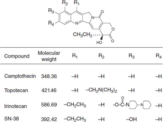
FIGURE 4-1 Structure of the camptothecins.
MECHANISM OF ACTION
Camptothecin and its analogs exert their antitumor activity by inhibiting the enzyme DNA topo I, a nuclear enzyme that relieves torsional strain in supercoiled DNA during replication, repair, and transcription (Figure 4-2). The enzyme forms a transient, intermediate complex with single-stranded DNA that opens the DNA strand and allows passage of an intact single strand through the nick. The strand break also allows for rotation about the intact strand. Camptothecins bind to and stabilize the otherwise transient DNA-enzyme complex and prevent resealing of the broken strand. The single strand break, when it encounters a replication complex, leads to a double-stranded break, and an accumulation of double strand breaks leads to apoptosis. Active synthesis of DNA is a prerequisite for this interaction (1). In addition to strand breakage, irinotecan causes a downregulation of HIF1-alpha, a transcription factor critical to cell survival in the presence of hypoxia; angiogenesis is inhibited as a result (2). The mechanism of drug effects on HIF1-alpha is not known.

FIGURE 4-2 Mechanism of the camptothecins. (Used with permission from www.scielo.br.)
MECHANISMS OF RESISTANCE
In preclinical studies, resistance to the camptothecins may result from multiple mechanisms: increased expression of the multidrug resistance (MDR) efflux pump, p-glycoprotein, and a related transporter, ABCG2, the breast cancer resistance transporter; downregulation of topo I expression; mutations in the catalytic or DNA binding sites of topoisomerase I; upregulation of topoisomerase II; upregulation of NFκB; and inhibition of apoptosis. The clinical significance of these mechanisms of resistance is unproven.
CAMPTOTHECINS: CLINICAL PHARMACOLOGY
TOPOTECAN
Pharmacokinetics
Topotecan is usually given as a 30-min intravenous infusion. Plasma concentrations of the inactive carboxylate species begin to predominate over the active lactone form within 5–10 min after the end of infusion. The plasma half-life of topotecan is 2.4–4.3 h. Topotecan is eliminated primarily by plasma hydrolysis to the inactive carboxylate form, followed by renal excretion. Approximately one-third to one-half of a dose of drug is excreted unchanged in the urine. The clearance of topotecan and the carboxylate metabolite is reduced by 33% in patients with renal dysfunction and a creatinine clearance between 40 and 59 ml/min, and by 75% in patients with a creatinine clearance between 20 and 39 m/min. There is no significant change in elimination or toxicity in patients with liver disease, even in patients with total bilirubin levels up to 10 mg/dl. Administration of cisplatin decreases topotecan clearance, presumably through renal tubular damage. Topotecan pharmacokinetics are not altered when it is combined with anthracyclines, cyclophosphamide, or cytarabine (1).
Dosing and Schedule
The standard dosing schedule for topotecan is 1.5 mg/m2 given as a 30-min intravenous infusion on 5 consecutive days, repeated every 21 days. Continuous infusion regimens up to 21 days in duration have attempted to take advantage of the in vitro observation that prolonged exposures of low concentrations are more efficacious than intermittent exposures to high concentrations. Results have been equivocal.
Toxicity
Neutropenia is the most significant dose-limiting toxicity for all schedules of topotecan administration, with grade 4 neutropenia in up to 81% of patients and febrile neutropenia in 26%. Renal dysfunction requires a dose reduction as indicated above. There is no dose modification required for patients with hepatic dysfunction. Non-hematologic toxicities of topotecan include nausea, vomiting, mucositis, elevated transaminases, fatigue, and rash. These side effects are generally minimal and easily managed (1).
IRINOTECAN
Pharmacokinetics
Irinotecan (Camptosar) is a congener of camptothecin specifically designed to facilitate generation of its lactone-stabilized metabolite, the 7-ethyl-10-hydroxy analog, SN-38, which is a 1000-fold more potent inhibitor of topo I than the parent drug. Irinotecan is primarily eliminated through the liver via two clinically relevant mechanisms (Figure 4-3). First, irinotecan is a substrate of the cytochrome p450 system and is metabolized to inactive derivatives by CYP2B6 and CYP3A4. These inactivation pathways are inducible by phenobarbital or phenytoin (1). Second, irinotecan is converted to SN-38 by ubiquitous esterases, and SN-38 is then cleared by glucuronidation and excreted in the biliary system. The half-life of the active lactone form of SN-38 is 11.5 h (1).
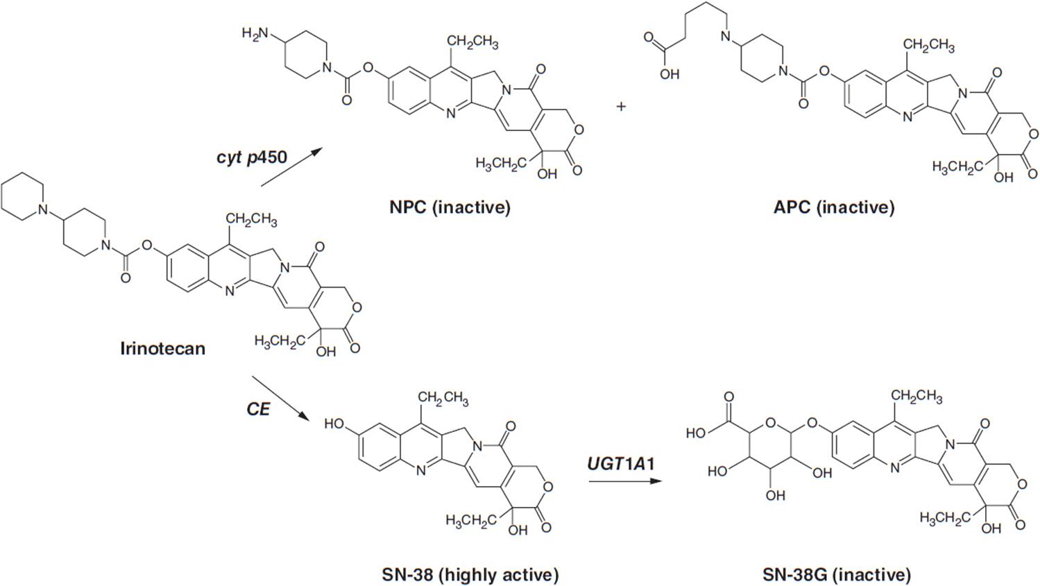
FIGURE 4-3 Metabolic pathway for irinotecan showing the conversion to inactive metabolites NPC and APC via cytochrome p450 enzymes and the conversion by liver carboyxylesterase (CE) to the active form, SN-38, and Its subsequent inactlvation to SN-38G by the enzyme UGT1A1.
SN-38 undergoes glucuronidation by the polymorphic enzyme uridine diphosphoglucuronosyl-transferase (UGT1A1), which is also responsible for bilirubin glucuronidation. The activity of this enzyme is significantly reduced in patients homozygous for the allele UGT1A1*28, the same defect seen in subjects with Gilbert’s syndrome. This homozygous deficiency is found in approximately 10% of patients. When treated with standard doses of irinotecan, such patients have slower clearance of the parent drug and higher levels of plasma SN-38 and encounter higher rates of toxicity, particularly neutropenia (3, 4). In patients with Gilbert’s syndrome, or with an unexplained elevated indirect bilirubin level, lower starting doses of irinotecan should be used. A commercially available test for the UGT1A1*28 polymorphism (Invader UGT1A1*28 Molecular Assay) can be used, although it has not been widely accepted in clinical practice. Common polymorphisms in ABC cassette drug transporters, which export the parent drug and SN-38 from intestinal epithelium, may account for the severe gastrointestinal toxicity that affects 20%–30% of patients (5).
Dosing and Schedule
Irinotecan, as a single agent, can be given at a dose of 125 mg/m2 over a 90-min intravenous infusion every week for 4 of 6 weeks or at a dose 350 mg/m2 over a 90-min intravenous infusion every 3 weeks. The weekly schedule appears to be equally effective, although there are lower rates of diarrhea with the every 3-week regimen (6).
Toxicity
The most common adverse effects of irinotecan are diarrhea, which can be life-threatening in some instances, myelosuppression, and an acute cholinergic syndrome of nausea, vomiting, mucositis, diarrhea, and flushing. The latter responds to atropine. Interstitial pneumonitis has been reported in Japanese patients receiving irinotecan. Grade 3–4 diarrhea was observed in up to 35% of patients in early clinical studies. Irinotecan, when given at 125 mg/m2 on a weekly basis for 4 of 6 weeks in combination with 5-FU and leucovorin (7), led to toxic deaths due to severe diarrhea and neutropenia. An alternative and better tolerated schedule of biweekly irinotecan at 180 mg/m2, whether as single agent or combined with 5-FU and leucovorin (FOLFIRI), has been widely adopted. Loperamide starting at 4 mg should be given at the first sign of diarrhea, and repeat doses of 2 mg every 2 h may be given until resolution of the diarrhea.
Myelosuppression is common with irinotecan; grade 3–4 neutropenia occurs in 14%–47% of patients.
Clinical Indications
Irinotecan is most commonly used in advanced colorectal cancer. As first-line therapy in metastatic disease, it is combined with 5-FU and leucovorin, with or without bevacizumab. It can also be given in the second-line therapy as a single agent, or in patients with k-ras wild-type tumors, it may be combined with cetuximab (8).
TOPOISOMERASE II INHIBITORS: ANTHRACYCLINES
Drugs of this class, derived from the fungal culture broths of Streptomyces peucetius, have become critical components of treatment for acute leukemias, lymphomas, breast cancer, and sarcomas. The two original members of this family, daunorubicin (DN) and doxorubicin (DX), remain in active clinical practice: DN for acute myelogenous leukemia (AML), and DX for lymphomas and solid tumor chemotherapy. Two semisynthetic derivatives, idarubicin (IDA) and epirubicin (EPI), have made inroads as valuable agents for leukemia and breast cancer, respectively. Doxil, a liposomal formulation of DX has found limited use, primarily for ovarian cancer, and a similar liposomal DN preparation is indicated for Kaposi’s sarcoma.
MECHANISM OF ACTION
The anthracyclines share a rigid planar four-ring structure complemented by a glycosidic substitution on the D ring and variable side groups on the A and D rings (Figure 4-4). The planar configuration allows anthracyclines to intercalate between strands of DNA, and this action was originally thought to be responsible for its inhibition of DNA synthesis. However, the anthracyclines possess other important features. The quinone on ring C readily undergoes oxidation/reduction cycling in the presence of Fe++, producing free radicals from oxygen and/or lipids (9). These free radicals are responsible for the cardiac toxicity inherent in this class of agents. The anthracyclines bind to and inhibit topo II, an enzyme that promotes DNA strand unwinding essential for DNA synthesis and repair. In its normal function, topo II binds to DNA and creates a double strand break that allows strand passage. The enzyme then reseals the break. Anthracyclines bind to and stabilize the DNA-topo II complex, preventing the resealing of the strand break. An accumulation of strand breaks signals the p53 system to halt cell cycle progression, and to initiate DNA repair. If the breaks are sufficiently numerous, the cell undergoes apoptosis. High levels of topo II expression correlate positively with response to DX in patients with breast cancer (10, 11). Cells become resistant to anthracyclines through diminished expression of topo II activity or through topo II mutations that decrease binding affinity of the enzyme for the drugs of this class. An interesting correlation has been observed between amplification of the isoenzyme topo IIa and the HER2/neu receptor. The topo IIa gene is located on chromosome 17 adjacent to the gene coding for the HER2/neu. HER2/neu amplification is found in one quarter of breast cancers. In one-third of patients with amplified HER2, topo IIa is co-amplified, and this subset of breast cancer patients has a higher response rate to DX.

FIGURE 4-4 Anthracyclines in current clinical use. For epirubicin and idarubicin, arrows point to the sites where these new drugs differ from doxorubicin and daunomycin, respectively.
DRUG RESISTANCE
Transporters that export anthracyclines and other natural products influence response to this class of agents. Expression of the MDR gene, which codes for the membrane transporter p-glycoprotein, increases resistance to anthracyclines, and confers drug resistance in patients with AML, multiple myeloma, and lymphomas. Other membrane exporters, including the MRP family, and the breast cancer resistance transporter (ABCG2) cause resistance in cell lines, but their clinical role is uncertain. IDA is less affected by the presence of MDR than is DX (12).
Other intracellular processes that recognize DNA strand breaks and initiate apoptosis may influence sensitivity to anthracyclines. High levels of BCL-2 expression, an anti-apoptotic factor, render cells insensitive to anthracyclines, as does a loss of function of the mismatch repair complex that recognizes defective strand pairing. Mutations in p53, mutations in ATM (a sensor of strand breaks), and high levels of MDM2 (an antagonist of p53) also confer resistance to DX (13).
CLINICAL PHARMACOLOGY
To a variable extent, anthracyclines are converted to an active alcohol (-ol) intermediate by the ubiquitous enzyme, aldoketoreductase, but for DX and EPI, the parent compounds are believed to be more potent and are responsible for their clinical efficacy. IDA is rapidly converted to its alcohol metabolite, and the alcohol becomes the predominant species in plasma 1–4 h after drug administration. The metabolite is slightly less potent than the parent drug and likely contributes to the drug’s antitumor activity in vivo.
The important pharmacokinetic features of the various anthracyclines are shown in Table 4-1. The parent compounds (DX, DN, and EPI), or in the case of idarubicin, the active alcohol metabolite, have a prolonged terminal half-life in plasma of 1 day or longer, thus allowing intermittent dosing once every week to once every 3 weeks. Clearance occurs primarily through hepatic non-microsomal conversion to sulfates, aglycones, and other inactive metabolites. Anthracycline semiquinone radicals may also be inactivated by enzymatic or chemically mediated conjugation with sulfhydryls such as glutathione. Because of the importance of hepatic enzymatic clearance of parent compounds and alcohol metabolites, hepatic dysfunction, with bilirubin greater than 1.5 mg/dl, is associated with delayed drug clearance and a probable increased risk of toxicity (9). In this case, most regimens call for a 50% dose reduction, with subsequent escalation if the dose is well tolerated. Renal dysfunction (creatinine clearance less than 60 ml/min) also slows DX and IDA clearance, probably through changes in hepatic blood flow or diminished hepatic clearance of parent drug.
TABLE 4-1 ANTHRACYCLINE PHARMACOKINETICS
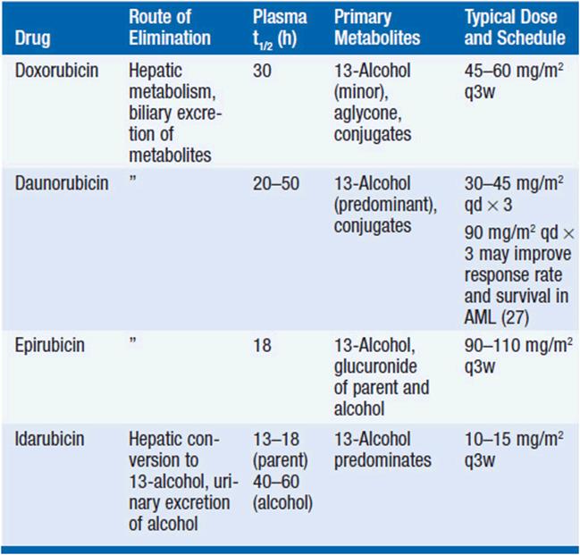
High doses of DN (90 mg/m2 vs. 45 mg/m2, both qd × 3) have improved complete response rates and survival in patients with AML, without an increase in life-threatening toxicity (15).
TOXICITY
All anthracyclines cause myelosuppression, mucositis, and alopecia. Recovery of peripheral blood counts occurs within 10–14 days. Their most significant late toxicity is cardiac injury. Initial clinical experience with DX, as documented by sequential endomyocardial biopsy, disclosed myocardial necrosis, both in animals and in patients receiving multiple doses of drug. Subsequent studies in children have revealed elevations of troponin T in the days following drug administration and an elevated risk of late cardiac events in patients demonstrating such elevations. Cardiac function is ordinarily monitored through tests of left ventricular ejection fraction (scans or echocardiography). Decreases of greater than 10% from baseline values, or a fall below 40%, signal a high risk of later congestive failure. These changes should prompt discontinuation of anthracycline treatment. Symptomatic cardiac disease, manifested primarily as congestive heart failure, usually does not occur until total doses of DX exceed 450 mg/m2, with a marked increase in risk above 550 mg/m2. However, in DX-treated children receiving a total dose of 300 mg/m2 or less, a significantly elevated risk of cardiac disease (arrhythmias, sudden death, myocardial infarcts, or congestive failure) emerges later in adult life (16).
EPI appears to be less cardiotoxic than DX in studies of breast cancer patients receiving adjuvant chemotherapy (17). The incidence of congestive failure following adjuvant therapy reaches 1%–1.5% of patients treated with EPI and is slightly higher (approximately 2%) for DX-containing regimens. It appears that cardiac toxicity may be less in patients receiving DX by continuous infusion over 4 days, or in small weekly doses, but the convenience of single bolus doses every 2–3 weeks has led to the use of this schedule in standard adjuvant therapy.
In both children and adults, oncologists usually limit total doses of DX to 300 mg/m2. In children, dexrozoxane, an iron chelating drug, clearly decreases the frequency of acute troponin T elevations and lessens the risk of late cardiac toxicity, and is routinely administered with DX to children.
Radiation therapy to the chest delivered with chemotherapy increases the risk of cardiotoxicity. Other chemotherapy drugs potentiate anthracycline cardiotoxicity. Paclitaxel, administered with DX, decreases the rate of DX clearance and significantly enhances the rate of DX cardiotoxicity, an effect attributed to inhibition of DX metabolism and/or biliary excretion (18). Trastuzumab, the anti-HER2/neu antibody, increases the risk of DX cardiotoxicity. Sequential administration of DX and cyclophosphamide (19), followed by trastuzumab, is associated with a more than twofold increase in heart failure, as compared to the heart failure risk of either drug alone. EPI given with trastuzumab or DX with docetaxel leads to no obvious increase in cardiotoxicity, although less data are available for these regimens (20).
In addition to cardiac toxicity, the anthracyclines as a class increase the risk of AML, and less commonly, acute promyelocytic leukemia (PML) (21). The onset of myelodysplasia (MDS) occurs within 1–3 years of treatment in patients receiving cyclophosphamide/EPI as adjuvant therapy, and the risk increases markedly in patients receiving greater than 720 mg/m2 EPI or 6300 mg/m2 cyclophosphamide. With alkylating agents and EPI, leukemias displayed either chromosome 5 or 7 deletions, or more commonly balanced translocations involved 11q23, a finding characteristic of leukemia secondary to topo II inhibitors. EPI also leads to (15:17) acute promyelocytic leukemia, perhaps due to a specific “hot spot” sensitive to topo II in exon 6 of the PML gene (22).
OTHER ANTHRACYCLINES AND ANTHRACENEDIONES
Mitoxantrone is a planar multi-ring quinine similar to anthracyclines but lacks the sugar linkage. It is less cardiotoxic, and is a less potent antileukemic agent than DN, but shares pharmacological properties with the anthracyclines: hepatic metabolism, long terminal half-life in plasma, myelosuppression as its major toxicity, susceptibility to MDR and other ABC cassette transporters, and a risk of causing acute AML and PML as a late toxicity (14, 22). It is rarely used in clinical oncology practice. The recommended doses are 12 mg/m2/day for 3 days for acute leukemia and 14–16 mg/m2 every 3 weeks for solid tumor therapy.
LIPOSOME ENCAPSULATED ATHRACYCLINES
In an effort to increase drug uptake selectively in tumor cells and to decrease cardiac toxicity, both DX and DN have been reformulated in lipid spheres (liposomes). In this form, the drug has a half-life in plasma of greater than 50 h. Liposomal DX (Doxil) has proven useful in platinum-refractory ovarian cancer, while liposomal DN is approved for treatment of Kaposi’s sarcoma. These preparations have less cardiotoxicity than the parent drugs. Late side effects of Doxil have included renal failure and oralpharyngeal carcinomas (23).
ETOPOSIDE
Topo II inhibitors are found in nature as potent cellular poisons. Etoposide, etoposide phosphate (rarely used), and teniposide (Figure 4-5) are semi-synthetic derivatives of podophyllotoxin, a plant product, which itself is an antimitotic without topo II inhibitory activity. Etoposide has been a valuable agent for the treatment of leukemias, lymphomas, and germ cell tumors in both conventional and high-dose regimens, while teniposide is used primarily for the treatment of AML in children.

FIGURE 4-5 Molecular structure of etoposide, etoposide phosphate, and teniposide.
MECHANISM OF ACTION AND RESISTANCE
As described above, topo II cleaves a DNA strand in a reaction mediated by ATP hydrolysis. Etoposide and teniposide bind to the complex of DNA and enzyme, inhibiting the resealing activity of the enzyme and perpetuating strand breaks (Figure 4-6) (24). Through p53, these strand breaks signal a halt to cell cycle progression and, if breaks are sufficiently numerous, prompt apoptosis.

FIGURE 4-6 Formation of single and double strand breaks by topoisomerase II (TOP II), and prevention of break resealing in the presence of etoposide ( ).
).
As natural products, the epipodophyllotoxin derivatives are subject to transport from tumor cells by the p-glycoprotein, a product of the MDR gene. Resistance may also arise through deletion of topo II (via methylation of the gene or loss of promoter activity) or through mutation of its binding site for these drugs. Finally, disruptions of apoptotic pathways, and the capacity to repair double strand breaks, may also determine the outcome of therapy.
CLINICAL PHARMACOLOGY
Etoposide is eliminated by both renal excretion and hepatic metabolism, and doses should be adjusted for dysfunction of either organ (25). Approximately 40% of a dose of etoposide is excreted unchanged in the urine, and dose should be reduced in proportion to changes in creatinine clearance. The remainder is eliminated by glucuronidation. A smaller fraction of drug undergoes CYP3A4 metabolism through demethylation, producing a cytotoxic catechol metabolite and other quinine derivatives of uncertain significance. The drug has a terminal half-life of 8 h in plasma in patients with normal renal and hepatic function. In patients with elevated serum bilirubin of 1.5–3.0 mg/dl, the dose should be reduced by 50%, while in those with higher bilirubin, the drug should be used with extreme caution and in lesser doses, and alternative therapies should be considered.
High-dose etoposide (1.5 g/m2 or above) is used alone, or in combination with cyclophosphamide, ifosfamide, or carboplatin (26). It is the only topo II agent that can be significantly escalated in dose without encountering irreversible nonmyeloid toxicity. At these high doses, mucositis and hepatic enzyme elevations become dose limiting. Its pharmacokinetics remain linear at these high doses.
Teniposide is eliminated primarily by the liver, with a variety of microsomal and other metabolites appearing in bile. Its half-life in plasma is 10–21 h. Very little drug is excreted unchanged in the urine, and no dose adjustment for renal dysfunction is needed.
TOXICITY
At usual doses of 100 mg/m2 per day for 3 days every 3 weeks, bone marrow suppression is the primary toxicity of etoposide, with recovery 10–14 days after treatment. Hypotension, fever, and asthmatic episodes may follow drug infusion, probably a response to the cremophor diluent in which the drug is administered. Liver function abnormalities and mucositis supervene at higher doses.
Etoposide causes acute myelogenous leukemia as a later toxic event, usually 2–3 years after treatment. The leukemia often involves a translocation (at 11q 23) in the MLL gene on the long arm of chromosome 11 at AT-rich sites favored for topo II cleavage. The leukemia may be preceded by a period of myelodysplasia. Less commonly, acute promyelocytic leukemia has also been reported following etoposide. The risk of AML increases with cumulative doses >6 g/m2 and with schedules of weekly or biweekly administration (27).
Tenoposide side effects follow the same pattern as these of etoposide; myelosuppression and mucositis are common toxicities. The incidence of acute hypersensitivity reactions is higher for teniposide, probably related to the greater concentration of lipid diluent used in its formulation. Like etoposide, it causes secondary AML.
REFERENCES
1. Garcia-Carbonero R, Supko JG. Current perspectives on the clinical experience, pharmacology, and continued development of the camptothecins. Clin Cancer Res. 2002; 8: 641–661.
3. Massacesi C, Terrazzino S, Marcucci F, et al. Uridine diphosphate glucuronsyl transferase 1A1 promoter polymorphism predicts the risk of gastrointestinal toxicity and fatigue induced by irinotecan-based chemotherapy. Cancer. 2006; 106: 1007–1016.
4. Zhou Q, Sparreboom A, Tan E-H, et al. Pharmacogenetic profiling across the irinotecan pathway in Asian patients with cancer. Br J Clin Pharmacol. 2005; 59: 415–424.
5. Di Martino MT, Arbitrio M, Leone E, et al. Single nucleotide polymorphisms of ABCC5 and ABCG1 transporter genes correlate to irinotecan-associated gastrointestinal toxicity in colorectal cancer patients. A DMET microarray profiling study. Cancer Biol Ther. 2011; 12: 780–787.
6. Fuchs CS, Moore MR, Harker G, et al. Phase III comparison of two irinotecan dosing regimens in second-line therapy of metastatic colorectal cancer. J Clin Oncol. 2003; 21: 807–814.
7. Saltz LB, Cox JV, Blanke C, et al. Irinotecan plus fluorouracil and leucov-orin for metastatic colorectal cancer. N Engl J Med. 2000; 343: 905–914.
8. Cunningham D, Humblet Y, Siena S, et al. Cetuximab monotherapy and cetuximab plus irinotecan in irinotecan-refractory metastatic colorectal cancer. N Engl J Med. 2004; 351: 337–345.
9. Doroshow JH. Anthracyclines and anthracenediones. In BA Chabner, DL Longo (eds.), Cancer Chemotherapy and Biotherapy: Principles and Practice. Lippincott, Williams and Wilkins, Philadelphia, PA, 2011; 356–391.
10. Durbecq V, Paesmans M, Cardoso F, et al. Topoisomerase-II expression as a predictive marker in a population of advanced breast cancer patients randomly treated either with single-agent doxorubicin or single-agent docetaxel. Mol Cancer Ther. 2004; 3: 1207.
11. Brase E, Schmidt M, Fischbach T, et al. ERBB2 and TOP2A in breast cancer: a comprehensive analysis of gene amplification, RNA levels, and protein expression and their influence on prognosis and prediction. Clin Cancer Res. 2010; 16: 2391–2401.
12. Kroschinsky R, Schleyer E, Renner U, et al. Increased myelotoxicity of idarubicin: is there a pharmacological basis? Cancer Chemother Pharmacol. 2004; 53: 61.
13. Chrisanthar R, Knappskog S, Lokkevik, et al. Predictive and prognostic impact of TP53 mutations and MDM2 promoter genotype in primary breast cancer patients treated with epirubicin or paclitaxel. PLoS One. 2011; 1–10.
14. Camaggi CM, Strocchi E, Carisi P, et al. Idarubicin metabolism and pharmacokinetics after intravenous and oral administration in cancer patients: a crossover study. Cancer Chemother Pharmacol. 1992; 30: 307.
15. Lowenburg B, Ossenkoppele GJ, van Putten W, et al. High dose-daunorubicin in older patients with acute myeloid leukemia. New Engl J Med. 2009; 361: 1235–1248.
16. Nysorn K, Holm K, Lipsitz SR, et al. Relationship between cumulative anthracycline dose and late cardiotoxicity in childhood acute lymphoblastic leukemia. J Clin Oncol. 1998; 16: 545.
17. Smith LA, Cornelius VR, Plummer CJ, et al. Cardiotoxicity of anthracycline agents for the treatment of cancer: systematic review and meta-analysis of randomised controlled trials. MBMC Cancer. 2010; 10: 337–351.
18. Partridge AH, Burstein HJ, Winer EP. Side effects of chemotherapy and combined chemohormonal therapy in women with early-stage breast cancer. J Natl Cancer Inst Monogr. 2001; 30: 135.
19. Bowles EJA, Wellman R, Feigelson HS, et al. Risk of heart failure in breast cancer patients after anthracycline and trastuzumab treatment: a retrospective cohort study. J Natl Cancer Inst. 2012; 104: 1293–1306.
20. Buzdar AU, Ibrahim NK, Francis D, et al. Significantly higher pathologic complete remission rate after neoadjuvant therapy with trastuzumab, paclitaxel, and epirubicin chemotherapy: results of a randomized trial in human epidermal growth factor receptor 2-positive operable breast cancer. J Clin Oncol. 2005; 23: 3676.
21. Praga C, Bergh J, Bliss J, et al. Risk of acute myeloid leukemia and myelodysplastic syndrome in trials of adjuvant epirubicin for early breast cancer: correlation with doses of epirubicin and cyclophosphamide. J Clin Oncol. 2005; 23: 4179.
22. Mays AN, Osheroff N, Xiao Y, et al. Evidence for direct involvement of epirubicin in the formation of chromosomal translocations in t(15;17) therapy-related acute promyelocytic leukemia. Blood. 2010; 14: 326–330.
23. Muggia F, Cannon T, Safra T, Curtin J. Delayed neoplastic and renal complications in women receiving long-term chemotherapy for recurrent ovarian cancer. J Natl Cancer Inst. 2011; 103: 160–161.
24. Minford J, Pommier Y, Filipski J, et al. Isolation of intercalator-dependent protein-linked DNA strand cleavage activity from cell nuclei and identification as topoisomerase II. Biochemistry. 1986; 25: 9–16.
25. D’Incalci M, Rossi C, Zucchetti M, et al. Pharmacokinetics of etoposide in patients with abnormal renal and hepatic function. Cancer Res. 1986; 46: 2566–2571.
26. Beyer J, Kramar A, Mandanas R, et al. High-dose chemotherapy as salvage treatment in germ cell tumors: a multivariate analysis of prognostic variables. J Clin Oncol. 1996; 14: 2638–2645.
27. Le Deley MC, Leblanc T, Shamsaldin A, et al. Societe Francaise d’Oncologie Pediatrique. Risk of secondary leukemia after a solid tumor in childhood according to the dose of epipodophyllotoxins and anthracyclines: a case-control study by the Societe Francaise d’Oncologie Pediatrique. J Clin Oncol. 2003; 21: 1074–1081.