
Olivia Foley, Marcela G. del Carmen
INTRODUCTION
Squamous cell carcinoma of the uterine cervix comprises an estimated 80% of all cervical cancers. The other histologies include adenocarcinoma (15%) and adenosquamous carcinomas (3%–5%), with only a small fraction of all cervical cancers having neuroendocrine or small cell histology. This chapter will focus on the diagnosis and management of primary squamous cell carcinoma of the uterine cervix. Amongst all malignancies, cervical cancer is the second most common cancer affecting women, with an estimated 52% case-fatality rate (1). Worldwide, cervical cancer is the most common gynecologic malignancy, accounting for 529,800 new cases (9%) and 273,200 deaths (8%) (1, 2). In developed countries, cervical cancer ranked tenth most common type of cancer in women (9.0/100,000 women) and below the top 10 causes of cancer mortality (3.2/100,000 deaths) (3). An estimated 86% of new cervical cancer cases are seen in the developing world, ranking as the second most common type of cancer (17.8/100,000 women) and cause of cancer deaths (99.8/100,000 deaths) (3). The highest incidence rates worldwide are observed in sub-Saharan Africa, Latin America and the Caribbean, South-Central Asia, and Southeast Asia (1). One-third of the cervical cancer burden in the world is experienced in South-Central Asia. Lastly, although cervical cytology is an excellent screening instrument for pre-invasive disease, the false negative rate for detecting invasive carcinoma is relatively high, reportedly 50%.
 INCIDENCE
The incidence of invasive cervical cancer is related to age, with a mean age at the time of diagnosis of 48 years in the United States (3). The reported age-adjusted incidence of cervical cancer in the United States in girls under 20 years of age is 0.1 per 100,000, 1.5 per 100,000 in women aged 20–24 years, and 11.0 per 100,000 for women aged 30 to over 85 years (3).
EPIDEMIOLOGY
Patients with squamous cell carcinoma of the cervix share the same risk factors as patients with cervical intraepithelial neoplasia or dysplasia (4). These factors include:
• Early onset of sexual activity
• Multiple sexual partners
• High-risk sexual partners
• History of sexually transmitted diseases
• Tobacco use
• Multiparity
• Low socioeconomic status
• Immunosuppression
• Previous history of vulvar or vaginal dysplasia
Perhaps the most significant risk factor for developing squamous cell cervical cancer is lack of cervical cytological screening. It is critical to underscore that infection with certain subtypes of the human papillomavirus (HPV) has been identified as the central causative factor in the development of cervical neoplasia (4). High-risk oncogenic types can be detected in almost all cervical cancers (4). Although most HPV infections are transient, chronic persistent HPV infection with the oncogenic subtypes is the central causative factor in the development of cervical neoplasia. The virus alone, however, is not sufficient to cause cervical neoplasia or cancer (4).
CLINICAL MANIFESTATIONS
SYMPTOMS
Because early cervical cancer is usually asymptomatic, screening is critical. Common symptoms, when they occur, include abnormal vaginal bleeding, bleeding after intercourse, and a vaginal discharge (watery, mucoid, malodorous, or even purulent). In the setting of advanced disease, patients may complain of back pain radiating to the lower extremities or pelvic pain. Other symptoms seen in the setting of advanced disease include bowel and urinary symptoms, such as hematuria, hemotochezia, or stool/urine passage per vagina.
PHYSICAL EXAMINATION
Findings at the time of physical examination may range from a normal appearing cervix to a grossly abnormal cervix with an exophytic, plaquelike, indurated, ulcerated, or endophytic lesion. Findings encountered in patients with regionally advanced stage disease include parametrial, paracervical, or vaginal involvement, lower extremity edema, and inguinal adenopathy. Distant disease may be manifest in ascites, pleural effusions, and supraclavicular adenopathy.
PATTERNS OF SPREAD
Squamous cell carcinoma of the cervix may spread via direct extension as well as lymphatic and hematogenous dissemination. It can spread directly to the parametria, uterine corpus, vagina, bladder, rectum, and peritoneal cavity. Although prior dictum described a predictable pattern of lymphatic spread, sentinel node mapping has demonstrated that the first site of metastasis may involve any one of the pelvic lymph node chains.
DIAGNOSIS
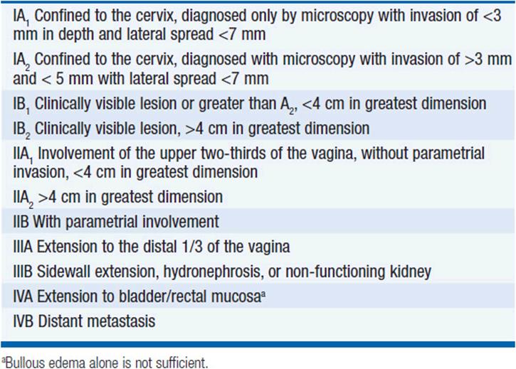
Squamous cell carcinoma of the uterine cervix is staged clinically based on the criteria delineated by the International Federation of Gynecology and Obstetrics (FIGO). Table 55-1 reflects the staging FIGO changes made in 2009.
TABLE 55-1 STAGING OF CERVICAL CANCER BASED ON CRITERIA FROM THE INTERNATIONAL FEDERATION OF GYNECOLOGY AND OBSTETRICS (FIGO)
CLINICAL STAGING PROCEDURES
After histological confirmation of an invasive cancer, a thorough physical examination is mandated. This survey should include careful inspection of the cervix, assessment of its size, and careful examination of the entire vagina. Cervical tumor size and parametrial involvement are best evaluated through a rectovaginal examination. The inguinal and supracervical regions should be inspected for the presence of adenopathy. It is reasonable to arrange for an examination under anesthesia in order to better appreciate the extent of local disease and to facilitate patient comfort. The following studies and procedures are allowed by FIGO as part of the staging of cervical cancer:
• Chest x-ray
• Intravenous pyelogram
• Barium enema
• Skeletal x-rays
• Colposcopy/biopsies
• Cervical conization
• Cystoscopy
• Proctoscopy
Other optional studies and procedures that can be obtained, but cannot alter FIGO staging, include the following:
• Computed tomography
• Magnetic resonance imaging
• Positron emission tomography (PET)
• Ultrasonography
• Radionucleotide scanning
• Laparoscopy
• Laparotomy
PROGNOSIS
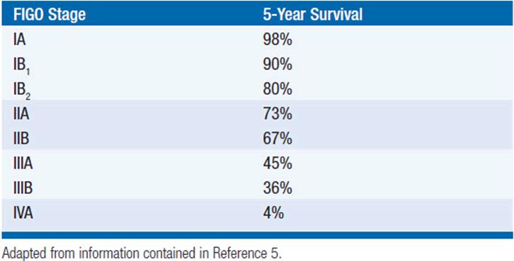
Prognosis for squamous cell carcinoma of the uterine cervix is influenced by numerous tumor-related factors, including stage, tumor volume, depth of invasion, lymph node involvement, lymph-vascular space involvement, histologic subtype, and tumor grade. FIGO tumor stage correlates well with 5-year survival (Table 55-2) (5). Lymph nodal status is also an important prognostic factor. Five-year survival in the presence of pelvic lymph node involvement is 45%–60% (6). Five-year survival in the setting of para-aortic lymph node involvement is estimated to be 15%–30% (7). The number of involved lymph nodes also plays a critical role. In the presence of one involved pelvic lymph node, the recurrence risk in 35% (7). When 2 or 3 pelvic lymph nodes are involved, the risk of recurrence is 59% and 69%, respectively (7).
TABLE 55-2 FIVE-YEAR SURVIVAL FOR SQUAMOUS CELL CARCINOMA OF THE UTERINE CERVIX BASED ON FIGO STAGING
TREATMENT
Treatment options for squamous cell carcinoma of the uterine cervix include surgery, chemoradiation therapy, and chemotherapy. Both surgical intervention and definitive radiation therapy with concomitant chemotherapy are appropriate alternatives for the treatment of early stage cervical cancer, including FIGO stage IIA (8).
SURGERY
For women with stage IA1 lesions, surgical treatment may be in the form of a loop electrosurgical excision procedure (LEEP) or cervical conization (if they desire to preserve fertility) or an extrafascial hysterectomy (8). For certain selected patients who want to preserve fertility and with stage IA2/IB1 lesions, a radical trachelectomy with lymphadenectomy may be an alternative to a radical hysterectomy. A radical trachelectomy involves removal of the entire cervix and the parametria, with placement of a cerclage in order to allow preservation of the uterine corpus with a competent vaginal-uterine junction (8). The radical trachelectomy can be performed abdominally or vaginally, and combined with a laparoscopic or open therapeutic lymphadenectomy. The radical trachelectomy appears to be a reasonable alternative for women with stage IA2/IB1 lesions desiring fertility preservation with tumors less than 2 cm in size, absence of lymph vascular space involvement, and absence of lymph nodal disease. The experience with this procedure indicates that it results in a similar oncologic outcome as a radical hysterectomy but allows for the possibility of future pregnancies.
For patients with stage IA2 lesions, surgical treatment may be in the form of a type II or modified hysterectomy (8). During this type of hysterectomy, the uterine artery is ligated where it crosses over the ureter, the uterosacral and the cardinal ligaments are divided midway toward their attachment to the sacrum and the pelvic side wall, respectively, and the upper one-third of the vagina is resected. For stage IB1, IB2, and IIA lesions, the recommended surgical treatment is a type III or radical hysterectomy. During this procedure, the uterine artery is ligated at its origin from the internal iliac artery. The uterosacral and the cardinal ligaments are divided at their insertion into the sacrum and the pelvic sidewall, respectively, and the upper one-half of the vagina is divided.
In premenopausal women, surgery, as compared to radiation therapy as the alternative treatment modality, offers the advantage of ovarian preservation and may avoid vaginal stenosis. It also allows for “debulking” of enlarged lymph nodes and may allow for the individualization and tailoring of the radiation treatment fields.
Part of the surgical treatment of cervical cancer for stage IA2-IIA includes a lymphadenectomy. For women with stage IA2 and small IB1 tumors, a pelvic lymphadenectomy should be performed at the time of hysterectomy (8). For those with enlarged lymph nodes, macroscopic stage IB1, IB2, or IIA tumors, or those with histologic confirmation of metastatic nodal disease at the time of frozen section, the surgical intervention should include both a pelvic and para-aortic lymphadenectomy.
PRIMARY RADIATION THERAPY
Since the oncologic results are similar, radical surgery and definitive radiation therapy are acceptable treatment modalities for stage IA, IB, and non-bulky IIA lesions (8). For women undergoing definitive treatment with radiation therapy, the use of concomitant cisplatin-based chemotherapy is also recommended. The progression-free survival and overall survival advantage of concomitant chemoradiotherapy over radiation alone in patients with early and locally advanced cervical cancer has been demonstrated in at least five randomized, controlled clinical trials and a meta-analysis.
Radiation therapy can be delivered in the form of external beam radiation or brachytherapy. Brachytherapy allows for treatment of centrally located disease, primarily the cervix, the vagina, and the parametria. It can be delivered via an intracavitary or interstitial needle system. The intracavitary systems include uterine tandems, vaginal colpostats, and vaginal cylinders. External beam radiation therapy involves radiation of the entire pelvis, with doses ranging from 4500 to 5000 cGy, given in daily fractions over several weeks (usually 180 cGy/fraction). As noted earlier, radiation therapy is administered with concomitant chemotherapy using cisplatin (40 mg/m2/week) (8). In a recent study of patients with bulky stage IIB to IVA, a novel regimen of concurrent cisplatin plus gemcitabine with radiation therapy followed by brachytherapy and adjuvant gemcitabine plus cisplatin significantly improved outcomes, with notable increased but acceptable toxicity, when compared to standard cisplatin-based chemoradiation therapy (9).
ADJUVANT CHEMORADIOTHERAPY
Patients with localized cervical cancer treated primarily with surgery and who have tumors with either intermediate- or high-risk factors for disease recurrence should receive adjuvant chemoradiotherapy (10). High-risk factors include positive or close resection margins, positive lymph nodes, and microscopic parametrial involvement (10). Intermediate-risk factors for disease recurrence include tumor size, deep stromal invasion, or the presence of lymph vascular space involvement (10). The recommended regimen includes radiation therapy with the use of concomitant cisplatin (40 mg/m2/week) (8, 10, 11).
POST-TREATMENT SURVEILLANCE
After completion of their treatment plan, patients should be evaluated every 3 months for the first 2 years, every 6 months for the subsequent 3 years, and annually thereafter. Evaluation should include a thorough review of systems and physical examination and a Pap smear at the time of each surveillance visit. Most vaginal recurrences are asymptomatic and may be only recognized through a Pap smear. For women with stage IIB or greater, an annual chest x-ray is recommended. Any palpable mass needs to be biopsied to rule out the presence of recurrent disease.
CONCLUSIONS
Squamous cell cancer of the cervix is staged clinically and can be treated through primary surgical therapy or radiation therapy with concomitant chemotherapy. The choice of treatment modality depends on numerous factors including the patient’s general condition, stage of disease, and desire for fertility preservation. stage IA1 tumors may be treated with a cervical conization or LEEP or with an extrafascial hysterectomy. Stage IA2 lesions may be treated with a modified radical hysterectomy and pelvic lymphadenectomy. The surgical treatment of choice for stage IB1, IB2, and small IIA lesions is a radical hysterectomy with a pelvic lymphadenectomy. Chemoradiation therapy may be offered to these patients instead of primary surgical therapy. Patients with stage IIB-IV disease should be treated with chemoradiation therapy. It is estimated that approximately 50% of recurrences from squamous cell carcinoma of the cervix occur within a year of completing treatment. Treatment options for recurrent disease depend on the modality of therapy utilized in the primary setting. Women treated surgically may be candidates for radiation therapy or systemic chemotherapy. Women treated initially with chemoradiation may be candidates for surgical resection or systemic chemotherapy at the time of disease recurrence. The choice of surgical treatment in the recurrent setting is generally limited to those women who are candidates for exenterative surgery. These patients are limited to those who have a central recurrence following primary treatment, in the absence of distant metastasis.
REFERENCES
1. Ferlay J, Shin HR, Bray F, et al. GLOBOCAN 2008 v1.2, Cancer Incidence and Mortality Worldwide: IARC CancerBase No. 10 [Internet] Lyon, France: International Agency for Research on Cancer, 2010. Available from: http://globocan.iarc.fr. Accessed February 8, 2012.
2. Jemal A, Bray F, Center MM, et al. Global cancer statistics. CA Cancer J Clin. 2011; 61: 69–90.
3. Ries LAG, Melbert D, Krapcho M, et al. SEER Cancer Statistics Review, 1975-2004. National Cancer Institute; Bethesda, MD. 2007; 2: 302–316.
4. Castle PE, Wacholder S, Lorincz AT, et al: A prospective study of high-grade cervical neoplasia risk among human papillomavirus-infected women. J Natl Cancer Inst. 2002; 94: 1406–1414.
5. Benedet JL, Odicino F, Maisonneuve P, et al. Carcinoma of the cervix uteri. J Epidemiol Biostat. 2001; 6: 7–43.
6. Averette HE, Nguyen HN, Donato DM, et al. Radical hysterectomy for invasive cervical cancer: a 25-year prospective experience with the Miami technique. Cancer. 1993; 71: 1422–1437.
8. Tanaka Y, Sawada S, Murata T. Relationship between lymph node metastases and prognosis in patients irradiated postoperatively for carcinoma of the uterine cervix. Acta Radiol Oncol. 1984; 23: 455–459.
9. Committee on Practice Bulletins-Gynecology. Diagnosis and treatment of cervical carcinomas, number 35, May 2002. Obstet Gynecol. 2002; 99: 855–867.
10. Duenas-Gonzalez JJ, Zarba JC, Alcedo P, et al. A Phase III study comparing concurrent gemcitabine (Gem) plus cisplatin (Cis) and radiation followed by adjuvant Gem plus Cis versus concurrent Cis and radiation in patients with stage IIB to IVA carcinoma of the cervix. J Clin Oncol. 2009; 27: 18s.
11. Peters WA III, Liu PY, Barrett RJ II, et al. Concurrent chemotherapy and pelvic radiation therapy compared with pelvic radiation therapy alone as adjuvant therapy after radical surgery in high-risk early-stage cancer of the cervix. J Clin Oncol. 2000; 18: 1606–1613.