
Richard T. Penson
INTRODUCTION
Ovarian cancer remains the most lethal of gynecologic malignancies in developed countries. Initial management is aimed at maximal cytoreduc-tion by surgery either before or after chemotherapy. Sequential palliative chemotherapy has enabled women to live with recurrent disease for years before suffering and dying with bowel obstruction. Despite advances, effective screening and cure remains elusive for most women.
INCIDENCE
Epithelial cancer of the ovary is the fifth most common tumor of women in the United States after cancer of the breast, colon, lung, and endometrium. It is the most lethal gynecologic malignancy, with approximately 22,240 cases and 14,030 deaths each year (1, 2). One in 70 of American women will develop ovarian cancer sometime during their lifetime. In premenopausal women ovarian cancer is uncommon and found in less than 5% of adnexal masses; indeed, only 30% of adnexal masses are malignant in postmeno-pausal women.
EPIDEMIOLOGY OF EPITHELIAL OVARIAN CANCER
The majority of ovarian cancer presents in postmenopausal women with only 10%-15% of cancers occurring in premenopausal patients. Median age at diagnosis is reported between 60 and 65 years. Possible risk factors identified in case controlled studies include white race, nulliparity, infertility, high fat diet, lactose, paracetamol, and asbestos contaminated talc. Oral contraceptives, pregnancy, tubal ligation, and lactation reduce the risk. The association with early menarche, late menopause, and nulliparity suggests that uninterrupted “incessant” ovulation may predispose to malignancy. The oral contraceptive pill may offer some protective benefit in preventing ovarian cancer in high-risk populations. However, hormone replacement therapy appears to double the risk of death from ovarian cancer. Initial reports of increased risks from fertility drugs have not been confirmed.
FAMILIAL OVARIAN CANCER
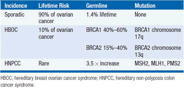
The vast majority of ovarian cancer is sporadic. However, a family history of ovarian cancer increases the risk of developing ovarian cancer two- to threefold, and it is estimated that 5%-15% of all epithelial ovarian cancer cases result from inherited predisposition. Approximately 75% of these families are linked to the breast ovarian cancer syndrome with loss of function of the tumor suppressor genes BRCA1 and BRCA2, involved in DNA repair, and inherited in an autosomally dominant fashion. Hereditary breast-ovarian cancer syndrome (HBOC) accounts for 65%-75% of all hereditary ovarian cancer cases, and is defined variably but typically with at least three cases of early onset (<60 years) breast or ovarian cancer. Mutations in BRCA1 (located on chromosome 17q) and BRCA2 (13q) are associated with a 40%-60% life-time risk of ovarian cancer. The founder mutations 185delAG, and 5382insC in BRCA1, and 6174delT in BRCA2 are present in 2.5% of Ashkenazi women.
Hereditary non-polyposis colon cancer syndrome (HNPCC, Lynch syndrome [type II]) results from mismatch repair (MSH2, MLH1, and PMS2) gene defects, low penetrance allele, and modifier gene defects. The American founder mutation is deletion of exons 1-6 of MSH2. HNPCC accounts for approximately 10%-15% of all hereditary ovarian cancer cases. A diagnosis of HNPCC can be made using the Amsterdam Criteria (colon cancer diagnosis in >3 relatives, one of whom is a first-degree relative of the other two, and one diagnosis before age 50 years). The risk of developing ovarian cancer associated with HNPCC appears to be associated with an approximately 3.7-fold increase in lifetime risk of developing ovarian cancer. The inherited predisposition syndromes are summarized in Table 54-1.
TABLE 54-1 GENETIC PREDISPOSITION SYNDROMES
Women from high-risk families should have genetic counseling and gene testing. At the present time a similar number of women elect biannual pelvic examinations, transvaginal ultrasound, and CA125 with no proven effect on mortality, as we have a prophylactic risk reducing salpingo-oophorectomy (RRSO), which reduces the lifetime risk of ovarian cancer by 95% (3).
BIOLOGY
Although commonly termed “epithelial,” the embryologic origin of ovarian cancer is mesodermal, from the peritoneal surface of the ovary. Previously, subepithelial inclusion cysts that form after ovulation were thought to be the initial focus of dyplastic change, but compelling evidence points to the fimbria of the fallopian tube as the origin of perhaps 50% of tumors. Primary peritoneal and fallopian tube carcinomas (5%) are rare, and behave and are treated in an identical fashion.
Tumor cells predominantly spread transcelomically to the pelvic structures, exfoliating and following the circulation of the peritoneal fluid throughout the abdomen. Cells also metastasize by lymphatic and hema-togenous routes. Extraperitoneal metastases are rare, but with increasing number of patients living for a protracted time, up to two-fifths of patients will develop metastases in the liver, lung, or the central nervous system during the later stages of their disease.
The majority of epithelial cancers are serous or papillary with a similar appearance to the lining of the fallopian tube (4, 5). Endometrioid carcinoma is associated with synchronous endometrial carcinoma and endome-triosis, and therefore more commonly present early, and have a better prognosis. Clear cell tumors have a particularly poor prognosis, are commonly associated with thromboses, and have distinct genetics that link the disease with other clear cell tumors. Mucinous tumors are more commonly bilateral and can be associated with pseudomyxoma peritonei and mucocele of the appendix. Brenner’s tumor has a histologic pattern similar to transitional cell carcinoma. Molecular evidence has confirmed that carcinosarcomas, previously called malignant mixed Mullerian tumors have a clonal proliferation of the epithelial element rather than being collision tumors and are treated as epithelial cancers. In women under the age of 40 years, but rarely in older patients, 60%-70% of non-benign ovarian neoplasms are borderline tumors, a neoplasm that has distinctly malignant cytological features without invasion. Ovarian tumor pathology is summarized in Table 54-2.
TABLE 54-2 OVARIAN CANCER PATHOLOGY

NON-EPITHELIAL OVARIAN MALIGNANCIES
Non-epithelial ovarian cancer accounts for <10% of ovarian tumors. The commonest of these, germ cell tumors represent almost 70% of ovarian tumors in the first two decades of life, with one-third of these being malignant. These rare tumors disseminate early and the doubling time of the tumor can be very short. The tumor markers βHCG and αFP are important in the treatment and surveillance of germ cell tumors. Dysgerminoma, essentially the female equivalent of testicular seminoma, is exquisitely sensitive to platinum, and this is now favored over the traditional treatment of radiotherapy. Teratoma is probably most effectively treated with combination platinum-based chemotherapy such as BEP (bleomycin, etoposide, and cisplatin) and surgical removal of residual masses. Yolk sac, embryonal, and choriocarcinoma subtypes are rarer, curable, and require specialized care.
Sex-cord-stromal tumors account for even fewer ovarian malignancies. Granulosa cell tumors are typically relatively low grade, secrete estrogen and in juveniles, tumors may be associated with pseudo-precocity. The tumors secrete estradiol, Mullerian inhibiting substance, and dimeric inhibin, which serve as tumor markers. Sertoli-Leydig tumors are associated with the production of androgens. Thecomas and fibromas are rare and nonfunctional tumors.
EPITHELIAL OVARIAN MALIGNANCIES
 DIAGNOSIS
The presentation of ovarian cancer is typically subtle, persistent (>2 weeks) abdominal or pelvic pain; bloating, nausea, change in bowel habit, as well as urinary and constitutional symptoms are typical of peritoneal involvement by advanced disease while early stage ovarian cancer is generally asymptomatic (Table 54-3). Unfortunately, most symptomatic women do not have a prompt diagnosis, and indeed it is not uncommon for these women to be misdiagnosed with irritable bowel syndrome, hiatal hernia, diverticu-losis, or endometriosis. Pelvic examination remains an essential part of the examination of women complaining of abdominal symptoms.
TABLE 54-3 CLASSICAL PRESENTATIONS

The diagnosis and initial management of ovarian cancer is surgical (Figure 54-1). Premenopausal women with a smooth, unilateral, mobile, cystic adnexal masses up to 8 cm can be managed conservatively with the use of oral contraceptives and serial ultrasound, typically performed at 6 weeks and at a different time of the menstrual cycle, as luteal cysts are common. In postmenopausal women, cystic masses larger than 5 cm in diameter warrant a tissue diagnosis. In postmenopausal women with a CA125 greater than 35 U/ml, adnexal masses are malignant in 80% of cases, although this should not be confused with the finding of a raised CA125 in an asymptomatic postmenopausal woman, as perhaps only 1 in 7 will have ovarian cancer. Occasionally there is uncertainty about the site of the primary and colonoscopy and mammography are appropriate. As early disease is typically asymptomatic, over 75% of tumors present as advanced stage disease (International Federation of Gynecology and Obstetrics [FIGO]) stage III/IV) (Table 54-4).

Figure 54-1 Advanced ovarian cancer. (Courtesy of Dr. Arlan Fuller.)
TABLE 54-4 OVARIAN CANCER STAGING (FIGO 1987)

The surface glycoprotein CA125 (gene MUC-16) is found at elevated levels in the blood of >80% of patients with epithelial ovarian cancer (abnormal >35 u/ml) (6). CA125 is not useful in screening for occult carcinoma but can be used as a tumor marker. Human epididymis protein 4 (HE4) is another marker elevated in ovarian cancer and when both markers are elevated, adnexal masses are more often malignant. High levels of both markers are associated with disease bulk. Changes in levels correlate with response to therapy or disease progression. The OVA1TM diagnostic is the first FDA-approved blood test to evaluate the likelihood that an ovarian mass is malignant using 5 tumor markers including CA125.
The differential diagnosis of adnexal masses includes simple hemorrhagic physiologic cysts (follicular or corpus luteal); endometrioma; theca luteal cysts; benign, malignant, or metastatic tumors; and extraovarian masses such as paraovarian or peritoneal cysts and pedunculated fibroids, extopic pregnancy, hydrosalpinx, tuboovarian, diverticular, or appendiceal abscesses.
SCREENING
Screening for ovarian cancer is an attractive proposition because of the significant morbidity and mortality associated with advanced disease, the availability of apparently sensitive and specific diagnostic tests (ultrasound and CA125), and the better outcome for patients with early-stage disease. However, there is no evidence at the present time that screening improves survival. The serum marker CA125 has been shown to be elevated in only 50% of patients with stage I disease, but in 90% of stage II-IV ovarian cancers. CA125 has a sensitivity of 20%-58% and a specificity of 97%-99%. With a 1/70 lifetime risk, ovarian cancer is present in 1/2000 postmenopausal women and therefore the false-positive rate (1%-3%) for CA125 is unacceptably high, given the morbidity of laparotomy. In the largest study reported to date in 21,935 UK postmenopausal healthy women, the death rate was apparently halved by screening (18 of 10,977 vs. 9 of 10,958). However, this was not statistically significant (p = 0.083), and the massive U.S. study, Prostate, Lung, Colorectal, and Ovary Cancer Screening Trial (PLCO), in 150,000 subjects failed to confirm a survival advantage, with a 15% serious complication rate from laparotomy (7). There continues to be speculation about whether the sensitivity of screening can be improved by the use of a panel of markers such as HE-4, ROCA (Risk of Ovarian Cancer Algorithm), which evaluates change in CA125 over time, or proteinomics. At the present time, screening is only recommended in high-risk populations (+ve family history or BRCA carriers), where prophylactic surgery has a far greater impact on the risk.
MANAGEMENT OF EARLY STAGE OVARIAN CANCER
Approximately 2000-3000 patients a year in the United States will have disease confined to pelvis. While this group accounts for only 15% of ovarian cancer cases, more than half of all cured patients come from this group. Adequate staging requires that a surgeon with subspeciality expertise perform an exploratory laparotomy, total abdominal hysterectomy and bilateral salpingo-oophorectomy, omentectomy, careful examination of the peritoneal surfaces of the liver, diaphragm, pericolic gutters, and pelvic sidewalls with multiple biopsies and sampling of ascitic fluid and peritoneal washings with para-aortic and pelvic lymph node sampling. Patients diagnosed with apparently early-stage ovarian cancer without adequate staging should undergo reexploration for definitive staging.
The overall 5-year survival of patients with apparent stage I epithelial cancer was about 60% in earlier reports. With accurate staging, and migration of patients with clinically occult nodal or omental metastases to stage III, survival of stage I patients is now commonly reported as >90%. Approximately 30%-46% of cancers that appear to be confined to the pelvis (stages I and II) have occult metastatic disease in the upper abdomen or lymph nodes (stage III). In patients with stage IA and IB disease of low grade (well differentiated), no adjuvant treatment is warranted. For all other patients with high-risk histology or grade, adjuvant treatment is recommended with platinum-based chemotherapy based on Gynecologic Oncology Group (GOG-95), International Collaboration in Ovarian Neoplasia (ICON I), and the European Organization for the Research and Treatment of Cancer (EORTCs) ACTION study (8).
GOG-157 compared 3 with 6 cycles of carboplatin and paclitaxel in early-stage disease. While more cycles were associated with more toxicity, there was a survival advantage in serous tumors and so most patients still receive 6 cycles of chemotherapy if tolerated.
MANAGEMENT OF ADVANCED STAGE OVARIAN CANCER
The principle of therapy for patients with advanced ovarian cancer is to cytoreduce (debulk) with surgery and chemotherapy to a state of minimal residual disease. For some patients this will translate into cure, but for the majority of patients, it delays symptomatic relapse. Current standard of care has been defined as cytoreductive surgery and 6 cycles of a taxane with either cisplatin or carboplatin chemotherapy. Five-year survival rates for patients treated with platinum-based regimens are approximately 20%-40%. Figure 54-2 illustrates a treatment algorithm for advanced ovarian cancer.
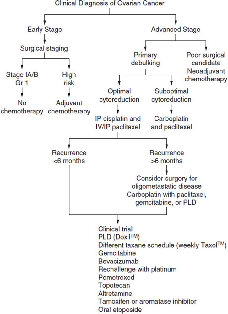
FIGURE 54-2 Treatment algorithm for ovarian cancer.
CYTOREDUCTIVE SURGERY
Meigs and Griffith are credited with the concept that successful surgical debulking to a residual tumor size of ≤1.5 cm maximum diameter results in superior survival, and that the volume left after surgery is more predictive of survival than the volume that enters the OR. More contemporary studies now strive for even smaller tumor volumes (<1 cm) called “optimal cytore-duction.” Surgical cytoreduction has two main advantages. First, it improves physiology with the immediate correction of the functional disorders and pain associated with bowel and ureteral obstruction, also improving the profound protein loss associated with exudative ascites. Second, optimal debulk-ing can remove 90% to many logs of tumor cells, thereby reducing the volume of residual disease to be treated by systemic or intraperitoneal chemotherapy. In theory cytoreduction may remove de novo chemotherapy-resistant clones and facilitate drug delivery, having removed tumor with a compromised blood supply. Multiple retrospective studies have demonstrated that patients with tumors that can be cytoreduced to minimal residual disease survive longer. However, this may simply reflect the biology of the tumor.
Van der Burg reported a 33% reduction in the risk of death (p = 0.008) in a phase III study of interval cytoreduction (after #3 cycles of chemotherapy) in patients with initially suboptimally cytoreduced disease (>1 cm). However, this advantage cannot be replicated if the initial attempt at deb-ulking was performed by a gynecologic oncologist who has already made a maximal attempt at cytoreduction (9). Standard of care is an attempt at deb-ulking by a gynecologic oncologist in every patient with advanced disease.
NEOADJUVANT CHEMOTHERAPY AND INTERVAL CYTOREDUCTIVE SURGERY
There is now level I evidence that supports equivalent survival and less morbidity when surgery is delayed until after #3 of primary or “neoadjuvant” chemotherapy. Vergote randomized 632 patients with bulky (>5 cm in 75% and >10 cm in 62%) stage IIIC or IV epithelial ovarian carcinoma, fallopian-tube carcinoma, or primary peritoneal carcinoma to primary debulking surgery followed by platinum-based chemotherapy or to neoad-juvant platinum-based chemotherapy followed by interval debulking surgery (10). Optimal debulking (to <1 cm) was achieved in 42% with primary debulking and in 82% at interval debulking. Postoperative rates of adverse effects and mortality were higher after primary debulking, but survival was the same in both groups (p = 0.01 for noninferiority). Complete resection of all macroscopic disease (at primary or interval surgery) was the strongest independent variable in predicting overall survival.
FIRST-LINE CHEMOTHERAPY
Chemotherapy plays an essential role in advanced stage ovarian cancer. Over the last 40 years, tumor response rates (shrinkage of the tumor by more than 50%) have increased from the 20% range with single agent mel-phalan used in the 1970s to 75% in recent studies using platinum with a taxane. Although overall survival has improved very little, median survival time has significantly increased over the last few decades, and the overall median survival of patients with suboptimally debulked advanced stage disease has nearly tripled over the past quarter century, coincident with an almost fourfold improvement in chemotherapy response rates. While some of this benefit is certainly due to improvements in supportive care and improved surgical staging, the primary reason for this improvement is modern chemotherapy, which is both less toxic and more effective. Following the model of Hodgkin’s disease, combination therapy logically followed single agent therapy with cyclophosphamide-based combinations, which produced response rates of approximately 35%. In the 1980s, cisplatin and subsequently carboplatin, non-classical alkylators, were demonstrated to have very significant activity in ovarian carcinoma with response rates of approximately 60%, thus redefining the combinations of chemotherapy used for advanced disease, with a significant survival advantage proven in 1986. Meta-analysis of randomized trials prior to 1991 concluded that cisplatin regimes were superior to non-cisplatin regimes, cisplatin combinations were superior to cisplatin alone, and cisplatin and carboplatin were equally effective.
Paclitaxel (TaxolTM), initially derived from the Western Pacific yew tree (Taxus brevofolia), inhibits microtubule depolymerization and demonstrated significant activity in patients with ovarian cancer refractory to platinum chemotherapy. Following the introduction of pre-medication that prevented a hypersensitivity reaction, McGuire reported a response rate of 24% using paclitaxel in heavily pretreated patients whose tumors were resistant to platinum, and reported a substantial survival advantage replacing cyclophosphamide with paclitaxel, in 410 randomly assigned women with suboptimally debulked advanced ovarian cancer (GOG-111). Cisplatin 75 mg/m2 and paclitaxel 135 mg/m2 over 24 h was associated with more alopecia, neutropenia, fever, and allergic reactions, but improved median overall survival from 24 to 38 months (p < 0.001) (11). This result was confirmed by OV10, which also demonstrated that combining a 3-h infusion of 175 mg/m2 paclitaxel with cisplatin produced unacceptable neurotoxicity. Two subsequent studies GOG-132 and ICON II suggested that platinum alone or sequential platinum and paclitaxel may be equally effective. The most common standard of care was defined in GOG-158, which compared cisplatin and paclitaxel with carboplatin and paclitaxel. Six cycles of carboplatin AUC 7.5 and paclitaxel 175 mg/m2over 3 h was a convenient outpatient regimen, which produced less gastrointestinal, renal, and metabolic toxicity, leukopenia, and a similar degree of peripheral neuropathy with a median overall survival of 57 months. As this regimen is associated with greater thrombocytopenia, the Area under the Concentration (AUC) Time Curve dosing, based on renal function, is now typically targeted at 5 or 6, and based on the Calvert formula (Total dose [mg] = target AUC [mg/ml/min] x [CrCl + 25] [ml/min]). Many studies have explored dose intensity as a strategy to improve outcomes, but moderate increases in platinum dose, even those that can be achieved by “high dose” chemotherapy, or more cycles of chemotherapy (10 vs. 5, or 12 vs. 6) do not improve survival.
The SCOTROC study demonstrated that docetaxel (TaxotereTM) was significantly less neurotoxicity than paclitaxel and equally effective in combination with carboplatin, and is a valid but less used popular alternative to paclitaxel. Dose-dense paclitaxel (weekly therapy at 80 mg/m2) with car-boplatin is associated with more anemia but has been reported to improve overall survival, and confirmatory studies are ongoing. Adding agents or replacing paclitaxel with pegylated liposomally encapsulated doxorubicin (DoxilTM), gemcitabine, or topotecan as first-line therapy has been widely investigated but have not improved cure rates.
CONSOLIDATION
In patients with suboptimally debulked disease, who are destined to have relapse, maintenance therapy is a rational strategy. GOG-178 compared monthly paclitaxel for 3 months to the same treatment for 12 months. Time to recurrence was delayed by 7 months with longer duration maintenance but the regimen had excess neurotoxicity. Consolidation with other agents such as topotecan have been shown not to improve survival.
GOG-218 and ICON-7 investigated the integration of bevacizumab (Avastin™) in the upfront treatment of advanced ovarian cancer administered with carboplatin and paclitaxel and as consolidation. Bevacizumab is a particularly promising agent as the response rate is highest in recurrent ovarian cancer compared with any other solid tumor. GOG-218 demonstrated a 3.8-month improvement in PFS, while the European study ICON-7, which had a similar design, reported a PFS of only 1.7 months, suggesting an overall survival in “high-risk” patients, although this remains a very controversial strategy (12, 13).
INTRAPERITONEAL CHEMOTHERAPY
One way to achieve high concentrations of cytotoxics is regional administration. Intraperitoneal (IP) infusion lends itself to this approach with a very high ratio of IP drug concentration that bathes the tumor compared with systemic concentrations (platinum 10×, paclitaxel 1000×).
Alberts initially reported a randomized trial of cyclophosphamide intravenously with either intraperitoneal or intravenous cisplatin (GOG-104), with an 8-month median survival advantage. Markman reported a second phase III study (GOG-114), which included intravenous paclitaxel with intraperitoneal cisplatin, but also included two initial cycles of moderate dose carboplatin as “medical” cytoreduction. The survival was extended 11 months but at a cost of greater toxicity. The third study (GOG-172) led to an NCI alert about the potential advantage of intraperitoneal therapy in patients with optimally debulked ovarian cancer because of an unprecedented 16-month, the longest ever reported, survival advantage (14). Armstrong’s regimen of paclitaxel 135 mg/m2 over a 24-h period (to reduce neurotoxicity) followed by intraperitoneal cisplatin 100 mg/m2 on day 2 with intraperitoneal paclitaxel 60 mg/m2 on day 8 given every 3 weeks for 6 cycles was associated with more fatigue, hematologic, gastrointestinal, metabolic, and neurologic toxicity, with significantly worse quality of life, but an improvement in median duration of overall survival from 50 to 66 months (p = 0.03). The biggest concerns are about catheter complications (infection, pain, and blockage), which are serious in a quarter of patients and prevented 58% of patients completing intraperitoneal therapy in GOG-172. Uptake of IP therapy has been dependent on local expertise.
RECURRENT DISEASE
The majority of patients who achieve a complete remission with first-line platinum-based chemotherapy will ultimately develop recurrent disease. Recurrent ovarian cancer is often considered a chronic disease, since active chemotherapy agents allow patients to live for years with their disease, and quality of life is one of the most important considerations. The definition of relapse is important as a rising CA125 typically has a lead-time of 2-6 months before symptoms develop. Patients with an asymptomatic rising marker can be managed expectantly, as palliative chemotherapy has toxicities. Rustin randomized clinicians and 529 patients to follow up with access to, or blinded to, CA125, and an elevation in marker led to treatment 5 months earlier that did not impact overall survival but did compromise quality of life (15). Treating an asymptomatic patient with a rising CA125 is too early, and waiting until they have bulk disease (>5 cm), which is associated with a poorer response to chemotherapy, may be too late.
The most important factor associated with poor prognosis at time of relapse is a short disease- and platinum-free interval. Platinum-resistant disease is arbitrarily defined as disease that relapses within 6 months of platinum, and platinum-sensitive disease after more than 6 months. Two-thirds of recurrences in the United States fall into this group; localized (oligometastatic on PET/CT scan) disease may be appropriate for surgery, which is being evaluated in GOG-213 and DESKTOP-III, and the role of surgery for recurrence remains controversial.
Rechallenge with a platinum-based combination is appropriate with a platinum-free interval of at least 6 months or a year. ICON 4 (carboplatin and paclitaxel) demonstrated an 18% reduction in risk of death (an absolute difference in 2-year survival of 7% [57 vs. 50%], p = 0.02) (16). Meanwhile, the CALYPSO study (carboplatin and pegylated liposomally encapsulated doxorubicin [PLD or DoxilTM]) and the AGO studies (carboplatin and gemcitabine) reported only PFS survivals. Concurrent bevacizumab with carboplatin and gemcitabine, followed by consolidation bevacizumab, in the OCEANS study was associated with a particularly long PFS advantage, but it is not clear that this will translate into a survival advantage.
The role of bevacizumab is controversial with no clearly proven survival advantage. However, bevacizumab is very effective against ascites. Toxicities in patients with recurrent disease have included hypertension, proteinuria, as well as arterial thromboses, and one study was halted with 5 of 44 patients developing bowel perforations. Bevacizumab is commonly given as monotherapy or in combination with weekly paclitaxel or metronomic low-dose oral cyclophosphamide.
Subsequent recurrences are typically treated with sequential single agent palliative chemotherapy. There are many options such as pegylated liposomally encapsulated doxorubicin hydrochloride (PLD or Doxil™), topotecan, a different taxane schedule (weekly Taxol™), rechallenge with platinum, gemcitabine, altretamine, or oral etoposide. Hormonal therapy, often tamoxifen, can be effective in ER +ve tumors. Many patients are appropriate for clinical trials, and an exciting number of agents are being investigated.
Obstructive symptoms typically herald the last chapter of patients’ lives. The constellation of difficult-to-treat symptoms requires multiprofessional care. Surgery should be limited to patients with chemotherapy-responsive disease, and for others a gastric venting tube (G-tube) alleviates vomiting. Total parenteral nutrition does not substantially alter the clinical course. Steroids and Otreotide™ may provide symptom relief. Attending to end of life issues is a vital part of holistic care.
NOVEL APPROACHES
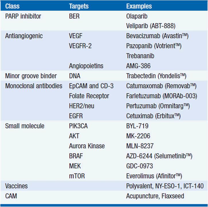
Genetic abnormalities underlie the development and progression of cancer. Tumors are increasingly recategorized by gene mutation or pathway activation, such as BRAF in low-grade serous tumors, which when treated with targeted therapy may be associated with better outcomes than chemotherapy. Patients with inherited BRCA mutations have “synthetic lethality” in that further inhibition of DNA repair with a PARP inhibitor triggers apoptosis and a response in approximately a third of these tumors. PIK3CA looks to be an important target in clear cell and endometrioid tumors. Table 54-5 lists some exciting new agents in late stages of development, with the hope that the impact of novel biologics will more than match the improvement in outcomes with radical surgery and the introduction of platinum.
TABLE 54-5 NOVEL THERAPIES IN CLINICAL TRIALS FOR EPITHELIAL OVARIAN CANCER
PROGNOSIS
With modern surgical cytoreduction followed by as little as 18 weeks of chemotherapy, overall survival rates at 5 years exceed 40%. Stage, grade, histologic subtype, age, and whether the patient can be optimally surgically cytoreduced predict prognosis. Although the ultimate long-term prognosis of patients with advanced disease remains poor, current therapy often provides excellent palliation for these women for many years (Table 54-6). Figure 54-3 illustrates the overall survival of patients with ovarian cancer.
TABLE 54-6 KEY POINTS
1. The presentation of ovarian cancer is often subtle.
2. A strong family history of breast or ovarian cancer should prompt consideration of gene testing and risk reducing salpingo-oophorectomy.
3. Surgical staging and cytoreduction is essential in the successful management of ovarian cancer and should be undertaken by a trained Gynecologic Oncologist.
4. Combination taxane and platinum chemotherapy is standard and should be delivered intraperitoneally in patients with optimally cytoreduced disease.
5. Chemotherapy provides effective palliation with an increasing number of therapeutic options.
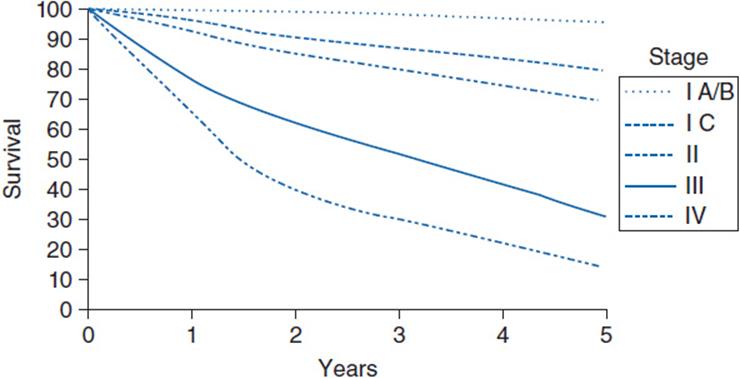
FIGURE 54-3 Overall survival for patients with ovarian cancer.
REFERENCES
1. Cannistra S, McGuire W. Progress in the management of gynecologic cancer. J Clin Oncol. 2007: 2865–2866.
2. Siegel R, Naishadham D, Jemal A. Cancer statistics, 2013. CA Cancer J Clin. 2013; 63: 11–30.
3. Kauff ND, Satagopan JM, Robson ME, et al. Risk-reducing salpingo-oophorectomy in women with a BRCA1 or BRCA2 mutation. N Engl J Med. 2002; 346: 1609–1615.
4. Levanon K, Crum C, Drapkin R. New insights into the pathogenesis of serous ovarian cancer and its clinical impact. J Clin Oncol. 2008; 26: 5284–5293.
5. Young RH, Scully RE. Differential diagnosis of ovarian tumors based primarily on their patterns and cell types. Semin Diagn Pathol. 2001; 18: 161–235.
6. Bast RC Jr, Klug TL, St John E, et al. A radioimmunoassay using a monoclonal antibody to monitor the course of epithelial ovarian cancer. N Engl J Med. 1983; 309: 883–887.
7. Buys SS, Partridge E, Black A, et al. Effect of screening on ovarian cancer mortality: the Prostate, Lung, Colorectal and Ovarian (PLCO) Cancer Screening Randomized Controlled Trial. JAMA. 2011; 305: 2295–2303.
8. Colombo N, Guthrie D, Chiari S, et al. International Collaborative Ovarian Neoplasm trial 1: a randomized trial of adjuvant chemotherapy in women with early-stage ovarian cancer. J Natl Cancer Inst. 2003; 95: 125–132.
9. van der Burg ME, van Lent M, Buyse M, et al. The effect of debulking surgery after induction chemotherapy on the prognosis in advanced epithelial ovarian cancer. Gynecological Cancer Cooperative Group of the European Organization for Research and Treatment of Cancer. N Engl J Med. 1995; 332: 629–634.
10. Vergote I, Trope CG, Amant F, et al. Neoadjuvant chemotherapy or primary surgery in stage IIIC or IV ovarian cancer. N Engl J Med. 2010; 363: 943–953.
11. McGuire WP, Hoskins WJ, Brady MF, et al. Cyclophosphamide and cisplatin compared with paclitaxel and cisplatin in patients with stage III and stage IV ovarian cancer. N Engl J Med. 1996; 334: 1–6.
12. Burger RA, Brady MF, Bookman MA, et al. Incorporation of bevacizu-mab in the primary treatment of ovarian cancer. N Engl J Med. 2011; 365: 2473–2483.
13. Perren TJ, Swart AM, Pfisterer J, et al. A phase 3 trial of bevacizumab in ovarian cancer. N Engl J Med. 2011; 365: 2484–2496.
14. Armstrong DK, Bundy B, Wenzel L, et al. Intraperitoneal cisplatin and paclitaxel in ovarian cancer. N Engl J Med. 2006; 354: 34–43.
15. Rustin GJ, van der Burg ME, Griffin CL, et al. Early versus delayed treatment of relapsed ovarian cancer (MRC OV05/EORTC 55955): a randomised trial. Lancet. 2010; 376: 1155–1163.
16. Parmar MK, Ledermann JA, Colombo N, et al. Paclitaxel plus platinum-based chemotherapy versus conventional platinum-based chemotherapy in women with relapsed ovarian cancer: the ICON4/AGO-OVAR-2.2 trial. Lancet. 2003; 361: 2099–2106.