
Patrick W. Burke
Definition
• Monoclonal gammopathy of undetermined significance (MGUS): Premalignancy w/clonal plasma cell (PC) proliferation. No e/o other B-cell disease. <10% monoclonal PCs in BM, <3 g/dL serum M-protein, & no MM-related organ dysfunction s/s.
• Multiple myeloma (MM): Malignant neoplasm of clonal PC. Almost always in BM plus Ig or light chain production → M-protein in blood/urine. Arises from MGUS.
• Plasma cell Leukemia: Rare variant of MM (2–4% of MM) w/↑ aggressive course. Leukemic phase of MM → either 1° or 2° (antecedent MM). ↓↓ OS.
• Related organ or tissue impairment (ROTI): CRAB. C = ↑ Ca (>11.5 mg/dL); R = Renal insufficiency (Cr > 2 mg/dL);
A = Anemia (Hb < 10 g/dL or 2 g/dL < nl); B = Bone disease (lytic lesion/pathologic fx/sev. osteopenia). Also consider recurrent infxn, ↑ viscosity, & amyloidosis.

Epidemiology
• MGUS: ↑ incidence w/age & in Western countries → 3% of adults >50 y. 7.5% in adults >85 y. ∼0.5–3% per y progress to MM.
• MM: 1% all neoplasms. 13% of heme malignancies (2nd highest incidence in Western World. ∼5.6/100000/y). 2% of all CA death. U.S. 2012: ∼21700 new Dx. ∼10,710 death. ∼63000 total cases.
Median age ∼65 y. Risk of Asymptomatic MM → Sx MM ∼10–20%/y.
↑ Incidence African-Americans, Afro-Caribbeans, & Pacific Islanders.
Possible familial predisposition. Possible exposure risk (pesticides/herbicide).
Biology/Pathogenesis (N Engl J Med 2011;364:1046)
• Progression: MGUS → Asymptomatic (Smoldering) MM → Sx MM → PC Leukemia Multistep progression of genetic & BM microenvironment changes.
Genetic Abnl → MM PC altered expression of adhesion molecules/responses to microenvironment growth stimuli → ↑ cytokines/GF/cell cycle regulatory proteins/
antiapoptotic proteins → ↑ growth, survival, migration, drug resistance.
• MGUS: Asymptomatic proliferation of monoclonal PC from post-GCB.
CG: 50% hyperdiploid (48–74 chromosomes); 50% nonhyperdiploid.
Translocations at IgH switch region: Chromosome 14(q32.33) → IgH gene enhancers/promoter juxtaposed in proximity to oncogene locus.
↑ Prevalence IgH translocations w/↑ malignancy progression. MGUS → MM.
Three common translocation partners: MAF → t(14;16)(q32.33;q23); MMSET→ t(4;14)(p16;q32.33) (deregulation of MMSET & FGFR3); CCND1 → t(11;14)(q13;q32.33).
Molecular: ↑ expression of cyclins (D1, D2, & D3) & transcription factors.
• MM: CG: Hyperdiploidy & nonhyperdiploid (hypodiploid, near tetraploid, & pseudohypodiploid) & IgH translocations seen.
2° translocations: MYC (8q24), MAFB (20q12), IRF4 (6p25) ↑ MM; rare MGUS.
Del 18p, Del 17p13, Del 1p, amp 1q, Del 13 → only MM.
Molecular: NRAS & KRAS activation; FGFR3 & TP53 Mt; inactivation CDKN2A & CDKN2C in MM.
MM microenvironment: ↑ angiogenesis (↑ VEGF) + bone resorption (↓ Wnt signal → ↓ osteoblast action; ↑ RANK/MIP1α → ↑ osteoclast action).
Presentation
• M-protein: 97%; anemia: 70% (CKD, chronic disease, BM infiltration); bone
lytic lesion/osteopenia/pathologic fx: 80%; Renal impairment: 20–40% (M-Protein → tubular damage; dehydration; ↑ Ca; nephrotoxic meds).
Workup
• Initial H&P, CBC, basic metabolic profile (BMP), LFTs, electrolytes (esp. serum Ca), SPEP, UPEP, quantitative Igs, urine immunofixation (UIF), serum immunofixation (SIF), serum free light chain assay (SFLC), 24-h urine protein quantification, β2-microglobulin, albumin, BM aspirate & Bx (w/IHC, morphology, flow cytometry + CG w/karyotype + FISH), & x-ray skeletal survey. MRI or PET if bone sxs but negative skeletal survey.
• Diff Dx: MGUS, MM, PC leukemia, lymphoplasmacytic lymphoma (LPL)/WM, NHL, 1° amyloidosis, heavy chain deposition disease, light chain deposition disease, cryoglobulinemia, idiopathic cold agglutinin disease.
Staging
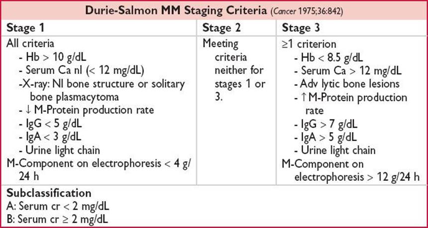
• Durie-Salmon (DS): MM “cell mass” c/w prognosis (response to CT & OS).
5 criteria (anemia, serum Ca, bone disease, M-protein, & serum Cr).
Significantly c/w MM cell mass & prognosis. DS staging performed at Dx.
3 prognostic categories: Low (Stage 1), INT (Stage 2), & High (Stage 3).
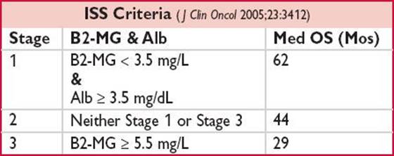
• International Staging System (ISS): Uses β2 microglobulin (B2-M) and albumin (Alb). Prognosis determined from start of systemic Tx.
Treatment (Tx)
• MGUS & Asymptomatic MM: No Tx required. Monitor q3–6mos w/MD visit & labs.
• Solitary osseous plasmacytoma: IFRT at ≥4500 cGy.
• Solitary extraosseous plasmacytoma: IFRT at ≥4500 cGy ± excision.
• Symptomatic MM: Tx indicated. Potential Tx algorithm below. Transplant vs. non transplant eligible dictates Tx choice. Avoid alkylators prior to stem cell collection (for possible two transplants). Differing RR w/diff CG & molecular. t(4;14) & p53 del/Mt a/w ↓ OS. Immunomodulators (IMIDs; thalidomide/lenalidomide) & proteasome inhib (bortezomib) used w/HDT → ASCR have ↑↑ RR, TTP, PFS, & OS. Conflicting results in ASCR followed by nonmyeloablative allogeneic HSCT (N Engl J Med 2007;356:1110; IFM 99–03; BMT CTN0102).

Figure 24-1 Palumbo A, et al. N Engl J Med 2011;364:1046.
• Post high-dose therapy → autologous stem cell rescue (ASCR) maintenance: Older studies w/steroids & melphalan →
↑ tox & ↑↓ efficacy. Recent/ongoing trials w/bortezomib, thalidomide, & lenalidomide. Lenalidomide a/w ↑ TTP, PFS, & event-free survival (EFS). Possible ↑ OS. A/w ↑ 2nd 1° malignancies (MDS/AML, HL, solid tumors). (CALGB 100104 N Engl J Med 2012;366:1770 & IFM 05–02 N Engl J Med 2012;366:1782). Esp. as immediate maintenance post-HD alkylator → ASCR.
• Adjunctive/Supportive Tx: Anemia: ESA if no response w/CT. Bone pain:
Analgesia (avoid nephrotoxins), steroids, IFRT, bisphosphonates (zoledronate w/↑ OS c/w clodronate in MRC Myeloma IX) (Lancet 2010;376:1989), bortezomib (↓ bone resorption independent of anti-PC activity) vertebroplasty, kyphoplasty. Infxn ppx: Vaccination, antimicrobials (consider antiviral, -pneumocystis, -bacterial, -fungal), IVIg, granulocyte colony stimulating factor (G-CSF). Thrombo-ppx: W/IMIDs (ASA vs. ppx anticoagulation).
↑ Viscosity: Plasmapheresis. ↑ Ca: Hypercalcemia Tx. Renal disease: IVF, Ca control, uricemia control, avoid nephrotoxins/IV contrast, plasmapheresis, HD, & Tx to ↓ paraprotein/amyloid.
Investigational: Carfilzomib (proteasome inhib) & Pomalidomide (IMID).
Current investigation of HDT → ASCR → T-cell depleted Allogeneic HSCT w/RIC Regimen in High-risk Pts.