
Parisa Momtaz and Lee M. Krug
Definition
• Malignant proliferation of the mesothelial cells that make up the lining of the pleura
• Note: Mesothelioma can also occur in lining of other sites (pleura most common 85% peritoneum 15%, pericardium & tunica vaginalis testis rare <1%)
Epidemiology/Risk Factors
• Rare; 2500 new cases dx annually in US
• Commonly develop in the 5th to 7th decade
• Men: Women ratio 4:1
• 70% are a/w asbestos exposure. Typically occupational (steam fitters, insulators, shipbuilders, brake mechanics) or environmental. However, approximately 5–10% of asbestos miners are dx w/mesothelioma
• Develops decades after asbestos exposure
• Other associations: Radiotherapy, erionite (mineral found in gravel roads), genetics
• Smoking does not ↑ the risk of mesothelioma, though it does markedly ↑ the risk of lung CA together w/asbestos exposure
Clinical Manifestations
• Most commonly p/w unilateral pleural effusion; other sx include dyspnea, cough, CP, CW mass, or other symtomatic 2° to local invasion
• Constitutional sx: Fever, wt loss, diaphoresis
• Distant mets to the contralateral lung, peritoneum, bone, or liver
Diagnostic Evaluation
• CT chest w/contrast
• Thoracentesis for cytologic assessment. Note: Cytologic samples are typically non diagnostic
• Pleural bx (thoracoscopic bx preferred)
• Serum marker SMRP level may be useful for monitoring response to tx (JCO 2010;28:3316)
• Following confirmation of mesothelioma workup includes: CT C/A/P w/contrast, PET-CT, mediastinoscopy or EBUS FNA of mediastinal LN, VATS and/or laparoscopy if suspicion of contralateral or peritoneal disease
Pathology
• Histologic subtypes: Epithelioid (most common 80%), biphasic or mixed, sarcomatoid
• Useful IHC markers noted below; of note TTF-1 & CEA w/c are negative in pleural mesothelioma are typically positive in adenoca of the lung

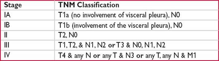
Staging (International Mesothelioma Interest Group [IMIG])
• Stage I/II: No nodal involvement
• Stage I divided into IA (T1a: No involvement of the visceral pleura) & IB (T1b: Involvement of the visceral pleura)
• Stage III: Nodal involvement (N1, N2) or locally adv resectable tumor w/limited ipsilateral extension (T3)
• Stage IV: Locally adv unresectable disease w/direct extension to surrounding ipsilateral or contralateral organs (T4), contralateral nodal involvement (N3), or met disease (M1)
Treatment
• Pre-tx workup: PFTs, Perfusion scanning (only if FEV1 <80%), cardiac stress test to determine if surgical candidate
• Trimodality tx is goal; however, a high proportion of pts are unable to complete all 3 tx modalities
• Clinical Trials
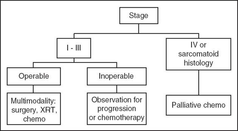
Figure 14-1
Surgery
• Pleurectomy/Decortication (P/D): Complete removal of the pleura & all gross tumor
• Extrapleural pneumonectomy (EPP): en bloc resection of the pleura, lung, ipsilateral Diaphragm, & often pericardium
• Both P/D & EPP should be performed w/mediastinal LN sampling (goal to obtain 3 nodal stations)
• Choice of P/D vs. EPP is controversial; EPP is a/w significant morbidity & mortality; P/D may not offer complete resection (J Thorac Cardiovasc Surg 2008;135:620)
RT
• Hemithoracic RT ↓ local recurrence after EPP (J Thorac Cardiovasc Surg 2001;122:788)
• May be used to treat surgical sites prophylactically to ↓ tracking through CW
• May be used for palliation to treat areas of CW invasion
Chemotherapy
• 1st line, gold standard: Peme (Alimta)/CIS (JCO 2003;21:2636)
• Carboplatin may be substituted for CIS (JCO 2006;20:1443)
• 2nd-line Rx has unclear benefit
Supportive Care
• Talc pleurodesis, pleural catheter, pain control
Prognosis
• Cure is rare; high risk of local & distant recurrence
• Median survival: 6–18 mos
• Epithelioid subtype has a better outcome
• Sarcomatoid subtype extremely poor prognosis, rarely responds to chemotherapy
• Screening has not been shown to ↓ mortality even in subjects w/asbestos exposure
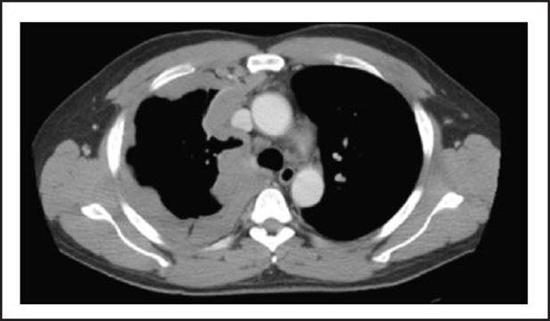
Figure 14-2 Courtesy of L. Krug MD, MSKCC