
William M. Mendenhall, Anthony A. Mancuso, Robert J. Amdur, and John W. Werning
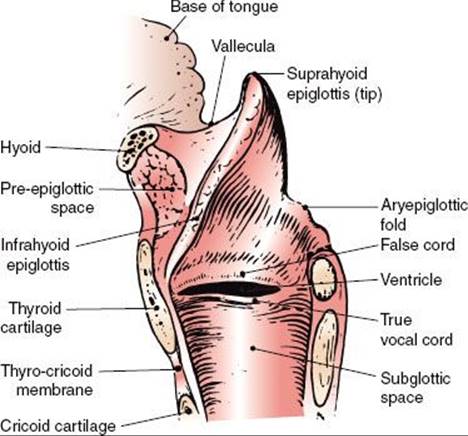
FIGURE 47.1. Diagrammatic sagittal section of the larynx. (Redrawn from Clemente CD. Anatomy: a regional atlas of the human body. Philadelphia: Lea & Febiger, 1975. Copyright Urban & Schwarzenberg, Munich, Germany, 1975.)
 ANATOMY
The larynx is divided into the supraglottis, glottis, and subglottis. The supraglottis consists of the epiglottis, false vocal cords, ventricles, aryepiglottic folds, and the arytenoids. The glottis includes the true vocal cords and the anterior commissure. The subglottis is located below the vocal cords (Figs. 47.1 and 47.2).1
The lateral line of demarcation between the glottis and supraglottic larynx is the apex of the ventricle. The demarcation between the glottis and subglottis is ill defined, but the subglottis is considered to extend from a point 5 mm below the free margin of the vocal cord to the inferior border of the cricoid cartilage or 10 mm below the apex of the ventricle.
The vocal cords vary from 3 to 5 mm in thickness and terminate posteriorly with their attachment to the vocal process. The posterior commissure is the mucosa between the arytenoids.
The shell of the larynx is formed by the hyoid bone, thyroid cartilage, and cricoid cartilage; the cricoid cartilage is the only complete ring. The more mobile interior framework is composed of the heart-shaped epiglottis and the arytenoid, corniculate, and cuneiform cartilages. The corniculate and cuneiform cartilages produce small, rounded bulges at the posterior end of each aryepiglottic fold.
The thyroid and the cricoid cartilages and a portion of the arytenoid cartilage are hyaline cartilage and may partially ossify with age, particularly in men. The epiglottis is elastic cartilage; ossification does not occur, and even focal calcification is rare.2
The external laryngeal framework is linked together by the thyrohyoid, the cricothyroid, and the cricotracheal ligaments or membranes (Figs. 47.3 and 47.4).1
The epiglottis is joined superiorly to the hyoid bone by the hyoepiglottic ligament. The epiglottis is joined to the thyroid cartilage by the thyroepiglottic ligament at a point just below the thyroid notch and above the anterior commissure. The arrangement of the ligaments that connect the cricoid and arytenoid cartilages and form the vocal ligaments, which are part of the true vocal cords, is shown in Figure 47.2B.1 The conus elasticus (cricovocal ligament) is the lower portion of the elastic membrane that connects the inferior framework. It connects the upper surface of the cricoid, the vocal process of the arytenoid, and the lower thyroid cartilage; its free border is thickened into the vocal ligament.
The vocal ligaments and muscles attach to the vocal process of the arytenoid posteriorly and the thyroid cartilage anteriorly. The intrinsic muscles of the larynx, which primarily control the movement of the cords, are presented in Figures 47.2 and 47.3.1 The extrinsic muscles are concerned primarily with swallowing. The cricothyroid muscle produces tension and elongation of the vocal cords and is innervated by the superior laryngeal nerve (Fig. 47.4).1
The pre-epiglottic and paraglottic fat spaces are essentially one contiguous space lying between the external framework of the thyroid cartilage and hyoid bone and the inner framework of the epiglottis and intrinsic muscles. Lam and Wong3 showed that there are thin membranous septa between the paraglottic and pre-epiglottic spaces that are capable of holding a tumor in check to a limited degree. The space is traversed by blood and lymphatic vessels and nerves. Because few capillary lymphatics arise in this area, invasion of the fat space should only indirectly be associated with lymph node metastases. The fat space is limited by the conus elasticus inferiorly, the thyroid ala, the thyrohyoid membrane, the hyoid bone anterolaterally, the hyoepiglottic ligament superiorly, and the fascia of the intrinsic muscles on the medial side. Posteriorly, it is adjacent to the anterior wall of the pyriform sinus.
The laryngeal surface of the epiglottis and the free margin of the vocal cords are squamous epithelium, and the remainder is usually pseudostratified ciliated columnar epithelium. Beneath the epithelium of the free edge of the vocal cord is the lamina propria, which can be divided into three layers. There is no true submucosal layer along the free margin of the vocal fold.4 The laryngeal arteries are branches of the superior and inferior thyroid arteries.
The intrinsic muscles of the larynx are innervated by the recurrent laryngeal nerve. The cricothyroid muscle—an intrinsic muscle responsible for tensing the vocal cords—is supplied by a branch of the superior laryngeal nerve; isolated damage to this nerve causes a bowing of the true vocal cord, which continues to be mobile, but the voice may become hoarse.
The supraglottic structures have a rich capillary lymphatic plexus; the trunks pass through the pre-epiglottic space and the thyrohyoid membrane and terminate mainly in the subdigastric (level II) lymph nodes; a few drain to the middle internal jugular chain (level III) lymph nodes.
There are essentially no capillary lymphatics of the true vocal cords; as a result, lymphatic spread from glottic cancer occurs only if tumor extends to supraglottic or subglottic areas.
The subglottic area has relatively few capillary lymphatics. The lymphatic trunks pass through the cricothyroid membrane to the pretracheal (Delphian) lymph nodes in the region of the thyroid isthmus. The subglottic area also drains posteriorly through the cricotracheal membrane, with some trunks going to the paratracheal (level VI) lymph nodes and others continuing to the inferior jugular (level IV) chain.
FIGURE 47.2. A: Cross section of the larynx at the level of the vocal cords. B: Framework of the larynx. (Redrawn from Clemente CD. Anatomy: a regional atlas of the human body. Philadelphia: Lea & Febiger, 1975. Copyright Urban & Schwarzenberg, Munich, Germany, 1975.)

FIGURE 47.3. Diagram of the coronal view of the larynx. (Redrawn from Clemente CD. Anatomy: a regional atlas of the human body. Philadelphia: Lea & Febiger, 1975. Copyright Urban & Schwarzenberg, Munich, Germany, 1975.)

FIGURE 47.4. External view of the larynx. (Redrawn from Clemente CD. Anatomy: a regional atlas of the human body. Philadelphia: Lea & Febiger, 1975. Copyright Urban & Schwarzenberg, Munich, Germany, 1975.)

EPIDEMIOLOGY AND RISK FACTORS
Cancer of the larynx represents about 2% of the total cancer risk and is the most common head and neck cancer (skin excluded). In 2010 in the United States, there were approximately 12,720 new cases of cancer of the larynx (10,110 men and 2,610 women) and about 3,600 deaths from laryngeal cancer.5 Based on 1973–1998 U.S. data, at diagnosis, about 51% of the cases remain localized, 29% have regional spread, and 15% have distant metastases.6The ratio of glottic to supraglottic carcinoma is approximately 3:1.
Cancer of the larynx is strongly related to cigarette smoking. The risk of tobacco-related cancers of the upper alimentary and respiratory tracts declines among former smokers after 5 years and is said to approach the risk of nonsmokers after 10 years of abstention.7 The role of alcohol in provoking laryngeal cancer remains unclear.8 Some evidence exists that heavy marijuana smoking may be associated with laryngeal cancer in young patients.
PATTERNS OF SPREAD
Local Spread
Although supraglottic and glottic lesions tend to remain confined to their original compartments, there is no anatomic barrier to growth from one area to the next. Glottic lesions tend to be slow growing, but as they increase in size, they extend to the supraglottic and subglottic areas. Supraglottic lesions do not often start near the vocal cords. Involvement of the cords on their external epithelial surface is a late phenomenon, but submucosal extension by way of the paraglottic space occurs earlier.
The fat space is an important avenue of submucosal tumor spread for infrahyoid epiglottis, false cord, and true vocal cord lesions. As the false cord and the true vocal cord lesions penetrate anteriorly and laterally, they quickly encounter the tough perichondrium of the thyroid cartilage and may eventually be shunted by the conus elasticus (lateral cricothyroid membrane) out of the larynx via the cricothyroid space. Thyroid cartilage invasion usually occurs in the ossified section of the cartilage, commonly in the region of the anterior commissure tendon or the junction of the anterior one-fourth and the posterior three-fourths of the thyroid lamina.9
Fixation of the vocal cord from laryngeal cancer is usually caused by invasion or destruction of the vocal cord muscle, invasion of the cricoarytenoid muscle or joint, or, rarely, invasion of the recurrent laryngeal nerve. Perineural spread is uncommon.
Supraglottic Larynx
Suprahyoid Epiglottis
A lesion of the suprahyoid epiglottis may produce a huge exophytic mass with little tendency to destroy cartilage or spread to adjacent structures. Other lesions may infiltrate the tip and destroy cartilage. The destructive lesions tend to invade the vallecula and pre-epiglottic space, the lateral pharyngeal walls, and the remainder of the supraglottic larynx.
Infrahyoid Epiglottis
Lesions of the infrahyoid epiglottis tend to produce irregular tumor nodules and simultaneously invade the porous epiglottic cartilage and thyroepiglottic ligament into the pre-epiglottic fat space and extend toward the vallecula and base of the tongue. The thick hyoepiglottic ligament is an effective tumor barrier. However, the tumor may present in the vallecula and base of tongue without involving the suprahyoid epiglottis.
Lesions of the infrahyoid epiglottis grow circumferentially to involve the false cords, aryepiglottic folds, medial wall of the pyriform sinus, and the pharyngoepiglottic fold. Invasion of the anterior commissure and cords and anterior subglottic extension usually occur only in advanced lesions. Infrahyoid epiglottic lesions that extend onto or below the vocal cords are at a high risk for thyroid cartilage invasion, even if the cords are mobile.10
False Cord
Early false cord carcinomas, which are usually submucosal with little exophytic component, are difficult to delineate accurately. They involve the paraglottic fat space early in their development and may spread a considerable distance beneath the mucosa without producing physical signs. These carcinomas extend to the perichondrium of the thyroid cartilage quite early, but cartilage invasion is a late phenomenon. Extension to the lower portion of the infrahyoid epiglottis and invasion of the pre-epiglottic space are common. Submucosal extension involves the true vocal cord, which may appear normal. Vocal cord invasion is often associated with thyroid cartilage invasion. Submucosal extension to the medial wall of the pyriform sinus occurs early.
Aryepiglottic Fold/Arytenoid
Early lesions of the aryepiglottic fold/arytenoid are usually exophytic. It may be difficult to decide whether the lesion started on the medial wall of the pyriform sinus or on the aryepiglottic fold. As the lesions enlarge, they extend to adjacent sites and eventually cause fixation of the larynx, which is usually a result of involvement of the cricoarytenoid muscle or joint or, rarely, invasion of the recurrent laryngeal nerve. Computed tomography (CT) may distinguish the cause of fixation. Advanced lesions invade the thyroid, epiglottic, and cricoid cartilages and eventually invade the pyriform sinus and postcricoid area.
Glottic Larynx
Most lesions of the true vocal cord begin on the free margin and upper surface of the cord. When diagnosed, about two-thirds are confined to the cords, usually one cord. The anterior portion of the cord is the most common site. Anterior commissure involvement, which is common, is said to occur when no tumor-free cord can be seen anteriorly; if the lesion crosses to the opposite cord, anterior commissure invasion is certain. Small lesions isolated to the anterior commissure account for only 1% to 2% of cases. Extension to the posterior commissure is uncommon, occurring only in advanced lesions.
Tumors at the anterior commissure may extend anteriorly via the anterior commissure tendon (Broyles’ ligament)11 into the thyroid cartilage. Kirchner,12 using whole-organ sections, showed that such extension is unusual unless the tumor extends off the vocal cord onto the base of the infrahyoid epiglottis and suggested that the tendon serves as more of a barrier than an avenue of tumor spread. Early subglottic extension is also associated with involvement of the anterior commissure, and tumor may grow through the cricothyroid membrane.
Lesions that arise on the posterior half of the vocal cord tend to extend along the submucosa toward the medial side of the vocal process and invade the cricoarytenoid joint and posterior commissure; this spread is difficult to appreciate by clinical examination.
Subglottic extension may occur by simple mucosal surface growth, but it more commonly occurs by submucosal penetration beneath the conus elasticus. One centimeter of subglottic extension anteriorly or 4 to 5 mm of subglottic extension posteriorly brings the border of the tumor to the upper margin of the cricoid, exceeding the anatomic limits for conventional hemilaryngectomy. Lesions may spread beneath the epithelium along the length of the vocal cord within Reinke’s space.13
As vocal cord lesions enlarge, they extend to the false cord, vocal process of the arytenoid, and subglottis. Infiltrative lesions invade the vocal ligament and muscle and eventually reach the paraglottic space and the perichondrium of the thyroid cartilage. Advanced glottic lesions eventually penetrate through the thyroid cartilage or via the cricothyroid space to enter the neck, where they may invade the thyroid gland. Lesions involving the anterior commissure often exit the larynx via the cricothyroid space after they extend subglottically.13
A fixed cord that is associated with a lesion having <1 cm of subglottic extension and no false cord involvement does not ordinarily indicate invasion of the thyroid cartilage.12 If the false cord is also involved, cartilage invasion is likely.
Subglottic Larynx
Subglottic cancers are rare. Most involve the inferior surface of the vocal cords by the time they are diagnosed, so it is difficult to know whether the tumor started on the undersurface of the vocal cord or in the true subglottic larynx. Because early diagnosis is uncommon, most lesions are bilateral or circumferential at discovery. They involve the cricoid cartilages in the early stage because there is no intervening muscle layer. Partial or complete fixation of one or both cords is common; misdiagnosis or diagnostic delay is frequent.
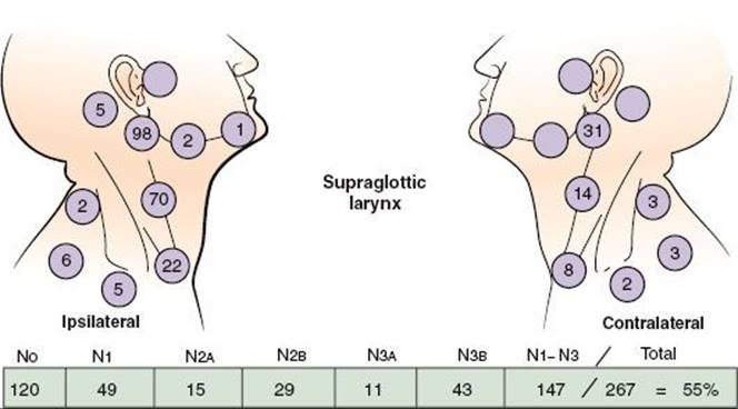
FIGURE 47.5. Nodal distribution on admission, MD Anderson Cancer Center, 1948–1965. (From Lindberg RD. Distribution of cervical lymph node metastases from squamous cell carcinoma of the upper respiratory and digestive tracts. Cancer 1972;29:1446–1449; with permission.)
Lymphatic Spread
The location and stage of neck nodes detected on admission for previously untreated patients with squamous cell carcinoma of the supraglottic larynx are given in Figure 47.5.14 The disease spreads mainly to the level II nodes. The level Ib nodes are rarely involved, and there is only a small risk of level V lymph node involvement. The incidence of clinically positive nodes is 55% at the time of diagnosis; 16% are bilateral.14 Elective neck dissection shows pathologically positive nodes in 16% of cases; observation of initially node-negative necks eventually identifies the appearance of positive nodes in 33% of cases.15,16 Spread to the pyriform sinus, vallecula, and base of the tongue increases the risk of lymph node metastases. The risk of late-appearing contralateral lymph node metastasis is 37% if the ipsilateral neck is pathologically positive, but the risk is unrelated to whether the nodes in the ipsilateral neck were palpable before neck dissection.
The incidence of clinically positive lymph nodes at diagnosis for vocal cord carcinoma approaches zero for T1 lesions and is <2% for T2 lesions.17 The incidence of neck metastases increases to 20% to 30% for T3 and T4 lesions. Supraglottic spread is associated with metastasis to the level II nodes. Anterior commissure and anterior subglottic invasion are associated with involvement of the midline pretracheal lymph node (level VI).
Lederman18 reported a 10% incidence of positive lymph nodes in 73 patients with subglottic carcinoma.
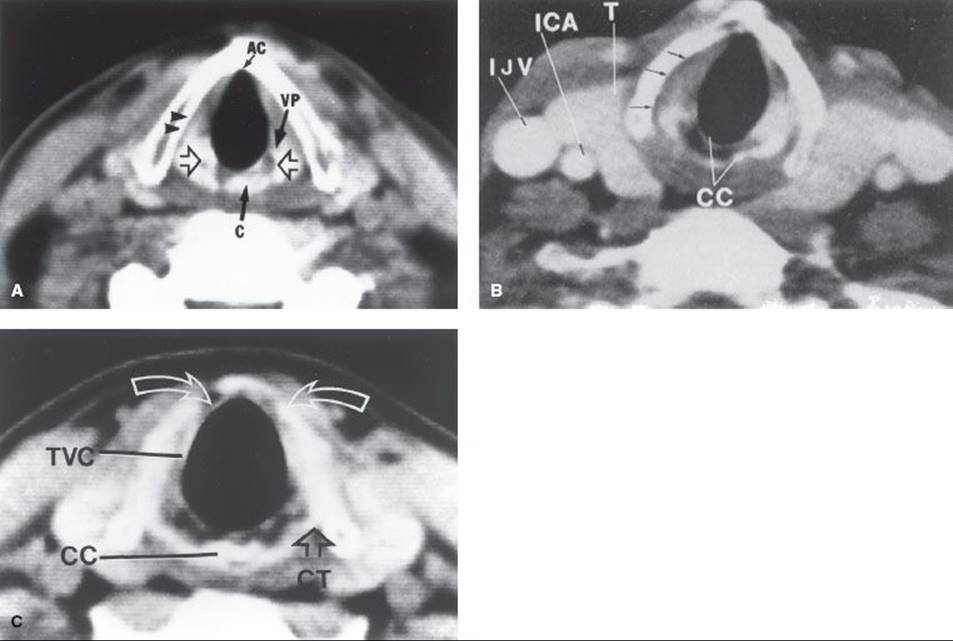
FIGURE 47.6. A: Normal CT anatomy of the midplane of the true vocal cords. Open arrows indicate arytenoid cartilages. The top of the cricoid cartilage (C) is partially visualized at this level. The vocal process (VP) of the left arytenoid cartilage is demonstrated. A narrow, low-density plane is seen between the right true vocal cord and the thyroid lamina (arrowheads); this is the inferior part of the paraglottic fat space. Notice the complete lack of tissue at the anterior commissure (AC). Any tissue density here should be considered abnormal. B: Normal CT anatomy just below the midplane of the vocal cords. Arrows indicate low-density lower paraglottic fat space. The fibrofatty tissue in this space facilitates separation of the vocal cord and the adjacent thyroid lamina. If this clear space is maintained in the face of the thyroid lamina irregularity adjacent to the tumor, the lamina abnormality can be attributed to uneven calcification rather than tumor destruction. The posterior portion (lamina) of the cricoid cartilage (CC) is seen. The outer and inner cortex of the cartilage is calcified; an intervening marrow space has lower density. The vertical height of the lamina is 2 to 3 cm. There is incomplete calcification of the thyroid cartilage anteriorly. ICA, internal carotid artery; IJV, internal jugular vein; T, thyroid gland. C: Normal CT anatomy 5 mm below the free margin of the true vocal cord (TVC). The vocal cord appears thin because of abduction during scanning. There is incomplete bilateral paramedian calcification and thinning of the thyroid lamina (arrows). Notice the normal lack of tissue density between the airway and the anterior arch of the thyroid cartilage. CC, cricoid cartilage; CT, cricothyroid joint. (Million RR, Cassisi NJ. Larynx. In Million RR, Cassisi NJ, eds. Management of head and neck cancer: a multidisciplinary approach. Philadelphia: JB Lippincott, 1984:315–364.)
CLINICAL PRESENTATION
Carcinoma arising on the true vocal cords produces hoarseness at a very early stage. Sore throat, ear pain, pain localized to the thyroid cartilage, and airway obstruction are features of advanced lesions.
Hoarseness is not a prominent symptom of cancer of the supraglottis until the lesion becomes extensive. Pain on swallowing, usually mild, is the most frequent initial symptom, often described as a sore throat. Some patients report a sensation of a “lump in the throat.” Pain is referred to the ear by way of the vagus nerve and auricular nerve of Arnold. A mass in the neck may be the first sign of a supraglottic cancer. Late symptoms include weight loss, foul breath, dysphagia, and aspiration.
DIAGNOSTIC WORKUP
Physical Examination
Flexible fiberoptic endoscopes are used routinely to complement the laryngeal mirror examination. The mirror often provides the best view of the posterior pharyngeal wall. The flexible fiberoptic laryngoscope is inserted through the nose and is useful in more difficult cases.
Determination of vocal cord mobility frequently requires multiple examinations because the subtle distinctions between mobile, partially fixed, and fixed cords are often challenging, apparently changing from examination to examination. A cord that appeared mobile before direct laryngoscopy may exhibit impaired motion or even fixation after biopsy.
Ulceration of the infrahyoid epiglottis or fullness of the vallecula is an indirect sign of pre-epiglottic space invasion. Palpation of diffuse, firm fullness above the thyroid notch with widening of the space between the hyoid and the thyroid cartilages signifies invasion of the pre-epiglottic space. The pre-epiglottic fat space is a low-density area on the CT scan, and changes resulting from tumor invasion are easily seen.
Postcricoid extension may be suspected when the laryngeal click disappears on physical examination. Postcricoid tumor may cause the thyroid cartilage to protrude anteriorly, producing a fullness of the neck.
Invasion of the thyroid cartilage remains a difficult clinical diagnosis. Localized pain or tenderness to palpation or a small bulge over one ala of the thyroid cartilage is suggestive.
Radiographic Studies
CT scan with contrast enhancement is the method of choice for studying the larynx (Fig. 47.6).19 The CT scan should be performed before biopsy so that abnormalities that may be caused by the biopsy are not confused with tumor. CT is preferred to magnetic resonance (MR) imaging because the longer scanning time for MR results in motion artifact.20 CT slices 1 to 2 mm thick are obtained at 1- to 2-mm intervals through the larynx and at 3-mm intervals for the remainder of the study. Thinner sections (1 to 2 mm through the larynx) facilitate high-quality multiplanar reformations. The gantry is angled so that the scan slices are parallel to the plane of the true vocal cords. It is also necessary to obtain a CT scan of the entire neck to detect positive, nonpalpable lymph nodes. Positive retropharyngeal nodes may be present at diagnosis in patients with laryngeal cancer who have advanced neck disease.21Retropharyngeal adenopathy is often not apparent on physical examination but is usually appreciated on CT scan.
Contrast enhancement helps to outline the blood vessels and thyroid gland. Tumor is often enhanced, probably because of reactive inflammatory changes. In addition to CT, MR may be obtained to define subtle exolaryngeal spread or early cartilage destruction. The value of MR for detecting early cartilage destruction is open to speculation. Sagittal MR may be useful in detecting early invasion of the base of the tongue.
Vocal Cord Carcinoma
Although the CT scan does not show minimal mucosal lesions and is generally not helpful for well-defined, easily visualized T1, or early T2 vocal cord carcinomas, it is almost always obtained. CT is excellent for determining subglottic extension and is often used in selected T1 and most T2 lesions for this reason alone. CT scanning is useful in the diagnosis of moderately advanced and advanced lesions; it is excellent for demonstrating extension outside the larynx into the soft tissues of the neck and has potential for determining thyroid or cricoid cartilage invasion, which tends to occur at the edges of the cartilage rather than on the faces. Early cartilage involvement is difficult to detect with axial scans, but it may be demonstrated by coronal or sagittal scanning techniques. If the low-density plane of the paraglottic space is intact, cartilage is probably not invaded by tumor.
Archer et al.22 correlated CT findings with the incidence of cartilage or bone invasion on whole-organ sections. For 12 of 14 patients with pathologic evidence of cartilage invasion, the average diameter of the tumor in two dimensions was >16 mm, and the lesion was located below the top of the arytenoid. Lesions in which the maximum diameter lay above the top of the arytenoid had a low incidence of cartilage invasion.22
Supraglottic Carcinoma
The CT scan provides an excellent means for viewing the pre-epiglottic and paraglottic fat spaces. Soft-tissue extension into the neck or base of the tongue can also be seen. The CT scan is also useful for determining extension to the subglottis.2
Diagnostic procedures for laryngeal cancer at the University of Florida are summarized in Table 47.1.23 A CT scan is usually performed for all patients; MR is obtained in a small subset of patients with questionable findings on CT. Positron emission tomography is not routinely obtained. Direct laryngoscopy and biopsy with frozen section are usually performed with the patient under general anesthesia. The ventricles, subglottis, apex of the pyriform sinus, and postcricoid area must be carefully examined because these areas are not consistently seen by indirect examinations. Fiberoptic telescopes (0 and 30 degrees) are introduced through the laryngoscope for inspection of these areas. A generous biopsy specimen is taken from the obvious lesion; additional biopsy specimens may be obtained from suspicious areas and from areas grossly involved. The mucosa of the margin of the cord may be stripped to provide adequate tissue if the lesion is distributed superficially along the cord and is not obviously a carcinoma.
TABLE 47.1 DIAGNOSTIC WORKUP FOR CARCINOMA OF THE LARYNX

TABLE 47.2 STAGING OF LARYNGEAL CANCER

STAGING
The 2010 American Joint Committee on Cancer (AJCC)24 staging system for laryngeal primary cancer is listed in Table 47.2. T2 glottic cancers are stratified into those with normal (T2A) and impaired (T2B) vocal cord mobility. For lesions arising in the supraglottis, the sites of origin include false cords, aryepiglottic folds, suprahyoid epiglottis, infrahyoid epiglottis, pharyngoepiglottic folds, and arytenoids. Only in the early T stages can one identify the specific site of origin with certainty. As the lesion enlarges, the site of origin is an educated guess based on the location of the greatest bulk of tumor. The major difference between the 1998 and 2010 staging systems is that a glottic cancer that invades the paraglottic space is upstaged to T3 in the latter system, even with mobile vocal cords, resulting in significant stage migration.25 In addition, T4 has been stratified into T4A and T4B, based on resectability.
PATHOLOGIC CLASSIFICATION
Nearly all malignant tumors of the larynx arise from the surface epithelium and therefore are squamous cell carcinoma or one of its variants.
Carcinoma in situ occurs frequently on the vocal cords. Differentiating among dysplasia, carcinoma in situ, squamous cell carcinoma with microinvasion, and true invasive carcinoma is a problem that the pathologist and the clinician frequently confront.
Most vocal cord carcinomas are well or moderately well differentiated. In a few cases, an apparent carcinoma and sarcoma occur together, but most of these are actually a spindle-cell carcinoma (i.e., squamous cell carcinoma with a spindle-cell stromal reaction).
Verrucous carcinoma occurs in 1% to 2% of patients with carcinoma of the vocal cord. The histologic diagnosis is difficult and must correlate with the gross appearance of the lesion.
Small-cell neuroendocrine carcinoma is rarely diagnosed in the supraglottic larynx, but it should be recognized because of its biologic potential for rapid growth, early dissemination, and responsiveness to chemotherapy.
Minor salivary gland tumors arise from the mucous glands in the supraglottic and subglottic larynx, but they are rare.26 Even rarer are paragangliomas, carcinoids, soft-tissue sarcomas, malignant lymphomas, and plasmacytomas. Benign chondromas and osteochondromas are reported, but their malignant counterparts are rare.
PROGNOSTIC FACTORS
The extent of the primary lesion and neck disease are the major determinants of prognosis. The likelihood of local control is determined primarily by T stage; there are conflicting data pertaining to a possible inverse relationship between N stage and local control. The likelihood of local-regional control is affected primarily by the overall AJCC stage, which accounts for both T stage and N stage. AJCC stage and N stage are the major determinants of cause-specific survival. In addition, within each N stage, patients with positive nodes in the low neck below the level of the thyroid notch tend to have a lower cause-specific survival rate than those with disease confined to the upper neck. In general, women tend to have a better prognosis than men.
TREATMENT SELECTION AND TECHNIQUE: VOCAL CORD CARCINOMA
Selection of Treatment Modality
In treating vocal cord carcinoma, the goal is cure with the best functional result and the least risk of a serious complication. Patients may be considered to be in an early group if the chance of cure with larynx preservation is high, a moderately advanced group if the likelihood of local control is 60% to 70% but the chance of cure is still good, and an advanced group if the chance of cure is moderate and the likelihood of laryngeal preservation is relatively low. The early group may be treated initially by radiotherapy (RT) or, in selected cases, by partial laryngectomy. The moderately advanced group may be treated with either RT with laryngectomy reserved for relapse or by total laryngectomy with or without adjuvant postoperative RT. The obvious advantage of the former strategy, which we use at the University of Florida, is that there is a fairly good chance that the larynx will be preserved.27 Although some patients may be rehabilitated with a tracheoesophageal puncture after laryngectomy, only about 20% of patients use this device long term, and the majority use an electric larynx.28 The advanced group is treated with total laryngectomy and neck dissection with or without adjuvant RT or by RT and adjuvant chemotherapy.29 Data suggest that if patients whose tumors show a partial or complete response to two to three cycles of neoadjuvant chemotherapy are then given high-dose RT, the cure rates are comparable with those obtained with initial total laryngectomy.30 Another less expensive and less toxic method to select patients likely to be cured by RT alone is to calculate the primary tumor volume on pretreatment CT or MR. Data indicate that primary tumor volume is inversely related to the probability of local control after irradiation.31,32 Recent data indicate that whereas induction chemotherapy probably does not improve the likelihood of local-regional control and survival, concomitant chemotherapy and RT results in an improved possibility of cure compared with RT alone.33–35 There is a subset of patients with high-volume (>3.5 cc), unfavorable, advanced cancers who may be cured by chemoradiation but have a useless larynx and permanent tracheostomy and/or gastrostomy.31 These patients are best treated with a total laryngectomy, neck dissection, and postoperative RT.
Carcinoma in Situ
Lesions diagnosed as carcinoma in situ may sometimes be controlled by stripping the cord. However, it is difficult to exclude the possibility of microinvasion on these specimens. Recurrence is frequent, and the cord may become thickened and the voice hoarse with repeated stripping. Localized carcinoma in situ can also be excised using the CO2 laser.
Early RT for carcinoma in situ often means a better chance of preserving a good voice, especially since many patients with this diagnosis eventually receive this treatment.36
Many patients with a diagnosis of carcinoma in situ have obvious lesions that probably contain invasive carcinoma. We have often proceeded with RT rather than put the patient through a repeated biopsy procedure.
Early Vocal Cord Carcinoma
In most centers, RT is the initial treatment prescribed for T1 and T2 lesions, with surgery reserved for salvage after RT failure.17,37,38 Although hemilaryngectomy or cordectomy produces comparable cure rates for selected T1 and T2 vocal cord lesions, RT is generally preferred.37,39 Supracricoid laryngectomy, as reported by Laccourreye et al.,40 is a procedure designed to remove moderate-sized cancers involving the supraglottic and glottic larynx. The larynx may be removed with preservation of the cricoid and the arytenoid with its neurovascular innervation; the defect is closed by approximating the base of the tongue to the remaining larynx. The oncologic and functional results of this procedure in selected patients are reported to be excellent. Transoral laser excision also may provide high cure rates for select patients with small, well-defined lesions limited to the mid one-third of one true cord.41–45 A small subset of transoral laser surgeons successfully use this technique in moderately advanced cancers.37 The major advantage of RT compared with partial laryngectomy is better quality of the voice. Partial laryngectomy finds its major use as salvage surgery in suitable cases after RT failure. Even if the patient has a local recurrence after salvage partial laryngectomy, there is a third chance with total laryngectomy, which may still be successful.
Verrucous lesions have the reputation of being unresponsive to RT and, in some instances, converting into invasive, often anaplastic, metastasizing lesions. Partial laryngectomy is recommended for early verrucous carcinoma of the glottis, but RT is recommended if the alternative is total laryngectomy. We have observed typical verrucous lesions that have disappeared with RT and not recurred. O’Sullivan et al.46 also made this observation. In addition, a variety of tumors that recur after unsuccessful treatment (with surgery, RT, and/or chemotherapy) are more likely to exhibit more aggressive behavior.
Moderately Advanced Vocal Cord Cancer
Fixed-cord lesions (T3) may be subdivided into relatively favorable or unfavorable lesions. Patients with unfavorable lesions usually have extensive bilateral disease with a compromised airway and are considered to be in the advanced group. Patients with favorable T3 lesions have disease confined mostly to one side of the larynx, have a good airway, and are reliable for follow-up. Some degree of supraglottic and subglottic extension usually exists. The extent of disease and tumor volume, in particular, are related to the likelihood of control after RT.31
The patient with a favorable lesion is advised of the alternatives of RT with surgical salvage or immediate total laryngectomy. Recent data suggest that the likelihood of local-regional control is better after some altered fractionation schedules compared with conventional once-daily RT.35,47 Follow-up examinations are recommended every 4 to 6 weeks for the first year, every 6 to 8 weeks for the second year, every 3 months for the third year, every 6 months for the fourth and fifth years, and annually thereafter. The patient must understand that total laryngectomy may be recommended purely on clinical grounds without biopsy-proven recurrence and that the risk of laryngeal osteochondronecrosis is about 5%.
Evaluation of cord mobility after 50.4 Gy or at the end of RT has not been helpful in predicting local control.32 Some patients in whom the vocal cord remained fixed have had local tumor control of the disease for 2 years or longer after RT.
The major difficulty in using RT for the more advanced lesions is distinguishing radiation edema from local recurrence during follow-up examinations.48 Progressive laryngeal edema, persistent throat pain, or fixation of a previously mobile vocal cord frequently signifies recurrent disease in the larynx, although a few patients with these findings remain disease-free with long-term follow-up.
Extended hemilaryngectomy has been used by a few surgeons in the treatment of well-lateralized fixed-cord lesions. A permanent tracheostomy is usually required because a portion of the cricoid is resected, but a useful voice may be retained.49
Advanced Vocal Cord Carcinoma
Advanced lesions usually show extensive subglottic and supraglottic extension, bilateral glottic involvement, and invasion of the thyroid, cricoid, and/or arytenoid cartilages.9,22 The airway is compromised, necessitating a tracheostomy at the time of direct laryngoscopy in approximately 30% of patients. Clinically positive lymph nodes are found in about 25% to 30% of patients.
The mainstay of treatment is total laryngectomy, with or without adjuvant RT. The most frequent sites of local failure after total laryngectomy are the tracheal stoma, the base of the tongue, the neck lymph nodes, and/or or soft tissues of the neck. If the neck is clinically negative before surgery and if postoperative RT is planned, neck dissection may be withheld, and RT may be used to treat both sides of the neck. However, in practice, most surgeons prefer to perform elective bilateral selective (levels II to IV) neck dissections in conjunction with a total laryngectomy for T3 N0 or T4 N0 laryngeal cancer, even if postoperative RT is planned. If the lymph nodes are clinically positive, a therapeutic neck dissection is performed at the time of laryngectomy.
The indications for postoperative RT include close or positive margins, significant subglottic extension (1 cm or more), cartilage invasion, perineural invasion, endothelial-lined space invasion, extension of the primary tumor into the soft tissues of the neck, multiple positive neck nodes, extracapsular extension, and control of subclinical disease in the opposite neck.50,51 Preoperative RT is indicated for patients who have fixed neck nodes, have had an emergency tracheotomy through tumor, or have direct extension of tumor involving the skin.
Definitive RT is prescribed for the patient who refuses total laryngectomy or is medically unsuitable for major surgery.
As previously stated, there is evidence that two to three cycles of induction chemotherapy followed by RT in patients obtaining at least a partial response may provide a moderate likelihood of larynx preservation without compromising cure.30 Data suggest that concomitant chemotherapy and RT is more efficacious than RT alone or induction chemotherapy followed by RT.33,34 The optimal combination of concomitant chemotherapy and irradiation is unclear.35
A randomized intergroup trial (RTOG 91-11) compared three treatment arms: arm A, three cycles of induction cisplatin and fluorouracil followed by RT in complete and partial responders; arm B, RT and concomitant cisplatin (100 mg/m2 on days 1, 22, and 43 of RT); and arm C, once-daily RT (70 Gy in 35 fractions over 7 weeks) alone.33 Five hundred forty-seven patients were randomized and followed for a median of 3.8 years; 518 patients were evaluable. The rates of larynx preservation were as follows: arm A, 72%; arm B, 84%; and arm C, 67%. The rates of larynx presentation were significantly improved for arm B; there was no significant difference between arms A and C. The 5-year survival rates were similar for the three treatment groups: arm A, 55%; arm B, 54%; and arm C, 56%. The likelihood of developing distant metastases was lower for the two groups of patients that received adjuvant chemotherapy.
Surgical Treatment
Cordectomy is an excision of the vocal cord and may be performed by the transoral approach usually with a laser or externally by a thyrotomy. Its use is usually confined to small lesions of the middle one-third of the cord. After cordectomy, a pseudocord is formed, and the patient has a useful, if somewhat harsh, voice.
Vertical partial laryngectomy (i.e., hemilaryngectomy) allows removal of limited cord lesions with preservation of voice. One entire cord with as much as one-third of the opposite cord with the adjacent thyroid cartilage is the maximum cordal involvement suitable for surgery in men; women have a smaller larynx, and usually only one vocal cord may be removed without compromising the airway. Partial fixation of one cord is not a contraindication to hemilaryngectomy; a few surgeons performed a hemilaryngectomy for selected fixed-cord lesions. The maximum subglottic extension suitable for hemilaryngectomy is 8 to 9 mm anteriorly and 5 mm posteriorly; this limit is necessary to preserve the integrity of the cricoid. Tumor extension to the epiglottis, false cord, or both arytenoids is a contraindication to hemilaryngectomy.
Supracricoid partial laryngectomy is used for selected T2 and T3 glottic carcinomas and entails removal of both true and false cords as well as the entire thyroid cartilage. The cricoid is sutured to the epiglottis and hyoid (cricohyoidoepiglottopexy).
Total laryngectomy with or without neck dissection is the operation of choice for advanced lesions and as a salvage procedure for RT failures in lesions that are not suited for conservation surgery. The entire larynx is removed, and the pharynx is reconstructed. A permanent tracheostomy is required. Speech may be reconstituted with a prosthesis or with an electrolarynx. One hundred four (63%) of 166 patients entered into the surgery and postoperative irradiation arm of the Veterans Affairs Laryngeal Cancer Study Group randomized trial were evaluable for communication status at 2 years after treatment.52 Ninety-six patients had undergone a total laryngectomy and communicated as follows: tracheoesophageal, 27 (28%); esophageal, 5 (5%); artificial larynx, 47 (50%); nonvocal, 7 (7%); and no data, 10 (10%).52 One hundred seventy-three patients underwent total laryngectomy and postoperative RT at the University of Florida, and 69 patients were evaluable for 5 years or longer.28 Voice rehabilitation was accomplished as follows: tracheoesophageal, 19%; artificial larynx, 57%; esophageal, 3%; nonvocal, 14%; and no data, 7%.
Radiation Therapy Technique
RT for T1 or T2 vocal cord cancer is delivered by small portals covering only the primary lesion.38 The cervical lymph node chain is not electively treated. For T1 lesions, RT portals extend from the thyroid notch superiorly to the inferior border of the cricoid and fall off anteriorly. The posterior border depends on the posterior extension of the tumor.20 For T2 tumors, the field is extended depending on the anatomic distribution of the tumor. The field size ranges from 4 × 4 cm to 5 × 5 cm (plus an additional 1.0 cm of “flash” anteriorly) and is occasionally 6 × 6 cm for a large T2 lesion. Portals larger than this increase the risk of edema without improving the cure rate.
A commonly used dose-fractionation schedule at many institutions is 66 Gy for T1 lesions and 70 Gy for T2 cancers given in 2-Gy fractions. Evidence suggests that increasing the dose per fraction may improve the likelihood of local control.53–57 Ample data suggest that 1.8 Gy once daily results in significantly lower local control rates compared with 2.0 Gy once daily.54 Yamakazi et al.58 reported a prospective trial in which patients with T1 N0 squamous cell carcinoma of the glottic larynx were randomized to definitive RT at 2.0 Gy per fraction or 2.25 Gy per fraction. The 5-year local control rates were 77% after 2.0 Gy per fraction and 92% after Gy per fraction (p = .004); there was no difference in either acute or late toxicity. Patients with T1 or T2 vocal cord cancer treated with once-a-day fractionation at the University of Florida are irradiated with 2.25-Gy fractions; the dose-fractionation schemes used are as follows: Tis–T2 A, 63.0 Gy in 28 fractions, and T2B, 65.25 Gy in 29 fractions.
At the University of Florida, patients are treated in the supine position; the field borders for a patient with a T1 N0 cancer are depicted in Figure 47.7.20 The field is checked by the physician at the treatment machine according to palpable anatomic landmarks. This allows the treatment volume to be kept at a minimum and reduces the risk of geographic miss. A three-field technique, using 4- or 6-MV x-rays, is used to deliver approximately 95% of the dose through opposed lateral wedged fields weighted to the side of the lesion; the remaining dose is delivered by an anterior field shifted 0.5 cm toward the side of the lesion (Fig. 47.8).20 The tumor dose is usually specified at the 95% normalized isodose line.
RT of T3 and T4 lesions requires larger portals, which include the levels II and III lymph nodes (Fig. 47.9).59,60 The level IV lymph nodes are included in a separate low-neck portal. Patients treated at the University of Florida are irradiated in a continuous course twice daily at 1.2 Gy per fraction to a total dose of 74.4 Gy. The portals are reduced after 45.6 Gy in 38 fractions; the reduced portals cover only the primary lesion.
Intensity-modulated RT (IMRT) is used if there is a clear advantage associated with this technique. Disadvantages associated with IMRT include increased dose inhomogeneity, increased total body dose, and increased labor and expense.61 The most common indications for IMRT for laryngeal cancers would be the occasional patients with a node-positive T3–T4 cancer, where the retropharyngeal nodes would be electively irradiated and the dose to the contralateral parotid gland reduced, and/or a difficult low match between the lateral fields used to treat the primary site and upper neck and the anterior low neck field in a patient with a short neck and large shoulders. In the latter instance, IMRT could be used to encompass the entire target volume and avoid the problem of field junctioning entirely. IMRT is especially useful for patients with extensive subglottic invasion, where achieving an adequate inferior margin with conventional lateral portals may not be possible.
FIGURE 47.7. Treatment portal for early glottic carcinoma. The top border is adjusted according to the lesion. The middle of the thyroid notch is the landmark for very early lesions, and the top of the notch is the marker for larger lesions or those with minimal supraglottic extension. The posterior border is 1 cm posterior to the back edge of the thyroid cartilage if the lesion is confined to the anterior two-thirds of the vocal cord; if the posterior one-third of the vocal cord is involved, the posterior border is placed 1.0 to 1.5 cm behind the cartilage. The inferior border is placed at the bottom of the cricoid cartilage if there is no subglottic extension. (From Million RR, Cassisi NJ, Mancuso AA. Larynx. In: Million RR, Cassisi NJ, eds. Management of head and neck cancer: a multidisciplinary approach, 2nd ed. Philadelphia, JB Lippincott, 1994;431–497.)

Evidence from both retrospective and randomized trials points to improved therapeutic ratios with altered fractionation schedules.35 Given that RT is effective treatment for head and neck primary squamous cell carcinoma, it should not be surprising that higher doses of RT given more intensively would be more effective at providing tumor control. Because most observers have noted no increase in late toxicity with the various regimens, it generally is concluded that these schedules yield an improved therapeutic ratio. A recently updated Radiation Therapy Oncology Group 90-03 trial reported on 1,073 patients who were randomly selected to receive one of four fractionation schedules47,62:
1. Standard fractionation: 2 Gy per fraction, once a day, 5 days a week, to a total dose of 70 Gy in 35 fractions over 7 weeks
2. Hyperfractionation: 1.2 Gy per fraction, twice daily (≥6 hours apart), 5 days a week, to a total dose of 81.6 Gy in 68 fractions over 7 weeks
3. Accelerated fractionation with split: 1.6 Gy per fraction, twice daily (≥6 hours apart), 5 days a week, to a total dose of 67.2 Gy in 42 fractions over 6 weeks, including a 2-week rest after 38.4 Gy
4. Accelerated fractionation with concomitant boost: 1.8 Gy per fraction, once a day, 5 days a week to a large field, plus 1.5 Gy per fraction once a day to a boost field given 6 or more hours after treatment of the large field for the last 12 treatments days, to a total dose of 72 Gy in 42 fractions over 6 weeks
The 5-year local-regional failure rates were as follows: standard fractionation, 59%; hyperfractionation, 51%; accelerated split course, 58%; and concomitant boost, 52%. Both the hyperfractionation and concomitant boost schedules yielded local-regional control rates that were significantly better than those with standard fractionation. There was a trend toward improved overall survival with hyperfractionation but no difference in cause-specific survival. Acute toxicity was increased with all three altered fractionation schedules; there was a modest increase in late effects with the concomitant boost schedule.
The treatment technique used for postoperative RT after total laryngectomy is depicted in Figure 47.10.50 The treatment technique for preoperative RT is essentially the same as that used for RT alone. Alternatively, IMRT may be employed for the indications discussed previously.
Treatment of Recurrence
Most recurrences appear within 18 months, but late recurrences may appear after 5 years. The latter are likely second primary malignancies. The risk of metastatic disease in lymph nodes increases with local recurrence.17
Recurrence After Radiation Therapy
With careful follow-up, recurrence is sometimes detected before the patient notices a return of hoarseness. There is often minimal lymphedema for 1 to 2 months after RT, which usually subsides or stabilizes. An increase in edema, particularly if associated with hoarseness or pain, suggests recurrence, even if there is no obvious tumor. Fixation of a previously mobile vocal cord usually implies local recurrence, but we have occasionally observed a patient who has experienced a fixed cord with an otherwise normal-appearing larynx and who has not shown evidence of recurrence.
It may be difficult to diagnose recurrence if the tumor is submucosal. Generous, deep biopsies are required. If recurrence is strongly suspected, laryngectomy may rarely be advised without biopsy-confirmed evidence of recurrence. Positron emission tomography may be useful to distinguish recurrent tumor from necrosis.
RT failures may be salvaged by cordectomy, hemilaryngectomy, supracricoid partial laryngectomy, or total laryngectomy. Biller et al.63 reported a 78% salvage rate by hemilaryngectomy for 18 selected patients in whom RT failed; total laryngectomy was eventually required in 2 patients. Only 2 patients died of cancer. These investigators offered guidelines for using hemilaryngectomy: contralateral vocal cord is normal, arytenoid is not involved, subglottic extension does not exceed 5 mm, and vocal cord is not fixed. In our experience, 14 patients irradiated for T1 or T2 vocal cord cancers underwent a hemilaryngectomy after local recurrence, and 8 were successfully salvaged.17
Recurrence After Surgery
The rate of salvage by RT for recurrences or new tumors that appear after initial treatment by hemilaryngectomy is about 50%. Lee et al.64 reported 7 successes among 12 patients; one lesion was later controlled by total laryngectomy. Total laryngectomy can be used successfully to treat hemilaryngectomy failures not suitable for RT. RT rarely cures patients with recurrence in the neck or stoma after total laryngectomy.
FIGURE 47.8. Normalized isodose distribution for three-field technique for treatment of a tumor involving the anterior two-thirds of one true vocal cord. The dose is specified at the 95% isodose line. (From Million RR, Cassisi NJ, Mancuso AA. Larynx. In: Million RR, Cassisi NJ, eds. Management of head and neck cancer: a multidisciplinary approach, 2nd ed. Philadelphia, JB Lippincott, 1994;431–497.)

FIGURE 47.9. A: Radiation treatment technique for carcinoma of glottic larynx, stage T3-T4 N0. The patient is treated supine, and the field is shaped with Lipowitz’s metal. Anteriorly, the field is allowed to fall off. The entire pre-epiglottic space is included by encompassing the hyoid bone and epiglottis. The superior border (just above the angle of the mandible) includes the jugulodigastric lymph nodes. Posteriorly, a portion of the spinal cord must be included within the field to ensure adequate coverage of the midjugular lymph nodes; spinal accessory lymph nodes themselves are at little risk of involvement. The lower border is slanted to facilitate matching with the low-neck field and to reduce the length of spinal cord in the high-dose field. The inferior border is placed at the bottom of the cricoid cartilage if the patient has no subglottic spread; in the presence of subglottic extension, the inferior border must be lowered according to the disease extent. B: Example of a low-neck portal for T3 N0 glottic carcinoma. The main nodes at risk are the low jugular and lateral paratracheal. The Delphian node would be in the primary portal. A very narrow and short midline shield is used. (A, from Parsons JT, Mendenhall WM, Mancuso AA, et al. Twice-a-day radiotherapy for squamous cell carcinoma of the glottic larynx. Head Neck 1989;11:123–128; with permission; B, from Million RR, Cassisi NJ, Mancuso AA, et al. Management of the neck for squamous cell carcinoma. In: Million RR, Cassisi NJ, eds. Management of head and neck cancer: a multidisciplinary approach, 2nd ed. Philadelphia: JB Lippincott Company, 1994:75–143.)

FIGURE 47.10. A: Typical simulation film for postoperative treatment of advanced cancer of the laryngopharynx. If the neck is pathologically negative, the superior field border is lowered to 2 cm above the angle of the mandible. The initial “off-cord” reduction (50 Gy) (broken line) and the final reduction (dotted line) are indicated. Wires mark the surgical scars and stoma. The slanting line used on the lower border reduces the length of spinal cord treated by the primary field, allows better caudal coverage of the mucosal surfaces while simultaneously bypassing the shoulders, and facilitates matching of the low-neck field. B: Schematic diagram of the low-neck field. The rectangle (solid line) represents the light field. The shaded areas represent the blocked portions of the field (stacked lead blocks). The superior border of the neck field is the inferior border of the primary field. The actual line is treated only in the primary field. The upper border of the low-neck field assumes a V shape. In the midline of the patient, the apex of the V generally is at or close to the central axis (broken lines), so that the portal that treats the spinal cord is not divergent in its upper portion and diverges away from the primary fields in its lower portion. At the junction of the three fields, a short (2 to 3 cm) segment of spinal cord remains untreated by any of the three fields. (From Amdur RJ, Parsons JT, Mendenhall WM, et al. Postoperative irradiation for squamous cell carcinoma of the head and neck: an analysis of treatment results and complications. Int J Radiat Oncol Biol Phys 1989;16:25–36; with permission from Elsevier.)

TREATMENT SELECTION AND TECHNIQUE: SUPRAGLOTTIC LARYNX CARCINOMA
Selection of Treatment Modality
Patients with supraglottic laryngeal carcinoma may be considered to be in an early or favorable group suitable for RT or conservation laryngectomy or an unfavorable group often requiring total laryngectomy.
Early and Moderately Advanced Supraglottic Lesions
Treatment of the primary lesion for the early group is by RT or supraglottic laryngectomy, with or without adjuvant RT.65 Transoral laser excision is effective in experienced hands for small, selected lesions.42 Total laryngectomy is rarely indicated as the initial treatment for this group of patients and is reserved for treatment failures.
RT and supraglottic laryngectomy are highly successful modes of therapy for early lesions.65 Approximately 50% of supraglottic laryngectomies performed at the University of Florida have been followed by postoperative RT because of neck disease and, less often, positive margins.
The decision to use RT or supraglottic laryngectomy depends on several factors, including the anatomic extent of the tumor, medical condition of the patient, philosophy of the attending physician(s), and inclination of the patient and family. Overall, about 80% of patients are treated initially by RT. Approximately half of the patients seen in our clinic whose lesions are technically suitable for a supraglottic laryngectomy are not suitable for medical reasons (e.g., inadequate pulmonary status or other major medical problems); these patients are treated with RT.
Analysis of local control by anatomic site within the supraglottic larynx shows no obvious differences in local control by RT for similarly staged lesions. Invasion of the pre-epiglottic space is not a contraindication to supraglottic laryngectomy or RT. Primary tumor volume based on pretreatment CT is inversely related to local tumor control after RT.31 A large, bulky (>6 cc) infiltrative lesion, especially one with extensive pre-epiglottic space invasion, is a common reason to select supraglottic laryngectomy.
The status of the neck often determines the selection of treatment of the primary lesion. Patients with clinically negative neck nodes have a high risk for occult neck disease and may be treated by RT or supraglottic laryngectomy and bilateral selective neck dissections (levels II to IV).
If a patient has an early-stage primary lesion but advanced neck disease (N2b or N3), combined treatment is frequently necessary to control the neck disease.29 In these cases, the primary lesion is usually treated by definitive RT, with surgery added to the treatment of the involved neck site(s). If the same patient were treated with supraglottic laryngectomy, neck dissection, and postoperative RT, the portals would unnecessarily cover the primary site and the neck. If the patient has early, resectable neck disease (N1 or N2a) and surgery is elected for the primary site, postoperative RT is added only because of unexpected findings (e.g., positive margins, multiple positive nodes, or extracapsular extension). We prefer to avoid routine high-dose preoperative or postoperative RT in conjunction with a supraglottic laryngectomy because the lymphedema of the remaining larynx may be considerable, although it eventually subsides. However, Lee et al.66 from the MD Anderson Cancer Center reported excellent results with combined supraglottic laryngectomy and postoperative RT for moderately advanced lesions.
Advanced Supraglottic Lesions
Although a subset of these patients may be suitable for a supraglottic or supracricoid laryngectomy, total laryngectomy is the main surgical option. Selected advanced lesions, especially those that are mainly exophytic, may be treated by RT and concomitant chemotherapy,34 with total laryngectomy reserved for RT failures.
For patients whose primary lesion is to be treated by a total or partial laryngectomy and who have resectable neck disease, surgery is the initial treatment, and postoperative RT is added if needed. If the neck disease is unresectable, preoperative RT is used. The indications for preoperative and postoperative RT have been previously outlined.
FIGURE 47.11. Example of the portal for a lesion of the lower epiglottis or false vocal cord and a clinically negative neck. The subdigastric nodes are included but not the junctional nodes. Depending on the anatomy and tumor extent, the anterior border may fall off (i.e., “flash”) or a small strip of skin may be shielded. (From Million RR, Cassisi NJ, Mancuso AA, et al. Management of the neck for squamous cell carcinoma. In: Million RR, Cassisi NJ, ed. Management of head and neck cancer: a multidisciplinary approach, 2nd ed. Philadelphia: JB Lippincott, 1994:75–143.)

Surgical Treatment
Supraglottic Laryngectomy
Supraglottic laryngectomy is voice-sparing surgery that can be used successfully for selected lesions involving the epiglottis, a single arytenoid, the aryepiglottic fold, or the false vocal cord. Extension of the tumor to the true vocal cord, the anterior commissure, or both arytenoids, fixation of the vocal cord, or thyroid or cricoid cartilage invasion precludes supraglottic laryngectomy. The supraglottic laryngectomy may be extended to include the base of the tongue if one lingual artery is preserved.
All patients have difficulty swallowing, with a tendency to aspirate immediately after surgery, but almost all learn to swallow again in a short time; motivation and the amount of tissue removed are key factors in learning to swallow again. Preoperatively, adequate pulmonary reserve is evaluated by blood gas determinations, function tests, chest roentgenography, and a work test involving walking the patient up two flights of stairs to determine tolerance to pulmonary stress. The voice quality is generally normal after supraglottic laryngectomy.
Supracricoid Laryngectomy
This procedure is an option for lesions extending from the supraglottis into one or both vocal cords. However, vocal cord fixation is a relative contraindication. At least one arytenoid must be preserved for successful decannulation and phonation. Extension to the cricoid and thyroid cartilage destruction also preclude its use. Phonation and respiratory function are reconstituted by approximating the cricoid to the hyoid (cricohyoidopexy).
Wide-Field Total Laryngectomy
Total laryngectomy is performed as previously described.
Radiation Therapy Technique
The primary lesion and both sides of the neck are treated with opposed lateral portals; wedges are used to compensate for the contour of the neck (Fig. 47.11).20 The lower neck nodes are irradiated through a separate anterior portal. IMRT may be employed to spare one or both parotids and to avoid a low match line in the occasional patient with a short neck and large shoulders. We currently use either the concomitant boost fractionation schedule or hyperfractionation when employing IMRT.
In the case of clinically positive nodes, an electron beam portal may be used to increase the dose to the posterior cervical nodes after the fields are reduced to avoid the spinal cord at 45 Gy. CT is obtained 4 weeks after completing RT, and a neck dissection is added if the residual cancer in the nodes is believed to exceed 5%; otherwise the patient is observed and a CT is repeated in 3 months.29
Patients experience a sore throat, loss of taste, and moderate dryness during RT. Edema of the arytenoids may occur and give a sensation of a lump in the throat. Tracheostomy is rarely necessary, even for bulky lesions.
Edema of the larynx may persist for several months to a year. Patients who continue to smoke heighten the side effects of dryness, dysphagia, and hoarseness.
Preoperative and Postoperative Treatment Technique
If total laryngectomy is required and the lesion is resectable, postoperative RT is preferred because there is no evidence that preoperative RT produces any better local-regional control or survival rates than surgery and postoperative RT. Irradiation is added for close or positive margins, invasion of soft tissues of the neck, significant subglottic extension (1 cm or more), thyroid cartilage invasion, multiple positive nodes, and extracapsular extension. The high-risk areas are usually the base of the tongue and the neck.
The dose for postoperative RT as a function of known residual disease is as follows: negative margins, 60 Gy in 30 fractions; microscopically positive margins, 66 Gy in 33 fractions; and gross residual disease, 70 Gy in 35 fractions. All patients are treated with a continuous course, one fraction per day, 5 days per week. The lower neck is treated with doses to 50 Gy in 25 fractions at Dmax. If there is subglottic extension, the dose to the stoma is boosted with electrons (usually 10 to 14 MeV) for an additional 10 Gy in five fractions. The treatment technique is shown in Figure 47.10.50 If postoperative RT is added after a supraglottic laryngectomy, the dose is lowered to 55.8 Gy given in 1.8-Gy fractions. This dose produces acceptable rates of local control and laryngeal edema.66
The treatment technique used for preoperative RT is essentially the same as that used for patients treated with RT alone, using doses of 50 to 60 Gy at 1.8 to 2.0 Gy per fraction. Thereafter, the dose is boosted to areas of unresectable disease (usually the neck) to total doses ranging from 65 to 70 Gy.
Treatment of Recurrence
Failures after supraglottic laryngectomy or RT can frequently be controlled by further treatment; therefore, recognition of recurrence should be vigorously pursued.65 Salvage of patients with recurrence after combined total laryngectomy and RT is uncommon. Stomal recurrences are occasionally controlled by RT or surgery.
TABLE 47.3 LOCAL CONTROL AFTER TRANSORAL LASER EXCISION

TABLE 47.4 LOCAL CONTROL AFTER OPEN PARTIAL LARYNGECTOMY

RESULTS OF TREATMENT
Vocal Cord Cancer
The local control and survival rates after treatment of early-stage glottic carcinoma are depicted in Tables 47.3 to 47.6.37 Sengupta et al.36 recently reported on 37 patients treated at the University of Florida with definitive RT for carcinoma in situ and observed the following 5 year outcomes: local control, 91%; local control with larynx preservation, 91%; and ultimate local control, 91%. Chera et al.38 reported on 585 patients at the University of Florida for T1-T2 N0 glottic carcinoma and observed the following 5-year local control rates: T1A1, 95%; T1B, 94%; T2 A, 81%; and T2B, 74%. Hinerman et al.27 recently reported an update of the University of Florida experience treating fixed-cord T3 glottic cancers with definitive RT and reported a 5-year local control rate after RT of 63%. The local control and survival rates are similar for transoral laser excision, open partial laryngectomy, and RT. Larynx preservation rates are also comparable. Voice quality depends on the amount of tissue removed with partial laryngectomy and is probably similar for patients with limited lesions treated with laser to those undergoing RT and poorer for patients undergoing open partial laryngectomy.37
Foote et al.67 reported on 81 patients who underwent laryngectomy for T3 cancers at the Mayo Clinic between 1970 and 1981. Seventy-five patients underwent a total laryngectomy and 6 underwent a near-total laryngectomy; 53 patients received a neck dissection. No patient underwent adjuvant irradiation or chemotherapy. The 5-year rates of local-regional control, cause-specific survival, and absolute survival were 74%, 74%, and 54%, respectively. The results of definitive RT patients with T3 glottic carcinoma are depicted in Table 47.759 and are similar to the surgical outcomes reported by Foote et al.67
The survival and control rates of patients with T3 fixed-cord lesions treated at the University of Florida are presented in Table 47.8.68 There was no relationship between subsequent local control and whether the vocal cord remained fixed or became mobile during irradiation. The incidence of severe complications, including those after the initial treatment and any later salvage procedures, was 15% after RT alone and 15% after surgery alone or combined with adjuvant irradiation. The vocal quality varied from fair to nearly normal.
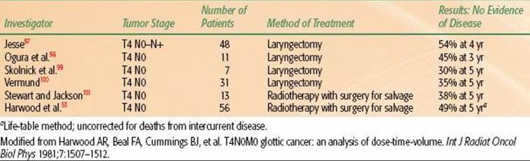
The results of treatment of T4 vocal cord carcinoma in four surgical series and two radiotherapy series are summarized in Table 47.9.53 Hinerman et al.27 observed an 81% 5-year local control rate in 22 selected patients with favorable T4 cancers treated with definitive RT at the University of Florida.
Parsons et al.69 reviewed the literature and reported a local control rate of 62% in a series of 87 patients treated with RT alone for T4 glottic carcinoma.
TABLE 47.5 LOCAL CONTROL AFTER RADIOTHERAPY
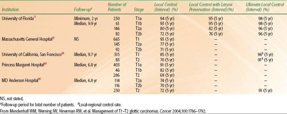
TABLE 47.6 SURVIVAL DATA

TABLE 47.7 STAGE T3 GLOTTIC CARCINOMA TREATED WITH IRRADIATION ALONE (NO CHEMOTHERAPY)

TABLE 47.8 T3 GLOTTIC CARCINOMA TREATED AT THE UNIVERSITY OF FLORIDA, 1965–1988: FIVE-YEAR RESULTS

TABLE 47.9 TREATMENT OF STAGE T4 GLOTTIC CARCINOMAS
Supraglottic Cancer
The proportion of patients suitable for a supraglottic laryngectomy is depicted in Table 47.10.65 Depending on the referral patterns, a modest subset of patients is suitable for this operation. The extent of neck disease for patients treated with either surgery or RT is shown in Table 47.11.65 In general, patients treated with supraglottic laryngectomy appropriately have earlier-stage neck disease and would be anticipated to have a lower risk of distant failure and improved survival. The local control rates after transoral laser, RT, and supraglottic laryngectomy are summarized in Tables 47.12 to 47.14.65 In general, the local control rates after transoral laser excision are fairly good for patients with T1–T2 tumors and tend to deteriorate for those with more advanced disease. The local control rate for patients selected for supraglottic laryngectomy is excellent. However, the incidence of severe complications tends to be higher after supraglottic laryngectomy compared with RT and transoral laser excision (Table 47.15).65,70
FOLLOW-UP POLICY
Follow-up of patients with early lesions is planned for every 4 to 8 weeks for 2 years, every 3 months for the third year, and every 6 months for years 4 and 5, and then annually for life.
Follow-up of patients with vocal cord or supraglottic larynx lesions treated by RT or conservative surgery is almost more important than the treatment itself because early detection of recurrence usually results in salvage that may include cure with voice preservation.
If recurrence is suspected but the biopsy is negative, patients are reexamined at 2- to 4-week intervals until the matter is settled. The value of follow-up CT scans for detecting early local recurrence is investigational.
Wagenfeld et al.71 studied 740 cases of glottic larynx cancer treated from 1965 to 1974 to determine the incidence of second respiratory tract malignancies. There was a minimum follow-up of 5 years. There were 48 second respiratory tract malignancies, although only 14 were expected. Twenty-five were in the lung, and 23 were scattered among other head and neck sites. Only 7 of the 23 second head and neck primary lesions resulted in death; these second lesions were frequently diagnosed in an early stage during routine follow-up for the glottic lesion.
Because the risk of a lethal lung primary lesion is nearly as great as that of dying of an early glottic carcinoma, it makes sense to obtain annual chest roentgenograms. Approximately 50% of patients who receive moderate- to high-dose RT to the entire thyroid gland will develop hypothyroidism within 5 years, and so thyroid functions are checked every 6 to 12 months and thyroid replacement is initiated if the thyroid-stimulating hormone level begins to rise.72
TABLE 47.10 PROPORTION OF PATIENTS SUITABLE FOR SUPRAGLOTTIC LARYNGECTOMY

TABLE 47.11 SUPRAGLOTTIC CARCINOMA: EXTENT OF NECK DISEASE VERSUS TREATMENT

SEQUELAE OF TREATMENT
Surgical Sequelae
Neel et al.73 reported a 26% incidence of nonfatal complications for cordectomy. Immediate postoperative complications included atelectasis and pneumonia, severe subcutaneous emphysema in the neck, bleeding from the tracheotomy site or larynx, wound complications, and airway obstruction requiring tracheotomy. Late complications included granulation tissue that had to be removed by direct laryngoscopy to exclude recurrences, extrusion of cartilage, laryngeal stenosis, and obstructing laryngeal web.
The postoperative complications and sequelae of hemilaryngectomy include chondritis, wound slough, inadequate glottic closure, and anterior commissure webs.74 The complications associated with supraglottic laryngectomy and total laryngectomy for supraglottic carcinomas include fistula (8%), carotid artery exposure or blowout (3% to 5%), infection or wound sloughing (3% to 7%), and fatal complications (3%).74 The risk of complication increased if tumor margins were involved by tumor; there was no change in risk associated with age, sex, race, laryngeal site, stage of primary tumor, size of primary tumor, use of low-dose preoperative RT, or status of the positive nodes.
The incidence of complications after treatment of supraglottic carcinoma is given in Table 47.15.65
TABLE 47.12 SUPRAGLOTTIC CANCER: LOCAL CONTROL AFTER TRANSORAL LASER EXCISION

TABLE 47.13 SUPRAGLOTTIC CANCER: LOCAL CONTROL AFTER RADIOTHERAPY
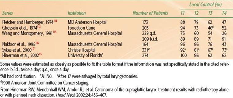
TABLE 47.14 LOCAL CONTROL AFTER SUPRAGLOTTIC LARYNGECTOMY

TABLE 47.15 SUPRAGLOTTIC CANCER: SEVERE COMPLICATIONS ACCORDING TO TREATMENT MODALITY

Radiation Therapy Sequelae
The acute reactions from the treatment of early vocal cord cancer using a tumor dose of 2.25 Gy per day to administer a total dose 63 Gy (4- or 6-MV photons, five fractions per week) are relatively mild. During the first 2 to 3 weeks, the voice may improve as the tumor regresses. The voice generally becomes hoarse again because of RT-induced changes, even though the tumor continues to regress. A mild sore throat develops beginning at the end of the second week, but medication is usually not required. The voice begins to improve approximately 3 weeks after completion of treatment, usually reaching a plateau in 2 to 3 months. Patients with extensive lesions often recover a normal voice, although not as frequently as those with small tumors.
Edema of the larynx is the most common sequela after RT for glottic or supraglottic lesions. The rate of clearance of the edema is related to the RT dose, volume of tissue irradiated, addition of a neck dissection, continued use of alcohol and tobacco, and size and extent of the original lesion. Edema may be accentuated by a radical neck dissection and may require 6 to 12 months to subside.
Soft-tissue necrosis leading to chondritis occurs in <1% of patients, usually in those who continue to smoke. Soft-tissue and cartilage necroses mimic recurrence, with hoarseness, pain, and edema; a laryngectomy may be recommended as a last resort for fear of recurrent cancer, even though biopsy specimens show only necrosis.
Corticosteroids such as dexamethasone (Decadron) have been used to reduce RT-induced edema after recurrence has been ruled out by biopsy. If ulceration and pain occur, administration of an antibiotic such as tetracycline may help. Of 519 patients with T1 N0 or T2 N0 vocal cord cancer treated at the University of Florida, 5 (1%) experienced severe complications,17 including total laryngectomy for a suspected local recurrence (1 patient), permanent tracheostomy for edema (3 patients), and a pharyngocutaneous fistula after a salvage total laryngectomy (1 patient).
In patients irradiated for supraglottic carcinoma, sore throat persists 1 to 2 months after completion of treatment. There is an associated dry mouth from RT of the salivary and parotid glands, a loss of taste, and a sensation of a lump in the throat. It is unusual for patients to require a tracheotomy before RT unless severe lymphedema develops at the time of direct laryngoscopy and biopsy. However, in patients who have recovered from the direct laryngoscopy and biopsy without obstruction, a tracheotomy has rarely been required during a fractionated course of RT.
Patients treated twice a day with 1.2-Gy fractions (continuous-course technique) to a total dose of 74.4 Gy usually have more brisk acute reactions than those treated once a day with 2-Gy fractions. Approximately 30% treated with twice-a-day RT require temporary gastrostomy feeding tubes because they have difficulty in swallowing.75
Examples of acute chondritis requiring discontinuation of treatment have not been seen, although most epiglottic lesions exhibit cartilage invasion.
The epiglottis, both suprahyoid and infrahyoid portions, remains thicker than normal for long periods of time, but this is not often associated with difficulty in swallowing, respiratory obstruction, or aspiration. The patient is cautioned to eat and drink slowly until the edema resolves. The false cord and arytenoids may develop some edema.
Lesions of the suprahyoid epiglottis frequently destroy the tip of the epiglottis, and it may require some time for the exposed cartilage to heal. Successful RT of infrahyoid epiglottis tumors is not associated with a high rate of necrosis, even though most of these lesions penetrate the porous epiglottic cartilage.
The incidence of severe late complications in 274 patients treated with RT alone or combined with neck dissection at the University of Florida was 4%.65
ACKNOWLEDGMENT
We thank the research support staff of the Department of Radiation Oncology for their help with statistics, editing, and manuscript preparation.
REFERENCES
1. Clemente CD. Anatomy: a regional atlas of the human body. Philadelphia: Lea & Febiger, 1975.
2. Mancuso AA, Hanafee WN. Head and neck radiology. Philadelphia: Williams & Wilkins, 2011.
3. Lam KH, Wong J. The preepiglottic and paraglottic spaces in relation to spread of carcinoma of the larynx. Am J Otolaryngol 1983;4(2):81–91.
4. Hirano M. Structure and vibratory behavior of the vocal folds. In: Sawashima M, Cooper FS, editors. Dynamic aspects of speech production: current results, emerging problems, and new instrumentation.Tokyo: University of Tokyo Press, 1977:13–27.
5. Jemal A, Siegel R, Xu J, et al. Cancer statistics, 2010. CA Cancer J Clin 2010;60(5): 277–300.
6. Ries LAG, Eisner MP, Kosary CL. SEER cancer statistics review, 1973-1998. Bethesda, MD: National Cancer Institute, 2001.
7. Wynder EL. The epidemiology of cancers of the upper alimentary and upper respiratory tracts. Laryngoscope 1978;88(1 Pt 2, Suppl 8): 50–51.
8. Vincent RG, Marchetta F. The relationship of the use of tobacco and alcohol to cancer of the oral cavity, pharynx or larynx. Am J Surg 1963;106:501–505.
9. Archer CR, Yeager VL, Herbold DR. Computed tomography vs. histology of laryngeal cancer: their value in predicting laryngeal cartilage invasion. Laryngoscope 1983;93(2):140–147.
10. Pillsbury HR, Kirchner JA. Clinical vs histopathologic staging in laryngeal cancer. Arch Otolaryngol 1979;105(3):157–159.
11. Broyles EN. The anterior commissure tendon. Ann Otol Rhinol Laryngol 1943;52:342–345.
12. Kirchner JA. Staging as seen in serial sections. Laryngoscope 1975;85(11, Pt 1):1816–1821.
13. Olofsson J, van Nostrand AW. Growth and spread of laryngeal and hypopharyngeal carcinoma with reflections on the effect of preoperative irradiation. 139 cases studied by whole organ serial sectioning. Acta Otolaryngol Suppl1973;308:1–84.
14. Lindberg R. Distribution of cervical lymph node metastases from squamous cell carcinoma of the upper respiratory and digestive tracts. Cancer 1972;29(6):1446–1469.
15. Fletcher GH. Elective irradiation of subclinical disease in cancers of the head and neck. Cancer 1972;29(6):1450–1454.
16. Ogura JH, Biller HF, Wette R. Elective neck dissection for pharyngeal and laryngeal cancers. An evaluation. Ann Otol Rhinol Laryngol 1971;80(5):646–650.
17. Mendenhall WM, Amdur RJ, Morris CG, et al. T1-T2N0 squamous cell carcinoma of the glottic larynx treated with radiation therapy. J Clin Oncol 2001;19(20):4029–4036.
18. Lederman M. The place of radiotherapy in the treatment of cancer of the larynx [in French]. Ann Radiol (Paris) 1961;4:433–454.
19. Million RR, Cassisi NJ. Larynx. In: Million RR, Cassisi NJ, ed. Management of head and neck cancer: a multidisciplinary approach, 1st ed. Phildelphia: JB Lippincott, 1984 315–364.
20. Million RR, Cassisi NJ, Mancuso AA. Larynx. In: Million RR, Cassisi NJ, ed. Management of head and neck cancer: a multidisciplinary approach, 2nd ed. Philadelphia: JB Lippincott, 1994:431–497.
21. McLaughlin MP, Mendenhall WM, Mancuso AA, et al. Retropharyngeal adenopathy as a predictor of outcome in squamous cell carcinoma of the head and neck. Head Neck 1995;17(3):190–198.
22. Archer CR, Yeager VL, Herbold DR. Improved diagnostic accuracy in laryngeal cancer using a new classification based on computed tomography. Cancer 1984;53(1):44–57.
23. Mendenhall WM, Parsons JT, Mancuso AA, et al. In: Carlos A, Perez LWB, eds. Principles and practice of radiation oncology, 3rd ed. Philadelphia: Lippincott-Raven, 1998:1069–1093.
24. American Joint Committee on Cancer, AJCC cancer staging handbook, 7th ed. New York: Springer, 2010.
25. Dagan R, Morris CG, Bennett JA, et al. Prognostic significance of paraglottic space invasion in T2N0 glottic carcinoma. Am J Clin Oncol 2007;30(2):186–190.
26. Gindhart TD, Johnston WH, Chism SE, et al. Carcinoma of the larynx in childhood. Cancer 1980;46(7):1683–1687.
27. Hinerman RW, Mendenhall WM, Morris CG, et al. T3 and T4 true vocal cord squamous carcinomas treated with external beam irradiation: a single institution’s 35-year experience. Am J Clin Oncol2007;30(2):181–185.
28. Mendenhall WM, Morris CG, Stringer SP, et al. Voice rehabilitation after total laryngectomy and postoperative radiation therapy. J Clin Oncol 2002;20(10):2500–2505.
29. Mendenhall WM, Villaret DB, Amdur RJ, et al. Planned neck dissection after definitive radiotherapy for squamous cell carcinoma of the head and neck. Head Neck 2002;24(11):1012–1018.
30. Induction chemotherapy plus radiation compared with surgery plus radiation in patients with advanced laryngeal cancer. The Department of Veterans Affairs Laryngeal Cancer Study Group. N Engl J Med1991;324(24):1685–1690.
31. Mendenhall WM, Morris CG, Amdur RJ, et al. Parameters that predict local control after definitive radiotherapy for squamous cell carcinoma of the head and neck. Head Neck 2003;25(7):535–542.
32. Mendenhall WM, Parsons JT, Mancuso AA, et al. Definitive radiotherapy for T3 squamous cell carcinoma of the glottic larynx. J Clin Oncol 1997;15(6):2394–2402.
33. Forastiere AA, Goepfert H, Maor M, et al. Concurrent chemotherapy and radiotherapy for organ preservation in advanced laryngeal cancer. N Engl J Med 2003;349(22):2091–2098.
34. Pignon JP, Bourhis J, Domenge C, et al. Chemotherapy added to locoregional treatment for head and neck squamous-cell carcinoma: three meta-analyses of updated individual data. MACH-NC Collaborative Group. Meta-Analysis of Chemotherapy on Head and Neck Cancer. Lancet 2000;355(9208):949–955.
35. Mendenhall WM, Riggs CE, Vaysberg M, et al. Altered fractionation and adjuvant chemotherapy for head and neck squamous cell carcinoma. Head Neck 2010;32(7):939–945.
36. Sengupta N, Morris CG, Kirwan J, et al. Definitive radiotherapy for carcinoma in situ of the true vocal cords. Am J Clin Oncol 2010;33(1):94–95.
37. Mendenhall WM, Werning JW, Hinerman RW, et al. Management of T1-T2 glottic carcinomas. Cancer 2004;100(9):1786–1792.
38. Chera BS, Amdur RJ, Morris CG, et al. T1N0 to T2N0 squamous cell carcinoma of the glottic larynx treated with definitive radiotherapy. Int J Radiat Oncol Biol Phys 2010;78(2):461–466.
39. O’Sullivan B, Mackillop W, Gilbert R, et al. Controversies in the management of laryngeal cancer: results of an international survey of patterns of care. Radiother Oncol 1994;31(1):23–32.
40. Laccourreye H, Laccourreye O, Weinstein G, et al. Supracricoid laryngectomy with cricohyoidoepiglottopexy: a partial laryngeal procedure for glottic carcinoma. Ann Otol Rhinol Laryngol 1990;99(6, Pt 1): 421–426.
41. McGuirt WF, Blalock D, Koufman JA, et al. Comparative voice results after laser resection or irradiation of T1 vocal cord carcinoma. Arch Otolaryngol Head Neck Surg 1994;120(9):951–955.
42. Steiner W. Results of curative laser microsurgery of laryngeal carcinomas. Am J Otolaryngol 1993;14(2):116–121.
43. Rodrigo JP, Suarez C, Silver CE, et al. Transoral laser surgery for supraglottic cancer. Head Neck 2008;30(5):658–666.
44. Peretti G, Piazza C, Cocco D, et al. Transoral CO(2) laser treatment for T(is)-T(3) glottic cancer: the University of Brescia experience on 595 patients. Head Neck 2010;32(8):977–983.
45. Rodel RM, Steiner W, Muller RM, et al. Endoscopic laser surgery of early glottic cancer: involvement of the anterior commissure. Head Neck 2009;31(5):583–592.
46. O’Sullivan B, Warde P, Keane T, et al. Outcome following radiotherapy in verrucous carcinoma of the larynx. Int J Radiat Oncol Biol Phys 1995;32(3):611–617.
47. Fu KK, Pajak TF, Trotti A, et al. A Radiation Therapy Oncology Group (RTOG) phase III randomized study to compare hyperfractionation and two variants of accelerated fractionation to standard fractionation radiotherapy for head and neck squamous cell carcinomas: first report of RTOG 9003. Int J Radiat Oncol Biol Phys 2000;48(1):7–16.
48. Parsons JT, Mendenhall WM, Stringer SP, et al. Salvage surgery following radiation failure in squamous cell carcinoma of the supraglottic larynx. Int J Radiat Oncol Biol Phys 1995;32(3):605–609.
49. Pearson BW, Woods RD 2 nd, Hartman DE. Extended hemilaryngectomy for T3 glottic carcinoma with preservation of speech and swallowing. Laryngoscope 1980;90(12):1950–1961.
50. Amdur RJ, Parsons JT, Mendenhall WM, et al. Postoperative irradiation for squamous cell carcinoma of the head and neck: an analysis of treatment results and complications. Int J Radiat Oncol Biol Phys1989;16(1):25–36.
51. Huang DT, Johnson CR, Schmidt-Ullrich R, et al. Postoperative radiotherapy in head and neck carcinoma with extracapsular lymph node extension and/or positive resection margins: a comparative study. Int J Radiat Oncol Biol Phys 1992;23(4):737–742.
52. Hillman RE, Walsh MJ, Wolf GT, et al. Functional outcomes following treatment for advanced laryngeal cancer. Part I—Voice preservation in advanced laryngeal cancer. Part II—Laryngectomy rehabilitation: the state of the art in the VA System. Research Speech-Language Pathologists. Department of Veterans Affairs Laryngeal Cancer Study Group. Ann Otol Rhinol Laryngol Suppl 1998;172:1–27.
53. Harwood AR, Beale FA, Cummings BJ, et al. T4NOMO glottic cancer: an analysis of dose-time volume factors. Int J Radiat Oncol Biol Phys 1981;7(11):1507–1512.
54. Kim RY, Marks ME, Salter MM. Early-stage glottic cancer: importance of dose fractionation in radiation therapy. Radiology 1992;182(1):273–275.
55. Schwaibold F, Scariato A, Nunno M, et al. The effect of fraction size on control of early glottic cancer. Int J Radiat Oncol Biol Phys 1988;14(3):451–454.
56. Woodhouse RJ, Quivey JM, Fu KK, et al. Treatment of carcinoma of the vocal cord. A review of 20 years experience. Laryngoscope 1981;91(7):1155–1162.
57. Mendenhall WM, Riggs CE, Cassisi NJ. Treatment of head and neck cancers. In: DeVita VT, Hellman S, Rosenberg SA, eds. Cancer: principles and practice of oncology, 8th ed. Philadelphia: Lippincott Williams & Wilkins, 2008:809–814.
58. Yamazaki H, Nishiyama K, Tanaka E, et al. Radiotherapy for early glottic carcinoma (T1N0M0): results of prospective randomized study of radiation fraction size and overall treatment time. Int J Radiat Oncol Biol Phys2006;64(1):77–82.
59. Parsons JT, Mendenhall WM, Mancuso AA, et al. Twice-a-day radiotherapy for T3 squamous cell carcinoma of the glottic larynx. Head Neck 1989;11(2):123–128.
60. Million RR, Cassisi NJ, Mancuso AA, et al. Management of the neck for squamous cell carcinoma. In: Million RR, Cassisi NJ, eds. Management of head and neck cancer: a multidisciplinary approach, 2 nd ed. Philadelphia: JB Lippincott, 1994:75–142.
61. Mendenhall WM, Mancuso AA. Radiotherapy for head and neck cancer—is the “next level” down? Int J Radiat Oncol Biol Phys 2009;73(3):645–646.
62. Trotti A, Fu KK, Pajak TF. Long term outcomes of RTOG 90-03: A comparison of hyperfractionation and two variants of accelerated fractionation to standard fractionation radiotherapy for head and neck squamous cell carcinoma. Int J Radiat Oncol Biol Phys 2005;63:S70–S71.
63. Biller HF, Barnhill FR Jr, Ogura JH, et al. Hemilaryngectomy following radiation failure for carcinoma of the vocal cords. Laryngoscope 1970;80(2):249–253.
64. Lee F, Perlmutter S, Ogura JH. Laryngeal radiation after hemilaryngectomy. Laryngoscope 1980;90(9):1534–1539.
65. Hinerman RW, Mendenhall WM, Amdur RJ, et al. Carcinoma of the supraglottic larynx: treatment results with radiotherapy alone or with planned neck dissection. Head Neck 2002;24(5):456–467.
66. Lee NK, Goepfert H, Wendt CD. Supraglottic laryngectomy for intermediate-stage cancer: U.T. M.D. Anderson Cancer Center experience with combined therapy. Laryngoscope 1990;100(8):831–836.
67. Foote RL, Olsen KD, Buskirk SJ, et al. Laryngectomy alone for T3 glottic cancer. Head Neck 1994;16(5):406–412.
68. Mendenhall WM, Parsons JT, Stringer SP, et al. Stage T3 squamous cell carcinoma of the glottic larynx: a comparison of laryngectomy and irradiation. Int J Radiat Oncol Biol Phys 1992;23(4):725–732.
69. Parsons JT, Mendenhall WM, Stringer SP, et al. T4 laryngeal carcinoma: radiotherapy alone with surgery reserved for salvage. Int J Radiat Oncol Biol Phys 1998;40(3):549–552.
70. Ganly I, Patel SG, Matsuo J, et al. Analysis of postoperative complications of open partial laryngectomy. Head Neck 2009;31(3):338–345.
71. Wagenfeld DJ, Harwood AR, Bryce DP, et al. Second primary respiratory tract malignancies in glottic carcinoma. Cancer 1980;46(8):1883–1886.
72. Garcia-Serra A, Amdur RJ, Morris CG, et al. Thyroid function should be monitored following radiotherapy to the low neck. Am J Clin Oncol 2005;28(3):255–258.
73. Neel HB 3rd, Devine KD, Desanto LW. Laryngofissure and cordectomy for early cordal carcinoma: outcome in 182 patients. Otolaryngol Head Neck Surg 1980;88(1):79–84.
74. Gall AM, Sessions DG, Ogura JH. Complications following surgery for cancer of the larynx and hypopharynx. Cancer 1977;39(2):624–631.
75. Al-Othman MO, Amdur RJ, Morris CG, et al. Does feeding tube placement predict for long-term swallowing disability after radiotherapy for head and neck cancer? Head Neck 2003;25(9):741–747.
76. Rudert HH, Werner JA. Endoscopic resections of glottic and supraglottic carcinomas with the CO2 laser. Eur Arch Otorhinolaryngol 1995;252(3):146–148.
77. Peretti G, Nicolai P, Redaelli De Zinis LO, et al. Endoscopic CO2 laser excision for Tis, T1, and T2 glottic carcinomas: cure rate and prognostic factors. Otolaryngol Head Neck Surg 2000;123(1, Pt 1):124–131.
78. Spector JG, Sessions DG, Chao KS, et al. Stage I (T1 N0 M0) squamous cell carcinoma of the laryngeal glottis: therapeutic results and voice preservation. Head Neck 1999;21(8):707–717.
79. Motta G, Esposito E, Cassiano B, et al. T1-T2-T3 glottic tumors: fifteen years experience with CO2 laser. Acta Otolaryngol Suppl 1997;527:155–159.
80. Gallo A, de Vincentiis M, Manciocco V, et al. CO2 laser cordectomy for early-stage glottic carcinoma: a long-term follow-up of 156 cases. Laryngoscope 2002;112(2):370–374.
81. Pradhan SA, Pai PS, Neeli SI, et al. Transoral laser surgery for early glottic cancers. Arch Otolaryngol Head Neck Surg 2003;129(6):623–625.
82. Giovanni A, Guelfucci B, Gras R, et al. Partial frontolateral laryngectomy with epiglottic reconstruction for management of early-stage glottic carcinoma. Laryngoscope 2001;111(4, Pt 1):663–668.
83. Crampette L, Garrel R, Gardiner Q, et al. Modified subtotal laryngectomy with cricohyoidoepiglottopexy—long term results in 81 patients. Head Neck 1999;21(2):95–103.
84. Thomas JV, Olsen KD, Neel HB 3rd, et al. Early glottic carcinoma treated with open laryngeal procedures. Arch Otolaryngol Head Neck Surg 1994;120(3):264–268.
85. Laccourreye O, Weinstein G, Brasnu D, et al. A clinical trial of continuous cisplatin-fluorouracil induction chemotherapy and supracricoid partial laryngectomy for glottic carcinoma classified as T2. Cancer 1994;74(10):2781–2790.
86. Spector JG, Sessions DG, Chao KS, et al. Management of stage II (T2N0M0) glottic carcinoma by radiotherapy and conservation surgery. Head Neck 1999;21(2):116–123.
87. Wang CC. Carcinoma of the larynx. In: Wang CC, ed. Radiation therapy for head and neck neoplasms, 3rd ed. New York: Wiley-Liss, 1997:221–255.
88. Le QT, Fu KK, Kroll S, et al. Influence of fraction size, total dose, and overall time on local control of T1-T2 glottic carcinoma. Int J Radiat Oncol Biol Phys 1997;39(1):115–126.
89. Warde P, O’Sullivan B, Bristow RG, et al. T1/T2 glottic cancer managed by external beam radiotherapy: the influence of pretreatment hemoglobin on local control. Int J Radiat Oncol Biol Phys1998;41(2):347–353.
90. Garden AS, Forster K, Wong PF, et al. Results of radiotherapy for T2N0 glottic carcinoma: does the “2” stand for twice-daily treatment? Int J Radiat Oncol Biol Phys 2003;55(2):322–328.
91. Harwood AR, Beale FA, Cummings BJ, et al. T3 glottic cancer: an analysis of dose time-volume factors. Int J Radiat Oncol Biol Phys 1980;6(6):675–680.
92. Wang CC. Radiation therapy of laryngeal tumors: curative radiation therapy. In: Thawley SE, Panje WR, eds. Comprehensive management of head and neck tumors. Philadelphia: WB Saunders, 1987:906–919.
93. Fletcher GH. Radiation therapy for cancer of the larynx and pyriform sinus. Eye Ear Nose Throat Digest 1969;31:58–67.
94. Skolyszewski J, Reinfuss M. The results of radiotherapy of cancer of the larynx in six European countries. Radiobiol Radiother (Berl) 1981;22(1):32–43.
95. Stewart JG, Brown JR, Palmer MK, et al. The management of glottic carcinoma by primary irradiation with surgery in reserve. Laryngoscope 1975;85(9):1477–1484.
96. Mills EE. Early glottic carcinoma: factors affecting radiation failure, results of treatment and sequelae. Int J Radiat Oncol Biol Phys 1979;5(6):811–817.
97. Jesse RH. The evaluation of treatment of patients with extensive squamous cancer of the vocal cords. Laryngoscope 1975;85(9):1424–1429.
98. Ogura JH, Sessions DG, Ciralsky RH. Supraglottic carcinoma with extension to the arytenoid. Laryngoscope 1975;85(8):1327–1331.
99. Skolnik EM, Yee KF, Wheatley MA, et al. Carcinoma of the laryngeal glottis therapy and end results. Laryngoscope 1975;85(9):1453–1466.
100. Vermund H. Role of radiotherapy in cancer of the larynx as related to the TNM system of staging. A review. Cancer 1970;25(3):485–504.
101. Stewart JG, Jackson AW. The steepness of the dose response curve both for tumor cure and normal tissue injury. Laryngoscope 1975;85(7):1107–1111.
102. Ogura JH, Sessions DG, Spector GJ. Conservation surgery for epidermoid carcinoma of the supraglottic larynx. Laryngoscope 1975;85(11, Pt 1):1808–1815.
103. Lutz CK, Johnson JT, Wagner RL, et al. Supraglottic carcinoma: patterns of recurrence. Ann Otol Rhinol Laryngol 1990;99(1):12–17.
104. Weems DH, Mendenhall WM, Parsons JT, et al. Squamous cell carcinoma of the supraglottic larynx treated with surgery and/or radiation therapy. Int J Radiat Oncol Biol Phys 1987;13(10):1483–1487.
105. Gregor RT, Oei SS, Baris G, et al. Supraglottic laryngectomy with postoperative radiation versus primary radiation in the management of supraglottic laryngeal cancer. Am J Otolaryngol 1996;17(5):316–321.
106. Spriano G, Antognoni P, Piantanida R, et al. Conservative management of T1-T2N0 supraglottic cancer: a retrospective study. Am J Otolaryngol 1997;18(5):299–305.
107. Bocca E. Sixteenth Daniel C. Baker, Jr, memorial lecture. Surgical management of supraglottic cancer and its lymph node metastases in a conservative perspective. Ann Otol Rhinol Laryngol 1991;100(4, Pt 1):261–267.
108. Isaacs JH Jr, Slattery WH 3rd, Mendenhall WM, et al. Supraglottic laryngectomy. Am J Otolaryngol 1998;19(2):118–123.
109. Davis RK, Kelly SM, Hayes J. Endoscopic CO2 laser excisional biopsy of early supraglottic cancer. Laryngoscope 1991;101(6, Pt 1): 680–683.
110. Zeitels SM, Koufman JA, Davis RK, et al. Endoscopic treatment of supraglottic and hypopharynx cancer. Laryngoscope 1994;104(1, Pt 1):71–78.
111. Rudert HH, Werner JA, Hoft S. Transoral carbon dioxide laser resection of supraglottic carcinoma. Ann Otol Rhinol Laryngol 1999;108(9):819–827.
112. Ghossein NA, Bataini JP, Ennuyer A, et al. Local control and site of failure in radically irradiated supraglottic laryngeal cancer. Radiology 1974;112(1):187–192.
113. Csanady M, Ivan L, Czigner J. Endoscopic CO(2) laser therapy of selected cases of supraglottic marginal tumors. Eur Arch Otorhinolaryngol 1999;256(8):392–394.
114. Fletcher GH, Hamberger AD. Causes of failure in irradiation of squamous-cell carcinoma of the supraglottic larynx. Radiology 1974;111(3):697–700.
115. Wang CC, Montgomery WW. Deciding on optimal management of supraglottic carcinoma. Oncology (Williston Park) 1991;5(4):41–46; discussion 46, 49, 53.
116. Nakfoor BM, Spiro IJ, Wang CC, et al. Results of accelerated radiotherapy for supraglottic carcinoma: a Massachusetts General Hospital and Massachusetts Eye and Ear Infirmary experience. Head Neck1998;20(5):379–384.
117. Sykes AJ, Slevin NJ, Gupta NK, et al. 331 cases of clinically node-negative supraglottic carcinoma of the larynx: a study of a modest size fixed field radiotherapy approach. Int J Radiat Oncol Biol Phys2000;46(5):1109–1115.
118. DeSanto LW. Early supraglottic cancer. Ann Otol Rhinol Laryngol 1990;99(8):593–597.
119. Steiniger JR, Parnes SM, Gardner GM. Morbidity of combined therapy for the treatment of supraglottic carcinoma: supraglottic laryngectomy and radiotherapy. Ann Otol Rhinol Laryngol1997;106(2):151–158.
120. Burstein FD, Calcaterra TC. Supraglottic laryngectomy: series report and analysis of results. Laryngoscope 1985;95(7, Pt 1): 833–836.
121. Weber PC, Johnson JT, Myers EN. Impact of bilateral neck dissection on recovery following supraglottic laryngectomy. Arch Otolaryngol Head Neck Surg 1993;119(1):61–64.
122. Beckhardt RN, Murray JG, Ford CN, et al. Factors influencing functional outcome in supraglottic laryngectomy. Head Neck 1994;16(3):232–239.