
Carlos A. Perez and Wade L. Thorstad
 GLOMUS TUMORS
Anatomy
Glomus bodies are found in the jugular bulb and along the tympanic (Jacobson) and auricular (Arnold) branches of the tenth nerve in the middle ear or in other anatomic sites (Fig. 48.1). Depending on the location, glomus tumors (chemodectoma or paraganglioma) are classified as tympanic (middle ear), jugulare, or carotid vagal or designated as originating from other locations, such as the larynx, adventitia of thoracic aorta, abdominal aorta, or the surface of the lungs (Fig. 48.2).1
Glomus tumors (GT) or chemodectomas consist of large epithelioid (smooth muscle) cells with fine granular cytoplasm embedded in a rich capillary network and fibrous stroma with reticulin fibers, which derive from embryonic neural crest cells. Although histologically benign, they may extend along the lumen of the vein to regional lymph nodes, but rarely to distant sites. These tissues are responsive to changes in oxygen and carbon dioxide tensions and pH.
FIGURE 48.1. Anatomy of the region of the glomus jugulare. (From Hatfield PM, James AE, Schulz MN. Chemodectomas of the glomus jugulare. Cancer 1972;30:1165–1168, with permission.)
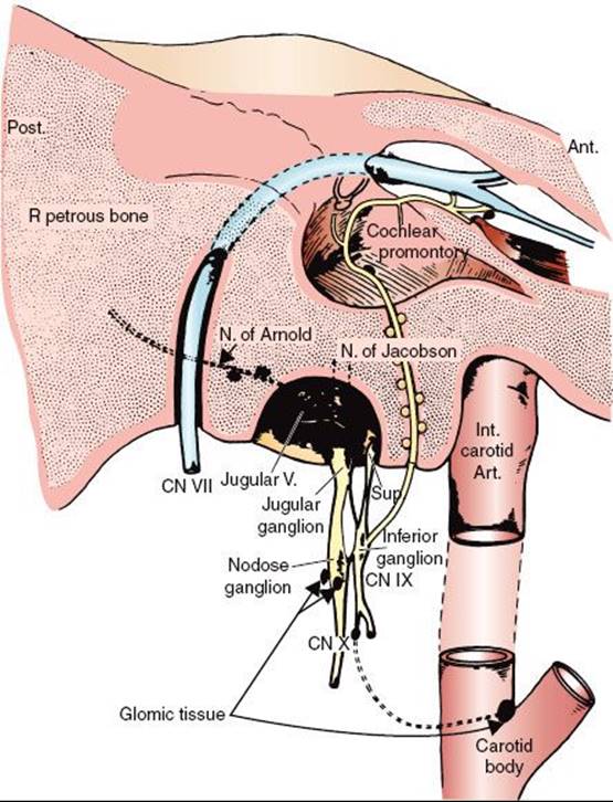
FIGURE 48.2. Distribution of paragangliomas of the head and neck region. Laterality was not specified in three patients with carotid body paragangliomas. The diagram does not include one left carotid body paraganglioma that was found incidentally at autopsy and a left vagal body paraganglioma that presented in a patient who had two other paragangliomas. (From Lack EE, Cubilla AL, Woodruff JM, et al. Paragangliomas of the head and neck region. Cancer 1977;39:3997–4009, with permission.)
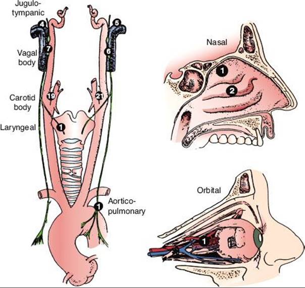
FIGURE 48.3. A: Late-phase arteriogram illustrating large glomus jugulare tumor with extension into the neck. B: Computed tomography scan with contrast enhancement showing intracranial component of lesion.
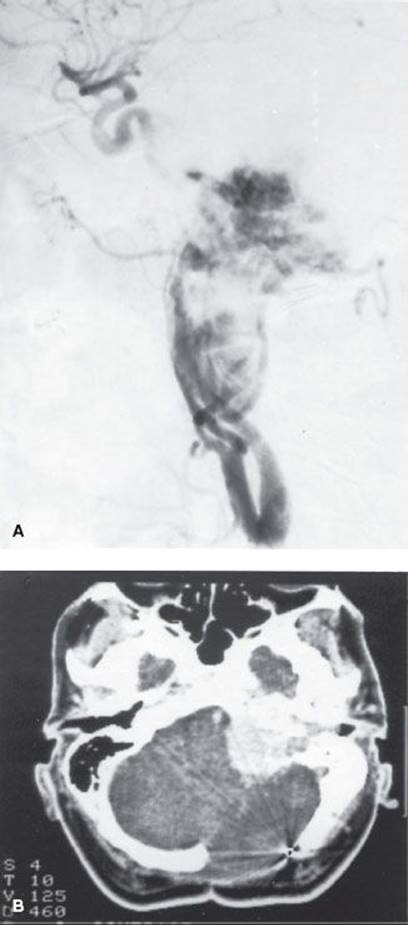
TABLE 48.1 DIAGNOSTIC WORKUP FOR GLOMUS TUMORS OF THE EAR AND BASE OF SKULL, HEMANGIOPERICYTOMA, ESTHESIONEUROBLASTOMA, EXTRAMEDULLARY PLASMACYTOMA, AND SARCOMA OF THE HEAD AND NECK

Epidemiology
The mean age at diagnosis has been reported to be 44.7 years for carotid body tumors and 52 years for glomus tympanicum.2 These tumors occur three or four times more frequently in women than in men, suggesting a possible estrogen influence.2,3,4 Glomus tumors may be familial; they occur in multiple sites in 10% to 20% of patients.5
Recent advances in genetics identified three loci associated with hereditary paragangliomas, and genetic screening may detect affected patients.6 Multiple paragangliomas of the head and neck are rare, with an incidence of 10% of all patients, but in familial cases it increases up to 35% to 50%.5 In the head and neck region, the most common association is bilateral carotid body tumors or carotid body tumor associated with tympanic–jugular glomus.7
Clinical Presentation
Glomus tumors may arise along the nerve roots. In the middle ear they may initially cause earache or discomfort.97 As they expand, eventually they produce pulsatile tinnitus, hearing loss, and, in later stages, cranial nerve paralysis resulting from invasion of the base of the skull in 10% to 15% of patients. If the tumor invades the middle cranial fossa, symptoms may include temporoparietal headache, retro-orbital pain, proptosis, and paresis of cranial nerves V and VI. If the posterior fossa is involved, symptoms may include occipital headache, ataxia, and paresis of cranial nerves V to VII, IX, and XII; invasion of the jugular foramen causes paralysis of nerves IX to XI. Chemodectoma of the carotid body usually presents as a painless, slowly growing mass in the upper neck. Occasionally the mass may be pulsatile and may have an associated thrill or bruit. As it enlarges, the mass may extend into the parapharyngeal space and be visible on examination of the oropharynx. Very rarely these tumors may be malignant.8 Metastases occur in 2% to 5% of cases.3
Diagnostic Workup
Diagnostic evaluation for glomus tumors of the ear and base of skull is outlined in Table 48.1. In the majority of glomus tympanicum, physical examination demonstrates a red, vascular middle ear mass, although occasionally it may be bluish or white (the latter resembling a cholesteatoma).2 Audiography may demonstrate conductive hearing loss in the ear involved by tumor as noted in 33 of 49 patients evaluated by Larson et al.;2 4 of 33 patients with conductive deficits also exhibited tympanic pulsations. Examination of the neck may occasionally demonstrate a mass in the neck that may be pulsatile or have a bruit or regional lymph node metastases.
Radiographic studies are invaluable in the diagnosis of these tumors. Plain mastoid radiographs never show the soft tissue mass in the middle ear, although they frequently demonstrate clouding of the mastoid air cells, suggesting mastoiditis.157 High-resolution computed tomography (CT) with contrast has a degree of sensitivity and specificity to diagnose this tumor when located in the middle ear or jugular bulb; masses as small as 3 mm have been demonstrated. Tumor enhancement is similar to that of the temporalis muscle (Fig. 48.3).2 In 46 patients with glomus tympanicum, there were no instances of local bony erosion; instead, the tumors engulfed the ossicular chain, bulged or protruded through the tympanic membrane, filled the middle ear, or extended into the eustachian tube orifice or aditus ad antrum. This pattern is in contrast to cholesteatomas, which typically destroy adjacent bony landmarks, including the ossicles, and progressively erode the petrous bones as they enlarge.2
Magnification angiography is a sensitive and specific means of detecting glomus tympanicum tumors. This procedure should be performed after high-resolution thin-section CT scan (with contrast material), only when there is a question regarding the nature of the lesion or the location of the carotid canal. Findings include a hypervascular middle ear mass that first appears in the middle to late arterial phase, persists through the capillary phase, and quickly disappears in the venous phase without demonstrably early draining veins. Biopsy of an aberrant internal carotid artery can result in major neurologic sequelae or death.
Vogl et al.9 reported on 40 patients with glomus tumors of the skull; diagnostic interpretations were correlated with histologic examination, digital subtraction angiography, CT, and clinical follow-up. Sixteen of 18 proven tumors were detected with spin-echo images alone. Although four high-flying jugular bulbs were misinterpreted as tumors because of similar signal intensity, combined evaluation allowed differentiation between tumor and sinusal blood flow in all cases.
Drape et al.10 described magnetic resonance imaging (MRI) findings in 31 patients with a clinical suspicion of glomus tumor; gadoterate meglumine was injected into 19 patients. Twenty-seven of 28 pathologically confirmed glomus tumors were detected with MRI; a peripheral capsule was present in most tumors. The investigators were able to differentiate three subtypes of glomus tumors (vascular, solid, and myxoid) on the basis of relaxation times and enhancement characteristics. Multidetector CT angiography was found to be more accurate in the diagnosis of six glomus tumors, with enhancement in the arterial phase, when compared with MRI.11
As GTs show high levels of somatostatin receptor (SSTR) subtypes 2 and 5, fluorine-(18F)-octreotate positron emission tomography (PET) may be useful for diagnostic purposes in a semiquantitative manner and for improving target volume delineation in radiation therapy planning. Astner et al.12 noted that preliminary findings with two different PET tracers for SSTR imaging have been reported: gallium-68 (68Ga)-DOTATOC (DOTA-d-Phe(1)-Tyr(3)-octreotide [somatostatin analog]) PET was shown to detect SSTR-expressing tumors with high sensitivity and specificity. A second PET tracer, Gluc-Lys18F-TOCA, allows fast, high-contrast imaging of SSTR-positive tumors with superior biokinetics and diagnostic performance as compared with indium-111 (111In)-DTPA (diethylene triamine pentaacetic acid)-octreotide and—as far as can be determined from the literature—comparable to 68Ga-DOTATOC.13,14,15
Cytochemical techniques demonstrate increased levels of serotonin, epinephrine, and norepinephrine in normal glomus tissue of the carotid body. Histologic staining techniques, including chromaffin and argentaffin reactions, identify patients with hormonally active tumors. This is important because the glomus tumor may coexist with a pheochromocytoma, which requires special preoperative preparation of the patient. Biopsy of glomus tumors may result in severe hemorrhage.
TABLE 48.2 GLASSCOCK-JACKSON CLASSIFICATION OF GLOMUS TUMORS
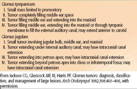
TABLE 48.3 MODIFICATION OF McCABE AND FLETCHER CLASSIFICATION OF CHEMODECTOMAS
Staging
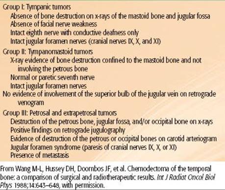
The prognosis of these tumors is closely related to the anatomic location and the volume of the lesion, which is reflected in the Glasscock-Jackson classification16 shown in Table 48.2. An alternative classification proposed by McCabe and Fletcher17 is presented in Table 48.3.
General Management
Li et al.18 published a historical perspective of various treatment modalities used to treat glomus tumors over the past 60 years.
Surgery
Surgery is generally selected for small tumors that can be completely excised. Glomus tympanicum tumors are particularly well managed with excision via tympanotomy or mastoidectomy. Percutaneous embolization of a low-viscosity silicone polymer has been used, frequently as preoperative preparation of the tumor embolization of feeding vessels allows meticulous microsurgery with virtually complete hemostasis.
Surgical treatment of a glomus tumor arising in the jugular bulb, however, often consists of piece-by-piece removal accompanied by significant intraoperative bleeding with damage to adjacent neurovascular structures and requires more complex surgical approaches involving the base of the skull. Preoperative embolization via a transarterial approach has proved beneficial but is often limited by vascular anatomy and unfavorable locations. Abud et al.19reported experience with preoperative devascularization using direct puncture and an intralesional injection of cyanoacrylate (acrylic glue) under fluoroscopic guidance in nine patients with head and neck paragangliomas. Ozyer et al.20 performed devascularization with intralesional injection of N-butyl-cyanoacrylate (seven carotid and three jugular paragangliomas). The tumors were subsequently surgically removed.
The local tumor control rate with surgery alone is only about 60%, and there is significant morbidity, particularly cranial nerve injury and bleeding.
In a retrospective review of all skull-base surgery cases treated at Baylor University, 175 jugulotympanic glomus tumors and 9 malignant cases (5.1%) were identified.8 The 5-year survival rate was 72%.
Radiation Therapy
Irradiation is frequently used in the treatment of glomus tumors, particularly for those in the tympanicum and jugulare bulb locations. Tumors with destruction of the petrous bone, jugular fossa, or occipital bone or patients with jugular foramen syndrome are more reliably managed with irradiation.2,4,21,22 Some surgeons, such as Glasscock et al.16 have questioned the effectiveness of radiation therapy in the treatment of chemodectomas because on histologic sections, obtained even many years after irradiation, it is possible to find chromophilic cells remaining in the tumor. However, there is also evidence of fibrosis and decreased vascularity.23 Suit and Gallager24 demonstrated in a murine mammary carcinoma model that morphologically intact cells may have lost their reproductive ability after irradiation, which is the ultimate end point of cell killing. Furthermore, it is extremely unusual to observe clinical regrowth of a glomus tumor after irradiation, even if they do not regress completely.
Some reports describe successful combinations of surgery with preoperative irradiation, in an attempt to make an unresectable tumor operable, or postoperatively when obvious tumor could not be resected.
Radiation Therapy Techniques
Radiation therapy techniques are determined by the location and extent of the tumor, which must be defined before treatment.21,25,26 Limited, usually bilateral, portals were used for relatively localized glomus tumors, whether or not the treatment is combined with surgery (Fig. 48.4). Dickens et al.27 used a three-field arrangement with a superior-inferior wedged and lateral open field, with a weighting of 1:1:0.33. A superior-inferior 60-degree and 45-degree wedged filtered field arrangement was also used. Electrons (15 to 18 MeV) with a lateral portal or combined with cobalt-60 (60Co) or 4- to 6-MV photons (20% to 25% of total tumor dose) render a good dose distribution (Fig. 48.5). Several prosthetic materials have been used to enhance irradiation dose homogeneity.28 In patients in whom tumor has spread into the posterior fossa, it may be necessary to use parallel opposed portals with 6- to 18-MV photons. Treatment is given at the rate of 1.8 to 2 Gy tumor dose per day with 5 treatments per week for a total tumor dose of 45 to 55 Gy in 5 weeks. Three-dimensional conformal radiation therapy (3D-CRT) or image-guided intensity-modulated radiation therapy (IMRT) are highly desirable techniques to treat these tumors, with excellent dose distributions (Fig. 48.5). Table 48.4 summarizes the doses of irradiation recommended by several investigators and the probability of tumor control for each.21,26,29
FIGURE 48.4. A: Portal used for relatively localized glomus tumor. B: Simulation film of patient with glomus tumor. C: Isodose distribution of a mixed-beam unilateral portal for a glomus tympanicum lesion (80% 16-MeV electrons, 20% 4-MV photons). (From Konefal JB, Pilepich MV, Spector GJ, et al. Radiation therapy in the treatment of chemodectomas. Laryngoscope 1987;97:1331–1335, with permission.)
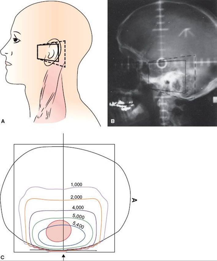
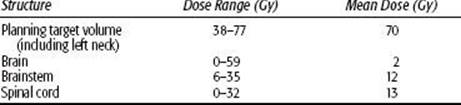
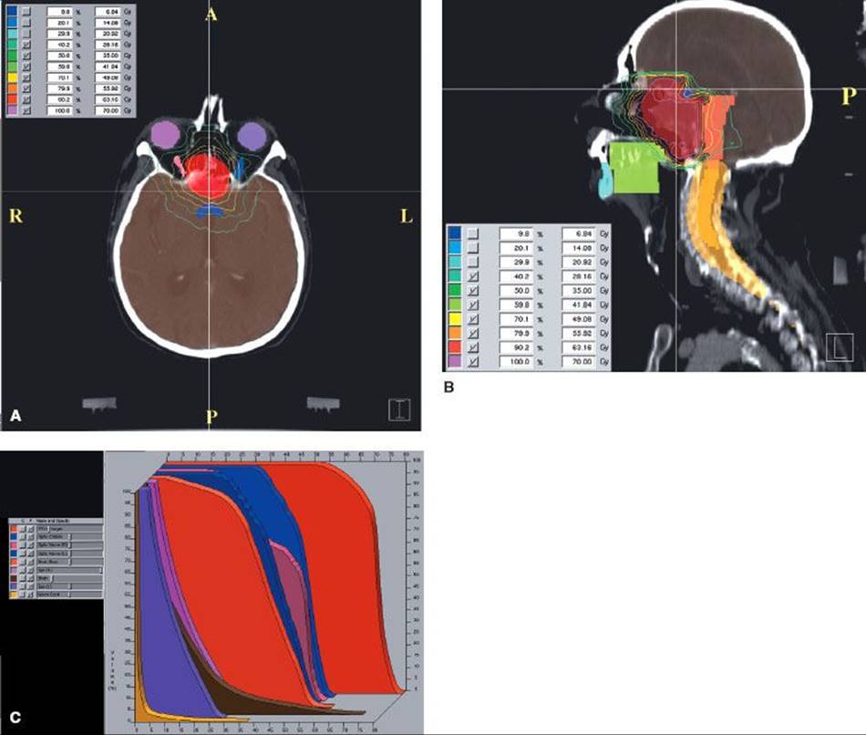
FIGURE 48.5. Fifty-nine-year-old woman with an unusual malignant left glomus jugulare, who had a metastatic left upper cervical lymph node. She was treated definitively with intensity-modulated radiation therapy (66 Gy in 2-Gy fractions). Cross (A), coronal (B), and sagittal (C) sections showing dose distributions at primary site and left neck, sparing normal structures (D) dose–volume histogram:

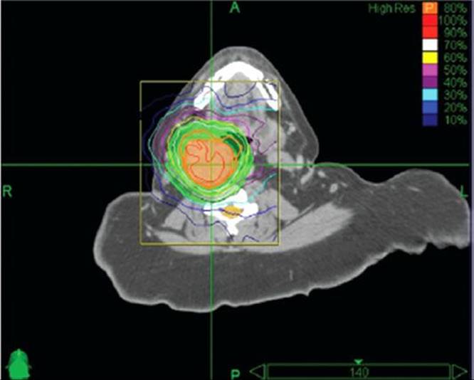
Leber et al.30 reported on 13 patients with glomus tumors treated with radiosurgery in 6, because of recurrences after surgical removal. Histology was not available in seven patients, diagnosis was made from neuroradiological features only. With mean follow-up of 42 months (range 14 to 72 months), there was no tumor progression and no clinical deterioration in any patient; 64% of the patients had improvement of symptoms, and in 36% the volume of the lesion decreased in size. There was no radiation-related morbidity. In recent years there has been an increasing number of reports in small series of patients with glomus tumors <2 or 3 cm treated with stereotactic radiation therapy, with tumor control over 80% and relatively minimal morbidity. The mean single dose is about 16 Gy (range 13 to 20 Gy).31,32,33,34,35,36,37 A report on fractionated stereotactic irradiation (6 MV x-rays) of 17 patients has been published, with a dose of 57 Gy.38 Pollock39 reported on 42 glomus tumors (19 primary treatment and 23 recurrences after initial surgery) treated with Gamma Knife (Elekta Corp, Stockholm) stereotactic radiation (12 to 24 Gy single dose at 50% isodose, depending on tumor size). Twelve lesions (31%) decreased in size and 26 were unchanged. The most common complication was hearing loss (19%). Wegner et al.40 treated 18 patients with carotid or jugular lesions with fractionated stereotactic CyberKnife (Accuray, Sunnyvale, CA) irradiation (21 Gy in 3 fractions or 25 Gy in 5 fractions) (Fig. 48.6). Tumor size was stable in 17 patients and decreased in 1 patient.
TABLE 48.4 LOCAL CONTROL WITH RADIATION THERAPY FOR CHEMODECTOMA OF THE TEMPORAL BONE (GLOMUS TYMPANICUM AND JUGULARE)

FIGURE 48.6. Dose distribution with stereotactic radiation therapy (radiosurgery) for glomus tumor. Patient was treated with 25 Gy in 5 fractions prescribed to the 80% isodose line; 98% of tumor received the prescribed dose. Thick orange line represents the 80% isodose. (From Wegner RE, Rodriguez KD, Heron DE, et al. Linac-based stereotactic body radiotherapy for treatment of glomus. Radiother Oncol 2010;97:395–398, with permission.)
Results of Therapy
The postirradiation change in tumor size is slow, with an increase in proliferative and perivascular fibrosis and minimal alterations in the chief epithelial cells. Histologic evaluation of tumor cell viability is not reliable.24 Despite the persistence of tumor both clinically and angiographically, amelioration of symptoms, absence of disease progression, and occasional return of cranial nerve function have been reported. Seventeen patients were treated for glomus tympanicum tumors at Washington University.3 In five patients, initial treatment consisted of irradiation alone, and all were tumor free at last follow-up (4.5 years in one patient) or at death. Seven of eight patients irradiated for surgical recurrence were free of disease 4.5 to 19 years after irradiation. The remaining four patients were treated preoperatively or postoperatively; only one had recurrence and was salvaged surgically and tumor free 10 years later. Of six patients with glomus jugulare lesions treated with irradiation, two with extensive lesions died of their disease, whereas the glomus tumor was controlled in four, including two patients with intracranial extension. Irradiation doses ranged from 46 to 52 Gy, with 86% to 100% tumor control with doses over 46 Gy and 50% (two of four) with doses below 46 Gy.
Wang et al.46 reported on 32 patients with tympanic chemodectomas; 13 treated with surgery alone, 15 with irradiation alone, and 4 with a combination of both modalities. The initial tumor control rate was 46% with surgery alone; ultimately 84% of patients were tumor free after salvage with additional surgery. Although 78% survived 10 years, 31% developed complications. Of the patients treated with irradiation, 84% had initial local tumor control; 77% survived 10 years, and only 11% developed complications. The doses of irradiation used were slightly higher than those reported by others (mean 58.32 Gy). However, no improvement in tumor control was noted with higher doses. Complications occurred in two patients receiving 66 Gy.
Zabel et al.47 described results in 22 patients with large chemodectomas of the skull base (8 after primary surgery and 4 after embolization), treated with fractionated stereotactic irradiation (median total dose 57 Gy with median fraction dose 1.8 Gy). With a median follow-up of 5.7 years, 5- and 10-year actuarial tumor control was 90%, with 7 patients (32%) having a partial response and 13 patients with (59%) stable tumors. No patient developed new neurological deficit.
In a compilation of several studies, Kim et al.48 noted a 25% local failure rate in 83 patients treated with <40 Gy and 1.4% local failure in 142 patients receiving >40 Gy.
Powell et al.22 reported on 84 patients with chemodectoma of the head and neck, 46 of which were in the glomus jugulare and tympanicum, treated with irradiation alone (45 to 50 Gy in 25 fractions). Local control of the lesion was 73% at 5 years. Thirty patients were treated with surgery after irradiation with no recurrences (median follow-up of 9 years). Four patients, treated with surgery alone, developed recurrences by 7 years. Four carotid body and glomus vagal tumors treated with irradiation were locally controlled at 1, 2, 8, and 11 years, respectively. In 13 patients treated with surgery alone, the 15-year local control rate was 54%.
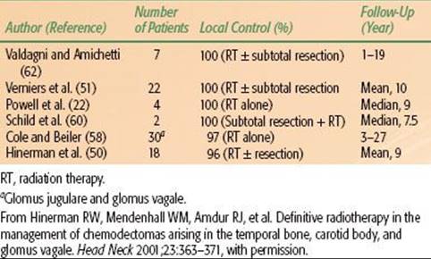
Hinerman et al.49,50 updated a previous report with 104 patients who had 121 chemodectomas of the temporal bone, carotid bone, or glomus vagal treated with radiation therapy alone in 104 patients or subtotal resection with or without radiation therapy (17 tumors). Seventeen patients had undergone a previous treatment (surgery 14, irradiation 1, or both 2). Eighty-nine patients were treated with megavoltage radiation therapy, 15 with stereotactic fractionated, 6 with stereotactic single dose, and 11 with IMRT. Median dose with fractionated irradiation was 45 Gy with daily fractions of 1.5 to 2 Gy delivered with 60Co, 6-MV, or 8-MV x-rays, or a combination of different beam energies.50 There were six tumor recurrences, with a local tumor control of 95%. No severe treatment complications were noted. In the initial report,49 18 patients had 25 chemodectomas of carotid body and/or glomus vagal; 15 tumors originated in the carotid body and 10 in the glomus vagal. Pathologic confirmation of chemodectoma was obtained in 10 patients, and diagnosis was based on physical and radiographic findings in the remaining 8 patients. Twenty-two lesions were treated with radiation therapy alone and two received postoperative radiation therapy after surgical resection for gross residual tumor with malignant changes and lymph node involvement. Patients with benign glomus tumors received 45 Gy in 25 fractions, in most instances, whereas patients with malignant carotid body tumors received 64.8 to 70 Gy in 1.8-Gy fractions. Local tumor control was obtained in 14 of 15 carotid body and 10 of 10 glomus vagal (overall 96% tumor control).51
Ivan et al.52 published a meta-analysis based on 869 patients in 46 studies reported with glomus tumors, with follow-up ranging from 6 to 256 months. The tumor control rates were, for subtotal resection 69%, gross tumor resection (GTR) 86%, subtotal resection and radiosurgery 71%, and stereotactic radiosurgery (SRS) alone 95%. Posttreatment cranial nerve deficit was observed in 26% to 40% of patients treated with GTR and in about 10% of the SRS group. Guss et al.53 reported on a meta-analysis of 19 studies (335 patients) with glomus tumors treated with stereotactic radiation therapy (radiosurgery), eight publications with a median follow-up of 36 months; tumor control (unchanged or reduced tumor volume) was 95% to 96%, with various stereotactic radiation therapy (RT) techniques.
In 29 patients with post surgical recurrent glomus tumors (16 jugular, 7 carotid, 5 tympanic and one thyroid) Elshaikh et al.54 reported that the 5 year tumor conrol was 100% in 12 treated with radiation therapy and 62% in 17 treated surgically.
Cheesman and Kelly55 emphasized the importance of evaluating preoperatively the swallowing function of patients with glomus jugulare undergoing surgery, as it is common for these patients to develop postsurgical dysphagia.
The results of primary treatment for temporal bone chemodectoma are summarized in Table 48.5.
The initial results of treatment for carotid body or glomus vagal are summarized in Table 48.6. Complications were rare in patients treated with chemodectoma of the head and neck.
TABLE 48.5 TEMPORAL BONE CHEMODECTOMAS: LOCAL CONTROL AFTER RADIATION THERAPY ALONE OR RADIATION THERAPY AND SURGERY
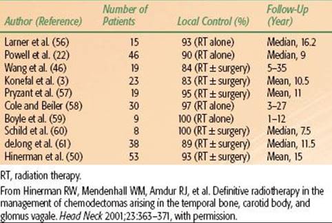
TABLE 48.6 CHEMODECTOMAS OF CAROTID BODY/GLOMUS VAGALE: RADIATION THERAPY ALONE OR RADIATION THERAPY AFTER SURGERY
HEMANGIOPERICYTOMA
Hemangiopericytomas are rare soft tissue neoplasms that account for 3% to 5% of all soft tissue sarcomas and 1% of all vascular tumors. Some 15% to 30% of all hemangiopericytomas occur in the head and neck, and of these, approximately 5% occur in the sinonasal area. They may resemble meningiomas in the central nervous system (CNS), clinically and on imaging studies.63 Vagal paragangliomas originate within the first 2 cm of the extracranial stretch of the vagus nerve and are associated with the inferior ganglion.64 These tumors are believed to originate from the pericytes of Zimmerman extravascular cells, morphologically resembling smooth muscle, found around the capillaries or from primitive mesenchymal cells. The function of the pericyte is uncertain but is believed to provide mechanical support for the capillaries having contractile function.65
Epidemiology
Hemangiopericytomas is an unusual tumor; it represents approximately 1% of all vascular neoplasms; it occurs in both genders with equal frequency and is found primarily in adults. Only 45 cases of primary hemangiopericytomas of bone were described in the world literature in 1988.
In the head and neck, the most common sites are the nasal cavity and the paranasal sinuses, and, less frequently, the orbital region, the parotid gland, and the neck.66,67,68,69 Hemangiopericytomas represent 3% to 4% of all meningeal and <1% of CNS tumors.
Pathology
Hemangiopericytomas are composed of a proliferation of tightly packed pericytes around thin-walled endothelial-lined vascular channels, ranging from capillary-sized vessels to large, gaping sinusoidal spaces.66 The tumor has a tendency to grow slowly and invade locally into adjacent structures.68,70 Although they are always well circumscribed and partially or completely surrounded by a pseudocapsule, benign tumors may be difficult to differentiate from malignant tumors. However, prominent mitoses (>4 per high-power field), foci of necrosis, and increased cellularity are suggestive of malignancy; the definitive sign is local recurrence or development of metastases. In general, tumors of the CNS, lower extremity, and mediastinum tend to be more malignant, with local recurrence occurring in up to 50% of cases.66
Kowalski and Paulino71 reviewed 12 cases of hemangiopericytomas. Proliferation index was assessed using an immunoperoxidase stain for MIB-1 (Ki-67). The mitotic index per 10 high power fields varied from 0 or 1 to 15. Proliferation indices using MIB-1 ranged from 2.6% to 52.5%. Clinical follow-up revealed three cases with recurrence all possessing proliferation indices of approximately 10%, indicating a more aggressive subset of hemangiopericytomas. Vuorinen et al.72 found the proliferation index to be a poor predictor of prognosis. In a review of 23 cases, Sundaram et al.63 found that all hemangiopericytomas were negative for epithelial membrane antigen and S-100 and all were positive for vimentin.
Meningeal hemangiopericytomas almost always recur, despite seemingly complete removal, due to infiltrative properties of hemangiopericytoma cells and not just higher proliferation potential. They often metastasize.
Clinical Presentation
Soft tissue hemangiopericytoma is a firm, painless, slowly expanding mass that is often nodular and well localized. The skin overlying the mass does not have any discoloration or redness to indicate its vascular origin because the capillaries are emptied of the blood by compression of massive numbers of pericytes surrounding them.64,66
In the head and neck, the tumor may constitute a polypoid, soft gray or red mass that grows slowly and may cause nasal obstruction. Epistaxis and nasal obstruction are common symptoms. Orbital hemangiopericytomas account for 3% of orbital malignancies and most frequently occur with painless proptosis.73 Hemangiopericytoma rarely originates in the lacrimal sac; it occurs in a younger age group than that of hemangiopericytoma of other locations. Charles et al.74 reported on seven cases previously described and added one case.
Hemangiopericytoma may occur intracranially. When it arises in the brain, it is a solid mass attached to the meninges that grossly resembles a meningioma. These intracranial hemangiopericytomas carry a high risk of local failure (80%), as well as higher potential for dissemination. The mean time for local recurrence is 75 months.75
The incidence of metastasis, which depends on the site of origin, can be 50% to 80%. Late metastases occurring 10 years after diagnosis are not uncommon.
On plain radiographs, hemangiopericytoma appears as a soft tissue mass in the nasal cavity or other portions of the head and neck. A defect caused by pressure erosion of the surrounding bones may occur, and calcifications are rare. In the neck, the tumor appears as a well-circumscribed, homogeneously and intensely enhancing mass on CT. On MRI, the mass is iso- to slightly hyperintense to muscle on T1- and T2-weighted imaging. Multiple, branching flow voids are typically seen within the tumor on both T1- and T2-weighted images. On T2-weighted imaging, the punctate black flow voids in cross-section within the relatively bright tumor, creating a characteristic “salt and pepper” appearance in tumors >2 cm in diameter. Additionally, flow voids of large feeding arteries are seen at the periphery of the mass. Angiography demonstrates the characteristic appearance of a vascular tumor, with large feeding arteries, intense tumor stain, and early draining veins.64 On arteriography, according to Yaghmai,76 hemangiopericytoma is the only vascular tumor that has radially arranged or spiderlike branching vessels around and inside the tumor and a long-standing, well-demarcated tumor stain. Intracranial tumors typically have arterial blood supply from both meningeal and cerebral connections, with one to three main feeders supplying many small corkscrew-like vessels.75 The most distinctive and constant feature of this tumor is its hypervascularity, which may be demonstrated with contrast-enhanced CT.67 Intracranially, the diffusely enhancing tumor may closely resemble a meningioma on CT. However, some CT signs may suggest hemangiopericytoma rather than meningioma, such as a lack of calcification, scarce surrounding edema, and ringlike enhancement. Both CT and MRI scans are of special value in the delineation of the full extent of the tumor.
General Management
Complete surgical resection, if possible, combined with preoperative embolization of the tumor, is the treatment of choice. More extensive surgery is required in tumors that show features of malignancy. Many patients undergo surgical treatment after embolization of the feeding artery(ies).
For incompletely resected tumors, postoperative radiation therapy is used.77,78 The role of chemotherapy in this tumor is not well determined; a few reports have described partial tumor regression in some lesions treated with cytotoxic agents. Doxorubicin, alone or in combination-drug regimens, is the most effective agent for metastatic hemangiopericytoma, producing complete and partial remissions in 50% of cases. Other drugs prescribed when metastasis occurs are cyclophosphamide, dacarbazine, vincristine, and actinomycin-D.79 Park et al.80 reported on 14 patients with soft tissue hemangiopericytoma treated with temozolomide 150 mg/m2 orally on days 1 to 7 and days 15 to 21 and bevacizumab 5 mg/kg intravenously on days 8 and 22, repeated at 28-day intervals. Median follow-up period was 34 months. Eleven patients (79%) achieved a Choi partial response, with a median time to response of 2.5 months. The estimated median progression-free survival was 9.7 months, with a 6-month progression-free rate of 78.6%. The most frequently observed toxic effect was myelosuppression.
Radiation Therapy Techniques
The role of radiation therapy alone in the management of hemangiopericytoma is controversial. The main role of irradiation is as an adjuvant after complete excision of the lesion or postoperatively for minimal residual disease.69,81,82 The tumor has been considered relatively radioresistant. Tumor doses of 60 to 65 Gy in 6 to 7 weeks are required to produce local tumor control in postoperative cases.83 Orbital hemangiopericytoma has been cured by surgery and postoperative irradiation to 65 Gy.73
There appears to be a definite role for postoperative irradiation to the brain for primary hemangiopericytoma when radical surgery is performed because these tumors tend to recur after seemingly complete removal. Jha et al.82reported local tumor control in all patients treated with adjuvant external-beam irradiation postoperatively. Radiation therapy also has been used as a salvage procedure after local recurrence following initial surgery or chemotherapy.
The fields of irradiation should be wide to encompass the tumor bed with a margin of at least 5 cm to safely avoid marginal recurrence. Portal arrangement and beam selection are similar to those used in treatment of malignant brain tumors or soft tissue sarcomas.
Results of Therapy
Billings et al.84 reported on 10 patients with hemangiopericytoma of the head and neck; seven tumors arose from soft tissue sites and three from the mucosa. All patients underwent wide excision of the primary lesion with a local recurrence rate of 40%. Three patients developed metastatic lung disease 0 to 8 years after initial diagnosis. Each patient who developed metastatic disease had abundant mitoses on pathological review compared with rare or absent mitoses in the lesions that took a more benign course.
Patrice et al.85 reported on 18 primary hemangioblastoma tumors (16 had no prior surgical resection and 2 were subtotally resected lesions) and 20 lesions treated after surgical failure with stereotactic irradiation (radiosurgery). Minimum tumor doses ranged from 12 to 20 Gy (median 15.5 Gy). With a median follow-up of 24.5 months (range 6 to 77 months), the 2-year actuarial survival was 88%, and the 3-year freedom from progression was 86%. Four of 22 patients died. Thirty-one of 36 evaluable tumors (86%) were controlled locally. None of the 18 primary tumors treated with definitive stereotactic irradiation failed. Of the 18 recurrent tumors, 13 (72%) were controlled. There were no significant permanent complications attributable to the stereotactic irradiation.
Spitz et al.69 published a report on 36 patients with hemangiopericytoma. The median follow-up was 57 months. Twenty-eight patients (78%) underwent complete and potentially curative resection. Of the nine patients (32%) who had local recurrences, four (44%) had epidural tumors and three (33%) had retroperitoneal tumors, but none had extremity tumors. Ten patients had recurrences at distant sites. Of the 13 patients who experienced any form of disease recurrence, four had recurrences after a disease-free interval of more than 5 years. The 5-year actuarial survival rate for the entire group of 36 patients was 71%.
Carew et al.86 reviewed the records of 12 patients with hemangiopericytomas of the head and neck: 5 had high or intermediate grade lesions and 7 had low-grade lesions. Nine patients were treated with curative intent; they underwent a variety of surgical resections dictated by tumor location and size. Four patients received postoperative radiation therapy, to a median dose of 60 Gy, for positive surgical margins (two patients), high-grade histology (one patient), or a recurrent lesion (one patient). The 5-year overall survival rate for patients treated surgically was 87.5%. A single mortality occurred in a patient with a recurrent high-grade lesion who failed at local, regional, and distant sites.
Staples et al.87 reported on 12 patients with localized hemangiopericytoma, 7 treated with surgery alone (only 1 had long-term tumor control and 2 were salvaged with radiation therapy), 4 with resection and postoperative irradiation (all with long-term tumor control), and 1 with surgery and chemotherapy. Local tumor control was achieved at all sites treated with doses >55 Gy. Mitotic activity was not a reliable predictor of biologic behavior.
Kim et al.48 evaluated 17 hemangiopericytomas in nine patients treated with Gamma Knife stereotactic radiation therapy. Mean and median marginal doses were 18.1 and 20 Gy (range 11 to 22 Gy), respectively, at the 50% isodose line. Mean clinical and radiological follow-up periods were 49 and 34 months, respectively. Successful tumor control was achieved in 14 of 17 lesions (82.4%). Actuarial local tumor control rates at 5 years was 67%. No adverse effects, such as radiation necrosis or marked peritumoral edema, were observed. Marginal dose (≥17 Gy) was the only statistically significant factor for local tumor control on univariate analysis.
Kano et al.88 in a retrospective review of 20 patients who had undergone stereotactic radiation therapy for 29 hemangiopericytomas. All patients had undergone previous surgical resection. In addition, 12 patients underwent fractionated radiotherapy before stereotactic radiation therapy. Of the 20 patients, 16 patients had low-grade hemangiopericytomas (20 tumors) and 4 had high-grade anaplastic hemangiopericytomas (9 tumors). The median target volume was 4.5 cm3 and the median marginal dose was 15 Gy (range 10 to 20 Gy). At an average of 48.2 months, the overall survival after radiosurgery was 85.9% and 13.8% at 5 and 10 years, respectively. Follow-up imaging studies demonstrated tumor control in 21 (72.4%) of 29 tumors. The progression-free survival rate after stereotactic radiation therapy at 3 and 5 years was 89.1% for low-grade hemangiopericytomas and 66.7% and 0%, respectively, for high-grade hemangiopericytomas. The factors associated with improved progression-free survival included lower grade and >14 Gy marginal radiation dose.
Olson et al.89 published a review of 21 patients with 28 recurrent or residual hemangiopericytomas on whom radiosurgery was performed. Prior treatments included embolization (6 cases), transcranial resection (39 cases), transsphenoidal resection (2 cases), and fractionated radiotherapy (8 cases). The mean prescription and maximum radiosurgical doses to the tumors were 17.0 and 40.3 Gy, respectively. Repeat radiosurgery was used to treat 13 tumors. With median follow-up of 68 months (range 2 to 138 months), local tumor control was 47.6% (10 of 21 patients). Of the 28 tumors treated, 8 decreased in size on follow-up imaging (28.6%), 5 remained unchanged (17.9%), and 15 ultimately progressed. Progression-free survival at 5 years was 28.7%, and it improved to 71.5% after multiple radiosurgery treatments. Prior fractionated irradiation or radiosurgical prescription dose did not correlate with tumor control. In 4 of 21 (19%) patients, extracranial metastases developed. Redmond et al.90 reported on 118 patients with hemangiopericytoma of the CNS, 9% of whom had distant metastases at the time of initial presentation; 112 patients underwent surgical resection (23% had GTR, 31% subtotal resection, and 46% had surgery not otherwise specified). Adjuvant RT was received by 31% of patients following GTR, 44% after subtotal tumor resection, and 50% of patients following surgery not otherwise specified. The 5- and 10-year overall survival for all patients was 76.7% and 50.1%, respectively. Patients receiving adjuvant RT (n = 42) had a significantly better overall survival than patients who did not (n = 67; 10-year overall survival was 66.2% vs. 40.7%; P = .05). There was no difference in 5- or 10-year overall survival for patients treated with subtotal resection plus RT (n = 16) compared with those treated with GTR alone (n = 14; 10-year overall survival 75% vs. 57.1%; P = .53).
CHORDOMAS
Anatomy
Chordomas are rare neoplasms of the axial skeleton that arise from the remnant of the primitive notochord (chorda dorsalis). About 50% arise in the sacrococcygeal area; 35% arise intracranially, where they typically involve the clivus, and the remaining 15% occur in the midline along the path of the notochord, primarily involving the cervical vertebrae.91
Epidemiology
Chordomas are more common in patients in their 50s and 60s but can occur in all age groups. In children and young adults, the prognosis and long-term survival appear to be better than in older patients. No risk factors have been identified. Male predominance is reported at a 2:1 to 3:1 ratio.
Natural History
Although slowly growing, chordomas are locally invasive, destroying bone and infiltrating soft tissues. Basisphenoidal chordomas tend to cause symptoms earlier and may be difficult to differentiate histologically from chondromas and chondrosarcomas and radiographically from craniopharyngiomas, pineal tumors, and hypophyseal and pontine gliomas. The lethality of these tumors rests on their critical location, aggressive local behavior, and extremely high local recurrence rate. The incidence of metastasis, which has been reported to be as high as 25%, is higher than previously believed and may be related to the long clinical history. The most common site of distant metastasis is the lungs, followed by liver and bone. Lymphatic spread is uncommon.
Pathology
Chordoma is a soft, lobulated tumor that may have areas of hemorrhage, cystic changes, or calcification. It is frequently encapsulated but may be nonencapsulated or pseudoencapsulated. Histologically, it is composed of cords or masses of large cells (physaliferous cells) with typical vacuoles and granules of glycogen in the cytoplasm and abundant intercellular mucoid material. Usually there are few mitotic cells.92 A chondroid variant of chordoma may exist, being prevalent in the spheno-occipital area. Patients with this type of histologic variant have improved survival.
Aside from the previously mentioned histologic features, the prognostic factors that most influence the choice of treatment are location and local extent of tumor.
Clinical Presentation
Chordomas tend to originate from the clivus and chondrosarcomas from the temporal bone.93 Clinical symptoms vary with the location and extent of the tumor. In the head, extension may be intracranial or extracranial, into the sphenoid sinus, nasopharynx, clivus, and sellar and parasellar areas, with a resultant mass effect. In chordomas of the spheno-occipital region, the most common presenting symptom is headache. Other presentations include symptoms of pituitary insufficiency, nasal stuffiness, bitemporal hemianopsia, diplopia, and other cranial nerve deficits. Volpe et al.94 reviewed the clinical features of 48 patients with chordoma and 49 patients with low-grade chondrosarcoma of the skull base. Twenty-five patients (52%) with chordoma and 24 patients (49%) with chondrosarcoma had ocular symptoms (diplopia or visual impairment) as the initial manifestation of the disease. Of the 59 patients (both groups) with diplopia, the diplopia was initially intermittent in 25 (42%). Headache and diplopia from abducens nerve palsy occurred in 22 patients (46%) with chordoma and 23 (47%) with chondrosarcoma.
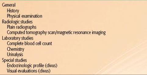
TABLE 48.7 DIAGNOSTIC WORKUP FOR CHORDOMA
Diagnostic Workup
The diagnostic workup varies with the primary location of disease. Most patients have significant bony destruction, and some may have calcifications in the tumor; hence, plain films and, specifically, CT scans or MRI are very useful (Table 48.7).95 In most cases, the soft tissue component is much more extensive than initially appreciated, and a CT scan with contrast enhancement is required (Fig. 48.7A). CT and MRI are equivalent for demonstration of the presence and site of these tumors. MRI is inferior to CT in its ability to demonstrate bony destruction and intratumoral calcification (Fig. 48.7B).96 MRI is superior to CT regarding the delineation of the exact extent of the tumor, which allows for better treatment planning.95 Because of availability and lower cost, CT appears to be the technique of choice for routine follow-up of previously treated patients.96
Reliable signs of chordoma of the skull base are posterior extension to the pontine cistern; a lobulated, “honeycomb” appearance after gadolinium; the swollen appearance of the bone in the early stages; bone erosion on CT; and frequent extension to critical structures such as the circle of Willis, cavernous sinuses, and brainstem.96
General Management
Because of their surgical inaccessibility and relative resistance to radiation therapy, clivus chordomas represent a formidable therapeutic challenge. The general management of the patient is dictated by the anatomic location of the tumor and the direction and extent of spread. A surgical approach is recommended (when feasible), but complete surgical extirpation alone is unusual.97 Regression of preoperative symptoms without additional postoperative morbidity could be achieved by radical transoral tumor extirpation documented by MRI. Intracranial spread usually requires steroid coverage and therapy directed to correction of neurologic deficits that may be present. Because of the high incidence of local recurrence, combined surgical excision and irradiation is frequently used. No effective chemotherapeutic agent or combination of drugs has been identified.
Radiation Therapy Techniques
Irradiation techniques vary considerably, depending on the location of the tumor along the craniospinal axis. Basisphenoidal tumors usually have been treated by a combination of parallel opposed lateral fields, anterior wedges, and photon and electron beam combinations, depending on the extent of the neoplasm. Precision radiation therapy planning, using CT and MRI, is required because high doses of external-beam radiation therapy are needed. Three-dimensional-CRT or IMRT provide optimal dose distributions.
The tumor usually surrounds the spinal cord and infiltrates vertebral bones. A combined technique using protons or electrons to boost the initial photon fields is generally applied. In the treatment of chordomas surrounding the spinal cord, IMRT can provide high-dose homogeneity and planning target volume (PTV) coverage (Fig. 48.8). Frequent digital portal image-based setup control reduces random positioning errors for head and neck cancer patients immobilized with conventional thermoplastic masks. Gabriele et al.98 treated a patient with incomplete resection of a vertebral chordoma surrounding C2-3 with a total dose of 58 Gy in 2-Gy daily fractions. Beam arrangement consisted of seven 6 MV nonopposed coplanar IMRT fields using 120-leaf collimator in sliding window mode. To verify the daily setup, portal images at 0 degrees and 90 degrees were compared with the simulation images before treatment delivery (manual matching) and after treatment delivery (automatic anatomy matching). The mean dose to the PTV was 57.6 Gy covering 95% of the PTV with the 95% isodose. The minimum dose to the PTV (D99) was 53.6 Gy. The maximum dose to the spinal cord was 42.2 Gy and to the spinal cord planning risk volume (8 mm margin) 53.7 Gy. The mean dose to the parotids were 37.4 Gy (homolateral gland) and 19.5 Gy (contralateral gland). Because of the slow proliferative nature of chordomas, high linear energy transfer may prove useful in their management, as it will be discussed later. Brachytherapy can be used for recurrent tumors of the base of skull or adjacent to the spine when a more aggressive surgical exposure is offered.
Results of Therapy
Photons
Although survival in some patients with chordoma may be long term, the salient feature of this unusual neoplasm is local recurrence with eventual death. The course may be indolent, with multiple treatments for recurrences, but the overall 5-year disease-free survival rate is <10% to 20%. Catton et al.99 analyzed the long-term results of treatment for patients with chordoma of the sacrum, base of skull, and mobile spine treated predominantly with postoperative photon irradiation. In 20 base of skull chordomas, most of them irradiated with conventionally fractionated radiation to a median dose of 50 Gy in 25 fractions for 5 weeks (range 25 to 50 Gy), median survival was 62 months (range 4 to 240 months) from diagnosis with no difference between clival and nonclival presentations. There was no survival advantage to patients receiving radiation doses >50 Gy (median 60 Gy) compared with lower doses <50 Gy (median 40 Gy). Hyperfractionation regimens did not influence the degree or duration of symptomatic response or progression-free survival. Median survival after retreatment was 18 months.
Forsyth et al.100 reported on 51 patients with intracranial chordomas (19 classified as chondroid) treated surgically (biopsy in 11 patients and subtotal removal or greater in 40); 39 patients received postoperative irradiation. At the time of the analysis, 17 patients were alive. The 5- and 10-year survival rates were 51% and 35%, respectively; 5-year survival was 36% for biopsy patients and 55% for those who had resection. Patients who underwent postoperative irradiation tended to have longer disease-free survival times.
Gay et al.101 analyzed the outcome of 46 patients with cranial base chordomas and 14 with chondrosarcomas after extensive surgical resection, 50% of them treated previously; 20% received postoperative irradiation. Nine patients with chordomas and two with chondrosarcomas died during the postoperative follow-up period. The 5-year recurrence-free survival for all patients was 76%. Chondrosarcomas had a better prognosis than chordomas (5-year recurrence-free survival of 90% and 65%, respectively; P = .09). Patients who had undergone previous surgery had a greater risk of recurrence than did those who had not undergone previous surgery (5-year recurrence-free survival rates of 64% and 93%, respectively; P <.05). Those with total or near-total resection had a better 5-year recurrence-free survival rate (84%) than did patients with partial or subtotal resection (64%; P <.05). Postoperative leakage of cerebrospinal fluid was the most frequent complication (30% of patients) and was found to increase the risk of permanent disability. Patients who had undergone previous irradiation had a greater risk of death in the postoperative period (within 3 months of operation) and during follow-up.
Tai et al.91 reviewed the results of irradiation combined with surgery, irradiation alone, and surgery alone in 159 patients reported in the literature. An analysis of the optimal biologically equivalent dose was performed using the linear-quadratic formula on 47 patients. With conventional photon irradiation, no dose–response relationship was shown. Survival improved in patients undergoing surgery followed by irradiation.
Chetty et al.102 reported on 18 chordomas, 61% of them occurred in the sphenoid region. Follow-up for 12 patients ranged from 3 to 170 months. Various combinations of surgery and radiation therapy were used. Mean survival was 73.4 months, with a survival rate of 50% (6 of 12 patients).
Keisch et al.103 reported on 21 patients with chordoma treated at the authors’ medical center: 5 had clival tumors, 2 had nasopharyngeal tumors, and 1 had a lumbar spine tumor. Nine patients were treated with surgery alone, eight had subtotal resection and postoperative irradiation, and four received irradiation alone after biopsy. The 5- and 10-year actuarial survivals were significantly better in patients treated with surgery alone or surgery and irradiation than in those treated with radiation therapy alone (52%, 32%, and 0%, respectively; P = .02). Disease-free survival of patients with base of skull tumors was not significantly different among the treatment groups.
FIGURE 48.7. A: Contrast material-enhanced axial computed tomography scan demonstrates a large chordoma with extension into the posterior fossa and left parasellar region. B: Computed tomography scan photographed at bone windows shows the bony destruction and intratumoral calcifications. C:Treatment planning field arrangement for illustrated clivus chordoma using standard irradiation techniques with wedges on lateral ports.
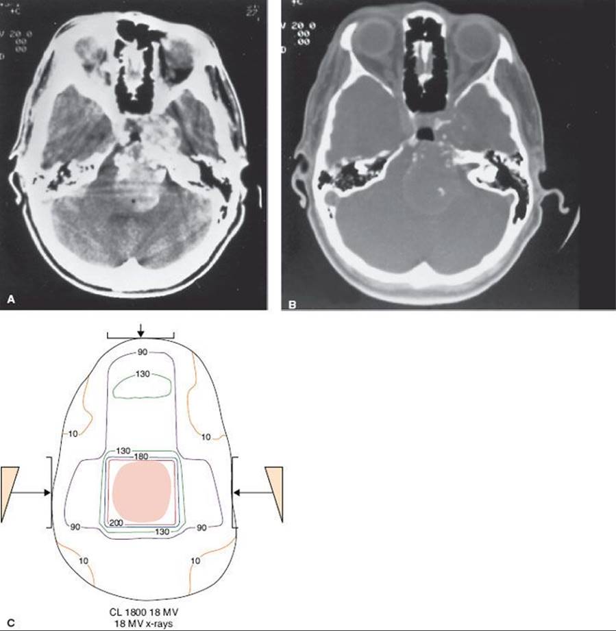
FIGURE 48.8. Chordoma of clivus in 81-year-old man treated with 70 Gy in 2-Gy fractions. Example of intensity-modulated radiation therapy plan: A: Cross-section in upper portion of planning target volume (PTV), demonstrating coverage of target volume with sparing of ocular structures. B: Sagittal plane dose distribution with excellent coverage of PTV. C: Dose–volume histogram:

Debus et al.104 reported on 45 patients treated for chordoma or chondrosarcoma with postoperative fractionated 3D stereotactic radiation therapy. Median dose at isocenter was 66.6 Gy for chordomas and 64.9 Gy for chondrosarcomas. All chondrosarcomas achieved and maintained local tumor and recurrence-free status at 5-years follow-up. Local control rate of chordomas at 5 years was 50% and survival was 82%. Clinically significant late toxicity developed in only one patient.
Bugoci et al.105 published results on 12 patients with skull-base chordoma treated with fractionated stereotactic RT and IMRT boost (total dose 74 Gy in 2-Gy fractions). With median follow-up of 34 months, local tumor control at 3 years was 65% and overall survival 92%.
Den et al.106 described a multi-institutional study of 31 patients, 28 with clivus chordoma or chondrosarcoma, treated with various photon techniques (single-dose stereotactic, fractionated 3D-CRT, IMRT). Median fractionated total dose was 65 Gy. Actuarial 3-year local tumor control was 68% and overall survival 63%. Patients receiving 65 Gy or a higher dose had a 5-year tumor control of 78% and overall survival of 90%. No grade 2 or greater toxicity was observed.
Muthukumar et al.107 published a report on 15 patients with skull-base chordoma and chondrosarcoma treated with stereotactic radiation therapy (13 had previous surgical resection). Median minimum marginal tumor single dose was 18 Gy (12 to 20 Gy) and the number of isocenters ranged from 1 to 10 (average, 4). Dose to optic nerve or chiasm was ≤9 Gy. With median follow-up of 40 months, eight patients had clinical improvement, three were stable, and four had died. No significant morbidity was noted.
Protons
The best results in the treatment of chordomas have been obtained with radical surgery followed by high-dose proton irradiation.108 Berson et al.109 described 45 patients with chordomas or chondrosarcomas at the base of the skull or cervical spine treated by subtotal resection and postoperative irradiation. Twenty-three patients were treated definitively by charged particles, 13 patients with photons and particles, and 9 were treated for recurrent disease. Doses ranged from 36 to 80 Gy equivalent (GyE). There appeared to be significant benefit for patients with smaller tumor volumes (80% vs. 33% actuarial survival rate at 5 years). Patients treated for primary disease had a 78% actuarial local tumor control at 2 years versus 33% for recurrent disease.
Austin et al.110 evaluated 141 patients with chordoma and chondrosarcoma of the base of skull and cervical spine treated with proton and photon irradiation. The local disease was controlled in 111 patients. They reviewed 26 patients who had recurrent disease (21 nonchondroid chordomas, two chondroid chordomas, and three chondrosarcomas). The prescribed doses ranged from 67 to 72 cobalt Gray equivalent (CGE). Approximately 25% (6 of 26) of the cases failed in the prescribed dose region. More than half (15 of 26) failed in regions where tumor dose was limited by normal tissue constraints. Approximately 10% of the patients recurred in the surgical pathway and 10% were judged to be marginal misses. Overall, 75% of the patients failed in regions receiving less than the prescribed dose. All tumors that failed in the high-dose region had recurrences (10 of 26) and larger tumors (average volume of 102 cc) than those with base of skull disease (16 of 115) with an average volume of 63 cc.
O’Connell et al.111 reported on 62 patients with base of skull chordomas treated with proton beam irradiation (65 to 73.5 GyE); 29 patients (19 women and 10 men) experienced local failure, and 14 women (48%) and 7 men (21%) died of disease. On histologic analysis, the presence of >10% necrosis, prominent nucleoli, and tumor >70 mm were significant predictors of short-term disease-specific survival. Chondroid chordoma and conventional chordomas had equivalent outcomes.
Fagundes et al.112 updated the Massachusetts General Hospital experience with 204 patients treated for chordoma of the base of the skull or cervical spine. Sixty-three patients (31%) had treatment failures, which were local in 60 patients (29%) and the only site of failure in 49 patients. Two patients had regional lymph node relapse, and three developed surgical pathway recurrence. Thirteen patients relapsed in distant sites (especially lungs and bones). The 5-year actuarial survival rate after any relapse was 7%. There was no significant difference in survival for patients who had a local or distant failure. Two patients (1.4%) with local tumor control developed distant metastases in contrast with 10 of 60 patients (16%) who failed locally and distantly.
Terahara et al.113 reported on 132 patients with skull-base chordoma treated with combined photon and proton irradiation; in 115 patients dose–volume data and follow-up were available. The prescribed doses ranged from 66.6 to 79.2 CGE (median 68.9 CGE). Dose to the optic structures (optic nerves and chiasm), the brainstem surface, and the brainstem center was limited to 60, 64, and 53 CGE, respectively. Local failure developed in 42 of 115 patients, with the actuarial local tumor control rates at 5 and 10 years being 59% and 44%, respectively. In a Cox multivariate analysis, the model’s equivalent uniform dose suggests that the probability of recurrence of skull-base chordomas depends on gender, target volume, and target dose inhomogeneity; equivalent uniform dose was shown to be a useful parameter to evaluate dose distribution for the target volume.
Hug et al.114 analyzed efficacy of fractionated proton radiation therapy for 33 skull-base chordomas and 25 chondrosarcomas. Following various surgical procedures, residual tumor was present in 91% of patients; 59% demonstrated brainstem involvement. Target doses ranged from 64.8 to 79.2 CGE (mean 70.7 CGE). The range of follow-up was 7 to 75 months (mean 33 months). In 10 patients (17%) the treatment failed locally, resulting in local control rates of 92% (23 of 25 patients) for chondrosarcomas and 76% (25 of 33 patients) for chordomas. All tumors with volumes of ≤25 mL remained locally controlled compared with 56% of tumors >25 mL (P = .02). Of patients without brainstem involvement, 94% did not experience recurrence; whereas with brainstem involvement (and dose reduction because of brainstem tolerance constraints), the tumor control rate was 53% (P = .04). Actuarial 5-year survival rates were 100% for patients with chondrosarcoma and 79% for patients with chordoma. Grade 3 and 4 late toxicities were observed in four patients (7%) and were symptomatic in three (5%).
Ares et al.42 treated 42 patients with chordomas and 22 with chondrosarcomas of the skull base using spot-scanning protons (median doses 73.5 and 68.4 Gy, respectively, at 1.8 to 2.0 Gy relative biological effect). With median follow-up of 38 months, 5-year tumor control was 81% and 94% and overall survival 100% and 91%, respectively. Late toxicity consisted of one grade 3 and one grade 4 unilateral optic neuropathy and two patients with grade 3 CNS necrosis. No patient experienced brainstem toxicity.
Noel et al.115 reported on 49 chordomas and 18 chondrosarcomas treated with high-energy photons (two-thirds of dose) and 201 MeV protons (one-third of dose). Median total dose was 67 CGE (60 to 70 CGE). With median follow-up of 32 months, 3-year local tumor control was 71% for chordomas and 85% for chondrosarcomas, and 4-year overall survival 88% and 75%, respectively. Fourteen tumors (21%) failed locally.
Recently, heavy particles have been used to treat some of these patients.116, 210 Hasegawa et al.79 reported on 54 patients with skull base or paracervical tumors (31 with chordomas) treated with carbon ions (escalating doses from 48 to 60.8 GyE in 16 fractions over 4 weeks). In the 31 chordoma patients, 5-year local tumor control and overall survival were 78% and 85%, respectively. Patients were divided into two groups; a low-dose group (n = 10) irradiated with doses ranging from 48 to 57.8 GyE, and a high-dose group (n = 21) irradiated with 60.8 GyE. The 5-year local tumor control was 60% for the low-dose group and 93% for the high-dose group and the overall survival was 90% and 84%, respectively. One late grade 2 brain sequela was noted in a patient treated with 60.8 GyE.
Likewise, Schulz-Ertner et al.43 treated 24 chordomas and 13 chondrosarcomas with 3D planning carbon ions (median dose 60 GyE). With mean follow-up of 13 months, local tumor control at 2 years was 90%. Progression-free survival was 83% for chordomas and 100% for chondrosarcomas. No significant toxicity was observed.
Benk et al.117 described results in 18 children 4 to 18 years of age with base of skull or cervical spine chordomas who received fractionated high-dose postoperative irradiation using mixed-photon and 160-MeV proton beams. Median tumor dose was 69 CGE with a 1.8-CGE daily fraction. With a median follow-up of 72 months, the 5-year survival was 68%, and the 5-year disease-free survival rate was 63%. Patients with cervical spine chordomas had a worse survival rate than did those with base of skull lesions (P = .008). The incidence of treatment-related morbidity was acceptable: two cases of growth hormone deficit corrected by hormone replacement, one temporal lobe necrosis, and one fibrosis of the temporalis muscle, improved after surgery.
A report on proton therapy for base of skull chordoma published by the Royal College of Radiologists118 concluded that outcome after proton irradiation is superior to that reported for conventional photon irradiation. Radiation therapy schedules involving a mixed schedule of protons and photons have achieved an approximately 60% local tumor control rate at 5 years.
Sequelae of Treatment
In patients treated with high irradiation doses, as well as with charged particles, there is an increasing probability of sequelae, including brain damage, spinal cord injury, bone or soft tissue necrosis, and xerostomia. In a report by Berson et al.,109 three patients experienced unilateral visual loss, and four patients had radiation injury to the brainstem.
Santoni et al.119 reported on the temporal lobe damage rate in 96 patients (75 primary and 21 recurrent tumors) treated with postoperative high-dose proton and photon irradiation for chordomas and chondrosarcomas of the base of the skull. All the patients were randomized to receive 66.6 or 72 CGE with conventional fractionation (1.8 CGE per day, 5 fractions per week) using opposed lateral fields for the photon component and a noncoplanar isocentric technique for the proton component. Of the 96 patients, 10 developed temporal lobe damage (lateral in 2 and unilateral in 8). The cumulative temporal lobe damage incidence at 2 and 5 years was 7.6% and 13.2%, respectively. CT and MRI scans were evaluated for white matter changes; the MRI areas suggestive of temporal lobe damage in 10 patients were always separate from the tumor bed.
In patients receiving high-dose proton therapy for clivus tumors, Slater et al.120 observed a 26% incidence of endocrine abnormalities at 3 years and 37% at 5 years, with hypothyroidism being the most frequent sequela. The dose to the pituitary in patients with abnormalities ranged from 63.1 to 67.7 GyE.
LETHAL MIDLINE GRANULOMA
Natural History and Pathology
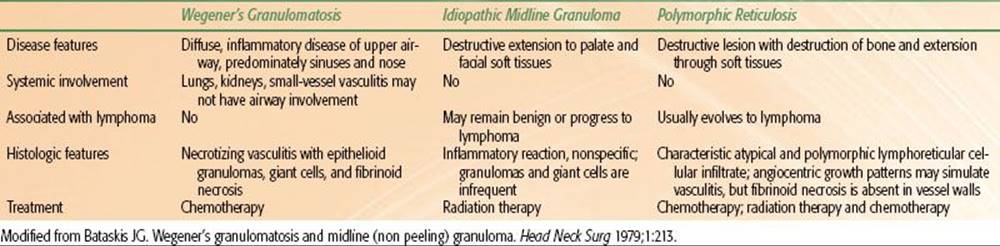
Lethal midline granuloma (LMG) or midline malignant polymorphic reticulosis is a clinical entity characterized by progressive, unrelenting ulceration and necrosis of the midline facial tissues.121,122 LMG is associated with Epstein-Barr virus, which has at least two subtypes with different biologic properties that can be identified by their genomic configuration. The occurrence of the rare subtype 2 in LMG may relate to a covert immune defect.123 Considerable controversy exists regarding various disorders characterized by a necrotizing and granulomatous inflammation of the tissues of the upper respiratory tract and oral cavity. It is now clear that if infections and other known agents such as cocaine use, sarcoidosis, environmental toxins, and various neoplasms can be excluded, three clinicopathologic entities remain: Wegener’s granulomatosis, LMG, and polymorphic reticulosis (PMR).15 A review of the literature suggests that cases described as idiopathic midline destructive disease and PMR are a large evolutionary spectrum from almost benign to fatal malignant lymphoma.124
Wegener’s granulomatosis is an epithelioid necrotizing granulomatosis with vasculitis of small vessels. Systemic involvement of the kidneys and lungs is common.
PMR is an unusual disorder with distinctive clinical and pathologic features. Histologically, PMR is characterized by an atypical mixed lymphoid infiltration of the submucosa with extensive areas of necrosis, sometimes extending to bone or cartilage. The lesion consists of variable zones of small lymphocytes with scattered immunoblastic forms, abundant plasma cells with occasional eosinophilia and histiocytosis.125 PMR has been considered a lymphoproliferative disorder; most, if not all, cases are peripheral T-cell lymphomas. Several authorities believe that PMR and systemic lymphomatoid granulomatosis are the same disease, with the latter predominantly involving the lungs.126
Idiopathic LMG describes a localized disorder not characterized by visceral lesions but by destruction of the midfacial area, which, if left untreated, is uniformly fatal. The histopathologic findings are nonspecific, with a relatively nondescript inflammatory reaction with acute and chronic inflammation and necrosis. Despite specific clinicopathologic features, the distinction between LMG and PMR is often difficult; although controversial, they may represent two phases of the same disease, with LMG remaining histologically benign or evolving into PMR. LMG occurs more frequently in men.98 Ages range from 21 to 64 years; almost half of the patients are in their 50s at presentation. Most patients have involvement of the nasal cavity (including destruction of the septum) and the paranasal sinuses (particularly maxillary antrum). The primary lesion may extend into the orbits, the oral cavity (palate, gingiva), and even the pharynx.
Characteristics of the three different diseases are outlined in Table 48.8.
Clinical Features and Diagnostic Workup
Clinical manifestations include progressive nasal discharge, obstruction, foul odor emanating from the nose, and, in later stages, pain in the nasal cavity, paranasal areas, and even in the orbits.
Examination discloses ulceration and necrosis in the nasal cavity, perforation or destruction of nasal septum and turbinates, and even ulceration of the nose. Edema of the face and eyelids may be noted, and the bridge of the nose may be sunken. Radiographic studies initially show soft tissue swelling, mucosal thickening, and findings consistent with chronic sinusitis.
CT is invaluable in demonstrating the full extent of the tumor, including bone or cartilage destruction. In 13 patients presenting with LMG, CT proved essential for determining the extent of the disease, guiding biopsy, and planning radiation therapy.127 MRI was also helpful for the latter because it could distinguish fluid retained within the paranasal sinuses from solid masses and tumor from granulation tissue; it was of little value for detecting bone lysis. Eight patients proved to have T-cell lymphoma, two had Crohn disease, in one the lesion was factitious, and two had granulomas without diagnostic histologic features.
TABLE 48.8 DIFFERENTIAL FEATURES OF THREE CLINICOPATHOLOGIC ENTITIES
General Management and Radiation Therapy Techniques
When treatment of these patients is planned, it is extremely important to exclude the diagnosis of Wegener’s granulomatosis, a benign process that is commonly treated with antimicrobial agents, steroids, and systemic chemotherapy.126 Bona fide LMG does not respond to steroids; the treatment of choice is radiation therapy.128,129,130
Target volume should encompass all areas of involvement, including adjacent areas at risk (i.e., for a lesion of the maxillary antrum, it will include the antrum as well as all of the paranasal sinuses) with a 2- to 3-cm margin.131Because marginal failures are a significant problem, wide margins are necessary for treatment of these patients.125
Irradiation techniques are similar to those described for tumors of the paranasal sinuses, nasal cavity, or nasopharynx. Several investigators have described complete responses with doses of 30 to 50 Gy; most patients are treated with 35 to 45 Gy in 3 to 4.5 weeks.129,132 The authors recommend 45 to 50 Gy in 4.5 to 5.5 weeks in 1.8- to 2-Gy daily fractions.
Results of Therapy
Because of the rarity of this tumor, experience is limited. Fauci et al.132 reported on 10 patients with extensive midline granuloma treated with irradiation. Three received 10 Gy, and all failed within 2 years (retreated with 40 to 46 Gy). The remaining seven patients received 40 to 50 Gy. Local control of disease was 77%; two patients had local recurrences, one outside the initially irradiated volume.
In a study of 34 patients with PMR treated with primary radiation therapy except for one patient, Smalley et al.125 found that a minimum dose of 42 Gy or a time-dose factor of 70 was necessary to achieve long-term local control. The most frequent failure site was within the original irradiation field. Systemic failure occurred in 25% of their patients initially presenting with limited disease. The salvage of this subset of patients requires effective systemic chemotherapy. Multimodality treatment using intensive chemotherapy and radiation therapy might improve the prognosis of these patients.
Fauci et al.132 published a prospective study of 15 patients with systemic lymphomatoid granulomatosis. Of 13 patients treated with cyclophosphamide and prednisone, seven sustained complete remission (mean duration of remission, 5.2 ± 0.6 years). Two patients receiving only prednisone and six receiving cyclophosphamide and prednisone died. Six deaths were associated with biopsy-proven lymphoma; one was caused by a lymphoma-like illness unproven by biopsy. The eighth death was caused by adenocarcinoma in a patient with lymphoma in remission. None of these patients received radiation therapy.
Chen et al.133 reported their experience in 92 cases of LMG or centrofacial malignant lymphoma treated with radiation therapy. Twenty-five patients received combination chemotherapy, usually containing doxorubicin, cyclophosphamide, vincristine, and prednisone (CHOP) or other combinations, including CHOP or nitrogen mustard, vincristine, procarbazine, and prednisone (MOPP) in some patients. The nose was the most frequently involved site at initial presentation (85% of patients). Immunophenotyping in 36 patients showed T-cell lineage in 25 (69%) and B-cell lineage in 6 (17%). The irradiation technique consisted of treating all involved and adjacent areas with doses of 30 to 75 Gy. Sixteen patients received neck irradiation (30 to 60 Gy). Daily fractions were 2 to 3 Gy in 5 weekly fractions. Actuarial survival rates were 59.5% at 5 years, 56.2% at 10 years, and 40.5% at 20 years. There was no significant difference in survival in patients receiving <50 Gy. A relapse in the midfacial region was noted in seven patients. Other relapse sites were lung and skin in three patients, para-aortic or inguinal lymph nodes in two patients, and brain in one. Survival of patients with recurrences was poor; 73% died within 8 months.
Hatta et al.134 reviewed 18 patients (15 males and 3 females) with LMG (polymorphic reticulosis), about 5.6% of patients with malignant head and neck tumors. Most of the 18 patients underwent both radiation therapy and chemotherapy (cyclophosphamide, vincristine, prednisone [COP], CHOP, methotrexate, leucovorin, doxorubicin, cyclophosphamide, vincristine, bleomycin, prednisone [MACOP-B]), but, because their disease had reached an advanced stage, three underwent radiation therapy only, three chemotherapy only, and one received no radical therapy. Of the 18 patients, 13 died of the disease; in 6 patients progress was confined to the local lesion. The 5-year cumulative survival rate was 15.7%. Fourteen autopsy studies revealed that tumor had invaded the liver (92.8%), lung (92.8%), and spleen (71.4%), and in all cases it was in leukemic patterns. Five cases were positive for ubiquitin carboxyl-terminal esterase L1 (ubiquitin thiolesterase) (CD45RO) and 10 cases were positive for lysozyme. All cases were positive for Ki-1 (CD30).
Sakata et al.135 reported on 107 patients with stage I and II non-Hodgkin lymphoma of the head and neck treated with involved field radiation therapy for orbital, nasal, or paranasal lymphoma and extended field radiation for Waldeyer’s ring or neck lymphoma (39 to 48 Gy). In the latter half of the study, adjuvant chemotherapy was administered. Of 107 patients, 95 achieved chemoradiation. Of the 12 patients who did not achieve chemoradiation, 9 had nasal T-cell lymphoma of the lethal midline granuloma (LMG-NTL) type. Only one patient who obtained chemoradiation relapsed in a previously irradiated area. LMG-NTL was the most significant prognostic factor on multivariate analysis (P <.001). Older patients also experienced a higher relative risk than patients aged ≤60 years (P = .0063). Dose of doxorubicin reached borderline significance (P = .0600). Radiotherapy is excellent for obtaining local control of head and neck non-Hodgkin lymphoma and LMG-NTL.
CHLOROMA
Natural History
Chloroma (granulocytic sarcoma, myeloblastoma) is a solid extramedullary tumor composed of early myeloid precursors usually associated with acute myelocytic or nonlymphocytic leukemia. These tumors have predilection for the skin, lymph nodes, and the spine; most common head and neck sites of presentation are in the orbit and other craniofacial bones. The name chloroma (from the Greek chloros,meaning green) derives from the green color of affected tissues resulting from the presence of myeloperoxidase. Because not all deposits exhibit the characteristic green tint, the term granulocytic sarcoma(GS) seems more appropriate.
GS, an extramedullary proliferation of malignant myeloid precursor cells, were identified in 3% of 478 patients with acute chronic granulocytic leukemia; they can be seen with other myeloproliferative disorders, including polycythemia vera, hypereosinophilia, and myeloid metaplasia. In the absence of acute leukemia, GS is usually an ominous sign, suggesting imminent conversion to acute myelocytic leukemia or blast crisis. As survival rates for myelogenous leukemias improve, the number of patients who relapse with chloromas is increasing.
Children are affected more often than adults. Of 33 patients with orbital chloromas reported by Zimmerman and Font,136 75% were in their first decade of life. Chloromas are found more frequently in children with the M4 and M5 acute myeloid leukemia subtypes of the French-American-British Cooperative Group Classification and are also associated with the 8:21 translocation. Chloromas may appear during bone marrow remission before an increase in blasts is detected in the bone marrow, so they may herald relapse.
Clinical Presentation and Diagnostic Workup
Intraorbital (retrobulbar) chloroma causes progressive exophthalmos or temporal swelling. CNS involvement causes both local pressure phenomena and generalized elevation of intracranial pressure with headaches, nausea, and vomiting. Intracerebral chloromas may manifest as the rare CNS (parenchymal) involvement of acute nonlymphocytic leukemia.137,138
Intracranial chloromas may exhibit intermediate or high attenuation in unenhanced CT scans, with intense, uniform enhancement with contrast material.139,140 Confusion with meningioma, hematoma, solitary metastasis, and lymphoma may occur on CT scans.125 MRI of GS is commonly used for a spinal or cranial location that demonstrates isointensity relative to gray matter on TI-weighted images and isointensity to white matter on T2-weighted images. GS demonstrate almost uniform enhancement with gadolinium, which further aids in delineating it.
General Management
Many of these patients are treated with anthracycline-based chemotherapy, although surgical excision or radiation therapy for masses are indicated.
Radiation Therapy Techniques
Chloromas are extremely radiosensitive; however, the optimal dose of irradiation has not been established. Response rates of leukemic infiltrates have been reported with doses as low as 4 Gy, yet the need for higher doses up to 30 Gy in certain locations of extramedullary leukemic infiltrates is well recognized. Although the literature is limited regarding the maximum dose needed for treatment of chloromas, Chak et al.,141 in a study of 23 patients with GS, reported that 20 to 30 Gy yielded 85% to 89% local tumor control. In the authors’ limited experience, there appears to be a relationship between the size of the chloroma and the total dose of irradiation required for control. The target volume is the tumor mass and an adequate margin (2 to 3 cm). Irradiation techniques depend on the location of the infiltrate. For superficial lesions, electron beam is recommended. Orbital chloroma may constitute a radiation therapy emergency because visual loss is possible if the patient is not treated promptly.
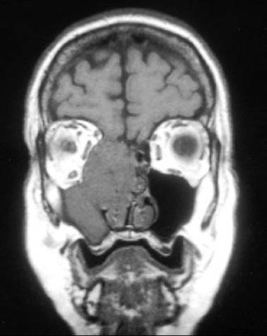
FIGURE 48.9. Coronal magnetic resonance imaging scan showing a large soft tissue mass and bone destruction in the right ethmoidal maxillary sinuses and nasal cavity secondary to extensive (Kadish stage C) esthesioneuroblastoma.
ESTHESIONEUROBLASTOMA
Esthesioneuroblastomas (ENB), first described by Berger and Luc,142 are rare tumors thought to arise in the olfactory receptors in the nasal mucosa or the cribriform plate of the ethmoid bone. The olfactory nerves perforate grooves in the ethmoid bone in the cribriform plate and continue into the subarachnoid spaces, accounting for the high incidence of intracranial extension.142
Epidemiology
ENB constitutes 3% of all endonasal neoplasms. In the United States,143 according to the data from the Surveillance, Epidemiology, and the End Results (SEER) program, 84 cases of ENB were registered from 1978 to 1990144 and about 945 cases have been reported in the world literature.145 The review authors’ cases accounted for 198 and collaborative efforts accounted for 747 cases. Sex distribution was 53.6% male and 46.64% female. Kadish classification was applied to 563 cases; 103 (18.3%) class A, 182 (32.2%) class B, and 278 (49.4%) class C cases.
There appears to be a slight male predominance. The age incidence has a bimodal distribution, with peaks at 11 to 20 years and 40 to 60 years, the highest incidence at 51 to 60 years.
Natural History
Although others thought that ENB were of ectodermal origin, most observers believe the tumor to be of neuroectodermal origin in the olfactory epithelium.14,146 Most of these tumors occur high in the nasal cavity or in the lateral wall adjacent to the ethmoid. The tumor may spread to the opposite ethmoid bone, superiorly to the frontal sinus and anterior cranial fossa, posteriorly to the sphenoid sinus, nasopharynx, and base of skull, laterally to the orbits, forward to the frontonasal angle, or inferiorly to the nasal cavity and antrum (Fig. 48.9). Lymphatic spread may be to the subdigastric, posterior cervical, submaxillary, or preauricular nodes, as well as to the nodes of Rouviere. The exact incidence of distant metastases is uncertain; it has been stated to be as high as 50%, but this rate is influenced by the use of chemotherapy in high-risk patients.
Clinical Presentation
These tumors tend to be friable and bleed easily. The most common clinical symptoms are epistaxis and nasal blockage. Patients also may have local pain or headache, visual disturbances, rhinorrhea, tearing, proptosis, or swelling in the cheek.147 The symptoms may be associated with a mass in the neck.
Diagnostic Workup and Staging
Physical examination may show the inferior aspect of a polypoid friable mass in the nasal cavity. Ocular findings or a mass in the nasopharynx may be present. With early lesions, radiographs or CT or MRI may show only nonspecific opacification, soft tissue swelling, and occasionally bone destruction.148 Octreotide is a somatostatin analog that, when coupled to a radioisotope, produces a scintigraphic image of neuroendocrine tumors (NETs) expressing somatostatin type-2 receptors. Octreotide scintigraphy may be useful in confirming the preoperative diagnosis of certain head and neck NETs, such as paragangliomas, Merkel cell carcinomas, medullary thyroid carcinomas, and esthesioneuroblastomas. Bustillo et al.149 carried out a retrospective study that compared the results of octreotide scintigraphy with the histopathologic diagnosis in 74 patients with head and neck NETs. Of the 60 patients undergoing evaluation for suspected paraganglioma, octreotide scintigraphy was correctly positive in 36 of 37 patients with paraganglioma and correctly negative in 19 of 23 patients who did not exhibit paraganglioma (sensitivity of 97% and a specificity of 82%). There were 14 patients in the nonparaganglioma group. Octreotide scintigraphy detected or diagnosed locoregional recurrences in two with esthesioneuroblastoma.
Table 48.1 outlines the suggested diagnostic workup. MRI, especially with gadolinium contrast, may be used as a supplement or alternative to CT scanning.39 CT provides the best information about the tumor and its local invasion into surrounding bone structures. MRI allows an estimate of tumor spread into surrounding soft tissue areas, such as the anterior cranial fossa and the retromaxillary space. Bone scintigraphy scan is useful in detecting distant metastases.
The expansile tendency of olfactory neuroblastoma is characterized by bowing of the sinus walls. The destructive aspect is manifested as tumor replacing the turbinates, septum, and sinus walls with extension into contiguous areas (Figs. 48.9 and 48.10). The density or signal and enhancement characteristics are nonspecific of olfactory neuroblastoma.
Although dopamine β-hydroxylase and catecholamines are produced by these tumors, their measurements or vanillylmandelic acid excretion levels have not proven clinically useful.
A staging system has been proposed by Kadish et al.147 (Table 48.9).
FIGURE 48.10. Sagittal (A) and coronal (B) views of a preoperative magnetic resonance imaging of a 56-year-old patient who was initially seen with a Kadish stage C tumor involving left nasal cavity and extending intracranially. (From Chao KSC, Kaplan C, Simpson JR, et al. Esthesioneuroblastoma: the impact of treatment modality. Head Neck 2001;23:749–757, with permission.)
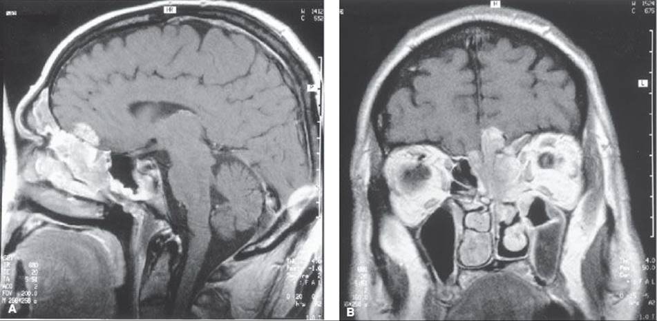
TABLE 48.9 KADISH SYSTEM FOR STAGING OF ESTHESIONEUROBLASTOMA

Pathologic Features and Prognostic Factors
ENBs are polypoid, frequently reddish, soft, and vascular tumors with neuroblasts and neurocytes. ENBs contain epithelial components serving as a supporting stroma and have a nerve component that corresponds to the olfactory cells. Rosettes are the main feature, consisting of several rows of cells arranged around the central area.14 ENBs may be confused with lymphoma or anaplastic carcinoma and have diffuse, regular distribution. ENBs contain many fibrils, which fill the central space of the rosette (called a pseudorosette). It has been suggested that the presence of chromaffin granules indicates a derivative from primitive neural crest cells. ENB must be distinguished from other poorly differentiated neoplasms, including sinonasal undifferentiated carcinoma, which is derived from the Schneiderian epithelium. Sinonasal undifferentiated carcinoma lacks rosettes and intercellular fibrils.14
Extension of the primary tumor based on the Kadish staging system147 has been identified as the most important determinant of treatment outcome, although this was not confirmed by Chao et al.150 High-grade tumors had worse outcome in the reports from the Mayo Clinic and the University of California–Los Angeles (UCLA).151
Argiris et al.152 found that in 16 patients with ENB, 11 of whom had Kadish stage C, 8 (50%) had brain involvement at presentation. Craniofacial resection was performed in 13 patients (81%); 14 received either preoperative or postoperative therapy (radiation therapy in 11 and chemotherapy in 4). The actuarial 5-year survival was 60%, disease-free survival 33%, with a median follow-up of 4.3 years. The first site of failure was locoregional alone in 10 of 12 patients who progressed, and in 6 patients involved the brain or the meninges. Two patients were successfully salvaged.
Hyams83 proposed a histologic grading system for ENB in which grade I tumors have an excellent prognosis and grade IV tumors are uniformly fatal. The Hyams grading system predated advanced craniofacial techniques, extensive use of immunohistochemistry, and the recognition of sinonasal undifferentiated carcinoma (SNUC) as a distinct entity. Miyamoto et al.,153 in a retrospective review of 12 patients with ENB and 14 with SNUC, used the Kadish clinical stage and Hyams histopathologic system. Kadish staging was available for 26 patients (2 patients with stage A tumors; 7 with stage B, and 17 with stage C). Of the eight evaluable patients with Kadish stage A or B tumors, six remained disease free for more than 2 years compared with only five of seven Kadish stage C tumors. Slides were available for Hyams grading in 21 patients (2 patients with grade I tumors, 4 with grade II, 4 with grade III, and 11 with grade IV). They concluded that both the Hyams grading and the Kadish staging system can be used as independent predictors of outcome; patients with either advanced clinical stage or pathologic grade of ENB or SNUC have poor prognosis, but long-term survival is possible in these patients if aggressive treatment is used.
Papadaki et al.154 analyzed 18 formalin-fixed paraffin-embedded olfactory neuroblastoma specimens (12 primary tumors and 6 recurrences or metastases) from 14 patients and concluded that p53 point mutation does not play an important role in the initial development of olfactory neuroblastoma; however, p53 wild-type hyperexpression may occur in subsets, show local aggressive behavior, and have a tendency for recurrence.
General Management
Surgery alone appears to be adequate treatment for small, low-grade tumors confined to the ethmoids in which negative surgical margins can be obtained. An ethmoidomaxillary resection with or without orbital sparing is usually necessary. This procedure is combined with preoperative or postoperative irradiation.151,155 A complete resection with preservation of vital structures is achievable by using a craniofacial approach.
Treatment, which could be classified in 898 reported cases in 1997, consisted of surgery alone in 24% (226 cases), radiation therapy alone in 18.4% (165 cases), combined surgery and radiation therapy in 43.2% (388 cases), chemotherapy in 13.2% (119 cases), and in 11 cases (1.2%) bone marrow transplant. In the reported cases follow-up could be evaluated in 477 cases, while in only 234 cases a 5-year follow-up was done; on these 20.5% had surgery only, 11.1% radiation therapy, and 68.4% combined surgery and radiation therapy. The best survival rates were obtained by combined therapy, 72.5% versus 62.5% with surgery alone and 53.8% with radiation therapy.31
Dias et al.156 reported on 35 patients with ENB treated with gross tumor resection through a transfacial approach with postoperative RT in 11 patients, craniofacial resection and postoperative RT in 7, exclusive RT in 14, craniofacial resection alone in 1, and a combination of chemotherapy and RT in 2 patients. Radiation therapy median dose was 48 Gy. Craniofacial resection plus postoperative RT provided a better 5-year disease-free survival rate (86%) compared with the other therapies (P = .05). The 5-year disease-specific survival rate was 64% and 43% for the low- and high-grade tumors, respectively (P = .20). At 5 and 10 years disease-free survival was 46% and 24%, respectively and overall survival was 55% and 46%, respectively.
Early lesions involving the ethmoids with little or no bony destruction or nerve invasion can be treated adequately by high-energy (photon or electron) radiation therapy with good cosmetic and functional results.81,103,157 Those with more extensive local disease benefit from surgery and adjuvant irradiation,156,158 although some have advocated against combined surgery and radiation therapy because of complications. Patients with locally advanced disease or high-grade tumors should receive aggressive treatment with combined modalities, such as surgery, radiation therapy, and chemotherapy.
Monroe et al.78 described treatment results in 22 patients who received RT for ENB (equal numbers of males and females, median age of 54 years). The modified Kadish stage was stage A in 1 patient, stage B in 4 patients, stage C in 15 patients, and stage D in 2 patients. Treatment modalities included primary RT in 6 patients, preoperative RT in 1 patient, postoperative RT after craniofacial resection in 12 patients, and salvage RT in 3 patients treated for recurrence after surgery. Elective neck RT was performed in 11 of 20 patients (2 patients had cervical metastases at presentation for RT). Rates of local tumor control, cause-specific survival, and absolute survival at 5 years were 59%, 54%, and 48%, respectively. The cause-specific survival rate at 5 years was lower after primary RT (17%) than after craniofacial resection and postoperative RT (56%). Cervical metastases occurred in 6 of 22 patients (27%). No neck recurrences occurred in 11 patients treated with elective neck RT compared with four neck recurrences in 9 patients (44%) not receiving elective neck RT (P = .02). Their data and review of the current literature suggest a higher cervical failure rate than previously recognized; elective neck RT seems to correlate with improved nodal tumor control and should be considered in the treatment of ENB.
Rosenthal et al.155 treated 72 adults with nonmetastatic, primary sinonasal neuroendocrine tumors (31 with ENB, 16 with SNUC, 18 with neuroendocrine carcinoma [NEC], and 7 with small cell carcinoma [SmCC]). Patients with ENB usually were treated with surgery and/or radiotherapy; only 3 of 31 patients (9.7%) received radiation to regional lymphatics, and only 5 of 31 received chemotherapy. In contrast, patients with non-ENB histologies usually received chemotherapy (10 of 16 patients with SNUC, 12 of 18 patients with NEC, and 5 of 7 patients with SmCC). With a median follow-up for surviving patients of 81.5 months, overall survival at 5 years was 93.1% for patients with ENB, 62.5% for SNUC, 64.2% for NEC, and 28.6% for SmCC (P = .0029). The local control tumor rate at 5 years also was superior for patients who had ENB (96.2%) compared with patients who had SNUC (78.6%), NEC (72.6%), or SmCC (66.7%) (P = .04). The regional failure rate at 5 years was 8.7% for patients with ENB, 15.6% for patients with SNUC, 12.9% for patients with NEC, and 44.4% for patients with SmCC. Additional late events increased the regional failure rate for patients with ENB to 31.9% at 10 years. The distant metastasis rate at 5 years was 0.0% for patients with ENB, 25.4% for patients with SNUC, 14.1% for patients with NEC, and 75.0% for patients with SmCC.
Eich et al.143 described 17 patients with ENB (4 Kadish stage B and 13 stage C), treated with incomplete surgery (2 patients), adjuvant radiation therapy (6 patients), definitive RT (7 patients), and for recurrent tumor (2 patients). Median postoperative dose was 56 Gy (40 to 60 Gy) and definitive 58 Gy (40 to 70 Gy). With a median follow-up of 7 years, 10 of 17 had no evidence of recurrence (5 of 6 treated with complete resection and postoperative RT and 3 of 7 treated with definitive RT).
Gruber et al.159 described 28 patients with ENB treated with RT (median dose 60 Gy). In 13 patients total tumor resection (recommended by the authors) was performed. Chemotherapy (cisplatin, etoposide, cyclophosphamide, and vincristine) combined with RT were used in five patients. With median follow-up of 68 months, 54% of the patients were free of local tumor progression (51% at 10 years). Disease-free survival at 10 years was 25%.
Ozsahin et al.160 described results of treatment in 13 European and North American centers for 77 patients with olfactory neuroblastoma, 11 with Kadish stage A, 29 with stage B, and 37 with stage C; 56 patients had surgery, 44 with total tumor excision. All but 5 patients received radiation therapy (50% with 3D-CRT) and 21 had chemotherapy. With median follow-up of 72 months locoregional tumor control was 62%, disease-free survival 57%, and overall survival 64%. Patients having total tumor resection or receiving ≥54 Gy had better overall survival than those treated with lower doses (Fig. 48.11). Six of the patients treated with RT (56 to 70 Gy) developed grade 3 or4 late complications (five osteonecrosis and one retinopathy).
Sperry et al.161 treated 30 patients with ENB (70% with Kadish stage C) with surgery in 27 (52% craniofacial resection), combined with postoperative RT in 75.9% and chemoradiation in 23%. Local tumor failure at 5 years was 43.6% and regional failure 15.7%. The 5-year disease-free survival was 40.7% and overall survival 80%.
Madani et al.162 published a report on 84 patients (73 with primary and 11 with local recurrent) sinonasal tumors, 9 of which were ENB, treated with IMRT (median dose 70 Gy in 35 fractions). Mean D50 to the optic chiasm was 37.2 Gy, to ipsilateral optic nerve 49.4 Gy, to contralateral optic nerve 47.1 Gy, and to the retina 37.7 and 28.4 Gy, respectively.
Protons have been used sparingly in the treatment of some of these patients. Nishimura et al.163 reported on 14 patients with olfactory neuroblastoma treated with proton beam (65 CGE in 2.5-GyE fractions) in 6 patients combined with surgery, sometimes combined with chemotherapy.164 With median follow-up of 40 months, 5-year local progression-free survival was 84%, disease-free survival 71%, and overall survival 93%.
For advanced lesions, in which disseminated disease is likely, chemotherapy may improve tumor control and decrease the incidence of distant metastases. A combination of thiotepa, cyclophosphamide, doxorubicin, vincristine, nitrogen mustard, and actinomycin-D has been used.165,166 A retrospective review of 10 patients with recurrent esthesioneuroblastoma treated with chemotherapy at the Mayo Clinic suggested that cisplatin-based chemotherapy is active in advanced, high-grade tumors.155 Survival from initial chemotherapy treatment was 44.5 months (range 3 to 130 months) in patients with low-grade tumors and 26.5 months (range 2 to 67 months) in patients with high-grade tumors (Table 48.10).
Elective Neck Treatment
ENB has been shown to metastasize to the neck and remote sites. Although the sites of metastases are widely variable and often atypical, Beitler et al.167 found cervical node metastasis to be as frequent as local recurrence. Davis and Weissler62 compiled a retrospective review of patients and found that the cumulative cervical metastasis rate reached 27% (55 of 207 patients). Noh et al.,168 in a report of 19 patients with ENB treated with combinations of surgery, RT, and/or chemotherapy, noted that 4 patients with high risk factors received elective neck RT (45 to 70 Gy). There were no cervical node failures in 5 patients with Kadish stages A and B, 3 of 10 in patients with Kadish stage C treated surgically, and 0 in 9 receiving chemotherapy.
In general, because of the low incidence of cervical lymph node metastasis (≤10%) in early-stage disease, elective irradiation of the neck or a dissection is not indicated. However, in patients with Kadish stage C disease, the cervical metastatic rate climbed to 44% (25 of 57 patients). As noted previously, Monroe et al.78 observed cervical node metastasis in 6 of 22 patients (27%). In 11 patients they treated with elective neck RT, no recurrences were noted, in contrast to 4 of 9 (44%) for patients not receiving elective neck RT. Thus, with advanced-stage disease, cervical nodes should be initially managed by irradiation, radical neck dissection, or a combination of both.169
FIGURE 48.11. Overall survival correlated with radiation therapy dose in 72 patients with olfactory neuroblastoma. (From Ozsahin M, Gruber G, Olszik O, et al. Outcome and prognostic factors in olfactory neuroblastoma: a rare cancer network study. Int J Radiat Oncol Biol Phys2010;78:993–997, with permission.)
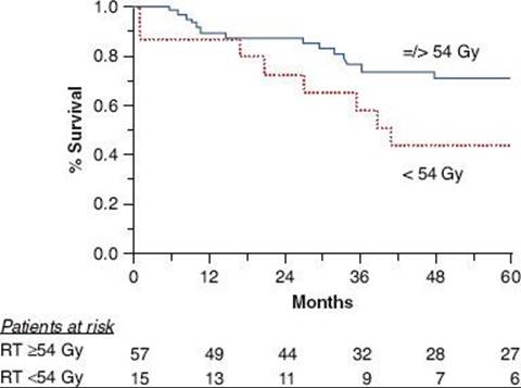
TABLE 48.10 RESULTS OF TREATMENT CORRELATED WITH MODALITY AND STAGE FOR ESTHESIONEUROBLASTOMA

Radiation Therapy Techniques
A combination of photons and electrons with conventional anterior fields provides good coverage for limited ethmoidal disease when the tumor is confined anteriorly. Beam arrangement can be modified for disease extending into the orbit or maxillary sinus. Obturator or bolus may be needed postoperatively to compensate for tissue deficit. When intracranial or posterior extension is present or tumor has spread into the maxillary sinus, a pair of perpendicular (anteroposterior and lateral) portals with wedges or two lateral wedge fields in conjunction with an open anterior photon field will give good coverage of the treatment volume, with the dose inhomogeneity around 10% to 20%. Incorporation of a vertex field eliminates the high inhomogeneous dose along the junction line of the conventional three-field technique. Treatment techniques are similar to those described for treatment of paranasal sinuses (see Chapter 42). The orbits can be spared or treated as the degree of extension dictates. Occasionally, an anterior electron beam field may be needed to supplement low-dose areas. When the electron beam is used over air cavities, some dosimetry problems result. Eye blocks must be positioned precisely to avoid undesirable side effects.
When combined therapy is used, preoperative doses of 45 Gy and postoperative doses of 50 to 60 Gy are indicated, depending on the status of the surgical margins. Doses of 65 to 70 Gy are delivered with irradiation alone in patients with inoperable tumors.170 Usual fraction dose is 1.8 to 2.0 Gy. Contrast-enhanced CT or MRI scans before initiation of treatment are crucial to demarcate extension of the tumor. Treatment planning with CT for determination of tumor extension is extremely important.171 Because of the proximity of esthesioneuroblastoma to the optic nerves, optic chasm, and the brainstem, the precision of treatment setup, target volume definition, and dose homogeneity dictate tumor control and the sequelae of treatment. Treatment techniques similar to those for paranasal sinuses may create “hot spots” along the optic tracks. High doses per fraction (exceeding 2 Gy) increase the possibility of late sequelae, such as blindness and bone and brain necrosis.
Three-dimension CRT or IMRT provides alternatives to the conventional three-field technique used to treat these tumors (Fig. 48.12). Special attention should be directed to reduce unnecessary irradiation to ocular structures, including optic nerve(s) and chiasma. When occasionally a patient presents with cervical node metastasis, IMRT is very helpful to optimally treat the primary tumor and the cervical lymphatics (Fig. 48.13).
FIGURE 48.12. Esthesioneuroblastoma in a 35-year-old woman, initially treated with a craniofacial surgical resection. Patient received postoperative intensity-modulated radiation therapy (2-Gy fractions). A: Cross-section illustrating coverage of ethmoid nasal and left maxillary antrum volume. B: Cross-section showing dose distribution in target volume with excellent sparing of ocular structures. C: Dose–volume histogram:


Results of Therapy
Surgery and Irradiation
Platek et al.,172 in an analysis of SEER data (1973–2005) of 135 cases of olfactory neuroblastoma, noted that 59% of the patients were treated with surgery and RT, 23% with surgery only, 12% with RT only, and 6% with neither. No data on chemotherapy administration were available. The 5-year survival with surgery plus RT was 66%, with surgery only 51%, with RT only 26%, and with other therapy 34% (P = .003).
Kased et al.170 reported on 17 patients with ENB (15 undergoing a surgical procedure, 7 receiving concurrent chemotherapy, and 4 adjuvant chemotherapy) treated with IMRT (median dose 66 Gy in 2-Gy fractions). With median follow-up of 44.5 months, the 5-year freedom of locoregional tumor progression was 91%, progression-free survival 83%, and overall survival 81%. Four patients had acute complications (meningitis, sepsis, sinusitis requiring surgery, and brain abscess) and two patients developed late brain abscess, requiring surgical treatment.
Radiation therapy is an important component in the management of ENB, but the optimal sequence when integrated with surgery is unknown. Eden et al.144 observed no significant difference in survival whether preoperative or postoperative irradiation was given, but suggested improved local tumor control with preoperative irradiation. Technical factors may have contributed to a higher incidence of postoperative radiation therapy failures because three of five postoperative cases received <50 Gy; all three patients were treated with a single anterior field, which gives less homogeneous dose distribution throughout the treatment volume.
Foote et al.151 updated the experience of the Mayo Clinic. Seventeen patients had disease confined to nasal cavity or paranasal sinuses (Kadish stages A and B), and 32 patients had more advanced disease. Treatment included gross total resection alone or combined with radiation therapy. The 5-year actuarial survival, disease-free survival, and local tumor control rates were 69.1%, 54.8%, and 65.3%, respectively. Local tumor control was improved in patients who received postoperative irradiation (55.5 Gy) even after complete tumor resection.
Levine et al.173 conducted a retrospective review of 35 patients; 6% of them presented with cervical metastasis, and ultimately 25.7% developed cervical metastases. Fourteen percent of the patients had a local recurrence at an average of 6 years after diagnosis, and in 37% at least one episode of metastatic disease occurred. The disease-free survival was 80.4% at 8 years. CNS complications occurred in 25.7% of patients, orbital complications in 22.9%, systemic posttreatment problems in 20%, and chemotoxic sequelae in 18%.
Eriksen et al.174 carried out a retrospective review of 13 patients with ENB (Kadish stage A 1 patient, stage B 5 patients, and stage C 7 patients). The 5-year disease-free survival was 51%. Forty-six percent of the patients experienced relapse, and despite intensive salvage therapy, median survival after recurrence was only 12 months.
Chao et al.150 reported on 25 patients with ENB (Kadish stage A in 3 patients, stage B in 13 patients, C in 8 patients, and modified D in 1 cervical nodal metastasis patient); 17 patients were treated with surgery and radiation therapy, 6 with irradiation alone, and 2 with surgery only. Eight patients received neoadjuvant chemotherapy. Median follow-up was 8 years. The 5-year actuarial overall survival, disease-free survival, and local tumor control rates were 66.3%, 56.3%, and 73%, respectively. Kadish stage was not a significant prognosticator for local control or disease-free survival. Five-year local tumor control was 87.4% for combined surgery with RT and 51.2% for irradiation alone. Two patients with Kadish stages A and B disease underwent surgical resection alone; both failed locally. In contrast, only three of nine patients with Kadish stage A or B disease who received adjuvant radiation therapy had a local recurrence. With adjuvant radiation therapy, the surgical margin status did not influence local tumor control.
Simon et al.171 reported on 13 patients with ENB or olfactory neuroblastoma (Kadish stage B 5 patients, and stage C 8 patients). The majority of the patients were treated with a craniofacial resection or tumor removal through a rhinotomy approach. Two patients received neoadjuvant chemotherapy before surgical resection (cisplatin, ifosfamide, and etoposide). Twelve of the 13 patients received RT either initially or for salvage. Median dose of postoperative irradiation was 59.4 Gy in 1.8-Gy fractions. The overall actuarial 5-year survival was 61% and 10-year survival 24%, and disease-free survival rates were 56% and 42%, respectively.
FIGURE 48.13. Patient with Kadish stage C esthesioneuroblastoma of ethmoid cells and nasal cavity who presented with a large left upper cervical lymph node metastasis. Intensity-modulated radiation therapy plans to deliver 70 Gy to primary tumor and cervical lymphadenopathy. (A) Coronal, (B), sagittal, and (C) cross-section dose distributions illustrate excellent coverage of all target volumes.
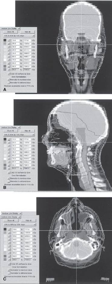
Chemotherapy and Irradiation
Eden et al.144 described results in 16 patients with stage A or B disease and 24 patients with stage C disease treated with irradiation (median dose 50 Gy) and surgery for stages A and B disease, with the addition of chemotherapy (cyclophosphamide and vincristine) for stage C disease. Actuarial survival rates at 5 and 10 years were 78% and 71%, respectively. Locoregional failure developed in 15 of 40 patients; 68% of the failures were locoregional (including brain, neck, facial bone, and sinus). They had no recurrences at the primary tumor bed; all recurrences were either outside the irradiation field or at distant sites.
Preoperative neoadjuvant therapy may provide a valuable complement to radical craniofacial resection.165
Forty patients were treated for ENB at Institut Gustave Roussy, France.146 Three patients had stage T1, 7 patients had T2, 15 patients had T3, and 15 patients had T4 lesions. At presentation the cervical metastatic rate was 18% and distant metastases were detected by bone marrow biopsy and bone scan in three patients. Treatment modalities included surgery alone in 8 patients, radiation therapy alone in 3 patients, surgery plus radiation therapy in 11 patients, chemotherapy alone in 2 patients, chemotherapy plus radiation therapy in 10 patients, and chemotherapy plus surgery and radiation therapy in 6 patients. The 5-year survival rate was 51%. Multimodality treatment offered better survival (63% at 5 years). Overall local, regional, and distant failure rates were 58%, 15%, and 40%, respectively. Distant metastases commonly occurred in bone (82%).
Noh et al.168 summarized reports on patterns of failure of ENB treated with or without chemotherapy. Although the indications for high-dose chemotherapy and bone marrow transplantation must be better defined, it may be a promising alternative for patients with large tumors or those with recurrent tumor to whom no further local therapy (e.g., surgery or irradiation) can be safely given.175
Sequelae of Treatment
In a few patients, depending on the dose of irradiation, long-term sequelae include bone necrosis, brain necrosis or abscess, blindness, or painful eye reactions requiring enucleation.176,177,178
Simon et al.,171 in 13 patients with olfactory ENB treated with surgery and radiation therapy, noted that one patient lost vision as a result of glaucoma and radiation retinopathy after 67.3 Gy in 34 fractions. One patient treated with 61.76 Gy in 34 fractions developed a visual field defect and optic atrophy; she also had a nasal cutaneous fistula. One patient sustained intraoperative rupture of the ocular globe and subconjunctival hemorrhage.
EXTRAMEDULLARY PLASMACYTOMAS
Solitary plasmacytomas are rare tumors of plasma cell origin, making up 4% of all plasma cell tumors. Multiple myeloma occurs about 40 times more frequently than solitary plasmacytoma. Monoclonal extramedullary plasmacytoma (EMP) is a rare, low-grade lymphoma found predominantly in the head and neck region. Only since the introduction of immunophenotyping techniques two decades ago has it been possible to differentiate EMP from benign polyclonal plasma cell proliferation. Hotz et al.179 reviewed the records of 24 patients with morphologically diagnosed EMP treated at their institution; only 14 patients had true monoclonal plasmacytoma. No EMP-related deaths occurred. Two patients had local recurrence, and two patients developed multiple myeloma. Diagnostic procedures exclude a benign polyclonal plasmacytoma, multiple myeloma, and solitary bone plasmacytoma. The slow natural progression of the disease and the rarity of secondary multiple myeloma favor nonmutilating local surgery whenever possible to avoid the long-term sequelae of radiation.
Epidemiology
The annual incidence of EMP is 0.04 cases per 100,000 population.180 They constitute only 0.5% of all upper respiratory tract malignancies. Male patients exceed female patients by a ratio of 4 to 1, and 75% of patients are 40 to 60 years of age.181
In a detailed literature search of more than 400 publications between 1905 and 1997, EMP mainly occurred between the fourth and seventh decades of life.182 Seven hundred fourteen cases (82.2%) were found in the upper aerodigestive tract.
The most common sites in the head and neck are the nasopharynx, nasal cavity, paranasal sinuses, and tonsils.
Clinical Presentation and Diagnostic Workup
EMP of the head and neck area should be considered a separate entity because of its clinical behavior. The most common symptoms are nasal obstruction, local pain and swelling, and epistaxis.
Grossly, plasmacytomas tend to be sessile in the nasal cavity and paranasal sinuses and pedunculated in the nasopharynx and larynx. The masses are soft, pliable, and pale gray. The lesion may remain localized or may infiltrate and destroy the surrounding soft tissue and bone. The usual criteria for solitary plasmacytomas, either medullary or extramedullary, include a biopsy-proven plasma cell tumor with one or, at the most, two solitary foci, absence of Bence-Jones protein in the urine, bone marrow taken some distance from the primary site not involved by tumor (<10% of plasma cells), hemoglobin of 13 g/mL or more, and a normal serum protein level or serum electrophoresis at the time of the diagnosis. Basically, the diagnosis of solitary plasmacytoma is made by exclusion, that is, by eliminating the possibility of multiple myeloma.183 Diagnosis is based on histology along with special immunoperoxidase staining for immunoglobulin -λ and -κ light chains.29
Strict staging criteria, including normal MRI studies of the axial skeleton and the long bones and absence of monoclonal plasma cells detected by flow cytometry or polymerase chain reaction, are required for diagnosis of solitary plasmacytoma. Careful microscopic and immunohistochemical studies are also required for the correct diagnosis, because this disease can be confused with other malignancies, particularly lymphomas.
Six patients with primary EMP in the head and neck were examined with MRI;184 five lesions were oval and sharply demarcated without signs of infiltration, while the other lesion filled the parapharyngeal space bilaterally. On T2-weighted sequence, the lesions had moderate signal intensity. On plain T1-weighted sequences, the tumors were isointense or slightly hyperintense with respect to surrounding muscles; after administration of contrast medium, four lesions showed notable enhancement, with distinct central inhomogeneity.
Bone destruction is not a particularly bad prognostic sign, although some investigators report that it adversely affects prognosis.184 Bony invasion is common in the more malignant types.14
Cervical lymph node metastasis from EMP varies with the site of the primary lesion and follows the same pattern of spread as squamous cell carcinoma arising in a similar site. The reported incidence of lymph node metastasis ranges from 12% to 26%. The diagnostic workup for EMP arising in the head and neck region is shown in Table 48.1. The exact relationship between EMP and multiple myeloma is unclear; however, approximately 20% to 30% of EMP cases will convert to multiple myeloma.179,183
General Management
Pedunculated EMP lesions may be treated by surgical excision because the chance of local recurrence is low. The treatment of choice for all other lesions is radiation therapy alone or combined with other modalities.185,186 In a review of 714 cases in the literature, the following therapeutic strategies were used to treat patients with EMP of the upper aerodigestive tract: radiation therapy alone in 44.3%, combined therapy (surgery and irradiation) in 26.9%, and surgery alone in 21.9%. The median overall survival or recurrence-free survival was longer than 300 months for patients who underwent combined intervention (surgery and irradiation), for surgical intervention alone (median survival time, 156 months), and for radiation therapy alone (median survival time, 114 months). Overall, after treatment for EMP in the upper aerodigestive tract, 61.1% of all patients had no recurrence or conversion to systemic involvement (i.e., multiple myeloma); however, 22% had recurrence of EMP, and 16.1% had conversion to multiple myeloma.
Radiation Therapy Techniques
Irradiation techniques vary with the location of the primary tumor. The techniques are similar to those used for primary tumors in comparable locations (i.e., nasopharynx, tonsil, paranasal sinuses). Solitary plasmacytomas respond well to doses of 50 to 60 Gy in 2-Gy fractions. The local tumor control rate with radiation therapy alone is about 85%. Harwood et al.186 summarized the literature but could not draw a dose–response curve from the data because of a lack of cases receiving low-dose radiation therapy. Nevertheless, there is a high risk of local recurrence with tumor doses below 30 Gy and a negligible risk for those treated at or above 40 Gy (Table 48.11).
TABLE 48.11 FAILURE PATTERNS OF ESTHESIONEUROBLASTOMA: LITERATURE REVIEW

Wax et al.29 reported on seven patients, three treated with radiation therapy (31.75 to 60 Gy). All patients have maintained local tumor control and had been followed for a minimum of 1.5 years, with an average of 3 years. One patient, treated with surgical excision, experienced a relapse at a distant site 6 years later.
The response to therapy of 32 patients with localized plasmacytoma were described by Shih et al.;192 22 patients had solitary plasmacytoma of bone and 10 had EMP. Median age for EMP was 63 years. Most EMPs occurred in the oronasopharynx (six cases) and paranasal sinuses (two cases). Seven patients with EMP received radiation therapy (47 to 65 Gy), and all achieved initial local tumor control. There was one local recurrence and multiple myeloma conversion in the EMP group. Local recurrence or dissemination was associated with the appearance of or an increase in myeloma protein.
Holland et al.193 reported on 14 cases of EMP, eight of which were in the head and neck. With doses of 46 to 62 Gy, the complete tumor response was 72%. No dose–response effect was observed.
Liebross et al.194 described results in 22 patients with solitary EMP, in the head or neck in 19 patients, usually in the nasal cavity or maxillary sinuses, and bone destruction was found in 10 of 11 patients. Among all patients, serum myeloma protein was present in three patients (14%) and Bence-Jones protein alone in two (9%). Radiation therapy was the sole treatment in 18 of 22 patients (median dose 50 Gy; range 40 to 60 Gy); 5 of 7 patients with an EMP of oral cavity, oropharynx, nasopharynx, parotid, or larynx also received elective neck irradiation. Local tumor control was achieved in 21 of 22 patients (95%), and disease never recurred in regional nodes. Disappearance of myeloma protein occurred in three of five patients with an evaluable abnormality. Multiple myeloma developed in seven patients (32%), all within 5 years. The 5-year rate of freedom from progression to multiple myeloma was 56% and the median survival was 9.5 years. Chao et al.150 reported on 16 patients with EMP and a median follow-up of 66 months. The head and neck region accounted for the majority of presentations (88%). A serum monoclonal paraprotein was found in three patients, and bone erosion was identified in seven patients. All patients received local RT, although two patients also received elective nodal irradiation. The median RT dose was 45 Gy (range 40 to 50.4 Gy). Local tumor control was achieved in all patients (100%), however, regional recurrence outside the RT fields occurred in 2 of 16. Multiple myeloma developed in five patients (31%) all within 5 years. The 10-year myeloma-free survival is 75% and 10-year overall survival is 54%.
Galieni et al.195 reviewed 46 cases of EMP most frequently localized in the upper airways (37 of 46 patients, 80%), with the mass being limited to a single site in all but seven patients in whom two contiguous sites were involved. The most frequent form of treatment was local radiation therapy. Thirty-nine patients (85%) achieved complete remission, five (11%) a partial remission, and two (4%) did not respond to therapy. Local recurrence or recurrence at other sites occurred in 7.5% and 10%, respectively. Seven patients (15%) developed multiple myeloma. The 15-year survival rate was 78%.
Michalski et al.180 described 10 patients with EMP treated with radiotherapy. One patient treated at relapse underwent surgical resection followed by postoperative RT. The disease was most frequently localized in the paranasal sinuses (50%). All nine patients who received definitive RT (40 to 50 Gy) achieved a complete response. Median follow-up period was 29 months. Four patients (40%) relapsed, three have died of their disease. Two patients with paranasal sinus disease subsequently relapsed with multiple myeloma at 10 months and 24 months, respectively. The relapse rate in neck nodes of 10% does not justify elective irradiation of the uninvolved neck.
Miller et al.196 reported that tumor arose in the sinonasal or nasopharyngeal region in 11 of 20 patients (55%). The primary modality of treatment was radiation therapy (45 to 60 Gy). The mean follow-up was 60.2 months. In 15 to 20 cases, immunohistochemistry staining for immunoglobulin light chain production was conducted. One of the two cases (50%) classified as medullary plasmacytoma demonstrated conversion to multiple myeloma, whereas only 2 of 18 cases of EMP (11%) converted to multiple myeloma.
Ozsahin et al.177 published a compilation of solitary plasmacytoma (42 cases) in the head and neck. There were 258 patients with bone (n = 206) or extramedullary (n = 52) plasmacytomas without evidence of multiple myeloma. Most (n = 214) of the patients received RT alone; 34 received chemotherapy and RT; and 8 had surgery alone. The median radiation dose was 40 Gy. Median follow-up was 56 months (range 7 to 245 months). The median time for multiple myeloma development was 21 moths (range 2 to 135 months), with a 5-year survival probability of 45% (Fig. 48.14A). The 5-year overall survival, disease-free survival, and local control rates were 74%, 50%, and 86% respectively (Fig. 48.14B). On multivariate analyses, favorable factors were younger age and tumor size <4 cm for survival; age, extramedullary localization, and RT for disease-free survival; and small tumor and RT for local control. Bone localization was the only predictor of multiple myeloma development. No dose–response relationship was found for doses >30 Gy, even for larger tumors.
FIGURE 48.14. A: Probability of progression to multiple myeloma according to bone (dotted line) or extramedullary (solid line) solitary plasmacytoma (P = .0009). B: Overall survival correlated with bone (dotted line) or extramedullary (solid line) solitary plasmacytoma (P = .04). (From Ozsakin M, Tsang RW, Poortmans P, et al. Outcomes and patterns of failure in solitary plasmacytoma: a multicenter rare cancer: study of 258 patients. Int J Radiat Oncol Biol Phys 2006;64:210–217, with permission.)

TABLE 48.12 EXTRAMEDULLARY PLASMACYTOMA OF HEAD AND NECK TREATED BY RADIATION THERAPY

Tournier-Rangeard et al.,197 in a review of 17 patients with solitary EMP in the head and neck, noted a local tumor control of 100% for patients who received ≥45 Gy dose to the CTV versus 50% with doses <45 Gy (P = .034). Prognostic factor for 5-year disease-specific survival (81.6%) was local tumor control (P = .058). Prognostic factors for disease-free survival (64.1%) were monoclonal immunoglobulin secretion (P = .008) and CTV dose >45 Gy (P= .056).
Bachard et al.185 published outcomes of 68 patients with EMP of the head and neck, 39 treated with radiation (median dose 35 to 37 Gy in 15 fractions), 14 with surgery plus RT, 8 with surgery, and 3 with chemoradiation. With median follow-up of 8 years, 5-year local recurrence-free survival was 81% and survival 76%. Local recurrence was equivalent in patients treated with surgery or RT (12.5%). Multiple myeloma developed in 23% of the patients. Sasaki et al.198 described results of radiation therapy in 67 patients with solitary plasmacytoma of the head and neck (in 44 combined with surgery) treated at 23 centers in Japan. Median RT dose was 50 Gy. With 63 months median follow up the 10 year local tumor control was 87% and overall survival 56%. The 10 year survival was 70% for patients treated with combined RT and surgery and 50% in those treared with RT alone (p = 0.004). Twelve patients (18%) developed distant metastasis and 8 (12%) converted to multiple myeloma.
Table 48.12 summarizes the doses of irradiation and probability of tumor control reported by various investigators. The authors’ limited experience confirms the efficacy of tumor doses of 45 to 50 Gy for local tumor control. In patients who had extensive disease, a higher dose (50 to 60 Gy) was used, as recommended by several investigators.203
NASOPHARYNGEAL ANGIOFIBROMA
Epidemiology
Juvenile nasopharyngeal angiofibroma (JNPAF) is found more frequently in young pubertal boys;204 it has been shown to contain androgen receptors205,206 and occasionally to regress with estrogen therapy. Hwang et al.,207 in 24 nasopharyngeal angiofibromas, detected androgen receptors in 18 of 24 (75%) cases, whereas only two (8.3%) were positive to progesterone. None of the 24 cases was positive for antibodies to estrogen.
The tumor is believed to originate from the posterolateral wall of the nasal cavity where the sphenoidal process of the palatine bone meets the horizontal ala of the vomer and the roof of the pterygoid process because it is always involved.208,209 Other investigators agree, because involution of tumor after irradiation usually occurs in this direction.210
JNPAF comprises <0.05% of head and neck tumors.162 Patient age at presentation ranges from 9 to 30 years,211,212 with a median of 15 years. Females comprise <4% of the total cases.213 Some investigators have suggested chromosomal studies in affected women because this is mainly a male disease.209
Clinical Presentation and Pathology
Symptoms usually occur 2 to 48 months before diagnosis.211 The most common complaints are nasal obstruction or epistaxis, followed by nasal voice or discharge, cheek swelling, proptosis, diplopia, hearing loss, and headaches.211Nasopharyngeal angiofibroma may initially extend into the nasal fossae and maxillary antrum and push the soft palate downward, then through the pterygopalatine fossa and superoanteriorly through the inferior orbital fissure or laterally through the pterygomaxillary fissure to the cheek and temporal regions.27
Beham et al.,214 in a study of 32 cases of JNPAF, noted that most of the tumor vessels, which lacked elastic laminae, were characterized by vascular walls of irregular thickness and variable muscle content. In places, endothelial cells were separated from the stroma by only a single attenuated layer of contractile cells; in some more fibrotic hyaline areas, the stromal cells displayed reactivity for smooth muscle actin. The irregularity of the vascular walls, together with the lack of elastic laminae and stromal fibers, explains the pronounced tendency for hemorrhage in these lesions.
Differential diagnosis includes fibrosarcoma, rhabdomyosarcoma, chronic sinusitis, arteriovenous malformation, lymphangioma, neurofibroma, pleomorphic adenoma, lymphoma, pyogenic granuloma, polyps, and hemangioma.
TABLE 48.13 STUDIES REPORTING TREATMENT OUTCOME AND LATE GRADE 3 VISUAL IMPAIRMENT AFTER INTENSITY-MODULATED RADIATION THERAPY FOR SINONASAL TUMORS

Diagnostic Workup
After the history and physical examination, CT scans with and without contrast should be obtained. Characteristic findings are a mass in the posterior nasal or pterygopalatine fossa and bone erosion in the sphenopalatine foramen and extension to the pterygoid plate.44 The pattern of enhancement in this highly vascular tumor is diagnostic,215,216 and many investigators believe carotid angiograms are unnecessary217 after CT diagnosis of the lesion, unless embolization, which is also controversial, is contemplated.
CT scans are especially helpful in regions involving thin bony structures (paranasal sinuses, orbits), where CT performs better than MRI. In the nasopharynx and parapharyngeal space, MRI is superior to CT. Obtaining tumor volumetric data with spiral CT or MRI facilitates 3D treatment planning.218
Seventy-two patients with JNPAF were evaluated with CT or MRI.215 Origin of the tumor was in the pterygopalatine fossa at the aperture of the pterygoid (vidian) canal. The tumor extended posteriorly along the pterygoid canal with invasion of the cancellous bone of the pterygoid base and greater wing of the sphenoid in 60% of the patients. The inability to remove the tumor in toto was principally due to deep invasion of the sphenoid; 93% of recurrences occurred with this type of tumor extension.
If intracranial extension is noted and radiation therapy is contemplated, no further studies are indicated. If the lesion is extracranial and surgery is indicated, bilateral carotid angiograms will identify the feeding vessels and delineate the boundaries of the tumor.
Biopsies are not indicated in all patients because of the potential for severe hemorrhage. It is important to perform a biopsy of the lesion when the clinical picture (sex, age, location, and behavior of the lesion) is not consistent with JNPAF because some lesions have proven to be sarcomas or chronic sinusitis.209 Two cases of fibrosarcoma have been reported in patients in their 40s.219
Staging and Prognostic Factors
Staging schemes have been proposed by Chandler et al.,209 a radiographic staging system by Sessions et al.,210 and a more detailed anatomical system by Radkowski et al.220 (Tables 48.13 and 48.14). In a retrospective review of 44 cases of JNPAF, invasion of the skull affected two-thirds of the patients, and the rate of recurrence was 27.5%.221 Extensions to the intratemporal fossa, sphenoid sinus, base of pterygoids and clivus, the cavernous sinus (medial), foramen lacerum, and anterior fossa were correlated with more frequent recurrence. In a review of 97 cases age at diagnosis, tumor size and Radkowski classification were significant prognostic factors for recurrence.222
General Management
Optimum management of these patients remains controversial223 and the decision of whether surgery or radiation therapy should be used depends in part on the initial extent of the disease. In patients with extracranial tumors,221surgery is the treatment of choice and yields near-zero mortality or any long-term morbidity.224 Tumor extension to the posterior infratemporal fossa or intracranially is associated with a higher risk of recurrence.225
Tumor remnants in symptom-free patients should be kept under surveillance by repeated CT scanning, because involution may occur. Recurrent symptoms may be treated by radiation therapy rather than by extended surgery or combined procedures.209,231,221
When there is intracranial tumor extension (seen in about 20% of patients), the risk of surgically related death increases. Some investigators recommend preoperative intra-arterial tumor vessel embolization at the time of diagnostic bilateral carotid angiography, claiming a decrease in operative bleeding.232 Salvage with embolization of polyvinyl alcohol has been described.233 Others have reported anecdotal evidence of partial regression with the use of estrogens, believed to be the result of feedback inhibition of the pituitary’s production of gonadotropin-releasing hormone.
Although radiation therapy is equally effective in extracranial tumors, the low but existing risk of secondary malignancies should limit its use to the more advanced tumors only, such as those involving the orbital apex or the base of the skull.225,234 In the experience of Cummings et al.231 covering 20 years, only two radiation-related malignancies were noted (one skin, one thyroid).
FIGURE 48.15. Example of conventional lateral portal used at the Mallinckrodt Institute of Radiology for nasopharyngeal angiofibroma.
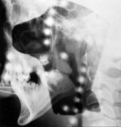
Radiation Therapy Techniques
Photon irradiation should be used for these patients, and fields must be individualized to cover the tumor completely with a margin (1 to 2 cm). Treatment portals are similar to those used in carcinoma of the nasopharynx (without irradiating the cervical lymph nodes) or carcinoma of the paranasal sinuses when these structures or the nasal cavity is involved. Opposing lateral portals are suitable in most patients, with larger fields and compensators used for tumors extending into the nose (Fig. 48.15). More extensive disease requires three-field or wedge-pair arrangements of 3D-CRT or IMRT that can yield excellent dose distributions, particularly when there is nasopharyngeal or intracranial tumor extension. In all cases, the eyes are protected as much as possible.
The recommended tumor dose ranges from 30 Gy in 15 fractions in 3 weeks to 50 Gy in 24 to 28 fractions in 5 weeks.235 A conventional setup uses 6- to 18-MV photons to treat the lesion with parallel-opposed fields to 50 Gy (2-Gy fractions).
The advantages of IMRT for the treatment of extensive or recurrent JNPAF were described in three patients on whom the tumor affected the base of skull, pterygopalatine, and intratemporal fossae, posterior orbit, and nasopharynx.232 Tumor dose varied from 34 to 45 Gy. Chakraborty et al.236 treated eight patients with IMRT for stage III tumors (median dose 39.6 Gy). Local control at 2 years was 87.5% and toxicity was minimal (persistent rhinitis in one patient).
Results of Therapy
In a surgical report, 18 patients were treated with gross tumor excision; 2 cases with intracranial involvement required a combined neurosurgical-otolaryngologic approach.237 Recurrent intracranial disease was detected by MRI in three patients, who were treated with 35-, 36-, and 45-Gy external-beam irradiation. Extracranial tumor recurrences were re-excised in seven patients. All patients (followed up with serial MRI) are living without evidence of active disease.
Cummings et al.231 treated 42 patients primarily with irradiation and 13 for postsurgical failures; all except 6 had biopsies. Nine had stage IV disease according to Chandler’s staging system. Dose was 30 to 35 Gy in 14 to 16 fractions over a 3-week period. Follow-up ranged from 3 to 26 years. The control rate was 80% and was equivalent for all dose ranges. Local control was 89% and 74%, respectively, when three fields versus two fields were used. When the field size was more than 6-by-6 cm, the control rate was 83% versus 55% for smaller portals, indicating the importance of accurately determining the target volume, including any potential tumor extension. Of 11 recurrences, 8 were controlled by a second course of irradiation and 3 by surgery. These tumors regress slowly, with 50% still present at 12 months. At 24 months, 23% of tumors were still present, and half of those recurred. Of the complete responders, only 1 of 33 had a recurrence. Robinson et al.238 also found that objective responses after irradiation were noted within 6 months in 60% of patients and within 6 to 20 months in the other 40%. Symptoms, however, resolved in all patients within 6 months of treatment.
At the Mallinckrodt Institute of Radiology, Fields et al.239 reviewed the authors’ experience with 13 patients: 11 surgical failures and 2 primarily treated with irradiation. Intracranial extension was noted in 38% of patients. Follow-up ranged from 40 to 173 months. Doses ranged from 36 to 52 Gy, with a median of 48 Gy (1.8 to 2 Gy per fraction, 5 days a week). The control rate was 85%; patients failing irradiation were salvaged with embolization. Late morbidity was mostly xerostomia and dental decay.
Ungkanont et al.212 described results in 20 patients treated before 1974 and 23 treated between 1975 and 1993: 31 had surgery (18 with preoperative embolization), 3 had irradiation, 7 received chemotherapy (4 combined with surgery), and 2 were observed. Disease-free survival was 67%; 28% of patients survived with residual tumor, and 4.6% died of surgical complications. Roche et al.240 treated 15 patients with JNPAF with various maxillofacial surgical resections, combined with Gamma Knife radiosurgery in 2 patients and external RT in 4 patients. With median follow-up of 108 months, 12 patients were tumor-free and 2 had no progression. All patients had normal or nearly normal quality of life.
Tumor regression usually occurs slowly after either irradiation231 or chemotherapy;241 therefore, the presence of tumor up to 2 years after treatment is not an invariable sign of failure unless it is symptomatic or progressing. McAfee et al.242 treated 22 patients with JNPAF with definitive RT (30 to 36 Gy); with median follow-up of 12.7 years, 20 patients (91%) had tumor control. The two patients failing were salvaged with surgery. No major treatment morbidity was noted. Kasper et al.,243 in 9 patients treated with RT (30 to 35 Gy), observed 25% to 50% initial tumor regression within 1 year. Eventually seven of the patients had complete clinical tumor regression, but on CT or MRI only two of seven had no residual disease.
The management of large JNPAF with intracranial extension is complex. In 18 patients with JNPAF, preoperative MRI, embolization of feeding branches from the external carotid artery, and attempted complete resection were used in seven patients with intracranial disease;237 serial MRI scans were used for follow-up. Intracranial disease that was persistent or recurrent and demonstrated subsequent growth was irradiated (35 to 45 Gy) or re-excised.
Wiatrak et al.234 reported on three patients with extensive intracranial extension treated primarily with radiation therapy doses of 36.6, 40.0, and 50.4 Gy, respectively, without surgical tumor resection. Although there was no complete resolution of the tumors, significant improvement of symptoms was obtained without serious sequelae.
Ochoa-Carrillo et al.216 reported on 31 patients treated with surgery or radiation therapy. Surgery was the treatment chosen in patients with stages II and III disease, while radiation therapy was the treatment in stage IV, but it had low effectiveness, indicating the need to carefully investigate the value of craniofacial approaches in these tumors. Radiation therapy (30 to 55 Gy) was administered to 16 patients; seven with stage III persistent or recurrent tumor, and eight patients as initial treatment for stage IV disease. The disease-free interval of patients with stages III and IV disease was 80.3% and 19%, respectively, after 36 months of follow-up.
Tranbahuy et al.244 reported on seven patients with juvenile angiofibroma who underwent direct tumoral embolization. This technique induced marked devascularization and necrosis of the tumor. No neurologic sequelae were encountered.
Goepfert et al.241 reported on five patients with aggressive nasopharyngeal angiofibromas recurrent after extracranial resection and irradiation who were treated with chemotherapy. Doxorubicin (60 mg/m2 intravenous push for 1 day) and dacarbazine (250 mg/m2 intravenous drip for 5 days) were given, with courses being repeated every 3 to 4 weeks. In a second regimen, vincristine, dactinomycin, and cyclophosphamide were administered at usual doses. Excellent tumor regression was noted in all patients. Patients were disease free at 2, 3, 6, and 10 years.
Sequelae of Therapy
Most investigators agree that surgical mortality increases with intracranial extension of the tumor. The most common radiation therapy sequelae include delayed growth secondary to hypopituitarism and decreased bone maturation.158
Malignant degeneration in JNPAF undergoing radiation therapy has been occasionally reported,245,246 and there are several well-documented cases of radiation-induced sarcomas in these patients,23,231 with doses ranging from 66 Gy to more than 90 Gy. Spagnolo et al.23 reported on four patients treated with irradiation who later developed sarcoma. Cummings et al.231 reported two neoplasms developing 13 and 14 years after irradiation; one was a basal cell carcinoma and one metastatic thyroid carcinoma. Both patients are alive without disease. Two patients developed cataracts.
NONLENTIGINOUS MELANOMA
Malignant melanoma accounts for 11% of primary head and neck malignancies.110 Of all malignant melanomas, 20% to 35% are located in the head and neck area.131
Cutaneous Melanomas
In a review of the literature, Batsakis et al.247 found that, of all head and neck malignant melanomas, 64% to 78% were cutaneous, 6% to 8% were mucosal, and 14% to 30% were ocular. The superficial spreading and nodular types of malignant melanoma have a metastatic potential of 10% to 30% and 50%, respectively.248 Prognosis is correlated with location and stage of the tumor.249 Neurotropic melanoma is an uncommon variant of cutaneous melanoma, with a higher propensity to invade peripheral nerves. A thorough evaluation with CT scans should determine if there is intracranial or base of the skull involvement.
Whole-body imaging with a CT or PET-CT scan is appropriate only for patients with regional nodal metastases. CT imaging detects occult disease in 0.5% to 3.7% of patients with microscopic nodal metastases on sentinel lymph node biopsy and in 4% to 16% of patients with clinically palpable nodal disease.250 Only one metastatic lesion was identified in over 500 patients with invasive, node-negative melanoma evaluated in two large studies.208
Treatment of cutaneous melanomas has typically been wide excision of the lesion with a minimum 3-cm margin.110 More recently, margins of at least 2 cm have been used in the head and neck for stage I melanomas, with equivalent success, with local failure rate of 3% to 6%.208 In the absence of palpable regional lymphadenopathy, the decision to proceed with lymph node sampling after excision of a primary melanoma is frequently based on the probability of detecting nodal micrometastatic disease, which increases monotonically with the depth of the primary lesion. In several large clinical trials and meta-analyses, nodal metastases were uncommon in patients with melanoma primary lesions under 1-mm thick, with positive nodes seen in only 1% to 5.6% of patients. In contrast, thick lesions with Breslow depths >4 mm were associated with nodal metastases, with estimates ranging from 35% to 45%.208
Radiation therapy, combined with surgery is increasingly used in the treatment of patients with malignant melanoma (cutaneous or mucosal).251 The Princess Margaret Hospital treated 16 patients with nodular melanomas with local excision and postoperative radiation therapy (50 Gy in 10 fractions over 2 weeks); 14 exhibited local tumor control, and 6 were alive and well 2 to 14 years after treatment. These results were comparable with those with wide local excision alone but with less morbidity and fewer cosmetic alterations. Later, at the same institution, Harwood and Cummings110 treated five patients with definitive radiation therapy for superficial spreading melanoma of the head and neck area. All five lesions were locally controlled; one patient had a lymph node metastasis that was later controlled, and one died of distant metastases. They recommend treating these patients with 45 Gy in 10 fractions over 2 weeks to 50 Gy in 15 fractions in 3 weeks.252,253
Harwood and Cummings248 also reported results in 74 patients treated with 3 fractions at 8 Gy given on days 0, 7, and 21 with shielding of the spinal cord, brain, and eye. Thirty patients were treated postoperatively after neck dissections if they had extracapsular tumor extension, multiple nodal involvement, a node >3 cm, or residual disease. Tumor control in the neck was achieved in 26 of 30 patients (86.6%) with follow-up of 1 to 4 years. In four patients with microscopic residual disease at the primary site, this postoperative regimen controlled three of four lesions with follow-up of 1 to 3.5 years. The other 40 patients were treated either for gross (13 patients) or recurrent (27 patients) cutaneous melanoma. Complete response was observed in 15 of 40 lesions (37.5%) and partial response in 12 lesions. An update of Harwood’s data (personal communication, 1989) showed a neck tumor control rate of 94% in 41 adjuvantly treated patients versus 57% in 48 patients with gross residual or recurrent tumors. He concluded that irradiation alone should be considered for treatment of superficial spreading melanomas when surgery is contraindicated or after a simple excision in all cases of nodular melanoma in which a wide excision may be contraindicated because of age, location, or medical condition. For nodal disease, patients with poor prognostic pathologic factors should receive postoperative irradiation. Recurrent or unresectable tumors also should be irradiated. Harwood et al.186 recommended high-dose fractions because the local control rate was 71% when the dose per fraction was >4 Gy and 25% with lower fractions.
Ang et al.205 reported on 174 patients with head and neck cutaneous melanoma high-risk features (three or more positive nodes, extracapsular tumor extension) who after surgery were treated with elective postoperative RT (30 Gy in 5 fractions of 6 Gy in 2.5 weeks).With median follow-up of 35 months, locoregional tumor control was 88%. Lesion thickness strongly affected 5-year survival (100% for <1.5 mm, 72% for >1.5 to 4.0 mm, and 30% for >4 mm). Bibault et al.254 treated 60 patients with cutaneous melanoma with node dissection (17 in the head and neck) and postoperative RT and 26 (4 in head and neck) with surgery alone. At 5 years, the regional tumor control was better in patients receiving >50 Gy (80% vs. 35% with lower doses), which was reflected on higher overall survival. Grade 2 toxicity was noted in 9% of the patients. In 49 patients with high-risk cutaneous melanoma in the head and neck, Chang et al.158 used postoperative RT (30 Gy in 5 fractions or 60 Gy in 30 fractions). With median follow-up of 1.7 years and 4.4 years for living patients, the 5-year locoregional tumor control was 87% (no difference with either RT schedule), cause specific survival 57%, and overall survival 46%. Two patients in the hypofractionated group developed major complications (osteonecrosis of temporal bone and brachial plexopathy). Strojan et al.255 reported on 83 patients with cutaneous melanoma in the head and neck, 40 treated with neck dissection only and 43 with dissection and postoperative RT (30 Gy in 5 fractions or 60 Gy in 30 fractions). In 20 patients, the primary site was included in the irradiated volume. With median follow-up of 2.1 years, the regional tumor control at 2 years was 56% with surgery alone and 78% with surgery plus RT, with survival 58% and 51%, respectively. Late toxicity was observed in 6 of 34 (17%) of surgery alone patients and in 10 of 36 (28%) of the surgery plus RT group. Chang et al.158 summarized reports published on results of postoperative RT in head and neck cutaneous melanoma.
Another approach to the treatment of recurrent or unresectable cutaneous melanomas is combined hyperthermia and high-fraction radiation therapy, as reported by Emami et al.255 and Engin et al.257 These data support the use of high fractions for melanoma because Overgaard’s complete response rate was 59% when fractions of more than 4 Gy were used and 33% for lower dose per fraction sizes. However, a randomized study by the Radiation Therapy Oncology Group comparing 4 fractions of 8 Gy given on days 0, 7, 14, and 21 and 20 fractions of 2.5 Gy in 5 weekly fractions showed no significant difference in tumor response (24.2% and 23.4% complete response and 35% partial response).258
Treatment with high doses of adjuvant interferon or interleukin-2 in high-risk stage II and III melanoma reduced the risk or disease recurrence and increased the median disease-free survival in several large trials.
Mucosal Melanomas
Primary mucosal melanomas of the head and neck area comprise 2% to 8% of the cases seen each year in the United States.247 They occur more commonly in countries such as Japan, where mucosal melanoma is found in 22% to 32% of patients with malignant melanoma.212 Most occur in the fifth to seventh decades of life; they are extremely rare in the first two decades (0.6% of mucosal melanomas).259 The male-to-female ratio approaches 1 to 1.259 A review by Batsakis14 of 204 mucosal melanomas showed 56.4% to be from the upper respiratory tract and 44% from the oral cavity and pharynx. Nasal cavity or paranasal tumors comprise <1% of malignant melanomas and 2% to 9% of head and neck melanomas.260 Pigmentation may precede the lesion in up to 28% of patients for more than 1 year.261 In the oral cavity, the most common location is the hard palate (up to 80%), followed in order of decreasing frequency by the upper gingiva and lower gingiva.
Diagnostic Workup
An excisional biopsy should be performed when feasible because some reports have suggested possible local or metastatic spread secondary to a punch or incisional biopsy,262 although this has not been noted in cutaneous melanomas.45 Batsakis14 found that one-third of these lesions were amelanotic, and Hoki et al.260 noted that 25% were amelanotic.
Metastatic melanoma to the mucosa of the head and neck area is less common. It can be differentiated from primary tumors by the presence of normal tissue between subepidermal tumor and the basal layer of melanocytes.14 The larynx, tongue, and tonsils are the most common locations for metastases.
Prognostic Factors
Batsakis et al.247 found >0.5 mm invasion to be a poor prognostic factor. Trapp et al.263 noted this to be true only in patients with >0.7-mm invasion. Lymph node involvement is not a prognostic factor. Mucosal melanomas fare worse than their cutaneous counterparts,147 suggesting a lack of immunologic competence.264
Management and Results of Therapy
Surgical excision is usually recommended for these lesions. Because of the poor results obtained and because 37% of patients had associated adjacent pigmentation, some investigators recommend prophylactic excision of all melanocytic nevi. Because the results with irradiation are comparable with those of surgical series and because of the poor survival of these patients due to distant metastases and not locoregional failure, irradiation alone, with surgery for salvage, should be seriously considered as the primary treatment for mucosal melanomas of the head and neck.253 Elective neck irradiation is not indicated in all patients, as only few develop nodal metastasis.97
Patients with nasal cavity or paranasal mucosal melanoma have a median survival of 24 months. Five-year disease-free survival rates of 25% have been reported.259 Patients with laryngeal melanoma had a 13% 5-year disease-free survival rate.147 In a review of the Japanese literature, Umeda et al.265 found a local tumor control rate for stages I and II disease of 58% (7 of 12) in surgically treated patients with oral melanomas and a minimum follow-up of 3 years. Similar rates of failure have been reported, even with radical en bloc excisions (20% to 42%). Because the main cause of treatment failure is distant metastases and because almost no patient has clinically evident nodal metastases at presentation, an elective neck node dissection is not consistently recommended. This subject is still controversial, as 30% to 60% of patients may later develop nodal disease.266
Harwood and Cummings248 treated 12 cases and added 12 cases from the literature for a total of 24 patients and 25 lesions. Local tumor control was achieved in 11 of 24 (9 to 54 months’ follow-up). Six of seven tumors treated with 4-Gy fractions or larger were controlled, versus 5 of 18 treated with smaller fractions. Saigal et al.97 treated 17 patients with mucosal melanomas (sinonasal tract in 11, oral cavity in 6) with surgery (16 combined with RT) and 1 with RT. Seven patients received adjuvant immunotherapy. With median follow-up of 35.2 months, local tumor control at 5 years was 81%, disease-free survival 44.5%, and overall survival 51.5%. Krengli et al.267 reported on 74 patients with upper aerodigestive tract mucosal melanomas (31 nasal and 12 oral), 17 treated with surgery alone, 42 with surgery and RT (median dose 60 Gy, 2-Gy fractions), 11 with RT alone and 4 with chemoimmunotherapy. At 3 years, the local recurrence was 43% with surgery alone, 29% with surgery plus RT. Grade 3 mucositis was noted in nine patients.
Kingdom and Kaplan268 described results in 13 patients with mucosal melanoma of the nasal cavity and paranasal sinuses treated with surgical resection. Eight had microscopically negative margins. Seven patients received postoperative irradiation (30 to 62 Gy). The neck was treated in three patients with doses of 30 to 50 Gy. The local tumor recurrence rate was 85% (11 of 13), with a mean interval from primary tumor treatment to recurrence of 16 months. Metastatic neck disease developed in two patients and distant metastases in four. Patients receiving postoperative irradiation had increased disease-free interval and prolonged survival. Negative surgical margins were not predictive of a more favorable outcome. The investigators recommend resection of tumor with negative margins and postoperative irradiation for the treatment of all patients with mucosal malignant melanoma.
Zenda et al.250 treated 14 patients with head and neck mucosal melanomas using protons (60 Gy in 15 fractions, 3 fractions per week). With median follow-up of 36.7 months, the 3-year local tumor control was 85.7% and overall survival 58%. The most frequent failure site was the cervical nodes (six patients). Two patients developed late decreased visual acuity. Carbon ion therapy was used by Yanagi et al.269 in the primary treatment of 72 patients with mucosal melanomas of the head and neck (dose ranging from 52.8 to 64 GyE in 16 fractions).With median follow-up of 49 months, local tumor control at 5 years was 84%, cause-specific survival 39.6%, and overall survival 27%. No grade 3 morbidity was observed. Jingu et al.270 treated 37 patients with head and neck melanomas with carbon ions (57.6 GyE in 16 fractions) and chemotherapy. With median follow-up of 19 months, the local tumor control at 3 years was 65.3% and overall survival 81%. MRI minimum apparent diffusion coefficient was a prognostic factor for survival.
LENTIGO MALIGNA MELANOMA
Natural History
Lentigo maligna (Hutchinson’s melanotic freckle271 or circumscribed precancerous melanosis of Dubreuilh) and its invasive counterpart, lentigo maligna melanoma (LMM), are well-recognized clinicopathologic entities. LMM comprises about 10% of all melanomas in the head and neck, occurs predominantly on the face and ears of elderly persons, and generally has a very long natural history, frequently reaching a large size before diagnosis. Approximately one-third of lentigo maligna lesions, if left untreated, will eventually transform into invasive LMM.
Tannous et al.272 hypothesized that lentigo maligna can be divided into two categories: one represents a pigmented lesion that is a precursor to melanoma, and the other melanoma in situ. Also, they hypothesized that in some patients there is a progression to malignant melanoma.
Clinical Presentation and Diagnostic Workup
These lesions appear as circumscribed and later as more diffuse areas of hyperpigmentation of the skin. They may develop some superficial nodularity and eventual ulceration as they become more invasive. In 10% of the latter patients, regional and distant metastases eventually develop. The 10% metastatic spread in LMM contrasts with the 25% metastatic tendency in nodular melanomas arising in superficial spreading melanomas and a 50% metastatic spread in nodular melanomas arising de novo.
The diagnostic workup of these patients is similar to that of patients suspected of having malignant melanoma. Biopsies of the lesion are required to obtain histopathologic confirmation of the diagnosis. Careful physical examination must rule out any areas of extension or regional or distant spread.
General Management
The usual treatment of lentigo maligna and LMM has been surgery, with approximately 5- to 10-mm margin of normal skin or Mohs surgery, although larger margins may be required for ill-defined lesions.273 Radiation therapy is used for more extensive lesions or for postsurgical recurrences.273 Hill and Gramp274 reported on 66 cases of LMM; 38% of which required two excisions or more to clear the tumor and 32% of cases showed evidence of invasive melanoma. Only one case has recurred thus far, and none have developed metastatic disease. For larger lesions, wider surgical excision with skin grafting has been reported to give poor cosmetic results.
Cohen et al.275 reported their experience with Mohs microsurgery, which was performed in 26 patients with lentigo maligna and 19 patients with LMM. After a median follow-up of 58 months (214.3 patient-years), there was one recurrence, in a patient with five prior recurrences before Mohs micrographic surgery. Kuflik and Gage176 treated 30 patients with cryosurgery. Lesions ranged from 1.3 to 4.5 cm in diameter. Lesions recurred in two patients (recurrence rate of 6.6%) who were successfully retreated with cryosurgery. Eleven patients observed for more than 5 years showed no recurrences.
Because of the low incidence of regional lymph node metastases, elective lymph node dissection is not indicated.
Radiation therapy with various techniques has been frequently used in the treatment of these patients, particularly those with larger lesions, because of minimal morbidity and generally excellent cosmetic results (Fig. 48.16).
Radiation Therapy Techniques
As in other skin lesions, the portals should be carefully designed to include the entire tumor with adequate margin (1 cm for lesions <2 cm and 2 cm for larger tumors). Because Miescher’s irradiation technique used very superficial x-rays, with 50% depth dose being at approximately 1 mm, there is the possibility of local recurrence if dermal extension is unrecognized. Therefore, Harwood and Lawson253 recommend using minimum x-ray energies of 100 keVp and preferably 140 to 175 keVp to treat these patients. Superficial x-rays (100 to 200 keVp) with adequate filtration or electrons (6 to 9 MeV) with appropriate thickness of bolus (1 to 1.5 cm) are adequate for most patients. Doses of 45 to 50 Gy in 15 to 25 fractions delivered over 3 to 5 weeks will control the disease in most patients. The authors recommend delivering 3 to 3.5 Gy, 3 times weekly, every other day, to a total of 50 Gy, depending on the size and thickness of the lesion. Elective irradiation of the regional lymphatics is not necessary.
Careful follow-up with clinical examinations and photographs of the lesion is essential to ascertain the continuing regression of the tumor.
In patients on whom surgical excision is performed, postoperative irradiation is recommended if positive margins are found.112 Doses are similar to those stated earlier.
Results of Therapy
Harwood and Lawson253 described 13 patients with lentigo maligna treated with radiation therapy: 11 had local tumor control, 1 had an edge recurrence salvaged by irradiation, and 1 had residual tumor (alive and well 11 years after treatment for the recurrence). One patient alive at 2 years refused further treatment. Of 19 patients irradiated for LMM, 17 had tumor control with radiation therapy alone for periods ranging from 6 months to 6 years. One patient had a central recurrence that was salvaged by surgery (alive and well 5 years after treatment of recurrence). No patient has developed lymph node or distant metastases in either group.
Tsang et al.259 described results in 54 patients treated with radiation therapy or surgery. Younger patients with smaller lesions were treated with surgical excision (18 patients) and achieved actuarial tumor control of 94% at 3 years. Older patients with larger lesions located in the head and neck area were treated by radiation therapy (36 patients), with an actuarial tumor control rate of 86% at 5 years. No patient developed metastatic melanoma. The late cosmetic appearance was acceptable in the majority of irradiated patients, with 11% showing poor cosmesis because of progressive skin pallor, atrophy, and telangiectasia in the treated area.
FIGURE 48.16. Lentigo maligna melanoma of face before (A) and 6 years after (B) 50 Gy in 25 fractions delivered with 9-MeV electrons and bolus.
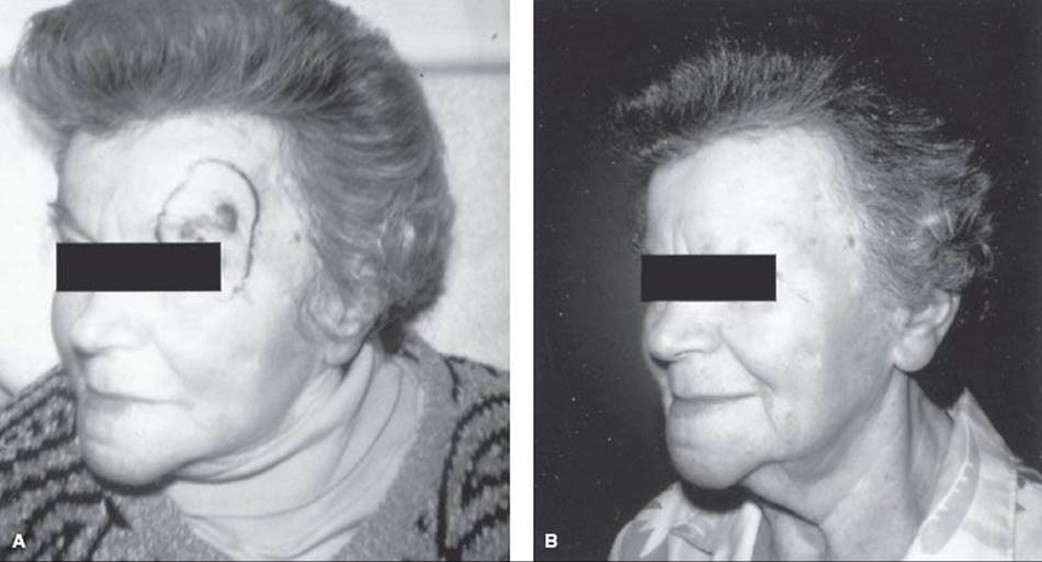
SARCOMAS OF THE HEAD AND NECK
Natural History
Sarcomas account for <1% of malignant neoplasms in the head and neck. The most frequent histological type is malignant fribohistiocytoma (29%), while the least common is liposarcoma (1%). The histology is complex and requires immunochemical analysis including angiosarcoma, chondrosarcoma, hemangiosarcoma, leiomyosarcoma, liposarcoma, malignant fibrous sarcoma, neurofibrosarcoma, osteosarcoma, rhabdomyosarcoma, malignant schwannoma, and synovial sarcoma. Fibrosarcoma, angiosarcoma, leiomyosarcoma, and rhabdomyosarcoma are the most common types, but this varies in published reports. Distribution of these sarcomas was 33% in the scalp or face, 26% in the orbit or paranasal sinuses, 14% arising from upper aerodigestive tract including larynx, and 27% in the neck. Synovial sarcomas are rare soft tissue malignancies in the head and neck region; they account for 3% to 5% of head and neck tumors. Histologic, immunohistochemical, and characteristic chromosomal translocation findings are necessary for diagnosis. The poor prognosis of this sarcoma justifies radical surgery with postoperative radiation.277
Radiation-induced sarcoma of the head and neck is a rare long-term complication of treatment. The rarity of this tumor is reflected in the very few series reported in the English language medical literature.278,279 When they do occur, most appear at least 10 years following radiation therapy. There is a possibility of a postirradiation sarcoma whenever a suspicious lesion is seen, regardless of the amount of time that has passed since radiation therapy was administered. The original pathology should be re-examined to ensure that the original tumor was diagnosed correctly. Electron microscopy can be useful in differentiating sarcomatous-appearing epithelial lesions from true soft tissue sarcomas.
The incidence of radiation-induced sarcomas of the head and neck is, however, likely to increase due to progressive aging of the population combined with improved survival in head and neck cancer patients. This problem can be extremely challenging, and the overall outlook has been reported to be very bleak. Patel et al.279 reviewed 69 cases reported in the English medical literature since 1966 and pooled this information with their experience in treatment of 10 patients. This group was compared for survival with 124 patients with a diagnosis of head and neck sarcoma registered on the Head and Neck Sarcoma database at the Royal Marsden Hospital. There was no site prediction for radiation-induced sarcoma of the head and neck, but malignant fibrous histiocytoma was the most common pathologic diagnosis. The period of latency between initial radiation therapy and diagnosis ranged from 9 to 45 years, with a median of 17 years. Surgery was the mainstay of treatment, and follow-up ranged from 6 months to 15 years with a median of 48 months. The actuarial 5-year disease-free survival rate in these patients was 60%.
Clinical Presentation and Diagnostic Workup
Clinical presentation varies with the primary site of disease. Tumors arising from the aerodigestive tract usually present with nasal bleeding, a palpable mass in the neck, or difficulty in swallowing or breathing. In tumors arising from the base of skull or the nerve sheath, cranial nerve deficit is the most common presentation. Diagnostic workup follows that of soft tissue sarcomas of other sites in the body. With early lesions, radiographs or CT may show only nonspecific opacification, soft tissue swelling, and occasionally bone destruction. Table 48.1 outlines the suggested diagnostic workup. MRI, especially with gadolinium contrast, may be used as a supplement or alternative to CT scanning.280,281 A CT scan of the chest is also mandatory for staging workup.
The American Joint Committee on Cancer staging system for soft tissue sarcomas is based on histologic grade, the tumor size and depth, and the presence of distant or nodal metastases. The staging system is the same as for sarcomas of the extremities, although specific staging for head and neck sarcomas is not standardized.213
Prognostic Factors
Prognostic factors for predicting local recurrence or disease-free survival include anatomic site, treatment modality, tumor histology and grade, tumor size, extension of disease, lymph node metastasis, and surgical margins.277,282,283,284
A report from Royal Marsden Hospital showed anatomic location and treatment modality to be independent prognostic factors for local recurrence; tumors of the head had a better local recurrence-free survival than did those of the neck.77 Patients treated with a combination of surgery and radiation therapy had a better recurrence-free survival than did those treated with surgery or irradiation alone. The only significant independent prognostic factor for overall survival was the implementation of definitive surgery versus biopsy. In the above report, the prognostic impact of tumor stage and grade did not reach statistical significance. In contrast, Tran et al.285 reported that 90% of patients with low-grade tumors were free of disease versus only 16% with high-grade lesions.
Bentz et al.157 reviewed 111 head and neck sarcoma patients; median duration of follow-up was 51 months; the actuarial 5-year relapse-free disease-specific, and overall survivals were 55%, 52%, and 44%, respectively. By multivariate analysis, size and grade significantly influenced all survivals, whereas margin status additionally influenced relapse-free survival.
In 109 soft tissue sarcomas of all sites, a French study demonstrated that quality of the surgery was one of the most important variables for predicting local recurrences. Tumor size, surgical margins, presence of tumor necrosis, and adequacy of the excision correlated with metastasis-free survival.286
General Management
Surgery is the preferred initial treatment modality for sarcomas.235,287 Unfortunately, it is often difficult to achieve complete resection of the tumor, and a high recurrence rate has been observed with surgery alone.288 Extracapsular enucleation of the tumor results in 90% local recurrence because of the presence of microscopic pseudopodia, which tend to grow through the pseudocapsule into the surrounding tissue, and the presence of skipped lesions some distance from the main tumor mass. Pathologic analysis of the surgical bed often discloses microscopic extension of tumor. Farhood et al.,288 in a review of 176 cases of adult head and neck sarcomas, reported that the pathologic margins of surgical specimens obtained by wide local excision were positive in >50% of cases. This resulted in inferior overall survival for sarcomas of the head and neck when compared with extremity sarcomas.238 Wide local excision, with a 5-cm margin around the pseudocapsule in extremity sarcomas, is associated with better outcome, although approximately 20% will have local recurrence. The criteria for surgical resection are impractical for head and neck sarcomas because of anatomic limitations;213 wide local excision is rarely possible because the tumors extend beyond the confines of origin and in the proximity of vital neurovascular structures. Some retrospective studies have suggested improved local tumor control when combined surgery and external irradiation are used. In 130 patients with soft tissue sarcomas of the head and neck treated with surgery alone at Royal Marsden Hospital, the overall 5-year survival was 50%; local tumor control was only 47%, and local recurrence was the cause of death in 63% of cases. Patients treated with combined-modality treatment (surgery and irradiation) had less extensive surgery, yet local recurrence-free survival was longer.283
Synovial sarcoma in the head and neck is rare. In a report of 36 patients Al-Daraji et al.289 noted there was a predilection for the parotid and the temporal regions, and nine involved skeletal muscle. Of 29 patients followed for a median 14 years after surgical treatment, 11 were alive and tumor free.
A multidisciplinary discussion before the initiation of treatment is required to formulate the best approach for radiation delivery, surgical technique, and mode of reconstruction.
Radiation Therapy
Radiation therapy, by external beam or brachytherapy, plays an important adjunctive role in disease management, especially for tumors where en bloc resection with negative margin is not possible.284,290 Chemotherapy regimens are available for soft tissue neoplasms, primarily designed to improve local tumor control.127 A systematic review of radiation therapy trials was performed by the Swedish Council of Technology Assessment in Health Care.290 This synthesis of the literature on radiation therapy for soft tissue sarcomas is based on data from five randomized trials. Moreover, data from 6 prospective studies, 25 retrospective studies, and 3 other articles were used. In total, 39 scientific articles were included, involving 4,579 patients. The results were compared with those of a similar overview from 1996, which included 3,344 patients. There was evidence that adjuvant radiotherapy improves local tumor control in combination with conservation surgery with negative, marginal, or minimal microscopic positive surgical margins. There are still insufficient data to establish that preoperative radiotherapy is favorable compared to postoperative radiotherapy in patients presenting primarily with large tumors. The preoperative setting results in more wound complications. There is no randomized study comparing external-beam radiotherapy and brachytherapy. These data suggest that external-beam radiotherapy and low–dose-rate brachytherapy result in comparable local control for high-grade tumors. Some patients with low-grade soft tissue sarcomas benefit from external-beam radiotherapy in terms of local control. Brachytherapy with a low-dose rate for low-grade tumors seems to be of no benefit, but data are sparse. In two small studies investigating hyperfractionation schedules, there was no indication of improvements compared to daily fractions of 2 Gy.
Mesenchymal chondrosarcoma of the sinonasal tract is a rare, malignant tumor of extraskeletal origin.93 Thirteen patients with sinonasal mesenchymal chondrosarcoma presented with nasal obstruction (n = 8), epistaxis (n = 7), mass effect (n = 4), or a combination of these. The maxillary sinus was the most common site of involvement (n = 9), followed by the ethmoid sinuses (n = 7) and the nasal cavity (n = 5). All cases were managed by surgery with adjuvant radiation therapy (n = 4) and/or chemotherapy (n = 3). The overall mean survival was 12.1 years, although five of six patients who developed local recurrences died of disease (mean survival, 6.5 years). Six patients were alive and disease free (mean survival, 17.3 years), and two patients were lost to follow-up.
Radiation Therapy Techniques
The general principles for radiation therapy of head and neck sarcomas are similar to those of soft tissue sarcomas. Complete coverage of the surgical bed and scar with adequate margins (3 to 5 cm) is required.291 However, because of the proximity of critical and radiosensitive organs (eyes, spinal cord, brainstem), selecting optimal portal margins without seriously compromising the functioning of these organs is an art. Techniques similar to those used in epithelial tumors of the head and neck can be applied to sarcomas. In general, 55 to 60 Gy is needed for postoperative adjuvant irradiation, and an additional 10- to 15-Gy boost is recommended if the surgical margins are close (≤3 mm) or involved by tumor. Some institutions prefer preoperative irradiation of 45 to 50 Gy. Special attention should be directed to limiting the dose to critical structures. Use of a 3D or IMRT treatment technique can be considered as demonstrated in Fig. 48.8.
Protons or heavy ions have been used in selected patients.292 Hug et al.293 reported on 27 patients treated at Massachusetts General Hospital in Boston (18 primary and 9 recurrent sarcomas of the head and neck close or abutting critical structures with 160 MeV protons; mean dose 68.5 GyE in 2.1-GyE fractions). Local recurrence was seen in eight patients (29%) and regional recurrence in six patients (22%). Tumor grade had a significant impact on outcome. One patient developed Lhermitte sign and another hypothyroidism.
Jingu et al.294 described results in 27 patients with head and neck unresectable bone and soft tissue sarcomas, treated with carbon ions (57.6, 64.0, or 70.4 GyE in 16 fractions). The 3-year local tumor control was 91.6% and survival 72%. Therapy was well tolerated.
TABLE 48.14 STAGING OF NASOPHARYNGEAL ANGIOBROMA
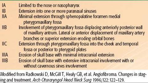
TABLE 48.15 TREATMENT RESULTS OF ADULT SOFT TISSUE SARCOMAS OF THE HEAD AND NECK

Results of Therapy
Because of the propensity for sarcomas to invade the surrounding tissues, complete surgical clearance may be difficult. In a series from UCLA, attempted en bloc resection left residual tumor at the surgical margins in 52 of 127 patients.285 The incidence of local recurrence was high (60%) with surgery alone.
In a retrospective report of 73 patients with sarcomas of the head and neck treated at Princess Margaret Hospital, the 5-year cause-specific survival was 62%, with a local recurrence rate of 41% and a distant metastasis rate of 31%.280 Extension to adjacent structures, high-grade tumor, and tumor >10 cm were associated with poor survival. Gross residual tumor after surgery was also associated with a high local recurrence rate (75%) despite the addition of radiation therapy. Patients with clear surgical margins or only microscopic involvement fared much more favorably and had a similar local tumor control rate (74% and 70%, respectively), provided adjuvant irradiation was given. Because of the difficulty in obtaining wide surgical margins, 68% of the patients died as a result of uncontrolled local disease. These data substantiate the importance of surgical margins as well as the contribution of adjuvant irradiation.280
Colville et al.213 reported on 41 male and 19 female patients treated with head and neck soft tissue sarcomas, with an overall 5-year survival of 60%. Twenty-five patients had surgery alone, 20 had surgery and pre- or postoperative radiation therapy, and 15 received nonsurgical treatment. With mean follow-up of almost 4 years, the 5-year local tumor control was 56% in the surgical group and 40% in the nonsurgical group (more advanced and aggressive tumors). The 5-year survival was 70% and 40%, respectively.
Penel et al.295 recorded their experience with 28 adult head and neck soft tissue sarcomas. The most common subtype was rhabdomyosarcoma (RMS) (seven cases). Twenty-two patients presented with previous inadequate resection performed elsewhere before admission. Nineteen patients had surgery (complete resection in 13 cases). Associated treatments were neoadjuvant chemotherapy, adjuvant chemotherapy, and postoperative radiotherapy in 4, 3, and 10 cases, respectively. The 2-year overall survival rate was 56%. Wolden et al.296 treated 28 patients with head and neck RMS using IMRT (50–55 Gy in 1.8 Gy fractions) combined with chemotherapy. With median follow up of 24 months local tumor control was 95–100% and 3 year survival McDonald et al.297 treated 20 children with RMS in the head and neck with IMRT (median dose 50.4 Gy, 1.8 per fraction). With 29 months median follow up the 3 year local tumor control was 100% and survival 76%.
Pandey et al.298 reported on 22 cases of head and neck sarcomas (neck, lower jaw, tongue, cheek, scalp, and maxilla were the most common sites affected). None of the patients had palpable neck nodes or distant metastasis at presentation. All the patients were treated with primary surgical resection, followed by adjuvant treatment in 14 cases (63.6%). After a median follow-up of 14.5 months, two patients died, six developed local recurrence, four developed metastatic disease, and another patient developed a second primary sarcoma. The overall 5-year survival was 80%, while the 5-year disease-free survival rate was 24.1%.
Barker et al.13 published a review of 44 patients with nonmetastatic soft tissue sarcoma in a head and neck. The most common tumor histologies included malignant fibrous histiocytoma (15 patients), angiosarcoma (9 patients), fibrosarcoma (6 patients), and leiomyosarcoma (6 patients). The median overall survival for all patients was 79 months. The actuarial 5-year local tumor control was 55% and was highly correlated with the extent of surgical excision: 25% for subtotal resection or debulking, 65% for wide local excision, and 100% for radical excision. Local tumor control at 5 years was 60% for patients treated with both surgery and radiotherapy, 54% surgery alone, and 43% for radiation alone. Adjuvant radiation therapy significantly improved the local control rates (from 25% to 54%) for patients with close (<2 mm) or positive surgical margins. Of 14 patients with locoregional failure in whom salvage was attempted, 9 (64%) were rendered disease free.
Rapidis et al.302 reported on 25 patients with head and neck sarcomas with follow-up ranging from 8 to 144 months. Twenty-three patients were treated with surgery as the primary modality; 14 with surgery alone. Clear margins were obtained in all of them and local control was achieved in 12 of 13. The 5-year survival for the entire group was 40%. Reported results of treatment of soft tissue sarcomas is summarized in Table 48.15.
Tumor Characteristics
Several series have shown that tumor grade and size dictate the outcome of patients with head and neck sarcomas such as leiomyosarcoma, rhabdomyosarcoma, and malignant fibrous sarcoma.275 Farhood et al.,288 in a review of 176 adult head and neck sarcomas, found that only 20% of the patients with high-grade tumors were alive 10 years after treatment, compared with 88% of patients with low-grade tumors. Weber et al.299 described a 45% 10-year survival rate for patients with tumors <5 cm versus 10% for those with tumors ≥5 cm.
Many series have reported that chondrosarcoma is not a radiosensitive tumor, and radiation therapy has no role in it treatment. However, some reports have demonstrated the contribution of radiation therapy in this histology. McNaney et al.303 described a 65% survival rate at 2 years in 20 chondrosarcoma patients who received primary radiation therapy. Tumor grade was the most important prognostic factor.
Osteogenic sarcoma of the head and neck has a pattern of recurrence different from similar tumors elsewhere in the body. Head and neck osteosarcomas are usually high grade; they have a very high incidence of local recurrence but a lower risk of distant metastases. Several studies have used adjuvant irradiation and chemotherapy, which commonly results in improved locoregional tumor control and survival. Tran et al.285 reported a 73% 5-year survival rate in patients with osteogenic sarcoma of the head and neck treated with high-dose preoperative irradiation followed by wide surgical excision.
Chemotherapy
Head and neck soft tissue sarcomas frequently metastasize; 25% of patients in a UCLA study had distant metastases.265 The role of adjuvant chemotherapy to improve disease-free survival in sarcoma of the head and neck is controversial. Unlike with soft tissue sarcomas of the extremities, in which distant metastasis is the most common cause of death, the majority of deaths in sarcomas of the head and neck are associated with local failure. Approximately half of the distant metastases were detected after local recurrence occurred.280 Chemotherapy did not appear to affect local tumor control.
In a series of 94 patients treated at UCLA,244 local control was achieved in 52% of patients treated with surgery alone and 90% of those receiving adjuvant irradiation with or without chemotherapy.
For preoperative neoadjuvant chemotherapy, which supplements radiation therapy to downstage disease before surgery, satisfactory results are available only for sarcomas of extremities.304 With the exception of rhabdomyosarcoma, postoperative adjuvant chemotherapy for head and neck sarcomas should be given only in a clinical trial setting.
REFERENCES
1. Davidson J, Gullane P. Glomus vagale tumors. Otol Head Neck Surg 1988;99: 66–70.
2. Larson TC III, Reese DF, Baker HL Jr, et al. Glomus tympanicum chemodectomas: radiographic and clinical characteristics. Radiology 1987;163:801–806.
3. Konefal JB, Pilepich MV, Spector GJH, et al. Radiation therapy in the treatment of chemodectomas. Laryngoscope 1987;97:1331–1335.
4. Mendenhall WM, Million RR, Parsons JT, et al. Chemodectoma of the carotid body and ganglion nodosum treated with radiation therapy. Int J Radiat Oncol Biol Phys 1986;12:2175–2178.
5. Magliulo G, Zardo F, Varacalli S, et al. Multiple paragangliomas of the head and neck. Anal Otorrinolaringol Ibero Am 2003;30:31–38.
6. Semaan MT, Megerian CA. Current assessment and management of glomus tumors. Curr Opin Otolaryngol Head Neck Surg 2008;16:420–426.
7. Gaut W, Jay AP, Robinson PA. Invasive glomus tumor of the nasal cavity. Am J Otolaryngol 2005;26:207–209.
8. Manolidis S, Shohet JA, Jackson CG, et al. Malignant glomus tumors. Laryngoscope 1999;109:30–34.
9. Vogl TJ, Juergens M, Balzar JO, et al. Glomus tumors of the skull base: combined use of MR angiography and spin-echo imaging. Radiology 1994;192:1003–1010.
10. Drape JL, Idy-Peretti I, Goettmann S, et al. Sublingual glomus tumors: evaluation with MR imaging. Radiology 1995;195:507–515.
11. Christie A, Teasdale E. A comparative review of multi-detector CT angiography and MRI in the diagnosis of jugular foramen lesions. Clin Radiol 2010;65: 213–217.
12. Astner STA, Essler MA, Bundschuch RAA, et al. 18F-Octreotate positron emission tomography (PET) for target volme delineation in stereotactic radiation therapy planning of glomus tumors. Int J Radiat Oncol Biol Phys2007;69(Suppl 3): S545–S546.
13. Barker JL Jr, Paulino AC, Feeney S, et al. Locoregional treatment for adult soft tissue sarcomas of the head and neck: an institutional review. Cancer J 2003; 9:49–57.
14. Batsakis JG. In: Tumors of the head and neck, 2nd ed. Baltimore: Williams & Wilkins, 1979:474–475.
15. Batsakis JG. Wegener’s granulomatosis and midline (non peeling) “granuloma.” Head Neck Surg 1979;1:213–222.
16. Glasscock ME III, Jackson CG, Johnson GD, et al. Radiation therapy in chemodectoma treatment [letter]. Laryngoscope 1988;98:465566.
17. McCabe BF, Fletcher M. Selection of therapy of glomus jungulare tumors. Arch Otolaryngol 1969;89:156–159.
18. Li G, Chang S, Adler JR, et al. Irradiation of glomus jugulare tumors: a historical perspective. Neurosurg Focus 2007;23:E13.
19. Abud DG, Mounayer C, Benndorf G, et al. Intratumoral injection of cyanoacrylate glue in head and neck paragangliomas. AJNR Am J Neuroradiol 2004;29: 1457–1462.
20. Ozyer U, Harman A, Yildirim E, et al. Devascularization of head and neck paragangliomas by direct percutaneous embolization. Cardiovasc Intervent Radiol 2010;33:967–975.
21. Cummings BJ, Beale FA, Garrett PG, et al. The treatment of glomus tumors in the temporal bone by megavoltage radiation. Cancer 1984;53:2635–2640.
22. Powell S, Peters N, Harmer C. Chemodectoma of the head and neck: results of treatment in 84 patients. Int J Radiat Oncol Biol Phys 1992;22:919–924.
23. Spagnolo DV, Papadimitiou JM, Archer M. Postirradiation malignant fibrous histiocytoma arising in juvenile nasopharyngeal angiofibroma and producing alpha-1-antitrypsin. Histopathology 1984;8:339–352.
24. Suit HD, Gallager HS. Intact tumor cells in irradiated tissue. Arch Pathol 1964; 78:648.
25. Krych AJ, Foote RL, Brown PD, et al. Long-term results of irradiation for paraganglioma. Int J Radiat Oncol Biol Phys 2006;65:1063–1066.
26. Springate SC, Weichselbaum RR. Radiation or surgery for chemodectoma of the temporal bone: a review of local control and complications. Head Neck 1990;12: 303–307.
27. Dickens WJ, Million RR, Cassisi NJ, et al. Chemodectomas arising in temporal bone structures. Laryngoscope 1982;92:188–191.
28. Onal C, Ozcelik TB, Sonmez S, et al. Radiation dose distribution in the external auditory canal with different prosthetic materials: a clinical report. J Prosthet Dent 2010;104:288–292.
29. Wax MK, Yun KJ, Omar RA. Extramedullary plasmacytomas of the head and neck. Otolaryngol Head Neck Surg 1993;109:877–885.
30. Leber KA, Eustacchio S, Pendl G. Radiosurgery of glomus tumors: midterm results. Stereotact Funct Neurosurg 1999;72(Suppl 1):53–59.
31. Bianchi LC, Marchetti M, Brait L, et al. Paragangliomas of head and neck: a treatment option with CyberKnife radiosurgery. Neurol Sci 2009;30:479–485.
32. Chen PG, Nguyen JH, Payne SC, et al. Treatment of glomus jugulare tumors with Gamma Knife radiosurgery. Laryngoscope 2010;120:1856–1862.
33. Genc A, Bicer A, Abacioglu U, et al. Gamma Knife radiosurgery for the treatment of glomus jugulare tumors. J Neurooncol 2010;97:101–108.
34. Hafez RF, Morgan MS, Fahmy OM. The safety and efficacy of Gamma Knife surgery in management of glomus jugulare tumors. World J Surg Oncol 2010;8:76.
35. Lim M, Bower R, Nangiana JS, et al. Radiosurgery for glomus jugulare tumors. Technol Cancer Res Treat 2007;6:419–423.
36. Maitz AH, Grills IS, Chen PY et al. Gamma Knife radiosurgery: A viable therapy for glomus jugulare tumors. Int J Radiat Oncol Biol Phys 2008;72(Suppl 1):S477.
37. Navarro MA, Maitz A, Grills IS, et al. Successful treatment of glomus jugulare tumors with Gamma Knife radiosurgery: clinical and physical aspects of management and review of the literature. Clin Transl Oncol 2010;12:55–62.
38. Henzel M, Hamm K, Gross MW, et al. Fractionated stereotactic radiotherapy of glomus tumors. Local control, toxicity, symptomatology and quality of life. Strahlenther Onkol 2007;183:557–562.
39. Pollock BE. Stereotactic radiosurgery in patients with glomus jugulare tumors. Neurosurg Focus 2004;17(2):E10.
40. Wegner RE, Rodriguez KD, Heron DE, et al. Linac-based stereotactic body radiotherapy for treatment of glomus. Radiother Oncol 2010;97:395–398.
41. Combs SE, Konkel S, Schulzertner D, et al. Intensity modulated radiotherapy (IMRT) in patients with carcinoma of the paranasal sinuses: clinical benefit for complex shaped target volumes. Radiat Oncol2006;1:23.
42. Ares C, Hug EB, Lomax AJ, et al. Effectiveness and safety of spot-scanning proton radiation therapy for chordomas and chondrosarcomas of the skull base: first long-term report. Int J Radiat Oncol Biol Phys 2009;75:1111–1118.
43. Schulz-Ertner D, Haberer T, Jakel O, et al. Radiotherapy for chordomas and low grade chondrosarcomas of the skull base with carbon ions. Int J Radiat Oncol Biol Phys 2002;53:36–42.
44. Lloyd G, Howard D, Lund VJ, et al. Imaging for juvenile angiofibroma. J Laryngol Otol 2000;114:727–730.
45. Griffiths RW, Briggs JC. Biopsy procedures, primary wide excisional surgery and long-term prognosis in primary clinical stage I invasive cutaneous malignant melanoma. Ann R Coll Surg Engl1985;67:75–78.
46. Wang M-L, Hussey DH, Doornbos JF, et al. Chemodectoma of the temporal bone: a comparison of surgical and radiotherapeutic results. Int J Radiat Oncol Biol Phys 1988;14:643–648.
47. Zabel A, Milker-Zabel S, Huber P. Fractionated sterotactic conformal radiotherapy in the management of large chemodectomas of the skull base. Int J Radiat Oncol Biol Phys 2004;58:1445–1450.
48. Kim JW, Kim DG, Chung HT, et al. Gamma Knife stereotactic radiosurgery for intracranial hemangiopericytomas. J Neurooncol 2010;99:115–122.
49. Hinerman RW, Mendenhall WM, Amdur RJ, et al. Definitive radiotherapy in the management of chemodectomas arising in the temporal bone, carotid body, and glomus vagale. Head Neck 2001;23:363–371.
50. Hinerman RW, Morris CG, Mendenhall WM, et al. Paragangliomas of the head and neck treated with external-beam radiotherapy. Int J Radiat Oncol Biol Phys 2007;69(Suppl 3): S434 (abstr 2416).
51. Verniers DA, Keus RB, Schouwenburg PF, et al. Radiation therapy, an important mode of treatment for head and neck chemodectomas. Eur J Cancer 1992;27:1028–1033.
52. Ivan ME, Sughrue ME, Clark AJ, et al. A meta-analysis of tumor control rates and treatment related morbidity for patients with glomus jugulare tumors. J Neurosurg 2011;114:1299–1305.
53. Guss ZD, Batra S, Limb CJ, et al. Radiosurgery of glomus jugulare tumors: a meta-analysis. Int J Radiat Oncol Biol Phys 2011;81(4):e497–e502.
54. Elshaikh MA, Mahmoud-Ahmed AS, Kinney SE, et al. Recurrent head and neck chemodectomas: a comparison of surgical and radiotherapeutic results. Int J Radiat Oncol Biol Phys 2002;52:953–956.
55. Cheesman AD, Kelly AM. Rehabilitation after treatment for jugular foramen lesions. Skull Base 2009;19:99–108.
56. Larner JM, Hahn SS, Spaulding CA, et al. Glomus jugulare tumors: long-term control by radiation therapy. Cancer 1992;69:1813–1817.
57. Pryzant RM, Chou JL, Easley JD. Twenty year experience with radiation therapy for temporal bone chemodectomas. Int J Radiat Oncol Biol Phys 1989;17: 1303–1307.
58. Cole JM, Beiler D. Long-term results of treatment for glomus jugulare and glomus vagale tumors with radiotherapy. Laryngoscope 1994;104:1461–1465.
59. Boyle JO, Shimm DS, Coulthard SW. Radiation therapy for paragangliomas of the temporal bone. Laryngoscope 1990;100:896–901.
60. Schild SE, Foote RL, Buskirk SJ, et al. Results of radiotherapy for chemodectomas. Mayo Clin Proc 1992;67:537–540.
61. deJong AL, Coker NJ, Jenkins HA, et al. Radiation therapy in the management of paragangliomas of the temporal bone. Am J Otolaryngol 1995;16:283–289.
62. Valdagni R, Amichetti M. Radiation therapy of carotid body tumors. Am J Clin Oncol 1990;13:45–48.
63. Sundaram C, Uppin SG, Uppin MS, et al. A clinicopathological and immunohistochemical study of central nervous system hemangiopericytomas. J Clin Neurosci 2010;17:469–472.
64. Paal E, Chung EM. Head and neck pathology-radiology classics: vagal paraganglioma. Head Neck Pathol 2007;1:35–37.
65. Stout AP. Hemangiopericytoma. Study of 25 new cases. Cancer 1949;2:1027–1054.
66. Enzinger FM, Weiss SW. Hemangiopericytoma. In: Enzinger FM, Weiss SW, eds. Soft tissue tumors, 2nd ed. St. Louis, MO: CV Mosby, 1988.
67. Palacios E, Restrepo S, Mastrogiovanni L, et al. Sinonasal hemangiopericytomas: clinicopathologic and imaging findings. Ear Nose Throat J 2005;84(2):99–102.
68. Rutkoeski MJ, Sughrue ME, Kane AJ, et al. Predictors of mortality following treatment of intracranial hemangiopericytoma. J Neurosurg 2010;113:333–339.
69. Spitz FR, Bouvet M, Pisters PW, et al. Hemangiopericytoma: a 20-year single institution experience. Ann Surg Oncol 1998;5:350–355.
70. Kanazawa T, Nishino H, Miyata M, et al. Haemangiopericytoma of infratemporal fossa. J Laryngol Otol 2001;115:77–79.
71. Kowalski PJ, Paulino AF. Proliferation index as a prognostic marker in hemangiopericytoma of the head and neck. Head Neck 2001;23:492–496.
72. Vuorinen V, Sallinen P, Haapsasalo H, et al. Outcome of 31 intracranial haemengiopericytomas: poor predictive value of cell proliferation indices. Acta Neurochir (Wien) 1996;138:1399–1408.
73. Setzkorn RK, Lee DJ, Iliff NT, et al. Hemangiopericytoma of the orbit treated with conservative surgery and radiotherapy. Arch Ophthalmol 1987;105:1103–1105.
74. Charles NC, Palu RN, Jagirdar JS. Hemangiopericytoma of the lacrimal sac. Arch Ophthalmol 1998;116:1677–1680.
75. Jaaskelainen J, Servo A, Haltia M, et al. Intracranial hemangiopericytoma: radiology, surgery, radiotherapy and outcome in 21 patients. Surg Neurol 1985;23: 227–236.
76. Yaghmai I. Angiographic manifestations of soft tissue and osseous hemangiopericytomas. Radiology 1978;126:652–659.
77. Lee J, Kim I. Roll of radiotherapy in intracranial hemangiopericytoma. Int J Radiat Oncol Biol Phys 2008;72(Suppl 1):S230 (abstr 2136).
78. Monroe AT, Hinerman RW, Amdur RJ, et al. Radiation therapy for esthesioneuroblastoma: rationale for elective neck irradiation. Head Neck 2003;25:529–534.
79. Hasegawa A, Mizoe J, Jingu K, et al. Carbon ion radiotherapy for skull base and paracervical chordomas. Int J Radiat Oncol Biol Phys 2009;75.
80. Park MS, Patel SR, Ludwig JA, et al. Activity of temozolomide and bevacizumab in the treatment of locally advanced, recurrent and metastatic hemangiopericytoma and malignant fibrous tumor. Cancer2011;117(21):4939–4947.
81. Espat NJ, Lewis JJ, Leung D, et al. Conventional hemangiopericytoma: modern analysis of outcome. Cancer 2002;95:1746–1751.
82. Jha N, McNeese M, Barkley HT, et al. Does radiotherapy have a role in hemangiopericytoma management? Report of 14 new cases and review of the literature. Int J Radiat Oncol Biol Phys1987;13:1399–1402.
83. Hyams VJ. Tumors of the upper respiratory tract and ear. In: Hyams VJ, Batsakis JG, Michaels L, eds. Atlas of tumor pathology, 2nd ed. series, Fascicle 25. Washington, DC: Armed Forces Institute of Pathology, 1988:240–248.
84. Billings KR, Fu YS, Calcaterra TC, et al. Hemangiopericytoma of the head and neck. Am J Otolaryngol 2000;21:238–243.
85. Patrice SJ, Sneed PK, Flickinger JC, et al. Radiosurgery for hemangioblastoma: results of a multi-institutional experience. Int J Radiat Oncol Biol Phys 1995; 32(Suppl 1):147(abstr).
86. Carew JF, Singh B, Kraus DH. Hemangiopericytoma of the head and neck. Laryngoscope 1999;109:1409–1411.
87. Staples JJA, Robinson RAA, Wen B-CA, et al. Hemangiopericytoma—the role of radiotherapy. Int J Radiat Oncol Bio Phys 1990;19:445–451.
88. Kano H, Niranjan A, Kondziolka D, et al. Adjuvant stereotactic radiosurgery after resection of intracranial hemangiopericytomas. Int J Radiat Oncol Biol Phys 2008;72:1333–1339.
89. Olson C, Yen CP, Schlesinger D, et al. Radiosurgery for intracranial hemangiopericytomas: outcomes after initial and repeat Gamma Knife surgery. J Neurosurg 2010;112:133–139.
90. Redmond KJ, Gullett NP, Kleinberg L, et al. Hemangiopericytoma of the central nervous system: analysis of current national patterns of care. Int J Radiat Oncol Bio Phys Proc 2010;78(suppl 3):S615.
91. Tai PT, Craighead P, Bagdon F. Optimization of radiotherapy for patients with cranial chordoma: a review of dose-response ratios for photon techniques. Cancer 1995;75:749–756.
92. Rich TA, Schiller A, Suit HD, et al. Clinical and pathologic review of 48 cases of chordoma. Cancer 1985;56:182–187.
93. Knott PD, Gannon FH, Thompson LD. Mesenchymal chondrosarcoma of the sinonasal tract; a clinicopathological study of 13 cases with a review of the literature. Laryngoscope 2003;113:783–790.
94. Volpe NJ, Liebsch NJ, Munzenrider JE, et al. Neuro-ophthalmologic findings in chordoma and chondrosarcoma of the skull base. Am J Ophthalmol 1993;115:97–104.
95. Doucet V, Peretti-Viton P, Figarella-Branger D, et al. MRI of intracranial chordomas. Extent of tumour and contrast enhancement: criteria for differential diagnosis. Neuroradiology 1997;39:571–576.
96. Oot RF, Melville GE, New PF, et al. The role of MR and CT in evaluating clival chordomas and chondrosarcomas. AJR Am J Roentgenol 1988;151:567–575.
97. Saigal K, Palmer JD, Reis I, et al. Mucosal melanomas of the head and neck: a modern experience at the University of Miami. Int J Radiat Oncol Bio Phys Proc 2010;78(suppl 3):S479.
98. Gabriele P, Macias V, Stasi M, et al. Feasibility of intensity-modulated radiation therapy in the treatment of advanced cervical chordoma. Tumori 2003;89: 298–304.
99. Catton C, O’Sullivan B, Bell R, et al. Chordoma: long-term follow-up after radical photon irradiation. Radiother Oncol 1996;41:67–72.
100. Forsyth PA, Cascino TL, Shaw EG, et al. Intracranial chordomas: a clinicopathological and prognostic study of 51 cases. J Neurosurg 1993;78:741–747.
101. Gay E, Sckhar LN, Rubinstein E, et al. Chordomas and chondrosarcomas of the cranial base: results and follow-up of 60 patients. Neurosurgery 1995;36: 887–896.
102. Chetty R, Levin CV, Kalan MR. Chordoma: a 20-year clinicopathologic review of the experience at Groote Schuur Hospital, Cape Town. J Surg Oncol 1991;46: 261–264.
103. Keisch ME, Garcia DM, Shibuya RB. Retrospective long-term follow-up analysis in 21 patients with chordomas of various sites treated at a single institution. J Neurosurg 1991;75:374–377.
104. Debus J, Schulz-Ertner D, Schad L, et al. Stereotactic fractionated radiotherapy for chordomas and chondrosarcomas of the skull base. Int J Radiat Oncol Biol Phys 2000;47:591–596.
105. Bugoci DM, Girvigian MR, Chen JCT, et al. Photon based fractionated stereotactic radiotherapy for postoperative treatment of skull base chordomas. Am J Clin Oncol (in press).
106. Den RB, Goldberg Y, Werner-Wasik M, et al. Photon based fractionated radiotherapy and radiosurgery for the treatment of chordomas and chondrosarcomas: a multi-institutional review. Int J Radiat Oncol Biol Phys2008;72(Suppl):S204 (abstr 2079).
107. Muthukumar N, Kondziolka D, Lundford LD, et al. Stereotactic radiosurgery for chordoma and chondrosarcoma: further experience. Int J Radiat Oncol Biol Phys 1998;41:387–392.
108. Suit HD, DeLaney T, Goldberg S, et al. Protons versus carbon ion beams in the definitive radiation treatment of cancer patients. Radiother Oncol 2010;95: 3–22.
109. Berson AM, Castro JR, Petti P, et al. Charged particle irradiation of chordoma and chondrosarcoma of the base of skull and cervical spine: the Lawrence Berkeley Laboratory experience. Int J Radiat Oncol Biol Phys1988;15:559–565.
110. Austin JP, Curie MM, Cardenosa G, et al. Probable causes of recurrence in chordomas and chondrosarcomas of the base of the skull and cervical spine. Int J Radiat Oncol Biol Phys 1993;25:439–444.
111. O’Connell JX, Renard LG, Liebsch NJ, et al. Base of skull chordoma: a correlative study of histologic and clinical features of 62 cases. Cancer 1994;74:2261–2267.
112. Fagundes MA, Hug EB, Liebsch NJ, et al. Radiation therapy for chordomas of the base of skull and cervical spine: patterns of failure and outcome after relapse. Int J Radiat Oncol Biol Phys1995;33:579–584.
113. Terahara A, Niemierko A, Goitein M, et al. Analysis of the relationship between tumor dose inhomogeneity and local control in patients with skull base chordoma. Int J Radiat Oncol Biol Phys1999;45:351–358.
114. Hug EB, Loredo LN, Slater JD, et al. Proton radiation therapy for chordomas and chondrosarcomas of the skull base. J Neurosurg 1999;91:432–439.
115. Noel G, Jauffret E, de Crevoisier R, et al. Photon and proton therapy for chordoma and chondrosarcoma of the base of the skull and cervical spine: prognostic factors and patterns of failure. Radiother Oncol 2002;S81 (abstr 241).
116. Jakel O, Land B, Combs SE, et al. On the cost-effectiveness of carbon ion radiation therapy for skull base chordoma. Radiother Oncol 2007;83:133–138.
117. Benk V, Liebsch NJ, Munzenrider JE, et al. Base of skull and cervical spine chordomas in children treated by high-dose irradiation. Int J Radiat Oncol Biol Phys 1995;31:577–581.
118. Royal College of Radiologists Proton Therapy Working Party. Proton therapy for base of skull chordoma: a report for the Royal College of Radiologists. Clin Oncol (R Coll Radiol) 2000;12:75–79.
119. Santoni R, Liebsch N, Finkelstein DM, et al. Temporal lobe damage following surgery and high-dose photon and proton irradiation in 96 patients affected by chordomas and chondrosarcomas of the base of the skull. Int J Radiat Oncol Biol Phys 1998;41:59–68.
120. Slater JD, Austin-Seymour M, Munzenrider J, et al. Endocrine function following high dose proton therapy for tumors of the upper clivus. Int J Radiat Oncol Biol Phys 1988;15:607–611.
121. Mendenhall WM, Olivier KR, Lynch JW Jr, et al. Lethal midline granuloma-nasal natural killer/T-cell lymphoma. Am J Clin Oncol 2006;29:202–206.
122. Rosignoli M, Pezzuto RW, Galli J, et al. Midline granuloma and Wegener’s granulomatosis. Acta Otorhinolaryngol Ital 1992;38(Suppl 12):1–46.
123. Borisch B, Hennig I, Laeng RH, et al. Association of the subtype 2 of the Epstein-Barr virus with T-cell non-Hodgkin’s lymphoma of the midline granuloma type. Blood 1993;82:858–864.
124. Berrettini S, Segnini G, Bruschini P, et al. Lethal midline granuloma: a case of Ki-1 lymphoma. Rev Laryngol Otol Rhinol (Bord) 1993;114:37–42.
125. Smalley RS, Cupps RE, Anderson JA, et al. Polymorphic reticulosis limited to the upper aerodigestive tract: natural history and radiotherapeutic considerations. Int J Radiat Oncol Biol Phys1988;15:599–605.
126. Gaulard P, Henni T, Marrolleau JP, et al. Lethal midline granuloma (polymorphic reticulosis) and lymphomatoid granulomatosis. Cancer 1988;62:705–710.
127. Mason M, Robinson M, Harmer C, et al. Intra-arterial adriamycin, conventionally fractionated radiotherapy and conservative surgery for soft tissue sarcomas. Clin Oncol 1992;4:32–35.
128. Fuller PS, Haferman DR, Byrol RB, et al. Use of irradiation in lymphomatoid granulomatosis. Chest 1978;74:105–106.
129. Shank BB, Kelley CD, Nisce LZ, et al. Radiation therapy in lymphomatoid granulomatosis. Cancer 1978;42:2572–2580.
130. Dickson RJ. Radiotherapy of lethal midline granuloma. J Chron Dis 1960;12: 417–427.
131. Halperin EC, Dosoretz MD, Goodman M, et al. Radiotherapy of polymorphic reticulosis. Br J Radiol 1982;55:645–649.
132. Fauci AS, Johnson RE, Wolff SM. Radiation therapy of midline granuloma. Ann Intern Med 1975;84:140–147.
133. Chen HHW, Fong L, Su I-J, et al. Experience of radiotherapy in lethal midline granuloma with special emphasis on centrofacial T-cell lymphoma: a retrospective analysis covering a 34-year period. Radiother Oncol 1996;38:1–6.
134. Hatta C, Ishida M, Matsumoto T, et al. Cases of lethal midline granuloma (polymorphic reticulosis) at our department in a recent 10-year period. J OtorhinoLaryngol Soc Jpn 1993;96:879–885.
135. Sakata K, Hareyama M, Oouchi A, et al. Treatment of localized non-Hodgkin’s lymphomas of the head and neck: focusing on cases of non-lethal midline granuloma. Radiat Oncol Invest 1998;6:161–169.
136. Zimmerman LCE, Font RL. Ophthalmologic manifestations of granulocytic sarcoma: a clinicopathologic study of 33 cases. Am J Ophthalmol 1975;80:975–990.
137. Woo E, Yue CP, Onann KE, et al. Intracerebral chloromas: report of a case and review of the literature. Clin Neurol Neurosurg 1986;88:135–139.
138. Byrd JC, Edenfield WJ, Shields DJ, et al. Extramedullary myeloid-cell tumors in acute nonlymphocytic leukemia: a clinical review. J Clin Oncol 1995;13: 1800–1816.
139. Pomeranz SJ, Hawkins HH, Towbin R, et al. Granulocytic sarcoma (chloroma): CT manifestations. Radiology 1985;155:167–170.
140. Sowers JJ, Moody PM, Naidich TP, et al. Radiographic features of granulocytic sarcoma (chloroma). J Comput Assist Tomogr 1979;3:226–233.
141. Chak LY, Saponzik D, Cox RS. Extramedullary lesions in non-lymphocytic leukemia: results of radiation therapy. Int J Radiat Oncol Biol Phys 1983;9: 1173–1176.
142. Berger L, Luc R. L’esthesioneuropitheliome olfacif. Bull Cancer (Paris) 1924; 13:410–421.
143. Eich HT, Staar S, Micke O, et al. Radiotherapy of esthesioneuroblastoma. Int J Radiat Oncol Biol Phys 2001;49:155–160.
144. Eden BV, Debo RF, Larner JM, et al. Esthesioneuroblastoma. Cancer 1994;73: 2556–2562.
145. Broich G, Pagliari A, Ottaviani F. Esthesioneuroblastoma: a general review of the cases published since the discovery of the tumour in 1924. Anticancer Res 1997;17:2683–2706.
146. Ganz JC, Abdelkarim K. Glomus jugulare tumors: certain clinical and radiologic aspects observed following Gamma Knife radiosurgery. Acta Neurochir 2009;151: 423–426.
147. Kadish S, Goodman M, Wang CC. Olfactory neuroblastoma: a clinical analysis of 17 cases. Cancer 1976;37:1571–1576.
148. Pickruth D, Heywang-Kobrunner SH, Speilmann RP. Computed tomography and magnetic resonance imaging features of olfactory neuroblastoma: an analysis of 22 cases. Clin Otolaryngol 1999;24:457–461.
149. Bustillo A, Telischi F, Weed D, et al. Octreotide scintigraphy in the head and neck. Laryngoscope 2004;114:434–440.
150. Chao KSC, Kaplan C, Simpson JR, et al. Esthesioneuroblastoma: the impact of treatment modality. Head Neck 2001;23:749–757.
151. Foote RL, Morita A, Ebersold MJ, et al. Esthesioneuroblastoma: the role of adjuvant radiation therapy. Int J Radiat Oncol Biol Phys 1993;27:835–842.
152. Argiris A, Dutra J, Tseke P, et al. Esthesioneuroblastoma: the Northwestern University experience. Laryngoscope 2003;113:155–160.
153. Miyamoto RC, Gleich LL, Biddinger PW, et al. Esthesioneuroblastoma and sinonasal undifferentiated carcinoma: impact of histological grading and clinical staging on survival and prognosis. Laryngoscope 2000;110:1262–1265.
154. Papadaki H, Kounelis S, Kapadia SB, et al. Relationship of p53 gene alterations with tumor progression and recurrence in olfactory neuroblastoma. Am J Surg Pathol 1996;20:715–721.
155. Rosenthal DI, Barker JL Jr, El-Naggar AK, et al. Sinonasal malignancies with neuroendocrine differentiation: patterns of failure according to histologic phenotype. Cancer 2004;101:2567–2573.
156. Dias FL, Sa GM, Lima RA, et al. Patterns of failure and outcome in esthesioneuroblastoma. Arch Otolaryngol Head Neck Surg 2003;129:1186–1192.
157. Bentz BG, Singh B, Woodruff J, et al. Head and neck soft tissue sarcomas: a multivariate analysis of outcomes. Ann Surg Oncol 2004;11:619–628.
158. Chang DT, Amdur RJ, Morris CG, et al. Adjuvant radiotherapy for cutaneous melanoma: comparing hypofractionation to conventional fractionation. Int J Radiat Oncol Biol Phys 2006;66:1051–1055.
159. Gruber G, Laedrach K, Baumert B, et al. Esthesioneuroblastoma: irradiation alone and surgery alone are not enough. Int J Radiat Oncol Biol Phys 2002;54:486–491.
160. Ozsahin M, Gruber G, Olszik O, et al. Outcome and prognostic factors in olfactory neuroblastoma: a rare cancer network study. Int J Radiat Oncol Biol Phys 2010; 78:993–997.
161. Sperry JL, Reis I, Casiano RR, et al. Esthesioneuroblastoma: a modern experience at the University of Miami. Int J Radiat Oncol Biol Phys 2010;78(Suppl 3): S472 (abstr 2603).
162. Madani I, Bonte K, Vakaet L, et al. Intensity modulated radiotherapy for sinasal tumors: Ghent University Hospital update. Int J Radiat Oncol Biol Phys 2009;73: 424–432.
163. Nishimura H, Ogino T, Kawashima M, et al. Proton-beam therapy for olfactory neuroblastoma. Int J Radiat Oncol Biol Phys 2007;68:758–762.
164. Bhattacharyya N, Thornton AF, Joseph MP, et al. Successful treatment of esthesioneuroblastoma and neuroendocrine carcinoma with combined chemotherapy and proton radiation. Results in 9 cases. Arch Otolaryngol Head Neck Surg 1997; 123:34–40.
165. McElroy EA, Buckner JC, Lewis JE. Chemotherapy for advanced esthesioneuroblastoma: the Mayo Clinic experience. Neurosurgery 1998;42:1023–1027.
166. Sohrabi S, Drabick JJ, Crist H, et al. Neoadjuvant concurrent chemoradiation for advanced esthesioneuroblastoma: a case series and review of the literature. J Clin Oncol 2011;29(13):e358–e361.
167. Beitler JJ, Fass DE, Brenner HA, et al. Esthesioneuroblastoma: is there a role for elective neck treatment? Head Neck 1991;13:321–326.
168. Noh OK, Lee S-w, Yoon SM, et al. Radiotherapy for esthesioneuroblastoma: is elective nodal irradiation warranted in the multimodality treatment approach? Int J Radiat Oncol Biol Phys 2011;79:443–449.
169. Davis RE, Weissler MC. Esthesioneuroblastoma and neck metastasis. Head Neck 1992;14:447–482.
170. Kased N, El-sayed IH, Weinberg VK, et al. Intensity modulated radiation therapy for esthesioneuroblastoma: clinical outcomes and toxicity. Int J Radiat Oncol Biol Phys Proc 52th ASTRO Annual Meeting S447 (abstr 2545).
171. Simon JH, Zhen W, McCulloch TM, et al. Esthesioneuroblastoma: the University of Iowa experience 1978–1998. Laryngoscope 2001;111:488–493.
172. Platek ME, Mashtare TL, Popat SR, et al. Improved survival following surgery and radiation for olfactory neuroblastoma: analysis of the SEER database. Int J Radiat Oncol Biol Phys 2009;75(Suppl 3):S396 (abstr 2478).
173. Levine PA, Gallagher R, Cantrell RW. Esthesioneuroblastoma: reflections of a 21-year experience. Laryngoscope 1999;109:1539–1543.
174. Eriksen JG, Bastholt L, Krogdahl AS, et al. Esthesioneuroblastoma: what is the optimal treatment? Acta Oncol 2000;39:231–235.
175. Polin RS, Sheehan JP, Chenelle AG, et al. The role of preoperative adjuvant treatment in the management of esthesioneuroblastoma: the University of Virginia experience. Neurosurgery 1998;42:1029–1037.
176. Astner STA, Bundschuch RAA, Beer AJ, et al. Assessment of tumor volumes in skull base glomus tumors using gluc-Lys[18F]Toca positron emission tomography. Int J Radiat Oncol Biol Phys2009;73:1135–1140
177. Ozsahin M, Tsang RW, Poortmans P, et al. Outcomes and patterns of failure in solitary plasmacytoma: a multicenter rare cancer network study of 258 patients. Int J Radiol Oncol 2006;64(1):210.
178. Payne BR, Prasad D, Steiner M, et al. Gamma surgery for hemangiopericytomas. Acta Neurochir (Wien) 2000;142:527–536.
179. Hotz MA, Schwaab G, Bosq J, et al. Extramedullary solitary plasmacytoma of the head and neck. A clinicopathological study. Ann Otol Rhinol Laryngol 1999; 108:495–500.
180. Michalski VJ, Hall J, Henk JM, et al. Definitive radiotherapy for extramedullary plasmacytomas of the head and neck. Br J Radiol 2003;76:738–741.
181. Woodruff RK, Whittle JM, Malpas JS. Solitary plasmacytoma. I. Extramedullary soft tissue plasmacytoma. Cancer 1979;43:2340–2343.
182. Alexiou C, Kau RJ, Dietzfelbinger H, et al. Extramedullary plasmacytoma: tumor occurrence and therapeutic concepts. Cancer 1999;85:2305–2314.
183. Corwin J, Lindberg RD. Solitary plasmacytoma of bone versus extramedullary plasmacytoma and their relationship to multiple myeloma. Cancer 1979;43:1007–1013.
184. Vogl TJ, Steger W, Grevers G, et al. MR characteristics of primary extramedullary plasmacytoma in the head and neck. AJNR Am J Neuroradiol 1996;17:1349–1354.
185. Bachard G, Goldstein D, Brown D, et al. Solitary extramedullary plasmacytoma of the head and neck-Long-term outcome analysis of 68 cases. Head Neck 2008;30:1012–1019.
186. Harwood AR, Knowling MA, Bergsagel DE. Radiotherapy for extramedullary plasmacytoma of the head and neck. Clin Radiol 1981;32:31–36.
187. Elkon D, Hightower SI, Lim ML, et al. Esthesioneuroblastoma. Cancer 1979;44:1087–1094.
188. Dulguerov P, Calcaterra T. Esthesioneuroblastoma: The UCLA experience 1970–1990. Laryngoscope 1992;102:843–849.
189. Zappia JJ, Carroll WR, Wolf GT, et al. Olfactory neuroblastoma: The results of modern treatment approaches at the University of Michigan. Head Neck 1993;15:190–196.
190. Eich HT, Hero B, Staar S, et al. Multimodality therapy including radiotherapy and chemotherapy improves event-free survival in stage C esthesioneuroblastoma. Strahlenther Onkol 2003;179:233–240.
191. Kim HJ, Kim CH, Lee BJ, et al. Surgical treatment versus concurrent chemoradiotherapy as an initial treatment modality in advanced olfactory neuroblastoma. Auris Nasus Larynx 2007;34:493–498.
192. Shih LY, Dunn P, Leung WM, et al. Localized plasmacytomas in Taiwan: comparison between extramedullary plasmacytoma and solitary plasmacytoma of bone. Br J Cancer 1995;71:128–133.
193. Holland J, Trenkner DA, Wasserman TH, et al. Plasmacytoma: treatment results and conversion to myeloma. Cancer 1992;69:1513–1517.
194. Liebross RH, Ha CS, Cox JD, et al. Clinical course of solitary extramedullary plasmacytoma. Radiother Oncol 1999;52:245–249.
195. Galieni P, Cavo M, Pulsoni A, et al. Clinical outcome of extramedually plasmacytoma. Haematologica 2000;85:47–51.
196. Miller FR, Lavertu P, Wanamaker JR, et al. Plasmacytomas of the head and neck. Otolaryngol Head Neck Surg 1998;119:614–618.
197. Tournier-Rangeard L, Lapeyre M, Graff-Caillaud P, et al. Radiotherapy for solitary extramedullary plasmacytoma in the head-and-neck region: a dose greater than 45 Gy to the target volume improves the local control. Int J Radiat Oncol Biol Phys 2006;64:1013–1017.
198. Sasaki R, Yasuda K, Abe E, et al. Multi-institutional analysis of solitary extramedullary plasmacytoma of the head and neck treated with curative radiotherapy. Int J Radiat Oncol Biol Phys2012;82(2):626–634.
199. Kotner LM, Wang CC. Plasmacytoma of the upper air and food passages. Cancer 1972;30:414–418.
200. Wiltshaw E. The natural history of extramedullary plasmacytoma and its relation to solitary myeloma of bone and myelomatosis. Medicine 1976;55:217–238.
201. Bush SE, Goffinet DR, Bagshaw MA. Extramedullary plasmacytoma of the head neck. Radiology 1981;140:801–805.
202. Kapadia SB, Desai U, Cheng VS. Extramedullary plasmacytoma of the head and neck: a clinicopathologic study of 20 cases. Medicine 1982;61:317–329.
203. Creach KM, Foote RL, Neben-Wittich MA, et al. Radiotherapy for extramedullary plasmacytoma of the head and neck. Int J Radiat Oncol Biol Phys 2009;73: 789–794.
204. Wu W, Shi JX, Cheng HL, et al. Hemangiopericytomas in the central nervous system. J Clin Neurosci 2009;16:519–523.
205. Ang KK, Peters LJ, Weber RS, et al. Postoperative radiotherapy for cutaneous melanoma of the head and neck region. Int J Radiat Oncol Biol Phys 1994;30: 795–798.
206. Farag MM, Ghanimah SE, Ragaie A, et al. Hormonal receptors in juvenile nasopharyngeal angiofibroma. Laryngoscope 1987;97:208–211.
207. Hwang HC, Mills SE, Patterson K, et al. Expression of androgen receptors in nasopharyngeal angiofibroma: an immunohistochemical study of 24 cases. Mod Pathol 1998;11:1122–1126.
208. Algazi AP, Soon CW, Daud AI. Treatment of cutaneous melanoma: current approaches and future prospects. Cancer Manag Res 2010;2:197–211.
209. Chandler JR, Goulding R, Moskowitz L, et al. Nasopharyngeal angiofibromas: staging and management. Ann Otol Rhinol Laryngol 1984;93:322–329.
210. Sessions RB, Bryan RN, Naclerio RM, et al. Radiographic staging of juvenile angiofibroma. Head Neck Surg 1981;3:279–283.
211. Cummings BJ. The treatment of juvenile nasopharyngeal angiofibroma: the case for radiation therapy. J Laryngol Otol 1983;8(Suppl 1):101–102.
212. Ungkanont K, Byers RM, Weber RS, et al. Juvenile nasopharyngeal angiofibroma: an update of therapeutic management. Head Neck 1996;18:60–66.
213. Colville R, Charlton F, Kelly G, et al. Multidisciplinary management of head and neck sarcomas. Head Neck 2005;27:814–824.
214. Beham A, Fletcher CD, Kainz J, et al. Nasopharyngeal angiofibroma: an immunohistochemical study of 32 cases. Virchows Arch 1993;423:2281–2285.
215. Lloyd G, Howard D, Phelps P, et al. Juvenile angiofibroma: the lesions of 20 years of modern imaging. J Laryngol Otol 1999;113:127–134.
216. Ochoa-Carrillo FJ, Carrillo JF, Frias M. Staging and treatment of nasopharyngeal angiofibroma. Eur Arch Otorhinolaryngol 1997;254:200–204.
217. Bremer JW, Neel HB III, DeSanto LW, et al. Angiofibroma: treatment trends in 150 patients during 40 years. Laryngoscope 1986;96:1321–1329.
218. Greese H, Nomayr A, Tomandl B, et al. 2D and 3D visualization of head and neck tumours from spiral-CT data. Eur J Radiol 2000;33:170–177.
219. Donald PJ. Sarcomatous degeneration in a nasopharyngeal angiofibroma. Otolaryngol Head Neck Surg 1979;87:42–46.
220. Radkowski D, McGill T, Healy GB, et al. Angiofibroma. Changes in staging and treatment. Arch Otolaryngol Head Neck Surg 1996;122:122–129.
221. Herman P, Lot G, Chapot R, et al. Long-term follow-up of juvenile nasopharyngeal angiofibroma: analysis of recurrences. Laryngoscope 1999;109:140–147.
222. Sun XC, Wang DH, Yu HP, et al. Analysis of reisk factors associated with recurrence of nasopharyngeal angiofibroma. J Otoraryngol Head Neck Surg 2010;39: 56–61.
223. Marsot-Dupuch K, Cabane J, Raveau V, et al. Lethal midline granuloma: impact of imaging studies on the investigation and management of destructive mid facial disease in 13 patients. Neuroradiology1992;34:155–161.
224. Lee JT, Chen P, Safa A, et al. The role of radiation in the treatment of advanced juvenile angiofibroma. Laryngoscope 2002;112:1213–1220.
225. Carrillo JF, Maldonado F, Albores O, et al. Juvenile nasopharyngeal angiofibroma: clinical factors associated with recurrence and proposal for a staging system. J Surg Oncol 2008;98:75–80.
226. Claus F, Boterberg T, Ost P, et al. Short term toxicity profile for 32 sinonasal tumor patients treated with IMRT: Can we avoid dry eye syndrome? Radiother Oncol 2002;64:205–208.
227. Duthoy W, Boterberg T, Claus F, et al. Postoperative intensity modulated radiotherapy in sinonasal carcinoma: clinical results in 39 patients. Cancer 2005;104: 71–82.
228. Hoppe BS, Stegman LD, Zelefski MJ et al. Treatment of nasal cavity and paranasal sinus cancer with modern radiotherapy techniques in the postoperative setting: the MSKCC experience. Int J Radiat Oncol Biol Phys 2007;67: 691–702.
229. Daly ME, Chen AM, Bucci MK, et al. Intensity modulated radiation therapy for malignancies of the nasal cavity and paranasal sinuses. Int J Radiat Oncol Biol Phys 2007;67:151–157.
230. Dirix P, Nuyts S, Vanstraelen B, et al. Post-operative intensity modulated radiotherapy for malignancies of the nasal cavity and paranasal sinuses. Radiother Oncol 2007;69:1042–1050.
231. Cummings BJ, Blend R, Fitzpatrick P, et al. Primary radiation therapy for juvenile nasopharyngeal angiofibroma. Laryngoscope 1984;94:1599–1604.
232. Kuppersmith RB, Teh BS, Donovan DT, et al. The use of intensity modulated radiotherapy for the treatment of extensive and recurrent juvenile angiofibroma. Int J Pediatr Otorhinolaryngol2000;52:261–268.
233. Jacobsson M, Petruson B, Svendsen P, et al. Juvenile nasopharyngeal angiofibroma: a report of eighteen cases. Acta Otolaryngol 1988;105:132–139.
234. Wiatrak BJ, Koopmann CF, Turrisi AT. Radiation therapy as an alternative to surgery in the management of intracranial juvenile nasopharyngeal angiofibroma. Int J Pediatr Otorhinolaryngol1993;28:51–61.
235. Cormier JN, Pollock RE. Soft tissue sarcomas. CA Cancer J Clin 2004;54:94–109.
236. Chakraborty S, Ghoshal S, Patil VM, et al. Conformal radiation therapy in the treatment of advanced juvenile nasopharyngeal angiofibroma with intracranial extension: an institutional experience. Int J Radiat Oncol Biol Phys2011; 85(5):1398–1404.
237. Deschler DG, Kaplan MJ, Boles R. Treatment of large juvenile nasopharyngeal angiofibroma. Otolaryngol Head Neck Surg 1992;106:278–284.
238. Robinson M, Barr L, Fisher C, et al. Treatment of extremity soft tissue sarcomas with surgery and radiotherapy. Radiother Oncol 1990;18:221–223.
239. Fields JN, Halverson KJ, Devineni VR, et al. Juvenile nasopharyngeal angiofibroma. Efficacy of radiation therapy. Radiology 1990;176:263–265.
240. Roche PH, Paris J, Regis J, et al. Management of invasive juvenile nasopharyngeal angiofibroma: the role of a multimodality approach. Neurosurgery 2007;61: 768–777.
241. Goepfert H, Cangir A, Lee YY. Chemotherapy for aggressive juvenile nasopharyngeal angiofibroma. Arch Otolaryngol 1985;111:285–289.
242. McAfee WJ, Morris CG, Amdur RJ, et al. Definitive radiotherapy for juvenile nasopharyngeal angiofibroma. Am J Clin Oncol 2006;29:168–170.
243. Kasper ME, Parsons JT, Mancuso AA, et al. Radiation therapy for juvenile angiofibroma: evaluation by CT and MRI, analysis of tumor regression, and selection of patients. Int J Radiat Oncol Biol Phys1993;25:689–694.
244. Tranbahuy P, Borsik M, Herman P, et al. Direct intratumoral embolization of juvenile angiofibroma. Am J Otol 1994;15:429–435.
245. Benghiat A. Juvenile nasopharyngeal angiofibroma treated by radiotherapy. J Laryngol Otol 1986;100:351–356.
246. Robinson ACR, Khoury GG, Ash DV, et al. Evaluation of response following irradiation of juvenile angiofibromas. Br J Radiol 1989;62:245–247.
247. Batsakis JG, Regezi JA, Solomon AR, et al. The pathology of head and neck tumors: mucosal melanomas. XIII. Head Neck Surg 1982;4:404–418.
248. Harwood AR, Cummings BJ. Radiotherapy for mucosal melanomas. Int J Radiat Oncol Biol Phys 1982;8:1121–1126.
249. Tseng WH, Martinez SR. Tumor location predicts survival in cutaneous head and neck melanoma. J Surg Res 2011;167:192–198.
250. Zenda S, Kawashima M, Nishio T, et al. Proton beam therapy as a non-surgical approach to mucosal melanoma of the head and neck: a pilot study. Int J Radiat Oncol Biol Phys 2011;81(1):135–139.
251. Khan N, Khan MK, Almasan A, et al. The evolving role of radiation therapy in the mangment of malignant melanoma. Int J Radiat Oncol Biol Phys 2011;80(3): 645–654.
252. Harwood AR. Role of radiation therapy in the treatment of melanoma. In: Larson DL, Ballantyne AJ, Guillamondegui OM, eds. Cancer in the neck: evaluation and treatment. New York: Macmillan, 1986:243.
253. Harwood AR, Lawson VG. Radiation therapy for melanomas of the head and neck. Head Neck Surg 1982;4:468–474.
254. Bibault J-E, Dewas S, Mirabel X, et al. Adjuvant radiation therapy in metastatic lymph nodes from melanoma. Radiat Oncol 2011;6:12–18.
255. Strojan P, Jancar B, Cemazar M, et al. Melanoma metastasis to the neck nodes: role of adjuvant irradiation. Int J Radiat Oncol Biol Phys 2010;77:1039–1045.
256. Emami B, Perez CA, Konefal J, et al. Thermoradiotherapy of malignant melanoma. Int J Hyperthermia 1988;4:373–381.
257. Engin K, Tupchong L, Waterman FM, et al. Hyperhtermia and radiation in advanced malignant melanoma. Int J Radiat Oncol Biol Phys 1993;25:87–94.
258. Radiation Therapy Oncology Group Protocol No. 8305. William T. Sause and Henry P. Plenk, Co-chairmen, 1988.
259. Tsang RW, Liu IF, Wells W, et al. Lentigo maligna of the head and neck: results of treatment by radiotherapy. Arch Dermatol 1994;130:1008–1012.
260. Hoki K, Sambe S, Asakuru K, et al. Malignant melanoma in the maxillary sinus: a case successfully treated with radiotherapy. Auris Nasus Larynx (Tokyo) 1985; 12:81–87.
261. Buchner A, Hansen LS. Pigmented nevi of the oral mucosa: a clinicopathologic study of 36 new cases and review of 155 cases from the literature. II. Analysis of 191 cases. Oral Surg 1987;63:676–682.
262. Rapini RP, Golitz LE, Greer RO, et al. Primary malignant melanoma of the oral cavity: a review of 117 cases. Cancer 1985;55:1543–1551.
263. Trapp TK, Fu YS, Calcaterra TC. Melanoma of the nasal and paranasal sinus mucosa. Arch Otolaryngol Head Neck Surg 1987;113:1086–1089.
264. Kato T, Takematsu H, Tomita Y, et al. Malignant melanoma of mucous membranes. Arch Dermatol 1987;123:216–220.
265. Umeda M, Mishima Y, Teranobu O, et al. Heterogeneity of primary malignant melanomas in oral mucosa: an analysis of 43 cases in Japan. Pathology 1988;20:234–241.
266. Berthelsen A, Andersen AP, Jensen TS, et al. Melanomas of the mucosa in the oral cavity and the upper respiratory passages. Cancer 1984;54:907–912.
267. Krengli M, Masini L, Kaanders JHAM, et al. Radiotherapy in the treatment of mucosal melanoma of the upper aerodigestive tract: analysis of 74 cases. A rare cancer network study. Int J Radiat Oncol Biol Phys 2006;65:751–759.
268. Kingdom TT, Kaplan MJ. Mucosal melanoma of the nasal cavity and paranasal sinuses. Head Neck 1995;17:184–189.
269. Yanagi T, Mizoe J-E, Hasegawa A, et al. Mucosal malignant melanoma of the head and neck treated by carbon ion radiotherapy. Int J Radiat Oncol Biol Phys 2009;74:15–20.
270. Jingu K, Kishimoto R, Mizoe J-E, et al. Malignant mucosal melanoma treated with carbon ion radiotherapy with concurrent chemotherapy: prognostic value of pretreatment apparent diffusion Coefficient (ADC). Radiother Oncol 2011;98: 68–73.
271. Hutchinson J. Notes on cancer and cancerous processes. Arch Surg 1890;2: 218–224.
272. Tannous ZS, Lerner LH, Duncan LM, et al. Progression to invasive melanoma from malignant melanoma in situ, lentigo maligna type. Hum Pathol 2000;31: 705–708.
273. Erickson C, Miller SJ. Treatment options in melanoma in situ: topcal and radiation therapy, excision and Mohs surgery. Int J Dermatol 2010;49:482–491.
274. Hill DC, Gramp AA. Surgical treatment of lentigo maligna and lentigo maligna melanoma. Australas J Dermatol 1999;40:25–30.
275. Cohen LM, McCall MW, Zax RH. Mohs micrographic surgery for lentigo maligna and lentigo maligna melanoma: a follow-up study. Dermatol Surg 1998;24: 673–677.
276. Kuflik EG, Gage AA. Cryosurgery for lentigo maligna. J Am Acad Dermatol 1994; 31:75–78.
277. VanDamme JP, Schmitz S, Machiels JP, et al. Prognostic factors and assessment of staging systems for head and neck soft tissue sarcomas in adults. Eur J Surg Oncol 2010;36:684–690.
278. Johns MM, Concus AP, Beals TF, et al. Early-onset postirradiation sarcoma of the head and neck: report of three cases. Ear Nose Throat J 2002;81:402–406.
279. Patel SG, See AC, Williamson PA, et al. Radiation induced sarcoma of the head and neck. Head Neck 1999;21:346–354.
280. LeVay J, O’Sullivan B, Catton C, et al. An assessment of prognostic factors in soft tissue sarcoma of the head and neck. Arch Otolaryngol Head Neck Surg 1994; 120:981–986.
281. Yankovitz M, Pinelt N, Warycha MA, et al. Role of radiologic imaging at the time of initial diagnosis of stage T1b-T3b melanoma. Cancer 2007;110:1107–1114.
282. Dudhat SB, Mistry RC, Varughese T, et al. Prognostic factors in head and neck soft tissue sarcomas. Cancer 2000;54:868–872.
283. Eeles R, Fisher C, Hern A, et al. Head and neck sarcomas: prognostic factors and implications for treatment. Br J Cancer 1993;68:201–207.
284. Willers H, Hug E, Spiro I, et al. Adult soft tissue sarcomas of the head and neck treated by radiation and surgery or radiation alone: patterns of failure and prognostic factors. Int J Radiat Oncol Biol Phys1995;33:585–593.
285. Tran L, Mark R, Meier R, et al. Sarcomas of the head and neck: prognostic factors and treatment strategies. Cancer 1992;70:169–177.
286. Mandard A, Petiot J, Marnay J, et al. Prognostic factors in soft tissue sarcomas: a multivariate analysis of 109 cases. Cancer 1989;63:1437–1451.
287. Ketabchi A, Kalavrezos N, Newman L. Sarcomas of the head and neck: a 10-year retrospective of 25 patients to evaluate treatment modalities, function and survival. Br J Oral Maxillofac Surg2011;49:116–120.
288. Farhood A, Hajdu S, Shiu M, et al. Soft tissue sarcomas of the head and neck in adults. Am J Surg 1990;160:365–369.
289. Al-Daraji W, Lasota J, Fossa R, et al. Synovial sarcoma involving the head: analysis of 36 cases with predilection to the parotid and temporal regions. Am J Surg Pathol 2009;33:1494–1503.
290. Strander H, Turesson I, Cavallin-Stahl E. A systematic overview of radiation therapy effects in soft tissue sarcomas. Acta Oncol 2003;42:516–531.
291. Pellitteri PK, Ferlito A, Bradley PJ, et al. Management of sarcomas of the head and neck in adults. Oral Oncol 2003;39:2–12.
292. Laramore GE. Role of particle radiotherapy in the management of head and neck cancer. Curr Opin Oncol 2009;21:224–231.
293. Hug EB, Hanssens PE, Liebsch NJ, et al. Soft tissue sarcomas of the head and neck: results of combined proton and photon radiation therapy using 3-D treatment planning. Int J Radiat Oncol Biol Phys1994;30(Suppl 1):222 (abstr 119).
294. Jingu K, Mizoe J-E, Hasegawa A, et al. Improvement in the prognosis of unresectable bone and soft tissue sarcomas in the adult head and neck by carbon ion radiation therapy: results of a phase I/I study. Int J Radiat Oncol Biol Phys 2009;75(Suppl 3):S524 (abstr 2760).
295. Penel N, Van Haverbeke C, Lartigau E, et al. Head and neck soft tissue sarcomas of adult: prognostic value of surgery in multimodal therapeutic approach. Oral Oncol 2004;40:890–897.
296. Wolden SL, Wexler LH, Kraus DH, et al. Intensity modulated radiation therapy for head and neck rhabdomyosarcoma. Int J Radiat Oncol Biol Phys 2005;61: 1432–1438.
297. McDonald MW, Esiashvili N, George BA, et al. Intensity modulated radiotherapy with cone-down boost for pediatric head and neck rhabdomyosarcoma. Int J Radiat Oncol Biol Phys 2008;72:884–891.
298. Pandey M, Chandramohan K, Thomas C, et al. Soft tissue sarcoma of the head and neck region in adults. Int J Oral Maxillofac Surg 2003;32:43–48.
299. Weber DC, Rutz HP, Pedroni ES, et al. Results of spot-scanning proton radiation therapy of chordoma and chondrosarcoma of the skull base: the Paul Scherrer Institute experience. Int J Radiat Oncol Bio Phys 2005;83:401–409.
300. Greager J, Patel M, Briele H, et al. Soft tissue sarcomas of the adult head and neck. Cancer 1985;56:820–824.
301. McKenna W, Barnes M, Kinsella T, et al. Combined modality treatment of adult soft tissue sarcomas of the head and neck. Int J Radiat Oncol Bio Phys 1987;13:1127–1133.
302. Rapidis AD, Gakiopoulou H, Stevrianos SD, et al. Sarcomas of the head and neck. Results from the treatment of 25 patients. Eur J Surg Oncol 2005;31: 177–182.
303. McNaney D, Linberg R, Ayala A, et al. Fifteen year radiotherapy experience with chondrosarcoma of bone. Int J Radiat Oncol Bio Phys 1982;8:187–190.
304. Pezzi C, Pollock R, Evans H, et al. Preoperative chemotherapy for soft-tissue sarcomas of extremities. Ann Surg 1990;211:476–481.