
Manisha Palta, Christopher G. Willett, and Brian G. Czito
 ANATOMY AND PATHWAYS OF SPREAD
The pancreas lies in the retroperitoneal space of the upper abdomen at about the level of the first two lumbar vertebrae. It is divided into head (including the uncinate process), neck, body, and tail (Fig. 59.1). It has intimate contact with surrounding organs, including stomach, duodenum, jejunum, kidneys, spleen, and major vessels, which can be involved by direct tumor extension. Tumors in the pancreatic head often invade or compress the common bile duct, causing jaundice and dilatation of the bile and pancreatic ducts and gallbladder.
The rich lymphatic drainage of the pancreas is interconnected with duodenal lymphatics. Regional drainage of the pancreatic head is to pancreaticoduodenal, porta hepatis, celiac, and superior mesenteric lymph nodes. The pancreatic body and tail drains to splenic artery, inferior pancreatic, celiac, superior mesenteric, and para-aortic nodal basins.1 With posterior, retroperitoneal tumor extension, para-aortic nodes are at risk. The main venous channels drain via the portal system to the liver. The lungs and pleura may be involved with posterior tumor extension into tissues with venous drainage by the vena cava or its tributaries. Generalized peritoneal involvement is more common with carcinoma of the body and tail than with carcinoma of the head of the pancreas.
 EPIDEMIOLOGY AND RISK FACTORS
In 2012 an estimated 43,920 new cases of pancreatic cancer were diagnosed in the United States, with 37,390 estimated deaths from the disease, making pancreatic cancer the fourth leading cause of cancer-related death in the United States.2 At present, surgery offers the only means of cure. Unfortunately, only 10% to 20% of patients present with tumors amenable to resection. Even among patients who present with localized disease, the 5-year overall survival (OS) is approximately 20%, and median survival ranges from approximately 13 to 20 months.3 Contemporary survival rates have reported improved results, particularly in patients with complete surgical resection (R0) and node negative (N0) disease.4–6 Patients who present with locally advanced, unresectable pancreatic cancer have a median survival of approximately 8 to 14 months, with rare long-term survival. Up to 60% of patients present with metastatic disease, which carries a shorter median survival of 4 to 6 months.7
The incidence of pancreatic cancer rises sharply after age 45, with higher rates in males than females (1.3:1) and in black males compared with the general population (14.8/100,000:8.8/100,000).8 Familial aggregation of pancreatic cancer is estimated in 5% to 10% of newly diagnosed cases. It is associated with BRCA1/2 mutations, familial atypical multiple mole melanoma syndrome, Peutz-Jeghers syndrome, familial adenomatous polyposis, hereditary nonpolyposis colorectal cancer, hereditary pancreatitis, ataxia telangiectasia, and Li-Fraumeni syndrome, with lifetime risk ranging from 5% to 40%.9,10 Described risk factors for pancreatic cancer include chronic pancreatitis, smoking, alcohol consumption, Helicobacter pylori infection, and factors associated with metabolic syndrome such as obesity and glucose intolerance.11,12
FIGURE 59.1. Pancreas and gallbladder duct anatomy. (Provided by the Anatomical Chart Company.)

CLINICAL PRESENTATION
Most patients with pancreatic cancer experience pain, weight loss, or jaundice. Classically, pain from pancreatic cancer is characterized by radiation to the back, given the organ’s retroperitoneal location. Jaundice, due to biliary obstruction, is often accompanied by pruritus, acholic stools, and dark urine color. The initial presentation varies according to tumor location. Patients with tumors in the pancreatic body or tail usually present with pain and weight loss, while those in the head of the gland typically present with jaundice, steatorrhea, weight loss, and pain. Recent onset of atypical diabetes mellitus, unexplained thrombophlebitis, or history of pancreatitis is often noted. Patients with pancreatic cancer frequently present with unresectable or metastatic disease, in part due to these nonspecific presenting symptoms and the tendency for early invasion and metastases. No effective screening method to detect disease at the localized stage has been identified. Positive physical findings, if any, generally reflect incurable disease. These may include a palpable abdominal mass (i.e., pancreas, liver, or gallbladder due to biliary obstruction), ascites, supraclavicular nodes, or palpable rectal shelf (i.e., peritoneal seeding).
EVALUATION
In recent years, significant advances have been achieved in the imaging and staging of pancreatic cancer.13 Currently, the principal diagnostic tools are helical computed tomography (CT) scan, endoscopic ultrasound (EUS), and laparoscopy. Magnetic resonance imaging (MRI) and positron emission tomography (PET) are emerging imaging techniques in initial evaluation of pancreatic malignancies. These tools have facilitated the characterization of the primary tumor (resectable or borderline resectable vs. unresectable) as well as the identification of metastatic disease so patients can be appropriately and reliably triaged to operative and nonoperative therapies.
The most commonly used staging and diagnostic examination tool is abdominal CT scan. Newer generation, multidetector, high-speed helical CT performed with contrast enhancement and thin section imaging allows high resolution, motion-free images of the pancreas and its surrounding structures to be obtained at varying phases of enhancement. This allows for adequate imaging of the pancreas and assessment of metastatic deposits in other intra-abdominal organs such as the liver.14 Over 90% of patients deemed unresectable due to vascular involvement by CT are truly inoperable at time of surgery.15 CT staging is limited, however, in detection of nodal involvement and peritoneal disease. Pathologic confirmation of malignancy is a necessary step prior to initiating therapy for patients with locally advanced tumors; CT can be utilized to facilitate fine-needle aspiration (FNA).
Another tool for staging and diagnosis is EUS. In this procedure, an endoscope with an ultrasound transducer at its tip is passed into the stomach and duodenum, where it provides high-resolution images of the pancreas and surrounding vessels. EUS facilitates FNA without exposing the peritoneum to potential tumor seeding, as may occur with CT-guided biopsy. Sensitivity for EUS is at least comparable to CT, with tumor detection reported as high as 97%.16 An advantage of EUS over CT is the ability to detect small lesions, which might not be well visualized on cross-sectional imaging.17 Interpretation of EUS is highly operator dependent. Frequently, endoscopic ultrasound is performed in conjunction with endoscopic retrograde cholangiopancreatography.18 This combined diagnostic approach allows for staging, therapeutic stenting of the common bile duct when indicated, and diagnostic FNA simultaneously.
A limitation of current imaging techniques is the inability to visualize small (1 to 2 mm) liver and peritoneal implants. Staging laparoscopy has been used preoperatively to assess for intraperitoneal metastases. A recent meta-analysis demonstrated that the adoption of staging laparoscopy and laparoscopic ultrasound will prevent up to 50% of patients from undergoing unnecessary laparotomy.19 Patients with locally advanced disease and involved peritoneal washings or positive peritoneal biopsies have the same prognosis as those with metastatic disease; these patients are more appropriately treated with systemic therapies.20
Advances in MRI, including high-resolution imaging, faster acquisition time, three-dimensional (3D) reconstruction, functional imaging, and MR cholangiopancreatography have led to an improved ability of MRI to diagnose and stage pancreatic cancer.14 This modality can be used in patients with poor renal function to assess the primary tumor and determine resectability; however, studies suggest that MRI is not as sensitive as EUS or CT in tumor detection.21 A potential advantage of MRI is identification of small foci of hepatic metastatic disease difficult to appreciate by CT or to further characterize ill-defined lesions seen on CT.22
Initial studies showed that PET has a higher sensitivity, specificity, and accuracy than CT in diagnosing pancreatic carcinomas.23–24,25 More recent studies have compared integrated PET-CT with PET or CT alone in pancreatic cancers. Lemke et al.23 evaluated 104 patients, with suspected pancreatic lesions. Integrated PET-CT had a higher sensitivity for malignancy detection than either PET or CT alone (96% vs. 84% vs. 77%) but did not improve specificity (64%). PET may also be a useful adjunct in the identification of benign versus malignant lesions and presence of metastatic disease. Although PET-CT may be useful in initial diagnosis, its role in determining resectability has been questioned as coregistration is often performed with lesser resolution CT images and high metabolism can obscure peripancreatic planes.26
High-resolution pancreatic CT and EUS remain the current standard for diagnosis and staging of pancreatic malignancies. Staging laparoscopy is useful in assessing for intraperitoneal and liver disease. MRI and PET-CT are emerging technologies that require further investigation. All image modalities are inadequate to stage lymph node involvement, and further advances may improve the ability to diagnose, stage, and appropriately select further therapies.
TUMOR STAGING
Staging is rarely used in published series, and tumors are instead characterized as resectable, unresectable, or metastatic. A new subclassification of localized pancreatic cancer (borderline resectable) has emerged.
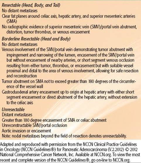
The current American Joint Committee on Cancer 2010 tumor, node, and metastasis staging system is described in Table 59.1. Common criteria of resectability are outlined in Table 59.2.
TABLE 59.1 AMERICAN JOINT COMMITTEE ON CANCER STAGING 2010 CLASSIFICATION FOR PANCREATIC CANCER

TABLE 59.2 CRITERIA DEFINING RESECTABILITY STATUS
PATHOLOGIC CLASSIFICATION
Approximately 85% to 90% of pancreatic cancers are adenocarcinomas. Other neoplasm histological cell types include intraductal papillary mucinous neoplasm, mucinous cystic, solid pseudopapillary, acinar cell, pancreaticoblastoma, neuroendocrine, squamous cell, serous cystadenocarcinoma, and lymphoma. Management and treatment sections will focus on pancreatic adenocarcinomas, given this is the predominate histologic subtype. The histopathology of lesions in the periampullary region of the head of the pancreas is of particular importance as different types of adenocarcinomas, originating from the pancreas, bile duct, ampullary region, or duodenum, respectively, portend different prognoses.27
Pancreatic adenocarcinomas evolve from noninvasive pancreatic intraepithelial neoplasias, acquiring various genetic and epigenetic mutations. Several molecular abnormalities have been implicated in contributing to the development of pancreatic cancer. The most frequent genetic alterations are activation of the oncogene KRAS and inactivation of tumor suppressor genes including TP53, p16 (CDKN2), DPC4 (SMAD4), and BRCA2, with reported incidences ranging from 50% to 95% in pancreatic tumors.28 These molecular changes have the potential to help in identifying precursor lesions with a high likelihood of progressing into carcinomas, characterizing pathologically ambiguous lesions, and useful in development of screening regimens.
GENERAL MANAGEMENT
Operative Considerations
Surgery, as part of a multimodality treatment approach for patients with resectable pancreatic cancer, represents the only potentially curative treatment strategy.29 More than 80% of patients present with advanced disease that is not amenable to curative resection, with roughly two-thirds presenting in the pancreatic head. The standard surgical treatment for pancreatic cancer of the head or uncinate is pancreaticoduodenectomy, first described by Whipple et al.30in 1935. For pancreatic tail lesions, distal pancreatectomy is employed, frequently with splenectomy. Initially, high operative morbidity and mortality rates led to technical modifications of the operation that, combined with improvements in anesthesia and critical care, have resulted in current perioperative mortality rates of ≤2% at high-volume centers.31–32,33 With reduced morbidity from the Whipple procedure, there is no age limitation, and selected patients, even at ages >80 years, may undergo pancreaticoduodenectomy.34–36
Several studies report low postoperative mortality rates and appropriate patient selection at high-volume institutions, highlighting the importance of performing such procedures at institutions with extensive experience.37–40 In addition, the definition of resectable lesions has broadened with the ability to perform venous reconstructions with minimal increase in morbidity or mortality.41,42 There are no universally accepted criteria for resectability other than absence of distant lymph node involvement or visceral, peritoneal, or extra-abdominal metastases, although the high likelihood of achieving R0 resection should be the ultimate goal.
The prognosis, even among resectable patients, remains poor, with 5-year survival rates of 10% to 25%. The number of nodes harvested appears to impact outcomes; data suggest that 12 to 15 lymph nodes constitutes an adequate assessment, although impact on outcomes may simply reflect accurate staging.43,44 Although appropriate lymph node dissection is desirable, trials of extended lymphadenectomy conveyed no survival benefit and compromised quality of life.45,46 Pathologic assessment provides important prognostic information, including margin status, tumor grade, tumor size, lymphovascular invasion, and lymph node involvement. The presence of positive operative margins, poor histologic grade, larger tumor size, lymphovascular invasion, and lymph node involvement portends a poorer prognosis.47–48,49,50–51 Similarly, patients with elevated pre- and postoperative carbohydrate antigen 19-9 (CA19-9) have inferior disease-free survival (DFS) and OS.51,52,53–54
In an effort to minimize surgical morbidity, pylorus-preserving pancreaticoduodenectomy and laparoscopic resection techniques have developed. Pylorus-sparing pancreaticoduodenectomy potentially improves gastrointestinal function without compromising oncologic management.55,56 Laparoscopic pancreatic resection was first explored in the late 1990s, and its use has increased dramatically.57,58 Although most experiences are small, retrospective, single-institution series, laparoscopic resection, in experienced hands, appears a reasonable option for distal pancreatic resections.59–62 The data on laparoscopic pancreaticoduodenectomy are more limited; the difficulty with this procedure arises from more complex dissection and the need for reconstruction.42,58 However, preliminary data suggest that despite prolonged operative times and potential increase in postoperative fistula formation, laparoscopic resection offers shortened hospitalization and equivalent oncologic results.58,63–65
Cause of Death and Patterns of Failure
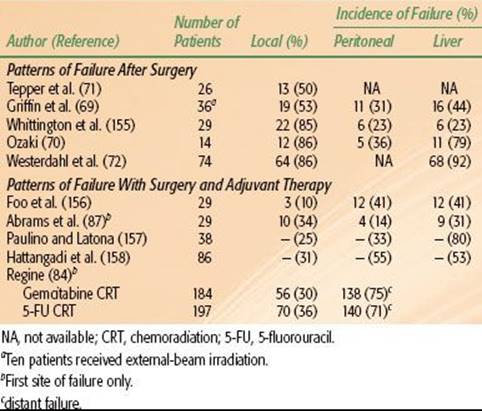
For patients with locally unresectable tumors or metastatic disease, death usually results from hepatic failure secondary to biliary obstruction by local tumor extension or hepatic replacement by metastases. For the 10% to 20% of patients undergoing a potentially curative pancreaticoduodenectomy, three major sites of disease relapse dominate: locoregional, peritoneal cavity, and liver (Table 59.3). High local failure rates of 50% to 86% occur despite resection because of frequent lymphatic and perineural involvement and cancer invasion into the retroperitoneal soft tissues, with an inability to achieve wide retroperitoneal soft tissue margins due to anatomic constraints (SMA and SMV, portal vein, and inferior vena cava).50,66,67 The incidence of microscopic residual disease after careful evaluation of the posterior peripancreatic soft tissue margin is as high as 40%.67 Therefore, tumor stage, grade, and resection margin status are the best predictors of survival after surgery.66,67 The use of adjuvant therapy after surgery can reduce the incidence of local failure, but distant disease recurrence rates remain high.
TABLE 59.3 PATTERNS OF FAILURE AFTER RESECTION OF PANCREATIC CANCER WITHOUT ADJUVANT RADIATION THERAPY OR CHEMOTHERAPY
RADIATION THERAPY TECHNIQUES
Dose-Limiting Tissues
The dose-limiting organs for irradiation of upper abdominal malignancies include the small bowel, stomach, liver, kidneys, and spinal cord. Various techniques, such as 3D conformal radiotherapy (3DCRT) and intensity-modulated radiotherapy (IMRT), can spare these organs. Given long-term survival is low, the actual number of patients at risk for late complications is small. Late small bowel and gastric effects may also be decreased through reduction of the volume of these organs within the high-dose field.
Treatment Volumes, Fields, and Doses
In patients undergoing surgery, clips should be placed to mark the extent of the lesion for postoperative irradiation. Used sparingly (e.g., a single small vascular clip placed to mark superior, inferior, lateral, and medial margins), small clips produce only minimal interference on CT scans.
The patient should be positioned supine during simulation and treatment. Until the mid-1990s, conventional simulation was the standard for radiotherapy treatment planning in pancreatic malignancies. During conventional simulation, an initial set of anterior-posterior/posterior-anterior (AP/PA) and cross-table lateral films is obtained after injection of renal contrast medium to identify both operative clips and renal position relative to the field center. Additional films can be obtained with contrast medium in the stomach and duodenal loop in unresected patients.
Three-dimensional conformal radiotherapy represents a major paradigm shift in radiation oncology treatment planning with better visualization of internal organs and target delineation. CT-based treatment planning allows the construction of normal tissue dose–volume histograms and the generation of treatment plans that optimize radiation dose delivery to tumor while sparing critical normal tissues. Despite this advancement, the intent of treatment remains the same. Multiple field, fractionated, external-beam techniques utilizing high-energy photons deliver 45 to 50 Gy in 1.8 Gy fractions to tumor bed, unresected or residual tumor, and lymph node–bearing areas at risk. After 45 to 50.4 Gy, a boost field can be designed to include unresected or gross residual disease, as defined by CT scans and clips, while excluding most of the stomach and small bowel.
With lesions in the head of the pancreas, major node groups include the pancreaticoduodenal, porta hepatis, celiac, and superior mesenteric nodes. Approximately two-thirds of the left kidney must be excluded from the AP/PA field because the right kidney is often in the field due to duodenal (bed) inclusion. The entire duodenal loop with margin is generally included, as pancreatic head lesions may invade the medial wall of the duodenum and place the entire circumference at risk, including the pancreaticoduodenal nodal basins. The superior field extent is often at the middle or upper portion of the T11 vertebral body for adequate margins on the celiac vessels (T12, L1) and the inferior limit at the level L2–3 to include the superior mesenteric lymph nodes and third portion of the duodenum. The upper field extent is sometimes more superior with body lesions to obtain adequate margin on the primary lesion. With lateral fields, the anterior field margin is 1.5 to 2.0 cm beyond gross disease. The posterior margin is often 1.5 cm behind the anterior portion of the vertebral body to allow adequate margins on para-aortic nodes, which are at risk with posterior tumor extension in head or body lesions. The lateral contribution usually is limited to 15 to 18 Gy because a moderate volume of kidney or liver may be in the irradiated volume (Fig. 59.2).
With pancreatic body or tail lesions, at least 50% of the left kidney may need to be included to achieve adequate margins on node groups at risk (i.e., lateral suprapancreatic, splenic artery, and splenic hilum nodes). Inclusion of the entire duodenal loop is often not indicated with body or tail lesions, and at least two-thirds of the right kidney can be preserved. With customized multileaf collimator (MLC) blocking, it is usually possible to cover pancreaticoduodenal and porta hepatis nodes adequately (Fig. 59.3).
After resection, AP/PA and lateral fields are designed on the basis of preoperative CT primary tumor volumes, preoperative duodenal loop location, operative clip placement, and postoperative CT nodal volumes (Radiation Therapy Oncology Group [RTOG] contouring atlas accessible on their website). The anterior border is determined by vascular or nodal boundaries (porta hepatis, superior mesenteric, and celiac), as demonstrated on CT, as well as preoperative tumor location.
FIGURE 59.2. External-beam four-field technique for pancreatic head lesion. A: The anteroposterior/posteroanterior (AP/PA) field, which includes gross tumor (red), duodenal loop (pink) (plus approximately 50% of the right kidney [light green]), liver (brown), and nodal areas at risk (porta hepatis: orange,superior mesenteric arteries: green, celiac: magenta). Most of the left kidney (blue) is excluded from the AP/PA field. B: The right lateral field with an anterior margin beyond gross disease and a posterior margin behind front edge of vertebral body. The liver contour (brown) has been removed from lateral field for visualization of other structures.

FIGURE 59.3. External-beam four-field technique for unresectable tail of pancreas lesion. A: The anteroposterior/posteroanterior (AP/PA) field showing lesion (red) and head of pancreas (green). The field is extended to the patient’s left to include coverage of the splenic hilum (cyan). Much of the right kidney (light green) is excluded from the field and multileaf collimators block most of the left kidney (blue). B: The right lateral field with an anterior margin beyond gross disease and a posterior margin at least 1.5 cm behind front edge of vertebral body.

TABLE 59.4 PROSPECTIVE, RANDOMIZED TRIALS FOR ADJUVANT THERAPY FOR PANCREATIC CANCER

RADIATION THERAPY RESULTS
Resectable Tumors: Adjuvant Therapy
After surgical resection of pancreatic cancer, local recurrence rates range from 50% to 86% and distant recurrence rates from 40% to 90%, most commonly to the liver or peritoneum.50,67–68,69–72 This provides the rationale for adjuvant therapy, including adjuvant external-beam radiation therapy (EBRT), chemotherapy, and chemoradiotherapy (CRT), which have been employed in an effort to improve patient outcomes (Table 59.4). Despite multiple randomized trials, a definitive role for adjuvant therapy for resected pancreatic cancer has not been established.
Prospective Trials
The Gastrointestinal Tumor Study Group (GITSG) conducted the first multicenter prospective trial of adjuvant CRT for patients with resected pancreatic cancer and negative surgical margins, laying the foundation for the adoption of CRT in the United States. Forty-three patients were randomized to observation or CRT to 40 Gy delivered in split-course fashion with concurrent 5-fluorouracil (5-FU) (500 mg/m2) as an intravenous bolus on the first 3 and last 3 days of radiation, followed by maintenance weekly 5-FU for 2 years or until disease progression. At interim analysis, improvements in median DFS and OS with CRT were observed: 11 versus 9 months and 20 versus 11 months, respectively. Two-year OS was 42% in the CRT group versus 15% in the surgery-alone arm.73 An additional 30 patients were later enrolled to receive adjuvant CRT. These additional patients confirmed the survival outcomes seen in the original trial, with median survival of 18 months and a 2-year survival of 46%.74
The GITSG trial has been criticized for a number of reasons. The trial closed prematurely following enrollment of 43 of an intended 100 patients due to slow accrual over the 8-year enrollment period. The EBRT approach in this trial was considered low dose and antiquated by contemporary standards due to a split-course technique and use of large treatment fields, which encompassed the entire pancreas or pancreatic bed and the celiac, pancreaticosplenic, peripancreatic, and retroperitoneal regional lymph nodes. Additionally, the inclusion of both CRT and adjuvant chemotherapy after surgery evaluated two treatment variables, making it difficult to discern the true effect of either treatment alone. In the CRT arm, there were issues of compliance, with 32% of patients assigned to CRT receiving inappropriate radiation and 25% of patients failing to initiate treatment within 10 weeks postsurgery, the protocol-specified time limit. Only 9% of patient completed the planned 2-year maintenance chemotherapy course. In addition, survival in the control arm was low compared to historical controls. Despite these limitations and the failure to reach the desired patient accrual, the GITSG trial demonstrated a benefit for CRT, which became standard adjuvant therapy, particularly in the United States.
A second study sponsored by the European Organisation for Research and Treatment of Cancer (EORTC) sought to confirm the findings of the original GITSG study. Two hundred eighteen patients with resected pancreas (n = 114) or nonpancreas periampullary cancers (n = 104) were randomly assigned to observation or CRT to 40 Gy in a split-dose fashion with concurrent continuous infusional 5-FU (25 mg/kg) without further adjuvant chemotherapy. This study showed no significant improvement (P = .208) in median survival (19 months [observation] vs. 24.5 months [CRT]) or 2-year survival (41% [observation] vs. 51% [CRT]).75 Long-term follow-up of the EORTC trial demonstrated no difference in 5-year OS with CRT use: 22% (surgery alone) versus 25% (CRT). Post hoc analysis of pancreatic head lesions failed to demonstrate a benefit with CRT, with a median OS of 1.3 years for CRT versus 1 year for surgery alone.76
This trial has been criticized for its heterogeneous patient population, which included patients with both pancreatic and other periampullary primary tumors. Periampullary carcinomas have a significantly better prognosis compared with pancreatic cancer; the two entities thus represent truly different diseases and potentially dilutes any evidence of benefit for adjuvant CRT.77 Similar to the GITSG trial, older EBRT techniques—split course and low total dose—were used. Also, >20% of patients in the CRT arm did not receive the intended treatment due to postoperative complications or patient refusal. The trial did not include maintenance chemotherapy in the treatment arm. Finally, although a small subset, patients undergoing noncurative resection and patients with positive surgical margins were still eligible for enrollment. The discordant results of the EORTC and GITSG trials have led some investigators to attribute the OS benefit seen in the GITSG trial to maintenance chemotherapy administration rather than to EBRT.
An additional European trial conducted by the European Study Group for Pancreatic Cancer (ESPAC) aimed to further explore the question of appropriate adjuvant therapy for “macroscopically” resected pancreatic cancers. Treating physicians were allowed to enroll patients into one of three parallel randomized studies:
1. Chemoradiation versus no chemoradiation (n = 68), consisting of 20 Gy over 2 weeks with 5-FU (500 mg/m2) on days 1 through 3, then repeated after a 2-week break;
2. Chemotherapy versus no chemotherapy (n = 188), consisting of bolus 5-FU (425 mg/m2) and leucovorin (20 mg/m2) given for 5 days every 28 days for 6 cycles; or
3. A 2-by-2 factorial design of 285 patients enrolled on chemoradiotherapy (n = 70), chemotherapy (n = 74), chemoradiotherapy with maintenance chemotherapy (n = 72), or observation (n = 69).
The data from the treatment groups from all three parallel trials were then pooled for analysis. There was no survival difference between the 175 patients who received adjuvant chemoradiation and the 178 patients who did not receive therapy (median survival, 15.5 vs. 16.1 months; P = .24). In the chemotherapy arm, however, a 35% reduction in death was seen in the group who received adjuvant chemotherapy (n = 238) compared with those who received no chemotherapy (n = 235), with a difference in median survival of 19.7 versus 14 months (P = .0005).78 On further follow-up of patients randomized using the 2-by-2 factorial design, the 5-year survival rate for the patients who received chemotherapy was 21% versus 8% for those who did not. Additionally, adjuvant CRT was associated with a deleterious effect on survival.79
A number of problems are associated with the interpretation of these data. First, the complex trial design has the potential for bias, because patients or physicians could select randomization for one treatment variable. Also, as in the aforementioned trials, the EBRT technique used was, by contemporary standards, considered outdated, using split course and low total dose. No details of EBRT delivery or central quality assurance for EBRT, surgery, or pathology are available. In addition, many treatment violations occurred, as only 62% of patients received full CRT treatment and only 42% of patients in the chemotherapy arms completed the predefined regimen. Although many have attempted to draw conclusions from the updated publication, the 2-by-2 factorial cohort of the study was not powered to detect OS differences. Additionally, patients receiving CRT in the ESPAC trial experienced poorer survival outcomes compared with those in other reported CRT series. Also, patients who had received prior “background” chemotherapy or radiation therapy were still eligible for enrollment. As in the EORTC trial, no maintenance chemotherapy was administered. Despite these critiques, some have speculated that the detriment seen in the CRT group may have resulted from delayed administration of systemic therapy shown to improve survival in the ESPAC trial.
A common critique of the aforementioned trials of adjuvant therapy is the lack of restaging to evaluate for the presence of persistent or metastatic disease after surgical resection and prior to the initiation of adjuvant therapy. The time between initial staging and the commencement of adjuvant treatment can be as long as 3 to 4 months, during which a significant number of patients would be expected to develop radiographically apparent metastases. Without interval restaging, these patients may inappropriately receive CRT. Although these three trials formed the foundation for adjuvant treatment approaches to resectable pancreatic cancer, perhaps because each trial was fraught with flaws, there continues to be little consensus regarding the “most appropriate” treatment. A relative dichotomy in adjuvant treatment approaches for resectable pancreatic cancer has emerged between the United States and parts of Europe, with adjuvant CRT frequently implemented in the United States and adjuvant chemotherapy alone administered in parts of Europe.
Building from the data showing potential benefit to adjuvant chemotherapy, investigators in Europe conducted a randomized phase III trial of observation versus adjuvant gemcitabine in patients with resected pancreatic cancer. Three hundred sixty-eight patients were enrolled on the Charite Onkologie (CONKO) trial and randomized to observation or adjuvant gemcitabine (1,000 mg/m2) intravenous days 1, 8, and 15 every 4 weeks for 6 months.80 The primary end point was DFS, and patients treated with gemcitabine achieved a statistically significantly longer DFS (14.2 vs. 7.5 months) than those observed after surgery. This improvement was seen in both the R0 and R1 subgroups. At initial publication, there was no difference in OS. With longer follow-up, median and 5-year OS were both improved in patients receiving gemcitabine (23 vs. 20 months, and 21% vs. 9%, respectively).81 These findings were supported by a smaller randomized study from Japan, which again demonstrated a DFS benefit for gemcitabine compared to surgery alone.82 At the same time the CONKO trial was enrolling patients, ESPAC-3, the largest randomized controlled trial in pancreatic cancer to date, enrolled 1,088 patients with pancreatic adenocarcinoma who underwent R0 or R1 resection. Patients were randomly assigned to 6 cycles of adjuvant gemcitabine (3 weekly infusions of 1,000 mg/m2) or 6 cycles of bolus 5-FU (425 mg/m2)/leucovorin (LV) (20 mg/m2). Patients receiving 5-FU/LV experienced significantly higher rates of grade 3 or 4 gastrointestinal toxicity (stomatitis and diarrhea), whereas patients receiving gemcitabine experienced significantly higher rates of grade 3 or 4 hematologic toxicity. At a median follow-up of 34.2 months, there was no difference between the two groups in the primary end point of OS. Given its favorable toxicity profile, in many parts of Europe, gemcitabine alone is considered the standard adjuvant treatment in patients with resectable pancreatic cancer.83 The focus of future European adjuvant therapies for resectable pancreatic cancer has centered on finding the ideal combination of systemic agents. The ongoing ESPAC-4 trial is accruing patients with resected pancreatic cancer for randomization to gemcitabine alone versus gemcitabine/capecitabine; the trial is expected to close in 2014.
In the United States, however, the GITSG trial laid the foundation for CRT as the predominate adjuvant treatment modality. The RTOG/Gastrointestinal Intergroup trial 9704 was a phase III randomized study comparing adjuvant 5-FU–based chemotherapy to gemcitabine-based chemotherapy, with both regimens followed by CRT. Four hundred fifty-one patients with resected pancreatic cancer were randomized to continuous infusion 5-FU (250 mg/m2/day) or gemcitabine (1,000 mg/m2 weekly) for 3 weeks prior to CRT and 12 weeks after CRT. CRT in both groups consisted of 50.4 Gy delivered with continuous infusion 5-FU (250 mg/m2/day).84 Prospective quality assurance of all radiation therapy plans was required. This trial was powered to demonstrate a survival benefit for the entire cohort and for the subgroup of patients with pancreatic head lesions. On initial analysis of the pancreatic head subgroup (n = 388), a nonsignificant trend toward improved median and 3-year OS was seen in the gemcitabine arm: 20.5 versus 16.9 months and 31% versus 22%, respectively. However, a higher incidence of grade 3 or higher hematologic toxicity was also seen in the gemcitabine group, with no significant differences seen in severe nonhematologic toxicities.
An update of this trial reported median and 5-year OS rates in patients with pancreatic head tumors of 20.5 months and 22% in those who received gemcitabine, compared with 17.2 months and 18% in those who received 5-FU. On multivariate analysis, in the subgroup of patients with pancreatic head lesions who received gemcitabine, there was a nonsignificant trend toward improved OS (P = .08).85,86
A secondary aim of RTOG-9704 was to assess the ability of post-resection CA19-9 levels to predict survival. When CA19-9 levels were analyzed in a cohort of 385 patients as a dichotomized variable (<180 IU/mL vs. ≥180 IU/mL, ≤90 IU/mL vs. >90 IU/mL), there was a significant survival difference favoring patients with CA19-9 levels of <180 IU/mL. This corresponded to a 72% reduction in the risk of death.52 Unlike preceding randomized CRT studies, a major strength of the RTOG study was the rigorous, centralized quality control of radiation therapy techniques and delivery. A recent analysis demonstrated that patients treated per study guidelines had a significant survival advantage, indicating the importance of centralized review and treatment technique in this disease.87
The high rates of local failure and need for more efficacious systemic therapy, as well as controversy surrounding the role of CRT is resected patients, led to the design of the current RTOG-0848/EORTC trial. This trial randomly assigns patients with resected pancreatic head adenocarcinoma (stratified based on CA19-9 level, nodal involvement, and margin status) to receive treatment with either gemcitabine alone or gemcitabine combined with erlotinib for 5 cycles. If no progression is seen on restaging following the completion of systemic therapy, patients are further randomized either to receive an additional cycle of the previously administered chemotherapy (for a total of 6 cycles) and no further treatment or to receive CRT (50.4 Gy with concurrent capecitabine or 5-FU) using modern techniques central radiation therapy quality assurance. This trial seeks to answer two primary questions: (a) the role of the small-molecule epidermal growth factor receptor (EGFR) inhibitor, erlotinib, in the adjuvant therapy of pancreatic cancer and (b) the role of CRT in the era of modern chemotherapy, particularly in patients who do not experience early disease progression. This is the only contemporary randomized clinical trial evaluating the role of CRT in the adjuvant setting.
Single Institution Experiences
Reports of single-institution experiences with adjuvant therapy for resected pancreatic cancer have provided additional evidence to the benefit of such. A review of the Mayo Clinic experience reported the outcomes of 472 patients who underwent R0 resection between 1975 and 2005. Despite more adverse prognostic features in patients receiving CRT (higher histologic grade and greater lymph node involvement), median, 2-year and 5-year OS were significantly improved in the CRT cohort compared with patients who received no adjuvant therapy: 25.2 versus 19.2 months, 50% versus 39%, and 28% versus 17%, respectively.88 A larger series from Johns Hopkins compared 908 patients who underwent pancreaticoduodenectomy between 1993 and 2005 and received surgery alone or CRT. Patients receiving CRT experienced a significant improvement in median, 2-year, and 5-year OS: 21.2 versus 14.4 months, 43.9% versus 31.9%, and 20.1% versus 15.4%, respectively.89 A pooled retrospective analysis of nearly 1,100 patients from both institutions demonstrated similar results in favor of adjuvant chemoradiation.90
Data with the highest survival after adjuvant therapy for pancreatic cancer come from a phase II trial conducted at the Virginia Mason Clinic. Forty-three of a planned enrollment of 53 patients were treated with CRT to 50 Gy with 5-FU (200 mg/m2/day) continuous infusion, weekly cisplatin (30 mg/m2), and interferon-α (3 million units) subcutaneously every other day. After completion of chemoradiation, patients received further adjuvant 5-FU (200 mg/m2/day) by continuous infusion.91 The 2-year and 5-year OS rates were 64% and 55%, respectively. Median survival was not reached at time of initial publication. With these encouraging data came significant toxicity, as 70% of patients experienced grade 3+ toxicities and 42% required hospitalization. The American College of Surgeons Oncology Group subsequently opened a multicenter, phase II trial evaluating the Virginia Mason adjuvant regimen. Although survival outcomes were promising (median survival 25 months), the trial closed early after accrual of 89 patients due to high toxicity rates. Ninety-five percent of patients experienced grade 3+ toxicity, and 50% failed to complete the entire treatment protocol.92
Neoadjuvant Therapy
Even after undergoing curative resection for pancreatic cancer, 80% to 85% of patients will recur. In addition, positive margins or nodal disease increases this rate of recurrence to approximately 90%.67,93 The use of neoadjuvant chemoradiation offers an alternative approach to improve on these figures for several reasons:
1. Approximately one-third of patients experience a significant delay or do not receive adjuvant therapy following resection.49,94,95
2. Twenty percent to 40% of patients will be spared the morbidity of resection as their metastatic disease becomes clinically apparent during course of neoadjuvant therapy.96–98
3. Preoperative therapy could theoretically be less toxic and more effective as the chemotherapy and radiation would be given without the postsurgical issues of small bowel in the radiation field, decreased oxygenation and decreased drug delivery to the remaining tumor bed.99,100
4. Patients with local, borderline and unresectable lesions may be able to be downstaged to allow for surgical resection and sterilization of the operative region, which potentially facilitaes R0 resection and reduces the risk of spread during surgical manipulation.
Unlike with adjuvant therapy, no phase III randomized trials of neoadjuvant CRT in resectable pancreatic lesions have been conducted, and the vast majority of data come from phase II and retrospective studies. At the MD Anderson Cancer Center, multiple trials of neoadjuvant 5-FU–based CRT have been performed. The earliest trial treated 28 patients with continuous infusion 5-FU (300 mg/m2/day) and concurrent EBRT to 50.4 Gy over 5.5 weeks. Patients who underwent surgical resection also received intraoperative radiation therapy (IORT). Twenty-five percent of patients had evidence of metastatic disease on preoperative restaging. Fifteen percent had metastatic disease that was found on laparoscopy. For the patients who underwent surgery, median survival was 18 months, and 41% had a pathologic partial response to therapy. However, 33% of patients treated in this study required hospitalization for gastrointestinal toxicity from therapy.101 In an effort to minimize hospitalization, this group subsequently focused on rapid fractionation EBRT. A prospective trial of 35 patients treated with EBRT to 30 Gy (3 Gy per fraction for 10 fractions) with concurrent continuous infusion 5-FU (300 mg/m2/day) found grade 3 nausea and vomiting in only 9% of patients with no grade 4 toxicities. Twenty-seven patients were taken to surgery and 20 patients underwent resection and IORT to 10 to 15 Gy. Locoregional recurrence occurred in only 2 of 20 resected patients. Median survival for patients who underwent surgery was 25 months, with a 3-year survival rate of 23%.102
Trials of neoadjuvant CRT with paclitaxel as a radiosensitizer have been conducted. In radiobiologic models, paclitaxel may result in enhanced radiosensitization through synchronization of tumor cells at G2/M, a relatively radiosensitive phase of the cell cycle, and tumor reoxygenation after apoptotic clearance of paclitaxel-damaged cells. In an MD Anderson Cancer Center study, 35 patients received weekly paclitaxel (60 mg/m2) with concurrent EBRT to 30 Gy. Eighty percent underwent resection with 21% of pathology specimens showing >50% tumor necrosis. The 3-year survival for the patients who underwent preoperative therapy and resection was 28%. Hospitalization was required in 11% of patients for toxicity, primarily nausea and vomiting. These preliminary data show an increased toxicity without a significant improvement in histological response rate or survival.103
The French Fédération Francophone de Cancérologie Digestive/Société Francophone de Radiothérapie Oncologique (SFRO-FFCD) 9704 trial incorporated further radiosensitizing chemotherapy. Forty-one patients received CRT to 50 Gy with 5-FU (300 mg/m2/day) and 2 cycles of CDDP (neoadjuvant cisplatin; 20 mg/m2) followed by surgical resection in patients without progression. Sixty-three percent underwent curative surgery, with 80% obtaining an R0 resection and 50% of specimens showing major histologic response. In patients who underwent curative surgery, median survival was 11.7 months, with a 32% 2-year overall survival.104
The evolution of CRT in the neoadjuvant setting appears to parallel advances made with adjuvant therapy, with transition to gemcitabine-containing regimens. In 2008, two published phase II studies from the MD Anderson Cancer Center evaluated the use of gemcitabine as part of a neoadjuvant regimen. One trial enrolled 86 patients with radiographically resectable adenocarcinoma of the pancreatic head or uncinate. Patients received weekly gemcitabine (400 mg/m2) with 30 Gy EBRT over 2 weeks. After restaging 4 to 6 weeks post-CRT, 73 patients (85%) underwent surgery, with 64 (74%) undergoing successful resection. Median survival in resected versus unresected patients was 34 versus 7 months, with corresponding 5-year OS rates of 36% and 0%, respectively. In patients undergoing pancreaticoduodenectomy, 11% experienced local failure, with distant failure representing the first site of failure in the majority of cases.105
Given the high incidence of distant disease development, a simultaneously conducted phase II trial incorporated induction CDDP and gemcitabine prior to initiation of CRT with gemcitabine. Induction chemotherapy consisted of CDDP (30 mg/m2) and gemcitabine (750 mg/m2) every 2 weeks for 4 cycles followed by 4 weekly infusions of gemcitabine (400 mg/m2) with 30 Gy EBRT. Sixty-two of 90 enrolled patients (78%) underwent surgery and 52 (66%) had successful resection. The median survival for patients undergoing surgery was 31 months, compared with 10.5 months in the unresected patients.106
A recent review of the Surveillance, Epidemiology, and End Results (SEER) database supports the use of neoadjuvant treatment. This analysis included 3,885 patients treated for resectable pancreatic cancer: 70 patients (2%) received neoadjuvant EBRT, 1,478 (38%) received adjuvant EBRT, and 2,337 (60%) were treated with surgery alone. Given that the SEER database does not provide information on administration of chemotherapy, this variable could not be assessed. Median OS was 23 months in patients receiving neoadjuvant EBRT, 17 months with adjuvant EBRT, and 12 months in the surgery-alone cohort.107
Despite the potential advantages and encouraging results using a neoadjuvant CRT approach, no randomized trial results exist comparing neoadjuvant to adjuvant therapy, and its role continues to be investigational. A multicenter, randomized phase II study evaluating neoadjuvant therapy in pancreatic carcinoma has been described. This multinational study is comparing outcomes of patients with resectable disease treated with neoadjuvant gemcitabine-based CRT followed by surgery to outcomes in patients undergoing upfront surgery. Resection is followed by adjuvant gemcitabine-based chemotherapy in both arms.108
Therapy for Locally Advanced Carcinoma
Approximately 30% of patients present with locally advanced carcinoma of the pancreas, comprising a group of patients with an intermediate prognosis between resectable and metastatic disease. These patients have pancreatic tumors that are defined as surgically unresectable but have no evidence of distant metastases. A tumor is usually considered to be unresectable if it has one of the following features:
1. Extensive peripancreatic lymph node involvement and/or distant metastases,
2. Encasement or occlusion of the SMV or SMV/portal vein confluence that is not amenable to reconstruction, or
3. ≥180 degree involvement of the SMA; involvement of the inferior vena cava, aorta, or celiac axis.
Recent advances in surgical technique allow for resection of selected patients with tumors involving the SMV.41,109 Treatment with radiation and chemotherapy increases median survival for patients with locally advanced cancers to approximately 8 to 14 months but rarely results in long-term survival. The therapeutic options of patients with locally advanced pancreatic cancer include chemotherapy alone, CRT, IORT, and more recently EBRT with novel chemotherapeutic and targeted agents. In evaluating the results of these various therapies, it is useful to remember that a median survival of 3 to 6 months has been reported for this subset of patients undergoing palliative gastric or biliary bypass only.110
Prospective Trials
Prospective trials have evaluated the role of EBRT versus CRT, variations in CRT regimens, and CRT versus chemotherapy alone. With conflicting results, there is little consensus as to the appropriate management of locally advanced patients, and all are considered reasonable options (Table 59.5). The Mayo Clinic undertook an early randomized trial in the 1960s in which 64 patients with locally unresectable, nonmetastatic stomach, large bowel, and pancreas adenocarcinoma received 35 to 40 Gy of EBRT alone or with concurrent bolus 5-FU (45 mg/kg). A significant survival advantage was seen for patients receiving EBRT with 5-FU versus EBRT only (10.4 vs. 6.3 months).111
The GITSG followed with a similar study comparing EBRT alone to EBRT with concurrent and maintenance 5-FU. One hundred and ninety-four eligible patients with surgically confirmed unresectable and nonmetastatic pancreatic adenocarcinoma were randomized to receive 60 Gy split course EBRT alone, 40 Gy split course EBRT with 2 to 3 cycles of concurrent bolus 5-FU chemotherapy (500 mg/m2), or 60 Gy split course EBRT using a similar chemotherapy regimen. Patients in the latter groups received 2 years’ maintenance 5-FU (500 mg/m2) after EBRT completion. The EBRT alone arm was closed early as a result of an inferior survival rate. The 1-year survival rate in the two combined modality therapy arms was roughly 40% (with no statistical difference between CRT arms) versus 11% in the EBRT alone arm.112
The Eastern Oncology Group’s trial ECOG-8282 randomized 114 patients to EBRT alone (59.4 Gy) with or without continuous infusion 5-FU (1,000 mg/m2/day) on days 2 to 5 and 28 to 31 and mitomycin-C (10 mg/m2) on day 2. There was no difference in response rates, DFS, or OS with the addition of concurrent chemotherapy. Higher rates of toxicity, primarily hematologic, where noted in the CRT group.113
Further studies sought to determine a more efficacious CRT regimen. A second GITSG trial randomized 157 and analyzed results for 143 eligible patients with unresectable disease to 60-Gy split course EBRT with concurrent and maintenance 5-FU (500 mg/m2) (as in the prior GITSG trial) or 40-Gy continuous course radiation with weekly, concurrent doxorubicin chemotherapy (10 mg/m2), followed by maintenance doxorubicin and 5-FU. A significant increase in treatment-related toxicity was seen in the doxorubicin arm, with no survival difference observed between the two groups (median survival 8.5 vs. 7.6 months). No clinical benefit was seen in substituting doxorubicin for 5-FU.114
TABLE 59.5 PROSPECTIVE RANDOMIZED TRIALS FOR LOCALLY ADVANCED, UNRESECTABLE PANCREATIC CANCER

As studies emerged demonstrating significant survival advantages with gemcitabine-based chemotherapy in the metastatic setting, it was incorporated into the locally advanced setting. A trial from Taipei randomized 34 patients to EBRT with concurrent 5-FU (500 mg/m2) or gemcitabine (600 mg/m2). Patients received 50.4 to 61.2 Gy EBRT and maintenance gemcitabine (1,000 mg/m2) at the completion of CRT. Median OS and time to progression were prolonged in the gemcitabine arm: 14.5 months and 7.1 months (gemcitabine) versus 6.7 months and 2.7 months (5-FU). There were no observed differences in grade 3 or 4 toxicity or hospitalization days between the two arms. Study criticisms include the small number of patients and relatively poor outcomes in the 5-FU arm compared with historical controls.115
A number of trials have compared CRT with chemotherapy alone. A subsequent GITSG trial compared combination streptozocin, mitomycin-C, and 5-FU (SMF) chemotherapy to chemoradiation with 5-FU. Forty-three patients were randomized to receive SMF chemotherapy or 54 Gy of EBRT with 2 cycles of concurrent bolus 5-FU chemotherapy followed by adjuvant SMF chemotherapy. The chemoradiation arm demonstrated a significant survival advantage over the chemotherapy-alone arm (1-year survival 41% vs. 19%, respectively).116
In contrast, the ECOG reported no benefit to chemoradiation versus chemotherapy only. In this study, 191 patients with unresectable, nonmetastatic pancreatic or gastric adenocarcinoma were randomized to receive either 5-FU chemotherapy (600 mg/m2) alone or 40 Gy EBRT with concurrent bolus 5-FU during week 1 (600 mg/m2) followed by maintenance chemotherapy. Patients with locally recurrent disease as well as patients undergoing surgery with residual disease were eligible for this trial. In the 91 analyzable pancreatic patients, no survival difference was observed between the two groups (median survival 8.2 vs. 8.3 months).117
The FFCD/SFRO trial randomized 119 patients to CRT consisting of 60 Gy EBRT with continuous infusion 5-FU (300 mg/m2/day) on days 1 to 5 for 6 weeks and intermittent CDDP (20 mg/m2/day) on days 1 to 5 in weeks 1 and 5 with maintenance gemcitabine (1,000 mg/m2) versus gemcitabine alone (1,000 mg/m2). Survival was inferior in the CRT arm at 8.6 months compared with 13 months with gemcitabine alone. Higher grade 3 or 4 toxicity rates were observed in the CRT arm during CRT (36% vs. 22%) and maintenance chemotherapy phases (32% vs. 18%). This trial closed early due to poor accrual. Many criticize the inferior survival in the CRT arm, which is similar to early GITSG trials, despite use of continuous infusion chemotherapy and modern EBRT techniques.118
The ECOG published results from a randomized trial of CRT (50.4 Gy with concurrent gemcitabine: 600 mg/m2/week, weeks 1 to 5) versus gemcitabine alone (1,000 mg/m2/week). The trial closed early due to poor accrual. Analysis of the eligible, randomized 71 patients demonstrated no difference in progression-free survival; however, a survival benefit was seen in the CRT arm: 11.1 versus 9.2 months. This benefit came at the expense of higher grade 4 or 5 toxicity rates in the CRT arm (mainly hematologic) but with no difference in quality of life.119
At present there is no consensus regarding the appropriate management of locally advanced patients. A meta-analysis of locally advanced patients demonstrated a survival benefit for CRT versus EBRT, but no such benefit was seen for CRT with further chemotherapy versus chemotherapy alone. Many consider upfront chemotherapy alone with subsequent response assessment and directed CRT in responders to facilitate selection of patients most likely to benefit from CRT, and is currently the topic of an ongoing phase III European study.120 However, many patients with locally advanced disease present with local symptoms (pain, obstruction), in which case EBRT can potentially offer more prompt and durable palliation. CRT (with continuous infusion 5-FU/capecitabine or gemcitabine), gemcitabine-based chemotherapy alone or a combination of both are considered reasonable options in patients with locally advanced disease.
Borderline Resectable Disease
As surgery of the primary tumor remains the only potentially curative treatment for pancreatic cancer; neoadjuvant therapy has been evaluated to assess its ability to convert locally unresectable pancreatic cancer to resectable disease. Although the definition of borderline resectable disease is contentious, the National Comprehensive Cancer Network definition is accepted by many institutions (Table 59.2). Approximately 30% of patients with borderline resectable disease become resectable after neoadjuvant therapy.121,122 Similarly, there appears to be higher rates of local control, R0 resection, and N0 disease in this patient subset after neoadjuvant therapy.123–126
A number of single institution retrospective studies have evaluated this patient subset. The earliest study from the MD Anderson Cancer Center assessed 160 patients with borderline resectable disease. Patients received 50.4 Gy in 28 fractions or 30 Gy in 10 fractions with concurrent 5-FU, paclitaxel, gemcitabine, or capecitabine at radiosensitizing doses. Forty-one percent underwent pancreatectomy with margin negative resection in 94%. Median survival in the 66 patients who completed preoperative therapy and surgery was 44 months, comparable to patients with initially resectable disease.124
A review from Fox Chase Cancer Center evaluated 109 patients who underwent resection of pancreatic adenocarcinoma with varying involvement of the portal vein or superior mesenteric vein as determined by CT. Patients received 5-FU or gemcitabine-based CRT. Median survival in the 74 patients who received preoperative therapy was 23 months compared with 15 months in the cohort undergoing upfront surgery. Preoperative CRT was associated with a higher rate of R0 resection and N0 disease.125
A smaller series from the University of Virginia reviewed the outcomes of 40 patients with borderline resectable disease. Patients received 50.4 Gy in 28 fractions or 50 Gy in 20 fractions with concurrent capecitabine. Forty-six percent of patients underwent surgery, with 75% undergoing an R0 resection. Patients undergoing surgery after neoadjuvant treatment had similar median survival rates as patients undergoing upfront surgery.126
The ECOG initiated a randomized phase II study of two neoadjuvant gemcitabine-containing regimens in patients with potentially resectable cancer. Patients received 50.4 Gy EBRT with weekly gemcitabine (500 mg/m2) or induction chemotherapy with gemcitabine (175 mg/m2), CDDP (20 mg/m2), 5-FU (600 mg/m2) prior to CRT with 5-FU (225 mg/m2). All patients received adjuvant gemcitabine (1,000 mg/m2). This trial, the only randomized trial of borderline resectable pancreatic cancer, closed early due to poor accrual with 21 patients.127 Although the subject of ongoing study, an approach utilizing neoadjuvant chemoradiotherapy in patients with borderline resectable disease, in efforts to facilitate R0 resection, seems appropriate.
Dose Escalation
Given the limited tolerance of normal tissue in the upper abdomen (liver, kidney, spinal cord, and bowel) to EBRT, total doses of only 45 to 54 Gy in 25 to 30 Gy fractions have traditionally been administered. For an unresectable tumor, this dose of radiation is inadequate, as demonstrated by the high rates of local tumor progression and poor survival seen in both prospective and retrospective studies. Local progression as first site of failure occurred in 58% of patients treated to 60 Gy with concurrent 5-FU in the second GITSG trial.114 Similarly, the Mayo Clinic reported a local failure rate of 72% for 122 patients with unresectable pancreatic cancer treated with an EBRT dose of 40 to 60 Gy.128 Attempts have been made to evaluate whether increasing dose of radiation may improve outcomes through 3DCRT, IORT, IMRT, and stereotactic body radiotherapy (SBRT).
In a report from Thomas Jefferson University Hospital, 46 evaluable patients with unresectable disease by laparotomy were treated with 63 to 70 Gy EBRT with or without chemotherapy. Despite high-dose EBRT, the local failure rate was 78%.129
IORT is an alternative method to deliver higher radiation doses. This technique allows for administration of a single high dose of radiotherapy with the advantage of enabling healthy tissues to be displaced and shielded from radiation. IORT has been utilized postoperatively and in treatment of locally advanced disease. Institutional reports and a randomized trial have shown that surgery followed by IORT yields lower rates of locoregional recurrence; however, no significant difference in survival has been seen.130–133 IORT has also been evaluated in patients with unresectable disease.134,135 A study from investigators at Massachusetts General Hospital reported the results of 150 patients with unresectable pancreatic cancer treated with IORT, EBRT, and chemotherapy. Although the study spanned nearly 25 years, it demonstrated that long-term survival is possible for patients with unresectable pancreatic cancer. Furthermore, this study shows that postoperative and late treatment-related toxicity rates were acceptable.135 A lower incidence of local failure in most series and improved median survival in some have been reported with these techniques when compared with conventional external-beam irradiation, but it is uncertain whether this is due to treatment superiority or case selection.128
Advances in radiation technique and delivery, such as IMRT and SBRT, also enable dose escalation and potential sparing of normal tissues. IMRT is a technique that breaks up a typical radiation treatment field into smaller beamlets. It is implemented either as dynamic IMRT (collimating leaves move in and out of the radiation beam path during treatment) or as “step-and-shoot” IMRT (leaves change field shape while the machine is off). The cumulative effect is that the prescription dose conforms around delineated target volumes, significantly reducing doses to adjacent normal tissues. This technology has increasingly been used in a number of gastrointestinal malignancies, including pancreatic cancer. Early clinical data support both the feasibility of this technique and its potential for reducing acute gastrointestinal toxicity.136–138
An analysis from the University of Maryland evaluated 46 patients treated with IMRT and concurrent 5-FU–based chemotherapy. Acute toxicities in these patients were compared with those in a control group enrolled in RTOG-9704 who received conventional 3D treatment. There was a statistically significant reduction in acute grade 3 or 4 gastrointestinal toxicity in the patients who received radiotherapy via IMRT compared with those who received 3D conformal radiotherapy.138 IMRT can also result in a significant reduction of dose to normal structures, including the liver, kidneys, stomach, and small bowel.137 This may allow alternate novel systemic agents to be administered with radiotherapy.136
Another radiation technique being investigated in the treatment of pancreatic cancers is SBRT, which involves the delivery of high dose-per-fraction radiation treatments over a small number of fractions (generally 1 to 5 treatments), utilizing techniques that allow very highly conformal dose delivery of external-beam radiotherapy. The postulated advantage of SBRT is potentially improving local control through the delivery of ablative doses of radiation, while minimizing associated side effects. Few institutions have published experience using this technique, and the data are largely in the setting of locally advanced disease.139,140 Stanford University has implemented an institutional protocol for the treatment of locally advanced pancreatic cancer using a 25-Gy single fraction with systemic therapy. This single, high dose of radiation has been estimated to result in delivery of a higher biologically equivalent dose compared with a more standard, protracted course of radiation therapy, albeit at the potential for increased risk of normal tissue injury. A report from these investigators described 77 patients who were treated with SBRT and found that freedom from local progression was 91% at 6 months and 84% at 12 months. No patients experienced grade 3 acute toxicity and 9% had grade 3 or higher late toxicity.140 Ultimately, the role of IMRT and SBRT in treatment of pancreatic cancer remains to be further defined and is currently investigational. The minimal gain with dose escalation and high rates of local and distant failures begs for improvements in both local and systemic therapies.
Chemoradiation Effects on Quality of Life
Despite the potential survival benefits for patients with locally advanced pancreatic cancer receiving therapy compared to supportive care, these gains are modest. With rare exception, all patients will ultimately succumb to their disease. In spite of this, significant palliative benefit can be achieved by chemoradiation. Pain, anorexia, fatigue, and cachexia are relatively common symptoms, which significantly impact a patient’s quality of life. When using EBRT with or without chemotherapy, approximately 35% to 65% of patients will experience pain resolution as well as some improvement in cachexia and obstructive symptoms.114,141,142 Definite but less dramatic improvements in performance status and anorexia may be observed as well.141,142 Palliation from therapy can take many weeks for maximal effect, and alternative treatments, such as biliary and duodenal stents, may provide more rapid relief of obstructive symptoms. Given the high mortality rate associated with pancreatic cancer, quality of life should be a study end point in trials for these patients.
TARGETED THERAPIES AND FUTURE DIRECTIONS
There is much room for improvement in the diagnosis and treatment of pancreatic cancer. Screening of high-risk individuals may allow for detection of disease in earlier disease states by means of novel imaging methods or assessment of serum biomarkers.143,144 As the biological basis of cancer is better understood, the successful use of cancer-specific targeted therapies in other cancers supports the need to identify new targets and better predictors of response to therapy in pancreatic cancer.
There is evidence of additive or synergistic effects for several targeted agents (such as antibodies against EGFR and vascular endothelial growth factor [VEGF] receptor) with both chemotherapy and radiation therapy, making these approaches promising. These targeted agents have been studied most extensively in the metastatic setting prior to evaluation in locally advanced and resectable disease. Currently, the only targeted agent that has shown a modest statistically significant survival benefit in the metastatic setting compared to chemotherapy alone is erlotinib, an anti-EGFR tyrosine kinase inhibitor. However, the survival benefit with addition of erlotinib is small, with median survival increased from 5.9 to 6.2 months and 1-year survival improvement from 17% to 24%.145 The clinical significance of this difference has been questioned by investigators and treating physicians.
Another EGFR/HER-1 inhibitor, cetuximab, initially showed promising phase II results in the metastatic setting. Efficacy was initially seen with the combination of cetuximab and EBRT in the treatment of locally advanced squamous cell carcinoma of the head and neck and subsequently tested in a phase III trial.146 However, in pancreatic cancer, a phase III randomized study of gemcitabine plus cetuximab versus gemcitabine plus placebo (Southwest Oncology Group S0205) as first-line therapy in locally advanced, unresectable, or metastatic disease enrolled over 700 patients from the United States and Canada. No survival improvement was seen with the addition of cetuximab to gemcitabine (6.3 months gemcitabine plus cetuximab vs. 5.9 months gemcitabine alone). Even among patients tested for EGFR expression (90% specimens positive for expression), no benefit to cetuximab was seen in this patient subgroup.147 An additional phase II study evaluated the efficacy and safety of multiagent chemotherapy in combination with cetuximab. Sixty-nine patients with locally advanced pancreatic cancer received cetuximab, gemcitabine, and oxaliplatin followed by cetuximab, capecitabine, and radiotherapy. Diagnostic cytology specimens were stained for Smad4 (Dpc4) expression. Median overall survival was 19.2 months, and the pattern of failure for patients with Smad4 (Dpc4) expression was primarily local, rather than distant, disease failure.148 The addition of EGFR inhibitors in localized disease is currently being evaluated in phase I and phase II trials.
VEGF inhibitors bind to receptors involved in tumor growth via vasculogenesis and angiogenesis pathways. Preclinical data have shown that inhibition of VEGF has radiosensitizing effects. Initially promising results in the metastatic setting, however, have ultimately not shown a benefit from the addition of an anti-VEGF antibody to chemotherapy. A phase II trial of the combination of gemcitabine and bevacizumab in 52 treated patients with advanced pancreatic cancer showed a response rate of 21%, a median progression-free survival of 5.4 months, and median overall survival of 8.8 months.149 In response to these positive results, a phase III randomized study of gemcitabine plus bevacizumab versus placebo (Cancer and Leukemia Group B CALGB-80303) was initiated. No significant difference was observed in OS or PFS.150 Bevacizumab has also been evaluated in combination with radiotherapy. An initial phase I study of radiotherapy, capecitabine, and bevacizumab led to incorporation in the phase II setting in patients with locally advanced pancreatic cancer (RTOG-0411).151 Eighty-two patients were treated with radiotherapy to the gross tumor, capecitabine (825 mg/m2), and bevacizumab (5 mg/kg on days 1, 15, 29) followed by adjuvant gemcitabine (1,000 mg/m2). The median and 1-year survival rates were 11.9 months and 47%, respectively. These results were similar to prior RTOG trials of locally advanced pancreatic cancer.152 An additional phase II trial assessed the efficacy of full-dose gemcitabine (1000 mg/m2), bevacizumab (10 mg/kg), and radiotherapy to 36 Gy (in 2.4 Gy fractions) with maintenance gemcitabine and bevacizumab in patients with unresected tumors with no evidence of disease progression. Median progression-free and overall survival were 9.9 and 11.8 months, respectively.153 The incorporation of bevacizumab with radiotherapy has not led to profound improvements in treatment outcomes compared to 5-FU–based CRT.
Although improvements with targeted agents have been modest, other pathways are being evaluated. Pancreatic cells deficient in BRCA repair pathways are sensitive to poly-adenosine diphosphate-ribose polymerase (PARP) inhibitors. PARP inhibitors have been evaluated in ovarian and breast cancer, with response rates of 40%; clinical trials of PARP inhibitors in treatment of pancreatic cancer are under way.7 Other therapeutic agents under evaluation include hedgehog pathway inhibitors, multikinase inhibitors (sorafenib), and agents targeting v-src sarcoma, γ-secretase, stem cell factor receptor (c-kit), secreted protein acidic and rich in cysteine, mitogen-activated protein kinase, mesothelin, RAS, prostate stem cell antigen, tumor necrosis factor-α, Mucin-1, mammalian target of rapamycin, tumor necrosis factor superfamily member 10, and type 1 insulin-like growth factor receptor.7,154
SUMMARY
Treatment of pancreatic cancer remains a challenge in oncology. Despite advances in many aspects of oncologic evaluation and management, including preoperative evaluation, surgical techniques, perioperative care, systemic therapy, and radiotherapy, the 5-year OS rate for patients with resectable pancreatic cancer remains around 20%. Local tumor control has been improved by the use of specialized radiation techniques, allowing safe dose escalation. Even with these techniques, it is not clear that a survival benefit is achieved given the proclivity of metastases in this malignancy. Further advances in systemic therapies, study of the optimal sequencing of therapies, earlier detection of disease, and development of new and novel therapeutic options are urgently needed in the treatment of this formidable disease. Despite the recognized limitations of current therapy, palliation can be achieved for a high percentage of patients through combined modality treatment. Quality of life should be considered a paramount end point in the care and protocol design of clinical trials for these patients. In patients with marginal or poor performance status, gemcitabine administration alone represents a reasonable alternative to combined modality therapy. Significant improvements in long-term survival will likely be achieved through ongoing attempts at exploitation of the malignancy’s basic biologic anomalies.
REFERENCES
1. American Joint Committee on Cancer. AJCC cancer staging manual. New York: Springer-Verlag, 2010.
2. Siegel R, Naishadham D, Jemal A. Cancer statistics, 2012. CA Cancer J Clin 2012;62(1):10–29.
3. Geer RJ, Brennan MF. Prognostic indicators for survival after resection of pancreatic adenocarcinoma. Am J Surg 1993;165(1):68–73.
4. Yeo CJ, et al. Pancreaticoduodenectomy for cancer of the head of the pancreas. 201 patients. Ann Surg 1995;221(6):721–733.
5. Wagner M, et al. Curative resection is the single most important factor determining outcome in patients with pancreatic adenocarcinoma. Br J Surg 2004;91(5):586–594.
6. Lim JE, Chien MW, Earle CC. Prognostic factors following curative resection for pancreatic adenocarcinoma: a population-based, linked database analysis of 396 patients. Ann Surg 2003;237(1):74–85.
7. Vincent A, et al. Pancreatic cancer. Lancet 2011;378(9791):607–620.
8. Ries LA, et al. The annual report to the nation on the status of cancer, 1973–1997, with a special section on colorectal cancer. Cancer 2000;88(10):2398–2424.
9. Brentnall TA. Management strategies for patients with hereditary pancreatic cancer. Curr Treat Options Oncol 2005;6(5):437–445.
10. Klein AP, et al. Familial pancreatic cancer. Cancer J 2001;7(4):266–273.
11. Maisonneuve P, Lowenfels AB. Epidemiology of pancreatic cancer: an update. Dig Dis 2010;28(4–5):645–656.
12. Lowenfels AB, et al. Pancreatitis and the risk of pancreatic cancer. International Pancreatitis Study Group. N Engl J Med 1993;328(20):1433–1437.
13. Willett CG, et al. Locally advanced pancreatic cancer. J Clin Oncol 2005;23(20):4538–4544.
14. Kinney T. Evidence-based imaging of pancreatic malignancies. Surg Clin North Am 2010;90(2):235–249.
15. Karmazanovsky G, et al. Pancreatic head cancer: accuracy of CT in determination of resectability. Abdom Imaging 2005;30(4):488–500.
16. Hunt GC, Faigel DO. Assessment of EUS for diagnosing, staging, and determining resectability of pancreatic cancer: a review. Gastrointest Endosc 2002;55(2):232–237.
17. Rosch T, et al. Endoscopic ultrasound in pancreatic tumor diagnosis. Gastrointest Endosc 1991;37(3):347–352.
18. Brugge WR, Van Dam J. Pancreatic and biliary endoscopy. N Engl J Med 1999; 341(24):1808–1816.
19. Hariharan D, et al. The role of laparoscopy and laparoscopic ultrasound in the preoperative staging of pancreatico-biliary cancers—a meta-analysis. Eur J Surg Oncol 2010;36(10):941–948.
20. Fernandez-del Castillo C, Rattner DW, Warshaw AL. Further experience with laparoscopy and peritoneal cytology in the staging of pancreatic cancer. Br J Surg 1995;82(8):1127–1129.
21. Bipat S, et al. Ultrasonography, computed tomography and magnetic resonance imaging for diagnosis and determining resectability of pancreatic adenocarcinoma: a meta-analysis. J Comput Assist Tomogr 2005;29(4):438–445.
22. Sica GT, Ji H, Ros PR. Computed tomography and magnetic resonance imaging of hepatic metastases. Clin Liver Dis 2002;6(1):165–179.
23. Lemke AJ, et al. Retrospective digital image fusion of multidetector CT and 18F-FDG PET: clinical value in pancreatic lesions—a prospective study with 104 patients. J Nucl Med 2004;45(8):1279–1286.
24. Sachelarie I, et al. Integrated PET-CT: evidence-based review of oncology indications. Oncology (Williston Park) 2005;19(4):481–490; discussion 490–492, 495–496.
25. Rose DM, et al. 18Fluorodeoxyglucose-positron emission tomography in the management of patients with suspected pancreatic cancer. Ann Surg 1999;229(5):729–738.
26. Grassetto G, Rubello D. Role of FDG-PET/CT in diagnosis, staging, response to treatment, and prognosis of pancreatic cancer. Am J Clin Oncol 2011;34(2):111–114.
27. Michelassi F, et al. Experience with 647 consecutive tumors of the duodenum, ampulla, head of the pancreas, and distal common bile duct. Ann Surg 1989;210(4):544–556.
28. Rozenblum E, et al. Tumor-suppressive pathways in pancreatic carcinoma. Cancer Res 1997;57(9):1731–1734.
29. Shaib Y, et al. The impact of curative intent surgery on the survival of pancreatic cancer patients: a U.S. Population-based study. Am J Gastroenterol 2007;102(7):1377–1382.
30. Whipple AO, Parsons WB, Mullins CR. Treatment of carcinoma of the ampulla of Vater. Ann Surg 1935;102(4):763–779.
31. Pellegrini CA, et al. An analysis of the reduced morbidity and mortality rates after pancreaticoduodenectomy. Arch Surg 1989;124(7):778–781.
32. Trede M, Schwall G, Saeger HD. Survival after pancreatoduodenectomy. 118 consecutive resections without an operative mortality. Ann Surg 1990;211(4):447–458.
33. Cameron JL, et al. One thousand consecutive pancreaticoduodenectomies. Ann Surg 2006;244(1):10–15.
34. Brozzetti S, et al. Surgical treatment of pancreatic head carcinoma in elderly patients. Arch Surg 2006;141(2):137–142.
35. Sohn TA, et al. Should pancreaticoduodenectomy be performed in octogenarians? J Gastrointest Surg 1998;2(3):207–216.
36. Hodul P, et al. Age is not a contraindication to pancreaticoduodenectomy. Am Surg 2001;67(3):270–276.
37. Bilimoria KY, et al. National failure to operate on early stage pancreatic cancer. Ann Surg 2007;246(2):173–180.
38. Bilimoria KY, et al. Multimodality therapy for pancreatic cancer in the U.S.: utilization, outcomes, and the effect of hospital volume. Cancer 2007;110(6):1227–1234.
39. Bilimoria KY, et al. Assessment of pancreatic cancer care in the United States based on formally developed quality indicators. J Natl Cancer Inst 2009;101(12):848–859.
40. Sosa JA, et al. Importance of hospital volume in the overall management of pancreatic cancer. Ann Surg 1998;228(3):429–438.
41. Tseng JF, et al. Venous resection in pancreatic cancer surgery. Best Pract Res Clin Gastroenterol 2006;20(2):349–364.
42. Martin RC 2nd, et al. Arterial and venous resection for pancreatic adenocarcinoma: operative and long-term outcomes. Arch Surg 2009;144(2):154–159.
43. Slidell MB, et al. Impact of total lymph node count and lymph node ratio on staging and survival after pancreatectomy for pancreatic adenocarcinoma: a large, population-based analysis. Ann Surg Oncol 2008;15(1):165–174.
44. Tomlinson JS, et al. Accuracy of staging node-negative pancreas cancer: a potential quality measure. Arch Surg 2007;142(8):767–724.
45. Yeo CJ, et al. Pancreaticoduodenectomy with or without distal gastrectomy and extended retroperitoneal lymphadenectomy for periampullary adenocarcinoma, part 2: randomized controlled trial evaluating survival, morbidity, and mortality. Ann Surg2002;236(3):355–368.
46. Farnell MB, et al. A prospective randomized trial comparing standard pancreatoduodenectomy with pancreatoduodenectomy with extended lymphadenectomy in resectable pancreatic head adenocarcinoma. Surgery2005;138(4):618–630.
47. Chang DK, et al. Margin clearance and outcome in resected pancreatic cancer. J Clin Oncol 2009;27(17):2855–2862.
48. Helm J, et al. Histologic characteristics enhance predictive value of American Joint Committee on Cancer staging in resectable pancreas cancer. Cancer 2009;115(18):4080–4089.
49. Sohn TA, et al. Resected adenocarcinoma of the pancreas—616 patients: results, outcomes, and prognostic indicators. J Gastrointest Surg 2000;4(6):567–579.
50. Raut CP, et al. Impact of resection status on pattern of failure and survival after pancreaticoduodenectomy for pancreatic adenocarcinoma. Ann Surg 2007;246(1): 52–60.
51. Kinsella TJ, et al. The impact of resection margin status and postoperative CA19-9 levels on survival and patterns of recurrence after postoperative high-dose radiotherapy with 5-FU-based concurrent chemotherapy for resectable pancreatic cancer. Am J Clin Oncol 2008;31(5):446–453.
52. Berger AC, et al. Postresection CA19-9 predicts overall survival in patients with pancreatic cancer treated with adjuvant chemoradiation: a prospective validation by RTOG 9704. J Clin Oncol 2008;26(36):5918–5922.
53. Ferrone CR, et al. Perioperative CA19-9 levels can predict stage and survival in patients with resectable pancreatic adenocarcinoma. J Clin Oncol 2006;24(18):2897–2902.
54. Hernandez JM, et al. CA19-9 velocity predicts disease-free survival and overall survival after pancreatectomy of curative intent. J Gastrointest Surg 2009;13(2):349–353.
55. Iqbal N, et al. A comparison of pancreaticoduodenectomy with pylorus preserving pancreaticoduodenectomy: a meta-analysis of 2822 patients. Eur J Surg Oncol 2008;34(11):1237–1245.
56. Diener MK, et al. Pylorus-preserving pancreaticoduodenectomy (pp Whipple) versus pancreaticoduodenectomy (classic Whipple) for surgical treatment of periampullary and pancreatic carcinoma. Cochrane Database Syst Rev2011; 5:CD006053.
57. Sussman LA, Christie R, Whittle DE. Laparoscopic excision of distal pancreas including insulinoma. Aust N Z J Surg 1996;66(6):414–416.
58. Kooby DA, Chu CK. Laparoscopic management of pancreatic malignancies. Surg Clin North Am 2010;90(2):427–446.
59. Kooby DA, et al. A multicenter analysis of distal pancreatectomy for adenocarcinoma: is laparoscopic resection appropriate? J Am Coll Surg 2010;210(5):779–787.
60. Kooby DA, et al. Left-sided pancreatectomy: a multicenter comparison of laparoscopic and open approaches. Ann Surg 2008;248(3):438–446.
61. Mabrut JY, et al. Laparoscopic pancreatic resection: results of a multicenter European study of 127 patients. Surgery 2005;137(6):597–605.
62. Fernandez-Cruz L, et al. Curative laparoscopic resection for pancreatic neoplasms: a critical analysis from a single institution. J Gastrointest Surg 2007;11(12):1607–1622.
63. Gumbs AA, et al. Laparoscopic pancreatoduodenectomy: a review of 285 published cases. Ann Surg Oncol 2011;18(5):1335–1341.
64. Zureikat AH, et al. Robotic-assisted major pancreatic resection and reconstruction. Arch Surg 2011;146(3):256–261.
65. Kendrick ML, Cusati D. Total laparoscopic pancreaticoduodenectomy: feasibility and outcome in an early experience. Arch Surg 2010;145(1):19–23.
66. Allema JH, et al. Prognostic factors for survival after pancreaticoduodenectomy for patients with carcinoma of the pancreatic head region. Cancer 1995;75(8):2069–2076.
67. Willett CG, et al. Resection margins in carcinoma of the head of the pancreas. Implications for radiation therapy. Ann Surg 1993;217(2):144–148.
68. Allema JH, et al. Results of pancreaticoduodenectomy for ampullary carcinoma and analysis of prognostic factors for survival. Surgery 1995;117(3):247–253.
69. Griffin JF, et al. Patterns of failure after curative resection of pancreatic carcinoma. Cancer 1990;66(1):56–61.
70. Ozaki H. Improvement of pancreatic cancer treatment from the Japanese experience in the 1980s. Int J Pancreatol 1992;12(1):5–9.
71. Tepper J, Nardi G, Sutt H. Carcinoma of the pancreas: review of MGH experience from 1963 to 1973. Analysis of surgical failure and implications for radiation therapy. Cancer 1976;37(3):1519–1524.
72. Westerdahl J, Andren-Sandberg A, Ihse I. Recurrence of exocrine pancreatic cancer—local or hepatic? Hepatogastroenterology 1993;40(4):384–387.
73. Kalser MH, Ellenberg SS. Pancreatic cancer. Adjuvant combined radiation and chemotherapy following curative resection. Arch Surg 1985;120(8):899–903.
74. Further evidence of effective adjuvant combined radiation and chemotherapy following curative resection of pancreatic cancer. Gastrointestinal Tumor Study Group. Cancer 1987;59(12):2006–2010.
75. Klinkenbijl JH, et al. Adjuvant radiotherapy and 5-fluorouracil after curative resection of cancer of the pancreas and periampullary region: phase III trial of the EORTC gastrointestinal tract cancer cooperative group. Ann Surg1999;230(6):776–784.
76. Smeenk HG, et al. Long-term survival and metastatic pattern of pancreatic and periampullary cancer after adjuvant chemoradiation or observation: long-term results of EORTC trial 40891. Ann Surg 2007;246(5):734–740.
77. Mehta V, Fisher GA, Ford JM, et al. Adjuvant chemoradiotherapy for “unfavorable” carcinoma of the ampulla of Vater. Arch Surg 2001;136:65–69.
78. Neoptolemos JP, et al. Adjuvant chemoradiotherapy and chemotherapy in resectable pancreatic cancer: a randomised controlled trial. Lancet 2001; 358(9293):1576–1585.
79. Neoptolemos JP, et al. A randomized trial of chemoradiotherapy and chemotherapy after resection of pancreatic cancer. N Engl J Med 2004;350(12):1200–1210.
80. Oettle H, et al. Adjuvant chemotherapy with gemcitabine vs observation in patients undergoing curative-intent resection of pancreatic cancer: a randomized controlled trial. JAMA 2007;297(3):267–277.
81. Neuhaus P, et al. CONKO-001: final results of the randomized, prospective, multicenter phase III trial of adjuvant chemotherapy with gemcitabine versus observation in patients with resected pancreatic cancer (PC). J Clin Oncol 2008;26:(abstr LBA4504).
82. Ueno H, et al. A randomised phase III trial comparing gemcitabine with surgery-only in patients with resected pancreatic cancer: Japanese Study Group of Adjuvant Therapy for Pancreatic Cancer. Br J Cancer2009;101(6):908–915.
83. Neoptolemos JP, et al. Adjuvant chemotherapy with fluorouracil plus folinic acid vs gemcitabine following pancreatic cancer resection: a randomized controlled trial. JAMA 2010;304(10):1073–1081.
84. Regine WF, et al. Fluorouracil vs gemcitabine chemotherapy before and after fluorouracil-based chemoradiation following resection of pancreatic adenocarcinoma: a randomized controlled trial. JAMA 2008;299(9):1019–1026.
85. Regine WF. Five-year results of the phase III intergroup trial (RTOG 97-04) of adjuvant pre- and postchemoradiation (CRT) 5-FU vs. gemcitabine (G) for resected pancreatic adenocarcinoma: implications for future international trial design. Int J Radiat Oncol Biol Phys 2009;75(3):S55–S56.
86. Regine WF, Winter KA, Abrams R. Fluorouracil-based chemoradiation with either gemcitabine or fluorouracil chemotherapy after resection of pancreatic adenocarcinoma: 5-year analysis of the U.S. Intergroup/RTOG 9704 phase III trial. Ann Surg Oncol2011;18:1319–1326.
87. Abrams RA, et al. Failure to adhere to protocol specified radiation therapy guidelines was associated with decreased survival in RTOG 9704-A phase III trial of adjuvant chemotherapy and chemoradiotherapy for patients with resected adenocarcinoma of the pancreas. Int J Radiat Oncol Biol Phys 2012;82(2):809–816.
88. Corsini MM, et al. Adjuvant radiotherapy and chemotherapy for pancreatic carcinoma: the Mayo Clinic experience (1975–2005). J Clin Oncol 2008;26(21):3511–3516.
89. Herman JM, et al. Analysis of fluorouracil-based adjuvant chemotherapy and radiation after pancreaticoduodenectomy for ductal adenocarcinoma of the pancreas: results of a large, prospectively collected database at the Johns Hopkins Hospital. J Clin Oncol 2008;26(21):3503–3510.
90. Hsu CC, et al. Adjuvant chemoradiation for pancreatic adenocarcinoma: the Johns Hopkins Hospital–Mayo Clinic collaborative study. Ann Surg Oncol 2010; 17(4):981–990.
91. Picozzi VJ, Kozarek RA, Traverso LW. Interferon-based adjuvant chemoradiation therapy after pancreaticoduodenectomy for pancreatic adenocarcinoma. Am J Surg 2003;185(5):476–480.
92. Picozzi VJ, et al. Multicenter phase II trial of adjuvant therapy for resected pancreatic cancer using cisplatin, 5-fluorouracil, and interferon-alfa-2b-based chemoradiation: ACOSOG trial Z05031. Ann Oncol 2011;22(2):348–354.
93. Cameron JL, et al. Factors influencing survival after pancreaticoduodenectomy for pancreatic cancer. Am J Surg 1991;161(1):120–125.
94. Spitz FR, et al. Preoperative and postoperative chemoradiation strategies in patients treated with pancreaticoduodenectomy for adenocarcinoma of the pancreas. J Clin Oncol 1997;15(3):928–937.
95. Aloia TA, et al. Delayed recovery after pancreaticoduodenectomy: a major factor impairing the delivery of adjuvant therapy? J Am Coll Surg 2007;204(3):347–355.
96. Evans DB, et al. Preoperative chemoradiation strategies for localized adenocarcinoma of the pancreas. J Hepatobiliary Pancreat Surg 1998;5(3):242–250.
97. Raut CP, et al. Neoadjuvant therapy for resectable pancreatic cancer. Surg Oncol Clin North Am 2004;13(4):639–661.
98. Wayne JD, et al. Localized adenocarcinoma of the pancreas: the rationale for preoperative chemoradiation. Oncologist 2002;7(1):34–45.
99. White RR, Tyler DS. Neoadjuvant therapy for pancreatic cancer: the Duke experience. Surg Oncol Clin North Am 2004;13(4):675–684.
100. Cheng TY, et al. Effect of neoadjuvant chemoradiation on operative mortality and morbidity for pancreaticoduodenectomy. Ann Surg Oncol 2006;13(1):66–74.
101. Evans DB, et al. Preoperative chemoradiation and pancreaticoduodenectomy for adenocarcinoma of the pancreas. Arch Surg 1992;127(11):1335–1339.
102. Pisters PW, et al. Rapid-fractionation preoperative chemoradiation, pancreaticoduodenectomy, and intraoperative radiation therapy for resectable pancreatic adenocarcinoma. J Clin Oncol 1998;16(12):3843–3850.
103. Pisters PW, et al. Preoperative paclitaxel and concurrent rapid-fractionation radiation for resectable pancreatic adenocarcinoma: toxicities, histologic response rates, and event-free outcome. J Clin Oncol 2002;20(10):2537–2544.
104. Le Scodan R, et al. Preoperative chemoradiation in potentially resectable pancreatic adenocarcinoma: feasibility, treatment effect evaluation and prognostic factors, analysis of the SFRO-FFCD 9704 trial and literature review. Ann Oncol 2009;20(8):1387–1396.
105. Evans DB, et al. Preoperative gemcitabine-based chemoradiation for patients with resectable adenocarcinoma of the pancreatic head. J Clin Oncol 2008;26(21):3496–3502.
106. Varadhachary GR, et al. Preoperative gemcitabine and cisplatin followed by gemcitabine-based chemoradiation for resectable adenocarcinoma of the pancreatic head. J Clin Oncol 2008;26(21):3487–3495.
107. Stessin AM, Meyer JE, Sherr DL. Neoadjuvant radiation is associated with improved survival in patients with resectable pancreatic cancer: an analysis of data from the surveillance, epidemiology, and end results (SEER) registry. Int J Radiat Oncol Biol Phys 2008;72(4):1128–1133.
108. Brunner TB, et al. Primary resection versus neoadjuvant chemoradiation followed by resection for locally resectable or potentially resectable pancreatic carcinoma without distant metastasis. A multi-centre prospectively randomised phase II study of the Interdisciplinary Working Group Gastrointestinal Tumours (AIO, ARO, and CAO). BMC Cancer 2007;7:41.
109. Leach SD, et al. Survival following pancreaticoduodenectomy with resection of the superior mesenteric-portal vein confluence for adenocarcinoma of the pancreatic head. Br J Surg 1998;85(5):611–617.
110. Gunderson LL, et al. Future role of radiotherapy as a component of treatment in biliopancreatic cancers. Ann Oncol 1999;10(Suppl 4):291–295.
111. Moertel CG, et al. Combined 5-fluorouracil and supervoltage radiation therapy of locally unresectable gastrointestinal cancer. Lancet 1969;2(7626):865–867.
112. Moertel CG, et al. Therapy of locally unresectable pancreatic carcinoma: a randomized comparison of high dose (6000 rads) radiation alone, moderate dose radiation (4000 rads + 5-fluorouracil), and high dose radiation +5-fluorouracil: the Gastrointestinal Tumor Study Group. Cancer 1981;48(8):1705–1710.
113. Cohen SJ, et al. A randomized phase III study of radiotherapy alone or with 5-fluorouracil and mitomycin-C in patients with locally advanced adenocarcinoma of the pancreas: Eastern Cooperative Oncology Group study E8282. Int J Radiat Oncol Biol Phys 2005;62(5):1345–1350.
114. Radiation therapy combined with Adriamycin or 5-fluorouracil for the treatment of locally unresectable pancreatic carcinoma. Gastrointestinal Tumor Study Group. Cancer 1985;56(11):2563–2568.
115. Li CP, et al. Concurrent chemoradiotherapy treatment of locally advanced pancreatic cancer: gemcitabine versus 5-fluorouracil, a randomized controlled study. Int J Radiat Oncol Biol Phys 2003;57(1):98–104.
116. Treatment of locally unresectable carcinoma of the pancreas: comparison of combined-modality therapy (chemotherapy plus radiotherapy) to chemotherapy alone. Gastrointestinal Tumor Study Group. J Natl Cancer Inst1988;80(10):751–755.
117. Klaassen DJ, et al. Treatment of locally unresectable cancer of the stomach and pancreas: a randomized comparison of 5-fluorouracil alone with radiation plus concurrent and maintenance 5-fluorouracil—an Eastern Cooperative Oncology Group study. J Clin Oncol 1985;3(3):373–378.
118. Chauffert B, et al. Phase III trial comparing intensive induction chemoradiotherapy (60 Gy, infusional 5-FU and intermittent cisplatin) followed by maintenance gemcitabine with gemcitabine alone for locally advanced unresectable pancreatic cancer. Definitive results of the 2000-01 FFCD/SFRO study. Ann Oncol 2008;19(9):1592–1599.
119. Loehrer PJ Sr, et al. Gemcitabine alone versus gemcitabine plus radiotherapy in patients with locally advanced pancreatic cancer: an eastern cooperative oncology group trial. J Clin Oncol 2011;29(31):4105–4112.
120. Huguet F, et al. Impact of chemoradiotherapy after disease control with chemotherapy in locally advanced pancreatic adenocarcinoma in GERCOR phase II and III studies. J Clin Oncol 2007;25(3):326–331.
121. Gillen S, et al. Preoperative/neoadjuvant therapy in pancreatic cancer: a systematic review and meta-analysis of response and resection percentages. PLoS Med 2010;7(4):e1000267.
122. Massucco P, et al. Pancreatic resections after chemoradiotherapy for locally advanced ductal adenocarcinoma: analysis of perioperative outcome and survival. Ann Surg Oncol 2006;13(9):1201–1208.
123. Greer SE, et al. Effect of neoadjuvant therapy on local recurrence after resection of pancreatic adenocarcinoma. J Am Coll Surg 2008;206(3):451–457.
124. Katz MH, et al. Borderline resectable pancreatic cancer: the importance of this emerging stage of disease. J Am Coll Surg 2008;206(5):833–848.
125. Chun YS, et al. Defining venous involvement in borderline resectable pancreatic cancer. Ann Surg Oncol 2010;17(11):2832–2838.
126. Stokes JB, et al. Preoperative capecitabine and concurrent radiation for borderline resectable pancreatic cancer. Ann Surg Oncol 2011;18(3):619–627.
127. Landry J, et al. Randomized phase II study of gemcitabine plus radiotherapy versus gemcitabine, 5-fluorouracil, and cisplatin followed by radiotherapy and 5-fluorouracil for patients with locally advanced, potentially resectable pancreatic adenocarcinoma. J Surg Oncol 2010;101(7):587–592.
128. Roldan GE, et al. External beam versus intraoperative and external beam irradiation for locally advanced pancreatic cancer. Cancer 1988;61(6):1110–1116.
129. Whittington R, et al. Multimodality therapy of localized unresectable pancreatic adenocarcinoma. Cancer 1984;54(9):1991–1998.
130. Hiraoka T, et al. Intraoperative irradiation combined with radical resection for cancer of the head of the pancreas. World J Surg 1984;8(5):766–771.
131. Sindelar WF, Kinsella TJ. Studies of intraoperative radiotherapy in carcinoma of the pancreas. Ann Oncol 1999;10(Suppl 4):226–230.
132. Showalter TN, et al. Does intraoperative radiation therapy improve local tumor control in patients undergoing pancreaticoduodenectomy for pancreatic adenocarcinoma? A propensity score analysis. Ann Surg Oncol2009;16(8):2116–2122.
133. Ogawa K, et al. Intraoperative radiotherapy for resected pancreatic cancer: a multi-institutional retrospective analysis of 210 patients. Int J Radiat Oncol Biol Phys 2010;77(3):734–742.
134. Ogawa K, et al. Intraoperative radiotherapy for unresectable pancreatic cancer: a multi-institutional retrospective analysis of 144 patients. Int J Radiat Oncol Biol Phys 2011;80(1):111–118.
135. Willett CG, et al. Long-term results of intraoperative electron beam irradiation (IOERT) for patients with unresectable pancreatic cancer. Ann Surg 2005; 241(2):295–299.
136. Ben-Josef E, et al. Intensity-modulated radiotherapy (IMRT) and concurrent capecitabine for pancreatic cancer. Int J Radiat Oncol Biol Phys 2004;59(2):454–459.
137. Milano MT, et al. Intensity-modulated radiotherapy in treatment of pancreatic and bile duct malignancies: toxicity and clinical outcome. Int J Radiat Oncol Biol Phys 2004;59(2):445–453.
138. Yovino S, et al. Intensity-modulated radiation therapy significantly improves acute gastrointestinal toxicity in pancreatic and ampullary cancers. Int J Radiat Oncol Biol Phys 2011;79(1):158–162.
139. Rwigema JC, et al. Stereotactic body radiotherapy in the treatment of advanced adenocarcinoma of the pancreas. Am J Clin Oncol 2011;34(1):63–69.
140. Chang DT, et al. Stereotactic radiotherapy for unresectable adenocarcinoma of the pancreas. Cancer 2009;115(3):665–672.
141. Dobelbower RR Jr, et al. Precision radiotherapy for cancer of the pancreas: technique and results. Int J Radiat Oncol Biol Phys 1980;6(9):1127–1133.
142. Haslam JB, Cavanaugh PJ, Stroup SL. Radiation therapy in the treatment of irresectable adenocarcinoma of the pancreas. Cancer 1973;32(6):1341–1345.
143. Larghi A, et al. Screening for pancreatic cancer in high-risk individuals: a call for endoscopic ultrasound. Clin Cancer Res 2009;15(6):1907–1914.
144. Greenhalf W, et al. Screening of high-risk families for pancreatic cancer. Pancreatology 2009;9(3):215–222.
145. Moore MJ, et al. Erlotinib plus gemcitabine compared with gemcitabine alone in patients with advanced pancreatic cancer: a phase III trial of the National Cancer Institute of Canada Clinical Trials group. J Clin Oncol2007;25(15):1960–1966.
146. Bonner JA, et al. Radiotherapy plus cetuximab for locoregionally advanced head and neck cancer: 5-year survival data from a phase 3 randomised trial, and relation between cetuximab-induced rash and survival. Lancet Oncol2010;11(1):21–28.
147. Philip PA, et al. Phase III study comparing gemcitabine plus cetuximab versus gemcitabine in patients with advanced pancreatic adenocarcinoma: Southwest Oncology Group–directed intergroup trial S0205. J Clin Oncol2010;28(22):3605–3610.
148. Crane CH, et al. Phase II trial of cetuximab, gemcitabine, and oxaliplatin followed by chemoradiation with cetuximab for locally advanced (T4) pancreatic adenocarcinoma: correlation of Smad4(Dpc4) immunostaining with pattern of disease progression. J Clin Oncol 2011;29(22):3037–3043.
149. Kindler HL, et al. Phase II trial of bevacizumab plus gemcitabine in patients with advanced pancreatic cancer. J Clin Oncol 2005;23(31):8033–8040.
150. Kindler HL, et al. Gemcitabine plus bevacizumab compared with gemcitabine plus placebo in patients with advanced pancreatic cancer: phase III trial of the Cancer and Leukemia Group B (CALGB 80303). J Clin Oncol2010;28(22):36173622.
151. Crane CH, et al. Phase I trial evaluating the safety of bevacizumab with concurrent radiotherapy and capecitabine in locally advanced pancreatic cancer. J Clin Oncol 2006;24(7):1145–1151.
152. Crane CH, et al. Phase II study of bevacizumab with concurrent capecitabine and radiation followed by maintenance gemcitabine and bevacizumab for locally advanced pancreatic cancer: Radiation Therapy Oncology Group RTOG 0411. J Clin Oncol2009;27(25):4096–4102.
153. Small W Jr, et al. Phase II trial of full-dose gemcitabine and bevacizumab in combination with attenuated three-dimensional conformal radiotherapy in patients with localized pancreatic cancer. Int J Radiat Oncol Biol Phys2011;80(2):476–482.
154. Hidalgo M. Pancreatic cancer. N Engl J Med 2010;362(17):1605–1617.
155. Whittington R, et al. Adjuvant therapy of resected adenocarcinoma of the pancreas. Int J Radiat Oncol Biol Phys 1991;21(5):1137–1143.
156. Foo ML, et al. Patterns of failure in grossly resected pancreatic ductal adenocarcinoma treated with adjuvant irradiation +/- 5 fluorouracil. Int J Radiat Oncol Biol Phys 1993;26(3):483–489.
157. Paulino AC, Latona C. Unresectable adenocarcinoma of the pancreas: patterns of failure and treatment results. Cancer Invest 2000;18(4):309–313.
158. Hattangadi JA, et al. Results and patterns of failure in patients treated with adjuvant combined chemoradiation therapy for resected pancreatic adenocarcinoma. Cancer 2009;115(16):3640–3650.
159. Phase II studies of drug combinations in advanced pancreatic carcinoma: fluorouracil plus doxorubicin plus mitomycin C and two regimens of streptozotocin plus mitomycin C plus fluorouracil. The Gastrointestinal Tumor Study Group. J Clin Oncol1986;4(12):1794–1798.