
Mirrorer M. Liu, Skye H. Cheng, and Andrew T. Huang
 LIVER CANCER
Hepatobiliary malignancies include hepatocellular carcinoma (HCC), gallbladder cancer, intrahepatic cholangiocarcinoma, extrahepatic cholangiocarcinoma, and rare neoplasms such as sarcoma and hepatoblastoma.1 There were an estimated 696,000 deaths caused by liver cancer in 2008.2 Liver cancer caused 1.2% of total death throughout the world in 2008. The male-to-female sex ratio was about 2.2 to 1.3 People who live in eastern Asia, middle Africa, and western Africa have higher incidence of liver cancer than those who live in developed countries.4
Patients with liver cancer usually are asymptomatic except those symptoms related to their chronic liver disease. Clinical symptoms are associated with advanced disease; the prognosis is dismal, with a 5-year survival of 0% to 10%. Only one of five patients was amenable to curative resection in the past.5,6 With the implementation of screening programs with α-fetoprotein (AFP) and ultrasonography, improvement of surgical technique, and liver transplantation, the resection rate can be increased to 30% to 50%.7,8 The primary curable treatment of hepatobiliary malignancies remains surgical resection, but most patients had inoperable or unresectable disease at diagnosis. For patients with unresectable disease, modalities such as transplantation, chemoembolization, local ablation, systemic chemotherapy, and molecular target therapy were taken into consideration.
 HEPATOCELLULAR CARCINOMA
Topographic Anatomy
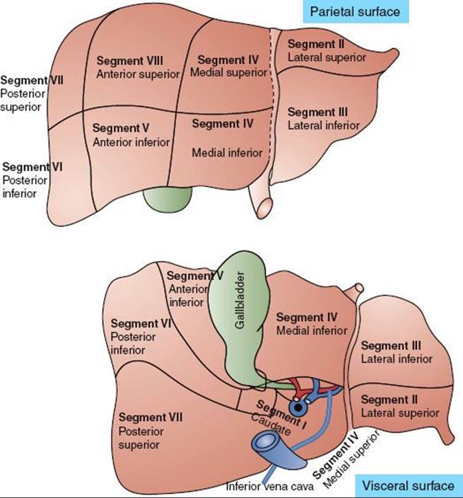
The liver is the largest solid organ in humans. Traditionally it is divided into left and right lobes separated by the falciform ligament. The surgeon needs to understand the spatial relationship of a tumor to the hepatic vascular system preoperatively to determine resectability. Fortunately, progress in imaging techniques has made segmental division of the liver based on the anatomy of portal and hepatic veins feasible. The most common segmentation scheme proposed by Couinaud divides the liver parenchyma into right and left liver with four segments each (Fig. 60.1). The left part of the liver consists of the caudate lobe (segment I), lateral segment (segments II and III), and medial segment (segment IV). The anatomic landmark between the medial segment and lateral segment is drawn between the gallbladder and inferior vena cava (the falciform ligament). The right part of the liver comprises the anterior segments (segments V and VIII) and posterior segments (segments VI and VII). The anatomic landmark that separates the anterior from the posterior segment is the right hepatic vein; the anatomic landmark that divides the anterior segment from the left medial segment is the middle hepatic vein. No good anatomic landmarks exist that further divide the anterior and posterior segments into superior and inferior subsegments.9
Dynamic computerized tomography is very useful in distinctly defining segmental anatomy because the portal vein, hepatic vein, and inferior vena cava can be opacified at the same time.10,11
Epidemiology
HCC is the most frequent primary cancer of the liver and ranks as the fifth most common cancer in the world and the third most common cause of cancer mortality.12 The age-standardized rate of incidence of liver cancer in 1990 worldwide was 14.7 per 100,000 men and 4.9 per 100,000 women.13 The male-to-female ratio was 3 to 1. The highest incidence rate is seen in the male population of South Korea, China, Gambia, and Senegal (28.5 to 48.8 per 100,000 populations).12 In low-risk areas such as Canada, Columbia, and the United Kingdom, HCC occurs in only 1 to 3 persons per 100,000.12 But the incidence rates of primary liver cancer have a trend of decrease among Chinese population in Hong Kong, Shanghai, and Singapore.12 In contrast, the incidence rates in some low-rate areas such as United States, United Kingdom, and Australia increased. The incidence of HCC approximately doubled between 1976 and 2000 in United States.14 The most likely reason for this rising incidence is related to a great prevalence of hepatitis C infection.14
Risk Factors
HCC is clearly associated with hepatitis B (HBV) and hepatitis C (HCV) viral infections and chronic liver disease. With persistent HBV infection, the relative risk of incidence of HCC could be 223.15 The risk of HCC is even higher in patients who are HBeAg (e antigen) positive compared with those who are HBeAg negative.16 As high as for HBV, the relative risk of HCC among persons with chronic HCV infection and cirrhosis is also approximately 100 times the risk of uninfected persons.17 There also exists a synergistic interaction between the two viruses to cause HCC.18
Other chronic liver-cell injury is also associated with the development of HCC. Chemical injury induced by ethanol, nitrites, hydrocarbons, solvents, organochlorine pesticides, primary metals, and polychlorinated biphenyls has been implicated.19,20 Ethanol is the most common culpable chemical agent and is thought to produce HCC through the development of liver cirrhosis or to play the role of a co-carcinogen. Chronic alcohol use of greater than 80 g per day for more than 10 years increases the risk for HCC approximately fivefold. In patients with HCV, alcohol use doubles the risk of HCC.21 Environmental toxins, including aflatoxin, contaminated drinking water, and betel nut chewing, may also be associated with the pathogenesis of HCC. Aflatoxins, well-known hepatotoxic agents, are produced by the fungi Aspergillus flavus and Aspergillus parasiticus. Aflatoxin could contaminate corn, soybeans, and peanuts. High rates of dietary aflatoxin intake have been associated with HCC.22,23 There is a synergistic interaction on HCC between chronic HBV infection and aflatoxin exposure.18 The risk of HCC increases dramatically when both factors are concurrently present. Patients with hereditary liver disease, such as hemochromatosis, Wilson disease, hereditary tyrosinemia, and type I glycogen storage disease, are at high risk of developing HCC.18,24 The common mechanism of developing HCC may be related to chronic injury and inflammation of the liver.
FIGURE 60.1. Segmentation schemes of the liver based on Couinaud’s proposal.
Prevention
Because the development of HCC is highly related to the viral hepatitis and chemical injuries, avoidance of viral infection and chemical exposure may be helpful in prevention of HCC. The HBV vaccine has been available for decades. Universal HBV vaccination in Taiwan has showed a significant decrease of incidence of HCC (from 0.70 to 0.36 per 100,000) in children aged 6 to 14.25 Because the development of HCV vaccination is difficult, the strategies to prevent HCV infection include blood screening, the use of disposable needles and syringes, the adoption of universal precaution for health care workers, and timely treatment of chronic HCV infection with interferon-alpha.26 Meta-analysis suggests that long-term interferon-alfa treatment may prevent the development of HCC in HCV-infected individuals. The preventive effect is more evident among sustained responders to interferon.27
Surveillance
Surveillance for patients with recognized risk factors remains controversial. There is only one randomized controlled trial of surveillance versus no surveillance that has shown a survival benefit,28 and other nonrandomized studies also showed survival benefit of surveillance in high-risk groups.29,30 But when taking the cost into consideration, some authors questioned whether adopting a surveillance policy toward HCC should rely on the prevalence of the disease and the resources of a particular country.30 According to the practice guideline of American Association for the Study of Liver Diseases (AASLD), surveillance is deemed cost-effective if the expected HCC risk exceeds 1.5% per year in patients with hepatitis C and 0.2% per year in patients with hepatitis B. Patients with hepatitis B virus, hepatitis C virus, and autoimmune hepatitis are candidates for surveillance. Current recommendations for surveillance according to the AASLD include:
1. Surveillance for HCC should be based on ultrasonography.
2. AFP alone should not be used for screening because of a lack of adequate sensitivity and specificity for effective surveillance and for diagnosis.
3. Patients should be screened at 6- to 12-month intervals.
4. The surveillance interval does not need to be shortened for patients at higher risk of HCC.31,32
Clinical Presentation
HCC in the early stage is asymptomatic; it is generally detected by elevation of AFP or ultrasonography screening or is an incidental finding in searching for other conditions such as chronic liver disease.33 Even in the advanced stage, some patients are still asymptomatic. Patients with symptoms usually suffer from chronic hepatitis and liver cirrhosis. Clinical symptoms include general fatigue, poor appetite, ascites, jaundice, upper gastrointestinal bleeding, splenomegaly, dilated abdominal veins, palmar erythema, gynecomastia, testicular atrophy, leg edema, and weight loss. Tumor-related symptoms include palpable mass in the upper abdomen (hepatomegaly), acute onset of pain (hemorrhage from tumor rupture), and dull pain in the right upper quadrant of the abdomen, abdominal fullness, low-grade fever, obstructive jaundice, and splenomegaly.34 Very few individuals with HCC present initially with metastatic disease involving extrahepatic organs such as lungs, bone, adrenal glands, pancreas, and neck lymph nodes.35
Diagnostic Workup
In a patient who is suspected of having HCC, the clinical history frequently includes a history of hepatitis, jaundice, blood transfusion, use of intravenous drugs, or exposure to aflatoxins. A family history of hepatitis or hemochromatosis is also an important indicator.36 Details concerning alcohol abuse and job descriptions related to industrial exposure to possible carcinogenic agents are also helpful. The physical examination should include a search for signs of underlying liver disease such as jaundice, ascites, ankle edema, spider angioma on the anterior chest wall, palmar erythema, splenomegaly, increasing abdominal girth, and weight loss. Evaluation of the abdomen for liver size, existence of tumor masses, tenderness, and abdominal bruits should also be performed.
Blood tests should include serology for HBV and HCV, and AFP. If HBV or HCV serology is positive, quantitative HBV DNA or HCV RNA should be obtained.37 Evaluation of hepatic functional reserve includes prothrombin time, activated partial thromboplastin time, and serum albumin. Platelet, red cell, and white blood cell counts also should be obtained to look for simultaneous existence of portal hypertension and hypersplenism from liver cirrhosis. Fifteen-minute retention rate of indocyanine green (ICG) before treatment is useful for determining resectability or the feasibility of radiation.38 The diagnosis of HCC would be included in the differential diagnosis for a patient with underlying liver disease such as cirrhosis or chronic viral hepatitis. In hepatitis or cirrhotic patients, any dominant solid nodule that is not clearly a hemangioma should be considered as HCC unless proven otherwise.39 Noninvasive criteria for diagnosing HCC suggested by the AASLD in 2005 consisted of serum AFP level >200 ng/mL or a typical enhancement pattern (arterial enhancement and portal or delayed washed out) on dynamic imaging of hepatic mass >2 cm in a cirrhotic liver.32 The AASLD further validated the diagnostic accuracy of a single dynamic technique showing intense arterial uptake followed by “washout’’ of contrast in the venous-delayed phases in patients of chronic hepatitis B or cirrhosis of any etiology.31 But histologic diagnosis of HCC is still recommended for patients who plan to have a nonsurgical therapy.40 For surgical patients, there is a concern over the possibility of tumor seeding from biopsy or fine-needle aspiration.40 The reported magnitude of the risk ranges from 1.6% to 5%.41–43 In patients who have coagulopathy or significant ascites, biopsy or fine-needle aspiration may be contraindicated. At the authors’ institution, fine-needle aspirations including cellblocks under ultrasound guidance are routinely performed.44 Core biopsy of the mass is reserved for patients in whom a definitive diagnosis cannot be made by fine-needle aspirations with cellblocks. Coaxial cannula is used with fine-needle or core biopsies to reduce the chance of needle track seeding.45 In patients who have small hepatic nodules with diagnostic possibilities ranging from well-differentiated malignancy to benign disease, ultrasonographically guided needle-core biopsy should be considered.46 Ultrasonography, computed tomography (CT), and magnetic resonance imaging (MRI) are the most common modalities used to evaluate tumors in the liver. Ultrasound examination of the liver is commonly used as a screening tool; small tumors are often hypoechoic. As the tumor grows, the echo pattern tends to become isoechoic or hyperechoic, and HCC can be difficult to distinguish from the surrounding liver.47 Nodules <1 cm should be followed with ultrasonography again at intervals of 3 months. Nodules >1 cm in a cirrhotic liver should be investigated further with four-phase dynamic CT scan. If the four-phase dynamic CT scan cannot prove the diagnosis of HCC, contrast-enhanced MRI or biopsy is the next tool. The contrast-enhanced ultrasound was not recommended in the diagnosis of HCC because it may provide false-positive result in patients with cholangiocarcinoma.31 Extrahepatic metastases are not common at presentation; they occur mainly in patients with T4 disease. The most common sites of metastases are lung, abdominal lymph nodes, and bone.48 Routine metastatic surveys in patients with early-stage HCC are not recommended.
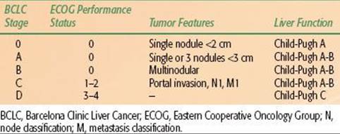
TABLE 60.1 BARCELONA CLINIC LIVER CANCER TAGING SYSTEM FOR HEPATOCELLULAR CARCINOMA
Staging
The prognosis in HCC patients is more complex because the underlying liver function also affects prognosis. Several factors have been identified as being important determinants of survival: the severity of underlying liver disease, the size and number of the tumor, vascular invasion, regional lymph node metastasis, and the presence of distant metastases. There is no worldwide consensus on the use of any given HCC staging system. However, most major trials of HCC therapy have chosen the Barcelona Clinic Liver Cancer Staging System (Table 60.1), making it the de facto reference staging system.31 Other systems, such as the American Joint Committee on Cancer’s TNM (tumor, node, metastasis) staging system,49 Okuda staging systems,50 and the Cancer of the Liver Italian Program scoring system,46 are also commonly used.
Pathologic Classification
Histologic classification of malignant tumors of the liver includes HCC (conventional), HCC (fibrolamellar variant), cholangiocarcinoma (intrahepatic bile duct carcinoma), mixed hepatocellular cholangiocarcinoma, undifferentiated carcinoma, and hepatoblastoma. The fibrolamellar variant of HCC has a relatively better prognosis. It occurs more frequently in adolescents or young adults and has a more indolent clinical course than conventional HCC.51Hepatoblastoma occurs most commonly in young children (median age, 13 to 16 months) and usually presents in an advanced stage.52,53
General Management
HCC is often a multicentric disease, especially when it is associated with HCV. The incidence of multicentric disease in HCV-related HCC (53.3%) is significantly higher (P <.05) than in the non-HCV-related HCC (7.7%).54 The risk of multicentric occurrence increases with the progression of chronic liver disease and cirrhosis.55,56 Although multiple tumors occur less often in HBV-associated HCC than in HCV-associated HCC, the incidence of intrahepatic recurrence of HCC is significantly higher in patients with a sustained HBeAg-positive and high serum concentration of HBV DNA.57 Despite these observations, the mainstay of therapy is surgical resection. The majority of patients, however, are not eligible for surgery because of the extent of tumor involvement or underlying liver dysfunction. Several other treatment modalities are available, including liver transplantation, radiofrequency ablation (RFA), percutaneous ethanol or acetic acid ablation, transcatheter arterial chemoembolization (TACE), cryoablation, radiation therapy, and systemic chemotherapy.
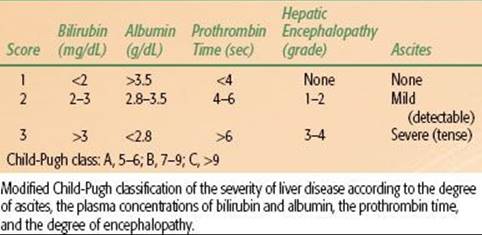
TABLE 60.2 GRADING SYSTEM FOR CIRRHOSIS: THE CHILD-PUGH SCORE
Surgical Resection
Surgical resection is a reliable method to obtain long-term disease control. The main limiting factor for resection is liver function. In virus-related HCC, the extent of surgical resection of hepatic tumor depends on the functional reserve of the residual liver after surgery. Previously, the selection of patients for resection has been based on the Child-Pugh classification (Table 60.2), but this method is known to be inconsistent. Many Japanese and other Asian investigators rely on the ICG retention test.57 In Europe and the United States, selection of optimal candidates for resection is usually based on the assessment of the presence of portal hypertension, which is assessed clinically, or by hepatic vein catheterization.58 Clinically significant portal hypertension is suspected when the platelet count is below 100,000/mm3, which is associated with significant splenomegaly. The goal of surgery is to remove gross tumor with a margin of 1 to 2 cm of normal liver. Today, however, the 5-year survival after resection can exceed 50%.
Liver Transplantation
Many patients are inappropriate for definitive surgery because of underlying liver dysfunction. The increasing availability of liver transplantation has made this procedure an alternative to tumor resection for selected patients. Based on the Milan study and others, liver transplantation is an effective option for HCC patients.59 The selection criteria are solitary tumor 5 cm or up to three nodules with tumor size <3 cm. When these selection criteria are strictly applied, excellent overall 3- to 4-year actuarial (75% to 85%) and recurrence-free survival rates (83% to 92%) can be achieved.59–61 Risk factors of recurrence after transplantation include tumor size, number of tumors, vascular invasion, and persistence of HBV infection.61–63 A major disadvantage with orthotopic liver transplantation is the unpredictable, potentially long waiting time for donor organs.63 Living donor transplantation can be offered for HCC if the waiting time would be so long that there is a high risk of tumor progression leading to exclusion from the waiting list.
Percutaneous Ablation
For patients with early-stage HCC who are not suitable for resection or transplantation, percutaneous ablation would be the treatment option.64,65 Destruction of tumor cells can be achieved by the injection of chemical substances (ethanol, acetic acid) or by modifying the temperature (radiofrequency, microwave, laser, cryotherapy). Ethanol injection is highly effective for small HCC. It achieves a necrosis rate of 90% to 100% of the HCC <2 cm, but the necrosis rate could be reduced to 50% in HCC >3 cm.66,67,68
RFA involves the local application of radiofrequency thermal energy to the lesion. RFA is a reasonable option for patients who do not meet resectability criteria for HCC yet are candidates for a liver-directed procedure based on the presence of liver-only disease. The best outcomes are in patients with a single tumor <4 cm in diameter. The efficacy in tumors >2 cm is better than with ethanol. Randomized control trials comparing RFA and ethanol injection have shown that RFA provides better local disease control that could result in an improved survival.69–72
Transcatheter Arterial Chemoembolization
HCC is a highly vascular tumor supplied mostly by the hepatic or adjoining arteries and has strong neoangiogenic activity during its progression. This character provides the pathologic basis for the radiological diagnosis of disease. Its also supports arterial obstruction as a treatment option. TACE combines selectively injecting chemotherapeutic agents through the tumor artery followed by intra-arterial embolization of tumor artery with lipiodol, an iodized oily contrast agent, despite two prospectively randomized studies that failed to show significant survival benefit over conservative management in patients with unresectable HCC.73,74 Systematic review of randomized prospective studies in more recent literature has shown TACE to have a positive impact on survival.75 For palliative purpose, TACE has been accepted as the standard treatment for patients with unresectable HCC, and it can be used selectively for tumors of different locations and can be repeated if necessary.1,76
Radiotherapy
HCC is a radiosensitive tumor.38 The major drawback of radiotherapy in treating HCC is the poor radiation tolerance of adjacent normal liver and the difficulty of tumor localization.77,78 Recent technological and conceptual developments in the field of radiation therapy, such as intensity-modulated radiation therapy, image-guided radiation therapy, respiratory gating, and stereotactic body radiation therapy, have the potential to improve radiation treatments by conforming the delivered radiation dose distribution tightly to the tumor or target volume outline while sparing normal liver tissue from high-dose radiation.79,80 For patients with liver-confined disease treated with conformal radiotherapy with or without TACE, local control response rates ranged from 40% to 90%, and the median survival ranged from 10 to 25 months.81 Indications for conformal radiotherapy include large unresectable HCC, relieving portal vein thrombosis and obstructive jaundice, failure of prior TACE and as part of combined modality treatment with TACE, and percutaneous ablation therapy.38,82,83 The combination of TACE and conformal radiotherapy in large unresectable HCC has not yet been defined in any randomized trial.
Radiation Doses
The tumor response to radiotherapy and survival of patients with HCC are related to the dose delivered.82,84 Partial response was 18% to 23% in unresectable HCC treated with 21 Gy in 7 fractions and increased to 48% with dose around 33 Gy.85,86 Seong et al.87 irradiated 27 HCC patients who failed TACE and observed an objective response rate of 67% after a mean dose of 51.8 Gy ± 7.9 Gy. Dawson et al.84 escalated radiation doses for unresectable hepatobiliary cancer and observed that patients who received radiation doses >70 Gy had better median survival (>16.4 months). It appears that the higher the radiation doses given, the higher the tumor response seen (Table 60.3).
Caution should be given in HBV-related HCC; local radiotherapy may deteriorate liver function by HBV activation, so prophylactic anti-HBV treatment should be considered.
TABLE 60.3 RADIOTHERAPY WITH AND WITHOUT CHEMOTHERAPY FOR UNRESECTABLE HEPATOCELLULAR CARCINOMA
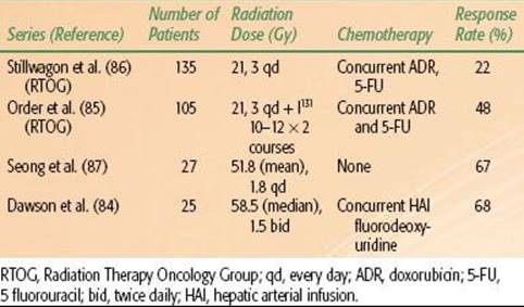
TABLE 60.4 RADIATION TREATMENT GUIDELINES FOR HEPATOCELLULAR CARCINOMA
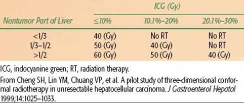
TABLE 60.5 COMBINATION TRANSCATHETER ARTERIAL CHEMOEMBOLIZATION AND LOCAL RADIOTHERAPY IN UNRESECTABLE HEPATOCELLULAR CARCINOMA

Treatment Volumes
Although higher radiation doses have been shown to produce a higher response, patients with HCC usually have liver cirrhosis, which will not allow such doses. Published reports on three-dimensional conformal radiation therapy (3D-CRT) with photon energy suggest that portions of the liver can be treated with higher doses and acceptable complications. McGinn et al.88 developed a normal tissue complication probability (NTCP) for intrahepatic malignancy. They designed a protocol in which each patient received the maximum possible dose while being subjected to a 10% risk of radiation-induced liver disease based on the NTCP model. The mean doses delivered according to this protocol were 56.6 ± 2.31 Gy (range, 40.5 to 81 Gy). They observed a complication rate of 4.8% (95% confidence interval, 0% to 23.8%), a number that did not differ significantly from the predicted 8.8% calculated on the basis of the NTCP model. This model, however, was derived using patients with relatively normal liver function, and only 4 of 21 of their study population had HCC. The NTCP model for patients with impaired liver function requires further evaluation.89
Multivariate analyses demonstrated that the severity of hepatic cirrhosis was the only independent predictor for radiation-induced liver disease. For cirrhotic patients with Child-Pugh grade A, the hepatic radiation tolerance was a mean dose to normal liver of 23 Gy.90 These authors also suggested a dose–volume histogram for irradiation to normal liver so that the irradiated hepatic volume over 10 Gy, 20 Gy, 30 Gy, and 40 Gy should be less than 68%, 49%, 28%, and 20%, respectively.
ICG retention can be used to guide the treatment of HCC. In HCC patients with normal bilirubin and without ascites, if the ICG retention 15 minutes (ICG 15) is normal, the resection volume can be trisegmentectomy or bisegmentectomy; if ICG 15 is 10% to 19%, the resection volume can be left lobectomy or right monosegmentectomy; if ICG 15 is 20% to 29%, the resection can be subsegmentectomy; if ICG 15 is 30% to 39%, the resection can be done to only a limited area of the liver.91 Therefore, the authors propose a treatment guideline using the ICG test for 3D-CRT for patients with impaired liver function (Tables 60.4 and 60.5). The authors advise no radiation treatment for patients with Child-Pugh class C liver cirrhosis or prolonged ICG retention unless only a very small portion of the liver is included in the radiation treatment fields.38
Stereotactic body radiotherapy (SBRT) to the intrahepatic lesion becomes the option of treatment with the improvement of planning system and linear accelerators. But the large fraction size makes a difference in normal tissue tolerance. There have been many retrospective studies but no universal consensus for hepatic tolerance of SBRT. Son et al.96 reported that at least 800 mL of normal liver had to receive a total dose <18 Gy to reduce the risk of the deterioration of hepatic function. For lesions beside the duodenum or stomach, Mahadevan et al.97 reported that 24 to 36 Gy (depending on the distance from the duodenum and stomach) in 3 fractions had acceptable side effects.
Design of Treatment Field
The goal of conformal radiotherapy is to precisely target the tumor(s) and to reduce damage to the surrounding normal tissue. Respiratory organ motion probably is the largest intrafractional organ motion. Aruga et al.98 studied organ motion involving the use of CT images obtained during both the static exhalation phase and static inhalation phase for upper abdomen irradiation. They found that the tumor shifted between the two respiratory phases. The variation ranged from 2.6 to 23.7 mm: from 0.4 to 5.9 mm in the lateral direction, 2.2 to 24.5 mm in the longitudinal direction, and 0.2 to 11.7 mm in the vertical direction. Breath-gating or breath-holding irradiation may help overcome the problem of respiratory movement during irradiation.99 In general, the radiation-field margin to the target in the lateral direction should be 6 to 9 mm, vertical direction 9 to 12 mm, superior direction 10 mm, and inferior direction 19 to 21 mm.100,101 Controlling, gating, or tracking respiratory motion or by the use of image-guided radiation therapy, as previously mentioned, is currently under investigation.102
Acute and Late Complications
The dose-limiting tissue injuries in radiation treatment for HCC include the liver, stomach, duodenum, bowels, and kidneys. Acute complications include general fatigue, transient elevation of liver function test, nausea and vomiting (mainly for tumors in the left lobe of the liver), fever, and pancytopenia.38,87 Subacute and late complications include hepatic failure, radiation pneumonitis, and gastrointestinal bleeding (especially in tumor located in the inferior portion of the right lobe of the liver and radiation doses >50 Gy).38,103 Hepatic failure can be avoided by an appropriate selection of patients and careful treatment planning.
Results of Combining Transcatheter Arterial Chemoembolization and Local Radiation Therapy
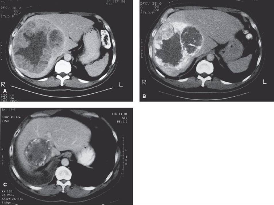
TACE alone rarely produces complete pathologic remission for HCC >5 cm, especially in the peripheral zone of the tumor.104,105 Additional therapy theoretically is required to eradicate the residual disease. The combination of TACE and conformal radiotherapy shows promising results in large HCC (see Table 60.5). Guo and Yu92 reported 107 patients with large unresectable HCC treated with TACE followed by external-beam irradiation. The greatest dimension of the tumors ranged from 5 to 18 cm. After a median follow-up interval of 24 months, the cumulative survival rates at 3 and 5 years were 28.4% and 15.8%, respectively, with a median survival of 18 months. Cheng et al.38 reported on 17 patients with unresectable HCC treated with TACE and conformal radiotherapy. The mean tumor size was 8.6 cm (range, 3.7 to 18 cm). The overall survival rate at 2 years was 58% and local progression-free tumor control was 83%. After 24 months of median follow-up, the median survival had not been reached, and 4 of 17 patients remained progression free (Fig. 60.2).
Another study that combined TACE and local radiotherapy in 50 unresectable HCC patients reported a partial response rate of 66% and survival rates at 3 years of 43%.103 Wu et al.95 reported 94 patients with HCC received 3D-CRT combined with TACE. The response rate was 90.5%. The overall survival rates at 1, 2, and 3 years were 93.6%, 53.8%, and 26.0%, respectively, with the median survival of 25 months.
Patients with branch portal vein thrombosis may benefit from combined treatment. Tazawa et al.106 reported on 19 patients with thrombus in the first branch of portal vein who received 3D-CRT with TACE. Eleven patients (58%) had an objective response, and the 1-year survival rate was 41% (eight patients). Although we do not know yet whether combined TACE and conformal radiotherapy is a superior treatment modality to TACE alone, the results of these studies suggest that in patients with large unresectable HCC, combined treatments could be a promising new treatment modality worthy of further investigation.
FIGURE 60.2. A: A 55-year-old man with hepatitis B virus infection developed a large hepatocellular carcinoma in right lobe of liver. The tumor measured 12 é 15 é 15 cm. Alfa-fetoprotein (AFP) was 901 ng/mL. B: After a first course of transcatheter arterial chemoembolization (TACE), his AFP dropped to 150 ng/mL. It rose to 440 ng/mL 3 months later. He had second TACE followed by three-dimensional conformal radiotherapy. This image was taken after the second TACE, revealing viable tumors in the peripheral zone of the main mass. C: Four years and 4 months after the first treatment, computed tomography revealed a nonenhancing tumor in the right lobe of the liver. His AFP remained <10 ng/mL after completion of radiotherapy.
Chemotherapy
Systemic chemotherapy for HCC has limited value in clinical practice because only a small portion of patients obtain significant benefits at the price of remarkable toxicity.107 In general, cytotoxic therapy should be reserved for medically appropriate patients with adequate hepatic function, preferably administered within the context of a clinical trial. The side-effect profile of any chemotherapy regimen should be considered carefully in patients with advanced liver disease and a short life-expectancy.
Several new drugs have been tested recently. One of the more promising of these drugs is gemcitabine. It has a low toxicity profile, but its antitumor activity in patients with advanced HCC is marginal: the response rate is 18% and the response duration is 35 weeks.108 Combination chemotherapy regimens have shown better responses. Leung et al.109 reported on 50 patients treated with intravenous chemotherapy composed of cisplatin, doxorubicin, 5-fluorouracil (5-FU), and interferon-alfa. The partial response was 26% (13/50) and four patients achieved pathologic complete remission. Louafi et al.110 reported 34 patients of advanced HCC treated with gemcitabine and oxaliplatin. The disease control rate was 76%. Median progression-free and overall survival times were 6.3 months and 11.5 months, respectively.
Molecular Targeted Therapy
Recent advances in molecular biology have uncovered the structures and functions of many cytokines thought to have a strong relationship with the mechanisms of the antitumor effect of biological therapies. Thalidomide, a sedative previously associated with severe fetal malformations, has limited value in the treatment of HCC.111,112 Molecular targeted therapy to the epidermal growth factor receptor and vascular endothelial growth factor has exhibited the potential to inhibit tumor growth of HCC.113 The randomized trial of sorafenib has showed statistic significance in median overall survival (10.7 vs. 7.9 months) and median time to radiological progression (5.5 vs. 2.8 months).114
Areas of Failure and Cause of Death
Survival of HCC after treatment depends on the clinical stage and liver function. Five-year survival in resectable HCC after partial hepatectomy is 50% to 73% in stage I patients, 30% to 56% in stage II patients, 10% to 29% in stage III patients, and ~10% in stage IV patients.115,116 The 5-year survival for patients with unresectable HCC is usually <10%.117 The major cause of failure after resection is tumor recurrence within the liver; this observation still holds true for patients who undergo TACE alone. Extrahepatic metastases in advanced-stage HCC have become more common in recent years, related to improvements in the local treatment of intrahepatic disease.118,119
Future Study
The overall treatment outcome in HCC is generally unsatisfactory. The main cause of failure is intrahepatic recurrence. Future efforts should be directed toward prevention of HBV and HCV infection through vaccination in hyperendemic regions and perhaps through chemoprevention for individuals who are already infected and are persistent carriers. Other approaches include early diagnosis in high-risk populations and development of effective adjuvant treatments after resection.
BILIARY TRACT CANCER
Biliary tract cancers consist of cancer of the gallbladder, the bile ducts, and the ampulla of Vater. They are highly lethal because most are locally advanced at diagnosis. Gallbladder cancer is the most common cancer of the biliary tract and accounts for two-thirds of these cancer patients, whereas bile duct cancer accounts for the remaining one-third.120 The term cholangiocarcinoma was used to describe cancers arising from the epithelial cells of the bile ducts, which include intrahepatic, perihilar, and distal extrahepatic biliary tree. At present, surgical excision of all detectable biliary tract cancers is associated with improvement in long-term survival. For unresectable tumors, the purpose of treatment is to palliate symptoms such as obstructive jaundice, biliary tract infection, pain, and ascites.
CHOLANGIOCARCINOMA
Topographic Anatomy
The bile ducts originate within the liver, with the left and right hepatic ducts joining to form the common hepatic duct. At the origin of the cystic duct, it becomes the common bile duct. The cystic duct drains bile from the gallbladder into the common bile duct. The gallbladder is adjacent to the undersurface of the liver.
There is a rich lymphatic network along the submucosa of bile ducts. The primary lymphatic drainage of the biliary tract is to the lymph nodes in the pericholedochal area, periportal region, hepatoduodenal ligament, common hepatic artery, and pancreaticoduodenal groups.80,121,122
Epidemiology and Risk Factors
Cholangiocarcinoma is a rare tumor in developed countries; there are approximately 2,000 to 3,000 cases per year in the United States. However, it is one of the most common cancers in endemic areas of developing countries, as high as 87 per 100,000 people in northeast Thailand.123 Cholangiocarcinoma accounts for about 15% of the primary liver cancer worldwide. But the proportion was different in different regions: 20% in Western countries, <10% in Asian nations, and as high as 90% in north Thailand.64,124
A number of risk factors have been identified as important in the development of cholangiocarcinoma, most of which share a history of long-standing inflammation and chronic injury of the biliary epithelium.125 The major risk factor in Western countries is primary sclerosing cholangitis, which is closely associated with chronic inflammatory bowel disease, particularly ulcerative colitis.126 The risk of developing cholangiocarcinoma is higher in patients with primary sclerosing cholangitis, ulcerative colitis, and colonic neoplasm than in patients with primary sclerosing cholangitis and ulcerative colitis without colonic neoplasm.127 Studies in Japan and the United States also showed that chronic hepatitis C infection elevated the incidence of intrahepatic cholangiocarcinoma, with the odds ratio between 4 and 17.128,129 In Asia, chronic infections of the biliary tract and infestation by certain liver flukes, such as Clonorchis sinensis and Opisthorchis viverrini, are associated with cholangiocarcinoma and hepatolithiasis.123 Hepatolithiasis itself is also a risk factor for cholangiocarcinoma; 5% to 10% of patients with intrahepatic stones develop this complication.130,131 Moreover, the combination of liver fluke infestation and nitrosamine exposure may explain the very high incidence of cholangiocarcinoma in northeast Thailand.132 Other risk factors, although rare, include congenital fibropolycystic disease of the biliary system such as choledochal cysts and Caroli disease (cystic dilatation of intrahepatic bile ducts).133 The observed incidence and mortality of intrahepatic cholangiocarcinoma has increased in the past 3 decades in Japan, the United States, and the United Kingdom.14,134,135 In the United States, the age-adjusted mortality rate increased from 0.07 per 100,000 in 1973 to 0.69 per 100,000 in 1997; the estimated annual percentage change of mortality was 9.44%. Better case ascertainment and diagnosis because of improved diagnostic imaging, use of image-guided biopsies, or increased use of endoscopic retrograde cholangiopancreatography (ERCP) cannot fully explain this observation.135,136
Diagnosis
The most common presenting symptoms of biliary tract cancer are caused by obstruction of the bile duct. Those include painless jaundice, clay-colored stool, tea-colored urine, and pruritus. Other signs and symptoms include abdominal pain, fever, general malaise, abdominal distention, fullness, anorexia, and weight loss. The spectrum of cholangiocarcinoma can be classified into three broad groups: (a) intrahepatic, (b) perihilar, and (c) distal tumors. The age of onset is similar among the three groups and ranges from 60 to 65 years.137 Patients with extrahepatic tumor usually present with jaundice and tea-colored urine. Patients with intrahepatic tumors are less likely to be jaundiced and more likely to present with abdominal symptoms.
There are no reliable screening methods; early diagnosis is almost impossible even in patients with high-risk situations such as primary sclerosing cholangitis and hepatolithiasis.138 Some patients are diagnosed when screening blood work demonstrates elevation of alkaline phosphatase and γ-glutamyl transferase. Ultrasonography and CT are the initial primary tools to evaluate biliary tract tumor (Table 60.6). Further tests include percutaneous transhepatic cholangiography, ERCP with brushing cytology, serum carbohydrate antigen-19-9 (CA-19-9) levels, radiologic imaging with dynamic CT scan, MRI, or both, and angiography.
A serum CA-19-9 value >100 U/mL has a sensitivity of ~75% and a specificity of ~80%.139 The optimal cutoff value for serum CA-19-9 that best discriminates between benign or malignant biliary tract diseases is influenced by the presence of cholangitis. Thus, in patients with symptoms of acute cholangitis, serum CA-19-9 concentrations should ideally be re-evaluated after recovery. The sensitivity of serum carcinoembryonic antigen (CEA) is low and helpful in only one-third of patients.140 Biliary CEA levels increase significantly in patients with cholangiocarcinoma and also in patients with intrahepatic cholelithiasis (average, 50.2 to 57.4 ng/mL) compared with patients with benign strictures (average, 10.1 ng/mL) and patients with sclerosing cholangitis and choledochal cysts (average, 20.0 to 21.6 ng/mL).139 Serum AFP may increase in some cases of cholangiocarcinoma, and this would suggest a diagnosis of mixed HCC and cholangiocarcinoma.141
Pathology
Cholangiocarcinomas arise from the epithelium of the biliary tract; the majority of these cancers (>90%) are adenocarcinomas. Squamous cell carcinoma is the second most histologic type. Rare histologies include mucoepidermoid carcinoma, cystadenocarcinoma, and carcinoid tumor.
Adenocarcinomas are divided into three type: sclerosing, nodular, and papillary. Sclerosing tumors are characterized by an intense desmoplastic reaction. This type of tumor tends to invade the bile duct wall early and, as a result, is associated with low resectability and cure rates. Most cholangiocarcinomas are of this type.137 The nodular cholangiocarcinomas are also highly invasive tumors. Most patients have advanced disease at diagnosis. The resection and cure rates are both very low. In contrast, papillary type tumors usually present as masses in the lumen of bile duct, causing biliary obstruction early in the course of disease. Therefore, these tumors have the most favorable prognosis.142 Microscopically, cholangiocarcinoma are classically well differentiated to undifferentiated. Cells tend to be cuboidal or low columnar and resemble biliary epithelium; mucin is always demonstrable in the cytoplasm. Bile duct obstruction can be associated with reactive hyperplasia of subepithelial mucous glands with or without cholangiocarcinoma.143 Cholangiocarcinomas frequently invade lymphatic, perineural, and periductal spaces and the portal tracts. Spread along the lumen of the large bile ducts can also be seen, especially in perihilar cholangiocarcinoma.144
The differential diagnosis of cholangiocarcinoma from HCC can be further affirmed by positive reaction with CEA, CA-19-9, and immunohistochemistry with cytokeratin-19.145 Mutations in the p53 tumor suppressor gene and K-ras proto-oncogene have been identified in cholangiocarcinoma.146–148 p53 overexpression and K-ras mutations are associated with a shortened survival.146
TABLE 60.6 DIAGNOSTIC WORKUP FOR CARCINOMA OF THE BILE DUCT

Pathways of Tumor Spread
Bile duct cancers commonly spread by direct extension through the bile duct and the abundant lymphatic network in the submucosa. These tumors also commonly involve the surrounding structures by direct invasion. Lymph node metastases in the porta hepatis and celiac axis are common. The lymph nodes in porta hepatis are more frequently involved with tumors in the intrahepatic duct and proximal extrahepatic bile duct, and the pancreaticoduodenal nodes are more often involved with tumors in the distal bile duct.144,149
The incidence of lymph node metastasis in intrahepatic cholangiocarcinoma ranges from 50% to 60%.150,151 Intrahepatic cholangiocarcinomas, irrespective of their intrahepatic location, mainly spread to the nodes in the hepatoduodenal ligament, then to the para-aortic nodes, retropancreatic nodes, or common hepatic artery node group. In addition, the left peripheral type or hilar type of cholangiocarcinoma tends to spread along the left gastric nodes through the lesser curvature.151 Lymph node metastasis in perihilar cholangiocarcinoma is common, which occurs in half of the patients, with the frequency of 43% in pericholedochal nodes, 31% in the periportal nodes, 27% in the common hepatic nodes, and 15% in the posterior pancreaticoduodenal nodes. The celiac and superior mesenteric nodes are rarely involved.144
Lymph node metastasis in distal duct cancer is commonly observed near the duodenopancreatic regions. Yoshida et al.149 examined 20 consecutive patients with distal bile duct cancer who underwent pancreaticoduodenectomy with extended lymph node dissection. Histologic evidence of lymph node metastasis was seen in 55% of the patients. The areas with frequent metastases were the posterior pancreaticoduodenal lymph nodes (35%), the nodes around the hepatoduodenal ligament (35%), and those around the common hepatic artery (30%). Para-aortic lymph node involvement occurred in 25% of patients and was significantly associated with pancreatic parenchymal invasion.
Staging
The American Joint Committee on Cancer TNM classifications for intrahepatic cholangiocarcinoma are similarly to HCC. The definition of T stage were
T1: solitary tumor without vascular invasion,
T2a: solitary tumor with vascular invasion,
T2b: multiple tumors, with or without vascular invasion;
T3: tumor perforating the visceral peritoneum or involving the local extra hepatic structures by direct invasion;
T4: tumor with periductal invasion.49
N0 means no regional lymph node metastasis, and N1 is with regional lymph node metastasis. M0 is without distant metastasis, whereas M1 is with distant metastasis. The complete stage groups were listed on Table 60.7.
The American Joint Committee on Cancer TNM classifications for both hilar and distal cholangiocarcinoma were revised in 2010 and similar to the TNM classification of intrahepatic cholangiocarcinoma (see Table 60.7). The exception was that they added N2 disease (metastasis to periaortic, pericaval, superior mesenteric artery, or celiac artery lymph nodes) for perihilar disease. The most important prognostic factors for cholangiocarcinoma are resectability, regional lymph node metastasis, and distant metastasis.
TABLE 60.7 AMERICAN JOINT COMMITTEE ON CANCER STAGE GROUPS BY TNM CLASSIFICATION

General Management
Surgical resection provides the only possibility of cure, but the resection rates for intrahepatic, perihilar, and distal lesions were 60%, 56%, and 91%, respectively, in one large series.137 The 5-year survival rates after resection range from 10% to 40%, depending on the location of the primary tumor.137 Patients with unresectable cholangiocarcinoma generally have a very poor prognosis; chemotherapy and radiotherapy have been used, but the results are disappointing.152
Intrahepatic Cholangiocarcinoma
Intrahepatic cholangiocarcinoma accounts for 5% to 10% of all biliary tract cancers and constitutes 10% to 20% of primary liver malignancies. The resectability rate in all patients is only 30% to 50%.153,154 The outcomes after surgery for patients with lymph node metastasis are poor regardless of the sites of nodal metastasis; the 5-year survival rate in patients with lymph node metastasis was lower than that in patients without lymph node metastasis (0% vs. 51%; P <.0001).151
Factors that affect tumor recurrence include lymph node metastasis, presence of satellite nodules, positive resection margin, tumor size, and bilobar distribution.151,155–157 The patterns of failure after resection of intrahepatic cholangiocarcinoma are primarily in the liver (56%), regional lymph node (20%), and peritoneal seeding (24%).158 The role of postoperative adjuvant radiotherapy with or without chemotherapy in the management of intrahepatic cholangiocarcinoma is controversial. The typical reports in the literature are retrospective, with small numbers of patients and marked variations in radiation field and dose.159 A delineation of radiation fields based on patterns of failure analysis and lymph node spread of the disease will be necessary to rationally define the role of radiotherapy in any proposed phase III study. For unresectable cholangiocarcinoma, the purpose of treatment is palliative; however, some long-term survivals (a 4-year rate of 20%) have been observed in unresectable cholangiocarcinoma treated with conformal radiotherapy and intra-arterial infusion of fluorodeoxyuridine.160
Total hepatectomy with orthotopic liver transplantation also can be considered in this situation. One systemic review combining all studies that included a minimum of 10 patients reported a median survival of 11.8 months. The 1-, 3- and 5-year overall survivals were 63%, 46%, and 22%, respectively. The 1-, 3- and 5-year disease-free survivals were 58%, 22%, and 13%, respectively.161 But orthotopic liver transplantation could not be considered as a standard treatment for localized cholangiocarcinoma at present. Such protocols should be preserved for selected patients with unresectable hilar disease only at transplantation centers.
Perihilar Cholangiocarcinoma
Perihilar cholangiocarcinoma is the most common biliary tract carcinoma and accounts for 65% to 70% of tumors of the biliary tract.137 Perihilar tumors involving the bifurcation of the hepatic duct are also called Klatskin tumors.162These tumors were further classified by Bismuth et al.163 as tumors below the confluence of the left and right hepatic ducts (type I), tumors reaching the confluence (type II), tumors occluding the common hepatic duct and the right or the left hepatic duct (types IIIa and IIIb, respectively), and tumors that are multicentric or that involve the confluence and both the right and left hepatic ducts (type IV).
Only 25% to 79% of patients are amenable for surgical resection. Definitive surgery may involve combined bile duct and liver resection with caudate lobe lobectomy.164 The 3-year survival rate was 55% for patients without involved nodes, 32% for patients with regional node metastasis, and 12% for patients with para-aortic node metastasis.144 In multivariate prognostic analysis, only lymph node metastases and curative resection have proven to be of independent prognostic significance.144
The role of postoperative radiotherapy with or without chemotherapy in patients with completely resected cholangiocarcinoma remains unproven. A retrospective analysis from Johns Hopkins suggests that postoperative adjuvant radiation therapy does not improve survival.165 However, postoperative radiotherapy or chemoradiotherapy reduces the rate of local recurrence in patients with incomplete resection.166,167 In addition, many retrospective series and small phase II studies suggest superior outcomes for resected patients who receive external-beam radiation therapy with or without concomitant chemotherapy.65,168,169–170 For patients with microscopic residual disease after resection, a retrospective analysis from 63 patients with stage IV Klatskin tumor revealed that postoperative radiotherapy (with or without intraoperative radiotherapy) yielded significantly higher 5-year survival rates than in the resection alone group (33.9% vs. 13.5%).167
An update report from the Mayo Clinic revealed that the 5-year survival in transplant patients of unresectable hilar cholangiocarcinoma was 80% and in resection-only patients it was 21%.171 Others also have reported similar promising results in these combined-modality treatments.172,173
In general, patients with inoperable perihilar cholangiocarcinoma usually have obstructive jaundice and should be treated with endoscopic or percutaneous drainage or stent placement initially. External-beam radiotherapy alone rarely controls advanced disease. Combinations of external-beam radiotherapy, chemotherapy, and intraluminal brachytherapy may relieve pain, contribute to biliary decompression, and sometimes achieve long-term survival. The most active agents include 5-FU, gemcitabine, docetaxel, and oxaliplatin.174,175 The median survivals range from 17 to 21 months. In some cases, combined chemotherapy and radiotherapy could delay the progression of cholangiocarcinomas while patients await the chance for liver transplant.94,168
Distal Bile Duct Carcinoma
Primary distal bile duct adenocarcinoma, which includes carcinoma of the ampulla of Vater, accounts for 25% to 30% of biliary tract carcinomas.137 Patients with distal duct carcinoma have the highest rate of curative resection as compared with proximal duct carcinoma. In an analysis of 171 patients who underwent surgical exploration at Mayo Clinic for extrahepatic cholangiocarcinoma from 1976 to 1985, the rate of curative resection (negative margins) by site of the primary tumor was 15% for proximal, 33% for middle, and 56% for distal duct lesions.176 The prognosis of patients with distal duct carcinoma is also better than that of patients with proximal duct carcinoma.127
The role of postoperative radiotherapy is uncertain. Investigators at Thomas Jefferson University found that postoperative radiotherapy did not improve survival in distal duct carcinoma after complete resection.177 However, studies of concurrent chemotherapy and radiotherapy show more promising results. Mehta et al.178 from Stanford University treated 12 patients having unfavorable (mainly lymph node metastasis) ampullary carcinoma with concurrent radiotherapy and protracted venous infusion of 5-FU. Actuarial overall survival at 2 years was 89%, and median survival was 34 months. Another study conducted by the Eastern Cooperative Oncology Group revealed that unresectable pancreaticobiliary carcinoma treated with concomitant radiotherapy and protracted intravenous infusion of 5-FU achieved a 2-year survival of 19%.170
Systemic Chemotherapy
Although some retrospective reports showed a benefit of postoperative chemotherapy,179,180 there was a multi-institutional randomized trial from Japan that showed only a trend toward better 5-year survival following a potential curative resection in those who received postoperative chemotherapy.166 But it was not a statistically significant difference.
For treatment of advanced cholangiocarcinoma, a randomized trial showed a survival benefit with 5-FU–based chemotherapy compared with best supportive care alone (median survival, 6 vs. 2.5 months, respectively).181 The most frequently used agents include 5-FU, gemcitabine, cisplatin, and oxaliplatin. Another phase III study enrolled 410 patients with locally advanced or metastatic cholangiocarcinoma, gallbladder cancer, or ampullary cancer. As compared with gemcitabine alone, cisplatin plus gemcitabine was associated with a significant survival advantage (11.7 vs. 8.1 months) without the addition of substantial toxicity. Cisplatin plus gemcitabine is an appropriate option for the treatment of patients with advanced biliary tract cancer.182
Radiation Therapy
The role of radiotherapy (with or without chemotherapy) in unresectable or recurrent cholangiocarcinoma is to relieve pain and biliary compression. At 1 year, 60% to 75% of patients are free of locoregional disease progression, and median survival was 7 to 12 months.183,184 But local failure remains the first site of disease progression in 50% to 75% of patients. To enhance treatment effects, radiotherapy given with 5-FU–based chemotherapy is generally recommended.170,178 The development of a treatment system of radiation oncology in the past few years made SBRT possible, but the experience is limited. One report included 27 patients with unresectable cholangiocarcinoma who underwent SBRT (45 Gy in 3 fractions). At a median follow-up of 5.4 months, only two patients were alive. The median progression-free and overall survival were 6.7 and 10.6 months, respectively. The local control was 84% in 1 year, but six patients had severe duodenal or pyloric ulceration and three patients developed duodenal stenosis.185 Radiotherapy technique for intrahepatic cholangiocarcinoma is similar to treatment of HCC. The dose-limiting surrounding organs include the liver, duodenum, stomach, and spinal cord.
Treatment Volumes
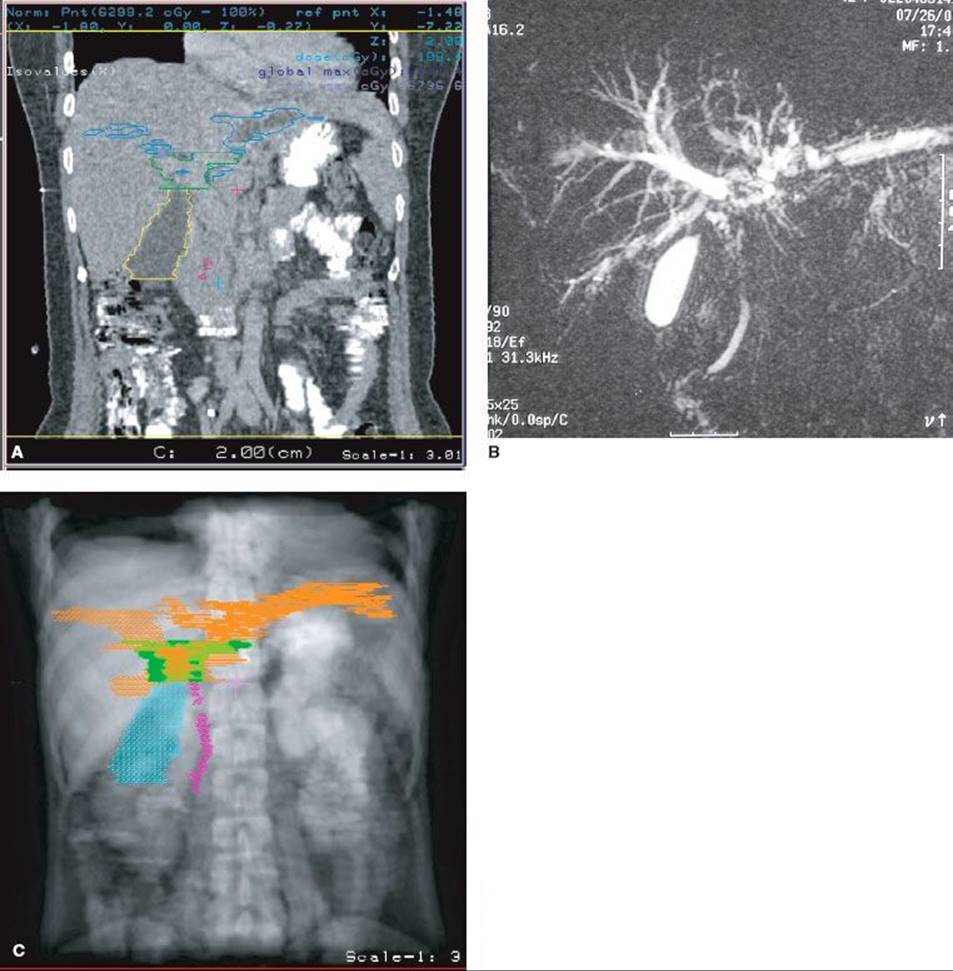
The extent of the tumor within the bile duct can be defined by percutaneous cholangiography, ERCP, and magnetic resonance cholangiopancreatography. However, extraductal disease is difficult to define by any noninvasive procedure. Clip placement at the time of surgery is useful in delineating the extrahepatic portion of ductal lesions and in defining the primary tumor bed. With the incorporation of CT in radiation-treatment planning, and to reduce errors to a minimum, the authors advocate placing patients in the treatment position when performing CT, using 1 to 3 mm per slice and contrast medium to reconstruct the bile ducts and gross tumor volume (Fig. 60.3). The gross tumor volume (GTV) is defined as any visible tumor by CT or MRI. Clinical target volume (CTV) is defined as a 1.5-cm margin to the GTV, especially along the bile duct and potential lymphatic drainage areas, which include nodes along the porta hepatis, pancreaticoduodenal system, and celiac axis.149,151,186,187 The planning target volume is defined by adding a margin of 0.5 to 1 cm to the CTV.188
Radiation Doses
Initial setup of radiation fields is to include GTV and CTV; the radiation fields can be coplanar or noncoplanar. The planning target volume will be treated to 45 to 50 Gy in 1.8- to 2-Gy fractions given 5 days a week, using blocks to exclude normal stomach, small intestine, kidney, and liver.168,189 Higher radiation doses are used only to treat the GTV with the application of 3D-CRT.84 If boost-dose irradiation is feasible with brachytherapy techniques, the GTV is carried to 45 to 50 Gy with external techniques, and 20 to 30 Gy is delivered via an intraluminal the catheter.79 The most commonly employed brachytherapy technique for the biliary tract begins with the placement, performed by an invasive diagnostic radiologist, of a percutaneous drainage catheter through the area of tumor. The radiation oncologist then threads a catheter inside the drainage system catheter.
When a low-dose–rate system is employed, a wire with iridium-192 is then placed at the desired location inside the catheter.190 If a remote high-dose–rate after-loading system is employed, then the appropriate dwell times and positions are selected and programmed. It is a matter of some debate as to where the brachytherapy prescription point should be placed. Some physicians select the tumor’s peripheral edge, away from the catheter, as determined by CT or MRI. Other practitioners prefer a point 0.5 or 1 cm away from the catheter. It is important to be familiar with the valves used to direct bile flow in the drainage system lest, during the administration of brachytherapy, bile leaks onto the patient’s skin and dressings. If the drainage system remains in place for a long period of time (i.e., a month), enteric bacterial colonization and biliary tract infection are common. The combination of external-beam irradiation and brachytherapy employed at Thomas Jefferson University resulted in 2-year and median survival rate, increasing twofold when doses were brought up to >55 Gy.190 However, investigators at the University of Amsterdam reported no improved survival at doses >55 Gy.191 The role of radiotherapy and its optimal dosage remain to be determined.
Acute and Late Complications
Acute complications of external-beam and intraluminal radiotherapy include nausea, vomiting, and transient elevation of transaminase. These effects are usually mild and tolerable.35
Late complications are associated with radiation dose to surrounding organs. The most common complications are gastrointestinal bleeding, biliary bleeding, and duodenal stenosis.35,190,191 With external-beam doses of <55 Gy to the duodenum or stomach, the risk of severe gastrointestinal complications varies from 5% to 10%. At doses >55 Gy, one-third of patients develop severe problems.
Future Directions
There is a paucity of phase III studies for cholangiocarcinoma, so physicians must rely on retrospective studies or prospective phase I or II trials. Further advances in treatment could potentially be made by (a) defining when to use radiation and chemotherapy for high-risk patients after curative resection, (b) investigating the use of higher doses of conformal radiotherapy and chemotherapy in unresectable cholangiocarcinoma, and (c) identifying novel chemotherapeutic agents. Patients with unresectable cholangiocarcinoma should be offered clinical trials.
FIGURE 60.3. A: Computed tomography (CT) was performed in a 39-year-old woman who had cholangiocarcinoma of perihilar area. This film was reconstructed from a CT, which was obtained with 2 mm per slice. The tumor involved right and left hepatic duct (green line). B: Magnetic resonance cholangiopancreatography affirmed tumor extent. C: Digital reconstruction radiography revealed the gross tumor volume (green), and clinical target volume (purple).
ADENOCARCINOMA OF GALLBLADDER
Gallbladder carcinoma is not common; it is the fifth most common cancer of the gastrointestinal tract. The incidence indicates a large geographic variation, and it accounts for approximately 5,000 new patients per year in the United States.192 Distinctly different from cholangiocarcinoma, the ratio of females to males is 2.5 to 1.133 Cholelithiasis, an anomalous junction of pancreaticobiliary ducts, and porcelain gallbladder are factors that predispose to gallbladder cancer.193 Cigarette smoking, alcohol consumption, and obesity may also increase the risk. Patients with polyps >10 mm in diameter may be at increased risk for gallbladder cancer.194,195 Chronic infection with Salmonella typhi and Helicobacter bilis is also associated with the risk of developing carcinoma of the gallbladder.196,197
The most common clinical presentation is pain, followed by anorexia, nausea, or vomiting. Patients with early invasive gallbladder carcinoma are most often asymptomatic or they have nonspecific symptoms that mimic or are due to cholelithiasis or cholecystitis. In general, gallbladder carcinoma is diagnosed late, which accounts for the poor prognosis.133 A French study of 724 patients revealed that only 4% of the patients had Tis lesions, 11% had T1 to T2 lesions, and 85% had T3 to T4 lesions.198 Five-year survival rates according to the National Cancer Data Base were Tis disease, 60%; T1N0, 39%; T2N0, 15%; and T3N0 or node-positive disease, 5%.199 However, improved outcomes have been noted in the past decade and attributed to more aggressive surgery and the use of postoperative adjuvant therapy.200 Studies from Japan report a 100% 5-year survival rate in stage I patients, with stage II at 50% to 78%, stage III at 0% to 69%, and stage IV at 0% to 11%.201,202
Lymphatic metastasis is initially to cystic and pericholedochal nodes and then to the pancreaticoduodenal system, with later potential spread to the rest of the celiac axis or the superior mesenteric or aortic nodes.201,203 The lymph node metastasis rate is associated with primary tumor stage: 0% to 2.5% in pT1 a disease (involvement limited to the mucosa), 15% in T1b, disease, 62% in pT2 disease, and 81% in pT3 or pT4 disease.201,204,205
Surgery is the only potentially curative therapy, but only 10% to 30% of patients are eligible for resection.206 The standard surgical procedure is removal of the gallbladder, resection of various amounts of liver surrounding the gallbladder bed, resection of the extrahepatic bile duct, and dissection of the regional lymph nodes. Patients with jaundice should be considered for preoperative percutaneous transhepatic biliary drainage for relief of biliary obstruction. Overall, the curative resection rates for gallbladder carcinoma range from 10% to 30% in Western countries.207 The prognosis is related to the possibility of curative resection, primary tumor extension, and regional lymph node metastasis.
Patients with T1 a disease often are cured after simple cholecystectomy and require no further adjuvant treatment.205 However, for patients with ≥T2 disease, many reports support the benefit of radical resection; re-exploration is generally recommended.200,204,208 In more advanced disease, the role of adjuvant radiotherapy is uncertain. After “curative” resection, locoregional relapse in the tumor bed or regional nodes is common. Factors predicting recurrence are positive surgical margins, lymph node metastasis, and perineural invasion.209 Several retrospective analyses demonstrate that postoperative radiotherapy improved local control and survival.210,211–212 Benefits are also seen in patients who have microscopic residual tumors.212 Combination radiotherapy and chemotherapy with 5-FU also reveal similar benefits.213–215 However, there is lack of phase III studies. Whether combination treatment is better than single modality is still unknown.
Patients with stage III and IV disease are at high risk for distant metastasis. The most frequent sites of involvement are liver, peritoneum, and lung, with less frequent spread to ovaries, spleen, bones, and other organs.188,214 In general, patients who are locally unresectable should be referred for chemoradiotherapy. Median survival is improved in several retrospective studies.216,217 Systemic chemotherapy has had limited success in the treatment of advanced gallbladder carcinoma. Objective response rates range from 25% to 50%. Active agents include infusion 5-FU in combination regimens, leucovorin-modulated 5-FU, capecitabine, cisplatin, oxaliplatin, gemcitabine, and docetaxel, 5-FU, and recombined interferon-alfa-2b.218,219–220,221–223 Currently, the standard of care for systemic chemotherapy is cisplatin and gemcitabine, as mentioned above in section on cholangiocarcinoma.182
OTHER RARE NEOPLASMS
Primary sarcomas of the liver are extremely rare in adults and represent only 1% to 2% of primary liver cancers.224 Leiomyosarcoma is the most common histologic type, followed by malignant fibrous histiocytoma, epithelioid hemangioendothelioma, and angiosarcoma.93 Some patients with angiosarcoma have a history of occupational exposure to vinyl chloride monomer.225 Histologic grade of sarcoma is the only factor significantly associated with overall patient survival. Complete resection offers a chance of long-term survival.93
Hemangioma is a benign lesion and generally is asymptomatic. For symptomatic hemangioma, the treatments of choice include steroids, interferon-alfa, arterial embolization, and surgery.226 If patients fail such treatments, radiotherapy may play a role. Long symptom-free survival times have been obtained with doses as low as 13 to 30 Gy during 2.5 to 4 weeks.227
Hepatoblastoma is the most common malignant liver tumor of childhood. Children who have familial adenomatous polyposis are at high risk to have hepatoblastoma. The risk of hepatoblastoma in these children is 700 to 7,500 times higher than in the general population.228,229 Surgery combined with chemotherapy has resulted in dramatic improvements in prognosis.230 With the combination of chemotherapy and surgery, 75% to 80% of patients may be cured.52 Preoperative chemotherapy often converts unresectable tumors to resectable and may facilitate surgery being performed with less blood loss and minimal technical complications.231
REFERENCES
1. Cance WG, Stewart AK, Menck HR. The National Cancer Data Base report on treatment patterns for hepatocellular carcinomas: improved survival of surgically resected patients, 1985–1996. Cancer 2000;88:912–920.
2. Ferlay J, Shin HR, Bray F, et al. Estimates of worldwide burden of cancer in 2008: GLOBOCAN 2008. Int J Cancer 2010;127:2893.
3. The WHO disease and injury country estimates for 2008. Available at: http://www.who.int/healthinfo/global_burden_disease/estimates_country/en/index.html.
4. Bosch FX, Ribes J, Díaz M, et al. Primary liver cancer: worldwide incidence and trends. Gastroenterology 2004;127:S5–S16.
5. Lin TY, Lee CS, Chen KM, et al. Role of surgery in the treatment of primary carcinoma of the liver: a 31-year experience. Br J Surg 1987;74:839–842.
6. Tsuzuki T, Sugioka A, Ueda M, et al. Hepatic resection for hepatocellular carcinoma. Surgery 1990;107:511–520.
7. Sasaki Y, Imaoka S, Nakano H, et al. Indications for hepatectomy for hepatocellular carcinoma: what stage of the disease is the best indication for surgery? J Hepatobiliary Pancreat Surg 1998;5:14–17.
8. Sotiropoulos GC, Lang H, Frilling A, et al. Resectability of hepatocellular carcinoma: evaluation of 333 consecutive cases at a single hepatobiliary specialty center and systematic review of the literature. Hepatogastroenterology2006;53:322–329.
9. Takayasu K, Okuda K. Imaging in liver disease. New York: Oxford University Press, 1997.
10. Hori M, Murakami T, Kim T, et al. Sensitivity of double-phase helical CT during arterial portography for detection of hypervascular hepatocellular carcinoma. J Comput Assist Tomogr 1998;22:861–867.
11. Kim T, Murakami T, Takahashi S, et al. Optimal phases of dynamic CT for detecting hepatocellular carcinoma: evaluation of unenhanced and triple-phase images. Abdom Imaging 1999;24:473–480.
12. El-Serag HB, Rudolph KL. Hepatocellular carcinoma: epidemiology and molecular carcinogenesis. Gastroenterology 2007;132:2557.
13. Parkin DM, Pisani D, Ferlay J. Global cancer statics. CA Cancer J Clin 1999:49:33.
14. McGlynn KA, Tarone RE, El-Serag HB. A comparison of trends in the incidence of hepatocellular carcinoma and intrahepatic cholangiocarcinoma in the United States. Cancer Epidemiol Biomarkers Prev 2006;15:1198–1203.
15. Beasley RP, Lin CC, Hwang LY, et al. Hepatocellular carcinoma and hepatitis B virus: a prospective study of 22,707 men in Taiwan. Lancet 1981;318:1129.
16. Yang HI, Lu SN, Liaw YF, et al. Hepatitis B antigen and the risk of hepatocellular carcinoma. N Engl J Med 2002;347:168–174.
17. Heintges T, Wands JR. Hepatitis C virus: epidemiology and transmission. Hepatology 1997;26:521–526.
18. Chen CJ, Yu MW, Liaw YF. Epidemiological characteristics and risk factors of hepatocellular carcinoma. J Gastroenterol Hepatol 1997;12:S294–S308.
19. Deuffic S, Poynard T, Buffat L, et al. Trends in primary liver cancer. Lancet 1998;351:214–215.
20. Haverkos HW. Viruses, chemicals and co-carcinogenesis. Oncogene 2004;23:6492–6499.
21. Morgan TR, Mandayam S, Jamal MM. Alcohol and hepatocellular carcinoma. Gastroenterology 2004;127:S87–S96.
22. Jackson PE, Groopman JD. Aflatoxin and liver cancer. Baillieres Best Pract Res Clin Gastroenterol 1999;13:545–555.
23. Yu SZ. Primary prevention of hepatocellular carcinoma. J Gastroenterol Hepatol 1995;10:674–682.
24. Fracanzani AL, Conte D, Fraquelli M, et al. Increased cancer risk in a cohort of 230 patients with hereditary hemochromatosis in comparison to matched control patients with non-iron-related chronic liver disease. Hepatology2001;33:647–651.
25. Chang MH, Chen CJ, Lai MS, et al. Universal hepatitis B vaccination in Taiwan and the incidence of hepatocellular carcinoma in children. Taiwan Childhood Hepatoma Study Group. N Engl J Med 1997;336:1855–1859.
26. Benvegnu L, Alberti A. Risk factors and prevention of hepatocellular carcinoma in HCV infection. Dig Dis Sci 1996;41:49S–55S.
27. Camma C, Giunta M, Andreone P, et al. Interferon and prevention of hepatocellular carcinoma in viral cirrhosis: an evidence-based approach. J Hepatol 2001;34:593–602.
28. Zhang BH, Yang BH, Tang ZY. Randomized controlled trial of screening for hepatocellular carcinoma. J Cancer Res Clin Oncol 2004;130:417–422.
29. Sangiovanni A, Del Ninno E, Fasani P, et al. Increased survival of cirrhotic patients with a hepatocellular carcinoma detected during surveillance. Gastroenterology 2004;126:1005.
30. Bolondi L, Sofia S, Siringo S, et al. Surveillance programme of cirrhotic patients for early diagnosis and treatment of hepatocellular carcinoma: a cost effectiveness analysis. Gut 2001;48:251.
31. Bruix J, Sherman M. Management of hepatocellular carcinoma: an update. Hepatology 2011;53:1020.
32. Bruix J, Sherman M. Management of hepatocellular carcinoma. Hepatology 2005;42:1208–1236.
33. Yuen MF, Cheng CC, Lauder IJ, et al. Early detection of hepatocellular carcinoma increases the chance of treatment: Hong Kong experience. Hepatology 2000;31:330–335.
34. Lee CS, Sung JL, Hwang LY, et al. Surgical treatment of 109 patients with symptomatic and asymptomatic hepatocellular carcinoma. Surgery 1986;99:481–490.
35. Vallis KA, Benjamin IS, Munro AJ, et al. External beam and intraluminal radiotherapy for locally advanced bile duct cancer: role and tolerability. Radiother Oncol 1996;41:61–66.
36. Yu MW, Chang HC, Liaw YF, et al. Familial risk of hepatocellular carcinoma among chronic hepatitis B carriers and their relatives. J Natl Cancer Inst 2000;92:1159–1164.
37. Buti M, Sanchez F, Cotrina M, et al. Quantitative hepatitis B virus DNA testing for the early prediction of the maintenance of response during lamivudine therapy in patients with chronic hepatitis B. J Infect Dis 2001;183:1277–1280.
38. Cheng SH, Lin YM, Chuang VP, et al. A pilot study of three-dimensional conformal radiotherapy in unresectable hepatocellular carcinoma. J Gastroenterol Hepatol 1999;14:1025–1033.
39. Gogel BM, Goldstein RM, Kuhn JA, et al. Diagnostic evaluation of hepatocellular carcinoma in a cirrhotic liver. Oncology (Williston Park) 2000;14:15–20.
40. Takamori R, Wong LL, Dang C, et al. Needle-tract implantation from hepatocellular cancer: is needle biopsy of the liver always necessary? Liver Transpl 2000;6:67–72.
41. Durand F, Regimbeau JM, Belghiti J, et al. Assessment of the benefits and risks of percutaneous biopsy before surgical resection of hepatocellular carcinoma. J Hepatol 2001;35:254–258.
42. Huang GT, Sheu JC, Yang PM, et al. Ultrasound-guided cutting biopsy for the diagnosis of hepatocellular carcinoma—a study based on 420 patients. J Hepatol 1996;25:334–338.
43. Kim SH, Lim HK, Lee WJ, et al. Needle-tract implantation in hepatocellular carcinoma: frequency and CT findings after biopsy with a 19.5-gauge automated biopsy gun. Abdom Imaging 2000;25:246–250.
44. Dusenbery D, Ferris JV, Thaete FL, et al. Percutaneous ultrasound-guided needle biopsy of hepatic mass lesions using a cytohistologic approach. Comparison of two needle types. Am J Clin Pathol 1995;104:583–587.
45. Maturen KE, Nghiem HV, Marrero JA, et al. Lack of tumor seeding of hepatocellular carcinoma after percutaneous needle biopsy using coaxial cutting needle technique. AJR Am J Roentgenol 2006;187:1184–1187.
46. Kanematsu M, Hoshi H, Yamada T, et al. Small hepatic nodules in cirrhosis: ultrasonographic, CT, and MR imaging findings. Abdom Imaging 1999;24:47–55.
47. Ishiguchi T, Shimamoto K, Fukatsu H, et al. Radiologic diagnosis of hepatocellular carcinoma. Semin Surg Oncol 1996;12:164–169.
48. Katyal S, Oliver JH 3rd, Peterson MS, et al. Extrahepatic metastases of hepatocellular carcinoma. Radiology 2000;216:698–703.
49. Edge SB, Byrd DR, Compton CC, et al. AJCC cancer staging manual, 7th ed. New York: Springer, 2009.
50. Okuda K, Ohtsuki T, Obata H, et al. Natural history of hepatocellular carcinoma and prognosis in relation to treatment. Study of 850 patients. Cancer 1985;56:918–928.
51. Pinna AD, Iwatsuki S, Lee RG, et al. Treatment of fibrolamellar hepatoma with subtotal hepatectomy or transplantation. Hepatology 1997;26:877–883.
52. Carceller A, Blanchard H, Champagne J, et al. Surgical resection and chemotherapy improve survival rate for patients with hepatoblastoma. J Pediatr Surg 2001;36:755–759.
53. Jung SE, Kim KH, Kim MY, et al. Clinical characteristics and prognosis of patients with hepatoblastoma. World J Surg 2001;25:126–130.
54. Hanazaki K, Wakabayashi M, Sodeyama H, et al. Surgical outcome in cirrhotic patients with hepatitis C–related hepatocellular carcinoma. Hepatogastroenterology 2000;47:204–210.
55. Kubo S, Nishiguchi S, Hirohashi K, et al. Clinicopathological criteria for multicentricity of hepatocellular carcinoma and risk factors for such carcinogenesis. Jpn J Cancer Res 1998;89:419–426.
56. Poon RT, Fan ST, Ng IO, et al. Different risk factors and prognosis for early and late intrahepatic recurrence after resection of hepatocellular carcinoma. Cancer 2000;89:500–507.
57. Kubo S, Hirohashi K, Tanaka H, et al. Virologic and biochemical changes and prognosis after liver resection for hepatitis B virus-related hepatocellular carcinoma. Dig Surg 2001;18:26–33.
58. Bruix J, Castells A, Bosch J, et al. Surgical resection of hepatocellular carcinoma in cirrhotic patients: prognostic value of preoperative portal pressure. Gastroenterology 1996;111:1018–1022.
59. Yamamoto J, Iwatsuki S, Kosuge T, et al. Should hepatomas be treated with hepatic resection or transplantation? Cancer 1999;86:1151–1158.
60. Mazzaferro V, Regalia E, Doci R, et al. Liver transplantation for the treatment of small hepatocellular carcinomas in patients with cirrhosis. N Engl J Med 1996;334:693–699.
61. Michel J, Suc B, Montpeyroux F, et al. Liver resection or transplantation for hepatocellular carcinoma? Retrospective analysis of 215 patients with cirrhosis. J Hepatol 1997;26:1274–1280.
62. Bismuth H, Chiche L, Adam R, et al. Liver resection versus transplantation for hepatocellular carcinoma in cirrhotic patients. Ann Surg 1993;218:145–151.
63. Philosophe B, Greig PD, Hemming AW, et al. Surgical management of hepatocellular carcinoma: resection or transplantation? J Gastrointest Surg 1998;2:21–27.
64. Chapman RW. Risk factors for biliary tract carcinogenesis. Ann Oncol 1999;10(Suppl 4):308–311.
65. Gerhards MF, van Gulik TM, Gonzalez Gonzalez D, et al. Results of postoperative radiotherapy for resectable hilar cholangiocarcinoma. World J Surg 2003;27:173–179.
66. Ferrari FS, Stella A, Gambacorta D, et al. Treatment of large hepatocellular carcinoma: comparison between techniques and long term results. Radiol Med (Torino) 2004;108:356–371.
67. Lin SM, Lin CJ, Lin CC, et al. Randomised controlled trial comparing percutaneous radiofrequency thermal ablation, percutaneous ethanol injection, and percutaneous acetic acid injection to treat hepatocellular carcinoma of 3 cm or less. Gut2005;54:1151–1156.
68. Meloni F, Lazzaroni S, Livraghi T. Percutaneous ethanol injection: single session treatment. Eur J Ultrasound 2001;13:107–115.
69. Lencioni RA, Allgaier HP, Cioni D, et al. Small hepatocellular carcinoma in cirrhosis: randomized comparison of radio-frequency thermal ablation versus percutaneous ethanol injection. Radiology 2003;228:235–240.
70. Lin SM, Lin CJ, Lin CC, et al. Radiofrequency ablation improves prognosis compared with ethanol injection for hepatocellular carcinoma < or = 4 cm. Gastroenterology 2004;127:1714–1723.
71. Livraghi T, Lazzaroni S, Meloni F. Radiofrequency thermal ablation of hepatocellular carcinoma. Eur J Ultrasound 2001;13:159–166.
72. Shiina S, Teratani T, Obi S, et al. A randomized controlled trial of radiofrequency ablation with ethanol injection for small hepatocellular carcinoma. Gastroenterology 2005;129:122–130.
73. A comparison of lipiodol chemoembolization and conservative treatment for unresectable hepatocellular carcinoma. Groupe d’Etude et de Traitement du Carcinome Hepatocellulaire. N Engl J Med 1995;332:1256–1261.
74. Pelletier G, Roche A, Ink O, et al. A randomized trial of hepatic arterial chemoembolization in patients with unresectable hepatocellular carcinoma. J Hepatol 1990;11:181–184.
75. Llovet JM, Bruix J. Systematic review of randomized trials for unresectable hepatocellular carcinoma: chemoembolization improves survival. Hepatology 2003;37:429–442.
76. Chuang VP, Wallace S. Chemoembolization: transcatheter management of neoplasms. JAMA 1981;245:1151–1152.
77. Dhir V, Swaroop VS, Mohandas KM, et al. Combination chemotherapy and radiation for palliation of hepatocellular carcinoma. Am J Clin Oncol 1992;15:304–307.
78. Friedman MA, Volberding PA, Cassidy MJ, et al. Therapy for hepatocellular cancer with intrahepatic arterial adriamycin and 5-fluorouracil combined with whole-liver irradiation: a Northern California Oncology Group Study. Cancer Treat Rep1979;63:1885–1888.
79. Gunderson LL, Haddock MG, Foo ML, et al. Conformal irradiation for hepatobiliary malignancies. Ann Oncol 10(Suppl 4):221–225.
80. Shirato H, Seppenwoolde Y, Kitamura K, et al. Intrafractional tumor motion: lung and liver. Semin Radiat Oncol 2004;14:10–18.
81. Hawkins MA, Dawson LA. Radiation therapy for hepatocellular carcinoma: from palliation to cure. Cancer 2006;106:1653–1663.
82. Lin CS, Jen YM, Chiu SY, et al. Treatment of portal vein tumor thrombosis of hepatoma patients with either stereotactic radiotherapy or three-dimensional conformal radiotherapy. Jpn J Clin Oncol 2006;36:212–217.
83. Yamada K, Soejima T, Sugimoto K, et al. Pilot study of local radiotherapy for portal vein tumor thrombus in patients with unresectable hepatocellular carcinoma. Jpn J Clin Oncol 2001;31:147–152.
84. Dawson LA, McGinn CJ, Normolle D, et al. Escalated focal liver radiation and concurrent hepatic artery fluorodeoxyuridine for unresectable intrahepatic malignancies. J Clin Oncol 2000;18:2210–2218.
85. Order SE, Stillwagon GB, Klein JL, et al. Iodine 131 antiferritin, a new treatment modality in hepatoma: a Radiation Therapy Oncology Group study. J Clin Oncol 1985;3:1573–1582.
86. Stillwagon GB, Order SE, Guse C, et al. 194 hepatocellular cancers treated by radiation and chemotherapy combinations: toxicity and response: a Radiation Therapy Oncology Group study. Int J Radiat Oncol Biol Phys1989;17:1223–1229.
87. Seong J, Park HC, Han KH, et al. Local radiotherapy for unresectable hepatocellular carcinoma patients who failed with transcatheter arterial chemoembolization. Int J Radiat Oncol Biol Phys 2000;47:1331–1335.
88. McGinn CJ, Ten Haken RK, Ensminger WD, et al. Treatment of intrahepatic cancers with radiation doses based on a normal tissue complication probability model. J Clin Oncol 1998;16:2246–2252.
89. Cheng JC, Wu JK, Huang CM, et al. Radiation-induced liver disease after three-dimensional conformal radiotherapy for patients with hepatocellular carcinoma: dosimetric analysis and implication. Int J Radiat Oncol Biol Phys2002;54:156–162.
90. Liang SX, Zhu XD, Xu ZY, et al. Radiation-induced liver disease in three-dimensional conformal radiation therapy for primary liver carcinoma: the risk factors and hepatic radiation tolerance. Int J Radiat Oncol Biol Phys2006;65:426–434.
91. Kawasaki S, Makuuchi M, Miyagawa S, et al. Results of hepatic resection for hepatocellular carcinoma. World J Surg 1995;19:31–34.
92. Guo WJ, Yu EX. Evaluation of combined therapy with chemoembolization and irradiation for large hepatocellular carcinoma. Br J Radiol 2000;73:1091–1097.
93. Yasuda S, Ito H, Yoshikawa M, et al. Radiotherapy for large hepatocellular carcinoma combined with transcatheter arterial embolization and percutaneous ethanol injection therapy. Int J Oncol 1999;15(3):467–473.
94. Minsky BD, Kemeny N, Armstrong JG, et al. Extrahepatic biliary system cancer: an update of a combined modality approach. Am J Clin Oncol 1991;14:433–437.
95. Wu DH, Liu L, Chen LH. Therapeutic effects and prognostic factors in three-dimensional conformal radiotherapy combined with transcatheter arterial chemoembolization for hepatocellular carcinoma. World J Gastroenterol2004;10:2184–2189.
96. Son SH, Choi BO, Ryu MR, et al. Stereotactic body radiotherapy for patients with unresectable primary hepatocellular carcinoma: dose–volume parameters predicting the hepatic complication. Int J Radiat Oncol Biol Phys2010;78:1073–1080.
97. Mahadevan A, Jain S, Goldstein M, et al. Stereotactic body radiotherapy and gemcitabine for locally advanced pancreatic cancer. Int J Radiat Oncol Biol Phys 2010;78:735–742.
98. Aruga T, Itami J, Aruga M, et al. Target volume definition for upper abdominal irradiation using CT scans obtained during inhale and exhale phases. Int J Radiat Oncol Biol Phys 2000;48:465–469.
99. Ohara K, Okumura T, Akisada M, et al. Irradiation synchronized with respiration gate. Int J Radiat Oncol Biol Phys 1989;17:853–857.
100. Balter JM, Lam KL, McGinn CJ, et al. Improvement of CT-based treatment-planning models of abdominal targets using static exhale imaging. Int J Radiat Oncol Biol Phys 1998;41:939–943.
101. Shimizu S, Shirato H, Xo B, et al. Three-dimensional movement of a liver tumor detected by high-speed magnetic resonance imaging. Radiother Oncol 1999;50:367–370.
102. Fuss M, Salter BJ, Herman TS, et al. External beam radiation therapy for hepatocellular carcinoma: potential of intensity-modulated and image-guided radiation therapy. Gastroenterology 2004;127:S206–S217.
103. Seong J, Park HC, Han KH, et al. Clinical results of 3-dimensional conformal radiotherapy combined with transarterial chemoembolization for hepatocellular carcinoma in the cirrhotic patients. Hepatol Res 2003;27:30–35.
104. Adachi E, Matsumata T, Nishizaki T, et al. Effects of preoperative transcatheter hepatic arterial chemoembolization for hepatocellular carcinoma. The relationship between postoperative course and tumor necrosis. Cancer1993;72:3593–5598.
105. Higuchi T, Kikuchi M, Okazaki M. Hepatocellular carcinoma after transcatheter hepatic arterial embolization. A histopathologic study of 84 resected cases. Cancer 1994;73:2259–2267.
106. Tazawa J, Maeda M, Sakai Y, et al. Radiation therapy in combination with transcatheter arterial chemoembolization for hepatocellular carcinoma with extensive portal vein involvement. J Gastroenterol Hepatol 2001;16:660.
107. Nagahama H, Okada S, Okusaka T, et al. Predictive factors for tumor response to systemic chemotherapy in patients with hepatocellular carcinoma. Jpn J Clin Oncol 1997;27:321–324.
108. Yang TS, Lin YC, Chen JS, et al. Phase II study of gemcitabine in patients with advanced hepatocellular carcinoma. Cancer 2000;89:750–756.
109. Leung TW, Patt YZ, Lau WY, et al. Complete pathological remission is possible with systemic combination chemotherapy for inoperable hepatocellular carcinoma. Clin Cancer Res 1999;5:1676–1681
110. Louafi S, Boige V, Ducreux M, et al. Gemcitabine plus oxaliplatin (GEMOX) in patients with advanced hepatocellular carcinoma (HCC). Cancer 2007;109:1384.
111. Patt YZ, Hassan MM, Lozano RD, et al. Durable clinical response of refractory hepatocellular carcinoma to orally administered thalidomide. Am J Clin Oncol 2000;23:319–321.
112. Patt YZ, Hassan MM, Lozano RD, et al. Thalidomide in the treatment of patients with hepatocellular carcinoma: a phase II trial. Cancer 2005;103:749–755.
113. Zhu AX. Development of sorafenib and other molecularly targeted agents in hepatocellular carcinoma. Cancer 2008;112:250.
114. Llovet JM, Ricci S, MazzaferroV, et al. Sorafenib in advanced hepatocellular carcinoma. N Engl J Med 2008; 359:378.
115. Dohmen K, Shirahama M, Onohara S, et al. Differences in survival based on the type of follow-up for the detection of hepatocellular carcinoma: an analysis of 547 patients. Hepatol Res 2000;18:110–121.
116. Poon RT, Ng IO, Fan ST, et al. Clinicopathologic features of long-term survivors and disease-free survivors after resection of hepatocellular carcinoma: a study of a prospective cohort. J Clin Oncol 2001;19:3037–3044.
117. Nakamura H, Mitani T, Murakami T, et al. Five-year survival after transcatheter chemoembolization for hepatocellular carcinoma. Cancer Chemother Pharmacol 1994;33(Suppl):S89–S92.
118. Cheng JC, Chuang VP, Cheng SH, et al. Local radiotherapy with or without transcatheter arterial chemoembolization for patients with unresectable hepatocellular carcinoma. Int J Radiat Oncol Biol Phys 2000;47:435–442.
119. Seong J, Keum KC, Han KH, et al. Combined transcatheter arterial chemoembolization and local radiotherapy of unresectable hepatocellular carcinoma. Int J Radiat Oncol Biol Phys 1999;43:393–397.
120. Landis SH, Murray T, Bolden S, et al. Cancer statistics, 1998. CA Cancer J Clin 1998;48:6–29.
121. Duda SH, Huppert PE, Schott U, et al. Percutaneous transhepatic intraductal biliary sonography for lymph node staging at 12.5 MHz in malignant bile duct obstruction: work in progress. Cardiovasc Intervent Radiol1997;20:133–138.
122. Shirabe K, Shimada M, Harimoto N, et al. Intrahepatic cholangiocarcinoma: its mode of spreading and therapeutic modalities. Surgery 2002;131:S159–S164.
123. Shin HR, Oh JK, Masuyer E, et al. Epidemiology of cholangiocarcinoma: an update focusing on risk factors. Cancer Sci 2010;101:579.
124. Shin HR, Lee CU, Park HJ, et al. Hepatitis B and C virus, Clonorchis sinensis for the risk of liver cancer: a case-control study in Pusan, Korea. Int J Epidemiol 1996;25:933–940.
125. Vatanasapt V, Martin N, Sriplung H, et al. Cancer incidence in Thailand, 1988–1991. Cancer Epidemiol Biomarkers Prev 1995;4:475–483.
126. Gores GJ. Early detection and treatment of cholangiocarcinoma. Liver Transpl 2000;6:S30–S34.
127. Bortolasi L, Burgart LJ, Tsiotos GG, et al. Adenocarcinoma of the distal bile duct. A clinicopathologic outcome analysis after curative resection. Dig Surg 2000;17:36–41.
128. Yamamoto S, Kubo S, Hai S, et al. Hepatitis C virus infection as a likely etiology of intrahepatic cholangiocarcinoma. Cancer Sci 2004;95:592–595.
129. Welzel TM, Graubard BI, El-Serag HB, et al. Risk factors for intra- and extrahepatic cholangiocarcinoma in the United States: a population based case-control study. Clin Gastroenterol Hepatol 2007;5:1221–1228.
130. Chijiiwa K, Ichimiya H, Kuroki S, et al. Late development of cholangiocarcinoma after the treatment of hepatolithiasis. Surg Gynecol Obstet 1993;177:279–282.
131. Kubo S, Kinoshita H, Hirohashi K, et al. Hepatolithiasis associated with cholangiocarcinoma. World J Surg 1995;19:637–641.
132. Parkin DM, Srivatanakul P, Khlat M, et al. Liver cancer in Thailand. I. A case-control study of cholangiocarcinoma. Int J Cancer 1991;48:323–328.
133. Chao TC, Greager JA. Primary carcinoma of the gallbladder. J Surg Oncol 1991;46:215–221.
134. Parkin DM, Pisani P, Ferlay J. Estimates of the worldwide incidence of 25 major cancers in 1990. Int J Cancer 1999;80:827–841.
135. Patel T. Increasing incidence and mortality of primary intrahepatic cholangiocarcinoma in the United States. Hepatology 2001;33:1353–1357.
136. Taylor-Robinson SD, Foster GR, Arora S, et al. Increase in primary liver cancer in the UK, 1979–94. Lancet 1997;350:1142–1143.
137. Nakeeb A, Pitt HA, Sohn TA, et al. Cholangiocarcinoma. A spectrum of intrahepatic, perihilar, and distal tumors. Ann Surg 1996;224:463–475.
138. Hultcrantz R, Olsson R, Danielsson A, et al. A 3-year prospective study on serum tumor markers used for detecting cholangiocarcinoma in patients with primary sclerosing cholangitis. J Hepatol 1999;30:669–673.
139. Nakeeb A, Lipsett PA, Lillemoe KD, et al. Biliary carcinoembryonic antigen levels are a marker for cholangiocarcinoma. Am J Surg 1996;171:147–153.
140. Bjornsson E, Kilander A, Olsson R. CA 19-9 and CEA are unreliable markers for cholangiocarcinoma in patients with primary sclerosing cholangitis. Liver 1999;19:501–508.
141. Nakamura S, Suzuki S, Sakaguchi T, et al. Surgical treatment of patients with mixed hepatocellular carcinoma and cholangiocarcinoma. Cancer 1996;78:1671–1676.
142. Jarnagin WR, Bowne W, Klimstra DS, et al. Papillary phenotype confers improved survival after resection of hilar cholangiocarcinoma. Ann Surg 2005;241:703–714.
143. Weinbren K, Mutum SS. Pathological aspects of cholangiocarcinoma. J Pathol 1983;139:217–238.
144. Klempnauer J, Ridder GJ, von Wasielewski R, et al. Resectional surgery of hilar cholangiocarcinoma: a multivariate analysis of prognostic factors. J Clin Oncol 1997;15:947–954.
145. Tsuji M, Kashihara T, Terada N, et al. An immunohistochemical study of hepatic atypical adenomatous hyperplasia, hepatocellular carcinoma, and cholangiocarcinoma with alpha-fetoprotein, carcinoembryonic antigen, CA19-9, epithelial membrane antigen, and cytokeratins 18 and 19. Pathol Int 1999;49:310–317.
146. Ahrendt SA, Rashid A, Chow JT, et al. p53 overexpression and K-ras gene mutations in primary sclerosing cholangitis-associated biliary tract cancer. J Hepatobiliary Pancreat Surg 2000;7:426–431.
147. Ohashi K, Nakajima Y, Kanehiro H, et al. Ki-ras mutations and p53 protein expressions in intrahepatic cholangiocarcinomas: relation to gross tumor morphology. Gastroenterology 1995;109:1612–1617.
148. Sturm PD, Baas IO, Clement MJ, et al. Alterations of the p53 tumor-suppressor gene and K-ras oncogene in perihilar cholangiocarcinomas from a high-incidence area. Int J Cancer 1998;78:695–698.
149. Yoshida T, Aramaki M, Bandoh T, et al. Para-aortic lymph node metastasis in carcinoma of the distal bile duct. Hepatogastroenterology 1998;45:2388–2391.
150. Tsuji T, Hiraoka T, Kanemitsu K, et al. Lymphatic spreading pattern of intrahepatic cholangiocarcinoma. Surgery 2001;129:401–407.
151. Yamamoto M, Takasaki K, Yoshikawa T. Lymph node metastasis in intrahepatic cholangiocarcinoma. Jpn J Clin Oncol 1999;29:147–150.
152. Garner PD, Hall LD, Johnstone PA. Palliation of unresectable hilar cholangiocarcinoma. J Surg Oncol 2000;75:95–97.
153. Chen MF, Jan YY, Jeng LB, et al. Intrahepatic cholangiocarcinoma in Taiwan. J Hepatobiliary Pancreat Surg 1999;6:136–141.
154. Chu KM, Fan ST. Intrahepatic cholangiocarcinoma in Hong Kong. J Hepatobiliary Pancreat Surg 1999;6:149–153.
155. El Rassi ZE, Partensky C, Scoazec JY, et al. Peripheral cholangiocarcinoma: presentation, diagnosis, pathology and management. Eur J Surg Oncol 1999;25:375–380.
156. Roayaie S, Guarrera JV, Ye MQ, et al. Aggressive surgical treatment of intrahepatic cholangiocarcinoma: predictors of outcomes. J Am Coll Surg 1998;187:365–372.
157. Valverde A, Bonhomme N, Farges O, et al. Resection of intrahepatic cholangiocarcinoma: a Western experience. J Hepatobiliary Pancreat Surg 1999;6:122–127.
158. Yamamoto M, Takasaki K, Otsubo T, et al. Recurrence after surgical resection of intrahepatic cholangiocarcinoma. J Hepatobiliary Pancreat Surg 2001;8:154–157.
159. Yeo CJ, Pitt HA, Cameron JL. Cholangiocarcinoma. Surg Clin North Am 1990;70:1429–1447.
160. Robertson JM, Lawrence TS, Andrews JC, et al. Long-term results of hepatic artery fluorodeoxyuridine and conformal radiation therapy for primary hepatobiliary cancers. Int J Radiat Oncol Biol Phys 1997;37:325–330.
161. Beavers KL, Bonis PAL, Lau J. Liver transplantation for patients with hepatobiliary malignancies other than hepatocellular carcinoma. Available at: https://www.cms.gov/medicare-coverage-database/.
162. Klatskin G. Adenocarcinoma of the hepatic duct at its bifurcation within the porta hepatis. An unusual tumor with distinctive clinical and pathological features. Am J Med 1965;38:241–256.
163. Bismuth H, Nakache R, Diamond T. Management strategies in resection for hilar cholangiocarcinoma. Ann Surg 1992;215:31–38.
164. Tsao JI, Nimura Y, Kamiya J, et al. Management of hilar cholangiocarcinoma: comparison of an American and a Japanese experience. Ann Surg 2000;232:166–174.
165. Pitt HA, Nakeeb A, Abrams RA, et al. Perihilar cholangiocarcinoma. Postoperative radiotherapy does not improve survival. Ann Surg 1995;221:788–798.
166. Takada T, Amano H, Yasuda H, et al. Is postoperative adjuvant chemotherapy useful for gallbladder carcinoma? A phase III multicenter prospective randomized controlled trial in patients with resected pancreaticobiliary carcinoma. Cancer 2002;95:1685–1695.
167. Todoroki T, Ohara K, Kawamoto T, et al. Benefits of adjuvant radiotherapy after radical resection of locally advanced main hepatic duct carcinoma. Int J Radiat Oncol Biol Phys 2000;46:581–587.
168. Morganti AG, Trodella L, Valentini V, et al. Combined modality treatment in unresectable extrahepatic biliary carcinoma. Int J Radiat Oncol Biol Phys 2000;46:913–919.
169. Verbeek PC, Van Leeuwen DJ, Van Der Heyde MN, et al. Does additive radiotherapy after hilar resection improve survival of cholangiocarcinoma? An analysis in sixty-four patients. Ann Chir 1991;45:350–354.
170. Whittington R, Neuberg D, Tester WJ, et al. Protracted intravenous fluorouracil infusion with radiation therapy in the management of localized pancreaticobiliary carcinoma: a phase I Eastern Cooperative Oncology Group trial. J Clin Oncol 1995;13:227–232.
171. Rea DJ, Heimbach JK, Rosen CB, et al. Liver transplantation with neoadjuvant chemoradiation is more effective than resection for hilar cholangiocarcinoma. Ann Surg 2005;242:451–461.
172. Heimbach JK, Gores GJ, Haddock MG, et al. Liver transplantation for unresectable perihilar cholangiocarcinoma. Semin Liver Dis 2004;24:201–207.
173. Sudan D, DeRoover A, Chinnakotla S, et al. Radiochemotherapy and transplantation allow long-term survival for nonresectable hilar cholangiocarcinoma. Am J Transplant 2002;2:774–779.
174. Alberts SR, Al-Khatib H, Mahoney MR, et al. Gemcitabine, 5-fluorouracil, and leucovorin in advanced biliary tract and gallbladder carcinoma: a North Central Cancer Treatment Group phase II trial. Cancer 2005;103:111–118.
175. Verderame F, Russo A, Di Leo R, et al. Gemcitabine and oxaliplatin combination chemotherapy in advanced biliary tract cancers. Ann Oncol 2006;17:vii68–vii72.
176. Nagorney DM, Donohue JH, Farnell MB, et al. Outcomes after curative resections of cholangiocarcinoma. Arch Surg 1993;128:871–879.
177. Alden ME, Waterman FM, Topham AK, et al. Cholangiocarcinoma: clinical significance of tumor location along the extrahepatic bile duct. Radiology 1995;197:511–516.
178. Mehta VK, Fisher GA, Ford JM, et al. Adjuvant chemoradiotherapy for “unfavorable” carcinoma of the ampulla of Vater: preliminary report. Arch Surg 2001;136:65–69.
179. Murakami Y, Uemu K, Sudo T, et al. Gemcitabine-based adjuvant chemotherapy improves survival after aggressive surgery for hilar cholangiocarcinoma. J Gastrointest Surg 2009;13:1470.
180. Liu YB, Fang CH, Jian ZX, et al. Surgical management and prognostic factors of hilar cholangiocarcinoma: experience with 115 cases in China. Ann Surg Oncol 2008;15:2113–2119.
181. Glimelius B, Hoffman K, Sjoden PO, et al. Chemotherapy improves survival and quality of life in advanced pancreatic and biliary cancer. Ann Oncol 1996;7:593.
182. Valle J, Wasan H, Palmer DH, et al. Cisplatin plus gemcitabine versus gemcitabine for biliary tract cancer. N Engl J Med 2010;362:1273–1281.
183. Crane CH, MacDonald KO, Vauthey JN, et al. Limitations of conventional doses of chemoradiation for unresectable biliary cancer. Int J Radiat Oncol Biol Phys 2002;53:969.
184. Ben-David MA, Griffith KA, Abu-Isa E, et al. External-beam radiotherapy for localized extrahepatic cholangiocarcinoma. Int J Radiat Oncol Biol Phys 2006;66:772.
185. Kopek N, Holt MI, Hansen AT, et al. Stereotactic body radiotherapy for unresectable cholangiocarcinoma. Radiothera Oncol 2010;94:47.
186. Kitagawa Y, Nagino M, Kamiya J, et al. Lymph node metastasis from hilar cholangiocarcinoma: audit of 110 patients who underwent regional and paraaortic node dissection. Ann Surg 2001;233:385–392.
187. Yamaguchi K, Chijiiwa K, Saiki S, et al. Carcinoma of the extrahepatic bile duct: mode of spread and its prognostic implications. Hepatogastroenterology 1997;44:1256–1261.
188. Kondo S, Nimura Y, Kamiya J, et al. Factors influencing postoperative hospital mortality and long-term survival after radical resection for stage IV gallbladder carcinoma. World J Surg 2003;27:272–277.
189. Urego M, Flickinger JC, Carr BI: Radiotherapy and multimodality management of cholangiocarcinoma. Int J Radiat Oncol Biol Phys 1999;44:121–126.
190. Alden ME, Mohiuddin M. The impact of radiation dose in combined external beam and intraluminal Ir-192 brachytherapy for bile duct cancer. Int J Radiat Oncol Biol Phys 1994;28:945–951.
191. Gonzalez Gonzalez D, Gouma DJ, Rauws EA, et al. Role of radiotherapy, in particular intraluminal brachytherapy, in the treatment of proximal bile duct carcinoma. Ann Oncol 1999;10(Suppl 4):215–220.
192. Baillie J. Tumors of the gallbladder and bile ducts. J Clin Gastroenterol 1999;29:14–21.
193. Sheth S, Bedford A, Chopra S. Primary gallbladder cancer: recognition of risk factors and the role of prophylactic cholecystectomy. Am J Gastroenterol 2000;95:1402–1410.
194. Moerman CJ, Bueno-de-Mesquita HB. The epidemiology of gallbladder cancer: lifestyle related risk factors and limited surgical possibilities for prevention. Hepatogastroenterology 1999;46:1533–1539.
195. Okamoto M, Okamoto H, Kitahara F, et al. Ultrasonographic evidence of association of polyps and stones with gallbladder cancer. Am J Gastroenterol 1999;94:446–450.
196. Dutta U, Garg PK, Kumar R, et al. Typhoid carriers among patients with gallstones are at increased risk for carcinoma of the gallbladder. Am J Gastroenterol 2000;95:784–787.
197. Matsukura N, Yokomuro S, Yamada S, et al. Association between Helicobacter bilis in bile and biliary tract malignancies: H. bilis in bile from Japanese and Thai patients with benign and malignant diseases in the biliary tract. Jpn J Cancer Res2002;93:842–847.
198. Cubertafond P, Mathonnet M, Gainant A, et al. Radical surgery for gallbladder cancer. Results of the French Surgical Association survey. Hepatogastroenterology 1999;46:1567–1571.
199. Donohue JH, Stewart AK, Menck HR. The National Cancer Data Base report on carcinoma of the gallbladder, 1989–1995. Cancer 1998;83:2618–2628.
200. Nakeeb A, Tran KQ, Black MJ, et al. Improved survival in resected biliary malignancies. Surgery 2002;132:555–564.
201. Shimada H, Endo I, Togo S, et al. The role of lymph node dissection in the treatment of gallbladder carcinoma. Cancer 1997;79:892–899.
202. Todoroki T, Kawamoto T, Takahashi H, et al. Treatment of gallbladder cancer by radical resection. Br J Surg 1999;86:622–627.
203. Tsukada K, Kurosaki I, Uchida K, et al. Lymph node spread from carcinoma of the gallbladder. Cancer 1997;80:661–667.
204. Shirai Y, Yoshida K, Tsukada K, et al. Inapparent carcinoma of the gallbladder. An appraisal of a radical second operation after simple cholecystectomy. Ann Surg 1992;215:326–331.
205. Wakai T, Shirai Y, Yokoyama N, et al. Early gallbladder carcinoma does not warrant radical resection. Br J Surg 2001;88:675–678.
206. Ruckert JC, Ruckert RI, Gellert K, et al. Surgery for carcinoma of the gallbladder. Hepatogastroenterology 1996;43:527–533.
207. Levin B: Gallbladder carcinoma. Ann Oncol 1999;10(Suppl 4):129–130.
208. Tashiro S, Konno T, Mochinaga M, et al. Treatment of carcinoma of the gallbladder in Japan. Jpn J Surg 1982;12:98–104.
209. Chijiiwa K, Nakano K, Ueda J, et al. Surgical treatment of patients with T2 gallbladder carcinoma invading the subserosal layer. J Am Coll Surg 2001;192:600–607.
210. Houry S, Haccart V, Huguier M, et al. Gallbladder cancer: role of radiation therapy. Hepatogastroenterology 1999;46:1578–1584.
211. Nadler LH, McSherry CK. Carcinoma of the gallbladder: review of the literature and report on 56 cases at the Beth Israel Medical Center. Mt Sinai J Med 1992;59:47–52.
212. Todoroki T, Kawamoto T, Otsuka M, et al. Benefits of combining radiotherapy with aggressive resection for stage IV gallbladder cancer. Hepatogastroenterology 1999;46:1585–1591.
213. de Aretxabala X, Roa I, Burgos L, et al. Preoperative chemoradiotherapy in the treatment of gallbladder cancer. Am Surg 1999;65:241–246.
214. Frezza EE, Mezghebe H. Gallbladder carcinoma: a 28 year experience. Int Surg 1997;82:295–300.
215. Kresl JJ, Schild SE, Henning GT, et al. Adjuvant external beam radiation therapy with concurrent chemotherapy in the management of gallbladder carcinoma. Int J Radiat Oncol Biol Phys 2002;52:167–175.
216. Czito BG, Hurwitz HI, Clough RW, et al. Adjuvant external-beam radiotherapy with concurrent chemotherapy after resection of primary gallbladder carcinoma: a 23-year experience. Int J Radiat Oncol Biol Phys 2005;62:1030–1034.
217. Hejna M, Zielinski CC. Nonsurgical management of gallbladder cancer: cytotoxic treatment and radiotherapy. Expert Rev Anticancer Ther 2001;1:291–300.
218. Gebbia V, Majello E, Testa A, et al. Treatment of advanced adenocarcinomas of the exocrine pancreas and the gallbladder with 5-fluorouracil, high dose levofolinic acid and oral hydroxyurea on a weekly schedule. Results of a multicenter study of the Southern Italy Oncology Group (G.O.I.M.). Cancer 1996;78:1300–1307.
219. Papakostas P, Kouroussis C, Androulakis N, et al. First-line chemotherapy with docetaxel for unresectable or metastatic carcinoma of the biliary tract. A multicentre phase II study. Eur J Cancer 2001;37:1833–1838.
220. Patt YZ, Hassan MM, Aguayo A, et al. Oral capecitabine for the treatment of hepatocellular carcinoma, cholangiocarcinoma, and gallbladder carcinoma. Cancer 2004;101:578–586.
221. Patt YZ, Jones DV Jr, Hoque A, et al. Phase II trial of intravenous fluorouracil and subcutaneous interferon alfa-2b for biliary tract cancer. J Clin Oncol 1996;14:2311–2315.
222. Penz M, Kornek GV, Raderer M, et al. Phase II trial of two-weekly gemcitabine in patients with advanced biliary tract cancer. Ann Oncol 2001;12:183–186.
223. Shirai Y, Ohtani T, Tsukada K, et al. Lymph node recurrence of gallbladder carcinoma successfully managed by systemic chemotherapy with 5-fluorouracil and mitomycin C: report of a 5-year survivor. Eur J Surg Oncol1997;23:457–458.
224. Cioffi U, Quattrone P, De Simone M, et al. Primary multiple epithelioid leiomyosarcoma of the liver. Hepatogastroenterology 1996;43:1603–1605.
225. Hozo I, Miric D, Bojic L, et al. Liver angiosarcoma and hemangiopericytoma after occupational exposure to vinyl chloride monomer. Environ Health Perspect 2000;108:793–795.
226. Iyer CP, Stanley P, Mahour GH. Hepatic hemangiomas in infants and children: a review of 30 cases. Am Surg 1996;62:356–360.
227. Park WC, Rhillips R. The role of radiation therapy in the management of hemangiomas of the liver. JAMA 1970;212:1496–1498.
228. Aretz S, Koch A, Uhlhaas S, et al. Should children at risk for familial adenomatous polyposis be screened for hepatoblastoma and children with apparently sporadic hepatoblastoma be screened for APC germline mutations? Pediatr Blood Cancer2006;47:811–818.
229. Giardiello FM, Offerhaus GJ, Krush AJ, et al. Risk of hepatoblastoma in familial adenomatous polyposis. J Pediatr 1991;119:766–768.
230. Douglass EC. Hepatic malignancies in childhood and adolescence (hepatoblastoma, hepatocellular carcinoma, and embryonal sarcoma). Cancer Treat Res 1997;92:201–212.
231. Seo T, Ando H, Watanabe Y, et al. Treatment of hepatoblastoma: less extensive hepatectomy after effective preoperative chemotherapy with cisplatin and Adriamycin. Surgery 1998;123:407–414.