
Karen M. Winkfield, Jose G. Bazan, Iris C. Gibbs, Tony Y. Eng, and Charles R. Thomas
Benign diseases generally include a class of localized tumors or growths that have a low potential for progression and do not invade surrounding tissue or metastasize to distant sites. Pathologically, they are composed of well-differentiated cells that are considered nonmalignant and usually do not require any treatment. However, clinically, not all benign diseases have benign consequences. Some untreated benign diseases can produce bothersome mass or secretory effects. Others can be locally aggressive and cause secondary debilitating symptoms. For example, Graves ophthalmopathy can lead to local pain and visual impairment without therapeutic intervention1; a hormonally active pituitary adenoma may cause growth abnormality in addition to blindness2; desmoid tumors, can be locally persistent even after surgical resection and some are therefore managed aggressively, similar to their malignant counterparts and may require adjuvant radiation therapy after radical resection.3
Documented empirical use of radiation in imaging and the treatment of benign diseases or conditions occurred soon after the discovery of x-rays by Wilhelm Röntgen in 1895.4 An estimate of over 1 million Americans, mostly young adults and children, received x-ray treatments to the head and neck region for benign conditions between 1920 and 1960.5,6 The painless x-ray treatment and its visible efficacy led to many benign conditions being treated with radiation, such as acne, body hair, scalp ringworm, enlarged tonsils, enlarged thymus, enlarged lymph neck nodes, whooping cough, and others. Radiation therapy was used in some instances due to a lack of effective alternative therapies.7
Over the past decades, advances in medical and surgical therapies have provided new treatment options for many diseases. With improved awareness of late radiation sequelae on normal tissue, particularly radiation carcinogenesis, there has been a gradual decline in the use of radiation therapy for treatment of benign conditions. However, with modern radiation therapy techniques and better understanding of radiobiology, judicial use of radiation still provides good local control in and relief of associated symptoms from a variety of benign diseases.
 RADIOBIOLOGICAL EFFECTS ON BENIGN DISEASES
The precise radiobiological mechanisms of radiation effects on benign diseases are not well defined. Radiation is believed to work through a complex of multicellular interactions that affect different cell types in our body system.8Specific cellular and functional mechanisms depend on the specific disease and site. Although most benign lesions have no known stimuli or causes, some benign lesions may be triggered by trauma as seen in keloid formation after body piercing or heterotopic bone formation after surgery. In conditions that arise following trauma, local inflammation and repair occur, which is often characterized by stimulation of growth factors and accelerated cellular proliferation. For example, in the development of keloids, fibroblast proliferation is responsible for most of the hyperproliferative process. Even with the lower doses commonly used in benign diseases, radiotherapy is clinically effective in inhibiting cell proliferation and suppressing cell differentiation without inducing cell death, as is typically seen with tumoricidal doses of radiation. Yet, radiation can induce apoptosis in selected target cells by influencing the expression of cytokines in macrophages, leukocytes, endothelial, and other cells and thereby modulating the inflammatory cascade.
Among the major sites of radiation effects are the blood vessels; vascular endothelial cells respond rapidly to radiation damage by up-regulating the cytokine-mediated cellular reactions responsible for inflammatory tissue response. Low-dose irradiation (<12 Gy) exerts anti-inflammatory effects on the endothelial cells of capillaries and mononuclear cells of the immune system.9
Cell adhesion molecules, selectins, are mobilized to the cell membrane and change the capillary permeability, allowing the inflammatory cells (lymphocytes, macrophages, monocytes) to migrate into interstitial space. The anti-inflammatory effect is attributed to the modulation of cytokine and adhesion molecule expression on the activated endothelial cells and leukocytes. These cells are known to be radiosensitive. They express proinflammatory cytokines (e.g., interleukin-1, interleukin-6) or necrosis factors (e.g., tumor necrosis factor-α), which influence the complement cascade and enzymes of inflammatory reaction. Interleukin-1 stimulates the production and release of proinflammatory prostaglandins, leading to a change in synthesis of inducible nitric oxide synthetase.
The radiation-induced modulation of nitric oxide production and oxidative burst in activated macrophages and native granulocytes lead to modification of the immune response and inflammatory process as well as clinical analgesic effects. Although endothelial cells possess a high proliferative potential and are sensitive to radiation damage at high doses, they are not prone to rapid mitotic radiation death at low doses.
Chronic inflammatory processes are triggered by antigen–antibody reactions and mediated by mononuclear peripheral blood cells in the immune system. Ionizing radiation helps suppress some of these cell populations, such as T-lymphocytes, in the inflammation process or modulate their effects. Although low doses of radiation can exert an anti-inflammatory response, high doses of radiation as used in malignant tumors can elicit proinflammatory effects and fibrotic change in normal tissue.10 At higher single or total doses, endothelial cell damage can lead to sclerosis and obliteration of blood vessels. In vascular disorders such as hemangiomas or arteriovenous malformations, high radiation doses may induce occlusion of pathologic vessels. In addition to inhibition of cell proliferation, cell killing may play a part in the management of benign meningiomas, pituitary adenomas, or neuromas where higher, tumoricidal doses of radiation may be required.
RISK OF SECOND MALIGNANCIES
The induction of cancer or genetic defects by radiation exposure is attributed to stochastic effects where there is no threshold level of radiation exposure below which cancer induction or genetic effects will not occur. Increasing the radiation dose or the volume of exposure will increase the probability that a cancer or genetic effect will occur. Sometimes, the radiation effects are difficult to separate from inherent genetic effects. For example, in patients with retinoblastoma, the Rb1 gene plays an important role in the development of radiation-induced sarcomas.11,12 In a study of 384 retinoblastoma patients treated with radiation, the actuarial risk for developing a sarcoma in the treatment field 18 years after treatment was 6.6%.11 In another study of 693 patients, the cumulative risk for any sarcoma 50 years after radiotherapy was 13.1%.12 Although most sarcomas were within the irradiated fields, 18 of 69 sarcomas developed outside the treatment fields. Rb1 mutations appear to confer a genetic predisposition to developing sarcomas, especially after radiation exposure.
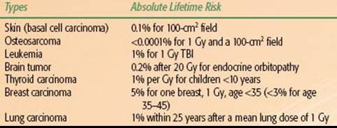
The risk of the induction of secondary tumors was overestimated in the past.13 Trott and Kamprad14 used the epidemiologic data from long-term follow-up studies on patients treated with radiotherapy for benign diseases to estimate the risk of cancer induction. Taking all known modifying and organ-specific factors into account, including doses of radiation and volume irradiated, the estimated absolute lifetime risk for sarcoma induction was <0.0001% for 1 Gy and a 100-cm2 field. Table 91.1 lists the absolute lifetime risk for other malignancies.
Jansen et al.15 applied the effective dose concept and estimated the carcinogenic risk in patients after radiotherapy of benign diseases (heterotopic ossification, omarthritis, gonarthrosis, heel spurs, and hidradenitis suppurativa). Special risk modifying factors, including age at exposure and gender, were taken into account. For an average-aged population, the estimated number of radiation-induced fatal tumors was between 0.5 and 40 persons per 1,000 patients treated. The range of effective doses was also found to be large (5 to 400 mSv). In addition to age and gender, the individual risk also depends on individual inherent sensitivity, anatomic site, type of disease, and treatment technique, such as dose and fractionation.
TABLE 91.1 THE ESTIMATED ABSOLUTE LIFETIME RISK FOR MALIGNANCIES AFTER RADIATION THERAPY FOR BENIGN DISEASES
INDICATION FOR RADIOTHERAPY
The majority of benign diseases can be classified as inflammatory, degenerative, hyperproliferative, or functional. Therefore, therapeutic approaches vary widely and are regionally customized, in part because of geographic traditions and differences in clinical training. Radiation treatment of benign diseases is less commonly used in the United States than in other parts of the world where variation in indications and treatment schedules are institutionally based.16Within Germany, a pattern of care study revealed significant geographic and institutional differences.17 Although most radiation treatments for benign disease are delivered in the low-dose range (<10 to 15 Gy), the prescribed dose varied widely and inconsistently within geographic regions and among institutions.
Degenerative processes in tendons, ligaments, and joints can cause pain by chronic inflammation and trigger secondary functional impairment of the involved musculoskeletal system. Although radiation does not halt the degenerative process, it may reduce the inflammation and provide partial or complete pain relief. This clinical effect is well established in reports of osteoarthritis, synovitis, and bursitis, where low-dose radiation therapy has improved the function of affected joints.17,18
Benign diseases may have a significant effect on self-image and self-esteem because of cosmetic appearance (e.g., facial keloids, juvenile angiofibroma) or have a lasting impact on quality of life because of chronic pain or other secondary symptoms (e.g., heterotopic bone, macular degeneration). When benign diseases become locally invasive with aggressive growth, therapeutic intervention can prevent or limit functional loss of organs. In rare cases of large hemangioma with associated thrombocytopenia and consumption coagulopathy (Kasabach-Merritt syndrome), potentially fatal complications can occur, and timely therapeutic intervention can be life-saving.19
Although there is a lack of international consensus, the German Working Group on Radiotherapy of Benign Diseases published their consensus guidelines for radiation therapy of nonmalignant diseases. The guidelines were to serve as a starting point for quality assessment, prospective clinical trials, and outcomes research.18 In brief, treatment is indicated when benign diseases are symptomatic or potentially symptomatic. When other methods are unavailable or have failed, radiation therapy should be considered. As medical professionals, we remain mindful of therapeutic gain and potential treatment side effects and complications. A thorough risk–benefit analysis is always pertinent. Organ-specific acute and chronic toxicities, including potential effects on fertility and induction of secondary tumors in the future, must be explained to and discussed with patients, especially those who are young and have a long life-expectancy. Informed consent, which is required for all medical interventions, is certainly required for treatment of benign diseases and should be obtained prior to the delivery of radiation therapy.
This chapter covers some of the more common benign conditions that are still encountered in the practice of radiation oncology. Details on the therapeutic approaches and data on radiation dose regimens for different benign diseases are summarized in the individual corresponding sections.
BENIGN NEOPLASMS OF THE BRAIN, HEAD, AND NECK
Nonmalignant tumors of the central nervous system (CNS) and neck can lead to severe, life-threatening symptoms due to pressure and mass effect on critical structures from tumor growth. However, depending on tumor growth rate and location, the surrounding tissue may adapt, leading to a delay in the clinical diagnosis.
Meningioma
Background and Clinical Aspects
Meningiomas are the most common benign tumors of the CNS. The incidence peaks in the seventh decade of life with a 2 to 1 female-to-male predominance. The majority (>90%) of meningiomas are benign and classified by the World Health Organization (WHO) as grade I tumors.20 WHO grade II meningiomas (atypical, clear cell, or chordoid) have a higher tendency for local recurrence, and WHO grade III malignant meningiomas (anaplastic, rhabdoid, papillary) are exceedingly rare.
The most common presenting symptom is headache, but patients may present with other localizing symptoms depending on the tumor location. The radiographic diagnosis of meningioma is often made on computed tomography (CT) or magnetic resonance imaging (MRI) based on the appearance of a homogeneously and intensely enhancing extra-axial mass with or without the presence of a dural tail.
Surgical Management
Surgical resection is the treatment of choice for the majority of patients, as this will relieve symptoms and also provide a pathologic diagnosis. The primary goal of surgery is to remove as much tumor burden as possible while minimizing the risk of neurologic deficits (maximal safe resection). Gross total resection (GTR) is generally attempted for patients with tumors in locations such as the convexity and olfactory groove.21,22 After GTR, the relapse rate is as low as 10%, but this depends on the Simpson classification, which grades tumors according to extent of resection and degree of dural involvement (Table 91.2).23 Local recurrence rates are as high as 40% for patients with incomplete resection,23 although these rates can be substantially reduced with the use of adjuvant radiotherapy.
Meningiomas tend to be highly vascularized tumors. In select patients, preoperative embolization is used to decrease blood loss and improve the extent of resection.24,25
Active Surveillance
Asymptomatic patients with small meningiomas may be observed clinically. At the time of tumor growth or the development of symptoms, patients can be treated with surgery or radiation therapy. The safety and reasoning for this approach was established in a large retrospective series from Japan that demonstrated that the majority of patients do not require intervention in the short term.26
TABLE 91.2 SIMPSON GRADING SYSTEM FOR POSTOPERATIVE MENINGIOMAS WITH ASSOCIATED RATES OF RECURRENCE

Systemic Therapy
Interest in the use of medical therapy to treat meningiomas stems from the observation that up to 67% of meningiomas express the progesterone receptor or androgen receptor, and approximately 10% express the estrogen receptor.27However, response rates to antihormonal agents are low. Overall, studies that have investigated the role of chemotherapy, such as hydroxyurea, in the management of recurrent disease have demonstrated little efficacy.27
Radiotherapy
Primary radiotherapy (RT) is indicated for tumors in locations in which complete resection is not feasible (i.e., optic nerve, cavernous sinus, major venous sinus) or for patients who are poor surgical candidates. Adjuvant RT is indicated for patients with subtotal resection (STR), recurrent disease, or for WHO grade II or III tumors. RT techniques include conventionally fractionated three-dimensional conformal radiotherapy (3D-CRT), conventionally fractionated intensity-modulated radiation therapy (IMRT), frame-based or linear accelerator–based fractionated stereotactic radiotherapy (FSRT), stereotactic radiosurgery (SRS), or protons and heavy ions.
The MRI sequences that best delineate the gross tumor volume (GTV) should be coregistered with the treatment-planning CT scan for optimal treatment planning and delivery. Particularly for patients receiving FSRT or SRS, it is important that a neuroradiologist and neurosurgeon be involved in assisting with GTV delineation, as enhancement from residual tumor versus postoperative change is often difficult to ascertain.
For 3D-CRT or IMRT treatments, the clinical target volume (CTV) is constructed by adding a 1- or 2-cm symmetric margin around the GTV, respecting normal tissue boundaries. An additional 3 to 5 mm is added for the final planning target volume (PTV). These margins may be modified based on institutional policy and other considerations, such as the availability of daily image guidance (i.e., kilovolt imaging or cone-beam CT).
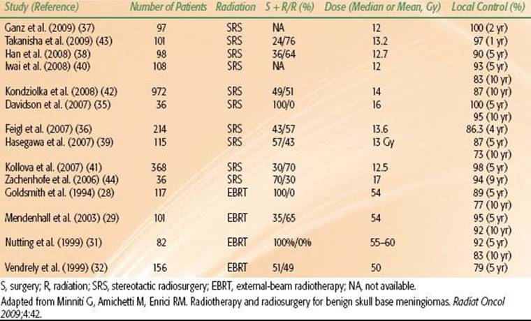
For benign meningiomas, the typical dose prescription to the PTV is 50 to 54 Gy given in 1.8- to 2-Gy daily fractions. Retrospective data suggest that local control is inferior for patients treated with doses of <52 Gy.28 For patients with more aggressive histology (WHO grade II or III tumors), the GTV is expanded by at least 2 cm, with a higher dose prescription in the range of 59.4 to 63 Gy. Several modern series of radiotherapy show 5-year local control rates ranging from 89% to 98%, with three-dimensional conformal therapy demonstrating local control rates >95% (Table 91.2).28–30,31–32
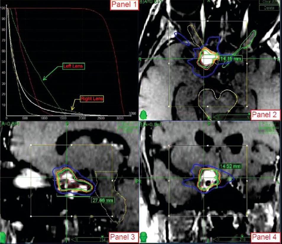
Because meningiomas are frequently noninvasive and well-circumscribed tumors, SRS and FSRT are increasingly being used in their treatment. The decision to fractionate depends largely on tumor size and proximity to critical structures, such as the optic apparatus or brainstem. Typical dose prescriptions for frame-based SRS range from 12 to 16 Gy prescribed to the 50% isodose line (IDL) and 14 to 18 Gy prescribed to the 80% IDL for a frameless robotic radiosurgery platform. In patients with tumors that require fractionated treatment, dose prescriptions vary and are dependent on the individual case. For example, at Stanford University, primary or residual meningiomas of the convexity and skull base are treated with 15 to 18 Gy in 1 or 2 fractions. Additionally, a select group of perioptic tumors, including meningiomas, have been treated with a prescription of 24 to 30 Gy in 3 to 5 fractions (to the 80% IDL) with high rates of tumor control and visual preservation (Fig. 91.1).33 Recent nonrandomized, prospective evidence indicates that FSRT should be the treatment of choice for optic nerve sheath meningiomas due to the high rate of preservation of visual acuity.34
Reported results with SRS are excellent, with 5-year local control rates as high as 98% to 100% (Table 91.3).35–41,42,43,44 DiBiase et al.45 demonstrated that male gender, conformality index <1.4, and size >10 mL predict for worse outcome after SRS. That article also showed improved disease-free survival in patients in which the dural tail was covered as part of the target volume. The benefit of including the dural tail has to be weighed against the risk of toxicity from increasing the target volume for each individual case.
Due to their physical properties, protons and heavy ions (i.e., carbon) are attractive choices for the treatment of meningiomas, particularly for those located near critical structures. Several studies have shown excellent local control rates with the combination of protons and photons or protons alone.46–49 In the study by Weber et al.,48 patients were treated to a median dose of 56 cobalt gray equivalents (GyE) given in 1.8 to 2.0 GyE per day.
FIGURE 91.1. Radiosurgery treatment plan of a patient with a right optic nerve sheath meningioma treated to a dose of 24 Gy in 3 fractions. The lesion is intensely enhancing on the postcontrast stereotactic magnetic resonance imaging sequences. Panel 1 demonstrates the dose–volume histogram for the patient. The maximum dose to the ipsilateral optic nerve was 22.3 Gy. Panels 2–4 demonstrate the isodose curves for the treatment in the axial, sagittal, and coronal planes, respectively. The 100% (24 Gy) isodose line is green, the 88% (21 Gy) isodose line is orange, and the 50% (12 Gy) isodose line is blue.
Pituitary Adenoma
Background and Clinical Aspects
Pituitary adenomas comprise 10% to 15% of all intracranial tumors. Approximately 75% of these tumors are functional (secretory), thereby producing increased amounts of hormones. Prolactinomas and growth-hormone (GH)–secreting adenomas are the most frequently encountered. Functional adenomas are more common in women, while nonfunctioning and GH-secreting adenomas are more common in men.
Adenomas are often classified by size, with a picoadenoma <0.3 cm, microadenoma <1 cm, and macroadenoma >1cm. Macroadenomas may exert mass effect upon the optic chiasm, leading to the classic sign of bitemporal hemianopsia. Headaches are seen in approximately 20% of patients. If the adenoma extends to the cavernous sinus, cranial nerve deficits may be present. Involvement of the hypothalamus by the adenoma results in hypopituitarism.
Patients with functional adenomas present with signs and symptoms that correspond to the excess hormone: galactorrhea, amenorrhea, diminished libido, and infertility in patients with prolactinomas; acromegaly or gigantism in patients with GH-secreting adenomas; Cushing disease in adrenocorticotropic hormone (ACTH)–secreting adenomas; hyperthyroidism in patients with thyroid-stimulating hormone (TSH)–secreting adenomas. In patients who have had bilateral adrenalectomy, up to 40% will develop Nelson syndrome, which is characterized by an ACTH-secreting adenoma and increased skin pigmentation secondary to increased release of α-melanocyte–stimulating hormone.
In addition to history and detailed physical examination (H&P), workup of a pituitary tumor includes laboratory analysis of pituitary hormone levels, contrast enhanced MRI with thin slices through the pituitary (Fig. 91.2A,B), and tissue diagnosis to rule out other causes of pituitary masses, including craniopharyngioma, meningioma, suprasellar germ cell tumor, metastatic disease, or a benign lesion (i.e., cyst).
TABLE 91.3 CLINICAL OUTCOMES OF STEREOTACTIC RADIOSURGERY OR EXTERNAL-BEAM RADIOTHERAPY (WITH OR WITHOUT SURGERY) FOR MENINGIOMAS IN MODERN SERIES
Surgical Management
Surgery is generally the treatment of choice for pituitary adenomas. Surgery provides immediate relief of compressive symptoms and helps to decrease hormone secretion. The most common surgical technique is through a transsphenoidal approach. In some cases, a more aggressive surgery (i.e., frontal craniotomy) may be indicated for patients with extensive intracranial and skull-based involvement. Overall, local control rates range from 50% to 80% after surgery alone for both functioning and nonfunctioning adenomas.50 In patients who continue to have abnormally elevated hormones after surgical resection, adjuvant treatment with pharmacotherapy or radiation therapy is pursued.
Pharmacotherapy
Pharmacotherapy, such as bromocriptine and cabergoline for prolactinomas, octreotide for GH adenomas and TSH adenomas, and ketoconazole for ACTH adenomas, is often used as an adjunct to surgery for patients with functioning adenomas. With the exception of prolactinomas, the use of these drugs as monotherapy is generally not curative. Prolactinomas can often be managed with pharmacotherapy alone, but a high proportion of patients are unable to tolerate bromocriptine for long periods of time due to nausea, headache, and fatigue.
Radiotherapy
Except for medically inoperable patients in which RT is used in the primary setting, the role of RT is generally in the adjuvant setting with the following indications: recurrent tumor after surgery; persistence of hormone elevation after surgery; residual disease after STR or debulking procedure. Tumor growth control is excellent, particularly for patients with nonfunctioning adenomas.51,52–53 Endocrine control after treatment of functioning adenomas, as demonstrated by normalization of pituitary hormone levels, takes years to develop. At a median of 2 years after RT, growth hormone levels stabilize quickest; normalization is slowest for TSH-secreting adenomas.54 Pharmacologic therapy should be discontinued 1 to 2 months prior to the initiation of RT based on evidence demonstrating lower RT sensitivity with concurrent medical treatment.55
RT techniques include 3D-CRT, IMRT, single-fraction SRS, and FSRT. Delineation of the GTV (or preoperative GTV in the case of GTR) should be performed by coregistration of the postoperative MRI to the treatment planning CT scan.
For 3D-CRT and IMRT, the CTV is constructed by adding 1 to 1.5 cm to the GTV; an additional 3 to 5 mm is added to the CTV to create the PTV. These margins may be modified based on institutional policy and other considerations, such as the availability of daily image guidance. Nonfunctional adenomas are typically prescribed a dose of 45 to 50.4 Gy given in 1.8- to 2.0-Gy daily fractions (Fig. 91.2C–E). Higher doses in the range of 50.4 to 54 Gy are recommended for secretory adenomas.
SRS remains an attractive option for the treatment of pituitary adenomas. General principles apply in that FSRT is used over SRS for large lesions (>3 cm) or lesions near critical structures (<1 or 2 mm from the chiasm). Similar to 3D-CRT or IMRT, higher doses are needed for functional adenomas compared with nonfunctional adenomas. Numerous retrospective studies have demonstrated excellent local control rates of 92% to 100% for nonfunctional adenomas, using doses of 14 to 25 Gy (at the edge of the tumor) in a single fraction.56 Commonly used prescriptions are 16 to 20 Gy in a single fraction for nonfunctional adenomas and 20 to 25 Gy in a single fraction for functional adenomas using a frameless robotic radiosurgery platform.
Craniopharyngioma
Background and Clinical Aspects
Craniopharyngiomas comprise 6% to 10% of pediatric CNS tumors, or approximately 300 to 350 cases per year in the United States. The median age of diagnosis is 5 to 10 years, with a second peak in patients >40 years old.
These benign tumors are epithelial, arising from remnants of Rathke’s pouch (hypophyseal-pharyngeal duct), and are most commonly located in the suprasellar region, although they may be found in the sella proper. Craniopharyngiomas generally abut the hypothalamus and third ventricle. Histologically, they are divided into the adamantinomatous and squamous subtypes. The adamantinomatous subtype is characterized by a solid and cystic pattern with the well-known description of “machine oil-like” cystic fluid.
FIGURE 91.2. A recurrent nonfunctioning pituitary adenoma 7 years after surgical resection in the axial (A) and coronal (B) planes. The yellow arrows denote invasion into the left cavernous sinus. Rapid arc intensity-modulated radiotherapy treatment plan for the same patient in the axial (C), coronal (D), and sagittal (E) planes. The planning target volume (purple shaded area) was prescribed at 50.4 Gy in 28 fractions.
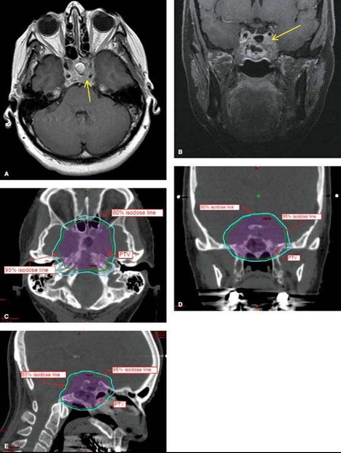
FIGURE 91.3. Axial, coronal, and sagittal magnetic resonance imaging of a patient with multicystic (yellow arrows) craniopharyngioma prior to treatment.

Presenting signs and symptoms include headache, nausea and vomiting, bitemporal hemianopsia, and endocrine dysfunction (diabetes insipidus, dwarfism, fat tissue disturbance, adrenal cortical insufficiency). The most common hormone deficiency is lack of GH. The workup is similar to that of pituitary adenoma and includes H&P, pituitary hormone levels, and brain MRI with thin slices through the sella (Fig. 91.3).
Surgery
The primary goal of surgery is complete resection. However, GTR may be associated with high rates of neurologic sequelae, including visual impairment and panhypopituitarism. In order to minimize morbidity, most patients are treated with maximal safe resection followed by adjuvant RT. In some cases, intralesional bleomycin may be directly injected into the cyst to decrease the rate of cyst recurrence.
Radiotherapy
Radiation therapy is often used in the adjuvant setting. In select patients (i.e., <3 years old), observation following STR may be an option as local control rates are similar with RT at the time of relapse (“salvage” RT) compared with adjuvant RT with no compromise in overall survival.57
RT techniques include 3D-CRT, IMRT, FSRT, proton therapy, and intralesional RT with β-emitting isotopes (yttrium-90, phosphorous-32). The GTV is the postoperative residual tumor volume, including the cyst wall, if present. The postoperative MRI should be fused with the treatment planning CT scan for optimal target delineation. A margin of 1 to 1.5 cm is added to the GTV to create the PTV. Dose prescriptions for 3D-CRT and IMRT are typically 54 Gy given in 1.8-Gy daily fractions.
Fractionated proton radiotherapy has demonstrated excellent results. The Loma Linda series treated 15 patients to a total dose of 50.4 to 59.4 GyE given in 1.8-GyE daily fractions.58 Local control was achieved in 14 of 15 patients, with few long-term complications. In a series from the Massachusetts General Hospital (MGH), no failures were seen in 24 patients who received fractionated proton radiotherapy to a total dose of 52.2 to 54 GyE in 1.8 GyE per fraction.59
It has been well established that cysts may regrow during the several weeks of fractionated treatment. The MGH proton study recommends that reimaging (CT, or MRI if the cyst is not well visualized on CT) be performed within 2 weeks of the treatment planning scan and every 2 weeks thereafter; for large cysts or those that demonstrate growth during RT, weekly reimaging is recommended.59 The emergence of image-guided radiotherapy techniques now allows for the convenient monitoring of cyst regrowth with cone-beam CT scans while patients are on the treatment table.
SRS and FSRT have been used with success in the treatment of craniopharyngioma. In one series from Stanford University using a frameless robotic platform,60 16 patients were treated postoperatively with doses of 18 to 38 Gy given over 3 to 10 fractions prescribed to mean IDL of 75%. Local control was 91% in this cohort of patients with no visual or neuroendocrine complications. Similar results have been demonstrated with use of a frame-based platform.61–64
Cystic craniopharyngiomas may also be managed by the use of intralesional radioactive isotope injection using a β-emitter. Typical prescriptions range from 200 to 250 Gy prescribed to the cyst wall. Optimal results are seen in patients whose tumors have one cyst and lack a large solid component.65,66
Acoustic Neuroma (Vestibular Schwannoma)
Background and Clinical Aspects
Acoustic neuromas (AN) represent 5% to 8% of primary CNS brain tumors. They are derived from Schwann cells of the neurilemma of the vestibulocochlear nerve (CN VIII). The vast majority of cases (90%) are unilateral and sporadic. Bilateral AN occur in about 10% of cases and are associated with the autosomal dominant disorder neurofibromatosis type II.
Symptoms include sensorineural hearing loss, tinnitus, and vertigo. Hearing loss is correlated more with tumor location (intracanalicular) rather than tumor size. In a minority of patients (5%), facial nerve symptoms may be present. As the AN grows, it may affect the trigeminal nerve (CN V) and brainstem.
During the initial workup, physical examination should include the Rinne test (air conduction greater than bone conduction on the affected side) and Weber test (vibratory sound louder on the unaffected side) to test for sensorineural hearing loss, as well as detailed examination of CN V and CN VII. All patients should undergo audiometry when the diagnosis of AN is suspected; this will often reveal asymmetric hearing loss more prominent at high frequencies as well as impairments in speech discrimination score. Imaging should include a contrast-enhanced MRI with thin slices through the internal auditory canal. The entire neuraxis should be imaged in patients with neurofibromatosis type II.
Surgery
For many years, the standard treatment for patients with AN was microsurgical resection, either via a translabyrinthine approach or middle cranial fossa approach. Surgery remains the preferred treatment for patients with large, symptomatic lesions. Hearing preservation is approximately 50% to 60% after surgery, and facial nerve preservation ranges from 80% to 90%.67,68
Active Surveillance
Observation is appropriate management for asymptomatic patients with small tumors. Serial MRI and audiometry (at least once per year) should be performed in this patient cohort for surveillance. Treatment is initiated when the lesion demonstrates rapid and significant growth or when the patient becomes symptomatic.
Radiotherapy
RT in the form of SRS or FSRT is an option for the primary treatment of AN, often with higher rates of hearing preservation and facial nerve presentation compared with surgery. Proton therapy has also been used to treat AN.
SRS doses using frame-based platforms are generally 12 to 13 Gy prescribed to the 50% IDL. Although earlier studies demonstrated lower hearing preservation rates with higher SRS doses,69 current results are significantly improved. Flickinger et al.70 demonstrated a local control rate of 98.6%, hearing preservation rate of 70.3%, trigeminal neuropathy rate of 4.4%, and no incidence of facial nerve dysfunction. Using a frameless robotic radiosurgery platform to treat 383 AN to a dose of 18 to 21 Gy given in 3 fractions, Stanford University researchers demonstrated a 98% local control, 76% hearing preservation, 2% incidence of trigeminal nerve dysfunction (transient in four of the eight affected patients), and no facial nerve dysfunction.71
Common dose prescriptions employing FSRT include 25 Gy in 5 fractions, 30 Gy in 10 fractions, and 50 to 55 Gy in 25 to 30 fractions. A nonrandomized prospective trial compared SRS (10 to 12.5 Gy) to FSRT (20 to 25 Gy in 5 fractions). This study demonstrated comparable rates of local control, hearing preservation, and CN V and CN VII preservation between the two groups.72
Proton beam SRS has also been used to treat AN, although with low rates of hearing preservation compared with other RT techniques. In a series from MGH, 88 patients were treated to a median dose of 12 GyE given in a single fraction prescribed to a median IDL of 70%. Tumor control rates at 2 and 5 years were 95.3% and 93.6%, respectively. Facial and trigeminal nerve preservation rates were 90%. Only 33% of patients retained serviceable hearing.73 In a study of FSRT using a proton beam, local control was excellent but the hearing preservation rate was poor at 42%.74
Chordoma
Background and Clinical Aspects
Chordomas are rare, slowly growing midline tumors originating from the embryonal notochord rests in the skull base (35%), vertebral column (15%), or sacral regions (50%). The most common sites of skull-based tumors include the clivus, dorsum sella, and nasopharynx.
Patients typically present with signs and symptoms attributable to the primary site of the tumor. Workup includes MRI with contrast enhancement. CT may complement MRI to assess for local bony destruction. A biopsy is necessary primarily to distinguish chordoma from chondrosarcoma, which has a better prognosis, and other malignancies. In children, biopsy is essential to distinguish chordoma from rhabdomyosarcoma, which can frequently present in the same location.
Surgery
Complete surgical resection is the mainstay of treatment. However, due to location, GTR is often not possible. Relapse rates are as high as 50% even after surgical resection with negative margins.75 Poor prognostic factors include large tumors, recurrent tumors, older age, and presence of necrosis on biopsy.76–77,78
Systemic Therapy
Approximately 25% of chordomas metastasize to the lungs, liver, or bone. In these patients, or in patients with recurrent disease after surgical resection and radiation therapy, molecularly targeted agents may be considered. Several small studies have demonstrated some benefit to the use of imatinib or the combination of imatinib and sirolimus in this situation.79–81
Radiotherapy
Adjuvant radiation therapy is indicated to reduce recurrence rates for skull-based chordomas. Retrospective data suggest that salvage RT is inferior to adjuvant RT with 5- and 10-year overall survival rates of 50% and 0%, respectively, for those treated with salvage RT compared with 80% and 65%, respectively, for patients treated with adjuvant RT.82 RT techniques for treatment of skull-based chordomas include conventionally fractionated 3D-CRT or IMRT, fractionated charged particle therapy (protons, carbon ions), and FSRT.
Determination of the GTV should be made by coregistration of the preoperative and/or postoperative MRI to the treatment planning CT scan. Particularly for pediatric cases in which IMRT will be used with tight margins, a neuroradiologist should be available at the time of treatment planning to assist in creating the GTV. Margins for CTV should be 1 to 2 cm with an additional 3 to 5 mm for the PTV. Dose prescriptions to the PTV for patients receiving photon-based treatment should be at least 60 Gy given in 1.8- to 2.0-Gy daily fractions.
Proton-based therapy can achieve higher doses with good results. In a series of 195 chordomas treated at the MGH, 5-year progression-free survival was 70% with doses of 63 to 79.2 GyE given in 1.8- to 2.0-GyE daily fractions.83Research at Loma Linda examined 33 cases of chordomas treated with a median of 70 GyE and found a 76% 5-year local control and 79% 5-year overall survival.78
In a report of 96 chordomas treated using carbon ion therapy at the University of Heidelberg, with median total dose of 60 GyE (range 60 to 70 GyE) delivered in 20 fractions within 3 weeks, good local control rates of 81% at 3 years and 70% at 5 years were observed.84 There was a trend to improved local control in patients who received >60 GyE.
FSRT and SRS are less well established than charged-particle therapy. The North American Gamma Knife Consortium published the experience of 71 patients who underwent Gamma Knife (Elekta Corp, Stockholm) SRS as primary, adjuvant, or salvage therapy for skull-base chordomas.85 The median dose to the tumor margin was 15 Gy (range 9 to 25 Gy). Five-year local control was 66% for the entire cohort (69% for the no prior RT group and 62% for the prior RT group). Debus et al.86 reported on 37 patients with chordomas treated with FSRT to a median dose of 66.6 Gy given in 1.8-Gy daily fractions; the target volume was encompassed by 90% of the dose. Local control was 82% at 2 years and 50% at 5 years.
Glomus Tumor, Chemodectoma, and Paraganglioma
Background and Clinical Aspects
Glomus tumors are rare, benign tumors that occur at and along the carotid artery near the bifurcation (carotid body tumor), the jugular bulb (glomus jugulare), or the middle ear (globus tympanicum). The peak age is in the fifth decade of life. Bilateral or multiple tumors occur in 10% to 20% of affected patients.
Symptoms include headache, cranial nerve dysfunction, dysphagia, pulsatile tinnitus, vertigo, and large, pulsating masses in the neck. In rare cases, patients present with episodic hypertension, which may be related to the secretion of vasoactive substances by the tumor. In this situation, urine and serum metanephrines should be measured. The clinical presentation coupled with imaging (high-resolution CT, MRI, or angiography) often establish the diagnosis. In patients in which multiple tumors are suspected, imaging with metaiodobenzylguanidine may be useful.
Surgery
In the carotid region, primary tumor resection after previous embolization is the therapy of choice. At the skull base or tympanum, neurosurgical intervention is often deferred due to the high rates of complications, including stroke and cranial nerve injury.87
Radiotherapy
RT is indicated for patients with tumors in unsuitable locations (i.e., skull base), as adjuvant therapy after STR, or as salvage therapy at the time of relapse after surgery. RT techniques include conventionally fractionated 3D-CRT or IMRT, SRS, and FSRT.
The diagnostic MRI should be coregistered with the treatment planning CT scan. The GTV is delineated and 1 to 1.5 cm is added for clinical and setup margin. With conventional techniques, doses are often 45 to 55 Gy given in 1.8- to 2.0-Gy daily fractions with local control rates near or >90% in several series.88–91
Results with SRS have been comparable to that of conventionally fractionated RT. Using a frame-based platform, reported tumor margin doses range from 12.5 to 20 Gy prescribed to the 50% IDL92,93–94 and 15 to 25 Gy for linac-based SRS.87 Local control rates in these series range from 90% to 100%. A recent meta-analysis of SRS published by the Johns Hopkins Hospital reviewed 335 patients treated with SRS across 19 different studies.95 In the eight studies with median follow-up >3 years, clinical control was 95% and tumor control was 96%. The control rates were equal among patients treated with linac-based platforms and frame-based platforms. Complications were rare and often transient in each of the studies examined. On the basis of these findings, the authors advocate for the use of SRS as primary treatment of glomus jugulare tumors.95
Juvenile Nasopharyngeal Angiofibroma
Background and Clinical Aspects
Juvenile nasopharyngeal angiofibroma (JNA) is a rare, benign, vascularized tumor in the head and neck, affecting mostly male adolescents. JNAs develop from the sphenoethmoidal suture and spread from the nasal cavity to the sphenopalatine foramen and pterygopalatine fossa. Other routes of local spread include the paranasal sinuses, infratemporal fossa, orbital space, and middle cranial fossa.
Symptoms initially include recurrent epistaxis and impaired nose breathing. As local extension occurs, patients may develop facial swelling, orbital symptoms (blindness), cranial nerve deficits, and headaches from intracranial extension.
Diagnosis is often made with the clinical presentation and CT or MRI-based imaging. Biopsy may cause massive bleeding. Numerous staging systems are used to categorize the tumors based on extent of local extension, including the Chandler et al.,96 Fisch,97 and Radkowski et al.98 systems (Table 91.4).
Surgery
Surgery combined with embolization is the preferred treatment. Through surgery, most JNAs without intracranial extension (i.e., Chandler stage I to III) have local control rates of near 100%. In patients with intracranial extension, complete resection is often not possible.
TABLE 91.4 CHANDLER STAGING SYSTEM FOR JUVENILE NASOPHARYNGEAL ANGIOFIBROMAA
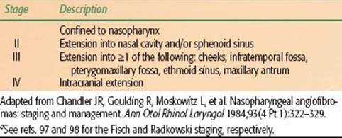
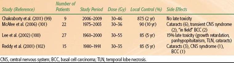
TABLE 91.5 CLINICAL RESULTS OF RADIOTHERAPY IN JUVENILE NASOPHARYNGEAL ANGIOFIBROMA
Radiotherapy
Tumors with intracranial extension or tumors in patients who are medically inoperable are generally treated with RT as the primary modality. Indications for postoperative RT include relapse after surgery. Fractionated IMRT is currently the RT technique of choice to limit collateral radiation to critical structures near the target volume.
The PTV is generally treated to 30 to 50 Gy given in fraction sizes of 2 to 3 Gy per day. In modern series, local control rates range from 85% to 100% (Table 91.5).99,100,101,102 After RT, JNA remission is slow, and late recurrences may occur.
Langerhans Cell Histiocytosis (Histiocytosis-X)
Background and Clinical Aspects
Langerhans cell histiocytosis (LCH) is a rare disorder that affects approximately 300 individuals with a higher incidence in children (3 to 5 per million) than adults (1 to 2 per million). Age is an important prognostic factor, with children having better outcomes than adults. In the past, it was felt that children younger than 2 years old were felt to have a poor prognosis, but recent data from the LCH-II study now refute that point.103
The disease is due to an accumulation or proliferation of cells that phenotypically resemble the Langerhans skin cell and can cause tissue damage by production of cytokines and infiltration. The actual Langerhans cell is a myeloid dendritic cell that expresses the same antigens (CD1a and CD207) as the Langerhans skin cell.104 On electron microscopy, Birbeck granules are the classic finding.
LCH can affect a variety of organ systems. Patients are typically stratified into groups based on the extent of disease: single-system disease at a single site, single-system disease involving multiple sites, or multisystem disease. The clinical presentation is dependent on the sites of disease. Involvement of the skeletal system is the most common site for children and may manifests as pain, palpable mass, motion deficit, or chronic otitis in the case of mastoid or middle ear involvement. Bony lesions from LCH are predominantly lytic in appearance, and the skull is the most frequently involved bony structure.
Cutaneous involvement affects primarily the skin of the scalp and groin and resembles a seborrheic dermatitis. Patients with cranial involvement (posterior pituitary or hypothalamus) may present with diabetes insipidus (DI). The disease may also involve the cervical lymph nodes. Pulmonary involvement is more typically seen in adults. Hepatomegaly, splenomegaly, bone marrow infiltration, and involvement of the gastrointestinal tract are further potential sites of disease.
Evaluation of the patient with suspected LCH should include complete H&P. Routine laboratory work should include white cell blood count with differential. Patients who have symptoms of DI should also undergo a water restriction test. A skeletal survey with or without bone scan should be performed to assess for potential lytic lesions. Further imaging with CT of the head should be performed in patients with skull, orbital, or mastoid involvement. MRI of the head is indicated for patients with DI and those suspected of having brain parenchymal disease involvement. CT of the chest is performed to evaluate patients with pulmonary involvement. An MRI of the abdomen should be performed for patients with palpable hepatomegaly or splenomegaly.
Management Principles
Treatment of LCH depends on the site and extent of disease. Asymptomatic lesions may be observed. For patients with involvement of only the skeletal system, treatment options include curettage, excision, or intralesional steroid injection. Response rates with curettage or excision alone range from 70% to 90%.105 Single system multifocal bone disease may be effectively treated with corticosteroids or chemotherapy, such as vinblastine. For skin-only disease, topical nitrogen mustard and methotrexate are considered effective treatments.
In patients with multisystem disease with symptoms (fever, pain, failure to thrive) or organ dysfunction, treatment with systemic therapy is indicated. In LCH-II, all patients received initial treatment with prednisone and vinblastine and were randomized to intensification with or without etoposide.103,106 The patients treated with intensification demonstrated superior rapid response rates and decreased mortality rates compared with the standard arm.106 Exogenous antidiuretic hormone (ADH; vasopressin) is used to treat children with DI.
Radiotherapy
Due to the excellent response rates to nonradiotherapeutic measures, the role of RT in the treatment of LCH bony lesions has decreased. Indications for radiation therapy to bony sites include relapse after surgery, no signs of clinical healing after other interventions, pain relief, potential compromise of critical structures from an expansile lesion (i.e., cord compression), or if the bony site is not amenable to other local therapies. Collapsed vertebral lesions should not be irradiated unless they are painful. DI is another recognized potential indication for treatment with RT. When the decision is made to treat, 3D-CRT should be the technique of choice.
The target volume for patients with bony disease should encompass the abnormality seen on imaging with a small margin. For children, low doses on the order of 5 to 10 Gy given in 1.5- to 2.0-Gy daily fractions should be sufficient to control most bony lesions. Higher doses can be used in adults. In an early study by Smith et al.,107 92% of patients received total doses in the range of 4.5 to 10.0 Gy with an 87% local control rate. A study at the University of California–Los Angeles found local control rates of 88% with doses in the range of 6 to 15 Gy for previously untreated lesions and 8 to 15 Gy for recurrent lesions.108
The target volume for patients receiving treatment for DI should encompass the hypothalamus and pituitary gland. The recommended prescribed dose is 15 Gy in 1.5-Gy daily fractions. In a report from the Mayo Clinic, 36% of 28 evaluable patients responded to radiotherapy.109 The response rates were 60% (3 of 5 patients) in those treated with >15 Gy compared with 30% (7 of 23 patients) treated with doses of <15 Gy. Six patients had a complete response to therapy, five of whom received treatment within 14 days from diagnosis of DI.
Controversy still remains regarding the role of RT in the management of DI. In a retrospective series from MGH, 14 of 17 patients with DI received irradiation to the hypothalamic–pituitary axis.110 Only two of these patients had a complete response (cessation of ADH therapy), and no patients had a partial response. The others argue that treatment of DI from LCH is “no longer justified.”110
 VASCULAR DISORDERS
Vascular disorders are broadly categorized into vascular tumors, most commonly benign hemangiomas, and vascular malformations, including arteriovenous malformations (AVMs) and cavernous hemangiomas. Radiosurgery has emerged as an important and common treatment option for AVMs. Although radiation was used commonly in the past to treat hemangiomas in children, the recognition of late effects associated with radiotherapy, especially secondary malignancies, has rendered this practice less common.
Arteriovenous Malformations
Background and Clinical Aspects
Intracranial AVMs are congenital vessel abnormalities consisting of widened arteries connected to the normal capillary bed. The nidus of an AVM is made up of tangled arteries and veins that are connected by one or more fistulas. The overall prevalence is low, affecting approximately 18 in 100,000 individuals, with age at presentation typically between 20 and 40 years old.
Clinical concern comes from the high risk of bleeding, estimated to be 2% to 4% per year. Approximately 50% of patients present with hemorrhage and 50% present with nonfocal (headache, nausea) symptoms or incidentally found focal neurologic deficits. The risk of death per bleed is up to 10%, and approximately 30% have serious morbidity associated with each bleed.
Diagnostic imaging includes angiography, which is invasive but allows for full grading of the AVM according to the Spetzler-Martin scale. MRI, MR angiography, and CT angiography are noninvasive and complementary studies that may be used to visualize the AVM.
Surgery
The goal of any therapy for AVM is to completely obliterate the nidus. Partial obliteration of the nidus does not decrease the bleeding risk. Complete surgical excision provides immediate cure but carries a risk of intraoperative bleeding, ischemic cerebrovascular accident, infection, and death. Surgery is particularly indicated for AVMs in superficial, noneloquent regions of the brain. Endovascular therapy (embolization) is not curative but may be used to decrease the risk of intraoperative bleeding or to decrease the size of the nidus before planned radiotherapy.
TABLE 91.6 FLICKINGER’S PREDICTED RATES OF IN-FIELD ARTERIOVENOUS MALFORMATIONS OBLITERATION BASED ON THE MINIMUM DOSE WITHIN THE TARGET VOLUME
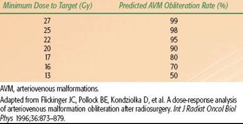
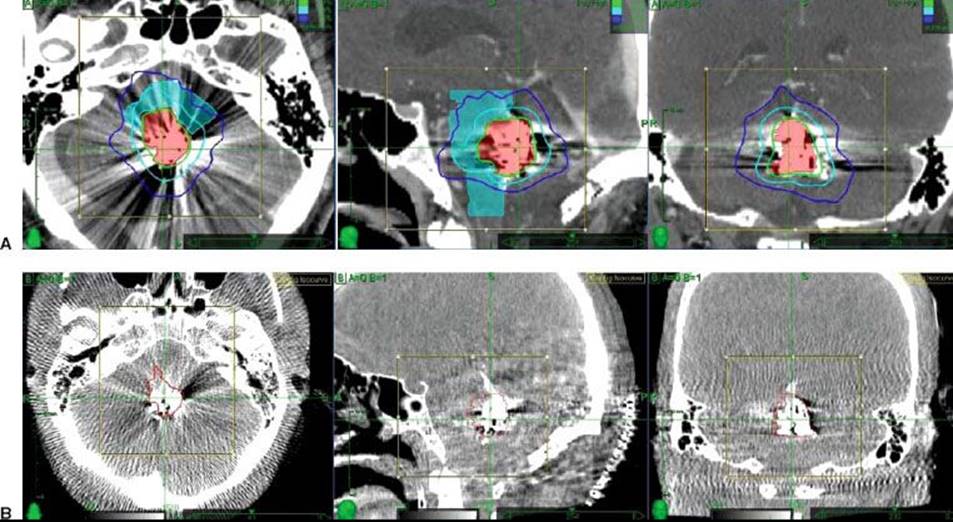
FIGURE 91.4. A: Stereotactic radiosurgery plan for an arteriovenous malformations (AVM) (red) in the dorsal pons treated with 22 Gy in 2 fractions. The prominent streak artifacts are present due to embolization 1 year prior to treatment. B: Computed tomography angiogram of the same patient used to assist in defining the AVM nidus (red contour).
Radiotherapy
SRS is the radiation modality of choice for the treatment of AVMs. SRS is indicated mostly for lesions in deep or eloquent regions of the brain and is particularly safe and successful for lesions that are <3 cm. Unlike surgery, the time to obliteration ranges from 1 to 4 years after SRS, so the patient remains at a continued bleeding risk. Even with time, Maruyama et al.111 demonstrated that the bleeding risk is not completely eliminated but reduced by approximately 88%.
Based on the Flickinger et al.112 dose–response data, typical prescriptions for treatment of AVM are 21 to 22 Gy prescribed to the 50% IDL for frame-based radiosurgery (Table 91.6). The prescription should be lowered for AVMs near the brainstem or larger lesions (>3 cm). For linac-based SRS, prescriptions generally range from 16 to 24 Gy in a single fraction to 20 to 22 Gy in 2 fractions for spinal AVMs (Fig. 91.4).113
Hemangioma and Kasabach-Merritt Syndrome
Background and Clinical Aspects
Hemangiomas are dynamic vascular tumors characterized by a proliferative phase followed by an involution phase. Approximately 20% of hemangiomas are present at birth, while the remaining 80% usually form within the first few weeks of life. Most hemangiomas present no problems to the patient and require no treatment. The spontaneous involution rate is approximately 10% per year.114 However, potential complications that may require treatment include obstruction of vision (eyelid hemangioma), ulceration and infection, cosmetic deformity from a facial hemangioma, and high-output cardiac failure.
Previously, it was felt that extremely large hemangiomas predisposed patients to Kasabach-Merritt syndrome (KMS). This syndrome consists of platelet trapping and destruction within the vascular tumor with a resultant consumptive coagulopathy (disseminated intravascular coagulation) that can be life-threatening. It is now recognized that this phenomenon is associated with a type of vascular tumor known as the kaposiform hemangioendothelioma, and not the classical infantile hemangioma.115
Systemic Therapy
Treatment options for hemangiomas include local and systemic pharmacotherapy, laser therapy, and surgery. Glucocorticoids have been the mainstay of systemic treatment for patients with hemangiomas. However, long-term use of steroids in children leads to many complications, including growth retardation, metabolic disorders, cushingoid facies, personality changes, and increased risk of infections. Recently, propranolol has been recognized as a potential therapeutic agent for hemangiomas. The first report of its use was published in the New England Journal of Medicine in 2008 when two patients with hemangiomas with resultant heart failure were treated with propranolol and were noted to have softening, change in color, and ultimately regression of the lesions.116 Both of these children were able to sustain a clinical response, even after being weaned off of steroids. Additional studies of propranolol have shown promising results, with most children exhibiting significant regression of their lesions and the ability to wean off of steroids without a rebound effect.117
For patients with KMS, vincristine and interferon-alfa have been used, particularly in situations in which a quick clinical response is needed or when the disease has become refractory to steroids and propranolol. Patients with KMS should also be managed with supportive measures, such as blood or platelet transfusions, as needed.
Patients with small lesions may be candidates for local therapy, such as intralesional or topical steroids. Topical timolol is also being investigated, given the promising results seen with propranolol.118 Select patients may be candidates for treatment with pulse-dye laser or excisional surgery.
Radiotherapy
Radiotherapy is indicated only in patients who have exhausted all other treatment options. When RT is used, responses are often quick and dramatic. With low-dose RT (<10 Gy), scarring should be minimal, but patients must be followed closely for secondary malignancies.
The CTV should include the clinically visible and palpable lesion with a margin. Imaging of the affected area with MRI is useful to delineate the depth and full extent of disease. Although there are no prospective trials to guide total dose and daily dose, most reports have used fractionation schedules of 1 to 3 Gy per day to a total dose of ≤10 Gy.19,119 When patients have not responded to low-dose RT, higher doses may be used to achieve a response.
FUNCTIONAL DISORDERS
SRS is widely used for the treatment of benign tumors. However, a significant number of patients benefit from the use of SRS to treat functional disorders such as trigeminal neuralgia, tremors, and epilepsy. Over the past several years, there have been increasing reports of the use of SRS for the treatment of refractory psychiatric disorders including obsessive-compulsive disorder and major depressive disorder.
Trigeminal Neuralgia (Tic Douloureux)
Background and Clinical Aspects
Trigeminal neuralgia (TN) is a common problem that affects approximately 15,000 patients each year in the United States. There is a slight female predominance (1.5 to 1). The disorder is currently classified into type I TN and type II TN, which is based on pain characteristics, as opposed to the traditional classification system, which divided patients into idiopathic TN versus secondary TN.120–121,122 Patients with type I TN describe pain as being predominantly (>50%) sharp, lancinating, and shock-like with pain-free intervals. Patients with type II TN predominantly experience burning, aching, or throbbing pain. This system also carries prognostic significance, with type I TN patients more likely to be pain free and have longer disease control than patients with type II TN after decompression.120
The classic clinical feature is recurrent episodes of sudden, brief, severe, stabbing, or lancinating pain in the area of the trigeminal nerve sensory distribution. It is most commonly unilateral, but some cases are bilateral. Common triggers for attacks include talking, chewing, brushing teeth, and cold air. The diagnosis is often suspected on the basis of the above clinical symptoms, but an MRI of the brain should be performed to rule out structural abnormalities that may be causing secondary TN.
Medical Therapy
TN is first treated with pharmacotherapy, with carbamazepine being the most common and extensively studied agent. Oxcarbazepine is an option for patients who are unable to tolerate carbamazepine. Numerous other agents have been used to treat carbamazepine-refractory patients including lamotrigine, gabapentin, pimozide, tizanidine, and topiramate.
Surgery
In patients who have medically refractory disease, microvascular decompression is the treatment of choice for immediate relief of symptoms. Other options include rhizotomy with either radiofrequency ablation, glycerol injection, or balloon compression.
Radiotherapy
SRS has emerged as a successful and minimally invasive procedure to treat classical TN. Treatment planning involves fusion of a contrast-enhanced MRI with thin cuts to the treatment planning CT scan. The target for SRS varies from the root entry zone of the trigeminal nerve as it enters the pons to the semilunar ganglion,123–129 Typical doses using a frame-based radiosurgery platform are 70 to 90 Gy, prescribed to the 50% IDL. This dose range is largely based on a trial that prospectively assigned patients to low-dose (60 to 65 Gy) or high-dose (70 to 90 Gy) SRS and demonstrated higher rates of pain relief in the high-dose arm (72% vs. 9% for patients treated with ≥70 Gy vs. <70 Gy), with a median time to pain relief of 1 month.125
The main concern with treating larger segments of the nerve with higher doses of radiation is the delayed onset of facial numbness. This is based on a prospective study by Flickinger et al.130 who randomized patients to 75 Gy targeted to a shorter (1 isocenter) or longer (2 isocenters) segment of the trigeminal nerve. The rates of pain relief were surprisingly identical between the two groups. There was a trend toward a higher incidence of numbness or paresthesias in the 2-isocenter patients, and overall, the nerve length irradiated was significantly correlated with the development of numbness and paresthesias. However, in a report by Adler et al.,131 46 patients received treatment with a frameless robotic radiosurgery platform to a 6-mm segment of the trigeminal nerve with a mean marginal prescription dose of 58.3 Gy and mean maximal dose of 73.5 Gy. In this cohort of patients, 85% experienced a complete response and, at a mean follow-up of 15 months, 96% reported excellent or good outcomes. Only 15% of patients experienced ipsilateral facial numbness.
Epilepsy
Background and Clinical Aspects
Epilepsy is a disorder characterized by recurring seizures. It affects 0.5% to 1% of the population, and its etiology is unknown in the vast majority of cases. The clinical presentation of patients with seizures is broad, depending on the type of seizure and the area of the brain involved. Seizures are generally classified into simple versus complex (loss of consciousness) and generalized versus partial (affecting only one focus in the brain).
Nonradiotherapeutic Treatment
Antiepileptic drugs are the treatment of choice for patients with epilepsy. Surgery is an option for patients who develop medically refractory disease, particularly for temporal lobe epilepsy.132
Radiotherapy
SRS may be an alternative to surgery in medically refractory epilepsy for patients who are not surgical candidates. In a multi-intuitional study, Regis et al.133 treated 20 patients with SRS for intractable mesial temporal lobe epilepsy. The mesial temporal lobe was treated to a dose of 24 to 25 Gy in a single fraction with Gamma Knife. At a follow-up of 2 years, 65% of the patients were seizure free. However, there was a 1-year lag between treatment and maximal effect, and there was a transient increase in seizures before the seizures started to diminish. In a study by Barbaro et al.,134 30 patients were randomized to low-dose (20 Gy) or high-dose (24 Gy) radiosurgery targeting the amygdala, hippocampus, and parahippocampal gyrus. At 3 years, the seizure-free rate was 67% among all patients with a trend toward a higher response rate (77% vs. 59%) and earlier responses in the high-dose compared with the low-dose group of patients. No serious toxicity was reported in the study.
In general, the use of SRS to treat epilepsy is not routine, and nonradiosurgical options should be employed first. Further study is needed to establish the long-term safety and efficacy of SRS in the treatment of epilepsy.
Parkinson Disease
Background and Clinical Aspects
Parkinson disease (PD) results from the loss of dopaminergic neurons in the substantia nigra. It is a debilitating and progressive neurodegenerative disorder that affects approximately 1 million people in the United States. The hallmark clinical symptoms include masked facies, resting tremor, slow movements, shuffling gait, and muscle rigidity. Some patients also develop dementia as part of the disorder.
Nonradiotherapeutic Treatment
Pharmacotherapy with dopamine agonists and other compounds is the treatment of choice for patients with PD. Once refractory to medical therapy, surgery (thalamotomy or pallidotomy) may be used to remove the overactive brain nuclei. Deep brain stimulation is another invasive procedure done under stereotactic guidance that can be used in medically refractory PD.
Radiotherapy
Patients who are poor candidates for surgery may receive SRS for the treatment of medically refractory PD. To relieve tremor, the target is the ventralis medialis nucleus with a dose in the range of 120 to 180 Gy. Young et al.135,136reported a long-term success rate of 80% to 90% in relieving symptoms from PD tremor with a very low rate of permanent complications.
Mixed results have been found with regard to the treatment of PD-related akinesia, dyskinesia, and rigidity. Treatment entails targeting the globus pallidus internus (pallidotomy) with doses in the range of 120 to 180 Gy. Rand et al.137 reported that four of eight patients received relief in rigidity with no serious complications. However, Friedman et al.138 achieved a response in only one of four patients, while causing dementia and psychosis in the only patient responder. Young et al.139 treated 29 patients with SRS pallidotomy with an 80% success rate at mean follow-up of 2 years and only one (3.4%) complication at 9 months (homonymous hemianopia).
Psychiatric Disorders
Background and Clinical Aspects
Psychiatric disorders such as obsessive-compulsive disorder (OCD), bipolar disorder, and major depressive disorder are debilitating illnesses. Radiosurgery has been used for the treatment of some of these psychiatric illnesses, with the majority of the experience in patients with OCD. OCD is characterized by intrusive thoughts (obsessions) that lead to repetitive behaviors (compulsions).
Nonradiotherapeutic Treatment
The combination of pharmacotherapy (i.e., selective serotonin reuptake inhibitors) and behavior therapy are the treatments of choice for patients with mood disorders and OCD. Surgical management and deep brain stimulation are used for extreme and severe cases.
Radiotherapy
Patient selection for the use of SRS to treat psychiatric disorders is complex. Friehs et al.140 recommend that such patients must be enrolled on an institutional protocol after carefully being evaluated by a multidisciplinary team. Similar strict criteria were used by Kondziolka et al.141
The treatment of OCD involves targeting the bilateral anterior capsules to a total dose of 120 to 140 Gy (Fig. 91.5). Kondziolka et al.141 delivered a maximum dose of 140 to 150 Gy to the anterior limb of the internal capsule in three patients. At a minimum follow-up of 28 months, all patients noted significant functional improvements with no treatment-related complications. In a pilot study by Lopes et al.,142 five patients were treated with SRS to a maximum dose of 180 Gy to the anterior limb of the internal capsule. At 3 years, three patients had a complete response to treatment, and one patient had a partial response based on changes in scores on an OCD assessment tool. Based on these findings, the group has opened a double-blind, randomized controlled study on the use of SRS to treat refractory OCD.
Summary
SRS is a well-established, safe, and effective treatment modality for TN. Its use in the treatment of PD, epilepsy, and psychiatric disorders remains an active area of investigation with limited data to support the widespread use of SRS in these situations. In particular, patients with psychiatric disorders should only be treated with SRS as part of a strict institutional protocol.
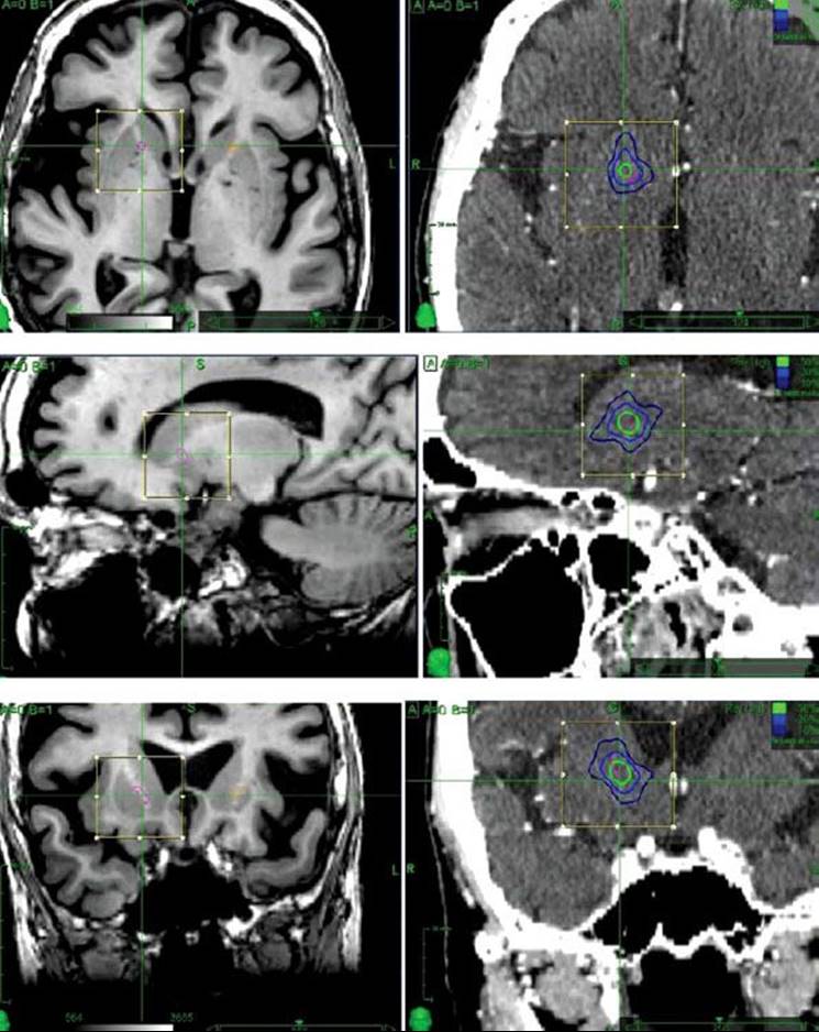
FIGURE 91.5. Stereotactic magnetic resonance imaging sequences (left side) demonstrating the contoured anterior limbs of the internal capsule bilaterally and the corresponding treatment plan for the right internal capsule (right side) for a patient with refractory obsessive-compulsive disorder. The right internal capsule was prescribed at 70 Gy to the 50% isodose line (140 Gy maximum dose) in a single fraction.
DISEASES OF THE EYE AND ORBIT
Pterygium
Background and Clinical Aspects
Pterygium is a chronic fibrovascular and degenerative process that arises from the conjunctival–corneal border that extends from the nasal corner of the eye to the cornea. Its name (“pterygium”) refers to the shape of the tissue, which is wing-like. The exact prevalence of this problem is unknown, but it is well established that the frequency is higher in tropical regions. Most patients are asymptomatic and present for medical attention on the basis of cosmetic concerns, but symptoms may include redness and irritation of the eye. Pterygium may impair vision by producing an irregular astigmatism as it grows onto the cornea.
Surgery
Treatment is indicated when vision is threatened and less commonly to improve cosmesis. The treatment of choice for pterygium is surgical excision with an adjunct to help improve local control rates (i.e., sliding conjunctival flap; rotational conjunctival autograft; free conjunctival or limbal autograft). Intraoperative or postoperative mitomycin-C has also been used to improve local control rates, although this leads to increased risk of scleral ulceration, secondary glaucoma, iritis, and cataracts.
Radiotherapy
Local radiation therapy with strontium-90 plays an important role as an adjunct to surgery to prevent relapse. Outcomes with radiotherapy have been excellent at decreasing relapse rates.
The first prospective randomized study comparing postoperative radiation to observation after surgery was performed by de Keizer.143 In this study, 19 pterygia were treated with bare scleral excision with a recurrence rate of 68% at 4 months compared with no recurrences in the 18 pterygia treated with postoperative fractionated irradiation (3 é 10 Gy, once a week). Numerous retrospective studies have also demonstrated the efficacy of postoperative radiation in preventing recurrence of pterygium, including a large series of 1,300 pterygia by Van de Brenk144 and 825 pterygia by Paryani et al.,145 both of which showed a low recurrence rate of 1.7% using fractionated radiotherapy.
In 2004, a European randomized trial compared single-dose (as opposed to fractionated) postoperative radiotherapy (25 Gy) compared with sham RT.146 Patients who received radiotherapy had a local control rate of 93.2% compared with 33.3% in the placebo arm, indicating that single-dose radiotherapy is effective. In another randomized study, Viani et al.147 compared low fractionation dose (2 Gy in 10 fractions) to high fractionation dose (5 Gy in 7 fractions) β-radiotherapy in the postoperative setting. Control rates were similar between the two groups (93.8% vs. 92.3%), with a significantly lower incidence of poorer cosmesis, photophobia, eye irritation, and scleromalacia in the low fractionation dose arm.
Choroidal Hemangioma
Background and Clinical Aspects
Choroidal hemangiomas (CH) are rare vascular tumors that arise from the choroid. CH can be classified as circumscribed, which occur in older patients, or diffuse, which are associated with the Sturge-Weber syndrome.148
Clinically, these lesions are often asymptomatic, but patients may present with a visual disturbance by several mechanisms, including retinal detachment, macular edema, and retinal pigment changes.149 Lesions are detected on funduscopic examination. Further workup includes ultrasonography, angiography with fluorescent dyes, and CT or MRI.
Surgery
Among the surgical treatment options available, CH that are not near the central visual structures (macula and papilla) are often treated with photodynamic therapy with a low rate of complications.148 Other treatment modalities include laser photocoagulation and transpupillary thermotherapy. In general, radiation therapy is preferred over photodynamic therapy for the treatment of diffuse CH, although several small studies have reported encouraging results with the use of photodynamic therapy.148
Radiation Therapy
RT is indicated to treat lesions near the macula and papilla and in cases that did not respond to other therapeutic maneuvers. RT techniques to treat CH include conventional 3D-CRT, proton-beam therapy, and brachytherapy.
Typical dose prescriptions for 3D-CRT are 18 to 20 Gy for circumscribed CH and 30 Gy for diffuse CH given in 1.8- to 2.0-Gy daily fractions. Schilling et al.150 irradiated 36 circumscribed CH with 20 Gy in 10 fractions. Retinal reattachment occurred in 64% of the cases with improved vision in 50% and stable vision in 50%.
Fractionated proton radiotherapy doses range from 16.4 to 30 Gy in 4 fractions.151–153 In the study by Zografos et al.,153 all 54 cases experienced retinal reattachment, and visual acuity was improved in 70%. A recent study from Paris also demonstrated a 100% rate of retinal reattachment and substantial improvement in visual acuity using proton-beam therapy.154
Plaque brachytherapy using cobalt-60, iodine-125, or ruthenium-106 has been used to treat circumscribed lesions.149,153,155,156 Typical doses prescribed to the apex of the lesion range from 25 to 50 Gy. Each isotope has advantages and disadvantages depending on the physical properties (i.e., energy, half-life), and there is no evidence to support the use of one over the other.
Age-Related Macular Degeneration
Age-related macular degeneration (AMD) is the leading causes of blindness in the developed world.157 The development of AMD is dependent on age, with a prevalence of up to 35% in the eighth decade of life. External-beam radiotherapy with photons or protons and brachytherapy have been used in the past to treat macular degeneration. Overall, results of radiotherapy in the management of this disease have been mixed. A Cochrane meta-analysis in 2010 analyzed 14 randomized trials utilizing RT as a treatment for AMD and concluded that the review “does not provide convincing evidence that radiotherapy is an effective treatment for neovascular AMD.”158 Given that there is no clear benefit or indication for RT, its use should be limited for the treatment of AMD.
Graves Ophthalmopathy
Background and Clinical Aspects
Graves ophthalmopathy (GO), also referred to as Graves orbitopathy or thyroid eye disease, is an autoimmune disorder affecting the musculature of the orbits. The presence of activated T-lymphocytes leads to an inflammatory reaction secondary to the release of cytokines. It is estimated that up to 50% of patients with Graves disease will develop orbitopathy, but 10% of patients are euthyroid and some are hypothyroid at presentation.1 Smoking is the greatest risk factor for the development of GO and also predicts for a poorer response to therapy.1
A multidisciplinary team, including an ophthalmologist, endocrinologist, and radiation oncologist, should be involved in the evaluation of the patient with GO. Clinical features of patients with GO include proptosis (measured by the Hertel exophthalmometer on physical examination), photophobia, upper eyelid retraction, periorbital edema (due to the accumulation of collagen and hyaluronan, which attract water), conjunctival erythema and tearing, and visual impairment (Fig. 91.6A,B). Patients may complain of a “gritty” sensation in their eyes. Several classification systems are available to document the extent of disease, although the one favored at Stanford University is the SPECS Ophthalmic Index (Table 91.7), which assigns a score of 1 to 3 on the basis of six categories: Soft tissue involvement, Proptosis, Extraocular movements, Corneal involvement, and Sight (visual acuity).
Imaging studies, such as CT or MRI, will demonstrate abnormalities, including enlargement of the extraocular muscles and fatty infiltration, in 70% to 80% of cases.1 The most commonly involved muscles include the inferior and medial rectus muscles.159
FIGURE 91.6. A 50-year-old woman with Graves ophthalmopathy before (A) and after (B) treatment with corticosteroids and radiotherapy for prominent eyelid edema and strabismus. C: Three-dimensional conformal radiotherapy treatment plan for a patient with Graves ophthalmopathy. The isocenter (yellow arrow) is placed a few millimeters posterior to the lenses (magenta), and the opposing fields are beam split anteriorly (white arrows). The extraocular muscles are contoured in red. The color wash display demonstrates that <10% of the dose reaches the lens.

TABLE 91.7 SPECS CLASSIFICATION SYSTEM FOR GRAVES OPHTHALMOPATHY
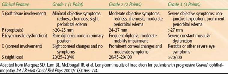
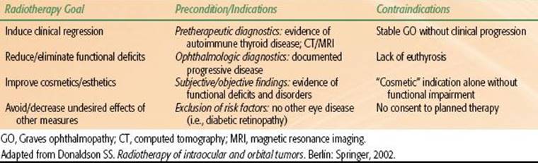
TABLE 91.8 CLINICAL GUIDELINES FOR USE OF RADIOTHERAPY IN GRAVES OPHTHALMOPATHY
Management Overview
Treatment options for GO include glucocorticoids, orbital radiotherapy, and surgery (orbital decompression, eye muscle surgery, eyelid surgery). Smokers should be encouraged to quit. Prior to the initiation of treatment, the patient’s thyroid function should be normalized, as this may help improve the GO.160 Radioiodine therapy, but not antithyroid drugs, may cause worsening of GO.161,162 Once thyroid function is stabilized, the treatment of GO depends on the severity of the disease.
Medical Management (Glucocorticoids)
Glucocorticoids (GCs) are a mainstay of treatment for GO. Immediate treatment with high-dose steroids (intravenous or oral) is required for patients whose vision is threatened by optic neuropathy.163 GCs may also be used to treat patients with moderate to severe active ophthalmopathy.163
Surgery
In the event that GCs fail to improve optic neuropathy, urgent orbital decompression is necessary.164,165 Another indication for urgent orbital decompression is when exposure keratopathy is not relieved by topical therapies.164 In order to improve extraocular muscle function and cosmesis, other procedures such as strabismus surgery and lid surgery may be performed. It is recommended that the GO be inactive for at least 6 months before pursuing these procedures.164
Radiotherapy
Indications for RT in the management of GO have been outlined by Donaldson166 and include inducing clinical regression, improving functional deficits, improving cosmesis, and avoiding side effects of other treatments (Table 91.8).
RT is generally administered with 3D-CRT. Both orbits, including the entire length of the extraocular muscles, are treated to a total dose of 20 Gy in 2-Gy fractions using opposed lateral fields with the isocenter placed a few millimeters posterior to the lenses using a beam-split technique (Fig. 91.6C).
In a double-blind, placebo-controlled study, untreated euthyroid patients with GO were randomized to oral steroids or 20 Gy orbital irradiation.167 Both groups experienced response rates of 50%, with greater improvements in eye motility and fewer side effects in the radiation arm. Two prospective studies have demonstrated a benefit of the combination of steroids and radiation compared with single modality treatment.168,169
For patients with progressive GO, retrospective data suggest that orbital RT is an effective treatment modality. Marquez et al.170 reviewed the records of 197 patients treated at Stanford University, all of whom received 20 to 30 Gy to the bilateral retrobulbar region. Outcomes assessed included SPECS score and patient satisfaction. There was a 96% overall response rate and 98% patient satisfaction rate, with the largest improvements in soft tissue findings (89%), extraocular muscle dysfunction (85%), and corneal abnormalities (96%).
Reactive Lymphoid Hyperplasia and Orbital Pseudotumor
Background and Clinical Presentation
Disease of the lymphoid tissue in the orbit is rare and may include orbital pseudotumor (OP) or malignant lymphomas. OP is an inflammatory condition of unclear etiology that affects the soft tissue of the orbits, most often unilaterally.159 Most patients present between the fifth and sixth decades of life.171
Clinical features of OP include periorbital edema, retrobulbar pain, extraocular muscle dysfunction, palpable mass, and exophthalmos.159 Symptoms usually develop acutely. Imaging with CT or MRI of the orbits should be obtained for further evaluation. Imaging findings include enlarged extraocular muscles, optic nerve thickening, and infiltrates in the retrobulbar adipose tissue with enhancement after administration of iodinated contrast or gadolinium.159 Biopsy should be obtained to establish the diagnosis, especially for lesions that are easily accessible.
Medical Therapy
Corticosteroids are the treatment of choice for the majority of patients. Response rates for optic neuropathy are as high as 92% with an overall response rate of 78%.159 However, only 33% of patients experience long-term control with a single course of steroids.172
Surgery
Surgical excision may be used for easily accessible lesions. Relapses are common after surgery.
Radiation Therapy
Indications for RT include recurrent lesions after surgery, steroid-refractory lesions, and lesions not amenable to other treatments. The RT technique of choice is 3D-CRT.
A planning CT should be obtained and coregistered with the diagnostic MRI or diagnostic CT to delineate the target volume when visible. Unilateral treatment is typically performed with a single lateral field or with an anterior and lateral field, weighted more heavily laterally. Bilateral orbital involvement is treated in a manner similar to GO. Occasionally, superficial lesions may be treated with electrons. Typically, the prescription dose is 20 Gy given in 10 fractions.159
In a modern retrospective series from the University of Oklahoma, 20 orbits in 16 patients were treated with RT for OP.173 With a mean dose of 20 Gy in 10 fractions, 87.5% of the patients experienced a response (clinical improvement or tapering of corticosteroid dose). Corticosteroid use was stopped or reduced in 81% of the patients. No significant late effects were reported.
BENIGN DISEASES OF SOFT TISSUE AND BONES
General Overview of Inflammatory Conditions of Joints and Tendons
The role of radiation therapy in the treatment of benign inflammatory conditions involving the joints or tendons is controversial. Osteoarthritis (OA), tendonitis, bursitis, rotator’s cuff syndrome, and tennis elbow are examples of inflammatory conditions for which radiation therapy has been used in the past. These soft tissue syndromes may result from repetitive activities that cause overuse or injury to the joint areas, incorrect posture, stress on the soft tissues due to an abnormal or poor positioned joint or bone, or other diseases, such as autoimmune diseases or infection.
Although the cause of each disorder may be different, the clinical presentation and general treatment plan are frequently similar. Symptoms include pain, swelling, or inflammation in the tissues and structures around a joint, such as the tendons, ligaments, bursae, and muscles. Treatment generally involves a combination of exercise, lifestyle modification, and analgesics. If pain becomes debilitating, joint replacement surgery may be used to improve the quality of life. In rare instances, low-dose radiation therapy (<10 to 15 Gy) can be employed. The low dose required to improve symptoms suggests the possible mechanism of action for radiation therapy (Table 91.9).
TABLE 91.9 RADIATION THERAPY MECHANISM OF ACTION DOSE CONCEPTS
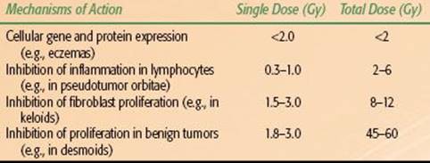
Osteoarthritis
OA is the most common joint disorder. It presents with pain associated with cartilage destruction, bone modification, and structural changes of capsule and synovia. Symptoms are caused by reactive inflammation of joint surface and joint capsule lining (synovia). Although in many instances the cause of OA is unknown, age is a major risk factor. Other risk factors include obesity, bone fracture or joint injury, whether by an accident or overuse from work or sports, and other medical conditions.
FIGURE 91.7. Dupuytren’s contracture of both hands and the left foot.

Nonradiotherapeutic Treatment
Hunter and Lo174 provide a general overview of the diagnosis, investigation, and treatment of OA. The treatment of early OA is intended to reduce the primary symptoms of joint pain and stiffness with the goal of maintaining and improving the functional capacity of the affected joint(s).175 Exercise, weight reduction, and joint braces, among other measures, have shown some success at unloading damaged joints and improving symptoms.176,177 For osteoarthritis of the hip and knee, exercises that strengthen muscles and improve aerobic condition are most effective.178
Oral analgesics are the mainstay of treatment for OA. Although acetaminophen is frequently offered due to its relative safety and effectiveness, a nonsteroidal anti-inflammatory drug (NSAID) may be added or substituted.179NSAIDs can be used in patients with symptomatic OA of the hand, hip, or knee. The goal is to administer the lowest effective dose for the shortest duration. The use of stronger analgesics, such as weak opioids and narcotics, may be considered when other methods have been ineffective or if certain drugs are contraindicated.176 Glucosamine and chondroitin sulfate are over-the-counter remedies that are frequently used to reduce pain, but their efficacy has not been proven.180 Corticosteroids and other analgesics may be injected directly into the joint; injections may temporarily reduce swelling and pain. Acupuncture and other complementary and alternative treatment modalities have been used, but their efficacy has yet to be proven.181
Surgery is reserved for patients with severe OA and those who have not responded to noninvasive therapies. Total or partial joint replacement is most commonly used for OA involving the knee, hip, and shoulder and is considered when structural damage is visible on x-rays. However, there are modern surgical procedures that can obviate or delay the need for joint replacement, including osteotomies and joint resurfacing.182 Joint fusion or arthrodesis may be used to treat arthritis of the spine, ankles, hands, and feet. Arthroscopy, or arthroscopic surgery, is a minimally invasive surgical procedure that can be used to examine and treat the interior surface of a damaged joint. Arthroscopic procedures can help relieve pain for a short time and allow the joints to move better. Although arthroscopy may delay the need for joint replacement surgery, it does not improve the arthritis itself.183
Radiotherapeutic Options
In nonsurgical candidates, low-dose RT may be considered if pharmacotherapy has failed. RT can lead to primary freedom from pain and secondary to improved joint function.184 Several single-institution studies have been published that report long-term pain relief and functional gain in 50% to 75% of patients. In Germany from 2006 to 2008, a pattern of care study investigated the use of RT for the treatment of OA of the knee (gonarthrosis).185 Almost 80% of institutions in Germany have used RT to treat OA in the 2-year period analyzed. Treatment of 4,544 patients was performed annually at 188 institutions. The median total dose was 6 Gy (range 3 to 12 Gy), with a median single dose of 1 Gy (0.25 to 3 Gy). Long-term clinical outcomes were available in 5,069 cases. The majority of patients experienced pain reduction for at least 3 months, but pain management for up to 12 months was reported. In 30% of patients, a second course of RT was used for inadequate pain response or early pain recurrence.
As with arthroscopy, radiation may reduce pain and pain-related dysfunction, but it does not improve the arthritis itself. Due to its efficacy and relative safety, RT may provide an alternative to conventional conservative treatment for patients who are not surgical candidates.
DISEASES OF CONNECTIVE TISSUE AND SKIN
Desmoid Tumors
Background and Clinical Aspects
Desmoid tumors (also called aggressive fibromatosis or deep musculoaponeurotic fibromatosis) are benign tumors of connective tissue tumors that arise from muscle fascias, aponeuroses, tendons, and scar tissue. They are slightly more predominant in females and tend to occur during the third and fourth decades of life, although children and the elderly can be affected. In the general population, desmoids are rare; the estimated incidence is 2 to 4 per million per year. Genetic factors, trauma, or surgery predispose the development of desmoids. Most desmoids arise sporadically; however, approximately 2% are associated with familial adenomatous polyposis (FAP). Desmoid tumors affect between 10% to 20% of patients with FAP. The development of desmoid tumors in patients with FAP is called Gardner syndrome.
Tumors can develop anywhere in the body, but most commonly involve the trunk or extremity, abdominal wall, and intra-abdominal sites, including the bowel and mesentery. Approximately 30% of patients with desmoid tumors have a history of prior trauma at the tumor site.186,187 Sporadic cases commonly involve the extremities, the shoulder girdle, and the buttock.187 In patients with FAP, intra-abdominal desmoids predominate and tend to be associated with surgical sites and anastomoses following colectomy.188 Desmoid tumors in women can occur during or after pregnancy and therefore may be associated with high estrogen states. Women who have been pregnant are more likely to have abdominal desmoid tumors that develop within 10 years of the last pregnancy.189
Although desmoids have no known potential for metastasis or dedifferentiation, they are locally aggressive and commonly have a high rate of recurrence even after complete resection. Diagnostics workup with MRI helps to estimate size and infiltration into other organs and should be obtained prior to incisional biopsy obtained to confirm diagnosis.
Nonradiotherapeutic Treatment
Observation is a viable option for stable, asymptomatic desmoids. Treatment is indicated for symptomatic patients, if there is risk to adjacent structures, or to improve cosmesis. Complete resection of the tumor with negative microscopic margins is the treatment of choice for most desmoid tumors. Due to the size and infiltrative nature of extra-abdominal desmoids, resection may require skin grafting or flap reconstruction. Desmoid tumors have a high rate of recurrence following even complete surgical removal, and the contribution of incomplete resection to local recurrence rates is unclear.190 Furthermore, resection does not appear to affect survival, which is not surprising in view of the histologically benign nature of desmoids. Given these issues, the overall surgical strategy should be an attempt at complete removal using function-preserving surgical approaches to minimize major morbidity (functional or cosmetic).191
Although extra-abdominal desmoid tumors can generally be treated effectively with local therapy, surgical intervention tends to be counterproductive in intra-abdominal variants, especially the ones associated with FAP. In some instances, systemic therapy may achieve significant and durable cytoreduction, obviating the need for resection. Patients with desmoid tumors have been treated with NSAIDs. The most widely used NSAID for treatment of desmoid tumors is sulindac. Hormonal agents such as tamoxifen, raloxifene, and progesterone have been used, often in combination with NSAIDs. Tamoxifen has been used most widely and is typically prescribed at doses similar to those used for breast cancer (10 mg daily). Much higher doses (120 mg daily) have been recommended,192 but high-dose tamoxifen is difficult to tolerate and there is no evidence to suggest that higher doses of tamoxifen are better than lower doses.
A variety of palliative chemotherapeutic regimens have been used.193–194,195–196 With the waxing and waning natural history of desmoids, it is difficult to say whether systemic therapy provides much benefit over observation. In one series, 142 patients presented with either a primary (n = 74) or recurrent (n = 68) desmoid tumor. Eighty-three patients were treated with observation alone, and 59 received either hormone therapy or chemotherapy. There was no statistically significant difference in progression-free survival between the two groups.
Desmoid tumors also respond to the tyrosine kinase inhibitor imatinib.197,198 The response is thought to be due to expression of one of Gleevec’s molecular targets, platelet-derived growth factor receptor, on desmoid tumors.199 In a phase II clinical trial to assess the efficacy of imatinib (400 mg per day for 1 year) in the treatment of progressive and recurrent aggressive fibromatosis, the 2-year progression-free and overall survival rates after the use of imatinib were 55% and 95%, respectively.198
Intralesional injections200 and radiofrequency ablation201 have also been used. Although the techniques led to some tumor shrinkage, the experience to date is limited and the long-term results are not yet known.
Radiotherapeutic Options
Radiation therapy is a viable option for inoperable patients and may also be used in combination with surgery or chemotherapy. Spear et al.202 retrospectively compared the efficacy of surgery alone, radiation alone, and combined modality therapy (radiation and surgery) in the treatment of desmoid tumors. Five-year local control rates among surgery, radiation therapy, and combined modality groups were 69%, 93%, and 72%, respectively. The study recommended radiation doses of 60 to 65 Gy for inoperable or recurrent desmoids. However, long-term results at another institution show increased posttreatment toxicity in patients who receive RT doses >56 Gy.191,203
Young age (≤30 years) was also associated with increased late toxicity. In a retrospective study of 30 patients under the age of 30, younger age (<18 years) is associated with inferior local-regional control following RT. Although actuarial control rates were better with RT doses of ≥55 Gy, almost 50% of patients experienced grade 3 or 4 complications, including pathologic fractures, impaired range of motion, pain, and in-field skin cancers.204 Because long-term results suggest that unresectable tumors respond to 56 Gy with a 75% expectation of local control, the lower dose may be more appropriate.
When an R0 resection is not possible, doses of 50 Gy postoperatively should be given to improve local control. RT is often not considered for intra-abdominal tumors because the dose and increased field size required increase risk of bowel injury. Due to the complexities involved in managing the disease, a multidisciplinary approach must be taken.205
Peyronie Disease
Background and Clinical Aspects
Peyronie disease (also known as Induratio penis plastica) is a chronic inflammatory connective tissue disorder involving the penile tunica albuginea that results in tissue proliferation and the development of hard plaques, most commonly on the dorsal surface of the penis, which may cause a curvature and changes in the length or circumference of the penis while erect. Symptoms may lead to difficult intercourse, penile pain, and erectile dysfunction.
Peyronie disease affects up to 10% of men, although a recent population-based study suggests the condition may be underreported in the United States.206 Although Peyronie disease can affect teenagers, peak incidence is between 40 to 60 years of age. The cause is unknown, but diabetes mellitus and arterial and venous vascular disease are risk factors, along with an assumed genetic predisposition. The disorder results in pain, abnormal curvature, erectile dysfunction, indentation, loss of girth, and shortening. Slow progression over several months is typical, but spontaneous remission may occasionally occur.
Nonradiotherapeutic Treatment
Results of nonsurgical treatment of Peyronie disease are mixed and are controversial.207,208 Some success has been reported with vitamin E supplementation, but results have not been confirmed in larger studies.209 A combination of vitamin E and colchicine may delay disease progression.210 Other agents that specifically target inflammatory pathways have also shown mixed benefit, including, tumor growth factor-β1 inhibitors,211 coenzyme Q10,212 and sildenafil, among others.213 Topical therapies have largely been ineffective, but penile injection with verapamil or collagenase, intended to break up scar tissue formed by the inflammation, have shown some efficacy.214 Physical therapy and extracorporeal shock treatments have also had limited benefit.
Surgical options for Peyronie disease are complex procedures that should only be performed by experienced urologists and are reserved for patients not responding to other therapies.215 Although the nonsurgical treatments discussed may not reliably treat the disease, they can be used to stabilize the scarring process and may result in some reduction of deformity. A combination of nonsurgical techniques may have even more efficacy.216
Radiotherapeutic Options
The largest experience with the use of RT in the treatment of Peyronie disease has been in Europe. Retrospective studies showed symptom improvement with the use of RT. Although some studies suggested improvement in curvature,217 the majority of studies suggest that radiation therapy primarily provides relief of pain associated with Peyronie disease. These data suggest that the benefit of RT might best be in the treatment of early stages of disease, when radio-responsive inflammatory cells and fibroblasts are still active in the disease. There may be little improvement in penile contracture once the plaques have fully formed.
As with radiation therapy for other rare benign conditions, the treatment regimens for Peyronie disease vary among institutions.17 A survey of European practices show that most practices give a total dose of approximately 20 Gy (3 to 30 Gy) in 2-Gy fractions (range 0.5 to 8.0 Gy). Most of the institutions used electrons (n = 44), however, orthovoltage was still used at a number of practices (n = 32). One retrospective study from the Netherlands indicated that low-dose RT, either 13.5 Gy (9 é 1.5 Gy, 3 fractions per week) or 12 Gy (6 é 2 Gy, daily fractions) resulted in pain relief in the majority of the 179 patients evaluated.217 Sexual dysfunction was a reported side effect, although this is confounded by the underlying disease.
As experimental models improve our understanding of the pathogenesis of the Peyronie disease, the use of radiation therapy may further decline, as concern regarding radiation induction of fibrosis surface and new more effective therapies to emerge.218,219
Dupuytren’s Contracture
Background and Clinical Aspects
Dupuytren’s contracture, also known as Morbus Dupuytren (MD) and Morbus Ledderhose (ML), depending on involvement of the hands or feet, respectively, is a connective tissue disorder that affects the palmar or plantar fascia. Incidence increases after the age of 40, and the condition affects men more often than women. Although there is a familial disposition, alcohol abuse, diabetes mellitus, epilepsy, and other conditions are associated. Initially, there is an inflammatory proliferative phase with fibroblast activity.
In the early stage, subcutaneous nodules appear, which may be fixed to the overlying skin. As the disease progresses, cords develop and become visibly predominant. With further progression, the cords reach the periosteum of the bones and lead to the characteristic appearance of palmar or plantar contraction. The fourth or fifth phalanges of the hand (MD) or the first or second toes of the foot (ML) are the most commonly affected digits (Fig. 91.7). With increased thickening of the fascia and progressive contracture, the fingers and toes begin to curl, resulting in impaired function. Flexion contractures in the metacarpal or proximal interphalangeal joints lead to difficulty grabbing (MD) or walking (ML).
Nonradiotherapeutic Treatment
Excision of diseased cords and fascia via limited or selective fasciectomy is widely considered the gold standard treatment for Dupuytren’s contracture.220,221 A 20-year review of open surgery for Dupuytren’s contracture showed that major complications occurred in 15.7% of cases and wound complications were seen in 22% of cases.222 Even with excellent surgical resection, relapse is common, with 30% to 50% recurrence rate at 3 years.
Modern minimally invasive techniques have substantially reduced the complication rates. Percutaneous needle fasciotomy is a technique where cords are weakened through the insertion and manipulation of a small 25-gauge needle mounted on a 10 mL syringe.223 The procedure is performed under local anesthesia and patients may return to full usage of the affected limb within 24 hours. Because the cords and nodules are not fully excised, minimally invasive surgery has an even higher recurrence rate than surgical excision. A randomized study comparing percutaneous needle fasciotomy with limited fasciectomy showed an 85% recurrence rate after 5 years with the minimally invasive procedure.224
During the early stage of Dupuytren’s, medication (steroids, allopurinol, nonsteroidals, vitamin E) may provide benefit, but the effects are temporary. Injectable collagenase extracted from Clostridium histolyticum has been approved for the treatment of Dupuytren’s contracture. Injection of small amounts of the enzyme collagenase weaken cords by breaking the peptide bonds in collagen.225 Treatments should only be applied by an experienced provider, as the amount of enzyme injected varies depending on the affected joint and also has a very high recurrence rate.226,227
Radiotherapeutic Options
Several clinical trials support the concept of prophylactic radiation therapy in the treatment of Dupuytren’s contracture.228–229,230,231 Radiotherapy is effective for prevention of disease progression in early stages of disease when only small lumps or cords are present and only moderate extension deficits (≤10 degrees) are present. Treatment can be administered with either electrons or orthovoltage radiation, and a variety of dose levels have been used. Because the target cells are proliferating and radiosensitive fibroblasts and inflammatory cells, low-dose radiation therapy can be applied.228,230
In a recent prospective trial involving 129 patients, two different dose regimens were compared for safety and efficacy. In group A, 63 patients received 10 é 3 Gy (30 Gy) via a split course (5 é 3 Gy) separated by 8 weeks; in group B, 66 patients were treated with 7 é 3 Gy (21 Gy) delivered over 2 weeks. There was no difference in treatment outcomes between the two groups. Regardless of dose regimen, approximately 90% of patients had stable or improved disease. Overall and mean number of nodules, cords, and skin changes decreased at 3 and 12 months. There was an 8% treatment failure rate at 1 year. Acute toxicity was more pronounced in group B, but long-term toxicity was comparable and included dryness, desquamation, skin atrophy, and altered sensation. Although long-term results of this study are pending, prior retrospective data indicated that prophylactic RT is well tolerated by patients and is effective at preventing disease progression. Irrespective of dose regimen, appropriate immobilization and shielding of unaffected joints is required (Fig. 91.8).
FIGURE 91.8. Immobilization for treatment of Dupuytren’s contracture with electrons.
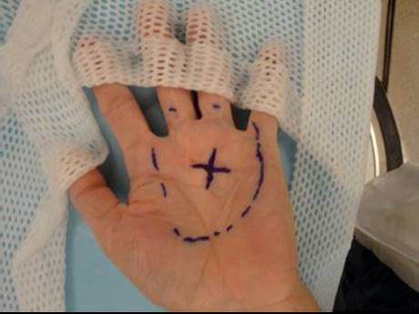
FIGURE 91.9. A: Keloid behind left earlobe. B: Status of keloid following resection plus 4 é 4 Gy radiotherapy.

Keloids and Hypertrophic Scars
Background and Clinical Aspects
Keloids are an excessive tissue proliferation around scars after skin injury from surgery, heat, chemical burns, inflammation (e.g., acne), or even spontaneous proliferation. They differ from hypertrophic scars by their typical infiltrative growth pattern, causing local pain and inflammatory reactions, and sometimes long-term progression; hypertrophic scars show thickening without surrounding reaction and can flatten spontaneously. Keloids appear mostly in the upper body and in regions with high skin tension (e.g., sternum, earlobes). The cause is still unknown, although there is a genetic and race-specific predisposition that is already noted during adolescence. Keloids at the earlobe after piercing are typical. In some patients, the resulting lesions are severely disfiguring and painful (Fig. 91.9). Recurrence is common after treatment.
Nonradiotherapeutic Treatment
Silicone bandages, pressure dressings, and cryosurgery have all been used to treat keloids, with varying efficacy.232–234 Intralesional injections remain the first-line therapy for most keloids. Corticosteroids, 5-fluorouacil, and verapamil have all been directly injected into keloid lesions with symptom improvement. Up to 70% of patients respond to intralesional corticosteroid injection with flattening of keloids, although the recurrence rate is high in some studies (up to 50% at 5 years).235
Surgical excision may be indicated if injection therapy alone does not result in improvement. In patients treated with excision alone, recurrence rates range from 45% to 100%,236 therefore, excision is typically combined with perioperative or postoperative injections of either triamcinolone or interferon.235
Radiotherapeutic Options
Radiotherapy should be considered in cases of repeat recurrences postoperatively or where there is a high-risk of recurrence (e.g., marginal resection, large lesion, unfavorable location). Primary RT can be considered in instances where resection would result in functional impairment and in actively proliferating disorders within about 6 months after the triggering trauma. Because proliferating fibroblasts and mesenchymal and inflammatory cells are the target cells for RT, fully matured keloids have minimal response to RT alone. Prophylactic RT immediately following excision is most effective and reduces the risk of recurrence to 20% or 25% in most series.
RT is initiated 24 hours after surgery. The target volume is limited to the scar plus a 1-cm margin; lead shielding can be constructed to protect normal tissue. An analysis of multicenter data on the use of postoperative RT for earlobe keloids shows that higher dose per fraction and use of deeper penetrating electrons are preferable to standard 2-Gy fractionation schemes or use of brachytherapy techniques that have rapid dose falloff.237 Radiation dose is typically 12 to 20 Gy, delivered in 3 or 4 fractions within 1 week.238 Single-fraction RT with 7.5 to 10 Gy is also effective.239 Clinical end points are long-term control, low relapse rate, and good cosmesis.
Diseases of Bone
Gorham-Stout Syndrome
Gorham-Stout syndrome, also known as disappearing bone disease or essential osteolysis, is a rare bone disorder of unknown etiology. It is characterized by painless bone destruction due to progressive proliferation of small blood or lymph vessels. There may also be significant osteoclast activation. The symptoms are nonspecific but include muscular weakness, limb tenderness, and pathologic fracture occurring after minimal trauma. Involvement of the cervical spine or skull base could be fatal. Case reports indicate limited efficacy of systemic therapies such as zoledronic acid and interferon-alfa.240,241 Radiation therapy has also been used.242,243 Heyd et al.244 completed a national patterns-of-care study and literature review that summarizes the scant data available for this rare disorder. The 38 articles listed therein provide evidence from treatment of 44 patients that indicate conventionally fractionated external-beam RT (total dose of 36 to 45 Gy) may prevent disease progression in 77% to 80% of cases.
Pigmented Villonodular Synovitis
Pigmented villonodular synovitis or tenosynovial giant cell tumor is a rare proliferative disorder of synovial tissue. Symptoms include sudden onset, unexplained joint swelling, and pain that frequently involves a single joint. The knee and foot are most commonly affected, but there are reports of shoulder, hand, and hip involvement.245 Decreased motion, joint stiffness, and increased pain occur as the disorder progresses. Surgical resection with either synovectomy or joint replacement is the treatment of choice.246,247
Radiation therapy is indicated in cases of diffuse disease, bulky disease resulting in bone destruction, or in the rare instance of multiple recurrences after resection. Although intrasynovial injection of radioactive isotopes postoperatively has been used in the past for high-risk patients,248 most institutions use external-beam radiation therapy. RT to a dose of 35 to 50 Gy has been effective.249,250 MRI is essential for delineating disease pre- and postoperatively. Final dose of RT should be tailored to amount of residual disease.251
Vertebral Hemangiomas
Hemangiomas are benign proliferations of blood vessels that can affect any tissue and are typically asymptomatic. About 50% of hemangiomas involving the vertebral body are associated with pain and therefore may require treatment. Treatment options include surgical resection or more conservative interventions such as vertebroplasty or intralesional injections.252 Radiation therapy either alone or postoperatively has been successful in reducing pain caused by vertebral hemangiomas.253 In this study, a total of 84 patients with 96 symptomatic lesions were irradiated for a symptomatic vertebral hemangioma. At a median 68 months’ follow-up, 90% of patients had either complete or partial pain relief. Radiation doses ≥34 Gy resulted in significantly improved pain relief. A total radiation dose of 36 to 40 Gy delivered in 2 Gy per fraction has been recommended.254
Heterotopic Ossification
Background and Clinical Aspects
Heterotopic ossification (HO) is a common complication of total hip arthroplasty, hip trauma, or acetabular fracture. HO occurs when the soft tissues around the hip become ossified. Following trauma, primitive mesenchymal cells in the surrounding soft tissues are transformed into osteoblastic tissue that then forms mature bone. The hip is the most common joint affected; HO typically occurs around the femoral neck and adjacent to the greater trochanter. The risk factors for development of HO are unknown, but the incidence is greater in men and occurs in more than 80% in patients who have a history of ipsilateral or contralateral HO. It is also more common in patients with a known history of osteoarthritis, ankylosing spondylitis, and diffuse and Paget disease.255 Hip stiffness is the primary symptom, and the diagnosis is made radiographically. Pain is typically not associated with HO.
FIGURE 91.10. Typical treatment field for heterotopic ossification.

Nonradiotherapeutic Treatment
The treatment for HO is surgical excision followed by some form of HO prophylaxis. Prophylaxis is only applied to patients at high risk for developing HO. A meta-analysis showed that NSAIDs are effective in reducing the risk of postoperative HO.256 Indomethacin is the most commonly used NSAID for HO prophylaxis. Indomethacin is a prostaglandin synthase inhibitor that also suppresses mesenchymal cells. The limited data available have not shown a clear benefit to the use of selective cyclo-oxygenase-2 inhibitors in HO prophylaxis.257,258 Bisphosphonates have been used for prophylaxis because they delay mineralization of osteoid and appear to have some efficacy in preventing HO if used at the appropriate time. In one study, the cost of bisphosphonate was prohibitive for routine use when compared with indomethacin.259
Radiotherapeutic Options
External-beam radiation is an effective method for prevention of HO after total hip arthroplasty. Prophylactic radiation therapy for the prevention of HO has been used since the 1970s. A single fraction of 7 or 8 Gy to the at-risk region (Fig. 91.10) is recommended and should be delivered in the perioperative period, either preoperatively (within 24 hours) or postoperatively (within 72 hours).260,261,262 When comparing radiation therapy and NSAIDs, there is no clear benefit for use of one modality over another. A prospective, randomized study demonstrated that radiation therapy and indomethacin are both effective in the prevention of postoperative HO.263 Although one meta-analysis of seven randomized studies concluded that radiotherapy is more effective than NSAIDs for HO prophylaxis,264 a more recent analysis of nine studies involving 1,295 patients found no statistically significant difference between the two.265 An economic analysis using the same nine studies and the meta-analysis suggests that radiation therapy is not cost-effective when compared with use of NSAIDs.266 This analysis has yet to be validated.
CONCLUSION
This chapter has reviewed the therapeutic approaches and supporting data for a variety of benign diseases and disorders. For some conditions, the use of radiation therapy has been well-established and the literature provides sufficient data regarding the radiation techniques, dose regimens, and outcomes. In instances where there are not yet sufficient data to make recommendations, the radiation therapy should be used cautiously, and preferably, in the context of a clinical trial.
Radiation therapy is a powerful tool and must always be used safely and judiciously. While radiation oncologists are trained to be mindful of acute toxicities, disease outcomes, and long-term treatment sequelae, special care must be taken when radiation is used for the treatment of benign disorders. When other therapeutic options exist, a thoughtful multidisciplinary approach should be considered to ascertain which modality is in a patient’s best interest.
SELECTED REFERENCES
A full list of references for this chapter is available online.
8. Trott KR, Kamprad F. Radiobiological mechanisms of anti-inflammatory radiotherapy. Radiother Oncol 1999;51(3):197–203.
9. Rodel F, Keilholz L, Herrmann M, et al. Radiobiological mechanisms in inflammatory diseases of low-dose radiation therapy. Int J Radiat Biol 2007;83(6):357–366.
13. Leer JW, van Houtte P, Seegenschmiedt H. Radiotherapy of non-malignant disorders: where do we stand? Radiother Oncol 2007;83(2):175–177.
14. Trott KR, Kamprad F. Estimation of cancer risks from radiotherapy of benign diseases. Strahlenther Onkol 2006;182(8):431–436.
18. Micke O, Seegenschmiedt MH. Consensus guidelines for radiation therapy of benign diseases: a multicenter approach in Germany. Int J Radiat Oncol Biol Phys 2002;52(2):496–513.
20. Louis DN OH, Wiestler OD, et al. WHO classification of tumours of the nervous system. Lyon: IARC Press, 2007.
23. Simpson D. The recurrence of intracranial meningiomas after surgical treatment. J Neurol Neurosurg Psychiatry 1957;20(1):22–39.
26. Yano S, Kuratsu J. Indications for surgery in patients with asymptomatic meningiomas based on an extensive experience. J Neurosurg 2006;105(4):538–543.
28. Goldsmith BJ, Wara WM, Wilson CB, et al. Postoperative irradiation for subtotally resected meningiomas. A retrospective analysis of 140 patients treated from 1967 to 1990. J Neurosurg 1994;80(2):195–201.
29. Mendenhall WM, Morris CG, Amdur RJ, et al. Radiotherapy alone or after subtotal resection for benign skull base meningiomas. Cancer 2003;98(7):1473–1482.
30. Minniti G, Amichetti M, Enrici RM. Radiotherapy and radiosurgery for benign skull base meningiomas. Radiat Oncol 2009;4:42.
33. Adler JR Jr, Gibbs IC, Puataweepong P, et al. Visual field preservation after multisession cyberknife radiosurgery for perioptic lesions. Neurosurgery 2008;62(Suppl 2):733–743.
34. Paulsen F, Doerr S, Wilhelm H, et al. Fractionated stereotactic radiotherapy in patients with optic nerve sheath meningioma. Int J Radiat Oncol Biol Phys 2012;82(2):773–778.
42. Kondziolka D, Mathieu D, Lunsford LD, et al. Radiosurgery as definitive management of intracranial meningiomas. Neurosurgery 2008;62(1):53–60.
44. Zachenhofer I, Wolfsberger S, Aichholzer M, et al. Gamma-Knife radiosurgery for cranial base meningiomas: experience of tumor control, clinical course, and morbidity in a follow-up of more than 8 years. Neurosurgery2006;58(1):28–36.
45. DiBiase SJ, Kwok Y, Yovino S, et al. Factors predicting local tumor control after Gamma Knife stereotactic radiosurgery for benign intracranial meningiomas. Int J Radiat Oncol Biol Phys 2004;60(5):1515–1519.
50. Laws ER, Sheehan JP, Sheehan JM, et al. Stereotactic radiosurgery for pituitary adenomas: a review of the literature. J Neurooncol 2004;69(1–3):257–272.
51. Brada M, Rajan B, Traish D, et al. The long-term efficacy of conservative surgery and radiotherapy in the control of pituitary adenomas. Clin Endocrinol (Oxf) 1993;38(6):571–578.
54. Littley MD, Shalet SM, Beardwell CG, et al. Hypopituitarism following external radiotherapy for pituitary tumours in adults. Q J Med 1989;70(262):145–160.
55. Landolt AM, Haller D, Lomax N, et al. Octreotide may act as a radioprotective agent in acromegaly. J Clin Endocrinol Metab 2000;85(3):1287–1289.
57. Stripp DC, Maity A, Janss AJ, et al. Surgery with or without radiation therapy in the management of craniopharyngiomas in children and young adults. Int J Radiat Oncol Biol Phys 2004;58(3):714–720.
59. Winkfield KM, Linsenmeier C, Yock TI, et al. Surveillance of craniopharyngioma cyst growth in children treated with proton radiotherapy. Int J Radiat Oncol Biol Phys 2009;73(3):716–721.
60. Lee M, Kalani MY, Cheshier S, et al. Radiation therapy and CyberKnife radiosurgery in the management of craniopharyngiomas. Neurosurg Focus 2008;24(5):E4.
62. Kobayashi T, Kida Y, Mori Y, et al. Long-term results of gamma knife surgery for the treatment of craniopharyngioma in 98 consecutive cases. J Neurosurg 2005;103(6 Suppl):482–488.
69. Kondziolka D, Lunsford LD, McLaughlin MR, et al. Long-term outcomes after radiosurgery for acoustic neuromas. N Engl J Med 1998;339(20):1426–1433.
70. Flickinger JC, Kondziolka D, Niranjan A, et al. Acoustic neuroma radiosurgery with marginal tumor doses of 12 to 13 Gy. Int J Radiat Oncol Biol Phys 2004;60(1):225–230.
71. Hansasuta A, Choi CY, Gibbs IC, et al. Multi-session stereotactic radiosurgery for vestibular schwannomas: single institution experience with 383 cases. Neurosurgery 2011;69(6):1200–1209.
73. Weber DC, Chan AW, Bussiere MR, et al. Proton beam radiosurgery for vestibular schwannoma: tumor control and cranial nerve toxicity. Neurosurgery 2003;53(3):577–588.
74. Vernimmen FJ, Mohamed Z, Slabbert JP, et al. Long-term results of stereotactic proton beam radiotherapy for acoustic neuromas. Radiother Oncol 2009;90(2):208–212.
78. Hug EB, Loredo LN, Slater JD, et al. Proton radiation therapy for chordomas and chondrosarcomas of the skull base. J Neurosurg 1999;91(3):432–439.
82. Carpentier A, Polivka M, Blanquet A, et al. Suboccipital and cervical chordomas: the value of aggressive treatment at first presentation of the disease. J Neurosurg 2002;97(5):1070–1077.
84. Schulz-Ertner D, Karger CP, Feuerhake A, et al. Effectiveness of carbon ion radiotherapy in the treatment of skull-base chordomas. Int J Radiat Oncol Biol Phys 2007;68(2):449–457.
85. Kano H, Iqbal FO, Sheehan J, et al. Stereotactic radiosurgery for chordoma: a report from the North American Gamma Knife Consortium. Neurosurgery 2011;68(2):379–389.
87. Li G, Chang S, Adler JR Jr, et al. Irradiation of glomus jugulare tumors: a historical perspective. Neurosurg Focus 2007;23(6):E13.
89. Hinerman RW, Amdur RJ, Morris CG, et al. Definitive radiotherapy in the management of paragangliomas arising in the head and neck: a 35-year experience. Head Neck 2008;30(11):1431–1438.
92. Foote RL, Pollock BE, Gorman DA, et al. Glomus jugulare tumor: tumor control and complications after stereotactic radiosurgery. Head Neck 2002;24(4):332–339.
95. Guss ZD, Batra S, Limb CJ, et al. Radiosurgery of glomus jugulare tumors: a meta-analysis. Int J Radiat Oncol Biol Phys 2011;81(4):e497–e502.
96. Chandler JR, Goulding R, Moskowitz L, et al. Nasopharyngeal angiofibromas: staging and management. Ann Otol Rhinol Laryngol 1984;93(4 Pt 1):322–329.
99. Chakraborty S, Ghoshal S, Patil VM, et al. Conformal radiotherapy in the treatment of advanced juvenile nasopharyngeal angiofibroma with intracranial extension: an institutional experience. Int J Radiat Oncol Biol Phys2011;80(5):1398–1404.
101. McAfee WJ, Morris CG, Amdur RJ, et al. Definitive radiotherapy for juvenile nasopharyngeal angiofibroma. Am J Clin Oncol 2006;29(2):168–170.
103. Gadner H, Grois N, Arico M, et al. A randomized trial of treatment for multisystem Langerhans’ cell histiocytosis. J Pediatr 2001;138(5):728–734.
107. Smith DG, Nesbit ME Jr, D’Angio GJ, et al. Histiocytosis X: role of radiation therapy in management with special reference to dose levels employed. Radiology 1973;106(2):419–422.
108. Selch MT, Parker RG. Radiation therapy in the management of Langerhans cell histiocytosis. Med Pediatr Oncol 1990;18(2):97–102.
109. Minehan KJ, Chen MG, Zimmerman D, et al. Radiation therapy for diabetes insipidus caused by Langerhans cell histiocytosis. Int J Radiat Oncol Biol Phys 1992;23(3):519–524.
110. Rosenzweig KE, Arceci RJ, Tarbell NJ. Diabetes insipidus secondary to Langerhans’ cell histiocytosis: is radiation therapy indicated? Med Pediatr Oncol 1997;29(1):36–40.
111. Maruyama K, Kawahara N, Shin M, et al. The risk of hemorrhage after radiosurgery for cerebral arteriovenous malformations. N Engl J Med 2005;352(2):146–153.
112. Flickinger JC, Pollock BE, Kondziolka D, et al. A dose–response analysis of arteriovenous malformation obliteration after radiosurgery. Int J Radiat Oncol Biol Phys 1996;36(4):873–879.
117. Sans V, de la Roque ED, Berge J, et al. Propranolol for severe infantile hemangiomas: follow-up report. Pediatrics 2009;124(3):e423–e431.
119. Ogino I, Torikai K, Kobayasi S, et al. Radiation therapy for life- or function-threatening infant hemangioma. Radiology 2001;218(3):834–839.
122. Miller JP, Acar F, Burchiel KJ. Classification of trigeminal neuralgia: clinical, therapeutic, and prognostic implications in a series of 144 patients undergoing microvascular decompression. J Neurosurg 2009;111(6):1231–1234.
130. Flickinger JC, Pollock BE, Kondziolka D, et al. Does increased nerve length within the treatment volume improve trigeminal neuralgia radiosurgery? A prospective double-blind, randomized study. Int J Radiat Oncol Biol Phys2001;51(2):449–454.
131. Adler JR Jr, Bower R, Gupta G, et al. Nonisocentric radiosurgical rhizotomy for trigeminal neuralgia. Neurosurgery 2009;64(2 Suppl):A84–A90.
132. Wiebe S, Blume WT, Girvin JP, et al. A randomized, controlled trial of surgery for temporal-lobe epilepsy. N Engl J Med 2001;345(5):311–318.
134. Barbaro NM, Quigg M, Broshek DK, et al. A multicenter, prospective pilot study of Gamma Knife radiosurgery for mesial temporal lobe epilepsy: seizure response, adverse events, and verbal memory. Ann Neurol2009;65(2):167–175.
139. Young RF, Vermeulen S, Posewitz A, et al. Pallidotomy with the Gamma Knife: a positive experience. Stereotact Funct Neurosurg 1998;70(Suppl 1):218–228.
140. Friehs GM, Park MC, Goldman MA, et al. Stereotactic radiosurgery for functional disorders. Neurosurg Focus 2007;23(6):E3.
142. Lopes AC, Greenberg BD, Noren G, et al. Treatment of resistant obsessive-compulsive disorder with ventral capsular/ventral striatal gamma capsulotomy: a pilot prospective study. J Neuropsychiatry Clin Neurosci2009;21(4):381–392.
143. de Keizer RJ. Pterygium excision with or without postoperative irradiation, a double-blind study. Doc Ophthalmol 29 1982;52(3–4):309–315.
147. Viani GA, De Fendi LI, Fonseca EC, et al. Low or high fractionation dose beta-radiotherapy for pterygium? A randomized clinical trial. Int J Radiat Oncol Biol Phys 2012;82(2):e181–e185.
149. Lopez-Caballero C, Saornil MA, De Frutos J, et al. High-dose iodine-125 episcleral brachytherapy for circumscribed choroidal haemangioma. Br J Ophthalmol 2010;94(4):470–473.
154. Levy-Gabriel C, Rouic LL, Plancher C, et al. Long-term results of low-dose proton beam therapy for circumscribed choroidal hemangiomas. Retina 2009;29(2):170–175.
156. Shields CL, Honavar SG, Shields JA, et al. Circumscribed choroidal hemangioma: clinical manifestations and factors predictive of visual outcome in 200 consecutive cases. Ophthalmology 2001;108(12):2237–2248.
158. Evans JR, Sivagnanavel V, Chong V. Radiotherapy for neovascular age-related macular degeneration. Cochrane Database Syst Rev 2010;5:CD004004.
159. Smitt MC, Donaldson SS. Radiation therapy for benign disease of the orbit. Semin Radiat Oncol 1999;9(2):179–189.
161. Bartalena L, Marcocci C, Bogazzi F, et al. Relation between therapy for hyperthyroidism and the course of Graves’ ophthalmopathy. N Engl J Med 1998;338(2):73–78.
164. Bartalena L, Baldeschi L, Dickinson AJ, et al. Consensus statement of the European Group on Graves’ Orbitopathy (EUGOGO) on management of Graves’ orbitopathy. Thyroid 2008;18(3):333–346.
165. Wakelkamp IM, Baldeschi L, Saeed P, et al. Surgical or medical decompression as a first-line treatment of optic neuropathy in Graves’ ophthalmopathy? A randomized controlled trial. Clin Endocrinol (Oxf) 2005;63(3):323–328.
173. Matthiesen C, Bogardus C Jr, Thompson JS, et al. The efficacy of radiotherapy in the treatment of orbital pseudotumor. Int J Radiat Oncol Biol Phys 2011;79(5):1496–1502.
174. Hunter DJ, Lo GH. The management of osteoarthritis: an overview and call to appropriate conservative treatment. Med Clin North Am 2009;93(1):127–143.
176. Zhang W, Nuki G, Moskowitz RW, et al. OARSI recommendations for the management of hip and knee osteoarthritis: part III: Changes in evidence following systematic cumulative update of research published through January 2009. Osteoarthritis Cartilage 2010;18(4):476–499.
182. Ronn K, Reischl N, Gautier E, et al. Current surgical treatment of knee osteoarthritis. Arthritis 2011;2011:454873.
185. Mucke R, Seegenschmiedt MH, Heyd R, et al. [Radiotherapy in painful gonarthrosis. Results of a national patterns-of-care study]. Strahlenther Onkol 2010;186(1):7–17.
191. Ballo MT, Zagars GK, Pollack A, et al. Desmoid tumor: prognostic factors and outcome after surgery, radiation therapy, or combined surgery and radiation therapy. J Clin Oncol 1999;17(1):158–167.
192. Hansmann A, Adolph C, Vogel T, et al. High-dose tamoxifen and sulindac as first-line treatment for desmoid tumors. Cancer 2004;100(3):612–620.
193. Gega M, Yanagi H, Yoshikawa R, et al. Successful chemotherapeutic modality of doxorubicin plus dacarbazine for the treatment of desmoid tumors in association with familial adenomatous polyposis. J Clin Oncol2006;24(1):102–105.
194. Garbay D, Le Cesne A, Penel N, et al. Chemotherapy in patients with desmoid tumors: a study from the French Sarcoma Group (FSG). Ann Oncol 2012;23(1):182–186.
198. Penel N, Le Cesne A, Bui BN, et al. Imatinib for progressive and recurrent aggressive fibromatosis (desmoid tumors): an FNCLCC/French Sarcoma Group phase II trial with a long-term follow-up. Ann Oncol 2011;22(2):452–457.
203. Guadagnolo BA, Zagars GK, Ballo MT. Long-term outcomes for desmoid tumors treated with radiation therapy. Int J Radiat Oncol Biol Phys 2008;71(2):441–447.
205. Lev D, Kotilingam D, Wei C, et al. Optimizing treatment of desmoid tumors. J Clin Oncol 2007;25(13):1785–1791.
206. Dibenedetti DB, Nguyen D, Zografos L, et al. A population-based study of Peyronie’s disease: prevalence and treatment patterns in the United States. Adv Urol 2011;2011:282503.
208. Hauck EW, Diemer T, Schmelz HU, et al. A critical analysis of nonsurgical treatment of Peyronie’s disease. Eur Urol 2006;49(6):987–997.
213. Trost LW, Gur S, Hellstrom WJ. Pharmacological management of Peyronie’s disease. Drugs 2007;67(4):527–545.
214. Kuehhas FE, Weibl P, Georgi T, et al. Peyronie’s disease: nonsurgical therapy options. Rev Urol 2011;13(3):139–146.
224. van Rijssen AL, Ter Linden H, Werker PM. 5-year results of randomized clinical trial on treatment in Dupuytren’s disease: percutaneous needle fasciotomy versus limited fasciectomy. Plast Reconstr Surg 2012;129(2):469–477.
226. Thomas A, Bayat A. The emerging role of Clostridium histolyticum collagenase in the treatment of Dupuytren disease. Ther Clin Risk Manag 2010;6:557–572.
230. Betz N, Ott OJ, Adamietz B, et al. Radiotherapy in early-stage Dupuytren’s contracture. Long-term results after 13 years. Strahlenther Onkol 2010;186(2):82–90.
237. Flickinger JC. A radiobiological analysis of multicenter data for postoperative keloid radiotherapy. Int J Radiat Oncol Biol Phys 2011;79(4):1164–1170.
239. Ragoowansi R, Cornes PG, Moss AL, et al. Treatment of keloids by surgical excision and immediate postoperative single-fraction radiotherapy. Plast Reconstr Surg 2003;111(6):1853–1859.
241. Kuriyama DK, McElligott SC, Glaser DW, et a;. Treatment of Gorham-Stout disease with zoledronic acid and interferon-alpha: a case report and literature review. J Pediatr Hematol Oncol 2010;32(8):579–584.
244. Heyd R, Micke O, Surholt C, et al. Radiation therapy for Gorham-Stout syndrome: results of a national patterns-of-care study and literature review. Int J Radiat Oncol Biol Phys 2011;81(3):e179–e185.
253. Heyd R, Seegenschmiedt MH, Rades D, et al. Radiotherapy for symptomatic vertebral hemangiomas: results of a multicenter study and literature review. Int J Radiat Oncol Biol Phys 2010;77(1):217–225.
260. Healy WL, Lo TC, DeSimone AA, et al. Single-dose irradiation for the prevention of heterotopic ossification after total hip arthroplasty. A comparison of doses of five hundred and fifty and seven hundred centigray. J Bone Joint Surg Am 1995;77(4):590–595.
262. Gregoritch SJ, Chadha M, Pelligrini VD, et al. Randomized trial comparing preoperative versus postoperative irradiation for prevention of heterotopic ossification following prosthetic total hip replacement: preliminary results. Int J Radiat Oncol Biol Phys1994;30(1):55–62.
264. Pakos EE, Ioannidis JP. Radiotherapy vs. nonsteroidal anti-inflammatory drugs for the prevention of heterotopic ossification after major hip procedures: a meta-analysis of randomized trials. Int J Radiat Oncol Biol Phys2004;60(3):888–895.