
Ray Lin and Prabhakar Tripuraneni
Vascular brachytherapy (VBT) continues to have a role in the treatment of coronary in-stent restenosis and has a promising role in the treatment of restenosis following intervention of the peripheral arterial system. With the introduction of drug-eluting stents (DESs) midway through the last decade, the incidence of coronary in-stent restenosis has declined following percutaneous coronary intervention.1 The future of VBT in the treatment of coronary in-stent restenosis at one point seemed uncertain. However, after several years of declining use of VBT for in-stent restenosis, some centers including Scripps Green/Scripps Clinic (La Jolla, CA) have recently seen a gradual rise in the number of patients needing VBT for coronary in-stent restenosis as some patients have developed in-stent restenosis following multiple DESs.
Patients with small vessel disease and diabetes are particularly prone to develop coronary in-stent restenosis, with rates as high as 10% or more.2 Repeated percutaneous coronary interventions and surgical revascularization procedures are also associated with high rates of restenosis, with rates as high as 14% being reported.3 DESs can reduce recurrences of in-stent restenosis by 50% to 70%.4 Nonetheless, restenosis still can occur following DESs at rates between 2% and 10%.5
There are more than 1.5 million coronary interventions performed worldwide each year for coronary stenosis.6 Coronary stenting with metallic stents has become a standard of care in treating patients with coronary stenosis. Restenosis has been the major complication of percutaneous transluminal coronary angioplasty since the introduction of balloon angioplasty by Gruentzig in the mid-1970s.7–9 This process has been thought to consist of three separate mechanisms: vascular recoil, neointimal hyperplasia, and negative remodeling.
VBT has been shown in several double-blinded randomized trials to demonstrate its efficacy in reducing rates of in-stent restenosis. The potential usefulness of vascular radiation therapy to prevent restenosis emerged rapidly, from positive preclinical studies carried out in animals in the late 1980s and early 1990s, to a large number of feasibility trials and randomized clinical trials in the mid- to late 1990s, to U.S. Food and Drug Administration (FDA) approval of the first commercial devices in the last quarter of 2000. Although initial preclinical studies have included evaluation of both teletherapy and brachytherapy approaches, only vascular systems have been tested extensively in human coronary vessels.
Of the 1 million angioplasties expected in the United States annually, approximately 80% to 90% of these patients will undergo stenting. In-stent restenosis will develop in approximately 15% of these patients, and these patients present a considerable management problem to the interventional cardiologist. Restenosis rates approaching 80% for long, diffuse lesions in small vessels have been reported.
Studies on DESs have now matured. Interventional cardiologists have implanted several million DESs worldwide.10 DESs using antiproliferative agents such as paclitaxel11 and immunosuppressive agents such as sirolimus12–15have been shown to be effective in treating in-stent restenosis. Published randomized trials show DESs to be superior to VBT in treating in-stent restenosis.16–18 DESs have replaced VBT as first-line therapy for coronary in-stent restenosis. Additionally, interventional cardiologists may simply prefer DESs over VBT in order to avoid the logistics and expense of having radiation oncology involved in the catheterization laboratory.12,14 However, adverse events such as acute stent thrombosis, aneurysm, and incomplete apposition do occur with DESs, although some randomized trials have not shown a difference in the incidence of stent thrombosis between patients with DESs or bare-metal stents (BMSs).19
VBT has found a niche in treatment of patients who fail DESs. The Checkmate system (Cordis Corporation, Miami Lakes, FL) and the Galileo system (Guidant Corporation, Indianapolis, IN) are no longer commercially available. The Novoste Beta-Cath 3.5F System (Novoste Corporation, Norcross, GA) using 90Sr is the only system clinically available for use in coronary artery in-stent restenosis following repeat intervention.
Approximately 400,000 peripheral vascular procedures are done each year in United States. The risk of restenosis after angioplasty and stent placement varies considerably in different parts of the peripheral arterial tree. Initial clinical trials of VBT in superficial femoral arteries and renal artery in-stent restenosis20,21 show similar results as observed in the coronary system. VBT appears to be safe and effective in certain patients with restenosis in the peripheral vascular system.
 HISTORICAL PERSPECTIVE
VBT was empirically tried in Frankfurt, Germany, by Liermann and colleagues on restenosed femoral popliteal arteries starting in 1990. A small cohort of patients has been followed for 10 years, and no long-term adverse events have been reported.22 The first coronary brachytherapy procedure was performed in Caracas, Venezuela, by Condado et al.23 in 1994, and the results of their initial feasibility trial were reported in 1997. At 5-year follow-up, the data from this landmark study are durable and demonstrate the feasibility of brachytherapy for preventing coronary artery restenosis. The first randomized trial of VBT was carried out by investigators at the Scripps Clinic in 1995, and the positive results from this trial were subsequently published in the New England Journal of Medicine.24 That led to GAMMA I, the first multi-institutional, double-blind, randomized pivotal trial using 192Ir, which led to the approval of the Checkmate system (Cordis Corporation, Miami Lakes, FL) for native coronary in-stent restenosis.25
In the mid-1990s, a Geneva group and a second group at Emory University began testing the feasibility of intracoronary brachytherapy using β-emitting sources.26,27 The Beta-Cath System (developed by Novoste Corporation, now owned by Best Vascular, Inc., Norcross, GA), which was piloted at Emory, became the focus of the START trial, the second pivotal trial. In November 2000, it was approved, along with the Checkmate system, for in-stent restenosis.28 The INHIBIT trial testing the Galileo system represents the third pivotal trial of VBT for in-stent restenosis and led to the approval of this device in November 2001.29 The only randomized trial comparing VBT for de novo lesions in native coronary vessels to stents used the Beta-Cath System and was reported as a negative trial.30
The approval of VBT is unique in radiation oncology in a number of ways. It is the first time that level I evidence supported by multi-institutional, randomized trials was required by FDA mandate before VBT became available in the routine clinical setting. Likewise, under FDA and Nuclear Regulatory Commission mandate, it was the first time that all specialists, including radiation oncologists (therapeutic radiologists and oncologists), interventional cardiologists, and medical physicists, were required to be part of the team delivering VBT. Since 1995, more than 6,000 patients have been enrolled in approximately 50 protocols testing the efficacy of VBT.31
CORONARY ANATOMY
(This section is modified with permission from Windecker S, Meier B. Basics of interventional cardiology. In: Tripuraneni P, Jani S, Minar E, et al., eds. Intravascular brachytherapy: from theory to practice. London: Remedica, 2001:83–86.) The coronary arteries originate as the only branches of the ascending aorta from the aortic root.32 Coronary arteries usually have an epicardial course and terminate as arterioles in the capillary network. The left and right coronary arteries surround the epicardial surface as a ring-loop system in two orthogonal planes defined by the fibrous skeleton of the heart. Thus, the right coronary artery (RCA) and the left circumflex coronary artery (LCX) run around the atrioventricular groove and form a circle between the atria and ventricles at the base of the heart. Perpendicular to this plane, the left anterior descending coronary artery (LAD) and the posterior descending RCA constitute a semicircle around the interventricular groove and encircle the left ventricular apex.
Left Coronary Artery
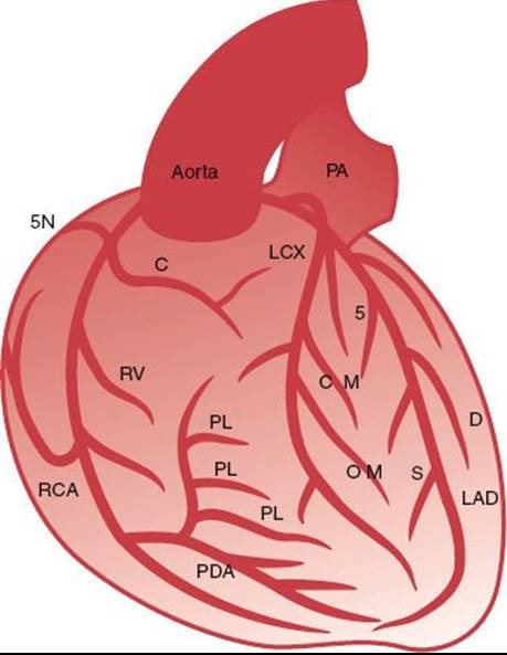
The left coronary artery usually originates from a single ostium in the middle portion of the left sinus of Valsalva (Fig. 92.1). The vessel originating from the left ostium is termed the left main coronary artery (LM) if it subsequently gives rise to both the LAD and the LCX. The LM measures 3 to 10 mm in diameter and is usually <40 mm long. The LM has no side branches and divides into the LAD and the LCX, although in 20% to 40% of cases a trifurcation with an intermediate branch between the LAD and LCX can be seen.
The LAD leads, in direct continuation of the LM, to the anterior interventricular groove toward the apex and supplies 40% to 60% of the left ventricular myocardium.
The LAD gives rise to septal branches, diagonal branches, and branches to the free right ventricular wall. The LCX originates at an almost vertical angle from the LM and courses posteriorly at variable length along the left atrioventricular groove, beneath the left atrial appendage and toward the crux of the heart. The LCX gives off one to three obtuse marginal branches, which supply the free lateral left ventricular wall and have the same course as the diagonal branches of the LAD.
Right Coronary Artery
Usually the RCA arises from the right sinus of Valsalva and runs in the right atrioventricular groove toward the crux of the heart. The segment from the ostium to the right-angled turn into the vertical part of the RCA is called the proximal RCA, the mid-RCA is defined as the vertical segment, and the distal RCA extends from the right-angled turn at the distal end of the vertical segment to the bifurcation into the posterior descending coronary artery and posterolateral branches.
FIGURE 92.1. Schematic representation of the major epicardial coronary arteries as seen in an anteroposterior projection. The left coronary artery originates left and posterior from the cusp of the aortic root as the left main (LM) coronary artery. The LM coronary artery divides after a variable length into the left anterior descending (LAD) artery and the left circumflex (LCX) artery. The LAD runs as a direct continuation of the LM anteriorly along the interventricular groove to the ventricular apex. The LAD gives rise to the septal branches, which take off at a vertical angle and immediately become intramural in the interventricular septum. The LAD also gives rise to the diagonal branches, which course epicardially over the anterolateral free wall. The LCX originates at a nearly vertical angle from the LM and courses posteriorly along the left atrioventricular groove. It gives rise to the obtuse marginal branches, which course epicardially and supply the free lateral wall. The right coronary artery (RCA) takes off anteriorly from the right coronary cusp and follows the right atrioventricular groove. The first branch of the RCA is the conal or infundibular artery, followed by the sinus node artery as the second branch. In the vertical portion of the atrioventricular groove, the RCA gives rise to the right ventricular marginal branches, which supply the right ventricular free wall. The RCA then courses posteriorly and divides at the crux of the heart base into the posterior descending artery (PDA) and a variable number of posterolateral branches. The PDA courses along the posterior interventricular groove toward the left ventricular apex. It closes a loop with the LAD along with the interventricular groove, whereas the posterolateral branches of RCA close a second perpendicular loop of blood supply with the LCX along with the atrioventricular groove. C, conus branch; D, diagonal branch; OM, obtuse marginal branch; PA, pulmonary artery; PL, posterolateral branch; RV, right ventricular branch; S, septal branch; SN, sinus node artery.
PERCUTANEOUS CORONARY INTERVENTION
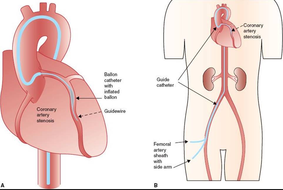
(This section is modified with permission from Windecker S, Meier B. Basics of interventional cardiology. In: Tripuraneni P, Jani S, Minar E, et al., eds. Intravascular brachytherapy: from theory to practice. London: Remedica, 2001:88–100.) The indications for percutaneous coronary intervention (PCI) have expanded during the past two decades, and there is currently no absolute contraindication to this technique. Arterial access is usually gained through an anterior wall stick of the right femoral artery using the Seldinger technique. The coronary guidewire is advanced by the coronary guiding catheter into the coronary artery and is cautiously navigated through the narrowing (stenosis) into the periphery of the vessel to be treated. It secures access to the coronary artery during the intervention and allows for the rapid exchange of balloons, stents, and other devices (Fig. 92.2).
The balloon catheter not only is central to balloon angioplasty but also serves as a complementary instrument for other intracoronary interventions, such as delivery of stents, local drugs, or radiation sources. Balloon catheters consist of a shaft providing support when pushing the catheter through vessels, a central lumen for the coronary guidewire, and an inflation channel for balloon expansion.
During conventional balloon angioplasty (percutaneous transluminal coronary angioplasty), a balloon catheter is advanced over the previously inserted coronary guidewire into the target stenosis and subsequently inflated until the balloon is fully expanded. The chief limitations to event-free survival after balloon angioplasty have been abrupt vessel closure (a short-term complication) and restenosis (a long-term complication).6 Abrupt vessel closure, defined as the sudden occlusion of the target vessel during or after angioplasty, has been reported in 4% to 8% of cases. Restenosis, defined as stenosis >50% diameter at follow-up angiography, has been the most important long-term limitation of balloon angioplasty, with an incidence of 30% to 50% and with 20% to 30% of patients requiring target vessel revascularization. Most restenoses occur during the first 4 months after balloon angioplasty.
The therapeutic effect of arterial vessel enlargement through PCI is accompanied by various degrees of arterial injury with exposure of thrombogenic components. This may result in intracoronary thrombus formation with its subsequent ischemic sequelae. Therefore, inhibition of platelets and the coagulation system has always been a central focus of interventional investigations. In patients undergoing PCI, aspirin is recommended at a low dose (75 to 325 mg/day), ideally administered at least 1 day before the procedure and continued indefinitely thereafter. Ticlopidine and clopidogrel are typically administered with a loading dose before or after the procedure. Additionally, they are prescribed for ≥6 months after VBT if a new stent was not placed and ≥12 months if a new stent was placed under the discretion of the interventional cardiologist.18
EXTERNAL-BEAM IRRADIATION STUDIES
Results using external-beam irradiation have been mixed.33–38 Studies of external-beam irradiation in the pig coronary balloon angioplasty model, done at Emory University, revealed that when 14 Gy was administered immediately before or after or 2 days after balloon injury, there was reduced neointima formation compared with controls, but the lumens were smaller owing to negative remodeling (contracture) of the vessel. Results from studies using 21 Gy after either angioplasty or stenting indicate a profound and consistent suppression of neointima formation.38,39 The lack of benefit seen with a 14-Gy external beam compared with 14 Gy delivered by VBT at a 2-mm radius from the source suggests that the minimum dose delivered to the vessel wall is not the only factor in determining outcome. Studies of 14- and 21-Gy external radiation treatment have shown focal myocardial necrosis, an effect never seen with endovascular irradiation at any dose. This suggests that sophisticated treatment techniques, limiting the dose to normal tissues, will be essential for external-beam therapy to be adopted.
ENDOVASCULAR IRRADIATION STUDIES
In contrast to the disparate results from studies using external radiation, numerous studies have consistently demonstrated remarkable suppression of neointima formation using radiation from a variety of isotopes delivered by an endoluminal approach. At least three groups have documented similar results in the pig coronary artery model of restenosis after balloon angioplasty, using the γ emitter 192Ir at roughly comparable doses.40–42
VBT typically reduces the neointima formation and maintains the patency of the lumen. Scanning electron microscopy of arteries irradiated to 14 Gy showed no morphologic differences from controls at 2 weeks; a confluent layer of endothelial or endothelial-like cells was present throughout the region of the angioplasty injury. However, at 28 and 56 Gy, neointima formation was nearly eradicated and endothelial coverage was incomplete. Inadequate endothelial recovery of an irradiated artery after angioplasty might render its luminal surface prothrombotic. In the setting of an appropriate physiologic stimulus, this can result in thrombotic occlusion. The problem of late thrombosis observed in clinical trials certainly is compatible with the delayed healing observed in the animal studies.
With stents playing an increasingly important role in the management of coronary stenosis, it became important to test whether radiation might prove a useful adjunct to coronary stenting. Studies carried out with β and γ emitters have demonstrated conclusively that the excess intimal hyperplasia occurring in a stent can be effectively eliminated by radiation delivered either before or after stenting.43
FIGURE 92.2. Schematic diagram of access to the coronary arteries during percutaneous coronary interventions. A: An introducer sheath with side arm is placed in the artery chosen for vascular access. A guiding catheter is introduced through the vascular access sheath and advanced to the coronary artery ostium chosen for the intervention. B: A guidewire is advanced through the guiding catheter into the coronary artery, navigated through the artery, and placed distal to the stenosis to be treated. A balloon catheter is then placed in the coronary stenosis for dilatation.
VASCULAR BRACHYTHERAPY PHYSICS AND DEVICES
The initial evaluation of radiation therapy in animal models of restenosis focused on testing the effect of radiation with commercially available radiation sources. Testing has included both teletherapy and brachytherapy. Although occasional positive results have been reported with teletherapy in experimental models of restenosis, consistently positive results emerged from a variety of brachytherapy approaches. These approaches have included both temporary (VBT) and permanent implants (radioactive stents). Although many isotopes have been proposed for VBT, only approximately half a dozen have been used in human clinical trials, and of those, only three have seen widespread use. The three isotopes, which have been used for well over 95% of all clinical trials of catheter-based systems, are 192Ir, 90Sr/Y, and 32P (Table 92.1).
BETA-CATH SYSTEM FOR CORONARY IN-STENT RESTENOSIS
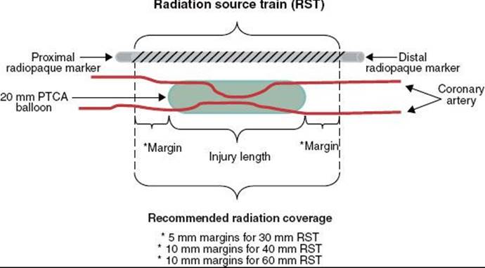
The Beta-Cath System is the only system currently manufactured that is offered for in-stent restenosis. 90Sr is a pure β emitter with a 28.8-year half-life (10,519.25 days) and 546-KeV maximum β energy. The daughter isotope 90Y is also a pure β emitter with a 64-hour half-life and 2.27-MeV maximum β energy. It is primarily the 90Y β emissions that are used for therapy because the 90Sr β particles are mostly absorbed by the stainless steel encapsulation and the surrounding catheter. The Beta-Cath System contains sources that are 0.38 mm in diameter and 2.5 mm in length confined within a flexible steel coil between radiopaque end plug markers that are maximum 0.495 mm in diameter and 2.5 mm in length. Source trains of 12, 16, and 24 sources (30-, 40-, and 60-mm active lengths, respectively) are commercially available. The nonradioactive marker plugs are located both proximally and distally to the radioactive sources in the jacketed train (Fig. 92.3). The markers permit fluoroscopic verification of the source train arriving at the end of the delivery catheter for treatment. For lesions plus margins longer than the available source train, a “pullback technique” is used in which the most distal portion of the lesion is treated first, and then the catheter is carefully pulled back to treat the more proximal lesion. At Scripps Clinic, we have the 60-mm source train available. A 60-mm source train can be used with a pullback technique to cover a distance of 120 mm. This technique could also be used for treatment of lesions at vessel bifurcations.44 Significant overlap or gaps between the treatment fields should be avoided. A careful review of the cine angiograms and a thorough understanding of the quantity, spacing, and positions of the radiopaque markers in the system (i.e., the two source train end markers, the delivery catheter stop marker, and the removable indicator of the source train marker wire) are pertinent when using this technique.
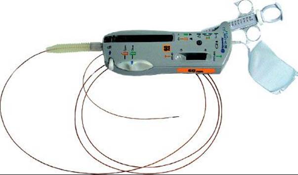
The Beta-Cath System consists of four main components: the source train, transfer device, delivery catheter, and accessories. The sources are stored in a hand-held transfer device and are advanced by a closed-loop hydraulic system that uses sterile water to send (and then return) the source train. The advantage of the Beta-Cath System is the relatively short treatment times (3 to 5 minutes) and the absence of radiation exposure to catheterization laboratory staff. The long half-life of the isotope permits a total shelf life of 12 months divided into two 6-month use periods, which allows for a decay correction of a few seconds in the second 6-month dwell times. A potential disadvantage of this system is the inferior depth–dose gradient compared with the γ source, attenuation by calcifications or stents, and lack of utility in larger vessels (Fig. 92.4).
A dose of 18.4 Gy is recommended at a 2-mm radius from the centerline of the source train axis for vessels with a reference diameter between 2.7 and 3.35 mm. For reference diameters between 3.35 and 4 mm and for most saphenous vein grafts, a dose of 23 Gy is recommended. The gross target volume is the stenotic area itself. The clinical target volume is the dilated part of the vessel. The planning target includes at least 5 mm proximal and distal to the clinical target volume45–47 for the 30-mm source train and at least 10 mm for the 40-mm and 60-mm source trains. Although both minimum 5-mm and minimum 10-mm treatment margins were proven safe and effective in the Beta-Cath System START trials, a cumulative comparison of efficacy outcomes out to 5 years suggests that longer treatment margins yield significantly lower target vessel revascularization (TVR) rates of 51% for minimum 5-mm margins versus 19% for 10-mm minimum margins versus 55% for the placebo, and with nonsignificant differences in major adverse coronary event (MACE) rates of 63% with minimum 5-mm margins versus 67% for minimum 10-mm margins versus 68% for the placebo (User’s Manual, Novoste Beta-Cath System).
TABLE 92.1 SUMMARY OF CATHETER-BASED SYSTEMS
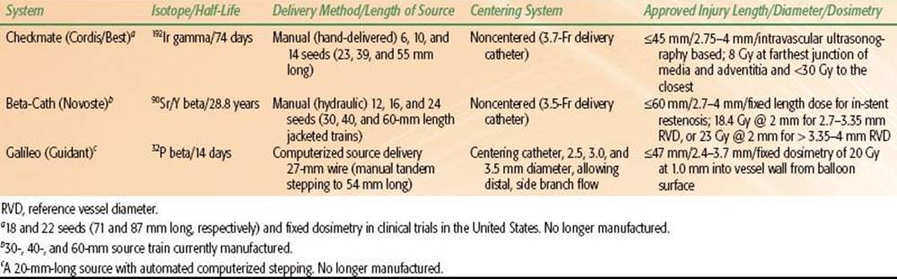
FIGURE 92.3. Radiation source train with proximal and distal radiopaque markers noted. (Courtesy of Novoste Corporation, Norcross, GA.)
Roles and Responsibilities
(This text is modified and printed with permission from Tripuraneni P. In: Tripuraneni P, Jani S, Minar E, et al., eds. Intravascular brachytherapy: from theory to practice. London: Remedica, 2001:272–274.) The FDA has mandated that VBT be carried out by a team consisting of an interventional cardiologist/radiologist, a radiation oncologist/therapeutic radiologist and oncologist, and a medical physicist. The roles and responsibilities of these specialists are listed in the following sections.31
Interventional Cardiologist/Radiologist
1. Perform preprocedure evaluation and communicate patient’s status (risk factors, interventions) with radiation oncologist.
2. Perform angioplasty with or without stenting as necessary.
3. Define the anatomic location of diseased vessel amenable to intervention, length/volume of intervention, angioplasty and stenting, and reference vessel diameter (including preintervention and postintervention vessel segment diameters), and communicate with radiation oncologist.
4. Determine the target volumes, jointly with the radiation oncologist and medical physicist.
5. Place delivery catheter and make final adjustment, in consultation with the radiation oncologist.
6. Advise the radiation oncologist of any changes in the delivery catheter position during the delivery of radiation.
7. Assist the radiation oncologist with any procedural details.
8. Assist the radiation oncologist in source removal as needed. In cases of medical or radiation emergencies, have procedures in place to ensure that radiation treatments are preplanned and nothing is left to chance.
Therapeutic Radiologist/Oncologist (Authorized User)
1. Review preprocedure evaluation, including patient’s status (including risk factors and interventions), with interventional cardiologist for the advisability of using intravascular radiation.
2. Review the anatomic location of diseased vessels amenable to intervention, length/volume of intervention, angioplasty, and stenting, and reference vessel diameter (including preintervention and postintervention vessel segment) with the interventional cardiologist.
3. Determine the target volumes together with the interventional cardiologist.
4. Obtain proper informed consent for VBT after discussions with the patient.
5. Review the final placement of the delivery catheter and any adjustments as needed.
6. Prescribe radiation dose and sign prescription.
7. Calculate treatment times, along with the medical physicist.
8. Insert radiation source.
9. Provide appropriate delivery of radiation.
10. Remove radiation source.
11. Supervise overall radiation delivery.
12. Participate in decisions and implementation of emergency source removal in case of medical or radiation emergencies.
Medical Physicist
1. Survey catheterization laboratory or radiation source delivery room and make appropriate preparations and modifications as needed.
2. Order radiation sources, under the direction of the radiation oncologist.
3. Calibrate sources upon arrival.
4. Ensure safe-keeping of radiation sources.
5. Prepare sources for clinical use.
6. Calculate treatment delivery times.
7. Conduct radiation survey of patient before and during treatment.
8. Assist radiation oncologist and interventional cardiologist in source removal in cases of radiation or medical emergencies.
9. Conduct radiation survey of patient after source removal.
10. In case of radiation or medical mishaps, inform appropriate regulatory authorities.
11. Participate in preparation of license application for medical use of VBT sources.
12. Develop and oversee quality assurance and improvement programs for efficacious and safe use of VBT sources in consultation with the radiation oncologist and interventional cardiologist as appropriate.
FIGURE 92.4. The Beta-Cath system is a hydraulic delivery system with a noncentered, 5-Fr, closed, over-the-wire delivery catheter. 90Sr seeds 24 source train (60 mm in length) is available. The delivery unit is shown. (Courtesy of Novoste Corporation, Norcross, GA.)
CLINICAL TRIALS OF CORONARY BRACHYTHERAPY
Nearly 5,000 patients have participated in clinical trials to determine the safety and efficacy of VBT.25,48,49 There have been seven double-blind randomized trials investigating the use of VBT on patients with in-stent restenosis.24,25,48,50–55 These trials led to the approval of one γ system using 192Ir and two β systems using 32P and 90Sr/Y isotopes.25,56,57
Of note, Rha et al.58 have reported that results from commercially available VBT are superior to the results obtained during investigational VBT trials as MACEs were lower with commercial radiation. This is likely from the lessons learned through clinical trials as dosimetry was optimized (higher doses, wider margins) and prolonged antiplatelet therapy was administered to reduce late thrombotic events. More recently, a rhenium-188–filled balloon source has also been shown to be effective for in-stent restenosis and improving long-term outcomes.59
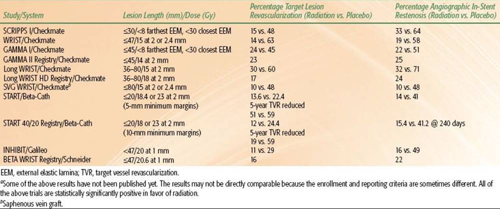
TABLE 92.2 SUMMARY OF PUBLISHED OR PRESENTED TRIAL RESULTS FOR CORONARY IN-STENT RESTENOSISA
-Radiation In-Stent Coronary Artery Restenosis Trials
The SCRIPPS I trial was the first double-blind, randomized radiation trial for coronary in-stent restenosis and restenosis without stents.24,43,52,60–62 It was a single-institution trial with off-site analysis involving the treatment of 55 patients during a 9-month period. Twenty-six patients were randomized to 192Ir and 29 to placebo. At 5-year follow-up, the target lesion revascularization (TLR) was significantly lower in the 192Ir group (23.1% vs. 48.3%; p = .05). There were two TLRs between years 3 and 5 in the treated patients, but none in the patients receiving placebo. The event-free survival rate (freedom from death, myocardial infarction, or TLR) was significantly lower in 192Ir-treated patients (34.5% vs. 61.5%; p = .028).
A second single-institution, randomized study of radiation for in-stent restenosis was carried out at the Washington Hospital Center in Washington, DC, and is known as the WRIST trial.55,63 The WRIST trial randomized 130 patients to either 192Ir or placebo. Fixed dosimetry was used, prescribing 15 Gy at either 2 or 2.4 mm depending on lumen diameter for lesions ≤47 mm. Six-month follow-up confirmed a statistically significant reduction in TLR from 63% to 14% and in angiographic restenosis from 58% to 19% in favor of radiation over placebo.
The GAMMA I trial was the first multi-institutional, randomized radiation trial for in-stent restenosis.25 A total of 252 patients at 12 centers were enrolled, and radiation therapy was delivered using intravascular ultrasound (IVUS)-based dosimetry (similar to the SCRIPPS trial dosimetry) for lesions ≤45 mm. A dose of 8 Gy was delivered to the farthest junction of the media and adventitia as long as the dose to the closest junction was <30 Gy, using IVUS. There were significant reductions in the rates of both TLR, from 45% to 24% at 9-month follow-up, and angiographic restenosis, from 50.5% to 21.6% at 6-month follow-up, in favor of radiation over placebo.
The GAMMA II registry included 125 patients at 12 centers and used fixed dosimetry for lesions ≤45 mm (same entry criteria as GAMMA I trial), with 14 Gy prescribed at a 2-mm radius for all patients.64 There was a TLR rate of 23% at the 9-month follow-up and an angiographic restenosis rate of 25% at the 6-month follow-up. This registry showed that the results with 192Ir were reproducible and similar to those in the GAMMA I trial with simplified dosimetry.65
The LONG WRIST trial was a two-institution, 120-patient randomized trial that used fixed dosimetry of 15 Gy at a 2- or 2.4-mm radius, for longer lesions of 36 to 80 mm. There was a statistically significant decrease in the 6-month angiographic restenosis rate (32% vs. 71%; p = .0002) in favor of radiation.66 The PLAVIX WRIST trial was a registry of 120 patients who received 6 months of clopidogrel. Six-month follow-up has demonstrated that extended antiplatelet therapy is able to eliminate the problem of late thrombosis observed in the WRIST and GAMMA studies67 (Table 92.2).
-Radiation In-Stent Coronary Artery Restenosis Trials
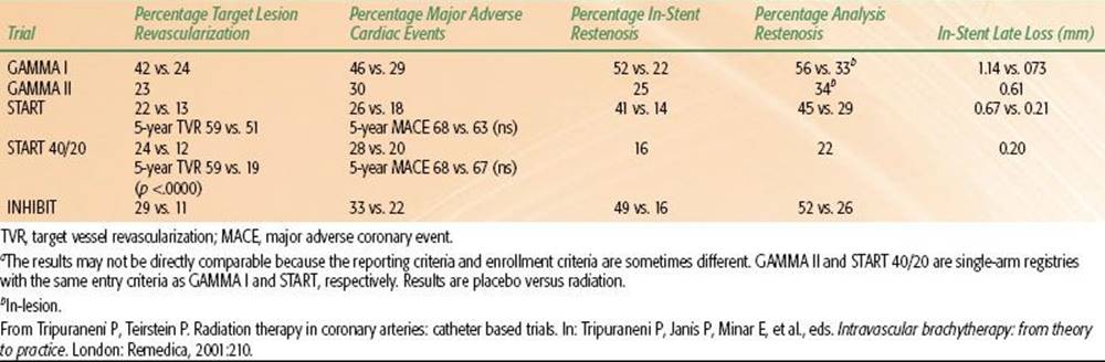
The START trial (Beta-Cath system) was a 50-center multi-institutional, double-blind, randomized trial of 476 patients with in-stent restenosis.68 A 30-mm-long 90Sr/Y source was used to treat lesions ≤20 mm; the dose prescribed was 18.4 Gy at a 2-mm radius for vessels ≥2.7 and ≤3.35 mm in diameter, and 23 Gy at a 2-mm radius for vessels ≥3.35 and ≤4 mm in diameter. At 8-month follow-up, the TLR rate was statistically decreased from 22% to 13% (p<.008), and the angiographic in-stent restenosis rate was also significantly decreased from 41% to 14% in favor of radiation over placebo.
The START 40/20 trial was a registry of 207 patients with the same entry criteria as START, except the source train was 40 mm long.69 The 8-month binary angiographic in-stent restenosis rate was 16%. The frequency of geographic misses with the longer source train in the START 40/20 trial was 6% compared with 15% in the START 30 trial.
The PREVENT trial (Galileo system) was a six-institution feasibility study involving 105 patients, 70% of whom had de novo lesions and 61% of whom were stented.51 The patients were randomized to placebo or 16, 20, or 24 Gy at 1 mm from the balloon surface for lesions ≤25 mm. The TLR rate was statistically decreased with 32P versus placebo from 24% to 10%, and the in-stent angiographic restenosis rate was decreased from 39% to 8%. The INHIBIT trial (Galileo system), a multi-institutional, double-blind, randomized study, enrolled 332 patients with in-stent restenosis at 27 centers. Fixed dosimetry was used to prescribe 20 Gy at 1 mm from the balloon surface for lesions with an injury length of ≤47 mm.29,49 Because the source is only 27 mm in length, manual repositioning of the balloon catheter was used to treat the longer lesions. At 9-month follow-up, the rate of MACEs was decreased from 31% to 15% (p = .0006), and the rate of angiographic in-stent restenosis was also significantly decreased from 49% to 16% (p <.0001) in favor of radiation over placebo. Sixty-four patients in the radiation group and 76 patients from the placebo group were treated with the pullback technique for lesions longer than 20 mm. No safety issues were identified in patients in whom there had been a significant overlap of active sources at the junction.
The Beta WRIST trial was a registry of 50 patients with in-stent restenosis treated with the Boston Scientific/Schneider system at the Washington Hospital Center using the same entry criteria as the WRIST trial.55 An angiographic restenosis rate of 22% and a TLR rate of 16% at 6 months were noted, which were similar to the results obtained in the radiation arm of the WRIST study (Tables 92.3 and 92.4).
Trials on Drug-Eluting Stents for Coronary In-Stent Restenosis
The role of VBT in coronary in-stent restenosis has clearly changed with the introduction of effective DESs. The first trial using DESs was a Brazilian study involving 31 patients. There was no incidence of restenosis reported on initial evaluation of patients at 4 months. No clinical events were reported at 8 months.15 There are also data showing that DESs can be safely and effectively delivered after VBT.70
The RAVEL (Randomized Study with Sirolimus-eluting Velocity Balloon-Expandable Stent) involved 238 patients randomized to either DESs or BMSs.13 At 3 years, event-free survival rates for target lesion revascularization were 93.7% in the DES arm versus 75% for the control group (p <.001).71
Paclitaxel-eluting stents have been investigated in the series of TAXUS trials I through IV.11,72–74 TAXUS I evaluated 61 patients randomized to 15-mm TAXUS NIRx paclitaxel-eluting stent (Boston Scientific, Natick, MA) or similar drug-free stent. At 6 months, there were no incidences of in-stent restenosis in the DES arm and three patients developed restenosis in the BMS arm (p = .112).11 TAXUS II investigated two different drug formulations and compared them to BMSs. No differences were noted between the two drug arms, but a statistical advantage was noted in the DES arms compared with BMSs.72 TAXUS IV randomized 1,314 patients to EXPRESS BMSs or EXPRESS DESs. Nine-month angiographic results were available in 559 patients. A relative risk of restenosis was reduced by 70% in the DES arm (p <.001).73 The TAXUS V and VI trials evaluated the EXPRESS DESs in patients with more complex diseases.
The two major trials comparing DESs to VBT have now been completed.75 The TAXUS V trial is a multicenter randomized trial with 396 patients enrolled. This study demonstrated that paclitaxel-eluting stents when compared to VBT significantly reduced the ischemic target lesion revascularization rate by 40% at 9 months: 6.3% for DESs versus 13.9% for VBT. Relative reductions in total target lesion (61%) and target vessel (49%) revascularization events were higher in patients treated with paclitaxel-eluting stents rather than those receiving VBT when considering only ischemic-related events. Additionally, the 9-month rate of major adverse coronary events was reduced by 43% in the DES arm versus the VBT arm.17 The SISR trial is a prospective, multicenter randomized trial with 384 patients enrolled. Patients were randomized to sirolimus-eluting stents versus VBT for restenosis following BMS implantation. At 6 months, the DES arm had superior clinical and angiographic outcomes when compared with VBT. At 3-year follow-up, patients treated with DESs had improved survival-free TLR and TVR compared to VBT. MACEs and stent thrombosis did not reach a statistical difference between the two groups.18
Nonetheless, there have been some reports of DES-associated arteriopathy with late stent malapposition, aneurysm formation, hypersensitivity reactions, and DES-induced spasms. The more recent VBT trials have seen very few of these complications.76,77 Still, short-term results of DESs compare favorable with VBT and long-term results will hopefully address these concerns.
TABLE 92.3 SUMMARY OF THE PIVOTAL MULTI-INSTITUTION RANDOMIZED TRIALS AND REGISTRIES FOR CORONARY IN-STENT RESTENOSIS

TABLE 92.4 RESULTS OF PIVOTAL TRIALS AND REGISTRIES FOR CORONARY IN-STENT RESTENOSISA
Vascular Brachytherapy for In-Stent Restenosis Following DESs
VBT following DES failure has been shown to be effective and safe. Up to 10% of patients develop restenosis following DESs. The optimal treatment remains unclear for patients who fail DESs. Options include balloon angioplasty, additional DESs with either the same or a different drug, or VBT.
The RESCUE Registry is a multicenter international electronic registry for patients who receive VBT for recurrent restenosis following DESs. The purpose of the registry is to evaluate the safety and efficacy of VBT following DESs and to compare it to repeat DESs following in-stenosis of DESs.
Sixty-one patients received VBT following DESs and were compared with 50 patients who received additional DESs (Taxus or Cypher stents) after DES restenosis. The patient’s demographic and angiographic characteristics were similar in both groups. Torguson et al.78 presented the data at the 2005 American Heart Association Scientific Sessions. VBT appeared to be safe. At 6 months, target vessel revascularization rates were 3.3% versus 16% (p = .04) in favor of VBT. TLR rates were 3.3% versus 8% (p = .24) in favor of VBT as well. At 8 months, VBT was associated with fewer MACEs compared with repeat DESs (9.8% vs. 24%; p = 0.044).79
At Scripps Clinic, the clinical experience on the first 41 patients treated with VBT following DESs has been reviewed. Median follow-up is 8 months. TLR rates are 22% and TVR (target vessel failure) rates are 2%.
Saphenous Vein Graft In-Stent Restenosis Clinical Trials
Saphenous vein grafts often are larger vessels with luminal diameters of >4 mm. Allowances must be made to deliver adequate doses when using IVUS-based or fixed dosimetry. All SCRIPPS trials included patients with saphenous vein grafts. However, the number of patients enrolled into SCRIPPS I was too small to reach any conclusions. Thirty of 130 patients in the WRIST trial were treated for in-stent restenosis of saphenous vein grafts. Subgroup analysis of WRIST suggests that radiation was as effective in saphenous vein grafts as in native coronary vessels. The more definitive study, SVG WRIST, a multi-institutional, randomized trial testing the efficacy of the Checkmate system with 192Ir, enrolled 120 patients with lesions <47 mm in length.80 A dose of 15 Gy at a 2-mm radius for vessels of 2.5- to 4-mm radius and 18 Gy at a 2-mm radius for vessels of 4- to 5-mm radius was delivered. A statistically significant reduction in the rate of in-stent restenosis of 43% versus 15% (p = .004), in favor of radiation over placebo, was observed. Likewise, the TLR rate at 6 months was statistically significantly decreased (10% vs. 48%; p<.01) in favor of radiation over placebo. At 36-month follow-up, TLR rate was still in favor of the radiation arm (43% vs. 66%; p = .02).81 With appropriate doses, the in-stent rate of saphenous vein grafts can be reduced with adjuvant brachytherapy after appropriate recanalization.
In summary, there are numerous multi-institutional, randomized trials confirming the efficacy of radiation in decreasing in-stent restenosis. The three pivotal trials, GAMMA I, START, and INHIBIT, led to the approval of VBT for use in native coronary in-stent restenosis after recanalization. The Beta-Cath system, the only commercial system that is currently available, was approved based on the START trial. The system has been used successfully and has a registry confirming that longer margins decrease edge restenosis. This system was initially approved for 20-mm-long injured length with 2-mm radius prescription dosimetry. Doses would need to be adjusted for vessels with larger diameters between 3 and 5 mm.82
De Novo Coronary Artery Stenosis Clinical Trials
There are limited data supporting the use of routine radiation therapy to prevent restenosis for de novo coronary artery stenosis after recanalization. However, data in the Beta-Cath trial indicate that radiation may indeed decrease the rate of restenosis in patients. This trial was a multi-institution, double-blind randomized study with 1,456 patients.30 After balloon angioplasty, patients received either radiation doses of 14 or 18 Gy depending on vessel diameter or no adjuvant radiation. The 8-month angiographic analysis showed a statistically significant decrease in restenosis in favor of radiation (34.3% vs. 21.4%; p = .003). However, with edge failures, this did not translate into a clinical benefit. Likewise, the GENEVA dose-finding trial83,84 confirmed the dose response with increasing doses, with the effect being more profound in the patients receiving angioplasty only without stenting. Nevertheless, with emerging data from the DES trials, it seems unlikely that VBT will be used routinely in de novo coronary artery stenosis. However, for special situations of high-risk de novo stenosis in which stents may not be optimal, such as small-diameter vessels, longer lesions, or branch vessels, VBT may possibly find a niche role.
RADIOACTIVE STENTS
Currently, the majority of patients undergoing PCI receive coronary stents. Coupling the radiation delivery to the stent appears attractive in that it simplifies the delivery of treatment. Most clinical investigations have been undertaken with the 32P β-emitting stent.85 Stents have been tested with activities ranging from 0.5 to 20 μCi. These stents are of extremely low activity and can be handled with the aid of a 1-cm acrylic shield. Unfortunately, clinical trials using the 32P stent demonstrated restenosis rates of approximately 50%, largely owing to intimal proliferation at the stent edges. These are often called candy wrapper edge failures. These clinical failures have inspired recent investigation of variations of the 32P-coated stent with “cold” ends, “hot” ends, low-pressure balloon deployment systems, and so forth; 103Pd (a γ-emitting isotope)-emitting stents; and 198Au-emitting stents. Because studies to date indicate a lack of efficacy, currently there are no clinical trials evaluating the efficacy of radioactive-coated stents.
PERIPHERAL VASCULAR BRACHYTHERAPY TRIALS
Peripheral vascular disease involves more organs than coronary artery disease (CAD), and hence there are more diverse clinical situations, manifestations, and end points.86 It is challenging to define measurable clinical end points for a diverse group of “host” organs (e.g., extremities, kidney, liver) that differ not only anatomically but also in function. Unlike coronary vessels, most peripheral vessels have a diameter >3 mm and, in fact, are typically approximately 7 to 10 mm. Peripheral vascular lesions tend to be much longer and are more likely to be multifocal. Because of the larger vessel diameter and increased thickness of the vessel wall, VBT in peripheral arteries is likely to require the use of either a more penetrating γ source (192Ir) or a β source that is in direct contact with the vessel wall.
Most trials in peripheral vessels have used an high–dose-rate (HDR) afterloader and treatment of the superficial femoral or popliteal artery. After successful percutaneous transluminal angioplasty (PTA) of these arteries, the restenotic rate at 6 months varies from 25% to 77%.87,88 In a Veterans Administration study, the actuarial restenosis rate was 41% at 36 months.89
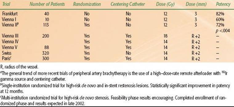
The feasibility of VBT for peripheral vascular systems in humans was first documented in a study from Frankfurt, Germany.22 In this study, 30 patients with in-stent restenotic lesions in femoropopliteal arteries were treated with repeat PTA, stent implant, and VBT. A dose of 12 Gy at 3 mm was administered with a 192Ir HDR afterloader through a 5-Fr, noncentered catheter. At last follow-up, the median follow-up time was 32.9 months for 28 patients (range, 7 to 84 months). There were no adverse effects from the brachytherapy. The 5-year vessel patency rate was 82% (23 of 28 patients) based on Doppler ultrasonography. Stenosis developed in the treated vessel in three of 28 patients (11%); two (7%) had complete occlusion of the vessel due to thrombosis after 16 and 37 months.
Investigators in Vienna, Austria, have mounted a series of trials exploring the use of VBT in similar patients.90,91 The Vienna II trial enrolled 107 patients with symptomatic de novo or restenotic femoropopliteal lesions treated with angioplasty and then randomly assigned patients to either additional radiation therapy or no further treatment. A fixed dose of 12 Gy at a 3-mm radius from the source center and a margin of 1 cm at each end of the injured segment were delivered with a 192Ir HDR afterloading system. At 6 months, the angiographic restenosis rate was significantly lower in the radiation group compared with the control group (28% vs. 54%; p <.05). The cumulative patency rate at 12 months was significantly higher in the radiation group than in the control group (64% vs. 35%; p <.005). Subgroup analysis demonstrated that restenotic lesions, occlusions, and long lesions benefit the most from VBT. Despite radiation, the recurrence rate in the radiation arm was 28%. It is postulated that this may be due to the relatively modest radiation dose and the absence of a centering catheter. At 5-year follow-up, the stenosis recurrence rate was similar in each group, 72.5% each arm, (p >.99).92 This showed that the radiation resulted in a delay but not an inhibition in restenosis.
The Vienna III trial enrolled 134 patients. This double-blind study randomized patients following angioplasty to brachytherapy or placebo irradiation. Patients had either de novo lesions ≥5 cm or restenosis following femoropopliteal angioplasty. A dose of 18 Gy (γ-irradiation) was prescribed 2 mm from the surface of the centering balloons. At 24 months, patency rates based on intention-to-treat analysis was 54% in the brachytherapy arm and 27% in the placebo arm (p <.005). VBT, however, reduced the restenosis rates for recurrent lesions only and not for de novo lesions.
The Vienna V trial studied 88 patients with femoropopliteal lesions at high risk for restenosis (mean treatment length, 16.8 ± 7.3 cm). This double-blind study randomized patients to receive either brachytherapy with 192Ir (14 Gy to 2 mm into arterial wall) or with nonradioactive seeds. In this trial, brachytherapy did not improve 6-month patency after femoropopliteal stent in high-risk patients mainly because of a high incidence of early and late thrombotic events.93
The Peripheral Artery Radiation Investigational Study (PARIS) is the pivotal multi-institutional, randomized trial of VBT in superficial femoropopliteal arteries using the Nucletron HDR afterloader (Nucleotron, The Netherlands). This study consists of two phases: an initial lead-in phase of 40 patients, followed by a second phase in which 300 patients were randomized to receive or not receive radiation after PTA. In the initial phase, 35 of 40 patients were successfully irradiated with no procedural complications.55 The angiographic restenosis rate at 6 months was 17.2%, which is very promising. The second phase started in early 1998 as the first multicenter, multinational, prospective, double-blind, randomized trial. Although initial results were promising at 6 months, the restenosis rates were similar in both groups (27.5% placebo and 28.6% brachytherapy) at 12 months (Table 92.5).
Krueger et al.94 reported on 30 patients who underwent PTA for de novo femoropopliteal stenosis. Patients received either 14-Gy centered VBT or no radiation. Rates of restenosis were statistically significantly lower in the radiated group at 6 (p = .006) and 12 (p = .042) months.
Compared with the femoropopliteal arteries, the tibioperoneal arteries are smaller; hence, post-PTA restenosis tends to occur more frequently. Long-term success is limited mostly by neointimal hyperplasia. In a study of 55 PTA lesions in 40 patients, 44% remained patent at an average follow-up of 25.8 months.95 There are no clinical data reported on the use of radiation therapy for the tibial-peroneal vessels, but this remains a potentially fertile site for further investigation.
There are two types of stenosis in renal arteries: ostial (at the origin of the renal artery from the aorta) and nonostial (beyond the ostium). Ostial stenoses are more difficult to treat and tend to recur; hence, they are often treated with stenting. The restenosis rates as determined by angiography are somewhat lower, at 23%, for nonostial regions compared with 30% for both ostial and nonostial lesions together.96,97 Renal artery stenosis also lends itself to exploration with VBT. In certain clinical subgroups, the prevalence of serious renal artery disease is as high as 43%, especially in the growing population of patients older than 50 years of age with multiple manifestations of atherosclerosis.98 Renal artery disease may account for up to 15% of patients with renal failure in the dialysis population older than 50 years of age. The morbidity and mortality among these patients are very high; hence, any potential benefit of VBT in the treatment of renal artery disease deserves investigation. In recent years, there have been some limited and selected cases published in the literature demonstrating the efficacy and safety of VBT for the treatment of renal artery in-stent restenosis.99–102
TABLE 92.5 DETAILS OF FEMORAL POPLITEAL ARTERY BRACHYTHERAPY TRIALSA
There are more than 120,000 patients with end-stage renal disease in the United States who require vascular access for hemodialysis. The most common forms of vascular access are arteriovenous (AV) grafts and central venous canalization. AV grafts typically fail within 14 to 19 months, with a reported primary occlusion rate of 15% to 50% at 1 year.103 The most common cause of failure is stenosis at the anastomosis. The most common causes are thrombosis and intimal hyperplasia. Development of intimal hyperplasia at the site of venous anastomosis is due to several factors, including high-flow turbulence, compliance mismatch, vessel vibration, and platelet activation The restenosis rates for AV dialysis grafts after PTA are 9% at 3 months, 29% at 6 months, and 61% at 12 months; the restenosis rate for the subclavian vein after PTA alone is 71% at 6 months.104 The restenosis rate after PTA and stenting for the subclavian vein is 30% to 53% at 1 year. An FDA-approved pilot study at New York Hospital used external-beam radiation (8 to 12 Gy, given in two equal fractions 48 hours apart) in a total of 10 patients to prevent restenosis. Unfortunately, all patients had restenosis by 18 months, suggesting no benefit from the therapy.90 A feasibility study involving a series of eight patients with restenosis of AV fistula in hemodialysis patients has been recently published.105 Although it appeared that radiation with 192Ir after PTA of fistula stenosis appeared as a safe and feasible method in these patients, the radiation did not seem to decrease the incidence of restenosis. Different fractionation schemes are currently under investigation.
Compared with trials in CAD, peripheral vascular disease clinical trials testing the efficacy of VBT in reducing restenosis are in earlier stages with mixed results. VBT has now been tried in numerous sites outside the coronary arteries, including vein grafts, renal artery in-stent restenosis, femoropopliteal arteries, and even the carotid arteries.106 The Vienna II and III trials, single-institution randomized trials, initially supported its efficacy in peripheral vascular disease. However, 5-year results were disappointing in the Vienna II trial. Certainly, there are opportunities to explore the role of VBT in peripheral arterial disease.
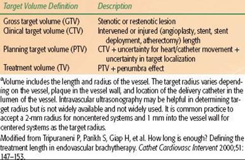
TABLE 92.6 VASCULAR BRACHYTHERAPY TREATMENT TERMINOLOGYA
ADDITIONAL CLINICAL CONSIDERATIONS
Edge Restenosis
When restenosis occurs after VBT, the renarrowing is found at the treatment edges in one-third to one-half of patients. The etiology of edge failure is likely to be multifactorial but most likely results from inadequate radiation dose delivered to injured lesion margins. Many factors can lead to higher-than-expected rates of edge failure. These include geographic miss, which arises from misalignment of the radioactive source in the injured segment of the vessel.107,108 In several studies using catheter-based radiation, careful, quantitative coronary angiographic measurements have documented a surprisingly high incidence of inadequate coverage of the injured region by the radioactive source. The balloon catheters used initially to open the stenotic segment can slip forward or backward (“watermelon seeding”), causing unintended injury to the lesion margins. Also, barotrauma from both angioplasty and stent deployment contributes to arterial wall injury beyond the nominal lengths of the balloons or stents.109 Longitudinal seed displacement may also contribute to higher-than-expected restenotic rates at lesion margins owing to the movement of the radioactive seeds relative to the coronary vessel during the cardiac cycle. In a seed movement analysis of 19 cineangiograms, proximal movement of 0 to 2 mm and distal movement of 1 to 5 mm were observed.110In addition to more movement in the distal portion of arteries, the movement also varies with the particular artery (possibly more in the circumflex, for example). Uncertainty in target localization can arise because of the difficulty in visual estimation of proximal and distal lesion ends. This uncertainty is compounded by different magnifications and obliquity of various projections during fluoroscopy and cineangiography and the relative lack of reference points available (branch vessels are commonly used as reference points for targeting). Last, the dose falloff and penumbra effect of the particular isotope used can contribute to marginal failure.111
VBT cannot be effective in regions injured by angioplasty or atherectomy where radiation is not delivered. Although the causes of edge failure are still unclear and most likely multifactorial, several strategies have been used to decrease edge failures. First, careful cineangiographic documentation of injury to the vessel should be carried out at every balloon angioplasty or stent placement (discouraged to minimize late thrombosis). Second, the most proximal and distal extents of the injury to the vessel should be carefully determined, ideally with a side-branch reference point. Finally, a very wide margin (i.e., 5 to 10 mm) of the radiation source should be provided on either side of the injured vessel region. These measures will not eliminate edge failure but will probably considerably reduce its occurrence.57
TERMINOLOGY
Based on the International Commission on Radiation Units and Measurements Report 50, terminology for VBT volumes was proposed that takes into consideration both radial and longitudinal dimensions.111 This terminology will help to define the target volume with attention to appropriate margins to decrease edge failures. The gross target volume (GTV) is the length of stenotic segment with an appropriate radius that may vary along the length. The clinical target volume (CTV) is the interventional length, which is delineated by the most proximal and distal extents of injury, and is always larger than GTV. The planning target volume (PTV) is the CTV plus a margin to account for both heart and catheter movements and inaccuracies in the visual delineation of the ends of the CTV. The uncertainty or magnitude of the margin depends on the location of the target in the vessel, the delivery system (centered or noncentered), and the cardiac cycle. The target volume is the volume irradiated based on the PTV and the penumbra of the isotope’s effect, which depends on the isotope, source design, and prescription distance. In practice, it is necessary to give at least a 4- to 8-mm margin to the longest injured length of the vessel (Table 92.6).
SUBACUTE THROMBOSIS
Similar to the first attempts at stent implantation, initial enthusiasm for VBT was dampened by reports of target thrombosis, particularly thrombosis occurring late (>30 days) after treatment. In early trials, late thrombosis after VBT was observed in 3% to 10% of patients independent of the isotope and delivery system tested.67,112 The thrombotic episode usually manifested itself as a sudden target vessel occlusion resulting in myocardial infarction 1 to 9 months after radiation treatment. There is no uniform definition or criteria for subacute thrombosis. Total occlusions can be subdivided into two groups: (a) symptomatic late thrombosis, occurring more than 30 days after the index procedure and resulting in myocardial infarction, and confirmed by angiography; and (b) silent late occlusions, occurring more than 30 days after the index procedure. These total occlusions are seen on the protocol-required follow-up angiogram without clinical symptoms of myocardial infarction.113
The emergence of this complication seriously jeopardized radiation as a viable treatment modality for CAD. Careful study, however, yielded two helpful clues that led to a dramatic reduction in radiation-associated late thrombosis: (a) the overwhelming majority of patients sustaining a late thrombosis had a new stent implanted at the time of the radiation procedure, and (b) almost all patients sustaining late thrombosis had discontinued antiplatelet therapy. Two strategies to prevent late thrombosis were initiated. First, the implantation of new stents during or immediately after treatment with brachytherapy was strongly discouraged. Second, antiplatelet therapy was extended for 6 to 12 months after the radiation therapy procedure. This strategy has now been tested with apparent success in several large series. In more recent trials using the aforementioned strategies, the incidence of late thrombosis was similar to that in the placebo arm, in the range of 1% to 3%. In the SCRIPPS III trial, the late thrombosis rate is zero.114
Stent placement at time of radiation delivery can cause both increased late thrombosis and possibly decreased efficacy of the brachytherapy itself. Pooled retrospective data from the SCRIPPS I, WRIST, and GAMMA I trials for patients with in-stent restenosis show an even more pronounced effect of brachytherapy in patients without new stent placement than in patients with stent placement. The Geneva dose-finding study of de novo stenosis confirmed these results, with decreased restenosis rates with radiation after angioplasty only compared with angioplasty and stenting.
SUMMARY
VBT is the first proven, clinically effective therapy in the management of in-stent restenosis. The Beta-Cath system using 90Sr/Y is currently the only available system used in the treatment of coronary in-stent restenosis.
Results of DES trials have decreased the need for VBT in coronary in-stent restenosis. The TAXUS V study and the SISR randomized trials demonstrate that DESs may be superior to VBT in treating coronary in-stent restenosis.74However, VBT can’t be entirely eliminated as there are still concerns with long-term complications with DESs. Long-term result studies of DESs are starting to address these concerns.
Outcomes from the RESCUE Registry have shown VBT to be safe and effective following in-stent restenosis of DESs. Overall MACEs were lower in the VBT group when compared to retreatment with DESs. Additionally, there are cost issues as well with multiple stents as there are concerns for stent thrombosis, increased DES failures, and additional interventional procedures. Also, it may be difficult to implant additional DESs in small vessel disease.
VBT is finding a niche role in the treatment of coronary artery disease. Although DESs may become even more efficacious as the next generation of DESs are released, VBT should be considered for earlier use in patients with DES failure, diffuse long lesions, small vessels, vein grafts, bifurcation lesions, possibly de novo lesions, and diabetic lesions. Although the initial cost for treatments with VBT may be high, cost-effective analysis performed from GAMMA I and INHIBIT studies show that these costs are offset in the long run due to reduced need for additional bypass surgeries and coronary interventions.
VBT may have an increased role in the treatment of vein graft in-stent restenosis and for peripheral vascular disease. The role of VBT for peripheral vascular disease will mainly be defined based on results of current clinical trials. The dose and volume to be treated will need to be refined to improve efficacy further. VBT has certainly come a long way, becoming more user-friendly and cost-effective.
REFERENCES
1. Babapulle NM, Joseph L, Belisle P, et al. A hierarchical Bayesian meta-analysis of randomized clinical trials of drug-eluting stents. Lancet 2004;364:583–591.
2. Lemos PA, Arampatzis CA, Saia F, et al. Treatment of very early vessels with 2.25mm diameter sirolimus-eluting stents. Am J Cardiol 2004;93:633–636.
3. Williams DO, Holubkov R, Yeh W, et al. Percutaneous coronary intervention in the current era compared with 1985–1986: the National Heart, Lung, and Blood Institute Registries. Circulation 2001;102:2945–2951.
4. Moses JW, Leon MB, Popma JJ, et al. Sirolimus-eluting stents versus standard stents in patients with stenosis in native coronary artery. N Eng J Med 2003;349:1315–1323.
5. Dobesh PP, Stacy ZA, Ansara AJ, et al. Drug-eluting stents: a mechanical & pharmacologic approach to coronary artery disease. Pharmacotherapy 2004;24:1554–1577.
6. Landau C, Lange RA, Hillis LD. Percutaneous transluminal coronary angioplasty. N Engl J Med 1994;330:981–983.
7. Hillegass WB, Ohman EM, Califf RM. Restenosis: the clinical issues. In: Topol EJ, ed. Textbook of interventional cardiology, vol 1, 2nd ed. Philadelphia: WB Saunders, 1994:415–435.
8. Klein LW, Rosenblum J. Restenosis after successful percutaneous transluminal coronary angioplasty. Prog Cardiovasc Dis 1990;32:365–382.
9. Ludbrook PA. Coronary restenosis: its mechanisms and modification—overview. Coron Artery Dis 1993;4:225–228.
10. Raja SG, Berg GA. Safety of drug eluting stents: current concerns and controversies. Curr Drug Saf 2007;2:212–219.
11. Grube E, Silber S, Hauptamann KE, et al. TAXUS I: six- and twelve-month results form a randomized double-blind trial on the slow release paclitaxel-eluting stent for de novo coronary lesions. Circulation 2003;107:38–42.
12. Degertekin M, Regar E, Tanabe K, et al. Sirolimus-eluting stent for treatment of complex in-stent restenosis: the first clinical experience. J Am Coll Cardiol 2003;41:184–189.
13. Morice MC, Serruys PW, Sousa JE, et al. A randomized comparison of a sirolimus-eluting stent with a standard stent for coronary revascularization. N Engl J Med 2002;346:1315–1323.
14. Saia F, Lemos PA, Sianos G, et al. Effectiveness of sirolimus-eluting stent implantation for recurrent in-stent restenosis after brachytherapy. Am J Cardiol 2003;41:184–189.
15. Sousa JE, Costa MA, Abizaid A, et al. Lack of neointimal proliferation after implantation of sirolimus-coated stents in human coronary arteries: a quantitative coronary angiography and three-dimensional intravascular ultrasound study. Circulation2001;103:192–195.
16. Holmes DR, Teirstein P, Satler L, et al. Sirolimus-eluting stents vs. vascular brachytherapy for in-stent restenosis within bare-metal stents. JAMA 2006;295:1264–1273.
17. Stone GW, Ellis SC, O’Shaughnessy CD, et al. Paclitaxel-eluting stents versus vascular brachytherapy for in-stent restenosis within bare metal stents: the TAXUS V ISR randomized trial. JAMA 2006;295:1253–1263.
18. Holmes DR, Teirstein P, Satler L, et al. 3-year follow-up of the SISR (sirolimus-eluting stents versus vascular brachytherapy for in-stent restenosis) trial. J A Coll Crdiol Intv 2008;1:439–448.
19. Mauri L, Hsieh WH, Massaro JM, et al. Stent thrombosis in randomized clinical trials of drug-eluting stents. N Engl J Med 2007;356:1020–1029.
20. Jahraus, CD, St Clair W, Gurley J, et al. Endovascular brachytherapy for the treatment of renal artery in-stent restenosis using a beta-emitting source: a report of five patients. South Med J 2003;96:1165–1168.
21. Waksman, R. an update on peripheral brachytherapy. Endovasc Today 2004;October:43–52.
22. Schopohl B, Leirmann D, Pohlit LH, et al. 192Ir endovascular brachytherapy for avoidance of intimal hyperplasia after percutaneous transluminal angioplasty and stent implantation in peripheral vessels: 6 years of experience. Int J Radiat Oncol Biol Phys1996;36:835–840.
23. Condado JA, Waksman R, Gurdiel O. Long-term angiographic and clinical outcome after percutaneous transluminal coronary angioplasty and intracoronary radiation therapy in humans. Circulation 1997;96:727–732.
24. Teirstein PS, Masullo V, Jani S, et al. Catheter-based radiotherapy to inhibit restenosis after coronary stenting. N Engl J Med 1997;336:1697–1703.
25. Leon MB, Teirstein PS, Moses JW, et al. Localized intracoronary gamma-radiation therapy to inhibit the recurrence of restenosis after stenting. N Engl J Med 2001;344:250–256.
26. King SB, Williams DO, Prakash C, et al. Endovascular beta-radiation to reduce restenosis after coronary balloon angioplasty: results of the Beta Energy Restenosis Trial (BERT). Circulation 1998;97:2025–2030.
27. Verin V, Urban P, Popowski Y, et al. Feasibility of intracoronary β-irradiation to reduce restenosis after balloon angioplasty. Circulation 1997;95:1138–1144.
28. Lansky A, Desai K, Costantino C, et al. Predictors of stent and stent-edge restenosis after Sr-90 radiation in the START trial. J Am Coll Cardiol 2001;37:54A.
29. Waksman R, Raizner A, Chiu K, et al. Beta radiation to inhibit recurrence of in-stent restenosis: clinical and angiographic results of the multicenter, randomized double blind study. Circulation Online 2000;102:e9046.
30. Kuntz RE, Speiser B, Joyal M, et al. Acute and midterm clinical outcomes after use of 90Sr/90Y beta radiation for the treatment of native coronary artery obstructions: acute results from the Novoste™ Beta-Cath™ System trial. Presented at the American College of Cardiology, 49th Annual Scientific Session, Anaheim, CA, March 12–16;2000.
31. Tripuraneni P, Berger B. Summary and future of vascular brachytherapy in 2000. In: Meyer JL, ed. Radiation therapy for benign diseases: current indications and techniques. Basel: Karger, 2001:211–215.
32. Windecker S, Meier B. Basics of interventional cardiology. In: Tripuraneni P, Jani S, Minar E, et al., eds. Intravascular brachytherapy: from theory to practice. London: Remedica, 2001:83–100.
33. Abbas MA, Afshari NA, Stadius ML, et al. External beam irradiation inhibits neointimal hyperplasia following balloon angioplasty. Int J Cardiol 1994;44:191–202.
34. Marijianowski M, Crocker I, Styles T, et al. Fibrocellular tissue responses to endovascular and external beam irradiation in the porcine model of restenosis. Int J Radiat Oncol Biol Phys 1999;44:633–641.
35. Schwartz RS, Koval TM, Edwards WD, et al. Effect of external beam irradiation on neointimal hyperplasia after experimental coronary artery injury. J Am Coll Cardiol 1992;19:1106–1113.
36. Shimatokahara S, Mayberg MR. Gamma irradiation inhibits neointimal hyperplasia in rats after arterial injury. Stroke 1994;25:424–428.
37. Styles T, Marijianowski MMH, Robinson KA, et al. Effects of external irradiation of the heart on the coronary artery response to balloon angioplasty injury in pigs. Proceedings from Advances in Cardiovascular Radiation Therapy 1997;11.
38. Verheye S, Salame M, Cui J, et al. High-dose external beam irradiation prevents lumen loss and inhibits neointima formation in stented pig coronary arteries. Int J Radiat Oncol Biol Phys 2001;51:820–827.
39. Robinson KA, Verheye S, Salame MY, et al. External radiation for restenosis. J Intervent Cardiol 1999;12:235–241.
40. Waksman R, Robinson KA, Crocker IR, et al. Endovascular low dose irradiation inhibits neointima formation after coronary artery balloon injury in swine: a possible role for radiation therapy in restenosis prevention. Circulation 1955;91:1553–1539.
41. Wiedermann JG, Marboe C, Amols H, et al. Intracoronary irradiation markedly reduces neointimal proliferation after ballon angioplasty in swine: persistent benefit at 6-month follow-up. J Am Coll Cardiol 1995;25:1451–1456.
42. Wiedermann JG, Marboe C, Schwartz A, et al. Intracoronary irradiation reduces restenosis after balloon angioplasty in a porcine model. J Am Coll Cardiol 1994;23:1491–1498.
43. Massullo VM, Teirstein PS, Jani SK, et al. Endovascular brachytherapy to inhibit coronary artery restenosis: an introduction to the Scripps Coronary Radiation to inhibit proliferation post stenting trial. Int J Radiat Oncol Biol Phys 1996;36:973–975.
44. Costa R, Joyal M, Harel F, et al. Treatment of bifurcation in-stent restenotic lesions with beta radiation using strontium 90 and sequential positioning pullback technique: procedural details and clinical outcomes. J Invasive Cardiol 2003;15:469–473.
45. Giap H, Massullo V, Teirstein P, et al. Theoretical assessment of late cardiac complication from endovascular brachytherapy for restenosis prevention. Cardiovasc Radiat Med 1999;1:233–238.
46. Giap H, Tripuraneni P, Teirstein P, et al. Theoretical assessment of dose-rate effect in endovascular brachytherapy. Cardiovasc Radiat Med 1999;1:227–232.
47. Tripuraneni P. Coronary artery radiation therapy for the prevention of restenosis after percutaneous coronary angioplasty, II: outcomes of clinical trials. Semin Radiat Oncol 2002;12:17–30.
48. Popma J, Suntharanlingam M, Lansky AJ, et al. Randomized trial of 90Sr/90Y b-radiation versus placebo control for treatment of in-stent restenosis. Circulation 2002;106:1090–1096.
49. Waksman R, Ajani A, White RL, et al. Two year follow up after beta and gamma intracoronary radiation therapy for patients with diffuse in-stent restenosis. Am J Cardiol 2001;88:425–428.
50. Grise MA, Massullo V, Jani S, et al. Five-year clinical follow-up after intracoronary radiation: results of a randomized clinical trial. Circulation 2002;105:2737–2740.
51. Raizner AE, Oesterle SN, Waksman R, et al. Inhibition of restenosis with beta-emitting radiotherapy: report of the proliferation reduction with vascular energy trial. Circulation 2000;102:951–958.
52. Teirstein PS, Masullo V, Jani S, et al. Three-year clinical and angiographic follow-up after coronary radiation: results of a randomized clinical trial. Circulation 2000;101:360–365.
53. Waksman R, Ajani AE, White RL, et al. Intravascular gamma radiation for in-stent restenosis in saphenous-vein bypass grafts. N Engl J Med 2002;346:1194–1199.
54. Waksman R, Raizer AE, Yeung AC, et al. Use of localized intracoronary β radiation in treatment of in-stent restenosis: the INHIBIT randomized controlled trial. Lancet 2002;350:551–557.
55. Waksman R, White L, Chan RC, et al., for the Washington Radiation for In-Stent Restenosis Trial (WRIST) Investigators. Intracoronary γ-radiation therapy after angioplasty inhibits recurrence in patients with in-Stent restenosis. Circulation 2000;101:2165–2171.
56. Silber S, Popma JJ, Suntharalingam M, et al. Two-year clinical follow-up of 90 Sr/90 Y beta-radiation versus placebo control for the treatment of in-stent restenosis. Am Heart J 2005;149:689–694.
57. Urban P, Serruys P, Baumgart D, et al. A multicentre European registry of intraluminal coronary beta brachytherapy. Eur Heart J 2003;24:604–612.
58. Rha SW, Kuchulakanti P, Pakala R, et al. Real-world clinical practice of intracoronary radiation therapy as compared to investigational trials. Catheter Cardiovasc Interv 2005;64:61–66.
59. Hang CL, Hsieh BT, Wu CJ, et al. Six-year clinical follow-up after treatment of diffuse in-stent restenosis with cutting balloon angioplasty followed by intracoronary brachytherapy with liquid rhenium-188-filled balloon via transradial approach. Circ J2011;75:113–120.
60. Teirstein PS, Massullo V, Jani S. Radiation therapy to inhibit restenosis: early clinical results. Mt Sinai J Med 2001;68:192–196.
61. Teirstein PS, Masullo V, Jani S, et al. A subgroup analysis of the Scripps Coronary Radiation to Inhibit Proliferation Poststenting Trial. Int J Radiat Oncol Biol Phys 1998;42:1097–1104.
62. Teirstein PS, Masullo V, Jani S, et al. Two-year follow-up after catheter based radiotherapy to inhibit coronary restenosis. Circulation 1999;99:243–247.
63. Waksman R, Bhargava B, White LR, et al. Intracoronary beta radiation therapy inhibits recurrence of in-stent restenosis. Circulation 2000;101:1895–1898.
64. Tripuraneni P, Leon MB, Teirstein PS, et al. Gamma II, a prospective multicenter registry, with fixed dosing regimen compared to the gamma I trial, using Ir-192 in the treatment of coronary treatment of coronary in-stent restenosis. Int J Radiat Oncol Biol Phys 2000;48(Suppl 3):183–184.
65. Sanfilippo NJ, Tripuraneni P. Intravascular brachytherapy trials for coronary heart disease using gamma sources. Front Radiat Ther Oncol 2001;35:202–210.
66. Mehran R, Lansky A, Waksman R. Gamma radiation vs placebo in focal vs. diffuse in-stent restenosis: the length makes the difference. J Am Coll Cardiol 2001;35:82A.
67. Waksman R, Ajani AE, White RL, et al. Prolonged antiplatelet therapy to prevent late thrombosis after intracoronary gamma-radiation in patients with in-stent restenosis: Washington Radiation for In-Stent Restenosis Trial Plus 6 Months of Clopidogrel (WRIST PLUS). Circulation 2001;103:2332–2335.
68. Popma J, Heuser R, Suntharalingam M, et al., for the START Investigators. Late clinical and angiographic outcomes after use of 90Sr/Y beta radiation for the treatment of in-stent restenosis. J Am Coll Cardiol 2000;36:311–312.
69. Suntharalingam M, Laskey W, Lansky W, et al. Analysis of clinical outcomes from the START and START 40 trials:the efficacy of Sr-90 radiation in the treatment of long lesion in-stent restenosis. Int J Radiat Oncol Biol Phys2001;51(Suppl 3):142.
70. Zahn R, Hamm CW, Zeymer U, et al. Coronary stenting with the sirolimus-eluting stent in patients with restenosis after intracoronary brachytherapy: results from the prospective multicentre German Cypher Stent Registry. Clin Res Cardiol 2010;99:99–106.
71. Fajadet J, Morice MC, Bode C, et al. Maintenance of long-term clinical benefit with sirolimus-eluting coronary stents: three-year results of the RAVEL trail. Circulation 2005;111:958–960.
72. Colombo A, Drzewiecki J, Banning A, et al. Randomized study to assess the effectiveness of slow- and moderate-release polymer-based paclitaxel-eluting stent for coronary artery lesions. Circulation 2003;108:788–794.
73. Stone GW, Ellis SG, Cox DA, et al. A polymer-based, paclitaxel-eluting stent in patients with coronary artery disease. N Engl J Med 2004;350:221–231.
74. Tanabe K, Serruys PW, Grube E, et al. TAXUS III: in-stent restenosis treated with stent-based delivery of paclitaxel incorporated in a slow-release polymer formation. Circulation 2003;107:559–564.
75. Tripuraneni P. The future of CART in the era of drug-eluting stents: “It’s not over until it’s over.” counterpoint. Brachytherapy 2003;2:74–76.
76. Park S-W, Lee S-W, Koo BK, et al. Treatment of diffuse intent restenosis with drug-eluting stents vs. intracoronary beta-radiation therapy: INDEED study. Int J Cardiol 2008;31:70–77.
77. Ellis SG, O’Shaughnessy CD, Martin SL, et al. Two-year clinical outcomes after paclitaxel-eluting stent or brachytherapy treatment for bare metal stent restenosis: the TAXUS V ISR trial. Eur Heart J 2008;29:1625–1634.
78. Torguson R, Sabate M, Okubagzi P, et al. Intravascular brachytherapy for the treatment of patients with drug-eluting stent restenosis: the RESCUE Registry. Presented at Annual Meeting of the American Heart Association, November 13–16, 2005, Dallas, Texas.
79. Xiong-Jie L, Rha SW, Wani SP, et al. Vascular brachytherapy revisited for in-stent restenosis in the drug-eluting stent era: current status and future perspective. Chin Med J 2009;122(18):2174–2179.
80. Castagna M, Mintz G, Weissman N, et al. Intravascular ultrasound analysis of the impact of gamma radiation on the treatment of saphenous vein graft in stent restenosis. Am J Cardiol 2002;90(12):1378–1381.
81. Rha SW, Kuchulakanti P, Ajani AE, et al. Three-year follow up after intravascular gamma radiation for in-stent restenosis in saphenous vein grafts. Catheter Cardiovasc Interv 2005;65:257–262.
82. Schiele TM, Regar E, Silber S, et al. Clinical and angiographic acute and follow up results of intracoronary beta brachytherapy in saphenous vein bypass grafts: a subgroup analysis of multicentric European registry of intraluminal coronary beta brachytherapy (RENO). Heart 2003;89:640–644.
83. Erbel R, Verin V, Popowski Y, et al. Intracoronary beta-irradiation to reduce restenosis after balloon angioplasty: results of a multicenter European dose-finding study (abstr). Circulation 1999;100:1–154.
84. Verin V, Popowski Y, de Bruyne B. Endoluminal beta-radiation therapy for the prevention of coronary restenosis after balloon angioplasty: the Dose-Finding Study Group. N Engl J Med 2001;344:243–249.
85. Albiero R, Colombo A. Radiation therapy in coronary arteries: radioactive stent trials. In: Tripuraneni P, Jani S, Minar E, et al., eds. Intravascular brachytherapy: from theory to practice. London: Remedica, 2001:215–224.
86. Tripuraneni P, Giap H, Jani S. Endovascular brachytherapy for peripheral vascular disease. Semin Radiat Oncol 1999;9:190–202.
87. Johnsons KW. Femoral and popliteal arteries: reanalysis of results of angioplasty. Radiology 1992;183:767–771.
88. Murray RRJ, Hewes RC, White RIJ, et al. Long-segment femoro-popliteal stenoses: is angioplasty a boon or a bust. Radiology 1987;162:473–476.
89. Wilson SE, Wolf GL, Cross AP. Percutaneous transluminal angioplasty versus operation for peripheral arteriosclerosis: report of a prospective randomized trial in a selected group of patients. J Vasc Surg 1989;9:1–9.
90. Minar E, Parikh S. Peripheral trials of radiation therapy for prophylaxis of restenosis. In: Tripuraneni P, Jani S, Minar E, et al., eds. Intravascular brachytherapy: from theory topractice. London:Remedica, 2001:225–242.
91. Pokrajac B, Potter R, Maca T, et al. Intra arterial high dose rate brachytherapy for prophylaxis of restenosis after femoropopliteal percutaneous transluminal angioplasty: the prospective randomized Vienna 2 trial radiotherapy parameters and risk factor analysis. Int J Radiat Oncol Biol Phys 2000;48:923–931.
92. Wolfram RM, Budinsky AC, Pokrajac B, et al. Endovascular brachytherapy for prophylaxis of restenosis after femoropopliteal angioplasty: five-year follow-up-prospective randomized study. Radiology 2006;240(3):878–884.
93. Wolfram RM, Budinsky AC, Pokrajac B, et al. Endovascular brachytherapy: restenosis in de novo versus recurrent lesions of femoropopliteal artery-the Vienna experience. Radiology 2005;236:338–342.
94. Krueger K, Zaehringer M, Bendel M, et al. De novo femoropopliteal stenoses: endovascular gamma irradiation following angioplasty—angiographic and clinical follow-up in a prospective randomized controlled trial. Radiology2004;231:546–551.
95. Brown KT, Moore ED, Getrajdman GI, et al. Infrapopliteal angioplasty: long-term follow up. J Vasc Intervent Radiol 1993;4:139–144.
96. Martin LG, Rees CR, O’Bryant T. Percutaneous angioplasty of the renal arteries. In: Strandness DE, vanBreda A, eds. Vascular diseases: surgical and interventional therapy. New York. :Churchill Livingstone, 1994:721–742.
97. Schwarten DE. Percutaneous transluminal angioplasty of the renal arteries: intravenous digital subtraction angiography for follow-up. Radiology 1984;150:369–373.
98. Holley KE, Hunt JC, Brown AL, et al. Renal artery stenosis: clinical pathologic study. Am J Med 1964;37:14–22.
99. Aslam MS, Balasubramanian J, Greenspahn BR. Brachytherapy for renal artery in-stent restenosis. Catheter Cardiovasc Interv 2003;58:151–154.
100. Chrysant GS, Goldstein JA, Casserly IP, et al. Endovascular brachytherapy for treatment of bilateral renal artery in-stent restenosis. Catheter Cardiovasc Interv 2003;59:251–254.
101. Ellis K, Murtagh B, Loghin C, et al. The use of brachytherapy to treat renal artery in-stent restenosis. J Interv Cardiol 2005;18:49–54.
102. Jahraus CD, Meigooni AS. Vascular brachytherapy: a new approach to renal artery in-stent restenosis. J Invasive Cardiol 2004;16:224–227.
103. Palder SR, Kirkman RL, Whittermore AD, et al. Vascular access for hemodialysis: patency rates and results of revision. Ann Surg 1995;202:235–239.
104. Beathard GA. Percutaneous transvenous angioplasty in the treatment of vascular access stenosis. Kidney Int 1992;42:1390–1397.
105. Krueger K, Bendel M, Zaehringer M, et al. Centered endovascular irradiation to prevent postangioplasty restenosis of arteriovenous fistula in hemodialysis patients; results of a feasibility study. Cardiovasc Radiat Med2004;5:1–8.
106. Chan AW, Roffi M, Mukherjee D, et al. Carotid brachytherapy for in-stent restenosis. Catheter Cardiovasc Interv 2003;58:86–92.
107. Kim HS, Waksman R, Kollum M. Edge stenosis after intracoronary radiotherapy: angiographic, intravascular, and histological findings. Circulation 2001;103:2219–2220.
108. Sabate M, Costa MA, Kozuma K, et al. Geographic miss: a cause of treatment failure in radio-oncology applied to intracoronary radiation therapy. Circulation 2000;101:2467–2471.
109. Giap H, Teirstein P, Massullo V, et al. Barotrauma due to stent deployment in endovascular brachytherapy for restenosis prevention. Int J Radiat Oncol Biol Phys 2000;47:1021–1024.
110. Giap HB, Bendre DD, Huppe GB, et al. Source displacement during the cardiac cycle in coronary endovascular brachytherapy. Int J Radiat Oncol Biol Phys 2001;49:273–277.
111. Tripuraneni P, Parikh S, Giap H, et al. How long is enough? Defining the treatment length in endovascular brachytherapy. Cathet Cardiovasc Intervent 2000;51:147–153.
112. Waksman R, Bhargava B, Leon MB. Late thrombosis following intracoronary brachytherapy. Cathet Cardiovasc Intervent 2000;49:344–347.
113. Waksman R, Bhargava B, Mintz GS, et al. Late total occlusion after intracoronary brachytherapy for patients with in-stent restenosis. J Am Coll Cardiol 2000;36:65–68.
114. Teirstein PS, Moses JW, Casterella PJ, et al. Late thrombosis after coronary radiation may be eliminated by longer antiplatelet therapy and reduced stenting: the Scripps III results. J Am Coll Cardiol 2001;37(Suppl A):60 A.