
Kenneth Crump
Key points
· Anxiety about cancer and visits to the clinic can create a cycle of unpleasant expectations for the pet and the client.
· A behavioral history is important at initial presentation.
· Mark the front of an unpredictable patient’s record, cautioning others of the pet’s questionable behavior.
· Blood samples are drawn from the jugular vein of chemotherapy patients.
· It is important to protect the integrity of peripheral veins.
· A busy treatment room is not a suitable place for chemotherapy administration.
· The decision to use sedation is made by the veterinarian, with the informed consent of the owner.
· Proper restraint technique during chemotherapy administration is imperative to maintain control of the patient.
· Practice soft skills to ensure a successful chemotherapy experience for the pet.
· There are basic recommendations to follow when dispensing or administering chemotherapy tablets or capsules.
· Off-the-needle administration of any chemotherapy drug is strongly discouraged.
· Placement of a catheter for chemotherapy must be perfect on the first attempt.
· Available chemo-administration systems offer increased safety and decreased accidental exposure.
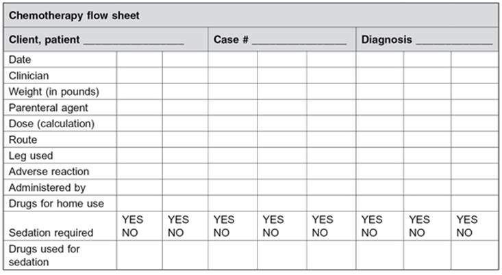
· A flow sheet is an effective way to track a patient’s individual progress.
· Providing clients with a one-page written overview of each treatment removes a layer of anxiety.
Introduction
The proportion of owners willing to pursue advanced diagnostics and treatment for their pets with cancer has grown significantly in recent years, making it increasingly important that veterinary practices gain expertise in the administration of chemotherapy. With the advanced safety devices available today, many chemotherapy protocols can be performed easily in a private practice setting with minimal specialized equipment. Veterinary technicians play a central role in the treatment of pets diagnosed with cancer. It is essential to take all necessary precautions to minimize the risks to the animal, as well as to the veterinary healthcare team. However, a successful chemotherapy experience for the pet, the owner, and the hospital staff requires more than careful attention to technical expertise. It also requires employing the soft skills of customer service.
The Greeting
One of the ways clients judge the quality of a veterinary hospital, its staff, and the services it provides is by how their animals respond to being treated. In other words, how much their pet enjoys its visits to the hospital can be as important to the client’s feeling of success as the efficacy of the treatment itself.
Visiting a veterinary clinic is stressful for some pets and therefore for their owners. Couple that with the anxiety that often accompanies a diagnosis of cancer in a loved one and a cycle of unpleasant expectations can begin to form. The owner may anticipate a chemotherapy visit with apprehension. Their pet will likely respond to its perception of the owner’s anxiety and “act out” behaviors of apprehension. Each visit that results in stress and unpleasantness for the pet ensures that future visits will become more difficult. Considering that most chemotherapy protocols require multiple visits to the clinic, it is to everyone’s benefit to help the owners and the patients feel relaxed at your veterinary clinic. One place to begin is during the initial patient history, when the pet first presents for cancer treatment. Getting to know a pet’s personality and behaviors in the beginning can go a long way toward preventing stress from mounting from one visit to the next. A behavior assessment should be a routine part of every pet’s history upon initial presentation.
Studies show that only 25% of veterinary practices routinely discuss behavior issues with clients. It is important to realize that an owner may never think to discuss his or her pet’s behaviors with you because he or she may not see how behaviors relate to cancer. Therefore, it is vital that you ask questions about his or her pet’s personality as part of your patient history. A behavior assessment can detect situations and can identify potential problems before they impact the quality of future visits. Consider questions similar to the following:
· Is there anything you are reluctant to do with (or to) your pet?
· What sorts of things frighten your pet?
· How does he or she react when he or she is afraid?
· How does he or she react to new people or situations?
Obtain behavioral descriptions, not interpretations. Owners often describe their pets’ behavior in vague subjective terms like, “He goes crazy when the doorbell rings.” Encourage a more specific description by asking, “What does that look like?”
Also, take note of the pet’s demeanor in the clinic. Record any observed occurrences of fearful, threatening, or aggressive behavior in the patient’s record, and share that information with other team members. It is a good idea to mark the front of an unpredictable patient’s record, cautioning others of the pet’s questionable behavior. Discuss behavior concerns with the doctor, so the doctor can discuss the pet’s behavior with the owner. Avoid taking it on yourself to develop a course of action for the client to follow, or making recommendations or referrals for appropriate treatment of behavior problems without approval from the veterinarian. In some cases, a script can be written for a difficult conversation with an owner, so as to discuss only the pet’s behavior and to avoid giving the impression that the pet is “bad.”
A veterinary technician can do a lot to calm a tense “first experience” for a pet receiving chemotherapy. Technicians know to avoid the behaviors most people use to greet dogs, which could be misunderstood by dogs as threats. A dog in a strange, emotionally charged environment might be “offended” by postures such as direct eye contact, direct frontal approach, reaching toward or over them, and leaning forward. Cats may find them equally threatening. Instead, when approaching a dog, turn your side toward the animal and try to approach at a slight angle rather than head-on. Look at the floor or off to the side, keeping the pet in your peripheral vision, but without looking him straight in the eyes. These and other simple techniques may be employed to ease a patient’s stress by keeping your behavior less threatening. When clients see their pets are calm and accepting of the new situation, they also relax. The cycle of unpleasant expectations then begins to unwind. Throughout your interactions with a chemotherapy patient, assume the role of the pet’s favorite “aunt” or “uncle,” doting on them and liberally rewarding the slightest positive behavior. Your goal is for the pet to drag its owner from the car to the front door of the clinic at every chemotherapy appointment.
Pretreatment Examination
Initial friendliness is not always predictive of how an animal will react to restraint and handling. Do not assume an animal will be tolerant of being handled for examination and other procedures. Careful observation of an animal’s posture is the best way to assess its emotional state and its intentions. Cancer patients are quite often older animals and may tolerate handling better if they are examined on the floor, rather than lifted onto a table. See Chapter 4 for details on the importance of a quality temperature, pulse, and respiration (TPR) and physical examination.
Many chemotherapy drugs affect the bone marrow’s ability to make cells, so blood is drawn from chemotherapy patients at nearly every visit to evaluate their white blood cell count. White blood cells called neutrophils form an integral part of the immune system. A low number of neutrophils could be a cause to postpone a chemotherapy treatment. Your clinic will establish a minimum acceptable number of neutrophils for treatment. Be sure you know that number.
Since many intravenous (IV) chemotherapy drugs are vesicants and damage tissues if they leak out of the vein, it is important to protect the integrity of peripheral veins as much as possible. Therefore, draw all blood samples for routine blood tests from the jugular vein.
Drawing blood from the jugular vein is an acquired skill and takes practice to perfect. One simple way to gain confidence in finding the jugular vein on a variety of sizes and shapes of animals is to raise the jugular vein of every patient you see. A successful blood draw from the jugular is rarely a matter of hitting the vein; it is usually a matter of finding the vein. Once it can be located, palpated, and visualized, you discover that it is larger than any other vein you can access. Sliding a needle into it is relatively easy.
Positioning the pet on the floor or on a table largely depends on the size of the animal. Have the handler restrain the patient in a manner that exposes its neck, yet still prevents the pet from backing up or trying to scratch you with its front or rear claws (Figure 6.1). If you are apprehensive about the pet’s behavior at all, use a muzzle to keep from having to divide your attention between the pet’s neck and its teeth.
Figure 6.1 To expose the jugular vein, hold the animal’s head in a near normal position.
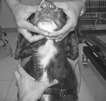
With the animal’s head in a near normal position, no more than slightly above a 90° angle from the neck, the jugular vein generally runs along a line from the base of the ear to the thoracic inlet. A quick visualization and palpation of the jugular vein of every patient you see, regardless of whether or not you draw blood from it, will increase your confidence remarkably when you need to use it.
Restraint for Chemotherapy
One of the first decisions to be made when preparing to administer chemotherapy is whether or not chemical restraint is indicated and appropriate. Sedatives and tranquilizers depress the central nervous system and reduce anxiety and struggling. They can help minimize stress during the administration of chemotherapy and reduce the need for physical restraint. The decision to use sedation is made by the veterinarian, with the informed consent of the owner. Although your patient’s disease usually does not merit any particular consideration when selecting the drugs to use for sedation, cancer patients are generally older animals and may have other age-related issues. When necessary, the ideal sedative for a chemotherapy patient has a rapid onset and short duration. The drug or drug combination should have minimal negative side effects and be reversible if possible. The rationale for using combination drug therapy for sedation is similar to the rationale for using combination drug therapy to fight cancer. Multidrug protocols are generally more effective than single-drug treatment regimens. When drugs are used in combination, several important points should be kept in mind:
· Each drug in the combination should be effective when used alone.
· Drugs with overlapping toxicities should be avoided.
· The drugs should be dosed in combination to produce the maximal effect, with the least side effects.
· Combined sedative drug regimens are most effective when they have different mechanisms of action and act at different receptor sites.
As mentioned earlier, we are all creatures of habit. And although our habits usually serve us well, it is important that you avoid just reaching for your usual sedation protocol and take that extra moment to make conscious decisions regarding sedatives for patients receiving chemotherapy. Whether or not to sedate and the choice of drugs to use are ultimately the veterinarian’s decisions to make. However, your awareness of the need to possibly divert from your usual sedation practice will go a long way toward enhancing the cancer patient’s chemotherapy experience.
A busy treatment room is the least suitable place for a chemotherapy treatment. Select a room that is out of the flow of traffic and is easy to regulate personnel moving in and out of the room. Clients often wish to “help” or watch a chemotherapy treatment. For the safety of the client, the patient, and the staff, this should not be allowed. See Chapter 3 for more details on preparing an appropriate place to administer chemotherapy.
Older, larger cancer patients may better tolerate receiving chemotherapy on the floor rather than being lifted onto a table. But lifting a pet onto the examination table is usually the first step in the administration procedure. Lifting smaller animals is usually fairly routine; however, larger animals present challenges. Their weight may be prohibitive, their bulk makes them awkward, and they are not used to being lifted. Also, their joints may be stiff from age-related changes, and any struggle they offer will likely hurt them, you, or both. For these reasons, it is better to have two people lift and position a larger pet onto the examination table for chemotherapy administration.
With two people, a larger patient can safely be lifted and positioned in one smooth motion. The two-step process of lifting the dog onto the table and then positioning it offers another opportunity for the patient to feel insecure and unstable standing on a slippery steel table. So the preferred method for a larger dog is to lift and position into lateral recumbency (lying on their side), all in one motion. Many large breeds of dogs are too deep-chested to adequately restrain for a chemotherapy infusion while positioned in sternal recumbency (lying on their chest). But lateral recumbency affords even the smallest handler the ability to safely and securely restrain the patient.
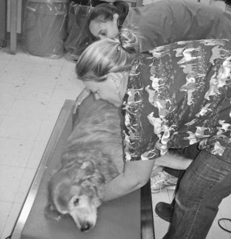
Before lifting the patient, decide which lateral recumbency is preferred for infusion: right or left. Then both handlers stand side-by-side, facing the side of the pet to be infused. Bending at the knees, both handlers then squat facing the dog. The handler near the pet’s head places an arm in front of the forelimbs across the sternum and the other arm in a forklift position about halfway down the abdomen. The other handler places an arm behind the hind legs and the other arm in a forklift position about halfway up the abdomen. Both handlers hold the dog securely against their own bodies and then lift and roll the pet onto the table into lateral recumbency, in one motion (Figure 6.2). The handler near the head remains in position over the dog, holding it in place on the table, while the second handler circles to the other side of the table and establishes the proper position to restrain the patient for an infusion.
Figure 6.2 Two handlers hold the dog securely against their own bodies and then lift and roll the pet onto the table into lateral recumbency.
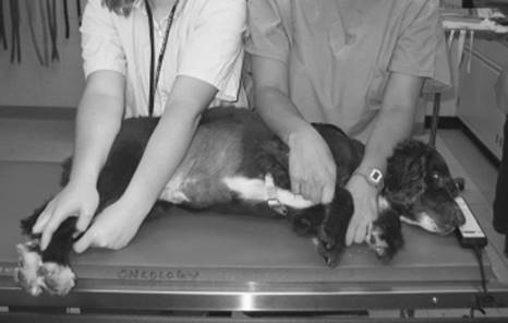
To properly restrain a dog in lateral recumbency, the holder stands at the dog’s back with one forearm pressed across the animal’s neck while that hand holds the “down” foreleg, which lies against the table. The other forearm presses across the dog’s flank, and the “down” hind leg, which lies against the table, is held in a similar manner (Figure 6.3). As mentioned before, if you are at all apprehensive about the pet’s behavior, use a muzzle. With a muzzle on the animal, you will be less concerned with the need for rigid restraint, and your hold will be more relaxed. It is finesse over force; animals respond better to being held securely rather than forcefully. Also, dogs who are not accustomed to being muzzled are often distracted by having a muzzle placed.
Figure 6.3 The proper technique to restrain a dog in lateral recumbency.
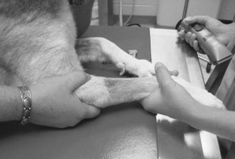
Held in sternal recumbency to use a cephalic (forelimb) vein for the infusion, the holder stands (or kneels) beside the dog facing the venipuncturist. Place one arm under the dog’s neck, and using the hand of that arm, hold the dog’s head against your shoulder, with the muzzle pointed away from your face. At the same time, wrap the other arm over the back of the dog and use that hand to encircle the patient’s foreleg. Cup the patient’s elbow in the palm of that hand and cover the cephalic vein with your thumb. Push slightly with your palm so the pet cannot withdraw its leg from the venipuncturist, and rotate your hand while pressing with your thumb to present the engorged cephalic vein to the most anterior (top) aspect of the foreleg (Figure 6.4). Once the catheter is seated in the vein, stop holding the vein with your thumb but continue to cup the elbow in your palm to maintain control of the patient’s leg.
Figure 6.4 Proper restraint to expose a cephalic vein. Note that the elbow is cupped by the holder’s palm to prevent withdrawal of the limb and that the dorsum of the forelimb is rotated laterally by the thumb so that the vein runs directly over the top of the limb.
Whether in sternal or lateral recumbency, using proper restraint techniques during chemotherapy administration is imperative to maintain control of the patient. Many of the cytotoxic drugs used to treat cancer are vesicants. Vesicants are highly reactive chemicals that combine with proteins, DNA, and other cellular components, resulting to destructive cellular changes. When vesicant medications leak out of a vein and make contact with the surrounding tissue, the damage begins. Tissue damage can range from mild skin irritation to gaping necrotic wounds, requiring multiple surgical repairs. See Chapter 3 for more information about managing vesicant drugs.
As a team, the venipuncturist and the holder work together to safely and effectively administer chemotherapy to the animal cancer patients. Neither job is more or less important than the other. Each person relies on the other to be focused and attentive to his or her responsibilities. Each is the star of the show. When the team is confident and relaxed, the patient will also be. But a successful chemotherapy experience for the pet does not end with expertly executed technical skills. For the pet to leave the experience feeling comfortable and wanting to do it again, the medical team must practice soft skills as well.
Soft skills refer to the intangibles that contribute to a positive experience. In a veterinary clinical setting, these intangibles include the sights, sounds, and smells of the environment. Avoid wearing strong-scented cologne or perfume. Create a quiet atmosphere away from slamming doors, barking dogs, and stressed cats. Also, remember that pets, especially dogs, are companion animals. They interact with all people, not just their owners. That means your demeanor can affect their comfort. While administering chemotherapy, keep in mind that your patient is responding to your actions and emotions. Avoid emotionally charged conversations with one another while holding a patient and administering chemotherapy. Monitor your surroundings for how things look (Are there bright lights? Fast, frantic motions?), sound (Are people shouting over the sound of a centrifuge?), and feel (Is someone recounting an angry conversation?). Assuming the perspective of a pet while administering chemotherapy is one way you can further unwind the cycle of unpleasant expectations for your patients’ visits.
Drug Administration
Oral Chemotherapy
Oral administration of chemotherapy drugs can be convenient, noninvasive, and, sometimes, economical. However, there are situations when drug delivery by this route is not possible. Follow these basic recommendations when dispensing or administering tablets or capsules:
· Never crush or break tablets and never open capsules.
o Although ill-advised, if a patient’s chemotherapy tablet must be split, and if the owner is willing to do it at home, instruct the owner to wear a mask and gloves, and to carefully cut the tablets with a tablet cutter.
o Provide owners with mask and gloves.
o Inform owners of the potential hazards associated with splitting and administering chemotherapy tablets.
o Have owners return unused medications to your practice for proper disposal.
· Wear appropriate personal protective equipment (PPE) when handling any tablet or capsule.
· When a capsule or tablet is administered in a food bolus, make sure the food bolus is completely consumed.
o It is a good practice to follow the drug-containing food bolus with a second “empty” food bolus to ensure ingestion of the first.
· When a capsule or tablet is administered directly per os (PO, in the mouth), take care to avoid dissolution of the outer protective coating of the tablet, and make sure the tablet or capsule is swallowed.
o It is a good practice to follow with a tidbit of food to ensure ingestion.
· Always wash hands after handling drugs.
· See Chapter 5 for more details on handling chemotherapy tablets and capsules.
Intravenous Chemotherapy
It should go without saying that eating, drinking, smoking, chewing gum, and applying lip balm are all prohibited in chemotherapy administration areas. Before administering injectable chemotherapy to any patient, review these simple steps to increase your awareness and protect your patients from being harmed by a chemotherapy-related error (see Chapter 3):
(1) Review the patient’s record.
(2) Recheck the patient’s blood test results.
(3) Recheck the drug name and dose.
(4) Recheck the dose calculation.
(5) Ask questions.
(6) Keep accurate records.
IV drugs can be administered through a traditional dedicated over-the-needle catheter or a butterfly-style catheter. Off-the-needle administration of any chemotherapy drug, no matter how small the volume, is strongly discouraged. The sites most frequently used are the cephalic and lateral saphenous veins in the dog, and the cephalic and medial saphenous veins in cats. The larger veins of the forelimb (cephalic veins) are often chosen, as many find them easier to catheterize. Saphenous veins have advantages also. Over-the-needle style catheters are used to infuse volumes of greater than a couple of milliliters. See Chapter 3 for criteria for selection of an appropriate style of catheter and for supplies to have ready in your “chemotherapy setup” prior to starting.
Placing a catheter for chemotherapy requires a level of skill greater than “novice.” Although a novice can likely place a catheter successfully, a seasoned veteran should stand at the ready to make any necessary second attempts. Always place a new, dedicated catheter for the administration of chemotherapy. Shave the area of the insertion site generously, as you would for a surgical incision. Perform a surgical prep with antiseptic scrub and solution. A full surgical prep (rather than a swipe with alcohol) and aseptic technique are both important because chemotherapy may lower a patient’s resistance to catheter-related infections. Repeated catheterization of an immunocompromised cancer patient courts disaster. Local anesthetic block is rarely indicated because the injection of anesthetic is likely as painful as the insertion of the catheter.
Ignore the general catheterization rule that suggests you start your first attempt distally and proceed proximally. The placement of a catheter for chemotherapy must be perfect on the first attempt. So, rather than beginning distally and working your way up the leg, begin where you have the best view of the vessel and the best chance of success. Should you fail an attempt, move to a different leg entirely. In this manner, you best protect your patient from being injured by vesicant drugs leaking from a vessel. You also minimize damage to any one vessel that might occur during multiple unsuccessful attempts to catheterize it.
Flush the catheter immediately before administration to confirm its patency and immediately after administration to assure the catheter is clear of any drugs before removal. Check patency with 0.9% sodium chloride solution. Avoid heparin–saline solution for flushing because it can precipitate some drugs, especially doxorubicin. Tape an over-the-needle catheter firmly in place. Do not tape a butterfly catheter in place (see Chapter 3). Leave the catheter uncovered to better visualize it during administration. Wear PPE during drug administration and check the catheter site often. If at any point you feel resistance to flow through the catheter, or see swelling or other reactions, stop until you have confirmed the catheter is displaced or until you are confident that the catheter is flowing as it should. After the infusion is complete, flush the catheter with at least 10 mL of 0.9% saline solution immediately upon completing the administration of chemotherapy to assure the catheter is clear of any drugs before removal. That way, the patient receives its entire intended dose and no drug remains in the catheter when it is removed. Then, remove the catheter immediately, place all materials into a sealed plastic bag, and discard as cytotoxic waste. Figures 6.5–6.17 demonstrate the step-by-step chemotherapy administration in a dog with lymphoma.
Figure 6.5 Review the record and check the drug to be administered. Do you have the right patient? Do you have the right drug? Do you have the right dose?
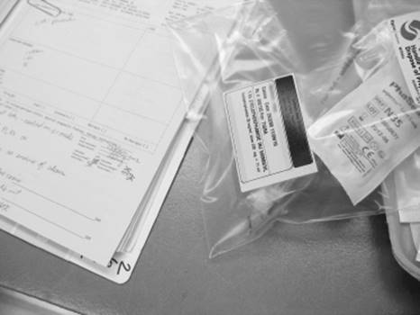
Figure 6.6 Shave the catheter site. A small shaved area is acceptable.
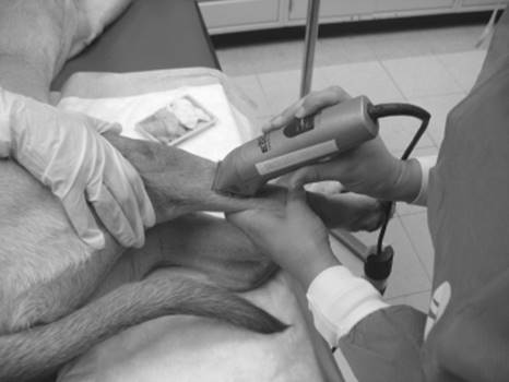
Figure 6.7 Prep the catheter site. A full surgical prep and aseptic technique are both important to prevent catheter-related infections.
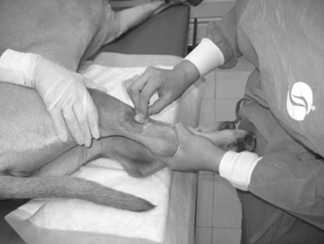
Figure 6.8 Ensure that all necessary supplies are close by and ready to go. This includes tearing strips of tape, opening catheters, T-ports, and protective supplies, and preloading the catheter and T-port with saline. Note the absorbent pad in place under the limb into which chemotherapy is going to be administered.
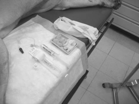
Figure 6.9 Insert the catheter. Remember the “one-and-done” rule for catheter attempts for chemotherapy administration!
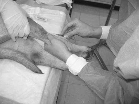
Figure 6.10 Attach the T-port. This allows the limb freedom to move without risk of the catheter being pulled out.
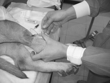
Figure 6.11 Tape the catheter securely in place.
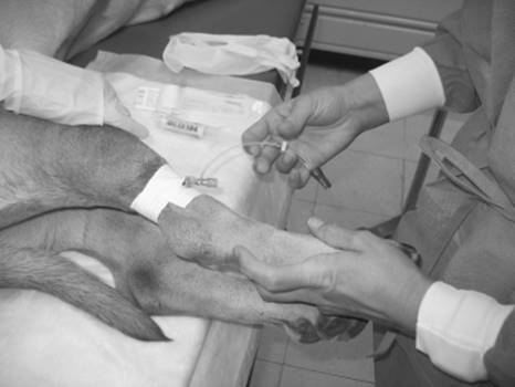
Figure 6.12 Flush the catheter to ensure patency. Watch carefully for blebs or swelling under the skin and note resistance to flushing, which could indicate that the catheter is improperly placed or is outside a vein. Check for a flashback of blood to ensure proper catheter placement.
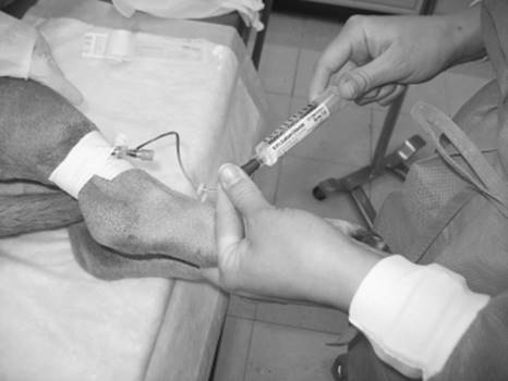
Figure 6.13 Attach the PhaSeal adapter or similar protective device to the tip of the extension set.
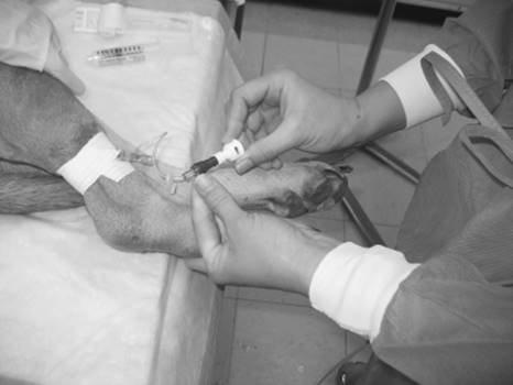
Figure 6.14 Administer chemotherapy. Speed of administration will vary depending on the agent.
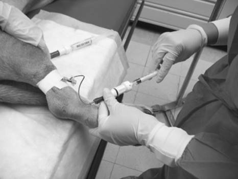
Figure 6.15 Flush thoroughly with saline after chemotherapy administration. This ensures that no chemotherapy drugs remain in the catheter, which could damage the skin or subcutaneous tissues upon catheter removal.

Figure 6.16 Remove the catheter and place the bandage. This usually consists of a small piece of flexible veterinary wrap or adhesive tape, and a cotton ball or folded gauze sponge.
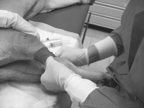
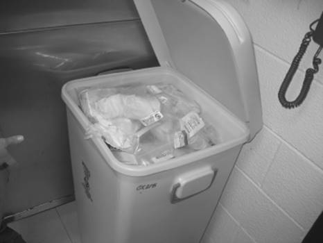
Figure 6.17 Dispose of all waste that came in contact with chemotherapy (catheters, syringes, T-ports, absorbent pad, etc.) in an appropriate cytotoxic waste container.
Two systems for administering chemotherapeutics are now available. The PhaSeal® system is considered a “closed-system drug transfer device.” The other system, Onguard®, is a “contained medication system.” Both systems offer increased safety for the hospital staff and decreased accidental chemo-exposure. Both systems have four primary components designed for nearly intuitive use. Because these systems can be easily integrated into your current practice, training can be completed easily and in a timely manner (see Chapter 5).
The rate of administration for chemotherapy is determined by the type and volume of drug to be administered, as well as the protocol designed by the veterinarian. Vincristine, for instance, is usually in small volumes and can be delivered in seconds by slow push through a butterfly catheter. Drugs, such as doxorubicin, that have higher extravasation risk and take longer to administer should be given through a securely placed over-the-needle catheter. The administration of cisplatin, however, involves a prescribed IV prehydration, followed by a long infusion of the drug, which is then followed by a prescribed IV posthydration. The entire chemotherapy administration session can take 6–8 hours. See Chapter 7 for general guidelines on chemotherapy routes and rates of administration.
Subcutaneous/Intramuscular Chemotherapy
Some chemotherapy drugs, such as cytarabine and asparaginase (see Chapter 7), are administered either subcutaneously (SQ) or intramuscularly (IM). They present additional contamination concerns to you because, although they can be reconstituted and the doses drawn into syringes using the PhaSeal or Onguard protective devices, a bare needle is required for subcutaneous or intramuscular injections. An unprotected, bare needle offers the opportunity for accidental drug spill, errant injection, and needlestick injuries. Be aware that you, your colleagues, and your facility are at their most vulnerable for contamination. It is imperative you wear PPE when administering chemotherapy drugs either SQ or IM.
When a subcutaneous injection is administered, tent a fold of skin and insert the needle at its base, parallel to the long axis of the fold. If the needle is inserted perpendicular to the fold, the needle may penetrate both sides, and you may empty the contents of the syringe into the pet’s hair. Aspirate slightly and check the needle hub for blood or air bubbles. Blood in the needle hub indicates that you have penetrated a vessel. Air in the hub is an indication that you have penetrated both sides of the “tent.” Either indicates you must reposition the needle.
Chemotherapy drugs are most often administered IM into the lumbar muscles, lateral to the dorsal spinous processes. Wearing PPE, uncap the 20- to 22-gauge needle attached to the syringe and isolate the muscle between your fingers and thumb. Slide the needle into the muscle and aspirate, checking the needle hub for blood. If blood is observed, withdraw the needle and insert into another spot. Once the proper needle placement into the muscle or under the skin is verified (no blood or air in the hub upon aspiration), slowly inject the drug. Then withdraw the needle.
A word of caution about needlestick injuries. The National Institute for Occupational Safety and Health (NIOSH) estimates that 62% of all reported needlestick injuries happen with hypodermic needles, and 42% of those injuries occur after the needle has been used and before its disposal. This means that the risk of a needlestick injury, and the consequential self-contamination with a cytotoxic drug, is very high as you withdraw the needle from the patient and move it toward disposal. Studies have shown that nearly 25% of needlestick injuries occur when recapping a used needle. The Occupational Safety and Health Administration (OSHA) prohibits recapping needles by hand, unless no other alternative exists. So, if you must recap a used chemotherapy needle before disposal, do it with extreme care and attention.
Chemotherapy Flow Sheet
A flow sheet is an effective way to track a patient’s individual progress through the course of chemotherapy. It consolidates the pertinent information from each visit into a single chart for easy reference. An example of an effective chemotherapy flow sheet is in Appendix 6.1. It contains
· the date,
· doctor’s name,
· patient’s weight,
· drug name,
· dose,
· route of administration,
· leg used,
· adverse reactions,
· name of the person who administered the drug, and
· drugs and doses of sedation if used.
Afterward
People don’t care how much you know until they know how much you care.
This often quoted statement by author John C. Maxwell holds an elemental truth with regard to medicine. A follow-up telephone call several days after each chemotherapy treatment goes a long way toward showing your clients how much you care for their pets and for them. It can also give you an early indication of how your patient tolerates chemotherapy and give you an opportunity to address complications before they become serious.
Providing clients with a one-page written overview of each treatment for them to take home also removes a layer of anxiety that many people feel about cancer therapy. An example of a posttreatment client information sheet can be found in Appendix 6.2. Such an overview includes
· an indication that a staff member will call in a few days for an update,
· the name of the drug the pet received during that visit,
· a list of the side effects that may be seen and how to manage them, and
· reminders on chemo-safety-related issues of concern at home.
Appendix 6.1 Sample Chemotherapy Flow Sheet
COMMENTS:
______________________________________________________________________________
______________________________________________________________________________
______________________________________________________________________________
Appendix 6.2 Sample Client Discharge Instructions
In the next few days, a member of our staff will call for an update on your pet and to address any questions or concerns you may have. Below is information about the chemotherapy your pet received, in addition to chemotherapy safety guidelines.
Vincristine (Oncovin®)
Vincristine (Oncovin) is an active chemotherapy drug used to fight many forms of cancer. The IV administration of vincristine usually takes only a few minutes. The procedure is painless, so the patient lies quietly on a padded table during administration and rarely needs any form of sedation.
Although serious adverse effects can occur with any chemotherapy, there is less than 5% chance that your pet will be hospitalized with side effects and less than 1% chance of fatality. As the owner, you play an important role in recognizing side effects, managing them at home whenever possible, and alerting the doctor if side effects persist. Listed below are some potential side effects of vincristine. Please consult your pet’s doctor with any questions you may have about this chemotherapy.
Potential Side Effects of Vincristine
· Hair loss (alopecia)
o Hair loss in pets is rare.
· Reduction in the number of white blood cells (neutropenia)
o Your pet’s ability to fight off infection is impaired by neutropenia.
· Stomach or intestinal (gastrointestinal) discomfort
o Many patients experience some form of stomach or intestinal discomfort 2–7 days after a chemotherapy treatment.
o If your pet shows signs of upset stomach (drooling, “smacking” lips, vomiting) or loss of appetite, administer the medicine your doctor prescribed for nausea.
o If your pet shows signs of diarrhea, administer the medicine your doctor prescribed for diarrhea.
o Keep water available at all times.
o Call your veterinarian if you have concerns, or if the condition persists for more than 24 hours.
· Tissue injury
o If irritation of the chemotherapy administration site appears in the form of pain or redness that persists for more than 24 hours, call your veterinarian.
Safety
The chemotherapy your pet received requires that you take special safety precautions after each visit. For 72 hours after each chemotherapy visit, your pet’s urine and feces should be considered contaminated. For your safety, please follow these guidelines:
· If you are pregnant or nursing, avoid cleaning the litter box or handling any other materials that may be contaminated.
· Wear gloves when cleaning urine or feces from the litter box, cage, and so on.
· Flush solid waste material or place it in a Ziploc® type bag for disposal.
· If your pet has an “accident,” remove the solid waste and rinse the area with water. Then wash the area with detergent, followed by a second rinsing with water.
· Soiled linens should be washed once separately prior to routine laundering.
· If accidental skin exposure occurs, wash with soap and water immediately.