Marissa L. Albano,
Robert P. Murphy
|
Key Features |
|||||||||||||||||||||||||||
|
Eales' disease is an idiopathic obliterative vasculopathy of the peripheral retina, initially presenting as retinal periphlebitis and later as retinal ischemia, leading to vascular alterations, neovascularization, and vitreous hemorrhage. It can affect persons of all ages and is usually bilateral; it most commonly affects young, healthy males in the Indian subcontinent and Middle East. The etiology is believed to be multifactorial, although it currently remains unclear. Recent immunological, biochemical, and histopathological studies have implicated Mycobacterium tuberculosis, retinal autoimmunity, human leukocyte antigen, and free radical mediated damage in the pathogenesis of this disease. Early manifestations include vascular sheathing and focal occlusion of peripheral retinal vessels. With progression, areas of nonperfusion can develop. Neovascularization can occur at the junction of the perfused and nonperfused retina, frequently resulting in recurrent vitreous hemorrhages. As the exact etiopathogenesis remains unclear, therapy is directed toward the presenting symptoms of the disease. Oral, peribulbar, and intravitreal corticosteroids are used in the active inflammatory stage, while retinal neovascularization in Eales' disease appears to respond well to laser photocoagulation. Vitrectomy is utilized for vitreous hemorrhage with or without retinal detachment.
HISTORY
The disease is named after Henry Eales, an ophthalmologist who in 1880 described a syndrome of recurrent vitreous hemorrhages in young men with epistaxis and constipation.[1] He termed this new entity primary recurrent retinal hemorrhage. Using the newly developed direct ophthalmoscope, Eales documented abnormal retinal veins and areas in the peripheral retina that were free of capillaries.[2] In spite of these astute early observations, the entity he described differs considerably from the disease that now bears his name. He did not observe any new vessels or inflammation preceding or accompanying the hemorrhages.
In 1887, Wadsworth described associated inflammation and neovascularization with this disease.[3] Since then, numerous authors have grouped periphlebitis retinae and idiopathic recurrent vitreous hemorrhages with or without retinal perivasculitis under the term Eales' disease.
Not all authors have believed that Eales' disease is a specific entity. Duke-Elder thought that Eales' disease represented the clinical manifestation of many diseases.[4] Indeed, refined diagnostic tests have demonstrated that many of the so-called idiopathic hemorrhages are the result of diseases with known causes, such as sarcoidosis, systemic lupus erythematosus, diabetes mellitus, sickle-cell disease, and collagen vascular disease.
However, after elimination of these causes, a group of patients remains with idiopathic peripheral nonperfusion and perivasculitis of the retina. Many investigators now agree that Eales' disease is a distinct entity comprising characteristic fundoscopic and fluorescein angiographic features.[5] Although this disease has been called periphlebitis retinae,[6] emphasizing the abnormalities of retinal venules, evidence suggests that the inflammation in this disease affects both arterioles and venules.[7]
EPIDEMIOLOGY
Eales' disease is uncommon in North America; however, this disorder is responsible for widespread visual loss in the Indian subcontinent. It often affects healthy young adults, with the average age of onset at 20-30 years. Patients usually present with symptoms of vitreous hemorrhage, such as floaters or decreased vision. The majority of patients experience bilateral, although often asymmetric, involvement.
Most reports, including Eales' original description, indicate a male predominance. However, Murphy and co-workers found an equal prevalence of men and women in their study of 55 patients in North America.[7]
CLINICAL FEATURES
|
Clinical Features of Eales' Disease |
||||||||||||
|
INFLAMMATION
Most patients with Eales' disease develop ocular inflammation, especially early in its course. Vascular sheathing is seen in most patients (Fig. 137.1). The degree of sheathing ranges from fine white lines on both sides of the blood column to thick exudative sheathing. The thin white lines tend to be continuous, whereas the heavy, exudative sheathing is often segmental.
|
FIGURE 137.1 (a) Thin white lines representing vascular sheathing surround a retinal venule in a patient with Eales' disease. (a) Extensive arteriolar sheathing with retinal vascular nonperfusion nasal to disk. |
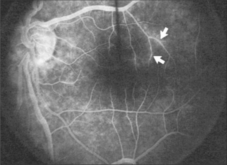
Areas of vascular sheathing frequently demonstrate hyperfluorescence in fluorescein angiography (Fig. 137.2). However, there is no direct correlation between the regions of sheathing and staining. The intensity of the hyperfluorescence seen on the fluorescein angiograms does not correlate with the intensity of inflammation.
|
|
|
|
FIGURE 137.2 Fluorescein angiogram demonstrates abnormal staining of a small retinal venule (arrows) in a patient with Eales' disease. There was venous sheathing in this area. |
In the century since Eales' observation of altered retinal veins, many investigators have described Eales' disease as a primary disease of altered retinal veins. Elliot and Harris suggested the term periphlebitis retinae for this disorder.[6] However, more recent studies have reported equal involvement of arteriolar and venular sheathing.[7] Because of the evidence of arteriolar involvement (Fig. 137.1b), this disease should be considered a retinal vasculitis or vasculopathy.
Keratic precipitates, anterior chamber cell and flare, and vitreous cells have been observed in patients with Eales' disease.[7] Cystoid macular edema occurs in eyes with extensive sheathing. Although the exact cause of the macular edema is unknown, it may be associated with low-grade inflammation.
NONPERFUSION
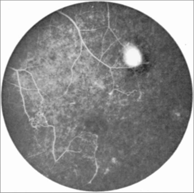
Peripheral retinal nonperfusion is present in all patients with this disease. The extent of the perfusion ranges from small areas in the far periphery to massive nonperfusion extending into the posterior pole (Fig. 137.3). The nonperfusion is generally confluent and sharply demarcated from the posterior perfused retina. The temporal retina is most commonly affected.
|
|
|
|
FIGURE 137.3 Fluorescein angiogram of the peripheral retina demonstrates the junction of normally perfused retinal vessels adjacent to an area of nonperfused retina. Note the vascular abnormalities and the small area of neovascularization at the junction. |
The microvascular abnormalities may be so severe that the fundus resembles Coats' disease. Fine white lines representing the remains of obliterated large vessels (ghost vessels) are often seen in the area of nonperfusion.
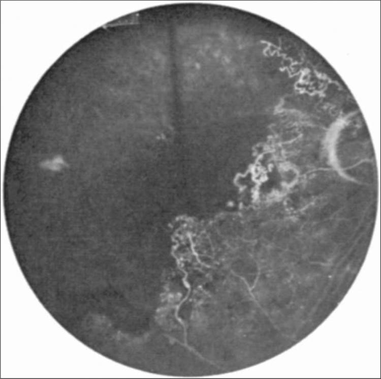
Elliot[8] and Spitznas and colleagues[9] have documented the vascular abnormalities at the junction of the anteroperipheral nonperfused retina and the posterior perfused retina. Intraretinal hemorrhages often first appear in the affected area, followed by an increase in vascular tortuosity with frequent collateral formation around occluded vessels (Fig. 137.3). Microaneurysms, arteriovenous shunts, and venous beading are commonly seen at the junction (Fig. 137.4). Fluorescein angiography enhances these abnormalities and often demonstrates staining at the stumps of obliterated vessels.
|
|
|
|
FIGURE 137.4 Fluorescein angiogram demonstrates severe nonperfusion of the retinal vasculature involving the macula. |
Patients with Eales' disease can also experience branch vein occlusion. The branch vein occlusion can be either single or multiple. Unlike patients with primary branch vein occlusion, in whom the pathologic condition is confined to one quadrant, patients with Eales' disease have more extensive involvement of the peripheral retina that does not respect the horizontal midline.
NEOVASCULARIZATION
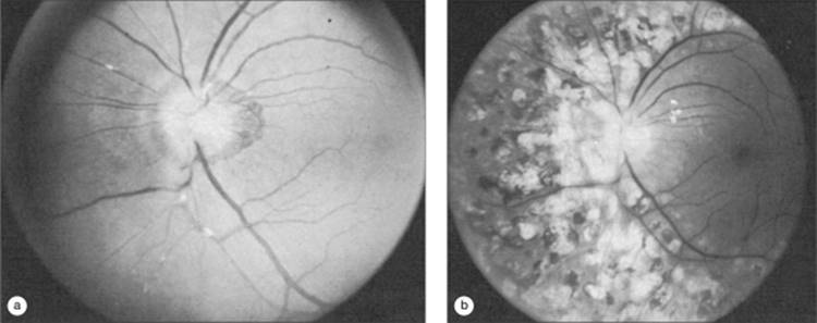
The retinal nonperfusion leads to the eventual development of new vessels. The neovascularization can form either on the disk or elsewhere in the retina. Neovascularization of the iris has also been described. These abnormal blood vessels frequently bleed and are the major cause of visual loss in this disease.
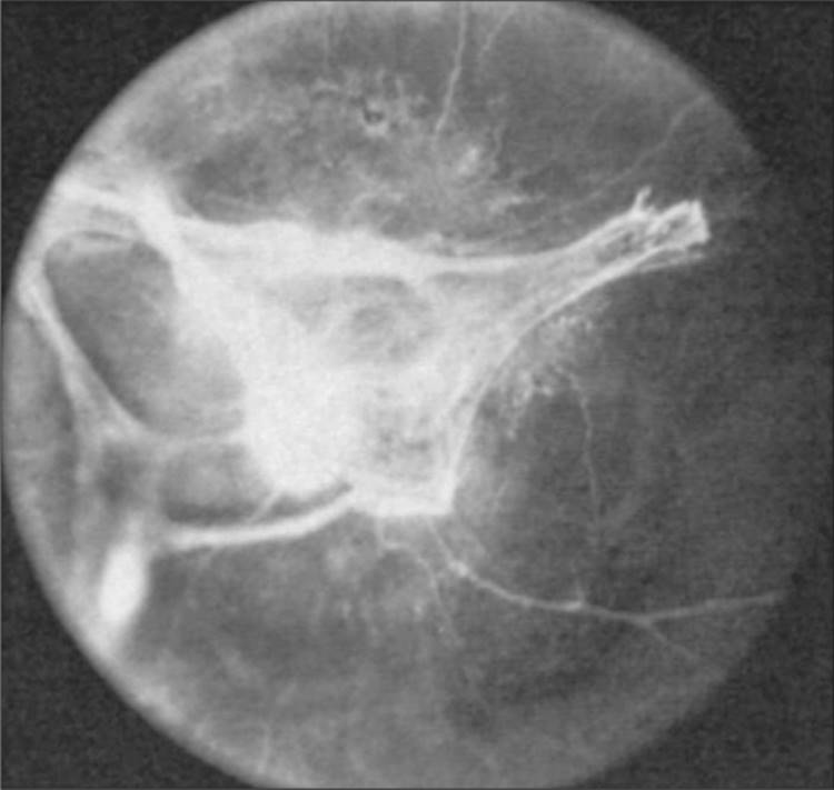
Ischemic retina produces a diffusible substance that stimulates neovascularization, vascular endothelial growth factor (VEGF). Perentes and colleagues demonstrated a close relationship between prominent neovascular proliferation and the intense expression of VEGF via histopathological and immunohistochemical examination of an enucleated eye from a patient with documented Eales' disease.[10] The neovascularization often occurs along the junction of perfused and nonperfused retina, similar to the appearance of sickle-cell retinopathy. The neovascularization frequently has a prominent fibrous component. Occasionally, patients have extensive retinal and vitreal proliferation of avascular sheets and strands of fibrous scar tissue (Fig. 137.5). The anteroposterior traction resulting from the fibrovascular membrane places these eyes at risk of developing traction retinal detachment. Badrinath and colleagues found multifocal fibrous and fibrovascular vitreoretinal adhesions in 83% of eyes undergoing vitreous surgery for complications of Eales' disease, in addition to noting type II collage in the epiretinal membranes causing tangential traction.[11]
|
|
|
|
FIGURE 137.5 Hypovascular fibroproliferation emanates from the disk of a patient with advanced Eales' disease. |
VISUAL PROGNOSIS
The natural course of this disease is variable. Although the visual acuity in patients with Eales' disease ranges from normal to no-light perception, most eyes retain good acuity. In spite of extensive anteroperipheral nonperfusion, the macula is usually spared, preserving central vision. Murphy and co-workers reported that 67% of their patients had final visual acuity in the better eye that ranged from 20/15 to 20/40.[7] Twenty-four percent had visual acuity that ranged from 20/50 to 20/200, and 9% had visual acuity worse than 20/200.
Vitreous hemorrhage is the most frequent cause of visual loss. Usually the hemorrhage settles to the lower portion of the vitreous and is gradually reabsorbed within several weeks or months, with the return of normal central vision. Severe, permanent visual loss usually results from complications associated with neovascularization, such as persistent vitreous hemorrhage, retinal detachment, and neovascular glaucoma. Occasionally, loss of vision is caused by cystoid macular edema, macular holes, retinal telangiectasia, or epiretinal membrane. In some patients, relentless nonperfusion progresses across the macula (see Fig. 137.4); visual acuity in these eyes is usually less than 20/400.
A long-term follow-up study of 130 patients with Eales' disease showed visual acuity improved in 20%, maintained in 43%, and worsened in 37%. The complications noted after 5 years were tractional detachment, cataract, rubeosis iridis, neovascular glaucoma, and phthisis bulbi.[12]
TREATMENT
Henry Eales offered his patients a mixture of laxative, digitalis, and belladonna.[1] Presumably, these remedies were not beneficial. Lowered levels of antioxidant vitamins in patients with Eales' disease and consequent accumulation of oxygen and lipid free radicals have prompted the belief that vitamins E, C, and A may be of value.[13] Bhooma and colleagues demonstrated an increase in serum thiobarbituric acid nactine substances, an indicator of lipid peroxidation products produced by oxygen and free radicals.[13] Some investigators believe there is an association of Eales' disease with tuberculoprotein hypersensitivity, therefore prompting the use of antituberculous therapy as well.[14-16] The benefit of these therapies is untested.
As the basic pathologic lesion of Eales' disease is inflammatory occlusion of retinal vessels, corticosteroids are becoming a mainstay of treatment. For the acute inflammatory stage, oral prednisone (1 mg/kg) is started and the dose tapered for 6-8 weeks. If the patient is intolerant to systemic corticosteroids or if macular edema develops, periocular depot corticosteroid/triamcinolone acetonide injections have been found to be beneficial.[17] Immunosuppressive agents are reserved for intolerance to systemic steroids or for lack of systemic steroid efficacy. In a prospective study by Bali, pulsed oral methotrexate therapy at a dose of 12.5 mg/week was shown to be clinically effective within 4 weeks and is associated with an acceptable safety profile.[18] Recently, intravitreal triamcinolone acetonide was successfully used in three eyes of two patients with Eales' disease in the acute inflammatory stage who were not responding to oral and peribulbar corticosteroids.[19]
Investigators generally agree that peripheral scatter photocoagulation of the ischemic retina is invaluable in the treatment of neovascularization (Fig. 137.6).[20,21] This is similar to the treatment used for other vasoproliferative diseases of the retina, such as diabetic and sickle-cell retinopathies. Several investigators have demonstrated favorable results with light-intensity, full-scatter argon laser photocoagulation to the nonperfused retina and to the junction of perfusion and nonperfusion.[21] Others have used a combination of cryotherapy and laser photocoagulation.[22] Because nonperfused retina is more fragile than perfused retina, caution should be exercised when treating these patients. There is evidence to show that prophylactic pan retinal photocoagulation in areas of ischemic retina in asymptomatic fellow eyes of patients is effective in controlling the progression and future complications of this disease.[23] Due to the intense expression of VEGF in Eales' disease patients, anti-VEGF therapy with either Avastin or Lucentis (Genentech Corp, San Francisco, CA) may be of some treatment benefit, however this remains to be studied.
|
|
|
|
FIGURE 137.6 (a) Neovascularization of the disk. Note the segmental exudative arteriolar sheathing. (b) Same patient as in (a) 2.5 years later. The patient has had total regression of the neovascularization after treatment with scatter photocoagulation of all areas of nonperfused retina. Note that the former areas of arteriolar sheathing have resolved. |
Vitrectomy can be employed for removing persistent vitreous hemorrhages and fibrosis, often with good results.[24] Kumar and colleagues' study demonstrated the importance of early vitrectomy for persistent hemorrhages. Delaying vitrectomy more than 6 months is believed to allow the development of cystoid macular edema, macular scar, and macular pucker formation with poorer visual outcome.[25] No treatment is known to prevent or reverse the nonperfusion or capillary drop-out.
DIFFERENTIAL DIAGNOSIS AND SYSTEMIC EVALUATION
As an idiopathic entity, Eales' disease must first be differentiated from retinal vasculopathies with known causes. Systemic diseases such as diabetes mellitus, sickle-cell hemoglobinopathies, sarcoidosis, and systemic lupus erythematosus can manifest retinal inflammation, nonperfusion, and neovascularization. Ocular diseases such as branch and central retinal vein occlusions, Coat's disease, parsplanitis, dragged disk syndrome, and macular telangiectasia.[17] However, one can rule out these conditions with a careful history and appropriate laboratory tests (Table 137.1).
TABLE 137.1 -- Laboratory Investigations for Eales' Disease
|
CBC |
|
Erythrocyte sedimentation rate |
|
Reticulocyte count |
|
Postprandial blood sugar |
|
Mantoux test (tuberculosis) |
|
Sickle cell preparation |
|
Hemoglobin electrophoresis (sickle cell retinopathy) |
|
Immunoglobulin profile VDRL and Treponema pallidum |
|
Hemagglutination Test |
|
Antinuclear antibody (SLE) |
|
Serum angiotensin-converting enzyme (sarcoidosis) |
|
Lysozyme (sarcoidosis) |
|
X-ray chest (tuberculosis and sarcoidosis) |
Many investigators have emphasized a relationship between Eales' disease and M. tuberculosis.[8,9,14-17] Although there is a higher than normal incidence of positive reaction to the tuberculin protein, no one has demonstrated any evidence that the ocular changes are related to infection of the eye or retina by tuberculin bacteria. The most widely held belief is that Eales' disease is an immune-mediated hypersensitivity process resulting from prior exposure to the tuberculoprotein.[15] Renie and associates noted in their group of 32 patients that 48% had either tuberculosis or a history of exposure to tuberculosis.[26] The detection of M. tuberculosis DNA by PCR in a significant number of vitreous fluid samples and epiretinal membranes of patients with Eales' disease compared to normal controls further emphasizes the association of tuberculosis with the pathogenesis of Eales' disease.[16]
Recently, a novel acute phase reactant 88-kDa protein has been isolated in the vitreous humor and serum of patients with Eales' disease.[14] This protein has not been seen in healthy controls or patients with diabetic retinopathy; however, it has been isolated from the serum of patients with posterior uveitis, tuberculosis, leprosy, and rheumatoid arthritis. Immunohistochemistry on epiretinal membranes of Eales' disease patients localized the 88-kDa protein to inflammatory cells and nonvascular endothelium.[27] Further investigation is necessary to decipher the role of this protein.
Electrophoretic study of serum proteins in patients with Eales' disease has shown a rise in ?-globulins and reduced albumin levels.[28] Isoelectric focusing of the serum samples has revealed several unique proteins in these patients.[13] It is possible that altered immune reactivity to an extraneous agent may play a role in the pathogenesis of Eales' disease. A small study by Saxena and associates found a lymphocyte proliferative response in 6 of 24 patients against S-antigen, its uveitogenic fragments or interphotoreceptor retinoid-binding protein, further suggesting that retinal antigens play a role in the pathogenesis of Eales' disease.[29]
Recent evidence may suggest that HLA haplotypes have a possible predilection for Eales' disease. HLA phenotypes DR3, A1, B8, B5(51), and DR15(2) occurred in the majority of cases of Eales' disease, while they are not found in normal controls. HLA DQ2, DR52, and Bw6 were found in higher frequency of Eales' patients, and are believed to be strongly associated with the disease.[30]
Some patients with Eales' disease have a concomitant vestibuloauditory dysfunction. Renie and associates discovered sensorineural hearing loss in 24% of their 35 patients with this disease and vestibuloauditory dysfunction in 50% of their patients.[26] Wagner and Fehrmann's case report documented a man diagnosed with Eales' disease presenting with concurrent visual acuity loss and Meniere-like vestibulocochlear symptoms, who responded well to high-dose IV corticosteroids. They hypothesized a common pathogenic cause such as autoimmune-mediated microvasculitis of retinal and vestibulocochlear vessels.[31]
There are multiple case reports of diseases of the central nervous system in patients with Eales' disease, including multiple sclerosis, cerebellar ataxia, myelopathy, hemiplegia, and stroke.[32] Internuclear ophthalmoplegia[33] and an internal carotid artery aneurysm[34] have also been reported in patients with Eales' disease. Additionally, several investigators have noted an increased prevalence of immunologic disorders.[35]
Eales' disease is a long-recognized, but poorly understood, disorder. Until we understand more about the cause and pathogenesis of Eales' disease, we will continue to be able to treat only the complications of this disorder.
REFERENCES
1. Eales H: Causes of retinal hemorrhage associated with epistaxis and constipation. Birm Med Rev 1880; 9:262.
2. Eales H: Primary retinal hemorrhages in young men. Ophthalmol Rev 1882; 1:41.
3. Wadsworth OF: Recurrent retinal hemorrhage, followed by the development of blood vessels in the vitreous. Ophthalmol Rev 1887; 6:289-299.
4. Duke-Elder WS: Diseases of the retina. In: Duke-Elder WS, ed. System of ophthalmology, St Louis, MO: Mosby; 1967.
5. Gieser SC, Murphy RP: Eales disease. In: Ryan S, ed. Retinal disease, St Louis, MO: Mosby; 1989.
6. Elliot AJ, Harris GS: The present status of the diagnosis and treatment of periphlebitis retinae (Eales disease). Can J Ophthalmol 1969; 4:117.
7. Murphy RP, Gieser SC, Fine SL, Patz A: Retinal and vitreous -ndings in Eales disease. Invest Ophthalmol Vis Sci 1986; 27:121.
8. Elliot AJ: Thirty-year observation of patients with Eales disease. Am J Ophthalmol 1975; 80:404.
9. Spitznas M, Meyer-Schwickerath G, Stephan B: The clinical picture of Eales disease. Graefes Arch Clin Exp Ophthalmol 1975; 194:73.
10. Perentes Y, Chan CC, Bovey E, et al: Massive vascular endothelium growth factor (VEGF) expression in Eales' disease. Klin Monatsbl Augenheilkd 2002; 219:311.
11. Badrinath SS, Gopal L, Sharma T, et al: Vitreoschisis in Eales' disease: pathogenic role and signi-cance in surgery. Retina 1999; 19:51.
12. Atmaca LS, Batioglu F, Atmaca Sonmez P: A long-term follow-up of Eales' disease. Ocul Immunol Inflamm 2002; 10:213.
13. Bhooma V, Sulochana KN, Biswas J, Ramakrishnan S: Eales' disease: accumulation of reactive oxygen intermediates and lipid peroxides and decrease of antioxidants causing inflammation, neovascularization and retinal damage. Curr Eye Res 1997; 16:91.
14. McCaughan F, Holmes A, Lynn W, Friedland JS: |ce:italic|Mycobacterium|/ce:italic| tuberculosis infection complicated by Eales disease with peripheral neuropathy. Clin Infec Dis 2002; 35:89.
15. Madhavan HN, Therese KL, Doraiswamy K: Further investigations on the association of |ce:italic|Mycobacterium|/ce:italic| tuberculosis with Eales' disease. Indian J Ophthalmol 2002; 50:35.
16. Madhavan HN, Therese KL, Gunisha P, et al: Polymerase chain reaction for detection of |ce:italic|Mycobacterium|/ce:italic| tuberculosis in epiretinal membrane in Eales' disease. Invest Ophthalmol Vis Sci 2000; 41:822.
17. Biswas J: Eales disease - an update. Survey Ophthalmol 2002; 47:197.
18. Bali T, Saxena S, Kumar D, Nath R: Response time and safety pro-le of pulsed oral methotrexate therapy in idiopathic retinal periphlebitis. Eur J Ophthalmol 2005; 15:374.
19. Agrawal S, Agrawal J, Agrawal T: Intravitreal triamcinolone acetonide in Eales' disease. Retina 2006; 26:227.
20. Spitznas M, Meyer-Schwickerath G, Stephan B: Treatment of Eales disease with photocoagulation. Graefes Arch Clin Exp Ophthalmol 1975; 194:73.
21. Magargal LE, Walsh AW, Magargal HO, Robb-Doyle E: Treatment of Eales disease with scatter photocoagulation. Ann Ophthalmol 1989; 21:300.
22. Das T, Namperumalsamy P: Combined photocoagulation and cryotherapy in treatment of Eales retinopathy. Indian J Ophthalmol 1987; 35:108.
23. Ishaq M, Niazi MK: Usefulness of laser photocoagulation in managing asymptomatic eyes of Eales' disease. J Ayub Med Coll Abbottabad 2002; 14:22.
24. Smiddy WE, Isernhagen RD, Michels RG, Glaser BM: Vitrectomy for nondiabetic vitreous hemorrhage. Retina 1988; 8:88.
25. Kumar A, Tiwari HK, Singh RP, et al: Comparative evaluation of early vs. deferred vitrectomy in Eales' disease. Acta Ophthalmol Scand 2000; 78:77.
26. Renie WA, Murphy RP, Anderson KC, et al: The evaluation of patients with Eales disease. Retina 1983; 3:243.
27. Rajesh M, Sulochana KN, Sundaram AL, et al: Presence of a 88 kDa Eales protein in uveitis, tuberculosis, leprosy and rheumatoid arthritis. Med Sci Monit 2003; 9:95.
28. Rengarajan K, Muthukkaruppan VR, Namperumalsamy P: Biochemical analysis of serum proteins from Eales patients. Curr Eye Res 1989; 8:1259.
29. Saxena S, Rajasingh J, Biswas S, et al: Cellular immune response to retinal S-antigen and interphotoreceptor retinoid-binding protein fragments in Eales' disease patients. Pathobiology 1999; 67:39.
30. Ishaq M, Karamat S, Niazi MK: HLA typing in patients of Eales disease. J Coll Physicians Surg Pak 2005; 15:288.
31. Wagner W, Fehrmann A: Association of retinal vasculitis (Eales' disease) and Meniere-like vestibulocochlear symptoms. Eur Arch Otorhinolaryngol 2006; 263:100.
32. Misra UK, Jha S, Kalita J, Sharma K: Stroke: a rare presentation of Eales' disease. A case report. Angiology 1996; 47:73.
33. Atabay C, Erdem E, Kansu T, Eldem B: Eales disease with internuclear ophthalmoplegia. Ann Ophthalmol 1992; 24:267.
34. Rangasetty UC, Tyagi S, Mukhopadhyay S, et al: Isolated extra-cranial internal carotid artery aneurysm in a young adult with Eale's disease. J Assoc Phys India 2003; 51:830.
35. Muthukkaruppan V, Rengarajan K, Chakkalath HR, Namperumalsamy P: Immunological status of patients of Eales disease. Indian J Med Res 1989; 90:351.