George N. Papaliodis,
Carol M. Lee,
Henry J. Kaplan
|
Key Features |
|||||||||
|
INTRODUCTION
Frosted branch angiitis was first described in 1976 by Ito and co-workers in a 6-year-old healthy boy with severe sheathing of all the retinal vessels, resembling the frosted branches of a tree.[1] Between 1976 and 2005 there have been 57 total cases[2-9] reported in the literature with some degree of uncertainty to the absolute number, given the confusion in the literature regarding the use of the term 'frosted branch angiitis'.[2-29] The initial clinical presentation of this rare entity may be similar to that of other more common retinal vasculitides,[30-33] and the term 'frosted branch angiitis' has been utilized to describe any condition which manifests with severe retinal vascular sheathing. It is unclear if this condition represents a distinct clinical syndrome or a sign of severe vasculitis/phlebitis.[72] The term 'frosted branch angiitis' has been used synonymously with 'diffuse acute retinal periphlebitis'.[6]
CLINICAL CHARACTERISTICS
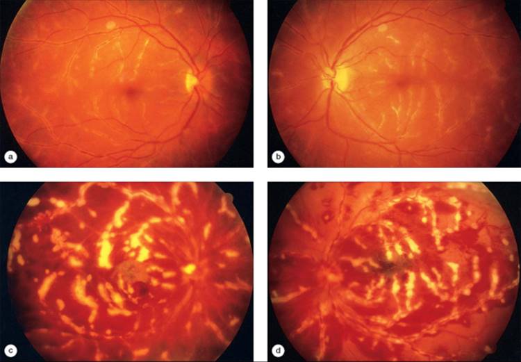
Acute frosted branch angiitis is most commonly a bilateral retinal vasculitis seen in young, otherwise healthy patients in the age range of 3-36 years and present with a chief complaint of decreased visual acuity. Unilateral cases have similarly been reported in the literature.[14,15,20,29] Vision is usually profoundly affected, with the majority of reported cases presenting with 20/200 vision or worse (range 20/20 to light perception). An antecedent viral illness has been reported in some cases. On examination, both anterior chamber and vitreous inflammation are present in almost all cases without pars plana deposits. The appearance of bilateral retinal phlebitis and arteritis extending from the posterior pole to the periphery with uninterrupted severe sheathing of all the vessels, established the term 'frosted branch angiitis' (Fig. 168.1). Prominent sheathing of the retinal veins is characteristic of this entity, but the retinal arteries may similarly be involved (more commonly noted in younger patients).
|
|
|
|
FIGURE 168.1 (a and b) The most frequent presentation of frosted branch angiitis is bilateral extensive venous sheathing, resembling the frosted branches of a tree. There is mild pallor of the left optic disk. (c and d) Bilateral retinal phlebitis extends from the posterior pole to the periphery with uninterrupted severe sheathing of all the vessels. Extensive perivenous exudates associated with intraretinal hemorrhage are noted in this patient. (Left figures, right eye; right figures, left eye.) |
Patients with frosted branch angiitis as described by Ito were young and otherwise healthy. This is in contrast to patients with 'frosted branch like appearance' of the retinal vasculature in patients with lymphomas or leukemias, and similarly distinct from patients with an associated viral or autoimmune disease in which the frosted branch appearance is a clinical sign of disease severity.[72]
DIAGNOSTIC TESTS
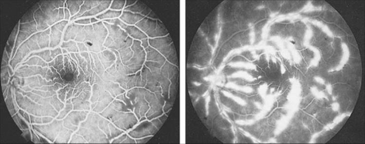
Fluorescein angiography shows normal blood flow without evidence of occlusion or stasis but with late staining and leakage of dye from affected vessels (Fig. 168.2). Additional fundus findings may include intraretinal hemorrhages, punctate hard exudates, macular edema, and serous exudative detachments of the macula or periphery. The retina can appear diffusely thickened and edematous. It is postulated that these thick perivascular infiltrates may be a result of immune complex deposition along the vessel walls.[29,34,35]
|
|
|
|
FIGURE 168.2 The fluorescein angiogram demonstrates normal venous flow in the early phase (left), with diffuse staining of the vein walls in the late phase (right). |
Electrophysiologic testing shows a reduction in the amplitude of the electroretinogram, which remains reduced even after convalescence. In contrast, the visual-evoked response is initially reduced but may return to normal.[5,8] Visual-field testing reveals concentric constriction or relative central defects that improve after clinical resolution of the vasculitis.
Laboratory investigations have not revealed a single causative agent in this disease. Moreover, as this entity must be distinguished from pathologic processes which present as an exaggerated sheathing of retinal vessels, any condition which can cause a retinal vasculitis should be included in the differential diagnosis. Included among the normal laboratory studies from case reports in the literature were: complete blood cell count and differential; erythrocyte sedimentation rate; skin test for tuberculin delayed-type hypersensitivity; determination of serum antibodies for syphilis, Lyme, toxoplasmosis, human immunodeficiency virus (HIV), herpes simplex, herpes zoster, and cytomegalovirus (CMV); urine and blood cultures for both bacteria and virus; antinuclear antibody determination; rheumatoid factor; serum protein electrophoresis; immunoelectrophoresis; and determination of serum angiotensin-converting enzyme levels. Chest radiograph films, lumbosacral X-ray films, cerebrospinal fluid examinations, and computed tomographic scans or magnetic resonance imaging of the head and orbits have all been normal. One patient demonstrated an increased serum antistreptolysin O titer.[6]
MANAGEMENT RECOMMENDATIONS
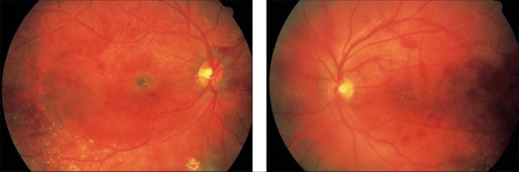
Frosted branch angiitis responds to systemic steroids with a rapid resolution of the vascular sheathing, retinal hemorrhages, and exudative neurosensory retinal detachment (Fig. 168.3). Systemic steroids (initial dose 80-100 mg oral prednisone in adults for 10 days) should be initiated once treatable causes in the differential diagnosis are excluded (most notably infectious etiologies which may worsen in the context of steroid therapy). The steroids should subsequently be tapered over several weeks. Funduscopic sequelae include attenuation of both the arteries and the veins, the development of sharply demarcated atrophic lesions in the periphery, and the deposition of yellow subretinal deposits in areas of previously detached retina.
|
|
|
|
FIGURE 168.3 Systemic steroids are associated with the rapid resolution of the vascular sheathing, intraretinal hemorrhages, and exudative neurosensory retinal detachments. A residual scar in the right macula (left figure) of this patient resulted in a permanent decrease in vision to 20/300. |
Recovery to normal vision usually occurs, with a range of 20/15 to ?20/40, the majority of cases improving to 20/20 or better within 2-3 weeks after initiation of treatment. Others have reported complications prohibiting full visual recovery including a fibrotic macular scar,[6] and central retinal vein occlusion.[19] Remote sequelae have included a horseshoe tear in one patient and multiple bilateral branch vein occlusions in another. It is not known whether these complications are adverse sequelae of frosted branch angiitis or are fortuitous in their occurrence.[6]
The natural history of the disease without the use of oral steroids is not known. One report has shown that the frosted branch angiitis resolved spontaneously without the use of oral agents and with only topical steroid drops for an anterior uveitis over a course of 1 week without any permanent retinal sequelae.[7]
DIFFERENTIAL DIAGNOSIS
Although no specific causative agent has been identified in frosted branch angiitis, treatable causes of retinal vasculitis should be excluded before the initiation of corticosteroid treatment (Table 168.1). Conditions associated with a predominant periphlebitis include tuberculosis, Eales' disease, sarcoidosis, multiple sclerosis, and HIV infection, whereas systemic lupus erythematosus (SLE) and syphilis have been associated with a predominant inflammation of the arterial tree. Retinal vasculitis has been reported as a consequence of Lyme borreliosis.[30] A workup for a presumed diagnosis of frosted branch angiitis revealed ultimately a diagnosis of ocular large cell lymphoma in one case.[31] Another study reported a case of frosted branch angiitis due to a relapsing acute lymphoblastic leukemia involving the central nervous system that resolved with appropriate systemic chemotherapy and local radiotherapy.[36] Case reports have similarly described secondary frosted branch angiitis in patients with Behçet's disease,[10] herpes simplex virus (types 1 and 2),[12,24] CMV retinitis,[33-35] toxoplasmosis,[13] Harada's disease,[16] rapidly progressive glomerulonephritis,[17] aseptic meningitis,[25] and Crohn's disease.[27]
TABLE 168.1 -- Differential Diagnosis of Frosted Branch Angiitis
|
Disease |
Laboratory Tests |
|
Sarcoidosis |
Chest radiograph film, serum calcium and phosphorus levels, serum angiotensin converting enzyme determination |
|
Multiple sclerosis |
Magnetic resonance imaging, cerebrospinal fluid examination |
|
Pars planitis |
|
|
Eales' disease |
|
|
Tuberculosis |
Chest radiograph film, tuberculin skin test |
|
Syphilis |
Venereal Disease Research Laboratory test, fluorescent treponemal antibody test |
|
Lyme disease |
Lyme serology, ELISA, Western blot analysis |
|
Systemic lupus erythematosus |
Antinuclear antibody, anti DNA determinations |
|
AIDS |
HIV antibody determination |
|
Bone marrow tumefaction |
Complete blood count with differential, vitreous biopsy, cerebrospinal fluid examination, magnetic resonance imaging |
|
Abbreviations: ELISA, enzyme linked immunosorbent assay; AIDS, acquired immunodeficiency syndrome; HIV, human immunodeficiency virus. |
The ocular manifestations of sarcoidosis occur in 15-33% of cases.[36-39] Posterior segment involvement is seen frequently (14-28%), usually in association with anterior segment disease. The perivascular sheathing of ocular sarcoid is seen in the midperiphery, usually without vascular occlusion, and affects mainly the veins. Severe perivascular sheathing appears as 'candle wax drippings'. Acute periphlebitis may be accompanied by intraretinal hemorrhages and edema. Occasionally, branch or central retinal vein occlusion may occur, with subsequent peripheral retinal or disk neovascularization.
The vitritis of sarcoidosis consists of clumps of vitreal inflammatory cells - 'snowballs' or 'strings of pearls'. These clumps of cells may remain adherent to the posterior vitreous face and may appear as a sheet. Sarcoid granuloma can be seen in the deep retina or choroid or on the optic nerve head. Although the diagnosis of sarcoid should be excluded by a chest CT scan and determinations of serum angiotensin-converting enzyme and serum calcium, the associated ophthalmic findings should help differentiate ocular sarcoidosis associated vasculitis from frosted branch angiitis.
Retinal venous sheathing is observed in 10-20% of patients with multiple sclerosis[40-45] and is seen either as active periphlebitis, with white patchy cuffs surrounding the blood vessel, or as venous sclerosis. Periphlebitis in multiple sclerosis is occasionally accompanied by pars planitis and frequently resolves spontaneously without visual symptoms or hemorrhages. Rarely, severe periphlebitis can lead to peripheral retinal ischemia and neovascularization. The perivascular infiltrates have been identified histopathologically to be composed of segmental lymphoplasmacytic infiltrates within and around the retinal veins.[40,44] The diagnosis of multiple sclerosis can be made readily by magnetic resonance imaging or cerebrospinal fluid examination with oligoclonal banding. Additionally, the fluffy perivenous cuffs in multiple sclerosis are quite small, focal, and interrupted, in contrast to the widespread sheathing of the vessels in frosted branch angiitis.
Retinal periphlebitis can be found in association with peripheral uveitis or pars planitis.[46-48] Snowbanks consisting of fibroglial proliferation and vascular exudation are seen over a broad area of the inferior peripheral retina; retinal edema, premacular fibroplasia, and cystoid macular edema are also observed. The peripheral terminal branches of the retinal veins are cuffed with white inflammatory material, differentiating this entity from the diffuse periphlebitis of frosted branch angiitis.
Tuberculosis commonly produces focal perivenous sheathing in the peripheral venules, which only occasionally involves the central retinal vein.[49,50] Eales' disease, which has been associated with tuberculosis,[51] characteristically produces early capillary closure in the periphery of multiple quadrants of the retina, with sheathing of the peripheral retinal veins and intraretinal hemorrhages. Neovascularization of the retinal periphery results from the ensuing ischemia, usually at the clearly defined border between perfused and nonperfused retina. The characteristic picture of extensive peripheral capillary nonperfusion in Eales' disease and the focal perivenular sheathing in tuberculosis should help differentiate these entities from frosted branch angiitis.
The vasculitis of ocular syphilis is generally a periarteritis with arteriolar sheathing, exudates, and intraretinal and preretinal hemorrhages,[52-55] although isolated venous periphlebitis has also been described.[55] The arteriolitis may become occlusive, with eventual sclerosis of the involved arterioles and the development of peripheral retinal neovascularization. Other more common manifestations of posterior syphilis include neuroretinitis, chorioretinitis, and papillitis. The course of ocular syphilis has been noted to be more aggressive in patients with associated HIV disease, and as such it is recommended that patients being evaluated or treated for ocular syphilis be tested for HIV infection.[53,56] Because ocular syphilis can mimic many diseases, patients with a vasculitis resembling frosted branch angiitis should have antibody tests for syphilis (the Venereal Disease Research Laboratory test and the fluorescent treponemal antibody test).
Retinal periphlebitis has also been described in patients with Lyme borreliosis.[30,57] Lyme disease is a tick-borne infection that causes a constellation of systemic complaints and manifestations usually within the dermatologic, rheumatologic, cardiac, and central nervous systems.[30,57-61] Ocular manifestations are generally uncommon, and these usually present in patients with either established systemic disease or systemic complaints referable to Lyme disease. The most frequent signs include conjunctivitis, optic neuritis and neuropathy, papilledema, and interstitial keratitis.[58-61] In the reported cases of retinal vasculitis with Lyme disease, the arteries and veins were sheathed but they also appeared occluded and extremely attenuated. This obliterative feature was confirmed on fluorescein angiography.[30,57] In the laboratory workup, patients seronegative for Lyme disease may have positive results with the more sensitive Western blot analysis.[57] A careful history should be taken to identify any exposure to ticks or to reveal any systemic complaints. The presence of associated systemic findings, in areas endemic for Lyme disease, and the more occlusive nature of the periphlebitis should help in differentiating this from frosted branch angiitis.
Retinal vasculitis is a common ophthalmic manifestation of SLE. Although the most common findings are cotton-wool spots with or without intraretinal hemorrhages, almost 30% of patients with SLE will have retinal vasculitis with microangiopathic small-vessel occlusion most often affecting the arterioles.[62] Occasionally, severe retinal occlusive disease can occur, affecting both the arteries and the veins in association with anticardiolipin antibodies,[63] with subsequent peripheral retinal neovascularization. Retinal arterial inflammation is also seen in segmental periarteritis of the retina, in which whitish plaques are scattered along the main arterial branches. Surprisingly, fluorescein leakage is seen from the venous but not from the arterial wall.[64] It can be distinguished from frosted branch angiitis by the generally more limited involvement of the retinal vascular tree.
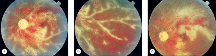
The perivasculitis in acquired immunodeficiency syndrome (AIDS) retinopathy should also be included in the differential diagnosis of frosted branch angiitis.[65-69] Diffuse perivasculitis of the far-peripheral veins and focal periarteritis can be seen in patients with AIDS or AIDS-related complex[67,68,70] without concomitant infectious retinopathy. These conditions are more commonly seen, however, with an associated infectious component such as CMV retinopathy (Fig. 168.4).[33,35,66] Frosted branch angiitis has been described in AIDS patients accompanying small areas of CMV retinopathy. Classically, CMV lesions appear as creamy yellow-white retinal opacifications generally following the vessel walls and often accompanied by variable amounts of intraretinal hemorrhage. The vessel walls may be narrowed, sheathed, or occluded.[60] In these patients, treatment for CMV infection provided resolution of the vasculitic component of their retinopathy. The perivascular infiltrates seen in CMV-related frosted branch angiitis may be due to direct infection by the CMV of the retinal vessel walls.[34] However, this infiltrate may also be a result of immune complex deposition within the vascular wall similar to that hypothesized in conventional cases of frosted branch angiitis.
|
|
|
|
FIGURE 168.4 (a) Severe sheathing of the vessels is present adjacent to a patch of CMV retinitis along the superotemporal arcade of the left eye. (b) This sheathing extends to the periphery, giving a frosted branch appearance. (c) Two weeks after induction with antiviral medications, the sheathing is resolving. |
A case of endogenous Fusarium endophthalmitis diagnosed by vitrectomy was reported in an intravenous drug user who initially presented with frosted branch angiitis and vitritis.[71] In this case, the presence of worsening vitritis and the social history were helpful in obtaining a correct diagnosis.
Secondary vasculitis with frosted branch appearance has been described in patients with ocular toxoplasmosis.[13,14,21] The vasculitis from toxoplasmosis is more commonly an arteritis in the vicinity of the active focus of retinitis.
Finally, bone marrow tumefactions such as leukemia and non-Hodgkin's lymphoma (i.e., reticulum cell sarcoma) should be considered in the presence of diffuse retinal vascular sheathing.[31,32] Intraretinal hemorrhages and cotton-wool spots are more frequently seen in leukemia, whereas retinal cell sarcoma can present as a diffuse vitritis, often with accompanying retinal pigment epithelial abnormalities such as multifocal, yellow, subretinal infiltrates or pigment epithelial detachments. Underlying the vitreal inflammation may be a diffuse retinal vasculitis resembling frosted branch angiitis. The diagnosis can be established by a vitreous biopsy if there is no obvious systemic evidence of disease.
CONCLUSIONS
In summary, frosted branch angiitis is a bilateral inflammation, in healthy young individuals, of the retinal arteries and veins, with the veins being more severely affected. Unilateral cases have been also described. No cause has been identified which is distinctly different from patients who have a frosted branch like appearance from underlying infectious or autoimmune mediated diseases. Vision is profoundly diminished in the acute phase, with dramatic improvement after oral corticosteroid administration. The differential diagnosis is extensive, but frosted branch angiitis can frequently be distinguished by its unique clinical presentation and course.
REFERENCES
1. Ito Y, Nakano M, Kyu N, et al: Frosted branch angiitis in a child. Jpn J Clin Ophthalmol 1976; 30:797.
2. Sakanishi Y, Kanagami S, Ohara K: Frosted retinal angiitis in children. Jpn J Clin Ophthalmol 1984; 38:803.
3. Yamane S, Nishiuchi T, Nakagawa Y, et al: A case of frosted branch angiitis of the retina. Folia Ophthalmol Jpn 1985; 36:1822.
4. Higuchi K, Maeda K, Uji T, et al: A case of infantile uveitis with frosted branch angiitis. Jpn Rev Clin Ophthalmol 1985; 79:2660.
5. Watanabe Y, Takeda N, Adachi-Usami E: A case of frosted branch angiitis. Br J Ophthalmol 1987; 71:553.
6. Kleiner RC, Kaplan HJ, Shakin JL, et al: Acute frosted retinal periphlebitis. Am J Ophthalmol 1988; 106:27.
7. Vander JF, Masciulli L: Unilateral frosted branch angiitis [letter; comment]. Am J Ophthalmol 1991; 112:477.
8. Hamed LM, Fang EN, Fanous MM, et al: Frosted branch angiitis: the role of systemic corticosteroids. J Pediatr Ophthalmol Strabismus 1992; 29:312.
9. Nakai A, Saika S: A case of frosted branch retinal angiitis in a child. Ann Ophthalmol 1992; 24:417.
10. Reynders S, Dewachter A, de Vriese AS: A case of secondary frosted branch angiitis in Behçet's disease. Bull Soc Belge Ophthalmol 2005; 298:41.
11. Liu Y, Ma Z, Tso MO, Zhang Q: Frosted branch angiitis secondary to macular choroidal neovascularization in a Chinese woman. Jpn J Ophthalmol 2005; 49:228.
12. Shenoy R, Elagib EN, Al-Siyabi H: Frosted retinal branch angiitis in an immunocompetent adult due to herpes simplex virus. Indian J Ophthalmol 2001; 49:56.
13. Oh J, Huh K, Kim SW: Recurrent secondary frosted branch angiitis after toxoplasmosis vasculitis. Acta Ophthalmol Scand 2005; 83:115.
14. Diaz-Valle D, Diaz-Rodriguez E, Diaz-Valle T, et al: Frosted branch angiitis and later peripheral retinochoroidal scar in a patient with acquired toxoplasmosis. Eur J Ophthalmol 2003; 13:726.
15. Agrawal S, Agrawal J, Agrawal TP: Unilateral frosted branch angiitis with vitreous hemorrhage. Indian J Ophthalmol 2001; 49:269.
16. Matsuo T, Kanata N, Itami M: Frosted branch angiitis associated with Harada disease-like manifestations recurs 10 years later. Jpn J Ophthalmol 2002; 46:682.
17. Gupta A, Narang S, Gupta V, et al: Frosted branch angiitis associated with rapidly progressive glomerulonephritis. Indian J Ophthalmol 2002; 50:317.
18. Huerva V, Puig T, Sanchez MC, et al: A new case of acute idiopathic frosted branch angiitis in Europe. Eur J Ophthalmol 2002; 12:127.
19. Kaburaki T, Nakamura M, Nagasawa K, et al: Two cases of frosted branch angiitis with central retinal vein occlusion. Jpn J Ophthalmol 2001; 45:628.
20. Fine HF, Smith JA, Murante BL, et al: Frosted branch angiitis in a child with HIV infection. Am J Ophthalmol 2001; 131:394.
21. Ysasaga JE, Davis J: Frosted branch angiitis with ocular toxoplasmosis. Arch Ophthalmol 1999; 117:1260.
22. Biswas J, Raizada S, Gopal L, et al: Bilateral frosted branch angiitis and cytomegalovirus retinitis in acquired immunodeficiency syndrome. Indian J Ophthalmol 1999; 47:195.
23. Luo G, Yang P, Huang S, et al: A case report of frosted branch angiitis and its visual electrophysiology. Doc Ophthalmol 1998-99; 97:135.
24. Markomichelakis NN, Barampouti F, Zafirakis P, et al: Retinal vasculitis with a frosted branch angiitis-like response due to herpes simplex virus type 2. Retina 1999; 19:455.
25. Johkura K, Hara A, Hattori T, et al: Frosted branch angiitis associated with aseptic meningitis. Eur J Neurol 2000; 7:241.
26. Seo MS, Woo JM, Jeong SK, et al: Recurrent unilateral frosted branch angiitis. Jpn J Ophthalmol 1998; 42:56.
27. Sykes SO, Horton JC: Steroid responsive retinal vasculitis with a frosted branch appearance in Crohn's disease. Retina 1997; 17:451.
28. Quillen DA, Stathopoulos NA, Blankenship GW, et al: Lupus associated frosted branch periphlebitis and exudative maculopathy. Retina 1997; 17:449.
29. Sugin SL, Henderly DE, Friedman SM: Unilateral frosted branch angiitis. Am J Ophthalmol 1991; 111:682.
30. Smith JL, Winward KE, Nicholson DF, et al: Retinal vasculitis in Lyme borreliosis. J Clin Neuroophthalmol 1991; 11:7.
31. Ridley ME, McDonald HR, Sternberg P, et al: Retinal manifestations of ocular lymphoma (reticulum cell sarcoma). Ophthalmology 1992; 99:1153.
32. Kim TS, Duker JS, Hedges TR: Retinal angiopathy resembling frosted branch angiitis in a patient with relapsing acute lymphoblastic leukemia. Am J Ophthalmol 1994; 117:806.
33. Geier SA, Nasemann J, Klauss V, et al: Frosted branch angiitis in a patient with the acquired immunodeficiency syndrome. Am J Ophthalmol 1992; 113:203.
34. Spaide RF, Vitale AT, Toth IR, et al: Frosted branch angiitis associated with cytomegalovirus retinitis. Am J Ophthalmol 1992; 113:522.
35. Rabb MF, Jampol LM, Fish RH, et al: Retinal periphlebitis in patients with acquired immunodeficiency syndrome with cytomegalovirus retinitis mimics acute frosted retinal periphlebitis. Arch Ophthalmol 1992; 110:1257.
36. Obenauf CD, Shaw HE, Sydnor CF, et al: Sarcoidosis and its ophthalmic manifestations. Am J Ophthalmol 1978; 86:648.
37. Jabs DA, Johns CJ: Ocular involvement in chronic sarcoidosis. Am J Ophthalmol 1986; 102:297.
38. Spalton DJ, Sanders MD: Fundus changes in histologically confirmed sarcoidosis. Br J Ophthalmol 1981; 65:348.
39. Smith JA, Foster CS: Sarcoidosis and its ocular manifestations. Int Ophthalmol Clin 1996; 1:109.
40. Arnold AC, Pepose JS, Hepler RS, et al: Retinal periphlebitis and retinitis in multiple sclerosis. Ophthalmology 1984; 91:255.
41. Barnford CR, Ganley JP, Sibley WA, et al: Uveitis, perivenous sheathing and multiple sclerosis. Neurology 1978; 28:119.
42. Rucker CW: Sheathing of the retinal veins in multiple sclerosis: review of the pertinent literature. Mayo Clin Proc 1972; 47:335.
43. Vine AK: Severe periphlebitis, peripheral retinal ischemia, and preretinal neovascularization in patients with multiple sclerosis. Am J Ophthalmol 1992; 113:28.
44. Kerrison JB, Flynn T, Green WR: Retinal pathologic changes in multiple sclerosis. Retina 1994; 14:445.
45. Birch MK, Barbosa S, Blumhardt LD, et al: Retinal venous sheathing and the blood-retinal barrier in multiple sclerosis. Arch Ophthalmol 1996; 114:34.
46. Brockhurst RJ, Schepens CL, Okamura ID, et al: Uveitis II. Peripheral uveitis: clinical description, complications, and differential diagnosis. Am J Ophthalmol 1966; 49:1257.
47. Wetzig RP, Chan CC, Nussenblatt RB, et al: Clinical and immunopathological studies of pars planitis in a family. Br J Ophthalmol 1988; 72:5.
48. Malinowski SM, Pulido JS, Folk JC: Long-term visual outcome and complications associated with pars planitis. Ophthalmology 1993; 100:818.
49. Fountain JA, Werner RB: Tuberculous retinal vasculitis. Retina 1984; 4:48.
50. Helm CJ, Holland GN: Ocular tuberculosis. Surv Ophthalmol 1993; 38:229.
51. Renie WA, Murphy RP, Anderson KC, et al: The evaluation of patients with Eales' disease. Retina 1983; 3:243.
52. Crouch ER, Goldberg MF: Retinal periarteritis secondary to syphilis. Arch Ophthalmol 1975; 93:384.
53. Tamesis RR, Foster CS: Ocular syphilis. Ophthalmology 1990; 97:1281.
54. Schlaegel RF, Kao SF: A review (1970-1980) of 28 presumptive cases of syphilitic uveitis. Am J Ophthalmol 1982; 93:412.
55. Lobes LA, Folk JC: Syphilitic phlebitis simulating branch vein occlusion. Ann Ophthalmol 1981; 13:825.
56. McLeish WM, Pulido JS, Holland S, et al: The ocular manifestations of syphilis in the human immunodeficiency virus type I-infected host. Ophthalmology 1990; 97:196.
57. Karma A, Seppala I, Mikkila H, et al: Diagnosis and clinical characteristics of ocular Lyme borreliosis. Am J Ophthalmol 1995; 119:127.
58. Winward KE, Smith JL, Culbertson WW, et al: Ocular Lyme borreliosis. Am J Ophthalmol 1989; 108:651.
59. Winterkorn JMS: Lyme disease: neurologic and ophthalmic manifestations. Surv Ophthalmol 1990; 35:191.
60. Rosenbaum JT, Rahn DW: Prevalence of Lyme disease among patients with uveitis. Am J Ophthalmol 1991; 112:462.
61. Breeveld J, Kuiper H, Spanjaard L, et al: Uveitis and Lyme borreliosis. Br J Ophthalmol 1993; 77:480.
62. Lanham JG, Barrie T, Kohner EM, et al: SLE retinopathy: evaluation by fluorescein angiography. Ann Rheum Dis 1982; 41:473.
63. Jabs DA, Fine SL, Hochberg MC, et al: Severe retinal vaso-occlusive disease in systemic lupus erythematosus. Arch Ophthalmol 1986; 104:558.
64. Orzalesi N, Ricciardi L: Segmental retinal periarteritis. Am J Ophthalmol 1971; 72:55.
65. Holland GN, Pepose JS, Pettit TH, et al: Acquired immune deficiency syndrome - ocular manifestations. Ophthalmology 1983; 90:859.
66. Jabs DA, Green WR, Fox R, et al: Ocular manifestations of acquired immune deficiency syndrome. Ophthalmology 1989; 96:1092.
67. Kestelyn P, Van de Perre P, Rouvroy D, et al: A prospective study of the ophthalmologic findings in the acquired immune deficiency syndrome in Africa. Am J Ophthalmol 1985; 100:230.
68. Kestelyn P, Lepage P, Van de Perre P: Perivasculitis of the retinal vessels as an important sign in children with AIDS-related complex. Am J Ophthalmol 1985; 100:614.
69. Holland GN: Acquired immunodeficiency syndrome and ophthalmology: the first decade. Am J Ophthalmol 1992; 114:86.
70. McClusky PJ, Wakefield D: Posterior uveitis in the acquired immunodeficiency system. Int Ophthalmol Clin 1995; 35:1.
71. Gabriele P, Hutchins RK: Fusarium endophthalmitis in an intravenous drug abuser. Am J Ophthalmol 1996; 122:119.
72. Kleiner RC: Frosted branch angiitis: clinical syndrome or clinical sign?. Retina 1997; 17:370.