Dennis P. Han
DEFINITION
Proliferative vitreoretinopathy (PVR) is defined as the growth and contraction of cellular membranes within the vitreous cavity and on both surfaces of the retina after rhegmatogenous retinal detachment (RD).[1] This process is extremely important, as these membranes exert traction and may cause recurrent detachment by reopening otherwise successfully treated retinal breaks, create new retinal breaks, and distort or obscure the macula. PVR is the primary cause of visual failure after surgical therapy for rhegmatogenous RD. Its incidence following repair of primary RD is estimated to be 5-11%.[2-8] It can also occur after severe penetrating trauma of the posterior segment of the eye, after which the risk of PVR is estimated at 25%.[9]
PATHOBIOLOGY
ORIGIN OF PVR MEMBRANES
Membranes occurring in PVR (PVR membranes) are composed of cells originating from the retinal glia[10] and retinal pigment epithelium (RPE), inflammatory macrophages, and collagen.[11] Continued dispersal and proliferation of RPE cells from the exposed pigment epithelium, combined with glial cell proliferation, ultimately results in a contracted collagenous membrane that covers both surfaces of the retina and the exposed vitreous matrix and produces the characteristic funnel-shaped RD of advanced PVR.
GLIAL CELL COMPONENT
The glial component comprises extensions of Müller cells and astrocytes[12] that form membranes on both surfaces of the retina. These membranes originate directly from the sustentacular glia and develop as a result of damage to the retinal surface[13]; they are also stimulated by platelet-derived growth factor (PDGF)[14] and proliferating RPE cells.[15, 16] PDGF is released by platelet aggregation in blood clots.[17]
RPE CELL COMPONENT
RPE cells are dispersed by three mechanisms into the vitreous cavity after RD:
|
1. |
During initial retinal tear formation, some RPE cells remain adherent to the retina and are avulsed from the pigment epithelium.[11] |
|
|
2. |
After retinal separation, some RPE cells elongate in a vertical direction and are eventually 'pinched off' of Bruch's membrane and form motile RPE macrophages.[18] |
|
|
3. |
RPE cells are released by cryotherapy and scleral depression during the course of surgery for RD repair.[19] |
RPE cells occur as three cell types in periretinal and vitreal membranes[11]: (1) pigment epithelial macrophages that form clumps rather than membranes but that may stimulate membrane formation,[20] (2) RPE cells with retained epithelial morphologic characteristics, and (3) RPE cells that have undergone epithelial-mesenchymal transformation into fibroblast-like cells. This transformation occurs in vivo when isolated RPE cells are not in contact with Bruch's membrane or when they are exposed to intravitreal fluid.[21] A similar transformation occurs in vitro when RPE cells are exposed to vitreous collagen[22] or fibrin.[23]
MACROPHAGE COMPONENT
Macrophages in periretinal membranes[24] are derived from circulating blood monocytes that enter the vitreous from ciliary,[25] retinal,[18] and choroidal[26] circulation and from free RPE cells that transform into macrophage-like cells.[17]
COLLAGEN COMPONENT
The stroma of PVR membranes is composed mainly of types I, II, and III collagen.[27] Type II collagen is a major component of normal vitreous, but RPE cells in vitro are also capable of synthesizing this type of collagen.[28] Therefore, its presence in PVR membranes may result from the development of membranes in areas of residual vitreous attachment or may represent newly secreted material. Types I and III collagen are not found in the normal retina or vitreous and therefore are presumably synthesized by cells within the membranes. Glial cells are capable of synthesizing type I collagen,[29] and RPE cells in culture can synthesize both type I and type III collagen.[28]
STIMULI FOR GROWTH OF PVR MEMBRANES
Conditioned media from RPE cell cultures[30] and glial cell cultures,[31] and vitreous macrophages,[32] bovine retinal extracts,[33] and human subretinal fluid from chronic RD[34] all stimulate RPE proliferation in vitro. It is probable that local cellular production of growth factors is responsible for these mitogenic effects. The evidence for growth control by local autocrine or paracrine mechanisms is well established in analogous biologic systems such as wound healing, in which, e.g., PDGF is synthesized by fibroblasts and then leads to their further proliferation.[35] With relevance to PVR, it has been shown that (1) PDGF-like proteins[36] are present in the conditioned media of RPE cells and are released by activated macrophages[20]; (2) RPE cells in culture produce transforming growth factor-? (TGF-?),[37] and vitreous aspirates from eyes with PVR have more than three times the amount of TGF-? found in eyes with uncomplicated RD.[38] TGF-? has multiple actions, including stimulation of fibrosis in wound healing[39] and synthesis of collagen and fibronectin by RPE cells.[40] The synthesis of growth factors by the cellular component of PVR membranes may explain the characteristic tendency of this disease to gather momentum once initiated and to recur after surgical intervention. However, the contribution of growth factors to the proliferation of cells in PVR is complex because the response of cells to growth factors is not necessarily growth but depends on a variety of other factors, such as the underlying matrix, contact with other cells, the presence of other growth factors, and the presence of specific receptors.[41] These mechanisms have been reviewed by Campochiaro.[42]
Although breakdown of the blood-ocular barrier (e.g., by cryotherapy) causes stimulation of cell proliferation,[43] it is unlikely that this is the source of growth factors because the concentration in plasma is either very low or nonexistent. For example, PDGF stimulates proliferation of RPE cells,[44] but its plasma level is ?1 ng/mL, less than that required to evoke proliferation. Presumably, disruption of the blood-ocular barrier is a general stimulus that evokes cellular production of growth factors.
MECHANISM OF CONTRACTION IN PVR
Transformed, fibroblast-like RPE cells are characteristically motile and move by extending lamellipodia covered with coated pits, which adhere to surrounding surfaces and then retract.[45] In vitreous, this cycle of extension and retraction leads to the gathering up of collagen fibers in the vicinity of the cell and thus to progressive contraction of the gel.[46] The ability of RPE cells to 'reel in' collagen fibers in this way is prodigious, with as much as 5 mm of fiber accumulating beneath a single cell in a 24-h period. It is for this reason that vitreous contraction can be severe, despite its relative hypocellularity.[47] On the surface of the retina on which RPE cells are entrapped in areas of residual vitreous, or when they encounter collagen produced by other RPE or glial cells, a similar process may produce contraction of the retina, although these membranes, at least initially, are generally more cellular.[48] In contrast, glial cells are only weakly contractile.[20] However, these cells create strong attachment points for RPE cells in PVR membranes and transmit contractile forces generated by the RPE component to the underlying retina.[49]
The generation of contraction by RPE cells depends on the presence of fibronectin (an extracellular matrix protein that mediates the adhesion of cells to one another and to structural proteins such as collagen)[50] and does not occur in serum-free media or in the presence of a peptide that specifically blocks cell binding to fibronectin.[51] A high concentration of fibronectin is typically found in the vitreous of eyes with PVR.[52] It is derived mainly from serum and results from cryotherapy[53] and associated breakdown of the blood-retinal barrier,[54] but RPE cells in culture also produce fibronectin,[28] and this additional source may further increase their ability to generate traction.[55] The amount of fibronectin within periretinal membranes may also have surgical consequences: Early membranes contain large amounts of fibronectin and are correspondingly difficult to peel from the retinal surface; later membranes, however, have much less fibronectin, and this coincides with their easier surgical removal.[56]Fibronectin also directly stimulates RPE motility.[57] Since, as we have previously noted, contraction is a result of cell movement, fibronectin therefore both facilitates and provokes contraction in PVR. PDGF also stimulates RPE motility and contraction,[58] and PVR occurs more frequently when RD is complicated by vitreous hemorrhage.[59, 60] Finally, activated macrophages produce fibronectin and interleukin-1,[20] which is found in the subretinal fluid of eyes with PVR[61] and stimulates the migration of RPE cells.[62]
CLINICAL DIAGNOSIS AND CLASSIFICATION
The widely used classification of PVR published by the Retina Society[1] has been revised.[63] The major changes are separate description and grading of PVR membranes in the anterior and posterior retina and elimination of the width of the retinal funnel as a component of the classification. The revised system of diagnosis and classification is further described in the remainder of this section.
|
Key Features Classification of Proliferative Vitreoretinopathy |
|||||||||||||||||||||||||||
|
|||||||||||||||||||||||||||
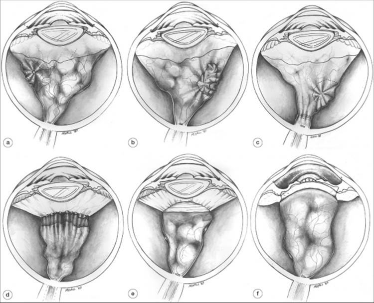
In the earliest manifestation of PVR, clumps of RPE cells and a protein flare are present in the vitreous (grade A). This is followed by the development of PVR membranes, which are initially almost transparent but cause distortion of the retina (grade B). These membranes subsequently become opaque and produce several characteristic patterns of contraction (grade C), in either the posterior or the anterior retina. In the posterior retina, defied as posterior to the retinal equator, these patterns of contraction consist of (1) star fold, which represents the contraction of a single focus of PVR (type 1) (see Fig. 183.1a); (2) irregular fixed folds, indicating multiple epicenters of contraction (type 2) (Fig. 183.1b); or (3) an elevated fold without visible preretinal membrane; either an annulus around the disk ('napkin ring') or an extended linear fold ('clothesline') (type 3) (Fig. 183.1c). In the anterior retina, the patterns of contraction consist of (4) a series of radial folds at the insertion of the posterior hyaloid-contraction in a circumferential direction creates an irregular equatorial fold (type 4) (Fig. 183.1d); (5) contraction of the posterior hyaloid creating a smooth circumferential equatorial fold (type 5) (Fig. 183.1e); and (6) anterior displacement of the peripheral retina as the result of PVR occurring on the surface of the residual basal vitreous remaining after vitrectomy. The basal vitreous is contracted in an anteroposterior direction. Since the anterior portion of the vitreous base is anchored to the pars plana, the peripheral retina is pulled forward, forming a trough in the extreme retinal periphery (type 6) (Fig. 183.1f).
|
|
|
|
FIGURE 183.1 Types of grade C PVR (see text). (a) Type 1. Regular folds radiate ('star fold') from a focal area of PVR. Traction is exerted centripetally toward the area of PVR. (b) Type 2. Irregular folds are present in the posterior retina. The retina is contracted in an anteroposterior direction, which flattens its normally bullous convex curvature, and in a circumferential direction, which creates a series of radial folds in the anterior retina. Traction is exerted on the remainder of the retina, pulling it in both a posterior direction and a circumferential direction. Traction in a perpendicular direction pulls the retina toward the center of the vitreous cavity, narrowing the funnel of retinal detachment over the optic disk. (c)Type 3. Subretinal membrane creates an annular constriction around the disk ('napkin ring') or irregular folds more anteriorly. In the napkin ring configuration, traction in a circumferential direction gathers the posterior retina together anterior to the optic disk. With irregular folds, traction is mainly perpendicular to the retinal surface, elevating the retina toward the center of the vitreous cavity. (d) Type 4. Irregular folds are present in the retina immediately behind the vitreous base, and the adjacent posterior hyaloid is contracted. Contraction of the retina in a circumferential direction causes radial folds posterior to the area of PVR. The anterior retina within the vitreous base is stretched inward to form a fold in the coronal plane because circumferential contraction exerted along the concavity of the retina produces a secondary traction vector perpendicular to the retinal surface, toward the center of the vitreous cavity. (e) Type 5. The posterior hyaloid is contracted. The anterior retina is pulled in a perpendicular direction toward the center of the vitreous cavity. Radial folds are also formed as a result of contraction in a circumferential direction. (f) Type 6. The anterior retina is pulled forward as a result of contraction in an anteroposterior direction of the posterior hyaloid, the anterior hyaloid, and the vitreous base that remain after vitrectomy. The underlying retina forms a trough within the vitreous base. |
PVR, grades B and C, are further illustrated in Figure 183.2.
|
|
|
|
FIGURE 183.2 Photographic illustration of types of PVR. (a) Grade B. The posterior lip of a large tear is rolled inward by PVR localized at its edge. (b) Grade C, type 1. A single epicenter of PVR creates a star fold in the posterior retina. (c) Grade C, type 2. Diffuse PVR in the posterior retina. (d) Grade C, type 3. Subretinal PVR closes the funnel of retinal detachment anterior to the disc. (e) Grade C, type 4. PVR at the retinal equator produces a circumferential fold immediately behind the insertion of the posterior hyaloid (left side of photograph); peripheral retina within the vitreous base is stretched inward (right side of photograph). (f) Grade C, type 5. Contraction of the posterior hyaloid (located more posteriorly than usual in this case, as indicated by the line of pigment immediately behind its insertion inferiorly) creates a circumferential fold in the coronal plane. (g) Grade C, type 6. An operative photograph shows a trough of retina anteriorly (immediately to the left of the vitrectomy probe), covered by an opaque PVR membrane. |
TREATMENT
Surgical therapy is the mainstay of treatment of PVR. Surgical goals include relief of retinal traction, intraoperative and postoperative retinal tamponade to facilitate treatment of retinal breaks, and removal of proliferative tissue likely to cause recurrent contraction and RD. Relief of retinal traction is most directly achieved by posterior vitrectomy and removal of preretinal and subretinal membranes. In most instances, scleral buckling is performed in conjunction with vitrectomy if there is no preexistent buckle. It can also be used alone for cases with minimal retinal traction. Relaxing retinotomies or retinectomies are considered if the above measures are insufficient to relieve traction, usually in cases with severe subretinal proliferation or vitreous base contraction. Large retinotomies may render scleral buckling unnecessary.
SURGICAL APPROACHES: CASE STUDIES
The following case examples are given to illustrate the general principles involved in selecting the appropriate surgical procedure for RD complicated by PVR.
Case 1
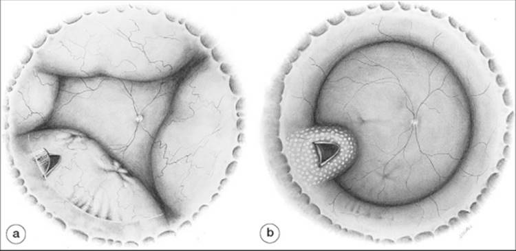
The RD in this case (Fig. 183.3a) is due to a single large horseshoe tear with a rolled posterior edge (grade B PVR) in the lower temporal quadrant. A focus of PVR in the posterior retina produces a star fold (grade C, type 1) and exerts traction on the tear. An area of more remote PVR, which is not affecting the tear, is present for 2 clock hours of the retinal circumference at and immediately behind the insertion of the posterior hyaloid (grade C, type 4).
|
|
|
|
FIGURE 183.3 Case 1 (see text). (a) Preoperative appearance. PVR is present in the inferotemporal quadrant. The large horseshoe tear has rolled edges (grade B PVR). Two star folds in the posterior retina (grade C PVR), radial folds between the 7:30 and the 6:30 meridians, and opacification of the posterior hyaloid in the anterior retina (grade C PVR) are present. (b) Postoperative result. The horseshoe tear is closed with a radial buckle and is surrounded by chorioretinal reaction. The eye has been encircled to maintain the height of the radial buckle and support the anterior retina. The star folds and radial folds are almost invisible. |
Such a detachment is treated by buckling the tear using a radial element and by draining subretinal fluid to approximate the tear to the buckle. Removal of the star fold may not be necessary if the mobility of the surrounding retina is sufficient to accommodate reattachment. The radial buckle facilitates reattachment by closing the break and reversing the traction vectors exerted on the tear from those of retinal separation from the underlying RPE to those of apposition.[64] An encircling band to support the vitreous base region and area of insertion of the posterior hyaloid is often used to counteract residual peripheral traction or the development of mild anterior PVR. The postoperative result is shown in Figure 183.3b.
Case 2
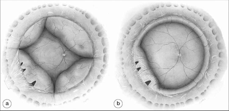
The RD in this case is illustrated in Figure 183.4a. Three horseshoe tears are present ?1 clock hour apart in the lower temporal quadrant. The eye has been treated previously by vitrectomy and the tears have occurred as a result of traction on the posterior hyaloid by residual vitreous incarcerated in the adjacent sclerotomy.[65] PVR has also occurred on the residual vitreous matrix adjacent to the tears, creating localized anterior traction (grade C, type 6). The combination of mechanical incarceration of residual hyaloid in the adjacent sclerotomy and localized PVR has led to gross elevation of the tears from the RPE.
|
|
|
|
FIGURE 183.4 Case 2 (see text). (a) Preoperative appearance. A vitrectomy has been performed previously. PVR associated with vitreous incarcerated in the lower temporal sclerotomy has caused opacification and contraction of the adjacent basal vitreous (grade C PVR) and resulted in three horseshoe tears. (b) Postoperative result. Traction has been relieved (1) directly by transecting and trimming the incarcerated vitreous and resecting the anterior flaps of the horseshoe tears and (2) indirectly by supporting the tears with a segmental buckle. |
It might be possible to treat this detachment simply by buckling the tears using a segmental explant supported by an encircling band (the band is needed for the same reasons as in case 1). This treatment may fail, however, because persistent traction on the breaks causes redetachment of the anterior portion of the breaks, leading to extension of fluid anteriorly and circumferentially around the buckle, similar to that occurring when a buckle is placed too posteriorly. An alternative approach would be to combine buckling with vitrectomy, visualizing the region of anterior dissection by means of coaxial illumination and scleral indentation.[66] This approach is more recently facilitated by the use of wide angle, binocular indirect ophthalmoscopic viewing systems. The postoperative result is depicted in Figure 183.4b
Case 3
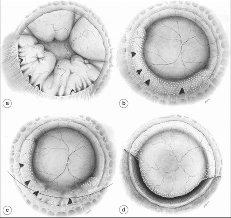
The RD in this eye (Fig. 183.5a), which has previously undergone vitrectomy, is caused by four horseshoe tears in the lower temporal quadrant. In addition, considerable PVR is present. The insertion of the residual posterior hyaloid membrane is opacified, and its contraction has led to a series of radial retinal pleats (grade C, type 4) for 5 clock hours of the retinal circumference, extending from the 9 o'clock to the 4 o'clock meridians. Within this zone of the retinal circumference, between the 8 o'clock and the 5 o'clock meridians, PVR spreads behind the equator, resulting in a diffuse area of contraction (grade C, type 2), and anterior to the equator, producing opacification of the residual vitreous matrix together with anterior displacement of the insertion of the remnant of the posterior hyaloid membrane (grade C, type 6). Vitrectomy, scleral buckling and anterior retinal membrane dissection may be sufficient to obtain posterior retinal attachment with localized, nonprogressive, anterior detachment (Fig. 183.5b). In others, diffuse and anterior contraction leads to persistent or recurrent anterior RD, even in the presence of silicone oil tamponade (Fig. 183.5c). These may progress and threaten macular function. In such cases, an anterior, circumferentially oriented relaxing retinectomy may be needed (Fig. 183.5d). Intraoperative positioning of the retinectomy flap and laser photocoagulation is facilitated by the temporary injection of perfluorocarbon liquid into the vitreous cavity (Fig. 183.5d).[67, 68] All retinal traction must be removed for the retinectomy to succeed. Large inferior retinectomies usually require silicone oil tamponade.
|
|
|
|
FIGURE 183.5 Case 3 (see text). (a) Preoperative appearance. A vitrectomy has been performed previously. PVR involves both inferior retinal quadrants. Several star folds are present in the posterior retina. PVR is present on the residual vitreous and immediately behind the insertion of the posterior hyaloid and has contracted the anterior retina in a circumferential direction. (b) Postoperative appearance after repeat vitrectomy with vitreous base dissection, scleral buckle, laser photocoagulation, and gas tamponade. Residual anterior PVR has resulted in localized anterior retinal detachment. Such cases may remain stationary or progress to more extensive detachment. (c) Residual anterior PVR and detachment has progressed through the barrier of laser photocoagulation and threatens to extend into the posterior retina, in spite of silicone oil tamponade. (d) Postoperative appearance after anterior relaxing retinectomy, intraoperative perfluorocarbon liquid injection, laser photocoagulation, and gas tamponade. Anterior PVR has been completely resected, and the posterior edge of the retinectomy has been sealed with laser treatment. Inferior large retinectomies usually require silicone oil tamponade. |
VISUAL OUTCOME IN PVR
Functional and anatomic outcomes are strongly dependent on the severity of the disease and its anteroposterior location. Posterior PVR is usually easily visualized, can often be completely removed, and is frequently relatively remote from retinal breaks. The converse may be true for anterior PVR, in which the more difficult visualization and surgical dissection may increase the risk of iatrogenic breaks or incomplete relief of traction. Overall, large surgical series utilizing vitrectomy as the primary modality report posterior retinal reattachment rates of 77-90%, with an approximate functional success rates (20/200 or better) of ?25%.[69-72] However, reported reattachment rates vary considerably, depending on the chosen surgical approach. These are discussed below.
SCLERAL BUCKLING
In two large series, scleral buckling alone was successful in 34%[73] and 47%[74] of cases, respectively.
VITRECTOMY
Compared to scleral buckling alone, vitrectomy combined with endolaser photocoagulation,[75] prolonged tamponade with gas,[76, 77] and more extensive dissection of anterior PVR[78] results in a larger proportion of cases with anatomic success, ranging from 53%[79] to 90%.[80, 81] However, additional surgery was required in 19-50% of cases to achieve success.
INTERNAL TAMPONADE
The Silicone Study showed that there was no major advantage to silicone oil compared with perfluoropropane (C3F8) gas for the management of RDs with PVR.[82] The anatomic results with C3F8 were somewhat better than those with silicone oil in eyes that had not undergone a previous vitrectomy but the visual results were basically equivalent. Eyes treated with gas were more likely to require additional procedures and more likely to become hypotonous; this difference probably neutralized the anatomic benefits of gas. Other results of the Silicone Study were that cases with extensive anterior PVR were more likely to become chronically hypotonous[83]; creation of a retinectomy did not affect the anatomic equivalence of the two tamponades[84]; in matched pairs of eyes, silicone oil removal led to a significant visual improvement[85]; the incidence rates of corneal abnormalities were similar in eyes treated with gas or silicone oil[86]; the type of tamponade did not influence the risk of postoperative macular pucker.[87]The long-term results of the Silicone Study have been published.[88] The authors conclude that success with one operation is more important than the choice of tamponade in determining the visual result and "if anatomically and visually successful at 3 years there is an excellent chance that the visual result will be maintained over the long-term."
RETINECTOMY
This procedure has been used by most surgeons only after alternative methods of relieving retinal traction have failed, although its use to permanently isolate the posterior retina from traction in the vitreous base has been described.[89] Using this technique, reattachment can be achieved in 58-83%[73, 90-92] of cases and can be compatible with good VA.
COMPLICATIONS OF PVR SURGERY
Each of the modalities used in PVR surgery are associated with particular risks which must be considered in the fial surgical approach. Assessment of such risks is a dynamic process that begins with the initial patient evaluation and continues intraoperatively throughout the surgical procedure as the response unfolds to the various interventions, sequentially applied.
SCLERAL BUCKLING
Relative to segmental scleral buckles often used in primary RD surgery, broad, encircling buckles are often used in PVR surgery to address larger areas of potential traction. Large buckles carry a higher risk of choroidal effusion, presumably due to impedance of the vortex venous outflow. Although effusions will usually reabsorb spontaneously, they may prolong postoperative breakdown of the blood-ocular barrier, a factor believed to contribute to recurrent PVR. Other well known complications of scleral buckling include ocular perforation during scleral suture placement, severe choroidal hemorrhage, strabismus, diplopia, extrusion, infection, and refractive error. Severe choroidal hemorrhage may impair macular function, cause severe glaucoma, and result in blindness or severe visual loss. In cases of strabismus with poor visual acuity (VA), sensory confusion, rather than frank diplopia may occur, in which a blurred eccentric image from the eye with PVR is superimposed on a sharp foveal image from the fellow eye. The buckle can extrude and become infected, particularly as the conjunctiva may have already been compromised from previous ocular surgeries. Due to these risks, a preexistent scleral buckle is often left undisturbed during vitreous surgery, with replacement or revision only for migrated buckles or for those having an inadequate buckling effect.
VITRECTOMY
Retinal breaks posterior to the sclerotomy entry sites are a potential complication of any vitrectomy procedure. They usually occur indirectly when instruments put traction on the posterior hyaloid,[65] but in surgery for PVR in which the retina is drawn forward over the pars plana, holes can be made directly through the retina. Such peripheral holes can be difficult to close because of persistent traction in the vitreous base. The posterior retinotomy used for endodrainage may also be a source of subsequent PVR.[93] In phakic eyes, transient cataract occurs because of the prolonged gas bubble in the vitreous cavity; less commonly, the gas bubble leads to either corneal decompensation or glaucoma in aphakic eyes. Progressive nuclear sclerosis occurs in virtually all patients undergoing vitrectomy.
RETINAL MEMBRANE DISSECTION, RELAXING RETINOTOMIES AND RETINECTOMIES
Iatrogenic retinal breaks and associated retinal bleeding may occur during retinal membrane dissection. Hemostasis is extremely important to maintain visualization and reduce a possible stimulus for reproliferation. In phakic eyes with anterior PVR, adequate relief of retinal traction may require lensectomy. In such cases, insertion of an intraocular lens is often best considered in a separate procedure after the retina has been stabilized.
Relaxing retinomies and retinectomies are associated with a significant risk of reproliferation, hypotony, or phthisis. Intraoperative slippage of retinal flaps or subretinal migration of silicone oil or perfluorocarbon liquids may also occur.[92, 94]
SILICONE OIL
Silicone oil can result in a number of complications, the prevention and management of which have been reviewed.[95] Cataract is probably inevitable[96] in eyes permanently filled with silicone oil, but there is some evidence that cataract may not develop if silicone oil remains in the eye only temporarily.[97] In eyes with good visual potential, subsequent cataract extraction can improve visual function.[95]
Glaucoma can result from pupillary block by the anterior surface of the silicone bubble or from the accumulation and subsequent fibrosis of multiple small droplets in the anterior chamber angle. Pupillary-block glaucoma is most commonly prevented by an inferior iridectomy,[98] although this has a tendency to close with time.[99] Droplet formation may be reduced by the use of more purified silicone of higher viscosity.[100] Interestingly, the Silicone Study showed that the risk of glaucoma is greater in eyes treated with gas than in those treated with silicone oil provided the silicone oil is ultimately removed.[88]
Keratopathy occurs if the anterior chamber is filled with silicone. This complication is unusual in eyes with normal aqueous production and a patent inferior peripheral iridectomy,[101] but it is common in eyes with severe hypotony.[102] The Silicone Study did not fid, as had been expected, a higher incidence of corneal abnormalities in eyes treated with silicone oil compared with those treated with gas.[88] This result suggests that some of the cases of keratopathy in eyes treated with silicone are not related directly to silicone oil but stem from other factors, e.g., previous surgery or hypotony.
Retinal toxicity from permanent placement of silicone oil has been suggested by several authors,[103-105] but remains unproved. It has also been proposed that silicone may stimulate PVR membranes by loculating growth factors in the preretinal space.[106] Although these membranes may lead to retinal breaks,[107] their clinical effect is, in fact, minimized by the presence of the silicone bubble.
Silicone removal is associated with a considerable risk of RD. In one study of the complications of prophylactic silicone oil removal, the retina redetached in 25% of cases.[108] The authors of this study concluded that "Ésilicone oil should be removed only in carefully selected cases.É patients with no significant vitreoretinal traction and those most likely to develop sight threatening anterior segment complications remain the obvious candidates."
PERFLUOROCARBON LIQUIDS
Perfluoro-n-octane or other perfluorocarbon liquids are useful intraoperative adjuncts in PVR surgery. Intraocular retention of perfluorocarbon liquids (PFCL) may occur and account for most secondary complications relating to this material. Symptomatic floaters, corneal toxicity, and subretinal migration are of primary concern. Small amounts of retained PFCL in the vitreous cavity are usually tolerated well, but can cause symptomatic floaters in patients who recover good VA. Short-term corneal contact with small amounts of retained PFCL in the anterior chamber are unlikely to cause significant toxicity.[109]Subretinal perfluorocarbon liquid may cause symptomatic scotoma, particularly if located in the macular region. These can be removed with fie (e.g., 39 gauge) cannulas used for macular translocation surgery, resulting in functional visual improvement.[110]
An increased risk of PFCL retention in the eye is associated with large retinotomies and failure to perform a saline rinse during the fluid-air exchange portion of the surgery.[94] Intraocular dispersion of the material may also make complete removal difficult. Such may occur if saline is infused directly into the PFCL, which can occur if there is overfilling of the eye to the point where PFCL rises above the level of the infusion cannula, or by rotation of the globe, such as during scleral buckle placement, in an eye filled with PFCL.
PREVENTION OF PVR
Pharmacologic means of PVR prevention have recently been evaluated. Effective prophylaxis of PVR requires identification of risk factors to optimize therapeutic effect. In patients with RD without established PVR, Kon et al[111] devised a regression equation to predict risk of PVR using various clinical characteristics. These were validated in a subsequent prospective study by Asaria et al[112] in 212 subjects undergoing primary vitrectomy for RD. The presence of a total detachment, grade C PVR, aphakia, larger extent of RD, and history of cryotherapy in the affected eye appeared to have a significant effect on PVR risk. Specifically, a greater than threefold risk of PVR (28% vs 9%, p< 0.001) was identified for patients with: (1) total RD with either grade B PVR (or worse), aphakia, or uveitis; (2) grade C PVR with 3 quadrants of detachment, or with 2 quadrants of detachment and a history of cryotherapy in the affected eye; or (3) aphakia with 3 quadrants of RD, or with 2 quadrants RD and a history of cryotherapy. Other factors that have been implicated in promoting PVR include the presence of giant retinal tears or multiple retinal breaks exposing large areas of RPE to the vitreous cavity, choroidal detachment, vitreous hemorrhage, preexisting PVR, and incomplete posterior vitreous detachment.[60, 61, 74, 113, 114]
Relatively few randomized clinical trials evaluating PVR prophylaxis have been performed. Asaria et al[115] performed a randomized clinical trial in high-risk cases without established PVR, assessing the safety and efficacy of adjuvant combination therapy using 5-fluorouracil (5-FU) and low-molecular weight heparin (LMWH) at the time of vitrectomy for repair of RD. They found a reduction in PVR rate from 26.4% (23/87) to 12.6% (11/87, p= 0.02), a trend toward reduction in reoperation rate due to PVR, and better fial VA in treated versus placebo groups (improved VA in 60% vs 46%, p = 0.048). Encouraging as this was, treatment with the above regimen in eyes with established PVR did not appear to influence the rate of surgical reattachment.[116] Treatment of established PVR with intraocular infusion of Daunorubicin was also evaluated in a randomized trial by Wiedemann et al.[117] The study showed a significant reduction of reoperations in the treated group compared to controls, but not a significant difference in the primary outcome measure of retinal reattachment at 6 months. Currently the role of pharmacotherapy in the treatment of PVR remains in debate.
CONCLUSIONS
PVR occurs after rhegmatogenous RD when ectopic RPE and glial cells proliferate and form contractile membranes on the retinal and vitreal surfaces. The clinical diagnosis of the established disease depends on the presence of characteristic patterns of retinal distortion or white or pigmented membranes on the retinal surface or within the vitreous. Surgical management specifically addressing PVR should be performed if it is severe enough to inhibit retinal reattachment or threaten redetachment. Such treatment consists of vitrectomy to allow removal of as much proliferative tissue as possible, scleral buckling, laser photocoagulation, long-acting gas or silicone oil tamponade, and, in severe cases, retinotomy/retinectomy. Despite good anatomic outcomes with these techniques, the challenge of improving visual outcome remains.
ACKNOWLEDGMENT
The author acknowledges Dr John S Lean, for his contributions to the original version of this chapter.
REFERENCES
1. The Retina Society Terminology Committee: The classification of retinal detachment with proliferative vitreoretinopathy. Ophthalmology 1983; 90:121.
2. Speicher MA, Fu AD, Martin JP, von Fricken MA: Primary vitrectomy alone for repair of retinal detachments following cataract surgery. Retina 2000; 20:459-464.
3. Duquesne N, Bonnet M, Adeleine P: Preoperative vitreous haemorrhage associated with rhegmatogenous retinal detachment: a risk factor for postoperative proliferative vitreoretinopathy?. Graefes Arch Clin Exp Ophthalmol 1996; 234:677-682.
4. Greven CM, Sanders RJ, Brown GC, et al: Pseudophakic retinal detachments. Anatomic and visual results. Ophthalmology 1992; 76:257-262.
5. Girard P, Mimoun G, Karpouzas I, Montefiore G: Clinical risk factors for proliferative vitreoretinopathy after retinal detachment surgery. Retina 1994; 14:417-424.
6. Gartry DS, Chignell AH, Franks WA, Wong D: Pars plana vitrectomy for the treatment of rhegmatogenous retinal detachment uncomplicated by advanced proliferative vitreoretinopathy. Br J Ophthalmol 1993; 77:199-203.
7. Bonnet M, Fleury J, Guenoun S, et al: Cryopexy in primary rhegmatogenous retinal detachment: a risk factor for postoperative proliferative vitreoretinopathy?. Graefes Arch Clin Exp Ophthalmol 1996; 234:739-743.
8. Heimann H, Bornfeld N, Friedrichs W, et al: Primary vitrectomy without scleral buckling for rhegmatogenous retinal detachment. Graefes Arch Clin Exp Ophthalmol 1996; 234:561-568.
9. Charteris DG, Sethi CS, Lewis GP, Fisher SK: Proliferative vitreoretinopathy: developments in adjunctive treatment and retinal pathology. Eye 2002; 16:369-374.
10. Laqua H, Machemer R: Glial cell proliferation in retinal detachment (massive periretinal proliferation). Am J Ophthalmol 1975; 80:602.
11. Machemer R, Laqua H: Pigment epithelium proliferation in retinal detachment (massive periretinal proliferation). Am J Ophthalmol 1975; 80:1.
12. McLeod D, Hiscott P: Age related cellular proliferation at the vitreoretinal juncture. Eye 1987; 1:263.
13. Foos R, Gloor BP: Vitreoretinal juncture: Healing of experimental wounds. Graefes Arch Clin Exp Ophthalmol 1975; 196:213.
14. Harvey A, Roberge F, Hjelmeland L: Chemotaxis of rat retinal glia to growth factors found in repairing wounds. Invest Ophthalmol Vis Sci 1987; 28:1092.
15. Peters M, Burke J, Clowry M, et al: Development of traction retinal detachment following intravitreal injections of retinal Müller and pigment epithelial cells. Graefes Arch Clin Exp Ophthalmol 1986; 224:554.
16. Rowen S, Glaser B: Retinal pigment epithelial cells release a chemoattractant for astrocytes. Arch Ophthalmol 1985; 103:704.
17. Ross R, Raines E, Bowen Pope D: The biology of platelet derived growth factor. Cell 1986; 46:155.
18. Johnson N, Foulds W: Observations on the retinal pigment epithelium and retinal macrophages in experimental retinal detachment. Br J Ophthalmol 1977; 61:564.
19. Singh A, Michels R, Glaser B: Scleral indentation following cryotherapy and repeat cryotherapy enhance release of viable retinal pigment epithelial cells. Retina 1986; 6:176.
20. Gilbert C, Hiscott P, Unger W, et al: Inflammation and the formation of epiretinal membranes. Eye 1988; 2:S140.
21. Lean J, Noorily S, Crisp A, et al: Response of the RPE monolayer to retinal hole formation. In: Heimann K, Wiedemann P, ed. Response of the RPE monolayer to retinal hole formation, Heidelburg: Kaden; 1989.
22. Vidaurri LJ, Hohman R, Glaser B: Effect of vitreous on morphologic characteristics of retinal pigment epithelial cells: A new approach to the study of proliferative vitreoretinopathy. Arch Ophthalmol 1984; 102:1220.
23. Vidaurri Leal J, Glaser B: Effect of fibrin on morphologic characteristics of retinal pigment epithelial cells. Arch Ophthalmol 1984; 102:1376.
24. Newsome D, Rodrigues M, Machemer R: Human massive periretinal proliferation. In vitro characteristics of cellular components. Arch Ophthalmol 1981; 99:873.
25. Cleary P, Ryan S: Histology of wound, vitreous, and retina in experimental posterior penetrating eye injury in the rhesus monkey. Am J Ophthalmol 1979; 88:221.
26. Grierson I, Forrester J: Vitreous haemorrhage and vitreal membranes. Trans Ophthalmol Soc UK 1980; 100:140.
27. Jerdan J, Pepose J, Michels R: Proliferative vitreoretinopathy membranes. An immunohistochemical study. Ophthalmology 1989; 96:801.
28. Campochiaro P, Jerdan J, Glaser B: The extracellular matrix of human retinal pigment epithelial cells in vivo and its synthesis in vitro. Invest Ophthalmol Vis Sci 1986; 27:1615.
29. Burke J, Kower H: Collagen synthesis by rabbit neural retina in vitro and in vivo. Exp Eye Res 1980; 31:213.
30. Bryan JI, Campochiaro P: A retinal pigment epithelial cell derived growth factor(s). Arch Ophthalmol 1986; 104:422.
31. Burke J, Foster S: Induction of DNA synthesis by co culture of retinal glia and pigment epithelium. Invest Ophthalmol Vis Sci 1985; 26:636.
32. Burke J, Twining S: Generation of stimulants for pigment epithelium in vitro. Invest Ophthalmol Vis Sci 1987; 28:1100.
33. Campochiaro P, Glaser B: A retina derived stimulator(s) of retinal pigment epithelial cell and astrocyte proliferation. Exp Eye Res 1986; 43:449.
34. Hackett SF, Conway BP, Campochiaro PA: Subretinal fluid stimulation of RPE migration and proliferation is dependent upon certain features of the detachment. Arch Ophthamol 1989; 107:391.
35. Sporn M, Roberts A: Introduction: Autocrine, paracrine and endocrine mechanisms of growth control. Cancer Surv 1985; 4:627.
36. Campochiaro P, Sugg R, Grotendorst G, et al: Retinal pigment epithelial cells produce PDGF like proteins and secrete them into their media. Invest Ophthalmol Vis Sci 1988; 29(Suppl):305.
37. Connor T, Roberts A, Sporn M, et al: RPE cells synthesize and release transforming growth factor beta, a modulator of endothelial cell growth and wound healing. Invest Ophthalmol Vis Sci 1988; 29(Suppl):307.
38. Connor TJ, Roberts A, Sporn MB, et al: Correlation of fibrosis and transforming growth factor beta type 2 levels in the eye. J Clin Invest 1989; 83:1661.
39. Mustoe T, Pierce G, Thomason A, et al: Accelerated healing of incisional wounds in rats induced by transforming growth factor?. Science 1987; 237:1333.
40. Sporn M, Roberts A, Wakefield L, et al: Some recent advances in the chemistry and biology of transforming growth factor beta. J Cell Biol 1987; 105:1039.
41. Burke JM: Cell interactions in proliferative vitreoretinopathy: Do growth factors play a role?. In: Heimann K, Wiedemann P, ed. Proliferative vitreoretinopathy, Heidelberg: Kaden; 1989.
42. Campochiaro PA: Pathogenic mechanisms in proliferative vitreoretinopathy. Arch Ophthalmol 1997; 115:237.
43. Campochiaro PA, Bryan JAI, Conway BP, et al: Intravitreal chemotactic and mitogenic activity: Implication of blood retinal barrier breakdown. Arch Ophthalmol 1986; 104:1685.
44. Bryan JA III, Campochiaro PA: Platelet derived growth factor facilitates but is not required for human RPE growth in vitro. Invest Ophthalmol Vis Sci 1986; 27(Suppl):305.
45. Glaser BM, Lemor M: Pathobiology of proliferative vitreoretinopathy. In: Glaser BM, Michels RG, ed. Surgical retina, St. Louis: CV Mosby; 1989.
46. Glaser BM, Cardin A, Biscoe B: Proliferative vitreoretinopathy. The mechanism of development of vitreoretinal traction. Ophthalmology 1987; 94:327.
47. Blumenkranz MS, Hartzer M: Contractile mechanisms in proliferative vitreoretinopathy (PVR). Invest Ophthalmol Vis Sci 1986; 27(Suppl):188.
48. Hiscott PS, Grierson I, McLeod D: Retinal pigment epithelial cells in epiretinal membranes: An immunohistochemical study. Br J Ophthalmol 1984; 68:708.
49. Hiscott PS, Grierson I, Trombetta CJ, et al: Retinal and epiretinal glia-An immunohistochemical study. Br J Ophthalmol 1984; 68:698.
50. Yamada KM: Cell surface interactions with extracellular materials. Ann Rev Biochem 1986; 52:761.
51. Avery RL, Glaser BM: Inhibition of retinal pigment epithelial cell attachment by a synthetic peptide derived from the cell binding domain of fibronectin. Arch Ophthalmol 1986; 104:1220.
52. Campochiaro PA, Jerdan JA, Glaser BM, et al: Vitreous aspirates from patients with proliferative vitreoretinopathy stimulate retinal pigment epithelial cell migration. Arch Ophthalmol 1985; 103:1402.
53. Csaky K, Hillenius J, Liggett P, et al: Effects of laser and cryotherapy on vitreal fibronectin (FN) concentrations in rabbits. Invest Ophthalmol Vis Sci 1987; 28(Suppl):209.
54. Jaccoma EH, Conway BP, Campochiaro PA: Cryotherapy causes extensive breakdown of the blood retinal barrier: A comparison with argon laser photocoagulation. Arch Ophthalmol 1985; 103:1728.
55. Grierson I, Boulton M, Hiscott P, et al: Human retinal pigment epithelial cells in the vitreous of the owl monkey. Exp Eye Res 1986; 43:491.
56. Morino I, Hiscott P, McKechnie N, et al: Variation in epiretinal membrane components with clinical duration of the proliferative tissue. Br J Ophthalmol 1990; 74:393.
57. Campochiaro PA, Jerdan JA, Glaser BM: Serum contains chemoattractants for human retinal pigment epithelial cells. Arch Ophthalmol 1984; 102:1830.
58. Campochiaro PA, Glaser BM: Platelet derived growth factor is chemotactic for human retinal pigment epithelial cells. Arch Ophthalmol 1985; 103:576.
59. Bonnet M: The development of severe proliferative vitreoretinopathy after retinal detachment surgery. Grade B: A determining risk factor. Graefes Arch Clin Exp Ophthalmol 1988; 226:201.
60. Yoshizumi MO, Kreiger AE, Sharp DM: Risk factors associated with the development of massive periretinal proliferation. In: Ryan SJ, Dawson AK, Little HL, ed. Retinal disease, Orlando, FL: Grune & Stratton; 1984.
61. Davis JL, Jalkh AE, Roberge F, et al: Subretinal fluid from human retinal detachment contains interleukin 1. Invest Ophthalmol Vis Sci 1988; 29(Suppl):396.
62. Kirchhof B, Kirchhof E, Sorgente N, et al: Interleukin 1 stimulates migration but not proliferation of pigment epithelial cells in vitro. Invest Ophthalmol Vis Sci 1987; 28(Suppl):208.
63. Lean JS, Stern WH, Irvine A, et al: Classification of proliferative vitreoretinopathy used in the silicone study. Ophthalmology 1989; 96:765.
64. Michels RG, Thompson JT, Rice TA, et al: Effect of scleral buckling on vector forces caused by epiretinal membranes. Am J Ophthalmol 1986; 102:449.
65. Carter JB, Michels RG, Glaser BM, et al: Iatrogenic retinal breaks complicating pars plana vitrectomy. Ophthalmology 1990; 97:848.
66. Rosen PH, Wong HC, McLeod D: Indentation microsurgery: Internal searching for retinal breaks. Eye 1989; 3:277.
67. Chang S, Ozmert E, Zimmerman NJ: Intraoperative perfluorocarbon liquids in the management of proliferative vitreoretinopathy. Am J Ophthalmol 1988; 106:668.
68. Han DP, Lewis MT, Kuhn EM, et al: Management of complex retinal detachment with combined relaxing retinotomy and intravitreal perfluoro-n-octane injection. Am J Ophthalmol 1994; 118:24.
69. Lewis H, Aaberg TM, Abrams GW: Causes of failure after initial vitreoretinal surgery for severe proliferative vitreoretinopathy. Am J Ophthalmol 1991; 111:8.
70. Lewis H, Aaberg TM: Causes of failure after repeat vitreoretinal surgery for recurrent proliferative vitreoretinopathy. Am J Ophthalmol 1991; 111:15.
71. The Silicone Study Group: Vitrectomy with silicone oil or sulfur hexafluoride gas in eyes with severe proliferative vitreoretinopathy: results of a randomized clinical trial. Arch Ophthalmol 1992; 110:770.
72. The Silicone Study Group: Vitrectomy with silicone oil or perfluoropropane gas in eyes with severe proliferative vitreoretinopathy: results of a randomized clinical trial. Arch Ophthalmol 1992; 110:780.
73. Grizzard WS, Hilton GF: Scleral buckling for retinal detachments complicated by periretinal proliferation. Arch Ophthalmol 1982; 100:419.
74. Yoshida A, Ho PC, Schepens CL, et al: Severe proliferative vitreoretinopathy and retinal detachment. II: Surgical results with scleral buckling. Ophthalmology 1984; 91:1538.
75. Parke DW, Aaberg TM: Intraocular argon laser photocoagulation in the management of severe proliferative vitreoretinopathy. Am J Ophthalmol 1984; 97:434.
76. Chang S, Coleman J, Lincoff H, et al: Perfluoropropane gas in the management of proliferative vitreoretinopathy. Am J Ophthalmol 1984; 98:180.
77. Blumenkranz M, Gardner T, Blankenship G: Fluid gas exchange and photocoagulation after vitrectomy: Indications, technique and results. Arch Ophthalmol 1986; 104:291.
78. Aaberg TM: Management of anterior and posterior proliferative vitreoretinopathy. XLV Edward Jackson Memorial Lecture. Am J Ophthalmol 1988; 106:519.
79. Lewis H, Aaberg TM: Anterior proliferative vitreoretinopathy. Am J Ophthalmol 1988; 105:277.
80. Hanneken AM, Michels RG: Vitrectomy and scleral buckling methods for proliferativevitreoretinopathy. Ophthalmology 1988; 95:869.
81. DeJuan EJ, McCuen BWI: Management of anterior vitreous traction in proliferative vitreoretinopathy. Retina 1989; 9:258.
82. The Silicone Study Group: Vitrectomy with silicone oil or perfluoropropane gas in eyes with severe proliferative vitreoretinopathy: Results of randomized clinical trial: Silicone Study Report 2. Arch Ophthalmol 1992; 110:780.
83. Barr CC, Lal MY, Lean JS, et al: Postoperative intraocular pressure abnormalities in the Silicone Study (Silicone Study Report 4). Ophthalmology 1993; 100:1629.
84. Blumenkranzm MS, Azen SP, Aaberg TM, et al: Relaxing retinotomy with silicone oil or long acting gas in eyes with severe proliferative vitreoretinopathy (Silicone Study Report 5). Am J Ophthalmology 1993; 116:557.
85. Hutton WL, Lee MBF, Blumenkranz MS, et al: The effects of silicone oil removal in the Silicone Study (Silicone Study Report 6). Arch Ophthalmol 1994; 112:778.
86. Abrams GW, Azan SP, Barr CC, et al: The incidence of corneal abnormalities in the Silicone Study. Arch Ophthalmol 1995; 113:764.
87. Cox MS, Azen SP, Bart CC, et al: Macular pucker after successful surgery for proliferative vitreoretinopathy (Silicone Study Report 8). Ophthalmology 1995; 102:1884.
88. Abrams GW, Azen SP, McCuen BWI, et al: Vitrectomy with silicone oil or long acting gas in eyes with severe proliferative vitreoretinopathy: Results of additional and long term follow up. Silicone Study Report 11. Arch Ophthalmol 1995; 113:930.
89. Haut J, Larricart JP, van Effenterre G, et al: Some of the most important properties of silicone oil to explain its action. Ophthalmologica 1985; 191:150.
90. Morse LS, McCuen BWI, Machemer R: Relaxing retinotomies. Analysis of anatomic and visual results. Ophthalmology 1990; 97:642.
91. Iverson DA, Ward TG, Blumenkranz MS: Indications and results of relaxing retinotomy. Ophthalmology 1990; 97:1298.
92. Han DP, Lewis MT, Kuhn EM, et al: Relaxing retinotomies and retinectomies: surgical results and predictors of visual outcome. Arch Ophthalmol 1990; 108:694.
93. McDonald HR, Lewis HA, Aaberg TM, et al: Complications of endodrainage retinotomies created during vitreous surgery for complicated retinal detachment. Ophthalmology 1989; 96:358.
94. Garcia-Valenzuela E, Ito Y, Abrams GW: Risk factors for retention of subretinal perfluorocarbon liquid in vitreoretinal surgery. Retina 2004; 24:746.
95. Leaver PK: Complications of intraocular silicone oil. In: Glaser BM, Michels RG, ed. Surgical retina, St. Louis: CV Mosby; 1989.
96. Billington BM, Leaver PK: Vitrectomy and fluid/silicone oil exchange for giant retinal tears: Results at 18 months. Graefes Arch Clin Exp Ophthalmol 1986; 224:7.
97. Casswell AG, Gregor ZJ: Silicone oil removal. I: The effect on the complications of silicone oil. Br J Ophthalmol 1987; 71:893.
98. Ando F: Intraocular hypertension resulting from pupillary block by silicone oil. Am J Ophthalmol 1985; 99:87.
99. Laganowski HC, Leaver PK: Silicone oil in the aphakic eye: The influence of a six o'clock peripheral iridectomy. Eye 1989; 3:338.
100. Gabel VP: Polydimethylsiloxane and the factors influencing its intraocular biocompatibility. In: Heimann K, Wiedemann P, ed. Proliferative vitreoretinopathy, Heidelberg: Kaden; 1989.
101. Beekhuis WH, Ando F, Zivojnovic R, et al: Basal iridectomy at 6 o'clock in the aphakic eye treated with silicone oil: Prevention of keratopathy and secondary glaucoma. Br J Ophthalmol 1987; 71:197.
102. McCuen BWI, de Juan EJ, Landers MBI, et al: Silicone oil in vitreoretinal surgery. Part 2: Results and complications. Retina 1985; 5:198.
103. Refojo MF, Chung H, Ueno N, et al: Solubility of retinol in intraocular silicone oil. Invest Ophthalmol Vis Sci 1987; 28(Suppl):210.
104. Gonvers M, Hornung JP, de Courten C: The effect of liquid silicone on the rabbit retina: Histologic and ultrastructural study. Arch Ophthalmol 1986; 104:1057.
105. Gray RH, Cringle SJ, Constable IJ: Fluorescein angiographic findings in three patients with long term intravitreal liquid silicone. Br J Ophthalmol 1989; 73:991.
106. Lambrou FH, Burke JM, Aaberg TM: Effect of silicone oil on experimental traction retinal detachment. Arch Ophthalmol 1987; 105:1269.
107. Glaser BM, de Bustros S, Michels RG: Postoperative retinal breaks occurring after intravitreal silicone oil injection. Retinal 1984; 4:246.
108. Casswell AG, Gregor ZJ: Silicone oil removal. II. Operative and postoperative complications. Br J Ophthalmol 1987; 71:898.
109. Green K, Cheeks L, Friedman JM: Perfluorocarbon liquid effects on corneal endothelial permeability. Lens Eye Toxic Res 1992; 9:1.
110. Roth DB, Sears JE, Lewis H: Removal of retained subfoveal perfluoro-n-octane liquid. Am J Ophthalmol 2004; 138:287.
111. Kon CH, Asaria RH, Occleston NL, et al: Risk factors for proliferative vitreoretinopathy after primary vitrectomy: a prospective study. Br J Ophthalmol 2000; 84:506.
112. Asaria RH, Kon CH, Bunce C, et al: How to predict proliferative vitreoretinopathy: a prospective study. Ophthalmology 2001; 108:1184.
113. Bonnet M: Clinical factors predisposing to massive proliferative vitreoretinopathy in rhegmatogenous retinal detachment. Ophthalmologica 1984; 188:148.
114. Leaver PK: Retinal detachment-When to refer? Identifying the complicated case. Eye 1989; 3:754.
115. Asaria RH, Kon CH, Bunce C, et al: Adjuvant 5-fluorouracil and heparin prevents proliferative vitreoretinopathy: results from a randomized, double-blind, controlled clinical trial. Ophthalmology 2001; 108:1179-1183.
116. Charteris DG, Aylward GW, Wong D, et al: A randomized controlled trial of combined 5-fluorouracil and low-molecular-weight heparin in management of established proliferative vitreoretinopathy. Ophthalmology 2004; 111:2240.
117. Wiedemann P, Hilgers RD, Bauer P, Heimann K: Adjunctive daunorubicin in the treatment of proliferative vitreoretinopathy: results of a multicenter clinical trial. Am J Ophthalmol 1998; 126:550-559.