Franz Grehn,
Thomas Klink
GENERAL CONSIDERATIONS OF COMBINED GLAUCOMA AND CATARACT MANAGEMENT
Chronic primary open-angle glaucoma, angle-closure glaucoma and cataract are diseases of the elderly population and hence can coincide in the different age groups and populations. While there is a high proportion of undetected chronic glaucoma cases, varying between 50% in industrialized countries to 90% in developing countries, the prevalence of cataract is, albeit not everywhere adequately treated, less hidden. The incidence of visually disturbing cataract per year in industrialized countries is grossly reflected by the number of cases operated per year, as only a small percentage of cases are not brought to surgery when vision is handicapped. However, the etiology and pathogenesis of both diseases although being mainly degenerative are considered different. Nevertheless, there are mutual influences between the two entities. The increasing volume of the ageing lens can narrow the chamber angle and contribute to pupillary block glaucoma. Mature cataract may leak proteins into the aqueous and thereby trigger autoimmune reaction and phagocytosis that will lead to secondary IOP elevation and glaucomatous damage. A subluxated lens may obstruct the pupil causing pupillary block. In pseudoexfoliation syndrome, both the lens zonule and the outflow apparatus are affected by the pseudoexfoliative material. On the other hand, glaucoma may also influence the transparency of the lens and may precipitate cataract: in acute angle closure, the lens may develop anterior subcapsular cataract. Glaucoma filtration surgery usually accelerates cataract formation. Medication has long been known to lead to earlier cataract formation, particularly with cholinergics. Even when untreated, eyes with glaucoma have a higher prevalence of cataract. Ischemic etiologies such as general ocular ischemia have a high incidence of cataract and develop neovascular glaucoma.
PREVALENCE AND INCIDENCE OF GLAUCOMA AND CATARACT
|
Cataract and glaucoma are a challenge in both diagnosis and therapy: While both diseases may coexist and may both have an impact on visual utility, surgical therapy may be either needed simultaneously, or only for one or the other of the conditions, or medical glaucoma therapy may be sufficient. It is the intention of this chapter to review the present knowledge on this complex matter. |
GLAUCOMA
Glaucoma prevalence varies among countries and ethnicities. In Europe and the United States, the general prevalence of glaucoma is ?0.8-1% for the Caucasian population. However, for population aged over 70 years, the prevalence was 6% in white and 16% in black populations, respectively.[1] Afro-American ethnicities have an earlier onset and progression is more severe. In the Asian population, including India and the Eskimo population, angle closure and angle closure glaucoma are more prevalent. Asians will contribute 87% of worldwide angle-closure glaucoma in 2010. A total of 8.4 million people will be bilateral blind from glaucoma worldwide in 2010 with a tendency to increase.[2]
CATARACT
The cataract incidence of white population over the age of 40 years was estimated to be between 15% and 19%[3] and the cataract surgery incidence per year was calculated to be 7.6-9.8%.[4]
COINCIDENCE
Hence, the coincidence of cataract and glaucoma is a rather frequent situation. The presentation of both diseases needs decisions that take age, severity of the individual disease of both glaucoma and cataract, and the individual risk into consideration.[5]
MONITORING GLAUCOMA IN THE PRESENCE OF CATARACT
INTRAOCULAR PRESSURE
Intraocular pressure measurement itself is not affected by the degree of cataract. However, certain conditions of advanced cataract may affect IOP: (1) Lens volume and lens intumescence can narrow the anterior chamber and therefore induce acute or chronic angle closure (see further ahead). (2) Phacoanaphylaxis or phacolysis are conditions where proteins of the lens become soluble and pass through the lens capsule inducing autoimmune inflammation or attract macrophages that become ingulfed with lens proteins thereby obstructing the trabecular meshwork. (3) Pseudoexfoliation is a general condition of the anterior segment of the eye that is rather frequent in glaucoma where fibrillar protein is visible on the anterior surface of the lens. The deposition of these fibrillar proteins in the outflow system cause high IOPs and large fluctuations of IOP. Nearly 15-20% of open-angle glaucoma cases in the elderly patients show signs of pseudoexfoliation. Up to 40% of glaucoma cases that undergo surgery present with pseudoexfoliation.
VISUAL FIELD
Visual field can be affected by both glaucoma and cataract. Glaucoma generally produces more localized defects, whereas cataract usually reduces sensitivity at all test locations of W/W standard automated perimetry (SAP). In SAP, the stimulus used corresponds to the III/4 stimulus if the Goldmann perimeter. This stimulus size has been shown to be relatively little affected by scattered light or shifts in refraction.
Separating Cataract Induced from Glaucomatous Visual-Field Defects
Generally, it is difficult to distinguish between glaucomatous and cataract induced visual-field defects. However, on the basis of several visual-field characteristics and indices, estimates can be made to separate the contribution of either cataract or glaucoma to the loss of sensitivity. With the Humphrey perimeter and SITA software, pattern standard deviation (PSD) and change probability maps give information to separate glaucoma defects from cataract induced sensitivity loss. Similarly, in the Octopus perimeter, loss variance (LV) is typical for glaucomatous damage. A high loss variance is a good indicator for localized visual-field defects. Some printouts also include Bebié curves which show parallel shift in cataract dominated visual-field loss and show steep decrease at the curve tail for localized glaucomatous defects. Correction factors have been introduced to separate cataractogenic from glaucomatous visual-field defects. However, these indices become less useful when very dense cataract or far advanced glaucoma damage are present.
Influence of Pupil Diameter
Pupil dilation can increase general sensitivity of W/W SAP, whereas a narrow pupil (either spontaneous or narrow by miotics) will decrease sensitivity according to the square of the pupil diameter. In addition, sensitivity loss strongly depends on the type of cataract. Nuclear cataract and other centrally located opacities will affect visual field more than peripheral opacities. However, localized opacities can sometimes mimic localized defects by reducing the sensitivity of one part of the visual field when the projection of the peripheral test point passes through a localized lens opacity from this direction. Whether this plays a role can be tested looking through a direct ophthalmoscope or retinoscope while observing the regredient light with dilated pupil from the direction from which the affected test points are projected. A lens opacity will show up as a dark area.
SWAP
Blue on yellow automated perimetry (B/Y or SWAP = short-wavelength automated perimetry) has been advocated for detection of early glaucomatous visual-field defects. However, as short wavelengths underly more light scatter than long wavelengths, cataract usually makes this method unreliable and unsuited for a condition where cataract and glaucoma coincide.
Frequency-Doubling Test
This method specifically tests the magnocellular pathway of visual propagation to the brain. The test area is large and the test quantifies the temporal transmission of visual information and hence is little affected by cataract.
Goldmann Kinetic Perimetry
Loss of transparency of the lens results in isopter constriction in kinetic perimetry. This effect is most pronounced for test points of small size and low luminance (I/4; I/3; I/2; I/1). Dilation of the pupil may reduce these artifacts.
Postcataract Surgery Visual Fields
Generally, mean visual-field sensitivities become better after cataract surgery. This cannot be taken as improvement of glaucoma and therefore poses a problem in the follow-up of glaucomatous visual fields. Artificial improvement will show up mainly in parameters such as mean sensitivity (MS) and mean defect (MD). However, the indices (pattern standard deviation or loss variance, Bebié curve) mentioned above allow to separate glaucomatous stability or progression from postcataract surgery improvements.
Performance and Compliance of the Patient
Elderly patients may have an increased reaction time when responding to visual-field test stimuli even with perfect optical conditions of the eye. However, if cataract is present, performance may even be more compromised. Particularly in SAP, the detectability of test points close to threshold can be severely affected by cataract and may mimic progression of glaucomatous visual-field defects. In late stage glaucoma cases, a representation of remaining visual-field areas is difficult in SAP when significant cataract is present. Kinetic Goldmann visual fields may help to document remaining peripheral and central fields but are less suited for follow-up.
OTHER PSYCHOPHYSIC MEASUREMENTS
As with the frequency-doubling test (FDT), flicker sensitivity testing is relatively robust against cataract. Contrast sensitivity tests, however, will be critically influenced by significant cataract. Pattern ERG and multifocal ERG or multifocal VEP, respectively, depend on image projection onto the retina and hence are dependent on the image properties of the eye.
OPTIC DISK
Ophthalmoscopy
Visualization of the optic disk is mainly a problem if small undilated pupil and significant cataract are present. Usually, indirect ophthalmoscopy, best performed with a 72-90 D lupe at the slit lamp, allows sufficient visualization of general optic disk parameters, whereas direct ophthalmoscopy may be more affected in these conditions. However, meticulous interpretation of the nerve fiber layer is also more difficult in indirect ophthalmoscopy as short-wavelength illumination (red-free light) is needed with this technique.
Imaging
Photography of the optic disk, HRT, GDxVCC, OCT are all routinely used to document or follow-up optic disk parameters. Imaging is usually affected by severe cataract. The wavelength of the illuminating and reflecting beam determines the suitability of the method used. Additional factors such as birefringence of the cataractous and the artificial lens will affect GDx, especially when variable compensation is not available.
MEDICAL MANAGEMENT OF GLAUCOMA IN THE PRESENCE OF CATARACT GENERAL PRINCIPLES
'Target pressure' is a term used in open-angle glaucoma to define the range of IOP for an individual patient low enough to halt or sufficiently slow down the progression on glaucomatous damage. Defintion of the 'target IOP range' should take into consideration IOP at which damage occurred, age, life expectancy, degree of optic disk and visual-field damage, previous glaucoma damage progression, and compliance. A target IOP is also useful to be defined when coincident cataract and glaucoma are present.
GLAUCOMA MEDICATIONS IN THE CATARACTOUS EYE
In eyes with mild cataract in which cataract surgery is not yet needed, all modern antiglaucoma medications can be used as if no cataract were present. ?-Adrenergic antagonists (i.e., timolol, betaxolol, and others), alpha-2-agonists (clonidine, brimonidine, apraclonidine), topical carbonic anhydrase inhibitors (dorzolamide, brinzolamide), and prostaglandin derivatives (latanoprost, bimatoprost, travoprost) and respective combinations are all suited for medical glaucoma treatment if cataract is present. Miotics (pilocarpine, carabachol, and cholinesterase inhibitors), however, are compromising visual acuity significantly by pupil constriction, particularly at night and when nuclear cataract is preponderant. Strong miotics, by contraction of the ciliary muscle, rotate the muscle anteriorly and thereby may decrease the anterior chamber depth to a critical degree. In addition, a cataractous lens may narrow the anterior chamber and the chamber angle by its higher volume and thereby can precipitate acute angle closure. Posterior synechiae frequently occur after long standing miotic therapy and may make cataract surgery more difficult. On the other hand, epinephrine derivates (dipivefrin) lead to mild pupillary dilatation and can occasionally improve visual acuity in cataract patients if local and systemic side effects are tolerable. General health problems such as asthma and heart failure have to be considered when using certain compounds, particularly in ?-blockers, brimonidine, and epinephrine. Local irritation by the drug or the preservative can limit tolerability of all topical antiglaucoma medications and this condition may influence the decision for combined glaucoma cataract surgery. Hyperosmotic agents can be used in acute angle closure before lens removal is indicated for narrow angle conditions.
|
In many cases of coincident glaucoma and cataract, medical therapy may be sufficient and allow control of glaucoma after cataract surgery |
LASER MANAGEMENT OF GLAUCOMA IN THE PRESENCE OF CATARACT
LASER TRABECULOPLASTY (LTP / ARGONLASERTRABECULOPLASTY = ALT)
Laser trabeculoplasty is usually performed with a green laser (argon (514 nm) or frequency-doubled Nd:YAG laser (532 nm)). LTP can be used as an adjunct to or a substitute of topical medical therapy. It is more effective in the phakic eye and when the trabecular meshwork is pigmented (pseudoexfoliative glaucoma and pigmentary glaucoma). Pseudophakic eyes usually are less responsive to LTP. Inflammation after cataract surgery should have subsided until LTP is performed. LTP is virtually ineffective in uveitic and neovascular glaucoma. It also has no role in all entities that show overgrowth of trabecular meshwork (i.e., ICE syndrome, severe Axenfeld-Rieger syndrome). These eyes may develop early cataract. LTP, however, may be helpful when glaucoma surgery is not yet indicated or when glaucoma surgery is considered too risky. However, the longevity of LTP is limited. For the old patient, it may serve for some years and may delay or substitute glaucoma surgery.
SELECTIVE LASER TRABECULOPLASTY
This procedure uses a Q-switched frequency-doubled Nd:YAG laser at 532 nm. Selective laser trabeculoplasty (SLT) works by selective absorption of the laser energy by pigmented trabecular cells. SLT has shown a similar IOP lowering effect as LTP. SLT technique may be more often repeated than LTP. The efficacy of SLT in pseudophakic eyes is not yet sufficiently investigated.
CYCLOPHOTOCOAGULATION
Cyclophotocoagulation (CPC) (mostly performed with a diode infrared laser (810 nm)) provides a moderate to substantial IOP lowering effect of variable duration. It has an intermediate role in pseudophakic eyes which insufficiently respond to medical therapy and/or LTP, and when surgery is not yet indicated or too risky. CPC is usually not used as primary surgery in phakic eyes. However, the indication for CPC varies among authors and changes continuously with upcoming new surgical techniques such as tube surgery, stents, and nonpenetrating glaucoma surgery. Repeatability of CPC is good and side effects are infrequent when CPC is properly performed.
|
LTP, SLT, and CPC can be combined with cataract surgery when moderate IOP decrease is needed or when incisional glaucoma surgery seems too risky. |
INDICATIONS FOR SURGERY WITH COINCIDENT GLAUCOMA AND CATARACT
Decision for combined cataract-glaucoma surgery, sequential surgery, or one of these procedures alone is dependent on the individual case present, on the experience of the surgeon, and on the preference of both patient and the surgeon.[6-14]
CATARACT SURGERY ALONE
Cataract surgery alone is generally considered when significant visual disturbance is caused by cataract and when glaucoma damage is only mild. Cataract surgery, particularly phacoemulsification, can decrease intraocular pressure to various degrees.[15] In ocular hypertension or cases with moderate glaucoma damage, phacoemusification alone with a clear cornea approach is used by many surgeons. Even with more advanced visual-field damage, cataract surgery alone may be justified when medical therapy allows adequate IOP control to the target IOP range.[16] IOP decrease after phacoemulsification usually occurs in less advanced glaucoma cases and in cases with ocular hypertension. On the other hand, also rise in IOP can occur, particularly in cases with advanced glaucoma damage and in myopes. Postoperative steroid medication as well as an inflammatory response may contribute to IOP spike or prolonged IOP elevation in these cases. In glaucoma patients undergoing cataract surgery alone, most surgeons prefer a temporal clear cornea approach for cataract surgery, which leaves the conjunctiva superiorly untouched and allows later filtration surgery if needed. Clear cornea no-stich techniques are usually combined with a foldable lens implantation. However, maximal medical therapy and borderline IOP control usually shift the decision to combined surgery or glaucoma surgery first. However, there are drawbacks of previous cataract surgery when filtration surgery becomes necessary. The increase of wound healing response in pseudophakic cases undergoing filtration surgery usually leads the surgeon to use Mitomycin C in most cases with previous cataract surgery.[15]
GLAUCOMA SURGERY ALONE
This approach is indicated when the cataract is visually insignificant and glaucoma surgery is needed. This is usually the case when borderline or uncontrolled IOPs with considerable visual-field damage are present or when progression of visual-field damage is documented. Accelerated cataract formation has to be taken into account and informed consent of the patient has to be documented. Although younger patients develop cataract less rapidly, their life expectancy is also longer and cataract formation can occur over a longer period. In general, the IOP-outcome of glaucoma filtration surgery alone is considered more successful than combined cataract-glaucoma surgery. However, taking into account the need for later cataract surgery, negative drawbacks of cataract surgery on the functioning filtering bleb have also to be considered (see further ahead). Therefore, the threshold for choosing combined surgery or single filtration surgery is largely dependent on the estimation of cataract progression after glaucoma surgery. Lower target IOPs should be aimed at if filtration surgery is used alone and cataract can be anticipated later. Increased wound healing may be compensated by using Mitomycin C also in glaucoma surgery alone.
COMBINED SURGERY
Generally, most surgeons combine cataract and filtration surgery when cataract is significantly disturbing vision and when the coincidence of poor medical IOP control, visual-field damage and progression makes filtration surgery necessary within a short period of time. In addition, poor IOP control in far advanced glaucoma is often accompanied by an IOP spike or permanent increase if cataract surgery is performed alone and therefore may make combined surgery more appropriate. The main drawback of combined cataract-glaucoma-filtration surgery is increased inflammatory response that has to be handled in the postoperative period. About 10-30% of cases develop significant fibrin reaction in the anterior chamber after combined filtration-cataract surgery. The etilogy is still to be elucidated, but postoperative hypotony and opening of the blood-aqueous barrier are considered causative. Both cataract surgery alone or glaucoma filtration surgery alone do not exhibit similar inflammatory responses, and combination of phacoemulsification with nonpenetrating glaucoma surgery or trabeculotomy shows also less inflammatory response. Topical mitomycin C of various concentration (0.2-0.5 mg/mL) is now used by many surgeons at the time of operation to overcome the negative influence of increased inflammation on the wound healing response of the filtering bleb.[5,17] Most surgeons apply MMC topically underneath the conjunctiva on the scleral surface before dissecting the scleral flap in order to prevent prenetration of this toxic substance into the anterior chamber and to the ciliary body. In addition, scar formation occurs predominantly on the level of Tenon fibroblasts and not at the scleral level. 5-Fluorouracil is less toxic but also less effective and is more prone to be used for postoperative subconjunctival injection. McNaught has shown loss of vision after phacotrabeculectomy.[18] When a very hard nuclear cataract is present, extracapsular cataract extraction may still be indicated. In these cases, also a clear cornea incision with sutures may be used to leave the conjunctiva untouched.
SPECIFIC FEATURES OF CATARACT SURGERY IN COINCIDENT GLAUCOMA
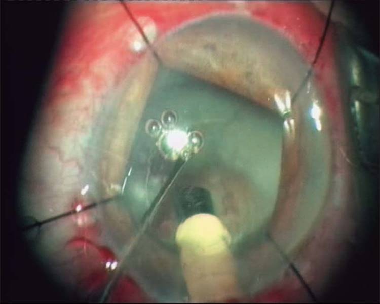
Some special features are present in many cases with coincident glaucoma and cataract. Pseudoexfoliation is more prevalent in cases that undergo glaucoma surgery than in average glaucoma cases. This is due to the fact that higher IOPs occur in pseudoexfoliative glaucoma and these cases are more likely to undergo glaucoma surgery than non-pseudoexfoliative open-angle glaucoma. Pupils dilate less favorably in most of the medically treated glaucoma eyes. This makes cataract surgery more difficult and sometimes more risky, particularly if pseudoexfoliation is present. Pupils in pseudoexfoliative glaucoma react poorly to parasympatholytic mydriatics and to intracameral epinephrine. In these cases, weak zonules and a hard nucleus are usually present. Therefore, the pupil often needs artificial dilation by stretching, iris hooks or diaphragms (see Fig. 222.3).
|
|
|
|
FIGURE 222.3 Management of the small pupil with iris hooks. |
NARROW ANGLES INDUCED BY LENS VOLUME OR LENS SWELLING
When monitoring glaucoma, particularly in open-angle glaucoma and angle-closure glaucoma, the condition of the lens plays an increasing role in aging glaucoma patient. The lens increases considerably in volume during life. This results in narrowing of the anterior chamber and particularly the chamber angle. A critical situation may arise when appositional closure or peripheral anterior synechiae develop. This is more frequent in the elderly hypermetropic patient. Peripheral prophylactic iridotomy may be advocated in narrow angles. However, if incipient cataract is present, phacoemulsification with intraocular lens implantation may be considered and will resolve both the relative pupillary block and a borderline plateau iris configuration. Acute angle closure due to pupillary block can be permanently cured after medically decreasing IOP and subsequent lens removal. This approach is particu-larly suited in hypermetropic eyes with incipient cataract. Phacoemulsification may improve the IOP also in ocular hypertension and in manifest glaucoma, possibly due to wash out of debri of the trabecular meshwork during anterior chamber irrigation (see section on Cataract Surgery Alone).
|
In summary, the combined cataract-glaucoma operation has the advantages of promptly restoring vision, controlling IOP better immediately after surgery and in long term and can control IOP without medications and their side effects in many cases. However, an experienced surgeon is needed to successfully overcome various problems such as inflammatory response and increased wound healing in these cases. |
TECHNIQUE OF COMBINED GLAUCOMA-CATARACT SURGERY
GENERAL CONSIDERATIONS
The Technique of the Glaucoma Part
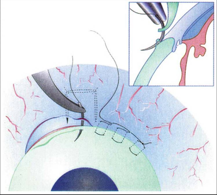
Fornix-based conjunctival dissection is the preferred approach by many surgeons in trabeculectomy alone and in combined glaucoma-cataract surgery (Fig. 222.1). This approach reduces the stimulation of wound healing response at the posterior conjunctiva and subconjunctival connective tissue.[19,20] However, every combined glaucoma-cataract procedure is to be considered a more risky situation for excessive wound healing than glaucoma surgery alone. Therefore, preoperative antiinflammatory treatment with topical steroids and wash out of glaucoma medications causing redness of the eye has to be considered. Intraoperative antimetabolites help to increase the success rate of the glaucoma part in combined surgery. Usually, mitomycin-C is applied topically using a concentration of 0.2-0.5 mg/ml over 3-5 min. 5-Fluorouracil was advocated postoperatively for combined glaucoma cataract surgery.[21] Postoperative modulation of wound healing may include frequent application of topical steroid drops and injections of 5 mg 5-fluorouracil if needed according to the development of the filtering bleb (intensified postoperative care).[21a]
|
|
|
|
FIGURE 222.1 Suture technique with Fornix-based conjunctival flap (schematic drawing). |
The Technique of the Cataract Part
Clear cornea or small incision corneoscleral approach for phacoemulsification are the procedures mostly used in combined cataract-glaucoma surgery.[22-24] This techniques provide the advantage the most effective IOP reduction with the least stimulation of inflammation and wound healing processes.
SPECIAL TECHNIQUES
Sequence of Cataract and Glaucoma Procedures in Combined Surgery
In combined glaucoma-cataract procedures, most surgeons start with cataract surgery and add glaucoma surgery second. If a two-site approach is used (see further ahead), it may be easier to compose the individual steps of the respective part. This may help the technical feasability and the technical success of combined cataract-glaucoma procedures, particularly when combined with penetrating surgery (see further ahead). In separate incision phacotrabeculectomy, the main problem may be (1) a soft globe, if clear cornea cataract surgery is completed first, making dissection of the scleral flap more difficult, (2) a ballooning filtering bleb due to excessive outflow during irrigation of the cataract part if glaucoma surgery is completed first. Therefore, some surgeons first start glaucoma surgery with dissection of the scleral flap without penetration. Second, clear cornea cataract surgery is completely performed. Third, penetration at the site of filtration (trabeculectomy, punching, or goniotrephination) is done and glaucoma surgery completed.[25] Many different glaucoma procedures can be combined with cataract surgery. It is beyond the scope of this chapter to describe all different techniques of combinations. The following list provides an overview on the various glaucoma procedures that can be combined (Table 222.1).
TABLE 222.1 -- Overview of Glaucoma Procedures Used for Combined Glaucoma/Cataract Surgery
|
One-Site versus Two-Site Approach
Formerly, when glaucoma surgery was performed with extracapsular techniques using a corneoscleral approach and rigid IOLs, the large wound could create excessive wound healing of the filtration procedure. With modern phacoemulsification and foldable lens implantation, however, the conjunctival wound is small and the site of glaucoma surgery can also be used as the approach for phacoemulsification without excessive wound healing response.[26,11,24,23]
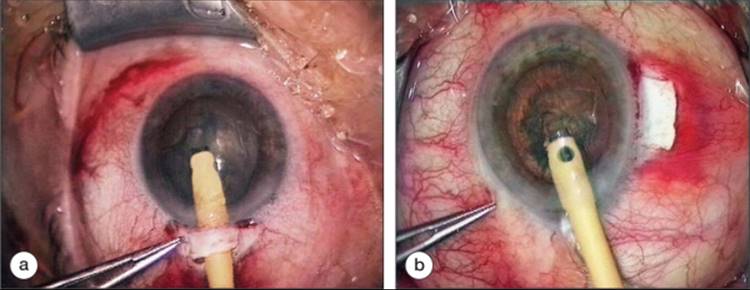
One-site approach: In this procedure, the scleral flap is dissected and the corneoscleral penetration of 3 mm width into the anterior chamber underneath the scleral flap is performed for cataract surgery. After completing phacoemulsification and foldable lens implantation, the trabeculectomy is either excised from the same incision with a diamond blade or is done with a punch (Fig. 222.2a).
|
|
|
|
FIGURE 222.2 Technique of phacotrabeculectomy. (a) One-site approach; (b) two-site approach. |
Two-site approach: This technique follows the concept that any additional intervention at the filtration site may create additional undesired wound healing. Therefore, the clear cornea phacoemulsification with lens implantation is positioned at the temporal site and the filtration site is located at the 12 o'clock position.[27] Clear cornea phacoemulsification from the temporal site is preferred when a two-site approach is performed.[28] With this approach it may be advisable to compose the different steps of the two procedures in a logic sequence (discussed earlier) (Fig. 222.2b).
|
In several randomized prospective studies, a trend toward better outcome after two-site approach was found.[25,28] However, in none of these studies, the difference was statistically significant, probably due to insufficient number of cases followed.[25,29,30,31] |
Extracapsular Cataract Extraction versus Phacoemulsification in Combined Glaucoma-Cataract Surgery
Extracapsular catract techniques are less suited for combined procedures and should only be used if phacoemulsification is not available or the nucleus is too hard for phacoemulsification.[32,33] The major disadvantage of larger wounds is increased wound healing. If possible, a clear corneal incision should be used instead of a corneoscleral approach.[34,35] Large amounts of visual-field defect must be considered in a special issue.[36]
Anesthesia
Both general and local anesthesia are used for combined glaucoma-cataract surgery. The patient should be informed that surgical time is longer than in single operations and should be asked whether he can comply with local anesthesia. Topical anesthesia can also be recommended.[37]
OTHER ADJUVANT TECHNIQUES
Management of the Small Pupil in Combined Surgery
In chronically treated glaucoma patients, the iris is usually rigid and small. Only occasionally, the pupil dilates sufficiently with hyaluronic acid and intracameral epinephrine. Therefore, artificial dilatation is often needed. If posterior synechiae are present, synechiolysis is usually performed using hyaluronic acid and blunt instruments to separate the iris from the lens. Artificial pupil dilatation can be done by (1) pupil stretching using two iris hooks (2) using an iris diaphragm, or (3) temporary implantation of iris hooks (Fig. 222.3).
Management of Pseudoexfoliation Related Problems in Cataract Surgery
Pseudoexfoliation is a risk factor for cataract surgery due to fragile zonule and small pupil. In addition, the nucleus may be harder in pseudoexfoliation than usual. Therefore, artificial pupil stretching should be considered also in borderline pupillary width. It is a common experience, however, that the lens nucleus in chronically treated glaucoma cases is not as hard as expected, particularly when pseudoexfoliation is absent. It is not clear whether this is due to chronic antiglaucoma medication or other factors.
RESULTS OF COMBINED GLAUCOMA-CATARACT SURGERY
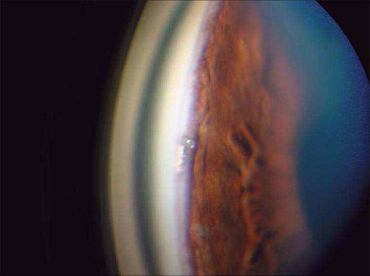
Virtually all glaucoma procedures have been used in combination with cataract extraction or phacoemulsification (see Table 222.1). A complete description of results of these studies is beyond the scope of this chapter. Results of the major techniques will be discussed here briefly. In a study comparing 44 patients undergoing phacotrabeculectomy to a matched group of trabeculectomy alone, a significant difference of success rates between the two groups was found in elderly white patients.[38] In a recent review,[39] the better outcome of trabeculectomy, compared to phacotrabeculectomy, was confirmed as found in previous retrospective or comparative studies.[40,41] However, since MMC is used, the outcome of phacotrabeculectomy improved significantly. In a study comparing MMC trabeculectomy with MMC phacotrabeculectomy, no difference in the 2-year IOP outcome was found.[24,42] Combined surgery using nonpenetrating glaucoma interventions and phacoemulsification seem to have less negative impact on outcome[39,43,44] but are less effective than phacotrabeculectomy with MMC.[39] Using mitomycin C in phacotrabeculectomy may improve success rate to the level of trabeculectomy alone.[42] Comparing nonpenetrating combined surgery with penetrating surgery, similar efficacy of both glaucoma procedures was found[45,46] Less astigmatism was found in the nonpenetrating group after 6 months postsurgery. MMC was useful in combining phacoemulsification with both trabeculectomy and deep sclerectomy.[47] In a Japanese retrospective study of 57 eyes with either primary open-angle glaucoma or primary angle-closure glaucoma, combined glaucoma-cataract surgery was particularly successful in angle-closure cases, possibly due to deepen-ing of the anterior chamber by lens removal.[48] The predictability of refraction was less precise and showed a myopic shift in phacotrabeculectomy as compared to phacoemulsification alone.[49] Scleral flap suture release can improve the outcome when perforemed in an adequate time frame.[50] Trabeculotomy was used as the glaucoma part for combined surgery with good control of IOP[51] and was found to have less incidence of fibrin reaction.[34,35] Endoscopic excimer laser trabeculopuncture (ELT) and Er:YAG laser goniotomy have be used as combined procedures for glaucoma-cataract surgery, the IOP reduction was more successful in combined cataract and ELT procedures than in ELT alone.[52,53] Trabecular aspiration is a method useful particularly in PEX glaucoma,[54] but the duration of IOP control is limited. Stent implantation into Schlemms canal was developed in an anterior segment model[55] and is now used in patients for single or combined glaucoma surgery. It is particularly useful when combined with phacoemulsification (Fig. 222.4).
|
|
|
|
FIGURE 222.4 Angle surgery in combined procedures (GlaukosT Stent). |
MANAGEMENT OF SPECIFIC COMPLICATIONS OF COMBINED GLAUCOMA-CATARACT SURGERY
MANAGEMENT OF INFLAMMATION AFTER COMBINED SURGERY STEROIDS
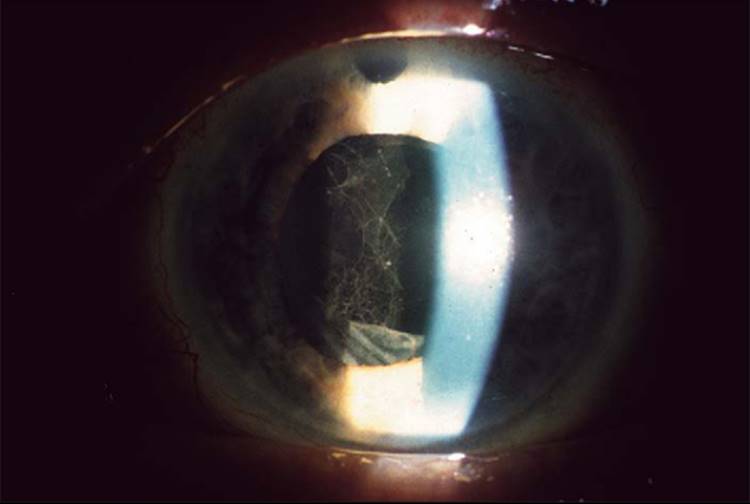
Hourly application of a potent steroid is advisable after combined cataract glauoma surgery to avoid increased flare or fibrinous exsudation into the anterior chamber (Fig. 222.5). If fibrin reaction occurs, systemic steroid treatment may be considered. Subconjunctival steroid use is less recommended as subconjunctival bleeding may enhance scar formation and thereby impair the proper development of the filtering bleb. However, fibrin usually persists for several days and may result in posterior synechiae that cannot be broken by medical pupil dilatation. Topical steroids often are not sufficient to control formed fibrin in the anterior chamber.
|
|
|
|
FIGURE 222.5 Fibrinous exsudation after phacotrabeculectomy and iris dilatation. |
RECOMBINANT TISSUE PLASMINOGEN ACTIVATOR
This enzyme dissolves fibrin and can be given into the anterior chamber through a paracentesis. A concentration of 0.2 mg/mL can be applied and dissolves fibrin within hours. However, late posterior synechiae cannot be broken by recombinant tissue plasminogen activator (rtPA). Additional steroids should be applied to avoid recurrence.
INCIDENCE AND MANAGEMENT OF BLEBITIS/ENDOPHTHALMITIS
There is no evidence that blebitis and endophthalmitis occur more frequently in combined cataract-glaucoma surgery than in single glaucoma procedures. Analysis and characterization of the bacteria involved is mandatory and instantaneous treatment should be started with topical antibiotics (hourly application or fortified drops). However, if hypopyon and vitritis are present, systemic antibiotics and vitrectomy are needed in many of these cases. In most cases, blebitis is caused by an external fistula of the filtering bleb due to a button hole of the conjunctiva, particularly after MMC trabeculectomy. Even quiet buttonholes should therefore be closed before blebitis or endophthalmitis occurs. In some cases, compression sutures may help. However, it is more effective and safer to interpose subconjunctival connective tissue (Tenon) underneath the conjunctiva. Buttonholes in avascular thin conjunctiva cannot be closed by stitches from outside due to the risk of additional holes created by the needle. Fibrin or cyanoacrylate glue applications are usually not effective.
MANAGEMENT OF GLAUCOMA SECONDARY TO CATARACT
PHACOLYSIS, PHACOANAPHYLAXIS
Mature or hypermature cataracts may leak proteins into the anterior chamber that are taken up by macropages. These engulfed macrophages block outflow at the level of the trabecular meshwork and can produce acute intraocular pressure elevation with similar symptoms as angle closure. Lens removal usually clears the situation. If the protein elicits immunogenic inflammation (phacoanaphylaxis), lens removal is also indicated but increased steroid doses are required to suppress and halt the immunogenic process. These mature lenses usually present with weak zonules. Extracapsular techniques and phacoemulsification sometimes are difficult to perform. IOP increase rarely persists after cataract operation and antiglaucoma medication for a limited period after lens removal is often sufficient to control the situation.
SUBLUXATION
Subluxated lenses may present with various problems related to intraocular pressure or glaucoma: The most frequent mechanism is blockade of aqueous flow by the lens trapped in the pupil. If the lens moves forward into the pupil, pupillary block and immediate rise of IOP will occur.[56] This can be treated best by intracapsular lens removal, but acute transient relief can be obtained by laser iridotomy. If the cataract is mature, in addition to mechanical block, leaking lens proteins may cause phacolytic or phacoanaphylactic IOP increase. Lens subluxation or luxation in elderly cataract patients are most frequently caused by pseudoexfoliation or trauma. Vitrectomy techniques are needed if the lens is luxated into the vitreous body. Not infrequently, the surgeon having seen a subluxated lens still in the pupillary plane at the slit lamp finds the lens dropped into the vitreous cavity when the patient is in supine position during surgery.
GENETIC DISORDERS
Genetic disorders can affect both lens and outflow system. Weill Marchesani syndrome may present with spherical crystalline lenses that either subluxate into the vitreous or are trapped in the pupil causing acute IOP rise. A similar situation of secondary glaucoma due to an ectopic lens and pupillary blockade may occur in Marfan's syndrome and homocystinuria, rarely in Retinitis pigmentosa.
CATARACT FOLLOWING GLAUCOMA SURGERY
Glaucoma surgery, particularly trabeculectomy, is considered to be cataractogenic. The mechanism by which this occurs is not fully understood.
|
1. |
In trabeculectomy, an iridectomy is needed to avoid prolaps of the iris into the filtration site. Hence, the aqueous humor is directly shunted to the anterior chamber through the iridectomy and is no more circulating around the lens. This mechanism may contribute to the faster progression of cataract. The same mechanism occurs after surgical or laser iridectomy without filtration surgery. |
|
|
2. |
Hypotony is not infrequent after trabeculectomy. Hypotony is believed to facilitate cataract formation. However, when uncomplicated low IOPs (5-8 mmHg) persist, cataract is not more frequent than in other trabeculoectomy cases. |
|
|
3. |
Many cases that develop accelerated cataract formation present with a history of other problems such as shallow anterior chamber, flat chamber with lens-cornea touch, increased irrigation during surgery, or direct trauma to the lens capsule. Nonperforating glaucoma drainage surgery, such as viscocanalostomy or deep sclerectomy, as well as trabeculotomy in adults have significantly lower cataract rates.[57] Cyclodestructive procedures, particularly kryotherapy of the ciliary body facilitate cataract formation, possibly due to reduced or altered aqueous humor supply. |
CATARACT SURGERY FOLLOWING SUCCESSFUL PREVIOUS GLAUCOMA SURGERY
RESULTS OF PROSPECTIVE STUDIES
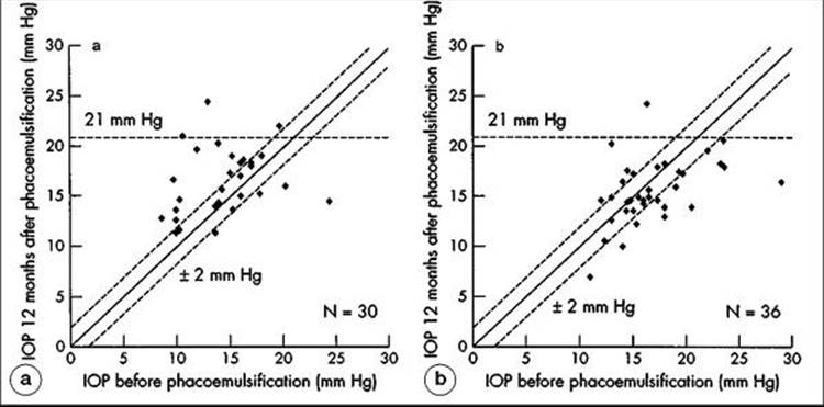
Several authors have recently shown that functioning filtering blebs may lose functionality by later uneventful clear cornea phacoemulsification.[15,32,58-60] In a prospective study using a control group of nonoperated glaucoma cases matched to visual-field damage and age, 1 year after clear cornea phacoemulsification an average increase of intraocular pressure by 2 mmHg was found. In the control group, an average IOP decrease of 2 mmHg was found.[15] It was found that 14 of 30 eyes with functioning filtering blebs had an IOP rise of more than 2 mmHg after clear cornea phacoemulsification, whereas only four of 30 eyes had an IOP decrease of more than 2 mmHg. In the control group, six out of 36 eyes had an IOP rise of more than 2 mmHg, whereas 13 of 36 eyes had an IOP decrease of more than 2 mmHg (Fig. 222.6).
|
|
|
|
FIGURE 222.6 (a) IOP before and after phacoemulsification in eyes with functioning filtering blebs comparing IOP before and after phacoemulsification (b) in matched glaucoma eyes without previous glaucoma surgery. |
MEASURES TO OVERCOME BLEB FAILURE AFTER PHACOEMULSIFICATION
There is no established regimen to overcome the unpredictable IOP change after clear cornea phacoemulsification when a functioning filtering procedure has been present before. Local steroids routinely administered after cataract surgery cannot be causative for the long-term IOP increase, as they were used in both groups and the control group did not show an IOP increase. Subclinical inflammation may contribute to a late scarring process. There is no information on factors released from lens proteins that could contribute to wound healing stimulation in this situation. A comparable reaction is known in corneal transplantation that can lead to considerable IOP elevation in glaucoma patients. Some surgeons use repeated 5-Fluorouracil injections after cataract surgery to compensate for the IOP rise after phacoemulsification in filtered glaucoma patients.
CONCLUSIONS
In general, the decision for combined surgery of glaucoma and cataract is based on the need to surgically approach both cataract and glaucoma at the same time period, the risk to postpone one of the procedures, or on comfort for the patient, particularly if the patient is old and would not tolerate two separate surgical interventions. On the other hand, if cataract surgery is not needed at the point when glaucoma surgery is indicated, it should be postponed. If, on the contrary, glaucoma is mild or ocular hypertension is present without visual-field defects, cataract surgery with phacoemulsification alone may provide an IOP lowering effect in many cases. The need of short-term glaucoma surgery when visually disturbing cataract is present is a good indication for combined surgery. It may also facilitate follow-up of glaucoma damage by better vizualization of the optic disk as well as better performance of visual-field testing.
REFERENCES
1. Rudnicka AR, Mt-Isa S, Owen CG, et al: Variations in primary open angle glaucoma prevalence by age, gender, and race: a Bayesian meta-analysis. Invest Ophthalmol Vis Sci 2006; 47:4254-4261.
2. Quigley HA, Broman AT: The number of people with glaucoma worldwide in 2010 and 2020. Br J Ophthalmol 2006; 90:262-267.
3. Acosta R, Hoffmeister L, Roman R, et al: Systematic review of population-based studies of the prevalence of cataracts. Arch Soc Esp Oftalmol 2006; 81:509-516.
4. Williams A, Sloan FA, Lee PP: Longitudinal rates of cataract surgery. Arch Ophthalmol 2006; 124:1308-1314.
5. Grehn F, Klink T: Combined glaucoma-cataract surgery. Ophthalmologe 2004; 101:661-666.
6. Scheie HG, Muirhead : Cataract extraction after filtering operations. Arch Ophthalmol 1962; 68:37-41.
7. McPherson Jr SD: Combined trabeculotomy and cataract extraction as a single operation. Trans Am Ophthalmol Soc 1976; 74:250-260.
8. Savage JA, Thomas JV, Belcher CD, Simmons RJ: Extracapsular cataract extraction and posterior chamber lens implantation in glaucomatous eyes. Ophthalmology 1985; 92:1506-1516.
9. Mc Guigan LJB, Gottsch J, Stark WJ, et al: Extracapsular cataract extraction and posterior chamber lens implantation in eyes with preexisting glaucoma. Arch Ophthalmol 1986; 104:1301-1308.
10. Longstaff S, Wormald RPL, Mazover A, Hitchings RA: Glaucoma triple procedures: efficacy of intraocular pressure control and visual outcome. Ophthalmic Surg 1990; 21:786-793.
11. Gregg FM: Phacoemulsification and modified trabeculectomy for managing combined cataracts and glaucoma. J Cataract Refract Surg 1992; 18:362-365.
12. Mamalis N, Lohner S, Rand AN, Crandall AS: Combined phacoemulsification, intraocular lens implantation and trabeculectomy. J Cataract Refract Surg 1996; 22:467-473.
13. Verges C, Cazal J, Lavin C: Surgical strategies in patients with cataract and glaucoma. Curr Opin Ophthalmol 2005; 16:44-52.1
14. Cohn H: Combined cataract and glaucoma surgery. J Fr Ophthalmol 2005; 28:52-54.
15. Klink J, Schmitz B, Lieb WE, et al: Filtering bleb function after clear cornea phacoemulsification: a prospective study. Br J Ophthalmol 2005; 89:597-601.
16. Bigger JF, Becker B: Cataract and primary open-angle glaucoma: the effect of uncomplicated cataract extraction on glaucoma control. Trans Am Acad Ophthalmol Otolaryngol 1971; 75:260-272.
17. Shin DH, Simone PH, Song MS, et al: Adjunctive subconjunctival mitomycin C in glaucoma triple procedure. Ophthalmology 1995; 102:1550-1558.
18. McNaught A: Loss of vision after phacotrabeculectomy. J Glaucoma 2005; 14:299-301.
19. Mandic Z, Bencic G, Zoric Geber M, Bojic L: Fornix vs limbus based flap in phacotrabeculectomy with mitomycin C: prospective study. Croat Med J 2004; 45:275-278.
20. Khaw PT: Advances in glaucoma surgery: evolution of antimetabolite adjunctive therapy. J Glaucoma 2001; 10:81-84.
21. Gandolfi SA, Vecchi M: 5-fluorouracil in combined trabeculectomy and clear-cornea phacoemulsification with posterior chamber lens implantation. A one-year randomized controlled trial. Ophthalmology 1997; 104:181-186.
21a. Marquardt D, Lieb WE, Grehn F: Intensified postoperative care versus conventional follow-up: a retrospective long-term analysis of 177 trabeculectomies. Graefe's Arch Clin Exp Ophthalmol 2004; 242:106-113.
22. Allan BDS, Barrett GD: Combined small incision phacoemulsification trabeculectomy. J Cataract Refract Surg 1993; 19:97-102.
23. Gimbel HV, Meyer D: Small incision trabeculotomy combined with phacoemulsification and intraocular lens implantation. J Cataract Refract Surg 1993; 19:92-96.
24. Shingleton BJ, Price RS, O'Donoghue MW, Goyal ODS: Comparison of 1-sire versus 2-site phacotrabeculectomy. J Cataract Refract Surg 2006; 31:799-802.
25. Borggrefe J, Lieb WE, Grehn F: A prospective randomized comparison of two techniques of combined cataract-glaucoma surgery. Graefes Arch Clin Exp Ophthalmol 1999; 237:887-892.
26. Cagini C, Murdolo P, Gallai R: Longterm results of one-site phacotrabeculectomy. Acta Ophthalmol Scand 2003; 81:233-236.
27. Weitzman M, Caprioli J: Temporal corneal phacoemulsification combined with separate - incision superior trabeculectomy. Ophthalmic Surg 1995; 26:271-273.
28. Gayton JL, Van der Karr MA, Sanders V: Combined cataract and glaucoma procedures using temporal cataract surgery. J Cataract Refract Surg 1996; 22:1485-1491.
29. El Sayyad F, Helal M, El Maghrabi A, et al: One site versus 2-site phacotrabeculectomy; a randomized study. J Cataract Refract Surg 1999; 25:77-82.
30. Rosetti L, Bucci L, Miglior S, Orzalesi N: Temporal corneal phacoemulsification combined with separate incision superior trabeculectomy versus standard phacotrabeculectomy; a comparative study. Acta Ophthalmol Scand Suppl 1997.224-239.
31. Wyse T, Meyer M, Ruderman JM, et al: Combined trabeculectomy and phacoemulsification; a one-site vs a two-site approach. Am J Ophthalmol 1998; 125:334-339.(1998).
32. Manoj B, Chako D, Khan MY: Effect of extracapsular cataract extraction and phacoemulsification performed after trabeculectomy on intraocular pressure. J Cataract Refract Surg 2000; 26:75-78.
33. Wishart P, Austin M: Combined cataract extraction and trabeculectomy: phacoemulsification compared with extracapsular technique. Ophthalmic Surg 1993; 24:814-821.
34. Schwenn O, Grehn F: Cataract extraction combined with trabeculotomy. Ger J Ophthalmol 1995; 4:16-20.
35. Hoffmann E, Schwenn O, Karallus M, et al: Long-term results of cataract surgery combined with trabeculotomy. Graefes Arch Clin Exp Ophthalmol 1999; 240:2-6.
36. Stewart WC, Crinkley CMC, Carlson AN: Results of trabeculectomy combined with phaoemulsification versus trabeculectomy combined with extracapsular cataract extraction in patients with advanced glaucoma. Ophthalmic Surg 1994; 25:621-627.
37. Ahmed II, Zabriskie NA, Crandall AS, et al: Topical versus retrobulbar anesthesia for combined phacotrabeculectomy: prospective randomized study. J Cataract Refract Surg 2002; 28:631-638.
38. Lochhead J, Casson RJ, Salmon JF: Long term effect on intraocular pressure of phacotrabeculectomy compared to trabeculectomy. Br J Ophthalmol 2003; 87:850-852.
39. Vass C, Menapace R: Surgical strategies in patients with combined cataract and glaucoma. Curr Opin Ophthalmol 2004; 15:61-66.
40. Krieglstein GK, Duzanec Z: Benefits and risks of the combined glaucoma-cataract operation. Fortschr Ophthalmol 1985; 82:357-361.
41. Hansen LL, Hoffmann F: Combination of phacoemulsification and trabeculectolmy. Results of a retrospective study. Klin Monatsbl Augenheilkd 1987; 190:478-481.
42. Murthy SK, Damji KF, Pan Y, Hodge WG: Trabeculectomy and phacotrabeculectomy, with mitomycin-C, show similar two-year target IOP outcomes. Can J Ophthalmol 2006; 41:51-59.
43. Lüke C, Dietlein TS, Lüke M, et al: Phaco-trabeculotomy combined with deep sclerectomy - a new technique in combined cataract and glaucoma surgery. Acta Ophthalmol Scand 2007; 85:143-148.
44. D'Eliseo D, Pastena B, Longanesi L, et al: Comparison of deep sclerectomy with implant and combined glaucoma surgery. Ophthalmologica 2003; 217:208-211.
45. Gianoli F, Schnyder CC, Bovey E, Mermoud A: Combined surgery for cataract and glaucoma: phacoemulsification and deep sclerectomy compared with phacoemulsification and trabeculectomy. J Cataract Refract Surg 1999; 25:340-346.
46. Kadowaki H, Mizoguchi T, Kuroda S, et al: Surgically-induced astigmatism following single-site phacotrabeculectomy, phacotrabeculectomy and advanced non-penetrating phacotrabeculectomy. Semin Ophthalmol 2001; 16:158-161.
47. Funnell CL, Clowes M, Anand N: Combined cataract and glaucoma surgery with Mitomycin C: phacoemulsification-trabeculectomy compared to phacoemulsification-deep sclerectomy. Br J Ophthalmol 2005; 89:694-698.
48. Lai JS, Tham CC, Chan JC, Lam DS: Phacotrabeculectomy in treatment of primary angle closure glaucoma and primary open-angle glaucoma. Jpn J Ophthalmol 2004; 48:408-411.
49. Chan JC, Lai JS, Tham CC: Comparison of postoperative outcome in phacotrabeculectomy and phacoemulsification with posterior chamber intraocular lens implantation. J Glaucoma 2006; 15:26-29.
50. Banitt M, Juzych MS, Chopra V, et al: The effect of timing of scleral flap suture release on the safety and long-term success of phacotrabeculectomy. Am J Ophthalmol 2006; 141:742-744.
51. Nagata M: Trabeculotomy ab externo, cataract extraction, and intraocular lens implantation: preliminary report. J Cataract Refract Surg 1996; 22:601-606.
52. Wilmsmeyer S, Philippin H, Funk J: Excimer laser trabeculotomy: a new minimally invasive procedure for patients with glaucoma. Graefes Arch Clin Exp Ophthalmol 2006; 244:670-677.
53. Beuerle S, Philippin H, Funk J: Combined cataract and glaucoma surgery. Trabeculectomy vs Er:YAG goniotomy. Ophthalmologe 2006; 103:605-608.
54. Jacobi PC, Dietlein TS, Krieglstein GK: The risk profile of trabecular aspiration versus trabeculectomy in glaucoma triple procedure. Graefes Arch Clin Exp Ophthalmol 2000; 238:545-551.
55. Bahler CK, Smedley GT, Zhou J, Johnson DH: Trabecular bypass stents decrease intraocular pressure in cultured human anterior segments. Am J Ophthalmol 2004; 138:988-994.
56. Inatani M, Tanihara H, Honjo M, et al: Secondary glaucoma associated with crystalline lens subluxation. J Cataract Refract Surg 2000; 26:1533-1536.
57. Gandolfi SA, Quaranta L, Ungaro N, et al: Deep sclerectomy versus trabeculectomy in open angle glaucoma. 7-year prospective randomized clinical trial. Invest Ophthalmol Vis Sci 2005; 46:ARVO abstract 1217.
58. Rebolleda G, Munoz-Negrete FJ: Phacoemulsification in eyes with functioning filtering blebs: a prospective study. Ophthalmology 2002; 109:2248-2255.
59. Derbalov A, Vass C, Menapace R, et al: Long term effect of phacoemulsification on intraocular pressure after trabeculectomy. J Cataract Refract Surg 2002; 28:425-430.
60. Sacu S, Rainer G, Findl O, et al: Correlation between the early morphological appearance of filtering blebs and outcome of trabeculectomy with mitomycin C. J Glaucoma 2003; 12:430-435.