Steven J. Gedde,
Donald L. Budenz
HISTORY
The concept of using an implantable material to shunt aqueous humor out of the anterior chamber dates back to 1906, when Rollet and Moreau[1] reported inserting horse hair through a paracentesis to drain aqueous humor. A variety of materials were subsequently used to serve as wicks allowing aqueous humor to flow from the anterior chamber to the subconjunctival or sub-Tenon's space. These materials included suture, glass, various metals, plastic, or biologic material used as a true solid (rather than tubular) 'seton'. However, because inflammation and fibrosis around the implanted material ultimately limited the success of these procedures, implants were developed consisting of a long tube connected to a distal end plate. The first of these modern glaucoma drainage implants was designed by Molteno,[2]and it is still in widespread use today.
PATHOPHYSIOLOGY
|
Key Features: Glaucoma Drainage Implants |
|||||||||||||||
|
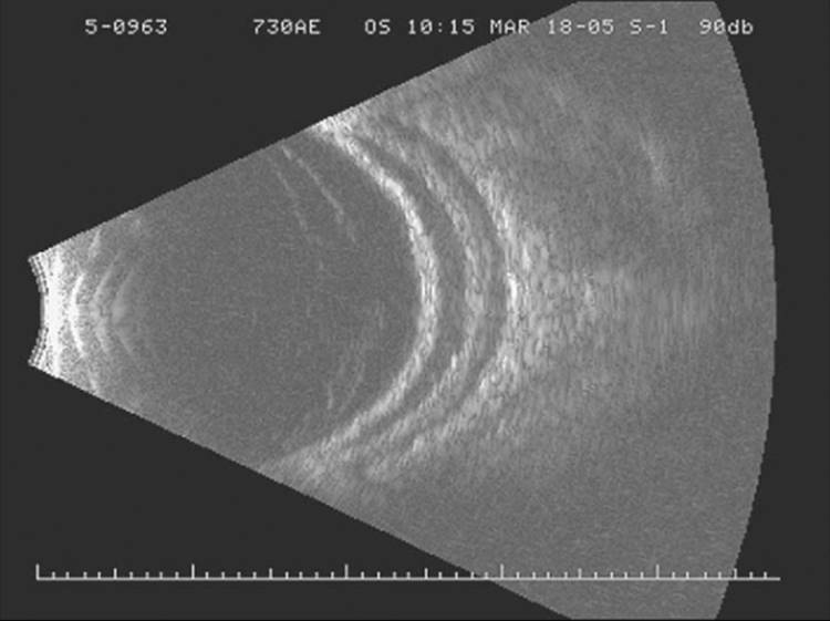
Glaucoma drainage implants have a silicone tube that shunts aqueous humor from the anterior chamber or posterior chamber to an end plate located in the equatorial region of the globe. Following surgical implantation, a fibrous capsule forms around the end plate over a period of several weeks. A feature common to all drainage implants is construction of the plate from biocompatible materials to which fibroblasts cannot adhere. As a result, aqueous draining through the device will pool in the potential space between the end plate and surrounding, nonadherent capsule. The reservoir of aqueous humor between the plate and overlying capsule may be demonstrated echographically[3] (Fig. 223.1). Aqueous passes through the capsule via passive diffusion and is absorbed by periocular capillaries.[4,5] It is the fibrous capsule around the end plate that offers the major resistance to flow with glaucoma drainage implants.[4,5] Therefore, capsule thickness and capsule surface area are the two major determinants of final intraocular pressure (IOP) after drainage implant surgery.[4,5] Lower postoperative IOP is expected with capsules that are thinner and have larger surface areas.
|
|
|
|
FIGURE 223.1 B-scan ultrasound showing collection of aqueous humor surrounding the end plate of a Baerveldt glaucoma implant. |
Surgeons have attempted to modulate capsule thickness with various glaucoma drainage implants by applying an antifibrotic agent in the region of the end plate, in a manner similar to standard filtering surgery. Randomized prospective studies have found similar IOP results with the intraoperative use of mitomycin C or balanced salt solution.[6,7] The benefit of mitomycin C as an adjunct of drainage implant surgery has not been clearly demonstrated. Furthermore, a higher incidence of hypotony, flat anterior chambers, choroidal effusions, and conjunctival melts has been reported with its use.[8-10]
The capsule surface area is directly related to the size of the end plate. Therefore, an implant with a larger end plate produces a larger surface area of encapsulation around the plate and greater IOP reduction. In a randomized clinical trial comparing the single-plate and double-plate Molteno implants, a higher success rate and lower IOP were reported with the double-plate implant presumably because of its larger surface area.[11] However, there appears to be an upper limit to end plate size beyond which an increase in surface area may not improve pressure control, and may even detrimentally affect surgical outcome. In a prospective study comparing the 350 and 500 mm2 Baerveldt glaucoma implants, no significant differences in surgical success and visual outcomes were found between the different implant sizes.[12] With longer follow-up, lower success with the 500 mm2 Baerveldt compared with the 350 mm2 implant was observed.[13]
INDICATIONS
Glaucoma drainage implants are generally used to manage medically uncontrolled glaucoma when trabeculectomy has failed to control IOP, or is deemed unlikely to succeed (Table 223.1). Drainage implants are indicated for several secondary glaucomas, including neovascular glaucoma, uveitic glaucoma, epithelial or fibrous downgrowth, and iridocorneal endothelial (ICE) syndrome. Eyes that have undergone previous ocular surgery are good candidates for drainage implant surgery, such as those with prior cataract extraction, failed glaucoma filtering surgery, penetrating keratoplasty, pars plana vitrectomy, or scleral buckling procedures. Implants offer a good surgical option in patients with scleral ectasia or cicatricial diseases of the conjunctiva, like the Stevens-Johnson syndrome and ocular cicatricial pemphigoid. These devices are also used in congenital glaucoma that has failed angle surgery and/or trabeculectomy. Drainage implants are usually reserved for eyes with ambulatory vision, and cyclodestructive procedures may be preferable in those with limited visual potential.
TABLE 223.1 -- Indications for Glaucoma Drainage Implant Surgery
|
Neovascular glaucoma |
|
Uveitic glaucoma |
|
Fibrous or epithelial downgrowth |
|
Iridocorneal endothelial (ICE) syndrome |
|
Prior cataract extraction |
|
Prior failed glaucoma filtering surgery |
|
Prior penetrating keratoplasty |
|
Prior retinal surgery (pars plana vitrectomy or scleral buckling procedure) |
|
Congenital glaucoma |
|
Scleral ectasia |
|
Cicatricial diseases of the conjunctiva (e.g., Stevens Johnson syndrome, ocular cicatricial pemphigoid) |
IMPLANT DESIGNS
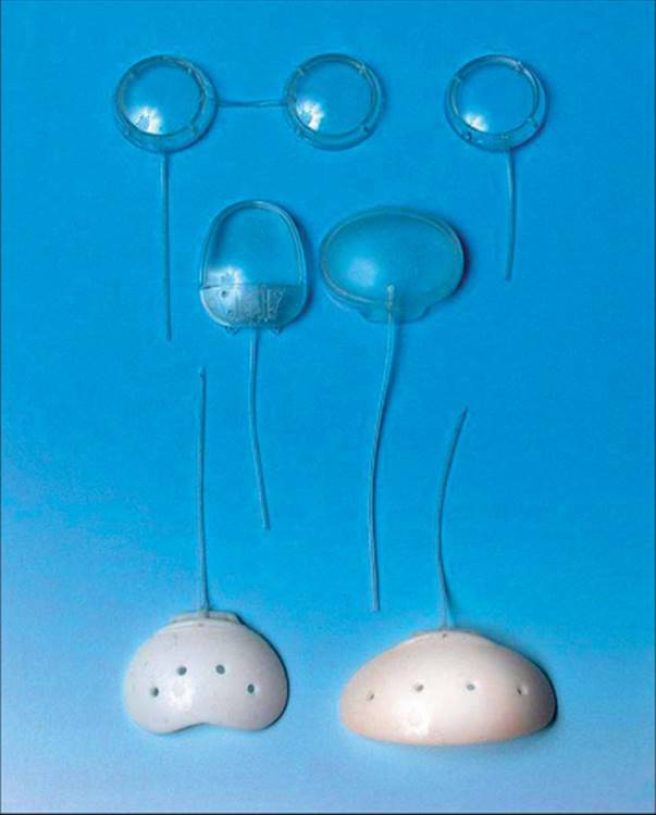
All modern glaucoma drainage implants consist of a tube that connects to an end plate (or explant). Drainage implants differ in their design with respect to the size, shape, and material from which the end plate is made. They may be further subdivided into valved and nonvalved implants, depending on whether or not a valve mechanism is present to limit flow through the tube to the plate if the IOP becomes too low. The implants currently in common use include the Ahmed glaucoma valve (New World Medical, Rancho Cucamonga, California), Baerveldt glaucoma implant (Advanced Medical Optics, Santa Ana, California), Krupin valve with disk (Hood Laboratories, Pembroke, Massachusetts), and Molteno implant (Molteno Ophthalmic Limited, Dunedin, New Zealand). Figure 223.2 shows these glaucoma drainage implants, and Table 223.2 lists the major design characteristics for each implant.
|
|
|
|
FIGURE 223.2 Glaucoma drainage implants in common use. Double-plate and single-plate Molteno implants (top row). Ahmed glaucoma valve and Krupin valve with disk (middle row). Baerveldt glaucoma implants - surface area 250-mm2 and 350-mm2 (bottom row). |
TABLE 223.2 -- Design Characteristics of Commonly Used Glaucoma Drainage Implants
|
Implant Type |
Size |
End Plate Material |
Valved/Nonvalved |
|
Ahmed glaucoma valve |
96 mm2 (S3) |
Polypropylene |
Valved |
|
184 mm2 (S2) |
Silicone |
||
|
364 mm2 (B1) |
|||
|
96 mm2 (FP8) |
|||
|
184 mm2 (FP7) |
|||
|
364 mm2 (FX1) |
|||
|
Baerveldt glaucoma implant |
250 mm2 |
Silicone |
Nonvalved |
|
350 mm2 |
|||
|
Krupin valve with disc |
183 mm2 |
Silastic |
Valved |
|
Molteno implant |
74 mm2 (pediatric) |
Polypropylene |
Nonvalved |
|
134 mm2 (single-plate) |
|||
|
268 mm2 (double-plate) |
NONVALVED IMPLANTS
Baerveldt Glaucoma Implant
The Baerveldt glaucoma implant has an end plate consisting of barium-impregnated silicone with a surface area of 250 and 350 mm2. The plate has fenestrations that allow growth of fibrous bands reducing the profile of the bleb. A pars plana version is available for placement of the tube in the posterior chamber. The Baerveldt implant offers the advantage of a large end plate that is implanted in only one quadrant. The lateral wings of the plate are designed to be positioned under adjacent rectus muscles.
Molteno Implant
The Molteno implant has a round polypropylene end plate with a surface area of 134 mm2 for the single-plate implant and 268 mm2 for the double-plate implant. The plates of the double-plate version are connected by a 10 mm silicone tube, which may be positioned over or under the superior rectus muscle. A pediatric size plate with a surface area of 74 mm2 is available. A dual-chamber Molteno implant has a ridge on the upper surface of the end plate and was designed to reduce postoperative hypotony. The greatest clinical experience is available with this implant.
VALVED IMPLANTS
Ahmed Glaucoma Valve
The Ahmed glaucoma valve has a scarab-shaped end plate made of polypropylene (models S2, S3, and B1) or silicone (models FP7, FP8, and FX1). Fenestrations were added to the plate of the silicone models. Single-plate versions of the Ahmed implant are available with a surface area of 96 mm2 (S3 and FP8) or 184 mm2 (S2 and FP7). The double-plate implants have a surface area of 364 mm2 (B1 and FX1). The valve mechanism consists of silicone elastomer membranes that separate with an IOP of 8-12 mm Hg.
Krupin Valve with Disk
The Krupin valve with disk has an oval silastic end plate with a surface area of 183 mm2. The distal end of the tube contains horizontal and vertical slits that function as a valve with an opening pressure of 9-11 mm Hg.
SURGICAL TECHNIQUE
The surgical technique for implanting glaucoma drainage implants is similar, irrespective of the type of implant used. Differences in technique relate to the size of the conjunctival incision needed for insertion of the device and methods for temporary restriction of flow with nonvalved implants. Careful examination of the anterior segment should be performed before surgery to plan the surgical approach. The procedure is usually performed under local anesthesia with a retrobulbar or peribulbar block.
CONJUNCTIVAL INCISION
The superotemporal quadrant is generally selected as the site for placement of single-plate implants because surgical exposure is better and postoperative strabismus is less frequent. The superonasal quadrant should be avoided because of the increased risk of producing motility disturbances.[14-16] A limbus-based or fornix-based conjunctival flap is dissected, depending on the surgeon's preference (Fig. 223.3). Double-plate devices require a 180° incision, while single-plate implants may be inserted through a 90-100° incision. A relaxing incision on either side of the conjunctival flap will improve exposure.
|
|
|
|
FIGURE 223.3 A fornix-based conjunctival flap is dissected. |
QUADRANT DISSECTION
The conjunctiva and Tenon's capsule is dissected from sclera to create space for the implant (Fig. 223.4). A corneal or scleral traction suture may be used to enhance exposure. Cautery is applied to bleeding episcleral vessels.
|
|
|
|
FIGURE 223.4 Conjunctiva and Tenon's capsule are dissected from sclera to create space for the implant. |
ATTACHMENT OF THE END PLATE
The implant is placed in antibiotic solution before insertion. Adjacent rectus muscles are identified with muscle hooks. The end plate is positioned between the rectus muscles. The lateral wings of the Baerveldt glaucoma implant is designed for positioning under the rectus muscles. The end plate is attached to sclera ?10 mm posterior to the limbus with nonabsorbable sutures through the fixation holes of the implant (Fig. 223.5). The knots are rotated into the fixation holes to prevent erosion through the conjunctiva. When using double-plate implants, one plate is positioned in each of two quadrants and the tube connecting them may be positioned under or over the rectus muscle.
|
|
|
|
FIGURE 223.5 The end plate is sutured to sclera through the fixation holes of the implant ?10 mm posterior to the limbus. |
PREPARATION OF THE IMPLANT
Restriction of Aqueous Flow in Nonvalved Implants
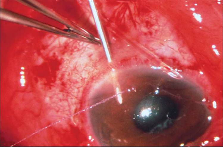
When using nonvalved implants, restriction of aqueous flow is required until fibrous encapsulation of the plate occurs. This is necessary to avoid overfiltration in the early postoperative period and to minimize the risk of hypotony. A two-stage implantation technique may be used. The end plate is attached to sclera during the first stage, but the tube is left in the subconjunctival space. The tube is inserted into the anterior chamber 4-8 weeks later in the second stage of the procedure, after a fibrous capsule has formed around the plate. More commonly, single-stage implantation is performed using one of several methods to temporarily occlude the tube. Temporary tube occlusion may be accomplished by ligating it with a 7-0 polyglactin suture near the tube-plate junction (Fig. 223.6). Complete closure is confirmed by attempting to irrigate balanced salt solution through the tube. The polyglactin suture reliably lyses 4-6 weeks postoperatively, causing spontaneous opening of the tube. Another technique for temporarily occluding the tube involves the use of a 4-0 chromic or polypropylene intraluminal suture, or placement of a 4-0 or 5-0 nylon or polypropylene suture alongside the tube and incorporating it within the ligating suture. These 'ripcord' sutures are positioned subconjunctivally in a different quadrant from the implant. Aqueous flow through the implant is initiated several weeks postoperatively by removing the ripcord suture with Vannas scissors and forceps. Alternatively, an intracameral 9-0 polypropylene suture may be used to ligate the tube. An argon or dye laser is subsequently applied to the suture to open the tube.
|
|
|
|
FIGURE 223.6 Temporary occlusion of the tube is accomplished by ligating it with a 7-0 polyglactin suture in a nonvalved implant. |
Priming Valved Implants
Valved implants must be 'primed' by injecting balances salt solution through the tube using a cannula. This serves to break the surface tension between the two silicone sheets of the Ahmed implant, so the valve mechanism can function. Irrigating with balanced salt solution confirms that the valve slits in the Krupin implant allow flow. It may be necessary to compress the valve end with tying forceps to ensure that each quadrant of the valve moves independently and are not adherent to one another.
INSERTION OF THE TUBE
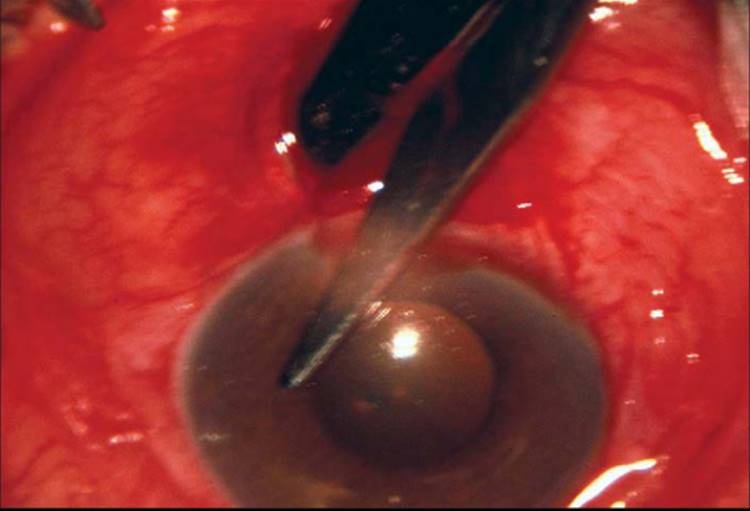
The tube is draped across the cornea and cut with an anterior bevel so that a 2-3 mm segment of tube extends into the anterior chamber from the site of limbal entry (Fig. 223.7). A 23-gauge needle is used to make an entry incision into the anterior chamber at the posterior limbus, parallel to the iris plane (Fig. 223.8). This size needle creates a tight entry wound for the tube, reducing leakage around the tube. The tube is inserted through the needle track with tying forceps or a tube insertion forceps. Proper positioning of the tube anterior to the iris and posterior to the cornea should be confirmed. Alternatively, the tube may be inserted through the pars plana in an eye that has had a complete pars plana vitrectomy.
|
|
|
|
FIGURE 223.7 The tube is cut with an anterior bevel so a 2-3 mm segment extends into the anterior chamber. |
|
|
|
|
FIGURE 223.8 A 23-gauge needle is used to make an entry incision into the anterior chamber. |
TUBE COVERAGE WITH A PATCH GRAFT
The limbal portion of the tube is covered with a patch graft measuring ?4 mm × 4 mm (Fig. 223.9). Donor sclera, cornea, pericardium, fascia lata, or dura mater have been used for the patch graft material. The graft is sutured in place with interrupted sutures at each corner.
|
|
|
|
FIGURE 223.9 The limbal portion of the tube is covered with a scleral patch graft. |
CONJUNCTIVAL CLOSURE
The conjunctiva is closed by reapproximating it to the limbus with mattress sutures when using a fornix-based flap, or using a running closure for limbus-based flaps and radial relaxing incisions (Fig. 223.10).
|
|
|
|
FIGURE 223.10 The conjunctiva is reapproximated to the limbus. |
SURGICAL RESULTS
Evaluation of the surgical results that are achieved with the various glaucoma drainage implants is made difficult because most clinical data are derived from retrospective studies with different study populations, follow-up periods, and criteria defining success. The specific implant used and type of glaucoma under treatment are factors that appear to influence surgical success.[17,18] Several studies have compared drainage implants with other surgical procedures in the management of medically uncontrolled glaucoma.
SURGICAL OUTCOMES BASED ON GLAUCOMA TYPE
Success rates with glaucoma drainage implant surgery vary depending on the underlying type of glaucoma being surgically treated. Tables 223.3 to 223.7 present surgical results reported with various drainage implants according to glaucoma type. Case series studying drainage implants have reported success rates ranging from 22% to 78% for neovascular glaucoma,[19-31] 75% to 100% for uveitic glaucoma,[24-26,3-34] 44% to 100% for developmental glaucoma,[19,20,23-26,35-48] 50% to 88% for eyes that have undergone cataract surgery,[11,19,20,23,25,26,29,30,49] and 44% to 88% for eyes with failed filters.[19,20,23,26,30,49] The poorest surgical results are observed in neovascular glaucoma. As with trabeculectomy, there appears to be attrition over time resulting in a trend toward lower success rates among studies with longer follow-up.
TABLE 223.3 -- Surgical Results with Glaucoma Drainage Implants in Eyes with Neovascular Glaucoma
|
Authors |
Procedure |
Success Rate |
IOP Success Criteria (mm Hg) |
Follow-Up (Months) |
|
|
Mean |
Range |
||||
|
Hodkin et al[19] |
Baerveldt |
43% (3/7) |
? 21 |
18.3 |
|
|
Minckler et al[20] |
SP Molteno |
47% (7/15) |
? 21 |
20.2 |
|
|
Krupin et al[21] |
Krupin long valve to band |
77% (30/39) |
? 21 |
20.2 |
12-36 |
|
Ancker and Molteno[22] |
SP Molteno |
67% (24/36) |
< 20 |
18 |
6-55 |
|
Lloyd et al[23] |
SP Molteno |
22% (4/18) |
? 21 and > 5 |
33.8 |
7-70 |
|
Siegner et al[24] |
Baerveldt |
71% (24/34) |
? 21 and > 5 |
13.6 |
4-37 |
|
Freedman and Rubin[25] |
SP Molteno |
67% (12/18) |
? 21 |
35 |
6-88.9 |
|
Mills et al[26] |
SP/DP Molteno |
50% (10/20) |
? 22 |
24 |
6-66 |
|
Sidoti et al[27] |
Baerveldt |
61% (22/36) |
? 21 and ? 6 |
15.7 |
6-28 |
|
Mastropasqua et al[28] |
Krupin-Denver valve |
36% (10/28) |
< 22 and > 5 |
58.4 |
10-108 |
|
Huang et al[29] |
Ahmed |
68% (19/28) |
< 22 and > 5 |
13.4 |
4-44 |
|
Broadway et al[30] |
SP/DP Molteno |
53% (10/19) |
< 22 and > 5 |
28 |
|
|
Krishna et al[31] |
Baerveldt |
78% (14/18) |
< 22 and 30% reduction |
24 |
|
|
DP, double-plate; IOP, intraocular pressure; SP, single-plate. |
TABLE 223.4 -- Surgical Results with Glaucoma Drainage Implants in Eyes with Uveitic Glaucoma
|
Authors |
Procedure |
Success Rate |
IOP Success Criteria (mm Hg) |
Follow-Up (Months) |
|
|
Mean |
Range |
||||
|
Siegner et al[24] |
Baerveldt |
91% (10/11) |
? 21 and > 5 |
13.6 |
4-37 |
|
Freedman and Rubin[25] |
SP Molteno |
80% (4/5) |
? 21 |
48 |
0.5-13.9 |
|
Mills et al[26] |
SP/DP Molteno |
75% (9/12) |
? 22 |
69 |
42-96 |
|
DaMata et al[32] |
Ahmed |
100% (21/21) |
? 21 |
24.5 |
|
|
Molteno et al[33] |
SP Molteno |
83% (30/36) |
? 21 and ? 6 |
85.2 |
20-240 |
|
Ceballos et al[34] |
Baerveldt |
92% (22/24) |
? 21 and ? 5 |
20.8 |
|
|
DP, double-plate; IOP, intraocular pressure; SP, single-plate. |
TABLE 223.5 -- Surgical Results with Glaucoma Drainage Implants in Eyes with Developmental Glaucoma
|
Authors |
Implant |
Age (Years) |
Success Rate |
IOP Success Criteria (mm Hg) |
Follow-Up (Months) |
|
|
Mean |
Range |
|||||
|
Molteno et al[35] |
SP Molteno |
95% (79/83) |
< 20 |
66 |
12-114 |
|
|
Goldberg[36] |
DP Molteno |
? 36 |
100% (15/15) |
< 20 |
18.4 |
6-24 |
|
Minckler et al[20] |
SP Molteno |
< 13 |
54% (7/13) |
? 21 |
22.8 |
|
|
Billson et al[37] |
DP Molteno |
78% (18/23) |
< 21 |
41.3 |
12-84 |
|
|
Hill et al[38] |
SP/DP Molteno |
< 21 |
62% (40/65) |
< 22 and > 5 |
22.7 |
6-59 |
|
Freedman and Rubin[25] |
SP Molteno |
50% (2/4) |
? 21 |
37 |
16-51 |
|
|
Munoz et al[39] |
SP Molteno |
< 12 |
68% (36/53) |
? 21 |
18 |
6-36 |
|
Nesher et al[40] |
SP/DP Molteno |
? 13 |
59% (16/27) |
? 21 |
20 |
6-36 |
|
Lloyd et al[23] |
SP/DP Molteno |
< 13 |
44% (7/16) |
? 21 and > 5 |
49.1 |
7-76 |
|
Netland and Walton[41] |
Molteno, Baerveldt |
? 10 |
80% (16/20) |
? 21 |
25 |
8-41 |
|
Hodkin et al[19] |
Baerveldt |
< 13 |
100% (3/3) |
? 21 |
19.2 |
|
|
Siegner et al[24] |
Baerveldt |
80% (12/15) |
? 21 and > 5 |
13.6 |
4-37 |
|
|
Fellenbaum et al[42] |
Baerveldt |
< 21 |
83% (25/31) |
? 21 and ? 6 |
15.0 |
6-25 |
|
Mills et al[26] |
SP/DP Molteno |
50% (2/4) |
? 22 |
36 |
10-99 |
|
|
Coleman et al[43] |
Ahmed |
< 18 |
71% (17/24) |
< 22 or 20% reduction |
16.3 |
|
|
Eid et al[44] |
SP/DP Molteno, Schocket, Baerveldt |
< 18 |
44% (8/18) |
? 21 and > 5 |
47.3 |
14-80 |
|
Englert et al[45] |
Ahmed |
< 18 |
85% (21/27) |
? 21 |
12.6 |
3-31 |
|
Djodeyne, et al[46] |
Ahmed |
< 15 |
69% (24/35) |
< 22 |
12.6 |
0-37.9 |
|
Pereira et al[47] |
SP/DP Molteno, Krupin-Schocket, Baerveldt |
? 3 |
60% (6/10) |
< 22 |
50 |
|
|
Budenz et al[48] |
Baerveldt |
< 18 |
71% (44/62) |
< 22 and ? 5 |
23.4 |
1-106 |
|
DP, double-plate; IOP, intraocular pressure; SP, single-plate. |
TABLE 223.6 -- Surgical Results with Glaucoma Drainage Implants in Aphakic/Pseudophakic Eyes
|
Authors |
Implant |
Age (Years) |
Success Rate |
IOP Success Criteria (mm Hg) |
Follow-Up (Months) |
|
|
Mean |
Range |
|||||
|
Minckler et al[20] |
SP Molteno |
A/P |
63% (26/41) |
? 21 |
16.2 |
7-30 |
|
Freedman and Rubin[25] |
SP Molteno |
A/P |
83% (20/24) |
? 21 |
22 |
8.1-53.3 |
|
Lloyd et al[23] |
SP/DP Molteno |
A/P |
56% (28/50) |
? 21 and > 5 |
48.6 |
7-78 |
|
Heuer et al[11] |
SP Molteno |
A/P |
50% (25/50) |
? 21 and > 6 |
14.9 |
6-29 |
|
DP Molteno |
75% (38/51) |
16.4 |
7-30 |
|||
|
Hodkin et al[19] |
Baerveldt |
A/P |
74% (26/35) |
? 21 |
16.3 |
6.1-26.1 |
|
Mills et al[26] |
SP/DP Molteno |
A/P |
58% (14/24) |
? 22 |
45 |
6-107 |
|
Huang et al[29] |
Ahmed |
A |
88% (28/32) |
< 22 and > 5 |
13.4 |
4-44 |
|
P |
88% (84/96) |
|||||
|
Broadway et al[30] |
SP/DP Molteno |
A |
70% (21/30) |
< 22 |
43 |
|
|
P |
66% (23/35) |
|||||
|
Roy et al[49] |
Baerveldt |
A |
75% (6/8) |
? 21 and > 6 |
37.6 |
12-68 |
|
A, aphakia; DP, double-plate; IOP, intraocular pressure; P, pseudophakia; SP, single-plate. |
TABLE 223.7 -- Surgical Results with Glaucoma Drainage Implants in Eyes with Failed Filters
|
Authors |
Procedure |
Success Rate |
IOP Success Criteria (mm Hg) |
Follow-Up (Months) |
|
|
Mean |
Range |
||||
|
Minckler et al[20] |
SP Molteno |
70% (7/10) |
? 21 |
12.3 |
6-25 |
|
Lloyd et al[23] |
SP/DP Molteno |
75% (9/12) |
? 21 and > 5 |
41.4 |
15-64 |
|
Hodkin et al[19] |
Baerveldt |
75% (9/12) |
? 21 |
16.1 |
7.1-26.1 |
|
Mills et al[26] |
SP/DP Molteno |
44% (4/9) |
? 22 |
42 |
8-78 |
|
Broadway et al[30] |
SP/DP Molteno |
58% (34/59) |
< 22 |
43 |
|
|
Roy et al[49] |
Baerveldt |
88% (15/17) |
? 21 and > 6 |
37.6 |
12-68 |
|
DP, double-plate; IOP, intraocular pressure; SP, single-plate. |
COMPARISON OF DIFFERENT IMPLANT TYPES
Randomized clinical trials have compared glaucoma drainage implants of differing size, but of the same type. Prospective comparisons of the single-plate versus double-plate Molteno implants[11] and the 350 mm2 versus 500 mm2 Baerveldt glaucoma implants[12,13] have provided important insight into the role of implant size on IOP reduction. Unfortunately, there are presently no published prospective studies comparing different implant types. Current data assessing different implant designs are limited to retrospective case series, which have selection bias inherent to any retrospective study design. A recently initiated prospective study called the Ahmed Baerveldt Comparison (ABC) Study promises to provide important clinical insight into the comparative efficacy and safety of two commonly used drainage implants (D. Budenz, personal communication).
Retrospective studies comparing the Ahmed glaucoma valve and the Baerveldt glaucoma implant have demonstrated no significant difference in IOP reduction and success rates with both implants.[50-53] The 350 mm2 Baerveldt implant and the double-plate Molteno implant produced similar IOP lowering, success rates, and visual outcomes in a nonrandomized comparison of these implants.[54] There was no significant difference in IOP reduction and success rates with the Ahmed and double-plate Molteno implants in a retrospective study.[55] A nonrandomized review of patients who received a double-plate Molteno implant, Krupin eye valve with disk, or Ahmed glaucoma valve with adjunctive mitomycin C found significantly greater pressure reduction with the double-plate Molteno implant.[56]
COMPARISON WITH OTHER GLAUCOMA PROCEDURES
The surgical options for patients with uncontrolled glaucoma who are at high risk for surgical failure generally include placement of a glaucoma drainage implant, trabeculectomy with an adjunctive antifibrotic agent, or cyclodestruction. Several studies have compared these different surgical procedures.
Trabeculectomy
Comparable results have been reported with the single-plate Molteno implant and trabeculectomy without an antifibrotic agent,[57] or trabeculectomy with adjunctive 5-fluorouracil[58] in retrospective studies. A case control study comparing trabeculectomy with mitomycin C and the single-plate Molteno in high-risk glaucoma patients found greater IOP lowering and fewer surgical complications with trabeculectomy.[59] However, a randomized clinical trial evaluating the Ahmed implant and trabeculectomy with or without an adjunctive antifibrotic agent in low-risk eyes found similar IOP reduction, visual acuity, visual fields, and surgical complications with both procedures after 3 years of follow-up.[60] The tube versus trabeculectomy (TVT) study is a multicenter randomized clinical trial comparing the safety and efficacy of trabeculectomy with mitomycin C and the Baerveldt glaucoma implant in eyes with previous cataract surgery and/or failed filtering surgery.[61] Tube shunt surgery was more likely to maintain IOP control and avoid persistent hypotony or reoperation for glaucoma than trabeculectomy during the first year of follow-up in the TVT Study.[62]
Cyclophotocoagulation
Drainage implants provided superior IOP control and preservation of vision compared with Nd:YAG cyclophotocoagulation in retrospective studies of eyes with neovascular glaucoma.[63,64] Placement of a drainage implant, Nd:YAG cyclophotocoagulation, and trabeculectomy with mitomycin C produced similar results in a retrospective series of eyes that had undergone penetrating keratoplasty, although there was a trend for patients treated with laser to have a higher incidence of graft failure, glaucoma failure, hypotony, and vision loss.[65] Glaucoma drainage implants provided greater IOP reduction than Nd:YAG cyclophotocoagulation in a nonrandomized study with matched controls of advanced uncontrolled glaucoma.[66]
COMPLICATIONS OF GLAUCOMA DRAINAGE IMPLANTS
Complications of glaucoma drainage implant surgery may be divided into intraoperative, early (<3 months) and late (>3 months) postoperative categories (Table 223.8).
TABLE 223.8 -- Complications of Glaucoma Drainage Implants
|
Intraoperative |
Early Postoperative |
Late Postoperative |
|
|
Tube malposition |
X |
X |
X |
|
Hypotony |
X |
X |
X |
|
Choroidal effusion |
X |
||
|
Shallow anterior chamber |
X |
X |
|
|
Hyphema |
X |
||
|
Scleral perforation |
X |
||
|
Retinal detachment |
X |
||
|
Suprachoroidal hemorrhage |
X |
X |
|
|
Vitreous prolapse |
X |
X |
|
|
Aqueous misdirection |
X |
X |
X |
|
Tube blockage |
X |
||
|
Erosion of tube or plate |
X |
||
|
Tube migration |
X |
||
|
Diplopia |
X |
X |
|
|
Endophthalmitis |
X |
X |
|
|
Corneal decompensation |
X |
INTRAOPERATIVE COMPLICATIONS
Complications that develop during implantation of glaucoma drainage devices may include problems with tube entry or placement, hyphema, scleral perforation, suprachoroidal hemorrhage, and vitreous prolapse. Fortunately, the prevalence of intraoperative complications with glaucoma drainage implants seems to be similar to trabeculectomy.[60,67]
Tube Insertion and Malposition
Tube insertion, as described previously, is a critical step in implant surgery, and complications related to tube entry can be minimized by using proper surgical technique. All drainage implant tubes have an external diameter of 0.58-0.63 mm.[68] Therefore, the ideal needle with which to make the entry track is a 23-gauge needle, which has a 0.6 mm external diameter. A smaller gauge may be used (like a 25-gauge, with an external diameter of 0.5 mm), but tube insertion is more difficult through such a small entryway. A larger needle (such as a 22-gauge, with an external diameter of 0.7 mm) can result in leakage around the tube and hypotony, with an increased risk of hypotony-related complications. The location and entry of the needle track is important as well, to prevent tube-corneal or tube-lens touch. The entry should be at the limbus, ideally just in front of the iris and parallel to the iris plane. If the tube is inserted and found to be touching the cornea or lens when the anterior chamber is formed, then it should be removed and a new entry made. The original track should be sutured tightly to prevent hypotony due to filtration from this site.
Tube insertion can be problematic in eyes with thin sclera, such as children with buphthalmos or patients who have had prior scleritis. In these cases, the sclera may be so thin that the needle track lacks enough integrity to keep the tube properly positioned. In these cases, the tube may be re-routed to an area with thick sclera, the tube may be inserted into the ciliary sulcus and positioned behind the iris (provided the patient is pseudophakic), or a different quadrant may be selected for implant placement.
Hyphema
Hyphema may occur with any anterior segment surgery, but this complication is especially common with glaucoma drainage implant surgery for neovascular glaucoma. Because the needle track and tube enter through the anterior chamber angle, any abnormal blood vessels in this location can produce a hyphema. If the hyphema is observed intraoperatively, an attempt may be made to irrigate out the blood through a paracentesis. If the blood clots at the tube tip, this may result in tube obstruction and IOP elevation.
Scleral Perforation
Scleral perforation may produce a retinal hole and subsequent detachment. The sclera is very thin near the equator, so care must be taken to visualize the needle throughout its course through the sclera and positioning holes while fixating the end plate. This can be difficult because of the awkward angle of approach, the crowded area in which the surgeon is working, and pooling of blood and fluid during this step of the procedure. One helpful tip is to lift the end plate off the eye with forceps during passage of the needle so that blood and fluid pass posteriorly, improving visualization.[68] If scleral perforation is suspected, retinal cryopexy should be performed intraoperatively and retinal consultation obtained to determine whether additional measures are needed to prevent a retinal detachment.
Suprachoroidal Hemorrhage
Intraoperative suprachoroidal hemorrhage is rare during glaucoma implant surgery. In the present authors' experience, this is most likely to occur in patients with very high preoperative IOP in whom there is a prolonged exposure to atmospheric pressure (e.g., during combined vitrectomy or penetrating keratoplasty). Preoperative intravenous Mannitol or slow decompression of the eye via a paracentesis at the beginning of the procedure may help to prevent this potentially devastating complication.
Vitreous Prolapse
Vitreous prolapse into the anterior chamber will commonly lead to tube obstruction, since there is a tendency for any material in the anterior chamber to get sucked into the tube. If vitreous is present in the anterior chamber preoperatively or encountered intraoperatively, it may be necessary to perform an anterior vitrectomy or pars plana vitrectomy to prevent blockage of the tube.
EARLY POSTOPERATIVE COMPLICATIONS
Hypotony-related complications, suprachoroidal hemorrhage, and aqueous misdirection are sometimes encountered in the first 3 months after glaucoma drainage implant surgery.
Hypotony-Related Complications
Hypotony-related complications, such as choroidal effusions and shallowing of the anterior chamber, are relatively common after glaucoma drainage implant surgery. Temporary restriction of aqueous flow is required when using nonvalved implants, to avoid overfiltration and profound hypotony in the early postoperative period. Although usually benign, choroidal effusions have been found to be a risk factor for suprachoroidal hemorrhage in postoperative glaucoma drainage implant patients.[69] The management of choroidal effusions is generally observation, although drainage may be required if the effusions are persistent, 'kissing', or associated with a flat anterior chamber. The surgical procedure and outcomes for draining choroidal effusions have been reviewed in detail elsewhere.[70,71] Shallow or flat anterior chambers may also occur in the early postoperative period. We define flat anterior chambers as those with lenticulo-corneal touch. Overfiltration is generally the cause, although choroidal effusions, aqueous misdirection, and suprachoroidal hemorrhage may also produce anterior chamber shallowing following glaucoma drainage implant surgery. Shallow anterior chambers can generally be observed and reform spontaneously, but flat anterior chambers must be reformed promptly to prevent lens and corneal decompensation. The underlying cause of the shallowing should also be addressed, so that the chamber does not shallow again.
Suprachoroidal Hemorrhage
Suprachoroidal hemorrhage may occur at any time in the early postoperative period, but the risk is highest in the first few days after surgery and immediately following the opening of the tube with nonvalved implants.[69] Risk factors for this serious complication include increased age, high preoperative IOP, low postoperative IOP, hypertension, and the presence of choroidal effusion postoperatively. Most of these factors are beyond the control of the surgeon, but prevention of hypotony in patients with very high preoperative IOPs is desirable.
Aqueous Misdirection
Aqueous misdirection is an uncommon complication in the early or late postoperative period after glaucoma drainage implant surgery and can be difficult to diagnose and treat.[72] Patients present with axial shallowing of the anterior chamber, and the IOP is generally normal to nominally elevated.[72] Occasionally, a patient may present with markedly elevated IOP making the diagnosis more obvious. Management with cycloplegics and aqueous suppressants is often unsuccessful, but laser or incisional surgery may be required.[72]
Tube Obstruction
Blockage of the tube with vitreous (Fig. 223.11), iris, fibrin (Fig. 223.12), or blood (Fig. 223.13) occurs most commonly in the early postoperative period. Although laser can help relieve obstruction of a tube by iris or vitreous, surgical removal of the material blocking the tube tip may be required. Injection of tissue plasminogen activator can dissolve fibrin or blood that is blocking a tube.
|
|
|
|
FIGURE 223.11 Tube blockage by vitreous. |
|
|
|
|
FIGURE 223.12 Tube blockage by fibrin. |
|
|
|
|
FIGURE 223.13 Tube blockage by blood. |
LATE POSTOPERATIVE COMPLICATIONS
Late complications of glaucoma drainage implant surgery include erosion of the tube or plate, migration of the tube, diplopia, endophthalmitis, and corneal decompensation.
Erosion of Tube or Plate
Tube erosion (Fig. 223.14) was a common occurrence prior to the use of tissue patch grafts to cover the anterior portion of the tube. Even with a patch graft, tube erosion through the conjunctiva with melting of the patch graft may occur. Melting of the patch over the tube may develop with any material. Evaluation for erosion of the tube or plate should be performed at every visit, since this represents a major risk factor for endophthalmitis.[73] Prompt repair of the exposed tube using another donor patch graft is advised. Once the plate has exposed, attempts at repairing it are almost uniformly ineffective due to poor tissue integrity. We almost always remove the exposed drainage implant and place a new one in a different quadrant.
|
|
|
|
FIGURE 223.14 Tube erosion. |
Tube Migration
Migration of the tube further into the eye, outside of the anterior chamber, or more posteriorly or anteriorly may occur postoperatively. This complication may require surgical treatment if tube-corneal touch or tube obstruction develops. If the tube migrates out of the anterior chamber, a tube extender (New World Medical, Rancho Cucamonga, California) may be used for revision. If one is not available, a tube spacer using a commonly available 22-gauge angiocatheter can be utilized.[74]
Diplopia
Diplopia is not uncommon after glaucoma drainage implant surgery, but usually disappears as the periocular swelling resolves. However, persistent strabismus may develop because of scarring between the rectus or oblique muscles,[14,15,75] or due to a crowding effect from a large bleb with limitation of extraocular motility.[76] Permanent diplopia is generally treated with prism spectacles. In rare instances, strabismus surgery or removal of the implant may be required.
Endophthalmitis
Late endophthalmitis is a very rare complication following glaucoma drainage implant surgery and is usually associated with exposure of the tube or plate.[73] Successful treatment of the infection may require explantation of the device, since the implant may be seeded with bacteria and resistant to sterilization.
Corneal Decompensation
Corneal decompensation appears to be uncommon in eyes with previously normal corneal endothelium, but occurs with higher frequency in patients with preexisting corneal endothelial cell dysfunction or penetrating keratoplasty.[77] The reasons for corneal decompensation after glaucoma drainage implants are unclear but may be mechanical or biological. Mechanical rubbing of the tube against the corneal endothelium can cause progressive endothelial cell loss and corneal decompensation.[78,79] Figure 223.15 shows a case of focal endothelial cell loss due to rubbing of the tube against the posterior cornea. This may be avoided by placing the tube as far posterior in the anterior chamber as possible. It has also been suggested that disruption of the blood-aqueous barrier allows the tube to carry inflammatory cells and factors into the eye causing an immune-mediated response.[79,80] A single nonrandomized retrospective comparative study suggested that placing the tube through the pars plana rather than the anterior chamber may result in less corneal decompensation,[80] so this should be considered in eyes with prior penetrating keratoplasty or endothelial cell-related corneal disease. However, a complete pars plana vitrectomy must be performed to prevent tube obstruction if the tube is placed into the vitreous cavity, and the tube length must be long enough (we suggest 6 mm from site of insertion[81]) to keep it away from the vitreous base.
|
|
|
|
FIGURE 223.15 Focal corneal edema due to tube-corneal touch in a patient who was rubbing his eye (a), which was treated with tube trimming to prevent further endothelial cell loss (b). |
It is often difficult to determine the cause of stromal corneal edema in a patient with hypotony and a glaucoma drainage implant. Hypotony alone may cause corneal edema, and corneal decompensation may occur in the presence of a glaucoma drainage implant tube at normal levels of IOP. The management of these two causes of corneal edema is drastically different. Endothelial cell decompensation would be treated with penetrating keratoplasty and repositioning of the tube, while hypotony would be treated by tying off or removing the tube. To distinguish between the two possible causes, viscoelastic may be injected into the anterior chamber to raise the intraocular pressure to determine if low pressure is the cause of the corneal edema. If the edema does not resolve, consideration should be given to penetrating keratoplasty and repositioning of the tube more posteriorly.
REFERENCES
1. Rollett M, Moreau M: Traitement de la hypopyon par le drainage capillaire de la chamber anterieure. Rev Gen Ophthalmol 1906; 25:481-489.
2. Molteno AC, Straughan JL, Ancker E: Long tube implants in the management of glaucoma. S Afr Med J 1976; 50:1062-1066.
3. Lloyd M, Minckler D, Heuer D, et al: Echographic evaluation of glaucoma shunts. Ophthalmology 1993; 100:919-927.
4. Minckler DS, Shammas A, Wilcox M, Ogden TE: Experimental studies of aqueous filtration using the Molteno implant. Trans Am Ophthalmol Soc 1987; 85:368-392.
5. Wilcox MJ, Minckler DS, Ogden TE: Pathophysiology of artificial aqueous drainage in primate eyes with Molteno implants. J Glaucoma 1994; 3:140-151.
6. Cantor L, Burgoyne J, Sanders S, et al: The effect of mitomycin C on Molteno implant surgery: a 1-year randomized, masked, prospective study. J Glaucoma 1998; 7:240-246.
7. Costa VP, Azuara-Blanco A, Netland PA, et al: Efficacy and safety of adjunctive mitomycin C during Ahmed glaucoma valve implantation: a prospective randomized clinical trial. Ophthalmology 2004; 111:1071-1076.
8. Perkins TW, Gangnon R, Ladd W, et al: Molteno implant with mitomycin C: intermediate-term results. J Glaucoma 1998; 7:86-92.
9. Susanna R, Nicolela MT, Takahashi WY: Mitomycin C as adjunctive therapy with glaucoma implant surgery. Ophthalmic Surg 1994; 25:458-462.
10. Ayyala RS, Zurakowski D, Smith JA, et al: A clinical study of the Ahmed glaucoma valve implant in advanced glaucoma. Ophthalmology 1998; 105:1968-1976.
11. Heuer DK, Lloyd MA, Abrams DA, et al: Which is better? One or two? A randomized clinical trial of single-plate versus double-plate Molteno implantation for glaucomas in aphakia and pseudophakia. Ophthalmology 1992; 99:1512-1519.
12. Lloyd MA, Baerveldt G, Fellenbaum PS, et al: Intermediate-term results of a randomized clinical trial of the 350- versus the 500-mm2 Baerveldt implant. Ophthalmology 1994; 101:1456-1463.
13. Britt MT, LaBree LD, Lloyd MA, et al: Randomized clinical trial of the 350-mm2 versus the 500-mm2 Baerveldt implant: longer term results: is bigger better?. Ophthalmology 1999; 106:2312-2318.
14. Prata J, Minckler D, Green R: Pseudo-Brown's syndrome as a complication of glaucoma drainage implant surgery. Ophthalmic Surg 1993; 24:608-611.
15. Ball S, Ellis G, Herrington R, et al: Brown's superior oblique tendon syndrome after Baerveldt glaucoma implant. Arch Ophthalmol 1992; 110:1368.
16. Smith S, Starita R, Fellman R, et al: Early clinical experience with the Baerveldt 350-mm2 glaucoma implant and associated extraocular muscle imbalance. Ophthalmology 1993; 100:914-918.
17. Hong C-H, Arosemena A, Zurakowski D, Ayyala RS: Glaucoma drainage devices: a systematic literature review and current controversies. Surv Ophthalmol 2005; 50:48-60.
18. Schwarz KS, Lee RK, Gedde SJ: Glaucoma drainage implants: a critical comparison of types. Curr Opin Ophthalmol 2006; 17:181-189.
19. Hodkin MJ, Goldblatt WS, Burgoyne CF, et al: Early clinical experience with the Baerveldt implant in complicated glaucomas. Am J Ophthalmol 1995; 120:32-40.
20. Minckler DS, Heuer DK, Hasty B, et al: Clinical experience with the single-plate Molteno implant in complicated glaucomas. Ophthalmology 1988; 95:1181-1188.
21. Krupin T, Ritch R, Camras CB, et al: A long Krupin-Denver valve implant attached to a 180 degrees scleral explant for glaucoma surgery. Ophthalmology 1988; 95:1174-1180.
22. Ancker E, Molteno AC: Molteno drainage implant for neovascular glaucoma. Trans Ophthalmol Soc UK 1982; 102:122-124.
23. Lloyd MA, Sedlak T, Heuer DK, et al: Clinical experience with the single plate Molteno implant in complicated glaucomas. Update of a pilot study. Ophthalmology 1992; 99:679-687.
24. Siegner SW, Netland PA, Urban RC, et al: Clinical experience with the Baerveldt glaucoma drainage implant. Ophthalmology 1995; 102:1298-1307.
25. Freedman J, Rubin B: Molteno implants as a treatmentfor refractory glaucoma in black patients. Arch Ophthalmol 1991; 109:1417-1420.
26. Mills RP, Reynolds A, Edmond JM, et al: Long-term survival of Molteno glaucoma drainage devices. Ophthalmology 1996; 103:299-305.
27. Sidoti PA, Dunphy TR, Baerveldt G, et al: Experience with the Baerveldt glaucoma implant in treating neovascular glaucoma. Ophthalmology 1995; 102:1107-1118.
28. Mastropasqua L, Carpineto P, Ciancaglini M, Zuppardi E: Long-term results of Krupin-Denver valve implants in filtering surgery for neovascular glaucoma. Ophthalmologica 1996; 210:203-206.
29. Huang MC, Netland PA, Coleman AL, et al: Intermediate-term clinical experience with the Ahmed glaucoma valve implant. Am J Ophthalmol 1999; 127:27-33.
30. Broadway DC, Iester M, Schulzer M, Douglas GR: Survival analysis for success for Molteno tube implants. Br J Ophthalmol 2001; 85:689-695.
31. Krishna R, Godfrey DG, Budenz DL, et al: Intermediate term outcomes of 350-mm2 Baerveldt glaucoma implants. Ophthalmology 2001; 108:621-626.
32. Da Mata A, Burk SE, Netland PA, et al: Management of uveitic glaucoma with Ahmed glaucoma valve implantation. Ophthalmology 1999; 106:2168-2172.
33. Molteno ACB, Sayanat N, Herbison P: Otago Glaucoma Surgery Outcome Study. Long-term results of uveitis with secondary glaucoma drained with Molteno implants. Ophthalmology 2001; 108:605-613.
34. Ceballos EM, Parrish RK, Schiffman JC: Outcomes of Baerveldt glaucoma drainage implants for the treatment of uveitic glaucoma. Ophthalmology 2002; 109:2256-2260.
35. Molteno ACB, Ancker E, Biljon GV: Surgical technique for advanced juvenile glaucoma. Arch Ophthalmol 1984; 102:51-57.
36. Goldberg I: Management of uncontrolled glaucoma with the Molteno system. Aust N Z J Ophthalmol 1987; 15:97-107.
37. Billson F, Thomas R, Aylward W: The use of two-stage Molteno implants in developmental glaucoma. J Pediatr Ophthalmol Strabismus 1989; 26:3-8.
38. Hill RA, Heuer DK, Baerveldt G, et al: Molteno implantation for glaucoma in young patients. Ophthalmology 1991; 98:1042-1046.
39. Munoz M, Tomey KF, Traverso C, et al: Clinical experience with the Molteno implant in advanced infantile glaucoma. J Pediatr Ophthalmol Strabismus 1991; 28:68-72.
40. Nesher R, Sherwood MB, Kass MA, et al: Molteno implants in children. J Glaucoma 1992; 1:228-232.
41. Netland PA, Walton DS: Glaucoma drainage implants in pediatric patients. Ophthalmic Surg 1993; 24:723-729.
42. Fellenbaum PS, Sidoti PA, Heuer DK, et al: Experience with the Baerveldt implant in young patients with complicated glaucomas. J Glaucoma 1995; 4:91-97.
43. Coleman AL, Smyth RJ, Wilson MR, Tam M: Initial clinical experience with the Ahmed glaucoma valve implant in pediatric patients. Arch Ophthalmol 1997; 115:186-191.
44. Eid TE, Katz LJ, Spaeth GL, Augsburger JJ: Long-term effects of tube-shunt procedures on management of refractory childhood glaucomas. Ophthalmology 1997; 104:1011-1016.
45. Englert JA, Freedman SF, Cox TA: The Ahmed valve in refractory pediatric glaucoma. Am J Ophthalmol 1999; 127:34-42.
46. Djodeyre MR, Calvo JP, Gomez JA: Clinical evaluation and risk factors of time to failure of Ahmed glaucoma valve implant in pediatric patients. Ophthalmology 2001; 108:614-620.
47. Pereira MLM, Araujo SV, Wilson RP, et al: Aqueous shunts for intractable glaucoma in infants. Ophthalmic Surg Lasers 2002; 33:19-29.
48. Budenz DL, Gedde SJ, Brandt JD, et al: Baerveldt glaucoma implant in the management of refractory childhood glaucomas. Ophthalmology 2004; 111:2204-2210.
49. Roy S, Ravinet E, Mermoud A: Baerveldt implant in refractory glaucoma: long-term results and factors influencing outcomes. Int Ophthalmol 2001; 24:93-100.
50. Tsai JC, Johnson CC, Dietrich MS: The Ahmed shunt versus the Baerveldt shunt for refractory glaucoma: a single-surgeon comparison of outcome. Ophthalmology 2003; 110:1814-1821.
51. Wang JC, See JL, Chew PT: Experience with the use of Baerveldt and Ahmed glaucoma drainage implants in an Asian population. Ophthalmology 2004; 111:1383-1388.
52. Syed HM, Law SK, Nam SH, et al: Baerveldt-350 implant versus Ahmed valve for refractory glaucoma: a case-controlled comparison. J Glaucoma 2004; 13:38-45.
53. Tsai JC, Johnson CC, Kammer JA, Dietrich MS: The Ahmed shunt versus the Baerveldt shunt for refractory glaucoma II. Longer-term outcomes from a single surgeon. Ophthalmology 2006; 113:913-917.
54. Smith MF, Doyle JW, Sherwood MB: Comparison of the Baerveldt glaucoma implant with the double-plate Molteno drainage implant. Arch Ophthalmol 1995; 113:444-447.
55. Ayyala RS, Zurakowski D, Monshizadeh R, et al: Comparison of double-plate Molteno and Ahmed glaucoma valve in patients with advanced uncontrolled glaucoma. Ophthalmic Surg Lasers 2002; 33:94-101.
56. Taglia DP, Perkins TW, Gangnon R, et al: Comparison of the Ahmed glaucoma valve, Krupin eye valve with disk, and the double-plate Molteno implant. J Glaucoma 2002; 11:347-353.
57. Hill RA, Nguyen QH, Baerveldt G, et al: Trabeculectomy and Molteno implantation for glaucomas associated with uveitis. Ophthalmology 1993; 100:903-908.
58. Bluestein EC, Stewart WC: Trabeculectomy with 5-fluorouracil vs single-plate Molteno implantation. Ophthalmic Surg 1993; 24:669-673.
59. El Sayyad F, Helal M, Elsherif Z, El-Maghraby A: Molteno implant versus trabeculectomy with adjunctive intraoperative mitomycin-C in high-risk glaucoma patients. J Glaucoma 1995; 4:80-85.
60. Wilson MR, Mendis U, Paliwal A, et al: Long-term follow-up of primary glaucoma surgery with Ahmed glaucoma valve implant versus trabeculectomy. Am J Ophthalmol 2003; 136:464-470.
61. Gedde SJ, Schiffman JC, Feuer WJ, et al: The tube versus trabeculectomy study: design and baseline characteristics of study patients. Am J Ophthalmol 2005; 140:275-287.
62. Gedde SJ, Schiffman JC, Feuer WJ, et al: Treatment outcomes in the tube versus trabeculectomy study after one year of follow-up. Am J Ophthalmol 2007; 143:9-22.
63. Chalam KV, Gandham S, Gupta S, et al: Pars plana modified Baerveldt implant versus neodymium:YAG cyclophotocoagulation in the management of neovascular glaucoma. Ophthalmic Surg Lasers 2002; 33:383-393.
64. Eid TE, Katz LJ, Spaeth GL, et al: Tube-shunt surgery versus neodymium:YAG cyclophotocoagulation in the management of neovascular glaucoma. Ophthalmology 1997; 104:1692-1700.
65. Ayyala RS, Pieroth L, Vinals AF, et al: Comparison of mitomycin C trabeculectomy, glaucoma drainage device implantation, and laser neodymium:YAG cyclophotocoagulation in the management of intractable glaucoma after penetrating keratoplasty. Ophthalmology 1998; 105:1550-1556.
66. Noureddin BN, Wilson-Holt N, Lavin M, et al: Advanced uncontrolled glaucoma. Nd:YAG cyclophotocoagulation or tube surgery. Ophthalmology 1992; 99:430-436.
67. Gedde SJ, Herndon LW, Brandt JD: Surgical complications in the tube versus trabeculectomy study during the first year of follow-up. Am J Ophthalmol 2007; 143:23-31.
68. Nguyen Q: Avoiding and managing complications of glaucoma drainage implants. Curr Opin Ophthalmol 2004; 15:147-150.
69. Nguyen Q, Budenz DL, Parrish RK: Complications of Baerveldt glaucoma implants. Arch Ophthalmol 1998; 116:571-575.
70. Budenz DL: Persistent choroidal effusion. In: Singh K, Smiddy W, Lee A, ed. Ophthalmology review: a case-study approach, New York: Thieme; 2001:119-123.
71. WuDunn D, Ryser D, Cantor LB: Surgical drainage of choroidal effusions following glaucoma surgery. J Glaucoma 2005; 14:103-108.
72. Greenfield DS, Tello C, Budenz DL, et al: Aqueous misdirection after glaucoma drainage device implantation. Ophthalmology 1999; 106:1035-1040.
73. Gedde SJ, Scott IU, Tabandeh H, et al: Late endophthalmitis associated with glaucoma drainage implants. Ophthalmology 2001; 108:1323-1327.
74. Smith MF, Doyle JW: Results of another modality for extending glaucoma drainage tubes. J Glaucoma 1999; 8:310-314.
75. Christmann LM, Wilson ME: Motility disturbances after Molteno implants. J Pediatr Ophthalmol Strabismus 1992; 29:44-48.
76. Wilson-Holt N, Franks W, Bourredin B, Hitchings R: Hypertropia following insertion of inferiorly sited double-plate Molteno tubes. Eye 1992; 6:515-520.
77. Lee RK, Fantes F: Surgical management of patients with combined glaucoma and corneal transplant surgery. Curr Opin Ophthalmol 2003; 14:95-99.
78. Arroyave CP, Scott IU, Fantes FE, et al: Corneal graft survival and intraocular pressure control after penetrating keratoplasty and glaucoma drainage device implantation. Ophthalmology 2001; 108:1978-1985.
79. Sidoti PA, Mosny AY, Ritterband DC, et al: Pars plana insertion of glaucoma drainage implants and penetrating keratoplasty in patients with coexisting glaucoma and corneal disease. Ophthalmology 2001; 108:1050-1058.
80. Kirkness CM, Moshegov C: Post-keratoplasty glaucoma. Eye 1988; 2(Suppl 26):S19-S26.
81. Smiddy WE, Rubsamen PE, Grajewski A: Vitrectomy for pars plana placement of a glaucoma seton. Ophthalmic Surg 1994; 25:532-535.