
JEFFREY NICHOLS
1 On a T2-weighted MRI, which would appear hyperintense?
A) Fat
B) Blood in the carotid
C) Bone
D) Vitreous
2 A 30-year-old man was hit in his left eye at work and complains of sudden visual loss. You measure his best acuity to be light perception in this eye. Ophthalmic examination is normal. Which test does not rely on the patient’s interpretation of visual information?
A) Red/green spectacles
B) Optokinetic nystagmus (OKN) drum
C) Stereo acuity
D) Color vision
3 A 24-year-old man presents with chemosis and proptosis of the right eye after direct trauma. MRI imaging reveals an abnormality (Fig. 4-1). What would be the most unlikely clinical finding?

FIGURE 4-1 From Brant WE, Helms CA. The Brant and Helms Solution: Fundamentals of Diagnostic Radiology. Third Edition. Philadelphia: Lippincott Williams & Wilkins, 2006, with permission.
A) Elevated intraocular pressure
B) Engorged conjunctival vessels
C) Conjunctival laceration
D) Abducens palsy
4 A 38-year-old white woman with multiple sclerosis was recently switched to a new medication for this disease. She now notes decreased vision in her right eye for one week. The new medication most likely associated with her visual change is:
A) interferon-β1-b
B) dimethyl fumarate
C) fingolimod
D) glatiramer acetate
QUESTIONS 5–10 (Figs. 4-2–4-6)

FIGURE 4-2 From Manusco AA. Head and Neck Radiology. Philadelphia: Lippincott Williams & Wilkins, 2010, with permission.

FIGURE 4-3 From Barkovich AJ, Raybaud C. Pediatric Neuroimaging. Fifth Edition. Philadelphia: Lippincott Williams & Wilkins, 2011, with permission.

FIGURE 4-4 From Miyoshi Y, Yunoki M, Yano A, et al. A case report. Neurosurgery. 2003;52[1]:224, with permission.

FIGURE 4-5 From Rowland LP. Merritt’s Neurology. Eleventh Edition. Philadelphia: Lippincott Williams & Wilkins, 2005, with permission.

FIGURE 4-6 From Penne RB. Wills Eye Institute–Oculoplastics. Second Editon. Philadelphia: Lippincott Williams and Wilkins, 2011, with permission.
5 In which condition can psammoma bodies be found?
A) Figure 4-2
B) Figure 4-6
C) Figure 4-3
D) Figure 4-4
6 Which lesion may be found in association with the left-eye optic nerve shown in Figure 4-7?

FIGURE 4-7 Reproduced from Tasman W, Jaeger E. The Wills Eye Hospital Atlas of Clinical Ophthalmology. Second Edition. Lippincott Williams & Wilkins, 2001, with permission.
A) Figure 4-4
B) Figure 4-3
C) Figure 4-5
D) Figure 4-2
7 Which lesion caused the visual field defect shown in Figure 4-8?

FIGURE 4-8
A) Figure 4-6
B) Figure 4-2
C) Figure 4-4
D) Figure 4-3
8 Figure 4-6 and the iris lesions shown in Figure 4-9 are found in the same patient. What syndrome does this patient have?

FIGURE 4-9
A) DeMorsier syndrome
B) von Recklinghausen disease
C) Down syndrome
D) Gradenigo syndrome
9 Which one of these conditions may need supplementation of pituitary hormones?
A) Figure 4-2
B) Figure 4-5
C) Figure 4-6
D) Figure 4-3
10 The optic nerve changes in Figure 4-10 are most frequently seen with which condition?

FIGURE 4-10 From Chern KC, Saidel MA. Ophthalmology Review Manual. Second Edition. Philadelphia: Lippincott Williams & Wilkins, 2012, with permission.
A) Figure 4-6
B) Figure 4-4
C) Figure 4-2
D) Figure 4-3
QUESTIONS 11–14 A 22-year-old white student presents with decreased visual acuity to 20/80 OU. He states that the vision in his left eye started to decline gradually over the past 3 months. His right eye has just recently become affected. His visual fields are shown in Figure 4-11.

FIGURE 4-11
11 All of the following are possible causes for this condition except:
A) alcohol–tobacco amblyopia
B) Leber hereditary optic neuropathy (LHON)
C) macular toxoplasmosis
D) bilateral occipital infarcts
12 You suspect LHON. What would be the best test to confirm your suspicion?
A) MRI scan of brain
B) Lumbar puncture
C) Serum electrophoresis
D) DNA analysis
13 Which fundus finding would be most suggestive of Leber’s?
A) Sectoral optic atrophy
B) Optic nerve drusen
C) Hyperemic optic nerve with telangiectatic capillaries
D) Papilledema and a macular star
14 What percentage of his children will also be affected?
A) 40%
B) None
C) 100%
D) 16%
15 Ingestion of all of the following can cause optic neuropathy except:
A) isoniazid
B) methanol
C) ethambutol
D) ganciclovir
16 Where is a lesion that produces the visual field defect shown in Figure 4-12?

FIGURE 4-12
A) Right temporal lobe
B) Right parietal lobe
C) Right optic tract
D) Right optic nerve
17 A 69-year-old man complains of intermittent diplopia for the past 3 years and denies any other systemic difficulties. Initially, the examination seems normal. The patient is asked to sustain left gaze (Fig. 4-13A), but has difficulty as shown at 30 seconds (Fig. 4-13B) and at 60 seconds (Fig. 4-13C). All of the following statements are true except:

FIGURE 4-13 A–C From Osher RH, Glaser JS. Myasthenic sustained gaze fatigue. Am J Ophthalmol. 1980;89;443–445, with permission.
A) He is more likely to develop dysthyroidism than an otherwise normal person his age.
B) The lack of acetylcholine (ACh)-receptor antibody in his blood makes myasthenia gravis an unlikely diagnosis.
C) Cerebral MRI is unnecessary.
D) He is unlikely (<20% chance) to develop systemic muscular weakness.
18 A 35-year-old woman was referred for evaluation of ptosis and abnormal eye movements (Fig. 4-14). You might expect her to have any of the following except:

FIGURE 4-14 From Miller NR. Walsh and Hoyt’s Clinical Neuro-ophthalmology, Volume 2: Autonomic nervous and ocular motor systems. Fourth Edition. Baltimore: Williams & Wilkins, 1985, with permission.
A) polychromatic lenticular deposits
B) sluggishly reactive pupils
C) 10-year-old photos showing bilateral ptosis
D) French–Canadian ancestry
19 A 47-year-old woman develops headache and double vision. One of her midline sagittal MRIs is shown in Figure 4-15. On examination, she may have all of the following except:
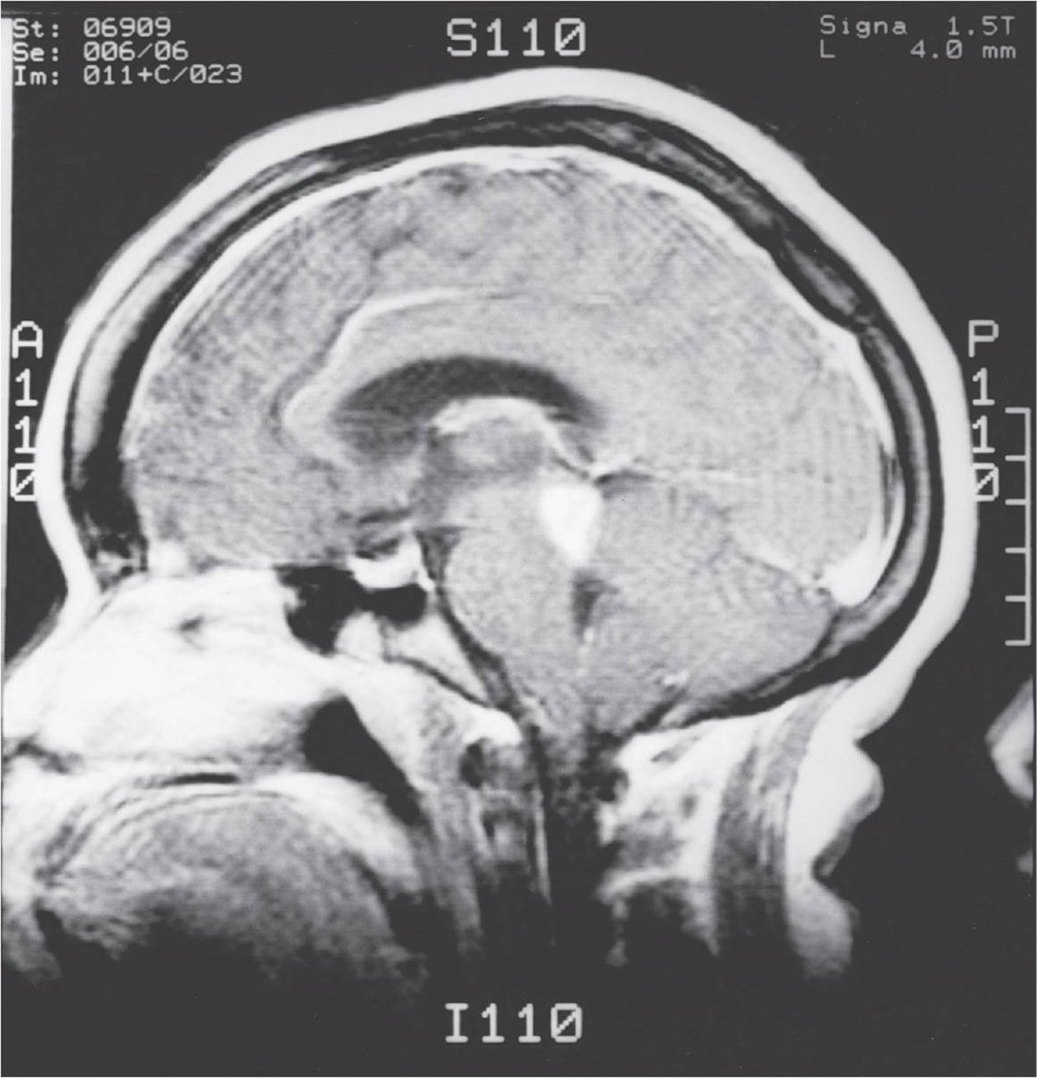
FIGURE 4-15 From Miller NR. Walsh and Hoyt’s Clinical Neuro-ophthalmology. Volume 2: Autonomic nervous and ocular motor systems. Fourth Edition. Baltimore: Williams & Wilkins, 1985, with permission.
A) sixth nerve palsy
B) skew deviation
C) lid retraction
D) pupils that react well to a near stimulus but not to light
20 The patient shown in Figure 4-16 has normal vertical eye movements. The left eye developed left jerk nystagmus on attempted left gaze. Where is the most likely location of the causative lesion?

FIGURE 4-16 From Miller NR. Walsh and Hoyt’s Clinical Neuro-ophthalmology. Volume 2: Autonomic nervous and ocular motor systems. Fourth Edition. Baltimore: Williams & Wilkins, 1985, with permission.
A) Left paramedian pontine reticular formation (PPRF)
B) Right third nerve nucleus
C) Left medial longitudinal fasciculus
D) Right medial longitudinal fasciculus
21 The patient in Figures 4-17 to 4-19 has normal vertical eye movements. Where can her problem be localized?

FIGURE 4-17 Attempted right gaze.

FIGURE 4-18 Primary position.

FIGURE 4-19 Attempted left gaze.
A) Right sixth nerve nucleus
B) Left PPRF
C) Both medial longitudinal fasciculi
D) Right sixth nerve fascicle
22 Which statement regarding the innervation of the extraocular muscles is false?
A) The levator palpebrae is innervated by a fused central nucleus.
B) The superior oblique is innervated by the ipsilateral IV nucleus.
C) The inferior oblique is innervated by the ipsilateral III nucleus.
D) The superior rectus is innervated by the contralateral III nucleus.
23 Which study would be least helpful in diagnosing the condition seen in Figure 4-20?

FIGURE 4-20 From Chern KC, Saidel MA. Ophthalmology Review Manual. Second Edition. Philadelphia: Lippincott Williams & Wilkins, 2011.
A) MRI of the orbits
B) B scan ultrasound
C) CT of the orbits
D) Fluorescein angiography
QUESTIONS 24–26 A 55-year-old man who was in a car accident several days ago complains of intermittent vertical diplopia since the accident. His monocular acuity is 20/20 in each eye. His motility is diagramed in Figure 4-21.

FIGURE 4-21
24 If only a single muscle is involved, which muscle is palsied?
A) Left superior rectus
B) Right inferior oblique
C) Right superior oblique
D) Left inferior rectus
25 What could be used to measure the amount of torsion that this patient has?
A) Double Maddox rods
B) Alternate cover test with prisms
C) Red filter and a light
D) Neutral density filters
26 Cover–uncover testing shows no shift (orthotropia), but alternate cover test shows a deviation neutralized with 12 PD base down prism in front of his right eye. What can this patient be told about his condition?
A) The injury from the accident is temporary and will resolve with time.
B) His symptoms and findings do not correspond to any organic neurologic condition.
C) This condition has been present for many years but has just recently been uncovered.
D) The accident has caused damage to nerves bilaterally.
QUESTIONS 27–35 Match each description with the corresponding structure from Figure 4-22.

FIGURE 4-22
27 Innervates the primary abductor of the eye:
A) structure A
B) structure B
C) structure D
D) structure F
28 Efferent pupil constrictor fibers:
A) structure B
B) structure A
C) structure E
D) none of the above
29 Visual information from the retina:
A) structure A
B) structure B
C) structure C
D) none of the above
30 Corneal sensation:
A) structure E
B) structure D
C) structure F
D) structure A
31 Innervates the inferior oblique muscle:
A) structure A
B) structure C
C) structure D
D) structure B
32 Closes eyelid:
A) structure F
B) structure B
C) structure A
D) none of the above
33 Efferent fibers controlling lacrimation:
A) structure A
B) structure F
C) structure E
D) none of the above
34 Enters the orbit through the inferior orbital fissure:
A) structure B
B) structure F
C) structure E
D) structure D
35 Which one of the following conditions would have positive forced ductions?
A) Myasthenia gravis
B) Thyroid eye disease
C) Comitant strabismus
D) Chronic progressive external ophthalmoplegia (CPEO)
QUESTIONS 36 and 37 A 63-year-old male diabetic patient has diplopia worse on upgaze. Ductions are full; however, on upgaze, the left eye only elevates halfway up (Fig. 4-23). His pupils and the remainder of the ocular examination are normal.

FIGURE 4-23
36 What finding would most likely accompany this examination?
A) Right upper lid retraction
B) Left nystagmus on upgaze
C) Miotic left pupil
D) Chin-down posturing
37 The patient is given a CT scan. Where would the lesion be located to cause this condition?
A) Brain stem
B) Orbital apex
C) Cavernous sinus
D) Juncture of the posterior communicating and internal carotid arteries
QUESTIONS 38–40 Match each condition to the location causing muscle weakness.
A) Primary myopathy
B) Neuromuscular junction
C) Nerve axon
D) Motor nucleus
38 Eaton–Lambert syndrome
39 Kearns–Sayre syndrome
40 Multiple sclerosis
41 Which syndrome includes cranial nerve III palsy, contralateral decreased sensation, and contralateral tremor in the extremities?
A) Benedikt syndrome
B) Weber syndrome
C) Nothnagel syndrome
D) Tolosa–Hunt syndrome
42 Which one of the following is not an example of aberrant regeneration?
A) Duane retraction syndrome
B) Crocodile tears
C) Superior oblique myokymia
D) Marcus Gunn jaw wink
43 Aberrant regeneration does not occur after injury to the oculomotor nerve with which one of the following conditions?
A) Trauma
B) Ischemia secondary to diabetes
C) Tumor compression
D) Aneurysm
44 Which cranial nerve is traumatized most commonly with a closed head injury?
A) Cranial nerve III
B) Cranial nerve II
C) Cranial nerve IV
D) Cranial nerve VI
QUESTIONS 45–48 Select the answer below that corresponds to the finding indicated.
A) Diabetic cranial nerve III palsy
B) Aneurysm
C) Both
D) Neither
45 Pupil commonly involved
46 Painful cranial nerve III palsy
47 Spontaneous resolution
48 Inability to abduct or adduct eye
49 A 74-year-old man developed difficulty reading and mild left-arm weakness yesterday. Visual acuity is 20/20 OU, and his visual fields are shown in Figures 4-24 and 4-25. What else would he be likely to have?

FIGURE 4-24

FIGURE 4-25
A) Poor OKN with the stripes moving to the left
B) Poor OKN with the stripes moving to the right
C) Unformed visual hallucinations
D) Formed visual hallucinations
50 Supranuclear vertical gaze abnormalities can be seen in all of the following conditions except:
A) myasthenia gravis
B) Parkinson disease
C) pineal region tumors
D) ataxia–telangiectasia
51 Downbeat nystagmus may be a result of all of the following except:
A) normal lithium use
B) a paraneoplastic syndrome
C) craniocervical junction abnormalities
D) pinealoma
52 A patient presents with irritation and conjunctival injection of the right eye. You find right orbicularis weakness, decreased ability to wrinkle the right forehead, slight right corneal anesthesia, and a small angle esodeviation develops on right gaze. What test would be most helpful?
A) Tensilon test
B) CT to rule out a left parietal lesion
C) Thyroid function tests
D) MRI to evaluate right cerebellopontine angle
53 What is the usual cause of hemifacial spasm?
A) Stroke
B) Dry eye
C) Facial nerve irritation by an adjacent blood vessel
D) Aberrant regeneration following Bell palsy
54 All of the following statements are true of relative afferent pupillary defects (APDs) except:
A) in general, media opacities do not cause a relative APD.
B) optic tract damage can result in an ipsilateral relative APD because of an asymmetric decussation in the chiasm.
C) the presence of a relative APD without any visual loss localizes damage to the contralateral brain stem.
D) anisocoria is never associated directly with a relative APD.
55 A patient seeks ophthalmologic evaluation because a friend said his eyes “don’t look right” (Fig. 4-26). More anisocoria was present in the dark than in the light. All of the following statements are true except:

FIGURE 4-26 From Miller NR. Walsh and Hoyt’s Clinical Neuro-ophthalmology. Volume 2: Autonomic nervous and ocular motor systems. Fourth Edition. Baltimore: Williams & Wilkins, 1985.
A) Neither pupil should constrict to pilocarpine 0.1%.
B) The responsible lesion could be interrupting the neural impulse as the neurons synapse in the ciliary ganglion.
C) The presence of a right abduction deficit would localize the lesion to the right cavernous sinus.
D) The right pupil would dilate after apraclonidine 0.5% instillation.
QUESTIONS 56 and 57 A 33-year-old recovery room nurse discovers her right pupil to be several millimeters larger than her left pupil. She denies diplopia, but she has had several headaches in the past week.
56 Findings helpful in diagnosing the etiology of her anisocoria include all of the following except:
A) 2 mm of right upper eyelid ptosis
B) a right relative APD
C) a small right hypertropia develops on downgaze
D) segmental contraction of the right iris
57 Her right pupil does not react to light, but otherwise the examination is normal. The next diagnostic step would be:
A) 1 drop of 1% pilocarpine OU
B) 1 drop of 0.1% pilocarpine OU
C) cerebral arteriogram
D) review of old photographs
58 The parasympathetic fibers to the lacrimal gland synapse in which ganglion?
A) Geniculate
B) Sphenopalatine
C) Ciliary
D) Superior cervical
59 Where is the cell body of the second-order neuron in the sympathetic pathway for the pupil?
A) Hypothalamus
B) Superior cervical ganglion
C) Ciliospinal center of Budge (C8–T2)
D) Ciliary ganglion
QUESTIONS 60–64 Select the answer below that corresponds to the finding indicated.
A) First-order Horner’s
B) Second-order Horner’s
C) Third-order Horner’s
D) All of the above
60 No dilation of the pupil after instillation of cocaine 10%
61 No dilation of the pupil after instillation of hydroxyamphetamine 1% (Paredrine)
62 Affected pupil is smaller
63 Carotid dissection
64 Pancoast tumor
65 Which one of the following is a sign of congenital Horner’s syndrome?
A) Iris heterochromia
B) Miotic pupil
C) Facial asymmetry
D) Unilateral epiphora
QUESTIONS 66–68 You see a patient in the ICU after cardiac bypass surgery. His right pupil is dilated and unreactive to light. You swing a light back and forth between his eyes.
66 What would be observed if he has a right APD?
A) When the light shines on the left eye, the left pupil dilates.
B) When the light shines on the right eye, the left pupil dilates.
C) When the light shines on the right eye, the left pupil constricts.
D) Cannot be determined because the right eye is dilated.
67 No APD is present. The ocular examination is unremarkable. Which drop placed in both eyes would provide the most additional information?
A) Cocaine 10%
B) Hydroxyamphetamine 1%
C) Pilocarpine 0.1%
D) Phenylephrine 2.5%
68 You decide to test the caloric response. Cold water is irrigated in the left ear. In what direction is the slow phase of the nystagmus?
A) Left
B) Right
C) Up
D) Down
69 What is the mechanism of action for edrophonium (Tensilon)?
A) Inhibits short acting cholinesterase inhibitor
B) Releases ACh from the presynaptic terminal
C) Directly binds to ACh sites on the receptor
D) Prevents reuptake of ACh
70 What is the mechanism of action for cocaine?
A) Inhibits catechol-O-methyltransferase (COMT)
B) Releases norepinephrine from the presynaptic terminal
C) Directly binds to norepinephrine sites on the receptor
D) Prevents reuptake of norepinephrine
71 What is the mechanism of action for hydroxyamphetamine (Paredrine)?
A) Inhibits COMT
B) Releases norepinephrine from the presynaptic terminal
C) Directly binds to norepinephrine sites on the receptor
D) Prevents reuptake of norepinephrine
72 What is the antidote for the crisis caused by an overdose of edrophonium (Tensilon)?
A) Atropine
B) Dantrolene
C) Epinephrine
D) Verapamil
73 Which one of the following is not a feature of Adie’s pupil?
A) Vermiform movement of iris border
B) Hypersensitivity to parasympathomimetic drugs
C) Light-near dissociation
D) Anisocoria greater in the dark
QUESTIONS 74–75 A 44-year-old black woman noticed a change in vision yesterday. Today, visual acuity is 20/20 OD and 20/200 OS. There is a left relative APD and a swollen left optic nerve head. The remainder of the examination is normal.
74 Of the following, which is the least helpful historical factor?
A) Hyperthyroidism treated 1 year ago
B) Recent 1-month episode of left-arm numbness
C) Hilar adenopathy on recent chest radiograph
D) Hypertension treated for 5 years
75 Additionally, she notes pain on eye movement. What is the best next step?
A) MRI
B) Oral prednisone
C) IV methylprednisolone
D) Observation and repeat examination in 1 month
QUESTIONS 76 and 77 A 72-year-old woman experienced three 10- to 15-minute episodes of “blurred vision” in the right eye over the past week. Her eye examination is normal.
76 The presence of which one of the following signs should elicit the most prompt attention to prevent permanent visual loss?
A) Right carotid bruit
B) Scalp tenderness
C) Blood pressure of 170/95
D) A cardiac murmur
77 This woman did not have any of the findings listed in the previous question. She also denied headache and numbness or weakness of her extremities. She did remember a 10-minute episode of double vision and recently has had difficulty chewing her breakfast because her jaw becomes tired. What would be the next step in the evaluation?
A) Carotid dopplers
B) Antiphospholipid antibody levels
C) Erythrocyte sedimentation rate
D) ACh-receptor antibody level
QUESTIONS 78 and 79 A 28-year-old woman developed diplopia on upgaze (Fig. 4-27). MRI of the brain was normal.

FIGURE 4-27
78 Compared with the normal population, she is more likely to develop:
A) proptosis
B) fatigable ptosis
C) nonfatigable ophthalmoplegia
D) all of the above
79 She was found to be hyperthyroid and was treated with radioactive iodine. Subsequently, she became hypothyroid and was treated with levothyroxine sodium (Synthroid). Now, 6 months later, she develops bilateral conjunctival injection and a right relative APD. What is the most appropriate next step in management?
A) Direct coronal CT scan of the orbits
B) Thyroid function tests
C) Oral prednisone
D) Follow-up in 2 months
80 Neuroimaging would most likely be normal in an individual with which of the following syndromes?
A) von Recklinghausen disease
B) Louis–Bar syndrome
C) Bourneville syndrome
D) Sturge–Weber syndrome
QUESTIONS 81–86 Select the phakomatosis that matches the conditions listed.
81 Seizures
A) von Hippel–Lindau syndrome
B) von Recklinghausen disease
C) Louis–Bar syndrome
D) Bourneville syndrome
82 Glaucoma
A) Wyburn–Mason syndrome
B) Sturge–Weber syndrome
C) Bourneville syndrome
D) von Hippel–Lindau syndrome
83 Retinal detachment
A) von Hippel–Lindau syndrome
B) Louis–Bar syndrome
C) Wyburn–Mason syndrome
D) von Recklinghausen disease
84 Astrocytic hamartomas
A) Louis–Bar syndrome
B) von Recklinghausen disease
C) Sturge–Weber syndrome
D) von Hippel–Lindau syndrome
85 Increased incidence of pheochromocytomas
A) Louis–Bar syndrome
B) von Hippel–Lindau syndrome
C) Sturge–Weber syndrome
D) Bourneville syndrome
86 Thymic aplasia
A) Wyburn–Mason syndrome
B) Sturge–Weber syndrome
C) von Hippel–Lindau syndrome
D) Louis–Bar syndrome
87 Ocular pulsations may be seen in all of the following except:
A) neurofibromatosis
B) carotid-cavernous sinus fistulas
C) orbitoencephaloceles
D) capillary hemangioma
88 A patient with multiple sclerosis could have all of the following except:
A) bitemporal visual field deficit
B) retinal venous sheathing
C) skew deviation
D) amaurosis
89 Uhthoff symptom describes:
A) the decrease in vision with an increase in body temperature
B) an electric shock sensation with neck flexion
C) the inability to distinguish faces
D) the ability to see moving objects but not stationary ones
90 A 29-year-old woman has had “migraine” headaches for several years. She recently developed episodes of “flashing lights off to the right” that affect her peripheral vision. Her automated perimetry is shown in Figures 4-28 and 4-29. The next step would be:

FIGURE 4-28

FIGURE 4-29
A) cerebral MRI
B) discontinue oral contraceptives
C) sumatriptan (Imitrex)
D) tangent screen
91 Which one of the following signs would not be expected with a classic migraine?
A) Premonitory aura
B) Scintillating lights
C) Headache
D) Persistent leg tingling and weakness
92 Which one of the following is not a criterion for the diagnosis of pseudotumor cerebri (idiopathic intracranial hypertension)?
A) Normal cerebrospinal fluid composition
B) Elevated opening pressure on lumbar puncture
C) Bilateral papilledema
D) Normal neuroimaging studies
93 A 22-year-old man presents to the emergency department with a 1-day history of diplopia. Examination reveals limitation of abduction of the right eye and limitation of elevation of the left eye. The pupils measure 7 mm OD and 8 mm OS with poor reaction to light and accommodation. Bilateral ptosis is present. The least useful test needed to confirm the diagnosis is:
A) Clostridium botulinum toxin test
B) tensilon test
C) lumbar puncture
D) GQ1b antibody test
94 A 68-year-old man developed a sudden onset of vomiting, imbalance, and double vision. On examination, he had a concomitant 10 PD left hypertropia and ataxia. What test should be ordered?
A) Cerebral arteriography
B) Cerebral MRI and arteriography
C) Tensilon test
D) Carotid ultrasonography
95 A 41-year-old woman presents with a 3-month history of shimmering photopsias in the left eye in association with a “dark spot” temporal to fixation in that eye which has gradually increased in size. The least likely diagnosis is:
A) cancer-associated retinopathy
B) acute zonal occult outer retinopathy (AZOOR)
C) MS
D) gyrate atrophy
96 Which muscles are affected most commonly in thyroid eye disease?
A) Medial rectus, inferior rectus
B) Superior rectus, inferior oblique
C) Lateral rectus, superior oblique
D) Inferior oblique, inferior rectus
97 A patient with thyroid eye disease has progressive loss of visual field. All of the following are possible treatments except:
A) radiotherapy
B) surgical decompression of the orbit
C) optic nerve sheath decompression
D) steroids
98 Which one of the following statements is a conclusion of the Ischemic Optic Neuropathy Decompression Trial (IONDT)?
A) One-third of the patients with optic nerve sheath decompression experienced improvement in acuity of three or more lines at 6 months.
B) The natural course of untreated non–arteritic ischemic optic neuropathy (NAION) is progressive visual field loss.
C) The efficacy of optic nerve sheath decompression is equivocal, and further study is needed.
D) Patients with optic nerve sheath decompression had better visual acuity at 6 months compared with the observation cohort.
99 Which treatment in the Optic Neuritis Treatment Trial (ONTT) had the highest rate of recurrence?
A) Oral prednisone alone
B) IV methylprednisolone alone
C) IV methylprednisolone and oral prednisone
D) Observation
100 Which one of the following is not involved with vertical eye movements?
A) Frontal eye fields
B) PPRF
C) Interstitial nucleus of Cajal
D) Trochlear nucleus
1 D) Vitreous
MRIs allow excellent soft tissue definition by varying radiofrequency pulse sequences and measuring the resulting signal produced by the tissue. T1- and T2-weighted MRIs are able to highlight structures by the intensity of the signal generated after the magnetic pulse. Table 4-1 lists some of the differences between T1- and T2-weighted images. Air, fast-moving blood, and bone generally produce no signal and are thus hypointense on the MRI. Fat and vitreous are opposite to one another in both the T1- and T2-weightings.
TABLE 4-1 Relative T1, T2, and DWI Signal Intensities by Tissue

2 B) Optokinetic nystagmus (OKN) drum
A number of tests can be used to determine whether a patient has functional visual loss. Direct tests do not require the patient to verbally respond or interpret the visual information. Tests such as the swinging light test, OKN response, and mirror test make it very difficult for a patient to feign disease. Indirect tests rely on the patient’s cooperation and allow more definite measurement of visual acuity in the “injured” eye.
3 C) Conjunctival laceration
The MRI image reveals an enlarged right superior ophthalmic vein, a typical finding in a carotid-cavernous fistula. The fistula directs arterialized carotid blood into the superior ophthalmic vein causing reversal of flow. Other common findings include raised intraocular pressure secondary to increased episcleral pressure, bright red arterialized conjunctival vessels, and an audible orbital bruit. Multiple cranial palsies (related to cavernous sinus pathology) may also be present but a sixth nerve palsy is most common. A conjunctival laceration may be present but is not typically seen with this disorder.
4 C) Fingolimod
Fingolimod (Gilenya) is a new oral agent used to treat multiple sclerosis. It has been associated with macular edema in 0.4% of patients using the medication, with a higher risk seen in patients with a history of diabetes or prior uveitis. Macular edema usually appears within the first 4 months of starting the medication, and a screening examination is recommended prior to or within the first few weeks of starting the medication. The other medications have not been associated with significant ocular side effects.
5 A) Figure 4-2
6 B) Figure 4-3
7 C) Figure 4-4
8 B) von Recklinghausen disease
9 D) Figure 4-3
10 C) Figure 4-2
|
Figure 4-2 |
= |
nerve sheath meningioma |
|
|
Figure 4-3 |
= |
absence of the septum pellucidum |
|
|
Figure 4-4 |
= |
suprasellar craniopharyngioma |
|
|
Figure 4-5 |
= |
sphenoid wing meningioma |
|
|
Figure 4-6 |
= |
optic nerve glioma |
Optic nerve sheath gliomas and meningiomas can readily be distinguished on CT because of their appearance (Fig. 4-30). The glioma produces fusiform enlargement, whereas the meningioma produces the railroad track sign on CT. Gliomas, Lisch nodules (see Fig. 4-9), absence of the sphenoid wing, plexiform neurofibromas, and café-au-lait skin lesions are all manifestations of neurofibromatosis, or von Recklinghausen disease.

FIGURE 4-30
Meningiomas have proliferation of benign meningothelial cells in whorls. Calcified psammoma bodies are commonly found. They cause damage by compression of adjacent structures. Lesions of the sphenoid wing can compress the optic nerve and cause visual field loss. Optic atrophy, proptosis, and optociliary shunt vessels (see Fig. 4-10) can be found with this condition.
Suprasellar craniopharyngiomas are found more commonly in children and young adults and are derived from remnants of Rathke pouch. They are located near the pituitary and optic chiasm and, with growth, compress these structures. A bitemporal hemianopsia (see Fig. 4-8) has resulted from such a lesion. These lesions are often calcified and can be seen readily on CT scan.
Absence of the septum pellucidum can be found as part of DeMorsier syndrome (septo-optic dysplasia) along with optic nerve hypoplasia (see Fig. 4-7) and pituitary abnormalities. These patients should have pituitary hormone studies because they may require supplementation.
11 C) Macular toxoplasmosis
Macular toxoplasmosis is a congenital infection that affects central vision from birth. All of the other conditions may cause acquired bilateral central scotomas. Bilateral occipital infarcts would be extremely rare in such a young patient.
12 D) DNA analysis
13 C) Hyperemic optic nerve with telangiectatic capillaries
14 B) None
LHON is caused by an abnormality in mitochondrial DNA. Several mutations have been identified, including a nucleotide substitution at position 11,778 of the mitochondrial DNA coding for subunit 4 of NADH dehydrogenase. Because mitochondrial DNA is only transmitted along the maternal line, men cannot pass this disease to their offspring.
In the acute phase, the optic nerve in LHON is hyperemic and swollen with telangiectatic capillaries. The nerve does not leak on fluorescein angiography. Later stages may only manifest optic atrophy.
15 D) Ganciclovir
A large number of medications have been associated with optic neuropathy. Among them are the antituberculous drugs isoniazid and ethambutol. Ganciclovir has not been described to cause an optic neuropathy.
16 Right temporal lobe
A homonymous visual field defect is caused by a lesion posterior to the chiasm. The homonymous left superotemporal quadrantanopia in Figure 4-31 is caused by injury to the inferior fibers that must detour through the temporal lobe to avoid the ventricles (Meyer loop). A lesion in the right optic tract would cause a similar defect but would be associated with a left APD and bilateral optic atrophy.

FIGURE 4-31
17 B) The lack of acetylcholine (ACh)-receptor antibody in his blood makes myasthenia gravis an unlikely diagnosis.
Figure 4-13 demonstrates fatigable ophthalmoplegia, a sign virtually pathognomonic for myasthenia gravis. The patient has no systemic weakness and thus has ocular myasthenia gravis. ACh receptor antibodies with no systemic involvement are only found in about 60% of patients. Dysthyroidism is more common in patients with myasthenia gravis. Tensilon testing, not MRI, should be obtained. When a patient has isolated ocular myasthenia gravis for more than 2 years, there is a less than 20% chance that he or she will develop systemic disease. ACh receptor binding antibodies are found in 90% of patients with generalized myasthenia and in approximately 50% of patients with ocular myasthenia. Modulating and blocking antibodies are detected less commonly in patients with myasthenia.
18 B) Sluggishly reactive pupils
Figure 4-14 shows generalized ophthalmoplegia and ptosis, findings compatible with either CPEO or myasthenia gravis. Neither of these conditions affects pupillary reactivity. Polychromatic lenticular deposits are seen in myotonic dystrophy, and patients with oculopharyngeal dystrophy often have a French–Canadian ancestry. CPEO can be seen in both of these conditions. Patients with CPEO typically have a long history of gradually worsening ptosis that can be documented in old photographs. This patient could also have cardiac conduction abnormalities and pigmentary retinopathy (Kearns–Sayre).
19 A) Sixth nerve palsy
The MRI shows abnormal signal in the area of the dorsal midbrain (Fig. 4-32, arrow). Skew deviation, lid retraction (Collier sign), and pupillary light-near dissociation are all signs of Parinaud dorsal midbrain syndrome. The sixth nerve arises in the pons (Fig. 4-32, curved arrow) and should not be affected by this lesion.

FIGURE 4-32
20 D) Right medial longitudinal fasciculus
Figure 4-33 shows normal horizontal movement of the left eye and poor adduction of the right eye. The combination of abnormal adduction and contralateral abducting nystagmus represents an internuclear ophthalmoplegia. Damage has occurred to the interneurons connecting the left sixth nerve nucleus and the right third nerve nucleus (medial rectus subdivision) traveling in the right medial longitudinal fasciculus. Poor adduction could also occur from third nerve dysfunction, but this patient has no ptosis, mydriasis, or involvement of the superior rectus, inferior rectus, or inferior oblique muscles. Damage to the left PPRF results in a left gaze palsy.

FIGURE 4-33
21 A) Right sixth nerve nucleus
Figures 4-17 to 4-19 show a right gaze palsy. The sixth nerve nucleus contains both sixth nerve axons and interneurons destined for the contralateral medial rectus subnucleus via the medial longitudinal fasciculus. Thus, a lesion of the sixth nerve nucleus would produce an ipsilateral gaze palsy. Left PPRF damage would result in a left gaze palsy. Damage to both medial longitudinal fasciculi would cause a bilateral internuclear ophthalmoplegia. A lesion of the right sixth nerve fascicle would produce a right abduction deficit.
22 B) The superior oblique is innervated by the ipsilateral IV nucleus.
As shown in Figure 4-34, the levator palpebrae are innervated by a single fused central nucleus. Unilateral ptosis as a result of a nuclear lesion is not possible. A third nerve nuclear lesion will therefore give either bilateral ptosis or no ptosis, depending on whether the central nucleus is affected. Ipsilateral nuclei innervate the inferior rectus, medial rectus, inferior oblique, and lateral rectus muscles. The nuclei controlling the superior rectus and superior oblique muscles have crossed projections. The superior oblique muscle is innervated by the contralateral IV nucleus.

FIGURE 4-34
23 A) MRI of the orbits
The figure demonstrates optic nerve head drusen, commonly confused with true optic nerve swelling. Drusen are refractile, often calcified nodules within the optic nerve thought to result from impaired ganglion cell axonal transport. Other associated findings on examination may include small crowded-appearing nerves with an anomalous vasculature pattern present. B scan ultrasound demonstrates high signal intensity at the nerve head when drusen are present. Fluorescein angiography may reveal autofluoresence in the preinjection frames of the angiogram. CT remains superior to MRI imaging for detection of drusen since calcium is poorly visualized with MRI. Optic disc drusen may be associated with retinitis pigmentosa and pseudoxanthoma elasticum.
24 C) Right superior oblique
The Park three-step test can be used to determine which muscle is palsied. The patient has a right hypertropia in primary position, which is worse on left gaze. The hypertropia also worsens on right head tilt. This pattern indicates a right superior oblique palsy.
25 A) Double Maddox rods
The Maddox rod takes a point source of light and converts it into a straight line. With a Maddox rod in front of each eye (in a trial frame), the patient can rotate one of the rods until the lines he sees in each eye are parallel. The difference in the axes of the two Maddox rods is the degree of torsion. The alternate cover test allows the measurement of horizontal and vertical tropias, but it cannot be used for torsion. Red filter and a muscle light will diagnose vertical and horizontal diplopia, not torsion. Neutral density filters reduce overall luminance and may be helpful for assessing APDs.
26 C) This condition has been present for many years but has just recently uncovered.
Vertical fusion amplitudes are useful for distinguishing congenital from acquired fourth nerve palsies. Normal vertical fusion amplitudes are 3 to 5 PD. In congenital IV palsy, patient can develop amplitudes of 10 to 25 PD, and large vertical fusion amplitudes are an indication of a long-standing vertical deviation usually since childhood. If the cover–uncover test shows no deviation but the alternate cover test discloses a deviation, this means a phoria, and indicates fusion amplitude. In this case, the patient has a right hyperphoria of 12 PD; this means that the vertical fusion amplitude is at least 12 PD (much more than normal), indicating a long-standing deviation. Although trauma is the most common cause of acquired IV palsy, it is also (often coincidentally) the trigger that allows congenital palsies to be manifest. In addition to increased vertical fusion amplitudes, these patients may have a head tilt to the contralateral side to reduce the hypertropia. Old photographs may be helpful in demonstrating this feature.
27 C) Structure D (Fig. 4-35)
The primary abductor of the eye is the lateral rectus, innervated by cranial nerve VI.

FIGURE 4-35
28 A) Structure B
Parasympathetic neurons to the pupil constrictor travel with cranial nerve III to the ciliary ganglion where they synapse and then join the short ciliary nerves.
29 D) None of the above
The optic nerve carries visual information from the retina. Cranial nerve II is not pictured on this section.
30 A) Structure E
Corneal sensation is subserved by branches of cranial nerve V1.
31 D) Structure B
The inferior oblique receives innervation from the inferior division of cranial nerve III.
32 D) None of the above
Eyelid closure is caused by action of the orbicularis muscles controlled by cranial nerve VII. Eyelid opening results from the levator palpebrae (cranial nerve III) and Müller muscle (sympathetics).
33 D) None of the above
Lacrimation is controlled by parasympathetic neurons that travel from the superior salivatory nucleus along the nervus intermedius through the greater petrosal canal. At the pterygopalatine ganglion, the nerves synapse and postganglionic branches join cranial nerve V2 to travel to the lacrimal gland.
34 B) Structure F
The superior orbital fissure transmits the branches of cranial nerves V1, III, IV, and VI. Cranial nerve V2 enters the orbit through the inferior orbital fissure.
35 B) Thyroid eye disease
Positive forced ductions indicate a restrictive process that may be caused by thyroid eye disease, blow-out fractures, fat adherence syndrome, and Brown syndrome, among others. Disorders of the neuromuscular junction (myasthenia) and of the muscle itself (CPEO) do not cause restriction.
36 A) Right upper lid retraction
This patient has an isolated superior division of cranial nerve III paresis affecting the left superior rectus and levator palpebrae. The most likely cause for this condition is a diabetic ischemic neuropathy. The weakened levator would cause a left ptosis. He can compensate for this by overstimulating the levator to raise the left lid, consequently raising the right lid higher. Mechanically lifting the left lid will produce a paradoxic right ptosis.
37 B) Orbital apex
The only location at which an oculomotor superior division nerve palsy could occur without affecting additional cranial nerves or the inferior division of cranial nerve III is in the orbital apex. Posterior to the orbital apex, the superior and inferior divisions are joined, and a compressive lesion selective to one division would be very improbable. A unilateral ptosis does not occur with a nuclear lesion.
38 B) Neuromuscular junction
Eaton–Lambert syndrome is thought to be an autoimmune disease similar to myasthenia gravis. Abnormalities of conduction at the neuromuscular junction lead to muscle weakness and fatigue. Ocular manifestations are less common with Eaton–Lambert than with myasthenia. Eaton–Lambert is often associated with cancer. In particular, patients with small cell lung cancer may develop this disorder.
39 A) Primary myopathy
CPEO and related conditions such as Kearns–Sayre syndrome have abnormalities caused by mutations in mitochondrial DNA. Biopsies of the muscles of these patients demonstrate “ragged red fibers.” Kearns–Sayre syndrome also includes pigmentary retinopathy, cardiac conduction abnormalities, and other systemic findings.
40 C) Nerve axon
The primary pathologic process in multiple sclerosis is demyelination of axons. Multiple sclerosis should be considered when the signs and symptoms cannot be localized to a single lesion.
41 A) Benedikt syndrome
The location of a lesion affecting the oculomotor nerve can be determined by the associated neurologic deficits. An oculomotor brainstem lesion also involving the ipsilateral cerebral peduncle causes contralateral hemiparesis (Weber syndrome). Cranial nerve III palsy with contralateral decreased sensation and contralateral tremor (red nucleus affected) is Benedikt syndrome. Nothnagel syndrome has involvement of the brachium conjunctivum and cerebellar ataxia.
Tolosa–Hunt syndrome, caused by inflammation of the cavernous sinus, involves multiple cranial nerves. Prompt resolution occurs with corticosteroid treatment.
42 C) Superior oblique myokymia
Superior oblique myokymia is an episodic twitching of the superior oblique muscle. Patients may experience a slight vertical/torsional diplopia or oscillopsia lasting from seconds to minutes. The exact mechanism is unknown but may related to vascular compression of the fourth cranial nerve or demyelinating plaques. Carbamazepine and propranolol have been effective treatments.
43 B) Ischemia secondary to diabetes
Damage to the oculomotor nerve with trauma or compressive lesions can cause aberrant regeneration. Ischemic neuropathy does not result in aberrant regeneration.
44 C) Cranial nerve IV
The trochlear nerve has the longest intracranial course and is the most commonly injured nerve following closed head injury. Cranial nerve VI palsy can also be injured with head trauma. Any condition that causes increased intracranial pressure (pseudotumor cerebri, tumor, hydrocephalus) may result in cranial nerve VI palsy.
45 B) Aneurysm
Aneurysms damage nerves by extrinsic compression. The parasympathetic pupillomotor fibers innervating the pupil sphincter are found on the outside of the oculomotor nerve and are the first to be injured with external pressure. Diabetic microvascular insults affect the central fibers of the nerve to a greater degree than the outer fibers, and as a result, pupil involvement is less common (<20%).
46 C) Both
Cranial nerve III palsy caused by aneurysm is almost invariably painful; palsy from diabetes may be painful or painless.
47 A) Diabetic cranial nerve III palsy
Diabetic nerve palsies will usually resolve over 2 to 6 months. There may be some slight residual damage. If the palsy does not resolve spontaneously, the possibility of an aneurysm or mass lesion must be investigated.
48 D) Neither
This indicates involvement of both the medial rectus (cranial nerve III) and lateral rectus (cranial nerve VI). It would be very uncommon for either diabetic microvascular or aneurysmal compressive lesions to involve both of these nerves simultaneously.
49 B) Poor OKN with the stripes moving to the right.
The visual fields show a left, inferior, homonymous hemianopsia. This field deficit, in combination with a left hemiparesis, indicates damage to the right parietal lobe. Acutely, ipsilateral pursuit may be affected by parietal lesions. Thus, the patient could have difficulty pursuing the optokinetic drum as the stripes move to the right. Abnormal OKN will be observed when the target moves toward the side of the lesion, opposite the hemianopsia since pursuit originates in the ipsilateral parietal region. Unformed visual hallucinations typically occur with occipital lobe damage, whereas formed visual hallucinations are associated with temporal lobe lesions.
50 A) Myasthenia gravis
Myasthenia gravis might produce ophthalmoplegia that mimics supranuclear vertical gaze abnormalities, but the pathophysiology is at the ACh receptor. Parkinson disease, pineal region tumors (dorsal midbrain syndrome), and ataxia–telangiectasia all can cause supranuclear gaze palsy.
51 D) Pinealoma
Downbeat nystagmus has been associated with both therapeutic and toxic lithium levels. The nystagmus does not necessarily resolve after the lithium is stopped. It has been reported as a remote effect of cancer (paraneoplastic cerebellar degeneration); gynecologic malignancy and small cell carcinoma of the lung are implicated most frequently. Craniocervical junction abnormalities (tumor, syrinx, Arnold–Chiari malformations) can be ruled out with MRI. Pinealomas or other lesions in the dorsal midbrain result in Parinaud dorsal midbrain syndrome. Features include contraction–retraction nystagmus, upgaze paralysis, and light-near dissociation.
52 D) MRI to evaluate right cerebellopontine angle
This patient has corneal exposure as a result of partial right facial nerve palsy (orbicularis weakness, inability to wrinkle forehead). Right corneal anesthesia indicates right trigeminal nerve dysfunction. A mild right abducens nerve palsy would produce an esodeviation on right gaze. These three cranial nerves are in close proximity in the cerebellopontine angle. Myasthenia gravis could produce facial weakness and ophthalmoplegia but not corneal anesthesia. Signs of Graves disease would include conjunctival injection, irritation, and ophthalmoplegia but not facial weakness.
53 C) Facial nerve irritation by an adjacent blood vessel
Hemifacial spasm is typically caused by irritation of the facial nerve as it exits the brain stem. Rarely, compression by tumor may cause this condition. Dry eye may result in blepharospasm but not spasm of the lower facial musculature. Aberrant regeneration of the facial nerve may result in inappropriate muscle contraction, but this should follow a pattern (i.e., orbicularis contraction on attempted smiling).
54 B) Optic tract damage can result in an ipsilateral relative APD because of an asymmetric decussation in the chiasm.
A contralateral relative APD would be present if the optic tract was damaged because relatively more nasal fibers from the contralateral eye cross in the chiasm. Approximately 52% of the optic nerve axons cross in the chiasm, whereas 48% remain ipsilateral.
Cataracts, vitreous hemorrhage, and hyphema generally do not produce relative APDs, although extremely dense opacities have been reported to produce small relative APDs. Damage to the pupillomotor fibers after they separate from the visual fibers in the midbrain can result in a small contralateral relative APD without visual loss because of the asymmetric decussation in the chiasm. Anisocoria is caused by asymmetric efferent pupillomotor input. Asymmetric afferent pupillomotor damage does not cause asymmetric input to the efferent pupillomotor system (Edinger–Westphal nuclei) because of its double decussation in the chiasm and posterior commissure.
55 B) The responsible lesion could be interrupting the neural impulse as the neurons synapse in the ciliary ganglion.
The patient has right ptosis and miosis, or a right Horner’s syndrome. The sympathetic axons travel through the ciliary ganglion but do not synapse. A right abduction deficit could represent a sixth nerve palsy. The postganglionic sympathetic chain and the sixth nerve travel together only in the cavernous sinus. Thus, a cavernous sinus lesion would be most likely if an abduction deficit were present. Parasympathetic denervation supersensitivity is not present in Horner’s syndrome, so neither pupil should constrict to dilute pilocarpine. Apraclonidine is an adrenergic agent with a weak α1-agonist action and a stronger α2-agonist action. Loss of sympathetic innervation leads to upgrading of α1-receptors causing denervation supersenstivity and pupillary dilation in a Horner’s patient. Cocaine testing also may be used to diagnose Horner’s. Cocaine inhibits the reuptake of norepinephrine and causes dilation if the sympathetic chain is intact. In Horner’s syndrome, no norepinephrine is present; thus, the pupil will not dilate with cocaine.
56 B) A right relative APD
A relative APD never causes anisocoria because the pupillary fiber decussations in the chiasm and posterior commissure ensure equal efferent input to both iris sphincter muscles. Right upper eyelid ptosis and a right hypertropia on downgaze could both be signs of third nerve dysfunction (levator palpebrae and inferior rectus weakness, respectively). Segmental iris contraction is a sign of Adie’s tonic pupil.
57 B) 1 drop of 0.1% pilocarpine OU
You suspect her anisocoria may be pharmacologic because of the lack of other findings. If so, her right pupil should not constrict to 1% pilocarpine. However, if you proceed with 1% pilocarpine and her pupils both constrict, she still could have either an Adie’s tonic pupil or a partial oculomotor nerve palsy. At that point, it is too late to use the 0.1% pilocarpine drops. Thus, testing for denervation supersensitivity (0.1% pilocarpine) should be done first. Cerebral arteriogram is appropriate if testing indicates third nerve dysfunction. Old photographs are most helpful if physiologic anisocoria is suspected.
58 B) Sphenopalatine
The primary parasympathetic nerve cell bodies are located in the superior salivatory nucleus. Their axons leave the brain with the nervus intermedius (glossopalatine) nerve to travel with cranial nerve VII through the geniculate ganglion and then emerge from the petrous portion of the sphenoid bone as the greater superficial petrosal nerve. The greater superficial petrosal nerve is then joined by the secondary sympathetics from the deep petrosal nerve and enter the pterygoid canal (vidian canal). The vidian nerve emerges from the pterygoid canal and enters the sphenopalatine ganglion (pterygopalatine) where the primary parasympathetic fibers synapse and then exit as secondary parasympathetic fibers. These secretomotor fibers then join the zygomatic nerve (a branch of the maxillary division of cranial nerve V), which sends a communicating branch that enters the lacrimal gland. Most references state that this communicating branch joins and travels with the sensory lacrimal nerve to the lacrimal gland. However, our dissections have shown that the communicating branch usually enters the lacrimal gland directly.
The geniculate ganglion is transversed by cranial nerve VII and contains the cell bodies that provide the sense of taste from the anterior two-thirds of the tongue.
The ciliary ganglion is the intraorbital location where the primary parasympathetic fibers from the Edinger–Westphal nucleus synapse with the secondary parasympathetic nerves. The secondary parasympathetic fibers innervate the ciliary body and iris sphincter muscle to provide accommodation and constriction of the pupil.
The cell bodies of the secondary sympathetic fibers, which provide innervation to the superior tarsal muscle, the pupillary dilator muscle, facial blood vessels, skin and sweat glands, reside in the superior cervical ganglion.
59 C) Ciliospinal center of Budge (C8–T2)
The sympathetic pathway consists of a chain of three neurons. The first neuron travels from the hypothalamus along the brain stem to synapse in the intermediolateral column of the spinal cord, the ciliospinal center of Budge. The second-order neuron exits the brain stem about the level of T1 and joins the cervical sympathetic chain. At the superior cervical ganglion, the second-order neuron synapses with the third-order neuron, which travels along the carotid plexus. Branches join the ophthalmic division of the trigeminal nerve and pass through the ciliary ganglion to the nasociliary and short ciliary nerves (Fig. 4-36).

FIGURE 4-36
60 D) All of the above
61 C) Third-order Horner’s
62 D) All of the above
63 C) Third-order Horner’s
64 B) Second-order Horner’s
Horner’s syndrome is a result of interruption of sympathetic input to the eye. The classic triad consists of ptosis, miosis, and anhydrosis. Pharmacologic tests with cocaine and hydroxyamphetamine help to localize the specific interrupted neuron that causes the Horner’s (Table 4-2). Cocaine prevents reuptake of norepinephrine into the presynaptic terminal from the synaptic cleft. With the sympathetics intact, cocaine increases the duration that norepinephrine remains in the synaptic cleft, causing pupillary dilation. In Horner’s syndrome (first, second, and third order), there is little activity at the cleft and hence no dilation. Hydroxyamphetamine causes the release of norepinephrine from the presynaptic terminal. In first- or second-order Horner’s, this results in pupillary dilation. If the third-order neuron is injured, it is unable to release norepinephrine and the pupil remains miotic.
TABLE 4-2 Evaluation of Anisocoria

Localization of the lesion in Horner’s syndrome can also be made by the accompanying findings. A Pancoast tumor at the apex of the lung is in close proximity to the location at which the sympathetics exit the spinal column. Neck surgery can disrupt portions of the cervical sympathetic chain. Carotid dissection can damage the sympathetic plexus as it ascends along the artery. Remember, a Horner’s at any level of the sympathetic chain will dilate with the use of apraclonidine and will not dilate with the use of cocaine 10%.
65 A) Iris heterochromia
Sympathetic innervation plays a role in the development of pigmentation of the iris. Congenital interruption of the sympathetics to one eye will result in the ipsilateral iris having less pigment than the fellow eye.
66 B) When the light shines on the right eye, the left pupil dilates.
One dilated pupil does not preclude the detection of an APD. Because the pupillary efferents are equal bilaterally, the consensual light response can be used instead of the direct response to detect an afferent defect. With damage to the right optic nerve, both pupils will dilate when the light swings from left to right.
67 C) Pilocarpine 0.1%
The differential of a dilated pupil includes traumatic mydriasis, Adie’s pupil, and pharmacologic dilation. Iris sphincter tears or a history of blunt trauma would be present with traumatic mydriasis. The other two possibilities can be differentiated by using dilute pilocarpine. Adie’s tonic pupil is hypersensitive to parasympathomimetics and will constrict with pilocarpine 0.1%. Acute Adie’s, however, may not constrict and can mimic pharmacologic dilation. Cocaine and hydroxyamphetamine drops are useful for the diagnosis of a Horner’s syndrome. Phenylephrine binds directly to the postsynaptic receptor, causing dilation.
68 A) Left
The mnemonic COWS—cold, opposite; warm, same (Fig. 4-37)—helps to give the direction of the fast phase of the nystagmus. In this case, the left eye is irrigated with cold water, so the fast phase will be toward the right. Consequently, the slow phase of the nystagmus is to the left. Bilateral cold water irrigation will produce nystagmus with fast phase upward (Table 4-3).

FIGURE 4-37
TABLE 4-3 Caloric Vestibular Stimulation
|
Eye Movement Direction Relative to Side Stimulated |
||
|
Warm Water |
Cold Water |
|
|
Awake (jerk nystagmus— fast phase) |
||
|
Bilateral irrigation |
Downward |
Upward |
|
Unilateral irrigation |
Same |
Opposite |
|
Comatose (tonic deviation—no nystagmus) |
||
|
Bilateral irrigation |
Upward |
Downward |
|
Unilateral irrigation |
Opposite |
Same |
69 A) Inhibits acetylcholinesterase
In myasthenia, there are fewer receptor sites on the postsynaptic terminal because of blockage of sites by immunologic complexes. Edrophonium is a short-acting anticholinesterase inhibitor. It prolongs the duration of ACh in the synaptic cleft. As a result, the muscles contract with more force.
70 D) Prevents reuptake of norepinephrine
71 B) Releases norepinephrine from the presynaptic terminal
Cocaine and hydroxyamphetamine (Paredrine) are used in the test for Horner’s syndrome. Cocaine blocks the reuptake of norepinephrine from the synaptic cleft, prolonging the effect of released neurotransmitter. In Horner’s syndrome, little norepinephrine is released, and as a result, cocaine has little effect. Paredrine, in contrast, causes the release of norepinephrine from the presynaptic terminal. In first- and second-order Horner’s, norepinephrine released from the third-order neuron causes dilation of the pupil. In third-order Horner’s, this neuron is damaged and no norepinephrine is released by Paredrine.
72 A) Atropine
Tensilon prolongs the effect of ACh in the synaptic cleft, causing a cholinergic crisis (sweating, nausea and vomiting, salivation, fever). Atropine blocks ACh receptor sites on the postsynaptic terminal.
73 D) Anisocoria greater in the dark
An Adie’s tonic pupil is dilated and poorly reactive to light. In the light, the fellow pupil constricts and the anisocoria is more pronounced. The cause of Adie’s pupil is unknown, but it may be caused by a miswiring of the parasympathetics in the ciliary ganglion. The vermiform movement of the iris border occurs as missynchronized neural impulses cause segmental iris sphincter contraction. These pupils are hypersensitive to parasympathomimetics and will constrict with dilute pilocarpine. A normal pupil will have minimal miosis with dilute pilocarpine.
74 A) Hyperthyroidism treated 1 year ago
This woman has a left optic neuropathy without any orbital signs. Compressive optic neuropathy associated with thyroid ophthalmopathy should be accompanied by the typical signs of proptosis, chemosis, and lid retraction. The optic neuropathy of Graves disease is usually of subacute onset as well.
Multiple sclerosis, sarcoidosis, and anterior ischemic optic neuropathy may cause isolated optic neuropathy and may be associated with arm numbness, hilar adenopathy, and hypertension, respectively.
75 A) MRI
This patient most likely has optic neuritis. The presence of periventricular plaques on MRI increases her risk of developing multiple sclerosis. Treatment of higher risk patients with IV methylprednisolone (Solu–Medrol) decreases the rate of developing multiple sclerosis over the next 2 years. Thus, observation would not be appropriate. Treatment with oral prednisone alone increases the rate of recurrence of optic neuritis and is thus contraindicated.
76 B) Scalp tenderness
Temporal arteritis must be considered in anyone with transient visual loss who is over 55 years of age. The presence of scalp tenderness should raise suspicions further. Permanent visual loss caused by ischemic optic neuropathy could occur at any moment, and prompt treatment with corticosteroids could be preventative. A carotid bruit or cardiac murmur may signify a potential embolic focus and should be evaluated. However, permanent visual loss would be much less likely. Similarly, hypertension is a risk factor for vascular disease and should be treated, but it need not be done urgently.
77 C) Erythrocyte sedimentation rate
Fatigue with chewing is a form of jaw claudication. Transient diplopia could be a result of ocular motor nerve ischemia. In combination, these symptoms are very suggestive of temporal arteritis.
Carotid Dopplers would be appropriate to evaluate transient visual loss if symptoms of temporal arteritis were not present. The antiphospholipid antibody syndrome (lupus anticoagulant, anticardiolipin antibody) can cause transient visual loss, typically in younger adults, and should be considered in the appropriate setting. Myasthenia gravis can cause transient diplopia and fatigue on chewing but not transient visual loss.
78 D) All of the above
Figure 4-27 shows bilateral eyelid retraction. This usually is a sign of thyroid orbitopathy, an overaction of Müller muscle. Eyelid retraction can also be seen in Parinaud dorsal midbrain syndrome (Collier sign), but then neuroimaging would be abnormal. Patients with thyroid orbitopathy may develop proptosis and nonfatigable ophthalmoplegia from extraocular muscle involvement. Myasthenia gravis is associated with dysthyroidism, and thus fatigable ptosis could occur.
79 A) Direct coronal CT scan of the orbits
The patient now has an optic neuropathy. Patients with thyroid dysfunction can develop or experience worsening of ophthalmopathy at any time after appropriate systemic treatment. Thus, the first consideration should be to rule out optic nerve compression by enlarged extraocular muscles. CT scan with direct coronal views would be the best test. Visual fields would also be helpful. Once the diagnosis has been established, a short course of prednisone can be used to decrease optic nerve compression until definitive treatment (orbital radiation, orbital decompression) can be instituted. Thyroid function tests to ascertain appropriate systemic treatment should be obtained but are not the first consideration. One should never simply observe a patient with an unexplained optic neuropathy.
80 B) Louis–Bar syndrome
Individuals with von Recklinghausen disease (neurofibromatosis) are prone to CNS tumors, including optic nerve glioma and meningioma, chiasmal glioma, and acoustic schwannoma. Bourneville syndrome (tuberous sclerosis) is characterized by seizures, mental retardation, and calcified CNS lesions (“tubers”). Individuals with Sturge–Weber syndrome (encephalo-trigeminal angiomatosis) typically have intracranial calcification associated with pial angiomatosis. Louis–Bar syndrome (ataxia–telangiectasia) is not associated with any CNS abnormalities detectable with neuroimaging.
81 D) Bourneville syndrome
82 B) Sturge–Weber syndrome
83 A) von Hippel–Lindau syndrome (related to serous exudation)
84 B) von Recklinghausen disease (although typically associated with Bourneville syndrome, they can also be found in Von Recklinghausen disease)
85 B) von Hippel–Lindau syndrome
86 D) Louis–Bar syndrome
The phakomatoses encompass a broad spectrum of diseases that have hamartomas affecting the eye, skin, CNS, and visceral organs.
Patients with Bourneville syndrome, or tuberous sclerosis, typically have mental retardation, seizures, and adenoma sebaceum. Astrocytic hamartomas found in the retina or brain are often calcified. Skin findings include café-au-lait spots, ash leaf depigmentation (shagreen patches), and periungual fibromas.
von Recklinghausen disease, or neurofibromatosis, describes a disorder with café-au-lait skin lesions, plexiform neurofibromas, CNS gliomas and meningiomas, pheochromocytomas, and acoustic schwannomas. Ocular manifestations include glaucoma, astrocytic hamartomas, and optic nerve gliomas. Absence of the sphenoid wing can give pulsatile proptosis.
Angiomatosis retinae (von Hippel–Lindau) presents with capillary angiomas of the retina and cerebellar hemangioblastomas. Cerebellar lesions may cause vertigo and ataxia. Associated visceral findings include pancreatic, hepatic, and renal cysts, renal cell carcinoma, and pheochromocytomas. Ocular manifestations of Wyburn–Mason are arteriovenous malformations with dilated tortuous vessels in the retina and brain. Louis–Bar syndrome, or ataxia–telangiectasia, presents with cutaneous and conjunctival telangiectasias, diffuse cerebellar atrophy, thymic aplasia, and recurrent sinopulmonary infections. Sturge–Weber (encephalotrigeminal angiomatosis) syndrome is characterized by the port-wine stain, ipsilateral intracranial hemangioma, seizures, mental retardation, and glaucoma from elevated episcleral venous pressure.
87 D) Capillary hemangioma
Pulsations are either from (a) abnormal vascular flow (arteriovenous malformations or carotid-cavernous sinus fistulas) or (b) from transmission of normal intracranial pulsations (mucocele encephalocoele), surgical removal of bone or sphenoid abnormalities in neurofibromatosis. Additionally, pulsation without bruits may be produced by neurofibromatosis meningoencephaloceles or as a result of the surgical removal of the orbital roof. Capillary hemangiomas consist of endothelial cells and small vascular spaces. Although primarily located periocular, a significant orbital component may be causing proptosis. However, the flow through these tumors is not high enough to cause pulsation.
88 D) Amaurosis
Bitemporal hemianopsia and skew deviation can occur if demyelination occurs in the optic chiasm or supranuclear vertical gaze pathway, respectively. Uveitis, including iritis, pars planitis, and retinal venous sheathing, has been reported in multiple sclerosis.
89 A) The decrease in vision with an increase in body temperature
Uhthoff symptom occurs with optic neuritis and is a decrease in vision with an increase in body temperature. Exercise or hot showers may trigger this symptom. Lhermitte sign is the electric shock sensation with neck flexion and is found in patients with multiple sclerosis. A bilateral medial occipitotemporal lesion causes prosopagnosia, the inability to distinguish faces. The Riddoch phenomenon occurs in patients with cortical blindness who are able to perceive objects in motion, but cannot see stationary objects.
90 A) Cerebral MRI
Figures 4-28 and 4-29 show a partial right homonymous hemianopsia. Although migraine can cause persistent visual field deficit, it is unusual. Cerebral arteriovenous malformations and tumors can mimic migraine and cause visual field loss. Therefore, neuroimaging is essential. Oral contraceptives can exacerbate migraine, and sumatriptan (Imitrex) is an effective form of migraine treatment in about 80% of patients. Both of these measures would apply only if the appropriate evaluation was normal. A tangent screen can be used if malingering is suspected.
91 D) Persistent leg tingling and weakness
The classic migraine has several components: a preceding aura, expanding scintillating scotoma, and throbbing headache. Neurologic deficits are not usually found, which may suggest alternate causes, such as a complex migraine, transient ischemic attack, or stroke.
92 C) Bilateral papilledema
Idiopathic intracranial hypertension (pseudotumor cerebri) is a diagnosis of exclusion. Obstructive, compressive, and infiltrative CNS lesions must be excluded. Papilledema, although commonly present in 90% of patients with the disease, is a result of the increased intracranial pressure and is not necessary for the diagnosis. The optic nerve swelling may be unilateral or asymmetric. The typical patient presenting with this disorder is a young obese woman.
93 B) Tensilon test
Tensilon test is useful in diagnosing myasthenia gravis. Pupil involvement however is not seen in myasthenia. The differential of motility deficit with pupil involvement includes Guillain–Barré syndrome and botulism. Guillain–Barré syndrome is an autoimmune disease following an infection or recent immunization. It presents as an acute polyneuropathy with ascending paralysis. The Miller–Fisher variant of Guillain–Barré syndrome presents with ataxia, areflexia, and ophthalmoplegia with pupil involvement. Lumbar puncture may reveal elevated protein, and anti-GQ1b antibodies are often present. Botulism may also present with ophthalmoplegia and pupillary involvement. Testing for toxins or organisms in the blood, stool, food, or a wound helps confirm the diagnosis.
94 B) Cerebral MRI and arteriography
This elderly man has symptoms (vomiting, imbalance) and signs (skew deviation, ataxia) of vertebrobasilar insufficiency. Cerebral MRI and MRA would be the best tests because the brain stem, cerebellum, and arteries (vertebral, basilar) could be evaluated. Cerebral arteriography may be necessary depending on the MRI and MRA results, but it should not be the first step. Tensilon test would be appropriate if the ocular misalignment were present without other signs and symptoms. Ultrasonography of the carotid arteries would not be appropriate because the patient has posterior circulation signs and symptoms.
95 D) Gyrate atrophy
The differential of disorders with blind spots associated with positive photopsias includes AZOOR, autoimmune retinopathy such as cancer-associated retinopathy and melanoma-associated retinopathy, as well as some of the white dot retinal syndromes. ERG is needed to confirm the diagnosis. Although not a typical presentation of multiple sclerosis, the disease remains in the differential. Gyrate atrophy is not associated with photopsias.
96 A) Medial rectus, inferior rectus
The medial and inferior recti are enlarged most commonly with thyroid eye disease. The lateral and superior recti are affected less commonly. The oblique muscles are almost never involved.
97 C) Optic nerve sheath decompression
Visual field loss from thyroid eye disease is usually the result of compression of the optic nerve from soft tissue swelling and enlargement of the extraocular muscles. Treatment options include radiotherapy, corticosteroids, and surgical decompression. Optic nerve sheath decompression does not relieve the orbital congestion and would not be effective in this condition.
98 A) One-third of the patients with optic nerve sheath decompression experienced improvement in acuity of three or more lines at 6 months.
The IONDT was an NIH-sponsored randomized, single-masked, multicenter trial comparing close observation with optic nerve sheath fenestration for nonarteritic anterior ischemic optic neuropathy. The study was terminated early by the Data and Safety Monitoring Committee. Patients in the surgery group did no better when compared with the observation group regarding improved visual acuity of three or more lines at 6 months. Approximately one-third of the surgery patients had improvement in acuity, whereas over 40% of the observation patients improved. Moreover, surgery was associated with a higher risk of loss of three or more lines of acuity (surgery: 24%, observation: 12%). The IONDT conclusively states that optic nerve sheath decompression is not effective.
99 A) Oral prednisone alone
Patients in the optic neuritis treatment trial were randomized to one of three arms: placebo, oral prednisone alone, or combination IV methylprednisolone and oral prednisone. Oral steroids alone did not have a significant difference in visual recovery compared with the control group; however, the recurrence rate of optic neuritis was increased. The IV steroid group had faster recovery of visual acuity and experienced a 2 year protective effect from developing clinically definite MS if demyelinating plaques were present on MRI.
100 B) PPRF
The supranuclear control of vertical saccades originates in the frontal eye fields or in the superior colliculus. They project to neurons in the rostral interstitial nucleus of the medial longitudinal fasciculus (riMLF) and on to the nuclei of cranial nerves III and IV. The interstitial nucleus of Cajal is involved with vertical pursuit control. The PPRF controls horizontal eye movements (Fig. 4-38).

FIGURE 4-38