
MEGAN E. COLLINS
1 Which statement is false about patients with homocystinuria?
A) Affected individuals have osteoporosis and progressive renal dysfunction.
B) Patients are abnormal from birth, with seizures and mental retardation.
C) Ectopia lentis may occur in over 30%.
D) Patients may benefit from a diet low in methionine and high in cysteine.
2 Which statement about galactosemia is true?
A) Disease effects are limited to the eye.
B) Cataracts are inevitably progressive.
C) It can result from a defect in galactokinase or galactose-1-P uridyl transferase.
D) It can lead to an accumulation of galactose in the lens, forming a snowflake cataract.
3 All of the following are characteristics of patients with Lowe syndrome except:
A) autosomal dominant inheritance
B) renal tubular acidosis
C) bilateral congenital cataracts
D) infantile glaucoma
4 True statements concerning the conjunctivitis in the neonate pictured in Figure 5-1 include each of the following except:
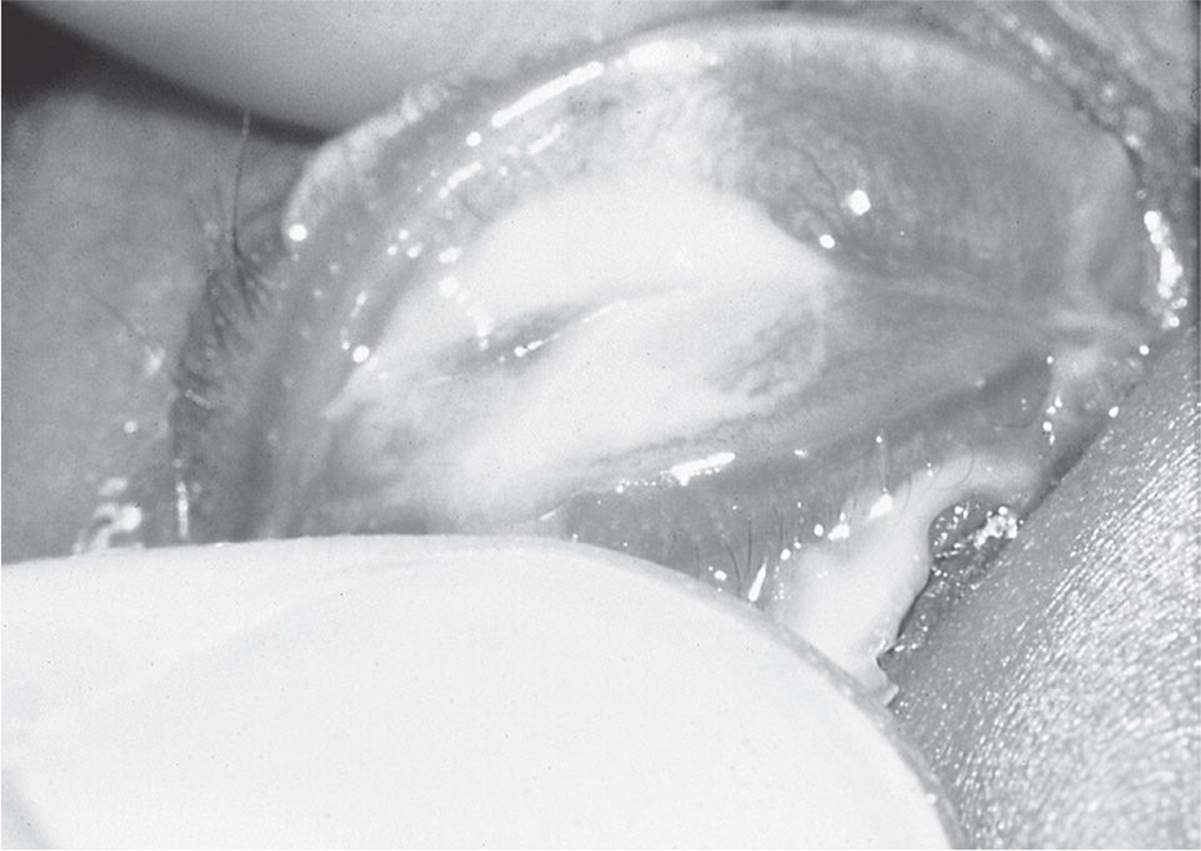
FIGURE 5-1
A) Definitive diagnosis can be made by seeing intracytoplasmic inclusion bodies on Giemsa stain.
B) Herpes simplex is not an important cause in infants under 4 weeks of age.
C) Differential diagnosis includes both viral and bacterial diseases.
D) Initial workup should include particular attention to the corneal epithelium.
5 A newborn infant presents with the condition shown in Figure 5-2. Which one of the following statements is least accurate with regard to the condition in Figure 5-2?

FIGURE 5-2
A) Differential diagnosis includes hemangioma, encephalocele, and dermoid cyst.
B) Patients present with significant retrograde discharge into the eye.
C) Swelling is caused by trapped amniotic fluid.
D) This condition can be associated with respiratory compromise.
6 The left eye of a 2-year-old child is shown in Figure 5-3. The punctum is cannulated, and fluorescein solution is irrigated (Fig. 5-4). Which one of the following statements is false?

FIGURE 5-3

FIGURE 5-4
A) This condition is associated with tear reflux and nasolacrimal duct obstruction.
B) The anomaly involves an abnormality of neural ectoderm.
C) If this condition is infected, topical antibiotics are usually sufficient to temporize the condition.
D) These tracts are lined with epithelium.
7 Which one of the following statements regarding megalocornea is false?
A) This condition is defined as a clear normal-appearing cornea with a diameter measuring greater than 13 mm.
B) This condition is often associated with anterior megalophthalmos, an autosomal dominant disorder.
C) The simple form of megalocornea is usually seen as a bilateral condition.
D) Tearing and IOP are important factors in the workup.
8 Which one of the following statements concerning Figure 5-5 is false?
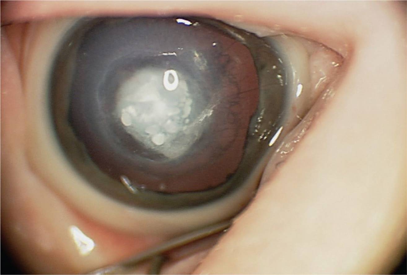
FIGURE 5-5
A) This developmental anomaly can demonstrate both lens and iris adhesions to the corneal endothelium.
B) The condition has progressive corneal opacification.
C) The peripheral cornea is unaffected.
D) This condition results from a developmental problem with neural crest cells.
9 A 3-year-old girl presents with the lesion shown in Figure 5-6. All of the following statements are true except:

FIGURE 5-6
A) This lesion is anterior to Bowman membrane and can be scraped off without concern of stromal involvement.
B) Associated findings include preauricular skin tags, upper eyelid coloboma, and vertebral anomalies.
C) Often, there is an associated arc of lipid in the cornea in advance of the lesion.
D) These lesions may have hair follicles or sweat glands in them.
10 All of the following are true regarding the condition shown in Figure 5-7, except:
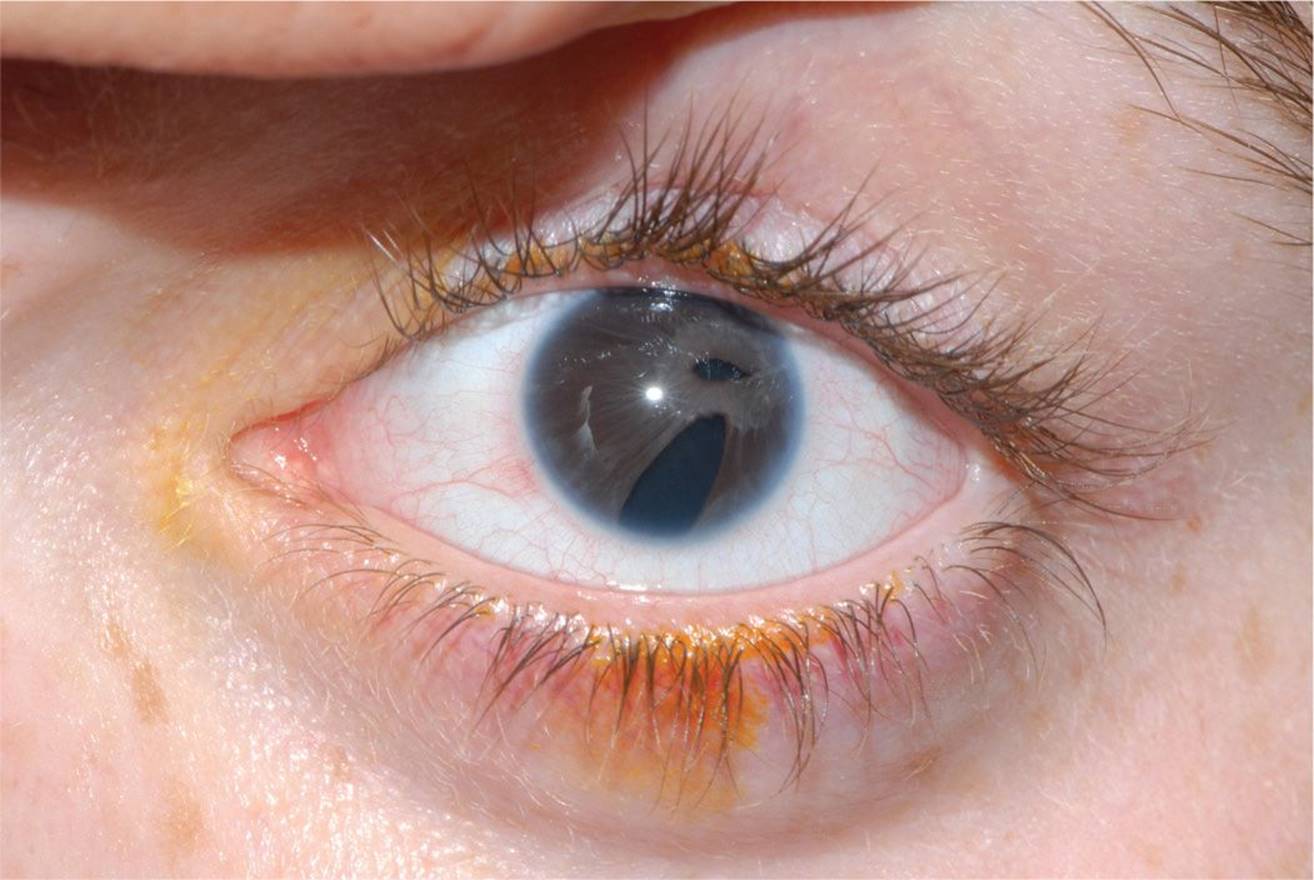
FIGURE 5-7
A) 50% of patients develop glaucoma.
B) Patients with this condition are found to have mutations in PITX2 and FOXC1.
C) Systemic abnormalities include redundant periumbilical skin, hypospadias, and microdontia.
D) Autosomal recessive inheritance is most common.
11 All of the following are causes of heterochromic irides except:
A) Horner syndrome
B) albinism
C) juvenile xanthogranuloma (JXG)
D) Waardenburg–Klein syndrome
12 Which one of the following ocular or systemic condition is not associated with ectopia lentis (Fig. 5-8)?
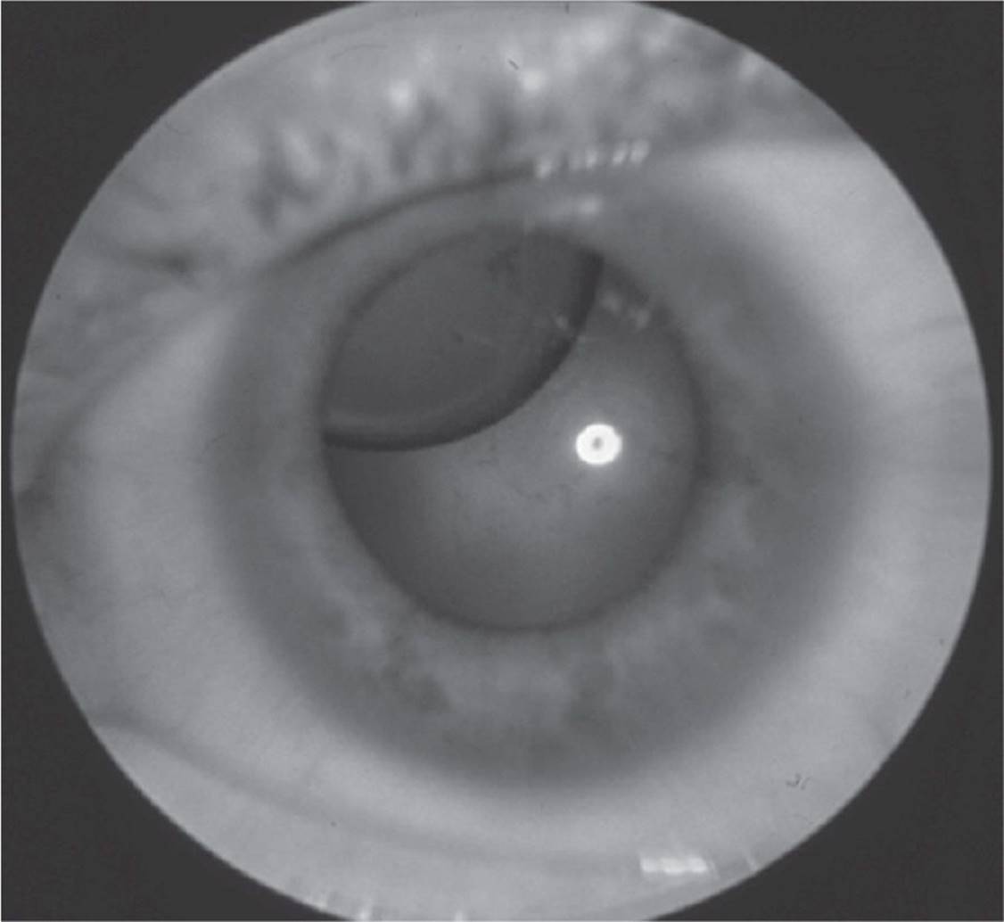
FIGURE 5-8
A) Homocystinuria
B) Aniridia
C) Microcornea
D) Weill–Marchesani
13 A 1-month-old infant presents with bilateral nuclear cataracts (Fig. 5-9). What is the most common identifiable cause for the cataracts?
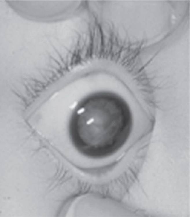
FIGURE 5-9
A) Hereditary autosomal dominant
B) Persistent hyperplastic primary vitreous (PHPV)
C) Galactosemia
D) Intrauterine infection
14 What is the optimum time to operate on a patient with bilateral dense congenital cataracts?
A) As soon as possible, even within the first few weeks of life
B) Between 2 months and 6 months of age
C) Between 6 months and 1 year of age
D) Between 1 and 2 years of age
15 Which one of the following is the preferred treatment for congenital cataracts in the 1-month-old infant with compliant parents?
A) Lensectomy, anterior vitrectomy, and fitting of contact lens
B) Intracapsular cataract extraction with contact lens fitting
C) Aspiration of lens and implantation of posterior chamber IOL
D) Lensectomy, anterior vitrectomy, and fitting with aphakic glasses
16 Each of the following statements regarding the disease depicted in Figure 5-10 is true except:

FIGURE 5-10
A) Photophobia and tearing may be the only presenting signs.
B) Although surgical therapy is usually indicated, medical therapy is often used initially.
C) Gonioscopy has clearly identifiable landmarks facilitating goniotomy as a first-line therapy.
D) Horizontally oriented breaks in Descemet membrane may be found in the buphthalmic eye.
QUESTIONS 17 and 18 A 3-year-old child with newly diagnosed juvenile idiopathic arthritis (JIA) is referred for an initial ophthalmic consultation.
17 The parents are anxious to know the risk of developing uveitis. Which historical information is least helpful in answering their question?
A) Gender of patient
B) Antinuclear antibody (ANA) positivity
C) Number of joints involved
D) Ocular complaints
18 The above patient is ANA-positive and has bilateral knee involvement. How often does she need to undergo ophthalmic evaluation?
A) Every 6 weeks
B) Every 3 months
C) Every 6 months
D) Annually
19 A 3-month-old infant presents with nystagmus and photophobia. Anterior segment and fundus photos are shown in Figures 5-11 and 5-12. All of the following statements regarding this condition are true except:

FIGURE 5-11
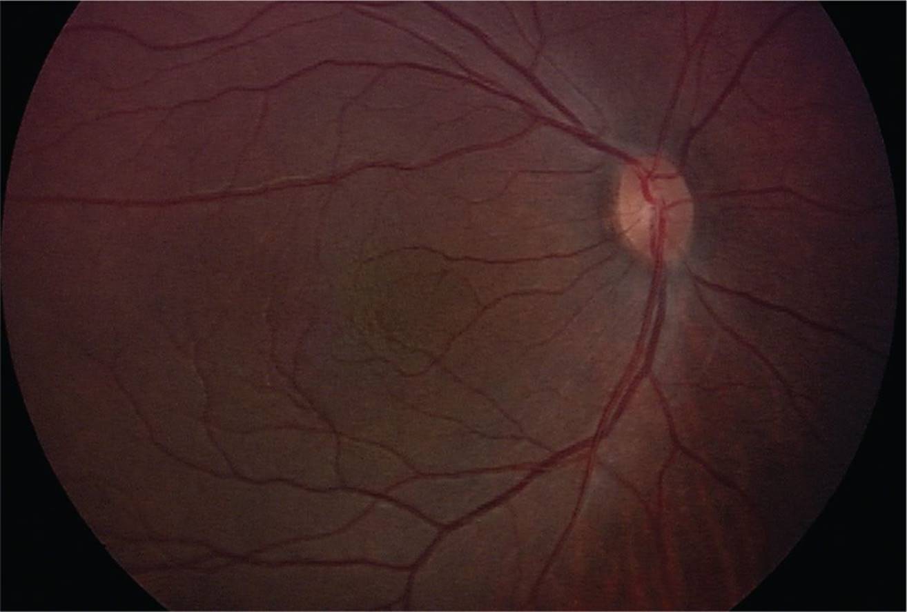
FIGURE 5-12
A) Both familial and sporadic cases have been reported.
B) Visual acuity is typically 20/100 or worse.
C) It is associated with corneal pannus.
D) The familial form of this condition is associated with nephroblastoma.
QUESTIONS 20 and 21 An infant girl is born 4 weeks prematurely and manifests jaundice, an intractable rash, persistent rhinitis, pneumonia, anemia, generalized lymphadenopathy, and bony abnormalities on radiograph.
20 Which one of the following historical features is the most important when questioning the mother regarding the history of the pregnancy?
A) Diet
B) Alcohol or drug use
C) Exposure to environmental toxins
D) Sexual history
21 Which one of the following findings would most likely be present on ophthalmic examination of this newborn child?
A) Interstitial keratitis
B) Segmental pigmentation of the retinal periphery and chorioretinitis
C) Scleritis
D) Anterior uveitis
22 All of the following would be considered in the differential diagnosis of vitreous hemorrhage in an 8-year-old child except:
A) trauma
B) juvenile X-linked retinoschisis
C) pars planitis
D) melanocytoma
23 An 8-year-old boy presents with 20/60 vision and exhibits the macular finding shown in Figure 5-13. Which one of the following is not true of this disorder?

FIGURE 5-13
A) Spoke-wheel configuration of the macula
B) Cleavage of the retina at the nerve fiber layer
C) Attenuated b wave on electroretinogram (ERG)
D) Macular microcysts exhibit classic petalloid leakage on fluorescein angiography.
24 A 4-year-old girl presents for evaluation of poor vision. Ophthalmologic examination reveals translucent irides, hypopigmentation of the fundi, and foveal hypoplasia. Each one of the following may be an associated finding except:
A) bleeding diathesis
B) recurrent sinopulmonary infections
C) oculodigital massage
D) sensory nystagmus
25 Figure 5-14 is representative of a child infected with a nematode that was contracted from a common house pet. Which one of the following statements regarding this process is false?
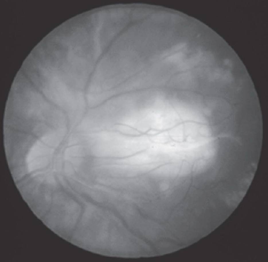
FIGURE 5-14
A) This infection may present with either anterior or posterior segment involvement.
B) The infection often manifests as an eosinophilic granuloma.
C) On CT, calcification is frequently present.
D) This infection can present as an apparent exotropia in which there are no refixation movements on alternate cover testing.
26 An infant is referred for poor tracking. On examination, the child does not fix or follow, there is searching nystagmus. The rest of the ocular exam is otherwise normal. Which one of the following is least likely in the differential?
A) Achromatopsia
B) Congenital stationary night blindness
C) Leber congenital amaurosis
D) Ocular albinism
27 Which one of the following statements regarding ocular toxoplasmosis is false?
A) Less than 10% of individuals younger than 5 years of age demonstrate antibodies against toxoplasmosis.
B) Toxoplasma oocysts shed by cats in their feces may remain infective for up to 1 year.
C) Maternal infection earlier in the course of pregnancy results in a greater risk of infection to the fetus.
D) Retinochoroiditis typically involves the retinal periphery and infrequently involves the macula.
28 According to the findings from the Early Treatment for Retinopathy of Prematurity Study (ET-ROP), laser therapy should be initiated for patients with which type of ROP?
A) Five contiguous clock hours of Zone II, Stage 3
B) Zone I, Stage 2
C) Zone I, Stage 3 with Plus disease
D) Zone III, Stage 1
29 An infant born prematurely at 27 weeks gestational age with a birth weight of 690 g demonstrates ROP as shown in Figure 5-15. The most appropriate next step would be:
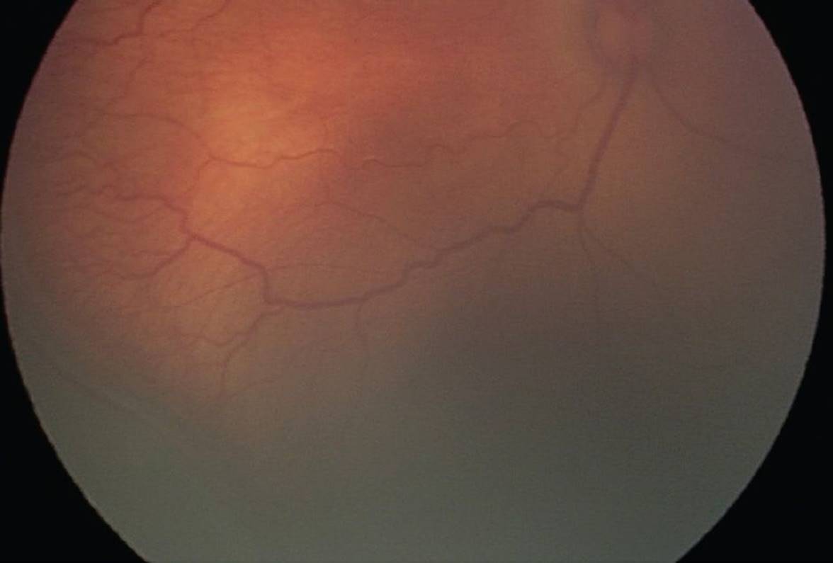
FIGURE 5-15
A) intravitreal bevacizumab within 72 hours
B) laser therapy within 72 hours
C) cryotherapy within 72 hours
D) close observation with repeat examination within 72 hours
30 Potential late complications of ROP include all of the following except:
A) strabismus
B) myopia
C) negative-angle-κ
D) glaucoma
QUESTIONS 31 and 32 A 11-month-old male presents with the examination findings shown in Figure 5-16 after an unwitnessed fall.
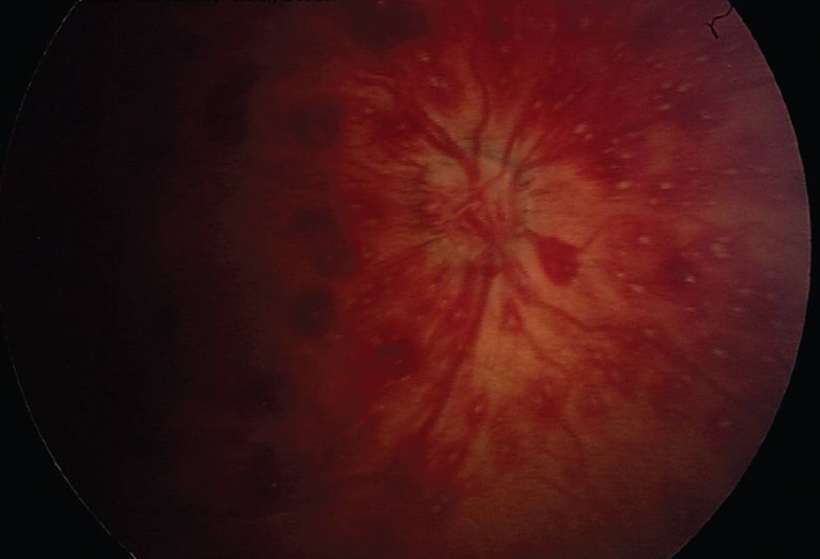
FIGURE 5-16
31 All of the following statements are true regarding this condition except:
A) Ocular findings are the presenting signs in 5% of child abuse cases.
B) Children affected are usually under 3 years of age.
C) Radiographic findings often include bilateral subdural hematomas.
D) Ocular findings typically resolve without long-term vision problems.
32 The next appropriate steps in management would include all of the following except:
A) contact child protective services to investigate for possible nonaccidental trauma
B) laboratory evaluation for coagulopathy
C) skeletal survey to evaluate for other signs of abuse
D) patching the contralateral eye for amblyopia management
33 Which one of the following statements regarding Coats disease is true?
A) It has an autosomal dominant pattern of inheritance with variable penetrance.
B) It is usually bilateral.
C) Males are affected more frequently than females.
D) It is usually diagnosed before 2 years of age.
34 A 5-year-old boy presents for evaluation of decreased vision. Ophthalmologic examination reveals degenerative changes involving the vitreous and retina and an optically empty vitreous. Which one of the following is the least likely diagnosis?
A) Wagner dystrophy
B) Stickler syndrome
C) Goldmann–Favre dystrophy
D) Kearns–Sayre syndrome
35 Which one of the following statements regarding retinitis pigmentosa is true?
A) The X-linked form is least common but most disabling.
B) Signs and symptoms typically precede ERG abnormalities.
C) Retinal pigmentary changes in the midperiphery are always present.
D) The initial visual field defect is a ring scotoma.
36 A 10-year-old girl is referred for an ophthalmologic examination after failing her school eye examination. Her best-corrected visual acuity is 20/50 in her right eye and 20/40 in her left. Examination reveals a normal anterior segment with an abnormal fundus appearance bilaterally. Her left eye is shown in Figure 5-17. Which one of the following would be most useful in establishing the diagnosis?

FIGURE 5-17
A) Electrooculogram (EOG)
B) ERG
C) Visually evoked cortical potential
D) Fluorescein angiogram
37 A 10-year-old boy is referred for evaluation of decreased vision in his left eye. Examination reveals an uncorrected visual acuity of 20/20 in his right eye and 20/40 in his left, which is correctable to 20/20 with glasses. Examination of the left fundus reveals a yellow-orange cystic macular lesion (Fig. 5-18). The father of this child reports that several members of his family including himself and a sister have suffered from mild to moderate deterioration of vision since youth. Which one of the following would be most helpful in establishing the diagnosis?
FIGURE 5-18
A) EOG
B) Ultrasound
C) Visual-evoked cortical potential
D) Fluorescein angiogram
QUESTIONS 38–42 Optic nerve disorders (Figs. 5-19 to 5-24).
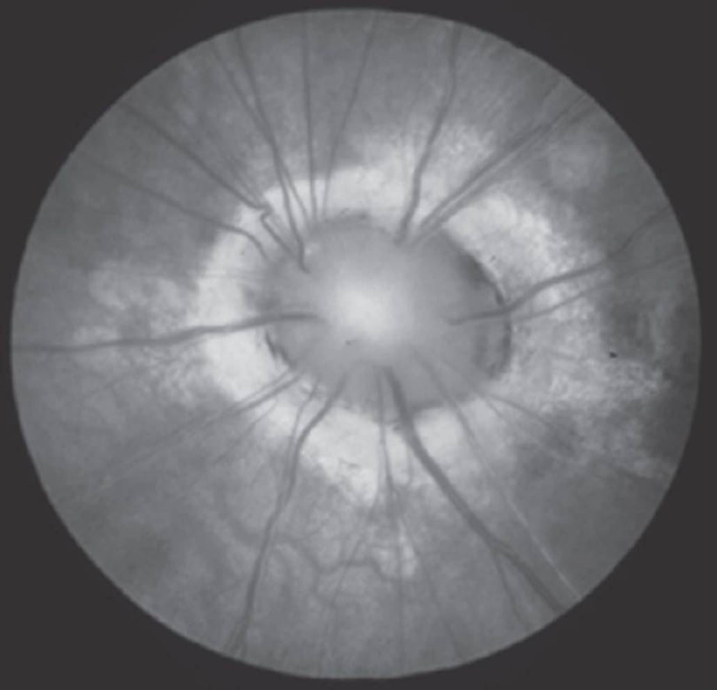
FIGURE 5-19
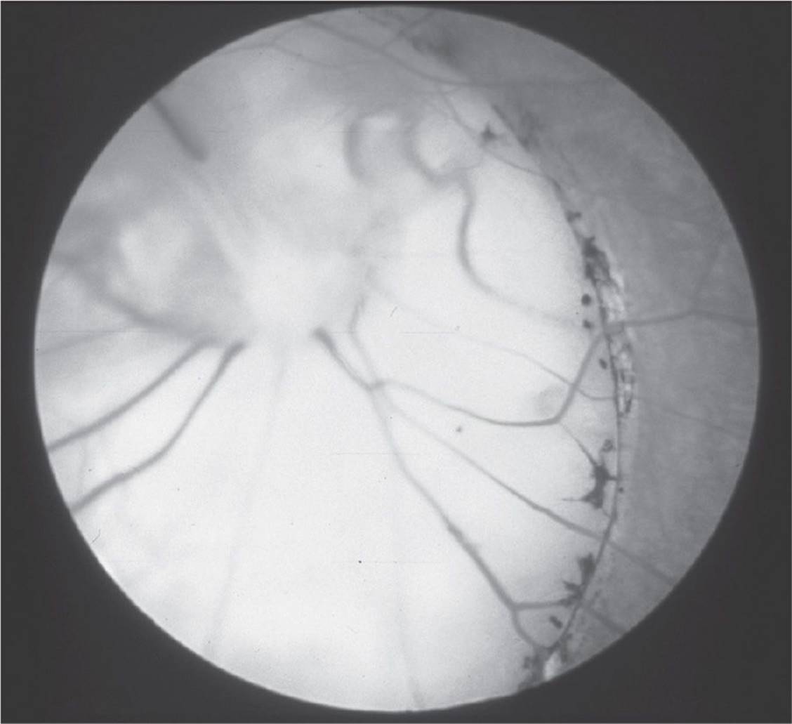
FIGURE 5-20

FIGURE 5-21

FIGURE 5-22
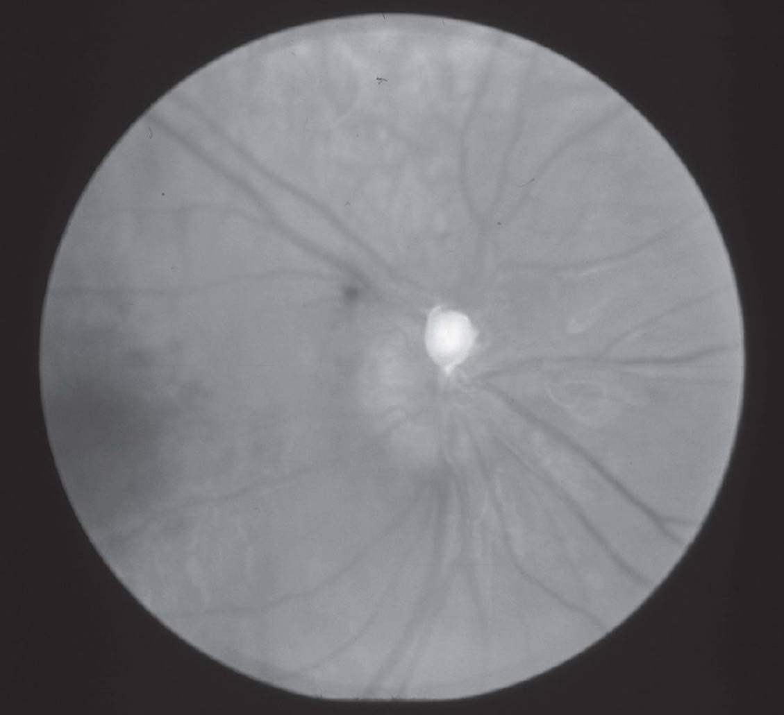
FIGURE 5-23

FIGURE 5-24
38 Which figure is associated with glial proliferation and folding of the retina?
A) Figure 5-19
B) Figure 5-20
C) Figure 5-22
D) Figure 5-23
39 Which figure is a result of faulty closure of the fetal fissure?
A) Figure 5-20
B) Figure 5-22
C) Figure 5-23
D) Figure 5-24
40 Which condition requires an endocrine evaluation and neuroimaging studies?
A) Figure 5-20
B) Figure 5-21
C) Figure 5-22
D) Figure 5-24
41 Which patient is at risk for developing serous macular detachments?
A) Figure 5-19
B) Figure 5-21
C) Figure 5-22
D) Figure 5-23
42 Which condition is found in conjunction with the posterior lens opacity shown in Figure 5-25?
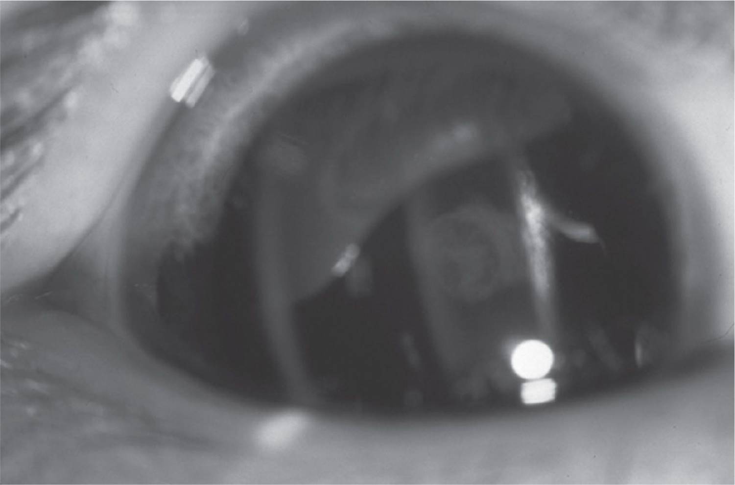
FIGURE 5-25
A) Figure 5-19
B) Figure 5-20
C) Figure 5-23
D) Figure 5-24
43 A 10-year-old asymptomatic male presents with the fundus finding seen in Figure 5-26. His vision, confrontational visual fields, color vision, pupils, motility, and anterior segment examination are within normal limits. What is the most appropriate next step in management?
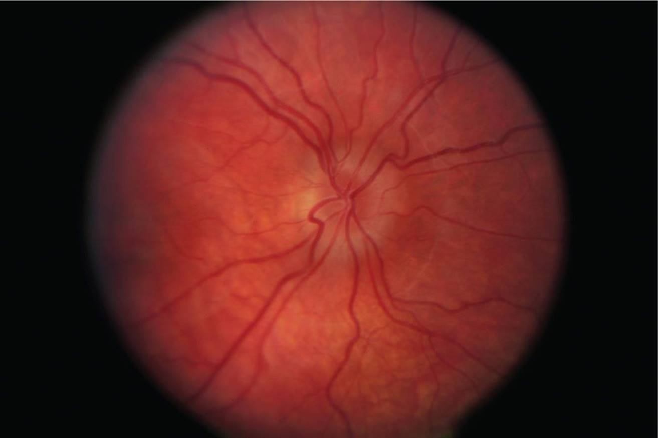
FIGURE 5-26
A) B-scan ultrasonography
B) Neuroimaging
C) Lumbar puncture
D) Ask if there is history of recent weight gain
44 All of the following are true regarding retinoblastoma (Rb), except:
A) Incidence is approximately 1 in 15,000 live births.
B) Presenting symptoms may include leukocoria, strabismus, cataract, and glaucoma.
C) Initial evaluation includes CT, lumbar puncture, and bone marrow biopsy.
D) Bilateral cases will typically present at <6 months and unilateral cases present at >12 months.
45 A 12-month-old male presents with the finding shown in Figure 5-27. All of the following statements are true regarding the inheritance of this condition, except:
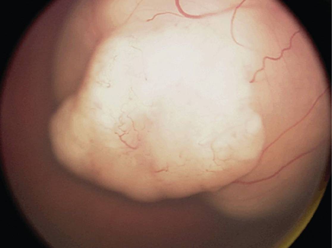
FIGURE 5-27 From Stocker JT, Dehner, LP, Husain AN. Stocker & Dehner’s Pediatric Pathology, 3rd ed. Philadelphia: Lippincott Williams & Wilkins, 2011.
A) 90% of newly diagnosed cases do not have a family history of this condition.
B) If the patient has a germline Rb1 mutation, there is a 50% chance the condition is bilateral.
C) If the patient has unilateral disease, he has a 15% chance of transmitting the condition to his offspring.
D) If the parent has a germline Rb1 mutation, there is an 85% chance the condition is bilateral.
46 The patient in Figure 5-28 has:
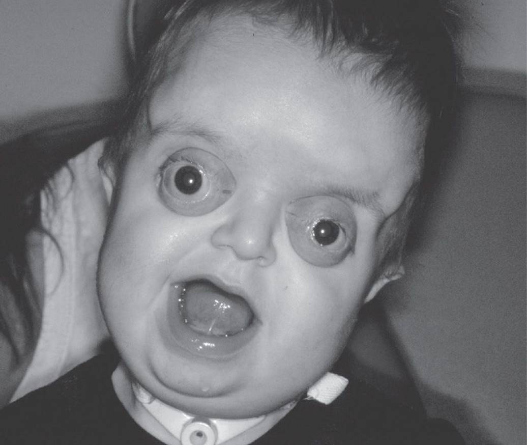
FIGURE 5-28
A) craniosynostosis—this patient would likely demonstrate midfacial hypoplasia, V-pattern exotropia, proptosis, and telecanthus.
B) Pierre Robin sequence—this patient would likely demonstrate micrognathia, glossoptosis, and cleft palate.
C) mandibulofacial dysostosis—this patient would likely demonstrate microstomia, coloboma, and malar and mandibular hypoplasia.
D) fetal alcohol syndrome—this patient would likely demonstrate an antimongoloid slant, deficiency of meibomian glands in the lower lid, and absent lower lid puncta.
47 A 9-month-old male presents with the finding seen in Figure 5-29. All of the following are true except:

FIGURE 5-29
A) significant risk of amblyopia
B) most commonly seen in the superotemporal quadrant
C) may contain hair follicles, sweat glands, and sebaceous glands
D) differential diagnosis includes rhabdomyosarcoma and orbital cellulitis
48 An infant presents with a hyphema with no history of trauma. Which one of the following is the least likely cause?
A) JXG
B) Herpes simplex uveitis
C) Lymphoma
D) Rb
49 A 2-year-old boy presents with periorbital ecchymosis. The differential diagnosis includes all of the following except:
A) neuroblastoma
B) leukemia
C) lymphangioma
D) dermoid cyst
50 Neuroblastoma is characterized by all of the following except:
A) metastasis from the adrenal gland
B) possible spontaneous regression
C) poor prognosis if diagnosed before 1 year of age
D) periorbital ecchymosis
51 A 15-year-old male presents with the finding shown in Figure 5-30. Additional exam findings would include:

FIGURE 5-30
A) hyperpigmented macules
B) adenoma sebacuem
C) port-wine stain
D) conjunctiva telangiectasia
52 Which one of the following statements regarding neurofibromatosis is false?
A) Café-au-lait spots appear in over 99% of patients with this disorder.
B) Tumors of the CNS, including optic nerve gliomas, astrocytomas, acoustic neuromas, meningiomas, and neurofibromas, occur in 5% to 10% of patients with neurofibromatosis.
C) Lisch nodules appear in over 90% of patients over the age of 6 years but are nondiagnostic because they may appear in normal patients as well.
D) Up to 50% of patients with plexiform neurofibromas involving the upper eyelid develop ipsilateral glaucoma.
QUESTIONS 53 and 54 A 2-month-old female presents with the lesion shown in Figure 5-31.

FIGURE 5-31
53 Characteristics of this lesion include all of the following, except:
A) clinically evident at birth
B) rapid proliferation and growth during first year of life
C) risk of anisometropic amblyopia
D) association with posterior fossa malformations
54 On examination, the patient has no fixation preference. Refractive error is +3.00 OU. Which treatment option is most appropriate for this patient?
A) Topical or systemic β-blocker
B) Corticosteroids
C) Refractive correction
D) Observation
55 The correct order of normal fusional amplitudes is:
A) convergence > divergence > vertical
B) convergence > divergence = vertical
C) vertical > divergence > convergence
D) convergence = divergence > vertical
56 All of the following can be used for to test for anomalous retinal correspondence (ARC) except:
A) red-glass test
B) afterimage test
C) titmus fly test
D) amblyoscope
57 A 75-year-old female presents with a stroke in the area of the inferior division of cranial nerve III. Which extraocular movement would not be affected?
A) Adduction
B) Elevation
C) Depression
D) Excyclotorsion
58 All of the statements regarding a pediatric ophthalmology evaluation are true except:
A) A child demonstrates eccentric fixation. This indicates poor vision, usually 20/200 or worse.
B) The cover–uncover test is used to identify a tropia.
C) The deviation measured with the alternate cover test is the phoria plus the tropia.
D) Temporal displacement of the corneal light reflex that does not shift during cover–uncover or alternate cover testing represents a positive-angle-κ.
59 The child in Figure 5-32 was brought in by her observant mother for evaluation of crossed eyes, which the mother has noticed for the past month. Which one of the following statements is least likely?

FIGURE 5-32
A) The cover–uncover test will demonstrate a tropia; when the fixing eye is covered, the other eye will display an abduction movement.
B) Both the cover–uncover test and the alternate cover test, even with excellent fixation on an accommodative target, will not demonstrate any deviation.
C) This child is unlikely to demonstrate inferior oblique overaction (IOOA) or dissociated vertical deviation (DVD). These findings generally develop after initial presentation of esotropia.
D) With time, this condition will spontaneously improve.
60 Which one of the following statements regarding optokinetic nystagmus (OKN) is false?
A) The slow phase occurs in the same direction as that in which the repetitive visual stimulus is moved.
B) The frontal lobes control the slow pursuit movement.
C) In patients with congenital motor nystagmus, a reversal of the OKN response can occur.
D) An OKN response elicited in an infant indicates that some visual input is present.
61 A 32-year-old woman presents with insidious onset of diplopia. On alternate cover testing, the patient has a right hypertropia, worse on right head tilt and left gaze. A palsy of which muscle might cause her symptoms?
A) Right superior oblique
B) Left superior rectus
C) Left inferior oblique
D) Right inferior rectus
62 Each of the following examination findings suggest a bilateral superior oblique palsy except:
A) excyclotorsion of greater than 10°
B) V-pattern exotropia
C) reversal of hypertropia in at least one position of gaze
D) absence of head tilt
63 A 3-year-old child with esotropia since birth has the deviation shown in Figure 5-33. The incomitance of this deviation is most likely secondary to a muscle that:

FIGURE 5-33
A) passes between the sclera and a rectus muscle
B) elevates, intorts, and adducts
C) passes below sclera and above an adjacent muscle
D) has its insertion near the macula
QUESTIONS 64 and 65
64 A 2-month-old boy (Fig. 5-34) with large-angle esotropia and apparent cross-fixation presents to your office. The least likely diagnosis on your differential is:
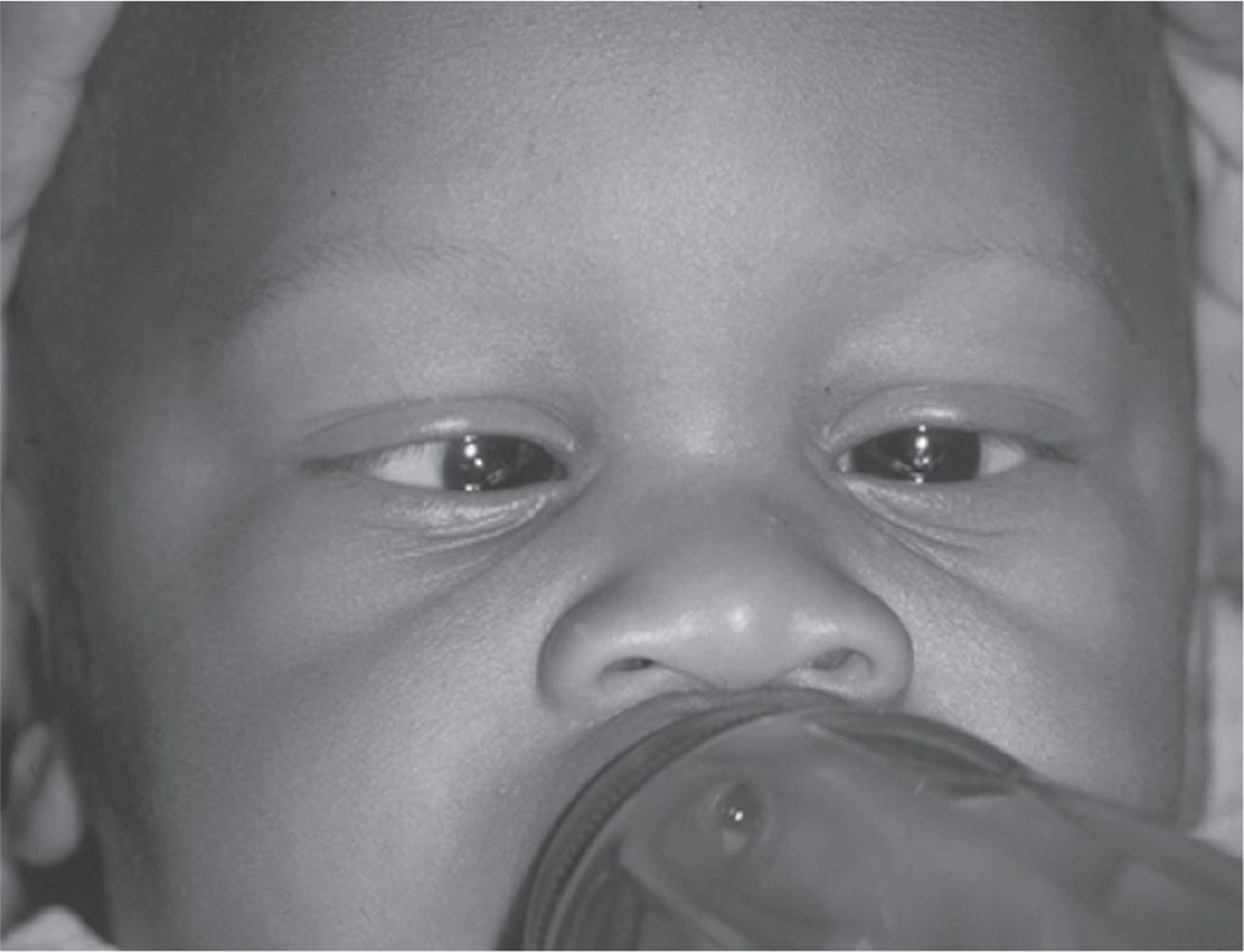
FIGURE 5-34
A) congenital esotropia
B) Möbius syndrome
C) dense amblyopia
D) congenital fibrosis syndrome
65 The patient has full abduction of both eyes and fixates well with each eye. His cycloplegic refraction is +2.00 sphere in each eye. The next appropriate step in management would be:
A) dispense +2.00 glasses
B) alternate patching for tight medial recti
C) patching or atropine for amblyopia
D) bilateral medial rectus recessions
66 Which of the following would you not expect to find in a patient with monofixation syndrome?
A) Eight prism diopter exotropia
B) Amblyopia
C) Fusion on Worth 4-dot testing at 10 feet
D) No movement when a 4Δ base-out prism is placed before the nonfixating eye
67 Each of the following is true regarding accommodative esotropia except:
A) always high AC/A ratio
B) usually intermittent at onset and becoming constant
C) amblyopia is very common (>95%)
D) rarely develop diplopia
68 Bifocals are appropriate for a patient with which one of the following measurements?
A) Dcc ET25 wearing the full distance correction of +3.00 D OU
Ncc ET40 wearing the full distance correction of +3.00 D OU
B) Dcc ortho wearing full distance correction of +1.00 D OU
Ncc ET15 wearing full distance correction of +1.00 D OU
C) Dsc ET20 first time visit, cycloplegic retinoscopy +3.00 D OU
D) Dcc ET10 wearing full cycloplegic correction of -2.50 D OU
Ncc ET13 wearing full cycloplegic correction of -2.50 D OU
69 Which is most consistent with accommodative esotropia?
A) DVD
B) Acquired onset from infancy to 4 years of age
C) Poor to no binocular potential
D) Cycloplegic refraction of +8.00 D
70 A 2-year-old child presents with intermittent exotropia. All of the following would most likely describe her condition except:
A) suppression
B) excellent stereopsis
C) large convergence amplitudes
D) amblyopia
71 An 8-year-old girl returns 1 day after bilateral lateral rectus recessions for intermittent exotropia. She measures 8 PD of consecutive esotropia and has diplopia. The parents are concerned. Your best course of action is:
A) begin convergence exercises
B) observe the child; tell the parents you are satisfied because this is the desired result postoperative day 1
C) prescribe prism glasses to maintain fusion
D) suggest that reoperation may be necessary
72 A 3-year-old boy measures XT35 distance and X(T)’15 at near fixation. How would this deviation be characterized?
A) True divergence excess
B) Pseudo-divergence excess
C) Basic exotropia
D) Cannot determine from the information provided
73 Which one of the following indications is the weakest for X(T) surgery?
A) Increasing degree of exodeviation in the tropia phase
B) Increasing ease of dissociation
C) Poor recovery of fusion once tropic
D) A deviation of greater than 15 PD
74 A 22-year-old emmetropic patient has difficulty reading. On alternate cover testing, she is orthophoric at distance and has an exodeviation of 15 PD at near. The best treatment option would be:
A) orthoptic therapy with a base-out prism or pencil push-up exercises
B) +2.00 D reading glasses
C) bilateral medial rectus recessions
D) unilateral recess-resect procedure
75 Which one of the following best illustrates an exception to Sherrington’s law?
A) DVD
B) Duane’s syndrome type I
C) Convergence
D) Alternating esotropia
QUESTIONS 76–79 A 5-year-old girl is referred by her pediatrician for evaluation of esotropia. Ophthalmologic examination reveals esotropia in the primary position with markedly limited abduction of the left eye, although there is minimal restriction of adduction of the left eye and lid fissure narrowing on attempted adduction. Right ocular motility and fissure height are normal.
76 This case most likely represents which one of the following syndromes?
A) Duane’s retraction syndrome (DRS) type I
B) DRS type II
C) DRS type III
D) Brown syndrome
77 Electromyography in this patient would most likely reveal which one of the following patterns of electrical activity?
A) Electrical activity of the left lateral rectus muscle both in abduction and adduction
B) Electrical activity of the left lateral rectus muscle only in abduction
C) Electrical activity of both the left medial and left lateral rectus muscles on both abduction and adduction
D) Absence of electrical activity in the left lateral rectus muscle on abduction, with paradoxical activity on adduction
78 Each of the following may be associated with DRS except:
A) thalidomide
B) Marcus Gunn jaw winking
C) Goldenhar syndrome
D) glaucoma
79 Which one of the following statements regarding DRS is true?
A) The incidence of amblyopia is high.
B) A Faden procedure may reduce the upshoot of the affected eye on adduction.
C) The lid fissure narrowing is secondary to abnormal innervation of the levator muscle.
D) The strabismus is comitant.
80 All of the following are characteristics of congenital third nerve palsy except:
A) esodeviation
B) abnormal pupillary function
C) hypotropia
D) ptosis
81 A 4-year-old boy presents with the measurements in Figure 5-35. He is 25 PD exotropic at near. He has 1+ overacting inferior obliques. He has no evidence of amblyopia. The most reasonable surgical approach is:

FIGURE 5-35
A) resection of both medial recti with supraplacement
B) recession of both lateral recti with supraplacement
C) recession of both lateral recti with inferior oblique weakening
D) recess-resect with supraplacement of the lateral rectus and infraplacement of the medial rectus
82 Which one of the following statements is true of DVD?
A) It is rare in patients with congenital esotropia.
B) It is usually a unilateral condition.
C) The deviated eye extorts as it elevates.
D) It obeys Hering’s law.
QUESTIONS 83–86
83 A 6-year-old boy presents with an exodeviation of 30 PD at distance and 10 PD at near on alternate cover testing. What is the next most appropriate step?
A) Recess the lateral recti for 30 PD.
B) Occlude one eye for 30 minutes and remeasure the deviation.
C) Dispense glasses prescription with bifocal.
D) Recess the lateral recti for an amount intermediate between the distance and near deviation.
84 A patch test is performed on this patient, and he now measures 30 PD at distance and 15 PD at near. What is the next most appropriate step?
A) Recess the lateral recti for 30 PD.
B) Remeasure the deviation with a +3.00 D add OU.
C) Recess the lateral recti for an amount intermediate between the distance and near deviation.
D) Dispense glasses prescription with bifocal.
85 This 6-year-old patient then undergoes a bilateral lateral rectus recession. One week later, he measures 15 PD consecutive esotropia. The child is complaining of diplopia. What is the next most appropriate step?
A) Observe and have the patient return in 2 weeks.
B) Start full-time patch OD.
C) Start penalization OD.
D) Start alternate patching.
86 At 3 weeks postoperative, the child is still 15 PD esotropic. What is the next most appropriate step?
A) Operate for 15 PD esotropia.
B) Prescribe enough base-out prism to fully neutralize the esotropia.
C) Prescribe enough base-out prism to alleviate the diplopia but leave a small residual esophoria.
D) Prescribe a miotic.
87 A 6-year-old child with 30 PD of intermittent exotropia and 40 seconds arc stereo acuity exhibits an A pattern with superior oblique overaction and a small right hyperphoria in primary gaze, a left hyperphoria in left gaze, and a larger right hyperphoria in right gaze. The right hyperphoria significantly increases on downgaze. What would be the most reasonable surgical approach?
A) Lateral rectus recessions plus superior oblique tenotomies
B) Lateral rectus recessions plus infraplace the lateral recti
C) Lateral rectus recessions plus supraplace the lateral recti
D) Lateral rectus recessions plus superior oblique tuck
88 A 5-year-old girl presents with the ocular motility pattern shown in Figure 5-36. All of the following are true of this condition except:

FIGURE 5-36 From Tasman W, Jaeger EA, Augsburger JJ, et al. Wills Eye Hospital Atlas of Clinical Ophthalmology. Philadelphia: Lippincott Williams & Wilkins, 2002.
A) an inelastic superior oblique muscle tendon complex
B) down shoot in adduction
C) superior oblique overaction with an A pattern is common
D) forced ductions in this case will be positive
89 An 8-year-old boy presents with the ocular motility shown in Figure 5-37. Which one of the following is not true of this condition?

FIGURE 5-37
A) Patients with this condition have normal vertical fusion amplitudes.
B) This condition often has IOOA.
C) The patient has a compensatory head tilt away from the pathology.
D) Early onset of this condition can result in facial asymmetry.
90 A 36-year-old female presents for evaluation of bilateral elevation deficit. All of the following would suggest a diagnosis of chronic progressive external ophthalmoplegia (CPEO), except:
A) diplopia
B) history of ptosis surgery
C) abnormal ERG
D) cerebellar dysfunction
91 Which one of the following statements concerning botulinum toxin (Botox) is false?
A) The effect of Botox lasts clinically for 3 months in extraocular muscles.
B) Botox interferes with cholinergic receptors preventing release of acetylcholine (ACh).
C) Botox has been proven to be effective as the primary treatment of most types of strabismus.
D) Side effects include ptosis, diplopia, and, rarely, globe perforation.
92 Botox injections have been found to be the treatment of choice in which of the following cases?
A) Nonresolving cranial nerve VI palsy
B) Moderate or large-angle strabismus
C) Blepharospasm
D) Brown syndrome
93 Latent nystagmus is characterized by all of the following statements except:
A) null point in adduction
B) fast phase to the fixing eye
C) increases with monocular occlusion
D) is often associated with intermittent exotropia
QUESTIONS 94 and 95 A 2-year-old girl is referred by her pediatrician for evaluation of a residual small-angle esotropia and face turn after strabismus surgery for infantile estropia. Examination reveals intermittent esotropia 15 PD with the fixing eye in adduction even when one eye is occluded. A horizontal nystagmus is present with the fast phase to the fixing eye and it increases when the fixing eye is abducted. Cycloplegic refraction shows +3.00 OU.
94 The most likely diagnosis is:
A) congenital nystagmus
B) spasmus nutans
C) latent nystagmus with face turn to place the fixing eye in the null point
D) dissociated nystagmus
95 The most appropriate treatment for the patient in Question 93 is:
A) Botox injection of both medial rectus muscles
B) bilateral medial rectus recessions
C) prescribe full hyperopic correction +3.00 OU
D) convergence exercises
96 A 6-year-old female fails a school vision screen. Her vision is 20/200 OD and 20/20 OS. Her anterior and posterior segment examinations are within normal limits. Refraction with 1% cyclopentolate is +5.00 sphere OD and +2.50 sphere OS. What is the most appropriate next step?
A) Perform atropine refraction
B) Initiate amblyopia management with patching or pharmacologic agents
C) Prescribe +3.50 OD and +1.00 OS
D) Neuroimaging to further evaluate decreased vision
97 Each of the following is a characteristic of spasmus nutans except:
A) monocular or dissociated nystagmus, which is rapid and of small amplitude
B) usually disappears within 2 years
C) head bobbing
D) optic atrophy
98 Characteristics of congenital motor nystagmus include each of the following except:
A) Oscillopsia is frequently present.
B) Affected patients typically have visual acuity ranging from 20/20 to 20/70.
C) A null point may exist.
D) The nystagmus does not occur while the patient is asleep.
99 Which one of the following is not a cause of congenital sensory nystagmus?
A) Congenital cataracts
B) Aniridia with foveal hypoplasia
C) Rod monochromatism
D) Arnold–Chiari malformation
100 The parent of a 5-year-old amblyopic patient reports that her son developed acute onset facial flushing, irritability, and vomiting. One pupil also appears dilated. The most likely diagnosis is:
A) atropine toxicity
B) third nerve palsy
C) Horner syndrome
D) traumatic mydriasis
1 B) Patients are abnormal from birth, with seizures and mental retardation.
Homocystinuria is an autosomal recessive inborn error of methionine metabolism. Patients exhibit elevated serum levels of methionine and homocystine. Lens dislocation is bilateral, with 30% occurring in infancy and 80% occurring by age 15. Normal lens zonules have a high concentration of cysteine, and deficiency results in abnormal, brittle zonules. A diet low in methionine and high in cysteine can reduce lens dislocation. Patients are normal at birth and develop seizures, mental retardation, and osteoporosis. The patients are usually tall with light-colored hair.
2 C) It can result from a defect in galactokinase or galactose-1-P uridyl transferase.
Galactosemia is an autosomal recessive inborn error of metabolism affecting the conversion of galactose to glucose caused by a defect in galactokinase, UDPgalactose-4-epimerase, or galactose-1-P uridyl transferase (most common). Excess galactose accumulation in body tissues with subsequent conversion to galactitol leads to the classic oil-droplet cataract, liver dysfunction, and mental deficiency within the first few weeks of life. If left untreated, it is fatal. Three-quarters of patients develop cataracts because the nucleus and deep cortex become increasingly opacified. In some cases, early cataract formation can be reversed with dietary intervention.
3 A) Autosomal dominant inheritance
Lowe syndrome is an X-linked recessive disorder characterized by renal tubular acidosis, bilateral congenital cataracts, glaucoma, mental retardation, muscular hypotonia, and failure to thrive.
4 B) Herpes simplex is not an important cause in infants under 4 weeks of age.
Ophthalmia neonatorum can be the manifestation of several different infections or chemical conjunctivitis. The differential diagnosis includes Chlamydia, Neisseria gonorrhea, herpes simplex, and other bacteria. Chlamydia can be diagnosed with Giemsa stain, which reveals intracytoplasmic inclusion bodies. Although Chlamydia conjunctivitis can be treated with topical erythromycin, the patient must be treated with systemic therapy to prevent an associated Chlamydia pneumonitis. The pneumonia usually has its onset 3 to 13 weeks later.Neisseria gonorrhea is well known for its potential to penetrate an intact corneal epithelium and cause a corneal perforation. Herpes simplex type 2 is an important cause of neonatal conjunctivitis with keratitis with involvement of the corneal epithelium. Untreated neonatal herpes keratitis can cause corneal scarring and dense amblyopia. In infants with immature immune systems, the typical hyperacute purulent conjunctivitis may not be present. Both topical and IV antibiotics are needed for this severe infection. Chemical conjunctivitis caused by topical silver nitrate application has onset within 24 hours of birth, and conjunctival scraping shows few to no polymorphonuclear neutrophils (PMNs).
5 B) Patients present with significant retrograde discharge into the eye.
The photograph demonstrates a dacryocystocele. This uncommon condition presents at birth with swelling below the medial canthal ligament. Differential diagnosis includes hemangioma, encephalocele, and dermoid cyst, which are more commonly seen above the medial canthal ligament. A membranous blockage over the valve of Hasner leads to lacrimal sac distention, which secondarily closes off entry to the sac from the common canaliculus. Retrograde discharge is blocked by the physiologic obstruction. At birth, the dacryocystocele often contains trapped amniotic fluid. Associated nasal mucoceles seen with this condition can result in significant respiratory compromise.
6 B) The anomaly involves an abnormality of neural ectoderm.
Pictured in Figure 5-3 is a congenital lacrimal fistula. Figure 5-4 shows fluorescein exiting from the fistula when the nasolacrimal duct is irrigated. This congenital anomaly represents an accessory epithelial-lined communication between the lacrimal system (usually the common canaliculus or the lacrimal sac) and the skin and is a problem of surface ectoderm not neural ectoderm. If it is associated with nasolacrimal duct obstruction, there is often discharge and reflux through the fistula. In this situation, topical antibiotics, probing of the nasolacrimal duct, and excision of the fistulas are appropriate therapy. There has been no association between this condition and a fistula to the maxillary sinus.
7 B) This condition is often associated with anterior megalophthalmos, an autosomal dominant disorder.
By 2 years of age, the cornea is approximately adult size. Simple megalocornea is defined as both corneas measuring greater than 13 mm in children older than 2 years, and greater than 12 mm in infants. Congenital glaucoma is associated with epiphora, and increased IOP and must be ruled out of the differential diagnosis. The most common type of megalocornea is associated with anterior megalophthalmos, an X-linked recessive disorder.
8 B) The condition has progressive corneal opacification.
Peters anomaly results from a developmental problem of faulty migration of neural crest cells. Neural crest cells usually migrate between the surface ectoderm of the cornea and the separating lens. As a result of faulty separation, both lens and iris may remain adherent to the central cornea, causing a central corneal opacity. Both Descemet membrane and layers of the posterior stroma may be absent. The peripheral cornea is characteristically clear because Descemet membrane and the endothelium are intact. In many cases, the corneal opacity will decrease over time.
9 A) This lesion is anterior to Bowman membrane and can be scraped off without concern of stromal involvement.
Corneal/limbal dermoids are hamartomatous lesions consisting of fibrofatty tissue, surrounded by keratinized epithelium, often containing hair follicles, sebaceous glands, and sweat glands. They are usually located at the inferotemporal limbus, and they are associated with lipid in the corneal stroma surrounding the edge of the lesion. Large dermoids can cause astigmatism and amblyopia. Excision may be difficult because they may involve Bowman layer and corneal stroma and, as a result, cause postoperative scarring, astigmatism, and amblyopia. Some require corneal patch grafts. Corneal dermoids can be found in association with Goldenhar syndrome, which also includes preauricular appendages (Fig. 5-38), aural fistulas, maxillary or mandibular hypoplasia, hemifacial microsomia, vertebral deformities, notching of the upper eyelid, and Duane syndrome.
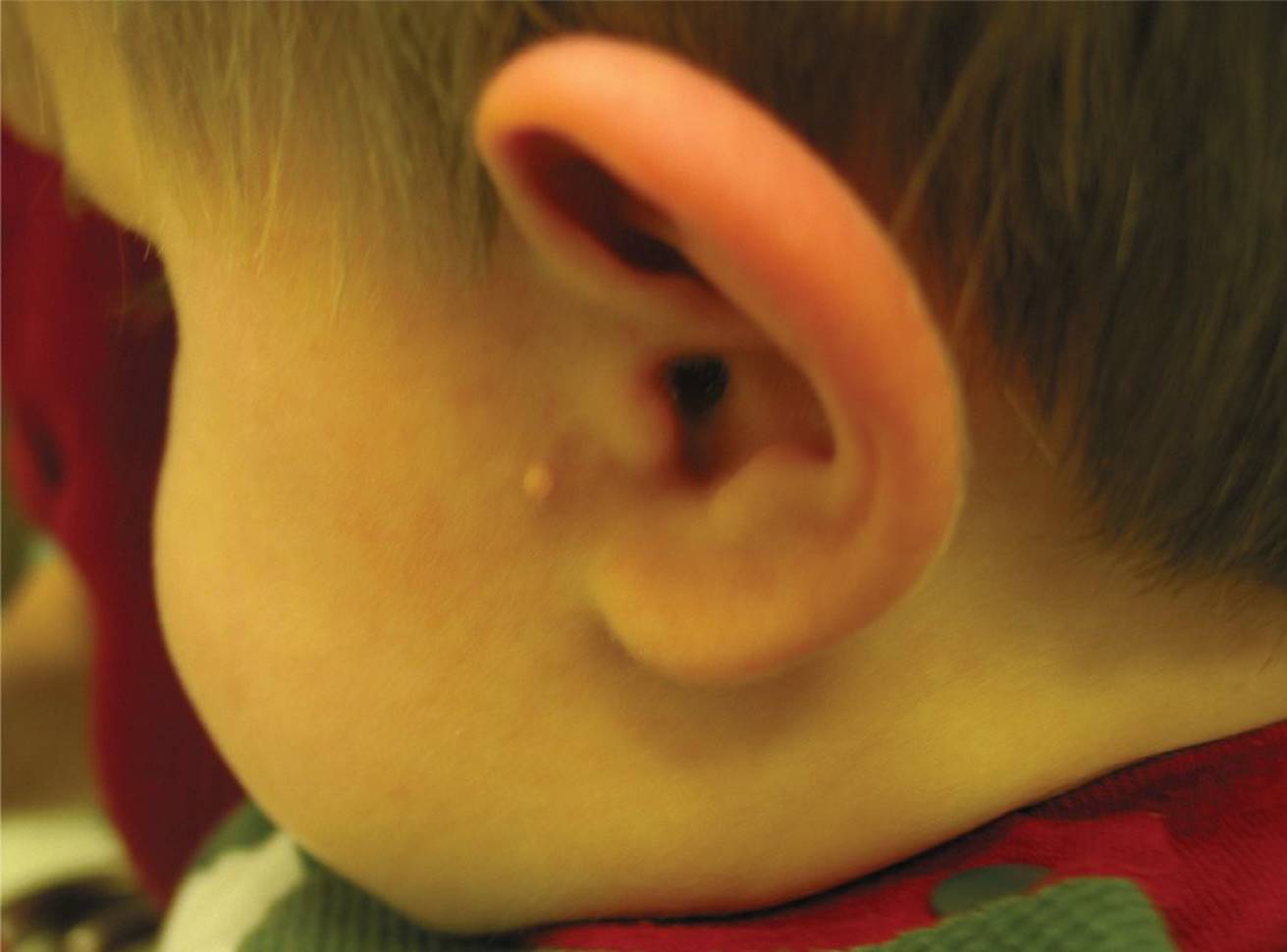
FIGURE 5-38 Dr. Barankin Dermatology Collection.
10 D) Autosomal recessive inheritance is most common.
The patient shown in Figure 5-7 has Axenfeld–Rieger syndrome (ARS), an autosomal dominant condition that can present with a spectrum of ocular and systemic findings. Mutations on the PITX2 and FOXC1 genes have been implicated in ARS. About 50% to 60% of patients will develop glaucoma due to angle dysgenesis. Systemic abnormalities include small teeth, redundant perumbilical skin, hypospadias, and pituitary abnormalities.
11 B) Albinism
JXG is primarily a cutaneous disorder characterized by benign histiocytic proliferation. Iris involvement is seen as richly vascularized orange nodules or as diffusely infiltrative, leading to heterochromia. Congenital Horner’s results in a hypochromic iris on the involved side. Waardenburg syndrome is an autosomal dominant disorder characterized by developmental anomalies of the eyelids, nasal root, and eyebrows along with heterochromia iridis, white forelocks, and sensorineural deafness. Albinism results in bilateral loss of iris pigmentation and therefore does not result in heterochromia.
12 C) Microcornea
Microcornea is the only listed diagnosis not associated with ectopia lentis. Other conditions associated with ectopia lentis are Marfan’s, hyperlysinemia, sulfite oxidase deficiency, trauma, syphilis, and Ehlers–Danlos.
13 A) Hereditary autosomal dominant
When determining the etiology of congenital cataracts, it is best to first determine whether they are unilateral or bilateral. Bilateral cataracts are often inherited in an autosomal dominant fashion. Bilateral disease may also indicate a metabolic or systemic disease such as diabetes mellitus, galactosemia, or Lowe syndrome. In contrast, unilateral congenital cataracts are caused by local dysgenesis and are not inherited. PHPV, anterior polar, and posterior lenticonus are commonly unilateral.
14 A) As soon as possible, even within the first few weeks of life
The critical period of visual development is the first few months of life. During this time, the visual areas of the brain are developing rapidly. Earlier treatment can result in better visual acuity. Bilateral visually significant cataracts can cause irreversible amblyopia and sensory nystagmus.
15 A) Lensectomy, anterior vitrectomy, and fitting of contact lens
There are different views on the “best” treatment of congenital cataracts. Weigert ligament, a connection between the peripheral posterior capsule and the anterior vitreous, is strong, and intracapsular surgery will most likely result in excessive vitreous loss and traction on the retina. IOL implantation has become a preferred method of treatment by many; however, most feel that an IOL should not be implanted in children younger than 2 years of age. This is partly a result of the increase in the size of the anterior segment during the first 2 years of life. Children also have high rates of capsular opacification. Contact lenses are preferred over aphakic glasses as the former reduces aniseikonia and astigmatism.
16 C) Gonioscopy has clearly identifiable landmarks facilitating goniotomy as a first-line therapy.
Congenital glaucoma may present with corneal edema and enlargement, epiphora, and photophobia. Associated horizontal breaks in Descemet membrane, Haab striae, are secondary to increased IOP. Vertical breaks in Descemet’s are often the result of forceps delivery. Congenital glaucoma is bilateral in up to two-thirds of cases. Vision may be poor in the involved eye or eyes, and there may be a secondary myopic shift from globe enlargement. On initial visit, the cornea is often cloudy, obscuring gonioscopy and prohibiting a goniotomy. Carbonic anhydrase inhibitors and β-blockers are used to lower IOP initially, and after the cornea clears, a goniotomy or trabeculotomy may be performed. When gonioscopy is possible, the normal landmarks are poorly recognizable, and some feel that a “Barkan membrane” overlies the angle. This membrane is incised at the time of goniotomy.
17 D) Ocular complaints
The majority of patients with JIA-associated uveitis are asymptomatic, and therefore, the presence or absence of symptoms is of limited predictive value. The patients at highest risk of ocular involvement are females with ANA-positive, pauciarticular (four or fewer joints) JIA.
18 B) Every 3 months
The American Academy of Pediatrics recommends that patients <7 years of age with ANA-positive pauciarticular or polyarticular arthritis of <4 years duration undergo eye examinations every 3 to 4 months. ANA-negative patients undergo examinations every 6 months.
19 D) The familial form of this condition is associated with nephroblastoma.
Figures 5-11 and 5-12 show a patient with aniridia and foveal hypoplasia. Both familial and sporadic cases are seen. Inheritance is autosomal dominant in the familial form. Visual acuity is often poor, usually less than 20/100. Aniridia is a panocular disorder. Ocular manifestations include glaucoma, optic nerve hypoplasia, cataract, pannus, limbal stem cell deficiency, and foveal hypoplasia. The sporadic form of this condition is associated with nephroblastoma (Wilms tumor).
20 D) Sexual history
These are the classic features of an infant with congenital syphilis. Maternal spirochetemia with Treponema pallidum may lead to fetal infection. In cases of maternal primary or secondary syphilis, approximately half of the offspring will be infected. In cases of untreated late syphilis, about 30% of offspring will contract the disease.
21 B) Segmental pigmentation of the retinal periphery and chorioretinitis
Some patients may manifest active chorioretinitis; in most, the only evidence of chorioretinitis is segmental pigmentation of the retinal periphery with a salt-and-pepper appearance to the fundus.
Interstitial keratitis represents an inflammatory response to treponemal antigens and usually presents between 7 and 17 years of age. Anterior uveitis and glaucoma may develop, but these are less common manifestations and are usually not found in the newborn. Hutchinson triad for syphilis includes interstitial keratitis; widely spaced, peg-shaped teeth; and deafness. Other systemic manifestations include saddle nose, saber shins, and rhagades (linear scars often found around the mouth).
22 D) Melanocytoma
Causes of vitreous hemorrhage in children include conditions such as pars planitis, juvenile X-linked retinoschisis, and trauma. Melanocytomas are elevated, deeply pigmented lesions most often found at the optic nerve head. Histologically, they appear as a benign proliferation of melanocytic cells. They are not known to cause vitreous hemorrhage.
23 D) Macular microcysts exhibit classic petalloid leakage on fluorescein angiography.
Juvenile X-linked retinoschisis is characterized by cleavage of the retina at the nerve fiber layer as opposed to senile retinoschisis where cleavage is in the outer plexiform layer. Because the photoreceptors are unaffected, the a wave on the ERG is intact, but both the scotopic and photopic b waves are reduced in proportion to the amount of retinal schisis. The EOG and dark adaptation test are normal or abnormal depending on the stage of disease. The macula is involved early, showing microcysts and radiating retinal folds, but fluorescein angiography exhibits no leakage. Peripheral schisis usually develops later. Vitreous veils and strands form. If a vessel is torn with these veils, vitreous hemorrhage results; this often is how children present. Typically, vision is reduced to the 20/50 to 20/100 level, but expressivity is variable.
24 C) Oculodigital massage
This child has albinism. In addition to iris transillumination defects, patients have decreased fundus pigmentation and may have foveal hypoplasia and, consequently, a sensory nystagmus.
Chediak–Higashi and Hermansky–Pudlak syndromes are potentially lethal forms of albinism, both of which are inherited in an autosomal recessive fashion. Chediak–Higashi syndrome is characterized by neutropenia, lymphocytosis, anemia, and thrombocytopenia. Neutrophils and other lysosome-containing cells characteristically have large granules and have impaired chemotaxis and microbial killing caused by poor fusion of lysosomes to phagosomes. Patients suffer from recurrent infections and are at increased risk of developing lymphoreticular malignancies. Hermansky–Pudlak syndrome is more common in Puerto Ricans, and patients with this syndrome have abnormal platelets and a susceptibility to bleeding and bruising.
Oculodigital massage is characteristic of Leber congenital amaurosis, another disease in the differential of sensory nystagmus but without iris transillumination defects.
25 C) On CT, calcification is frequently present.
Toxocara canis is a nematode larva ingested by children playing in the dirt or from improperly cleaned lettuce or carrots. The typical lesion on pathologic examination is an eosinophilic granuloma. Often mistaken for Rb, Toxocara does not have calcification on CT. PeripheralToxocara lesions can cause traction on the retina, dragging the macula temporally and causing an apparent exotropia. Although Toxocara predominantly affects the eye, it can also infect the lungs and liver.
26 B) Congenital stationary night blindness
When an infant presents with poor vision, searching nystagmus, and an apparently normal examination, conditions to consider include albinism, achromatopsia, and Leber congenital amaurosis. Achromatopsia, or rod monochromatism, is an autosomal recessive disorder with total lack of cones, with color blindness that results in 20/200 vision, photophobia, and nystagmus. The fundus is usually normal in infancy and the EOG is usually normal. In albinism, the signs in infancy are often subtle and may be missed. They would include iris transillumination defects, hypopigmented fundus, and foveal hypoplasia. Congenital stationary night blindness is marked by infantile onset of night blindness but not searching nystagmus. The ERG shows normal to near-normal photopic waveform but nearly nonrecordable scotopic waveform. Other diagnoses include optic nerve hypoplasia and aniridia, but they are usually diagnosed by the ocular exam.
27 D) Retinochoroiditis typically involves the retinal periphery and infrequently involves the macula.
Toxoplasmosis is one of several congenital infections that may cause damage to ocular structures. They are represented by the acronym TORCHS:
TO = toxoplasmosis, R = rubella, C = cytomegalic inclusion disease, H = herpes simplex, and S = syphilis.
Toxoplasmosis is caused by a protozoan with a propensity to infect the retina and other CNS structures. It is highly prevalent in North America. Cats shed oocysts in their feces, which may remain infective for up to 1 year. Ingestion of food contaminated by oocysts or undercooked meat containing tissue oocysts may result in human infection.
Maternal infection early in pregnancy results in a greater risk of transmission to the fetus. The organism may produce a retinochoroiditis, which is usually bilateral and frequently involves the macula. Inactive lesions may reactivate later in life, producing whitish, elevated lesions and a severe vitritis resulting in a “headlight through fog” appearance on ophthalmic examination.
28 C) Zone I, Stage 3 with plus disease
ET-ROP defined both Type 1 and Type 2 (prethreshold) ROP. The results of ET-ROP demonstrated that patients had better outcomes if eyes with Type 1 ROP were treated with laser therapy within 72 hours of diagnosis. Type 1 ROP includes (1) Zone I, any stage disease with concomitant Plus disease; (2) Zone I, Stage 3 ROP without Plus disease; and (3) Zone II, Stage 2 or 3 with Plus disease. Zone II, Stage 3; Zone 1, Stage 2; and Zone III, Stage 1 are all considered prethreshold disease and require careful monitoring.
29 B) Laser therapy within 72 hours
The patient shown in Figure 5-15 has Stage 3 ROP with Plus disease in Zone II. According to ET-ROP, laser therapy should be instituted within 72 hours of diagnosis. This infant meets criteria for treatment. Intravitreal bevacizumab, laser photocoagulation and cryotherapy have all been used in such cases, however, laser treatment is considered the standard of care. According to the BEAT-ROP study, bevacizumab demonstrated a benefit over laser for patients with Stage 3+ disease in Zone I, but statistical equivalence in Zone II. Becacizumab is not FDA approved for the treatment of ROP, however, according to the 2013 Policy Statement “Screening Examinations for Premature Infants with Retinopathy of Prematurity,” bevacizumab may be considered as a treatment option for patients with Stage 3+disease in Zone I. Cryotherapy as a treatment modality has largely been replaced by laser therapy. A repeat exam in 72 hours would not be appropriate in this case as the patient would be at risk for worsening ROP.
30 C) Negative-angle-κ
Children with ROP, either treated or untreated, are at risk for a number of ocular complications, including strabismus, pseudostrabismus, high refractive error, retinal folds, macular dragging, retinal detachment, and amblyopia. Patients with Stage 5 ROP can develop cataracts, glaucoma, and phthisis bulbi. Peripheral anterior synechiae can cause secondary angle closure glaucoma. Patients may demonstrate positive-angle-κ on examination, which is due to temporal dragging of the fovea.
31 D) Ocular findings typically resolve without long-term vision problems.
32 D) Patching the contralateral eye for amblyopia management
The details of this case are highly suspicious for nonaccidental trauma. Patients may present with a vague history of an unwitnessed fall or seizure. Children are usually younger than 3 years, with the highest risk in infants <12 months of age. Radiographic findings include bilateral subdural hematomas. Ocular findings are the initial presentation in approximately 5% of child abuse cases. A multidisciplinary approach, including collaboration with pediatrics and child protective services, should be sought to determine the etiology of the hemorrhages. Seeing blood in multiple layers of the retina, retinoschisis cavities and vitreous hemorrhage are all suggestive of nonaccidental trauma. Findings may be bilateral but asymmetric. It is important to rule out other etiologies, including coagulopathies. While amblyopia is a concern for patients with severe nonclearing vitreous hemorrhage, the first priority is to establish the diagnosis. Careful long-term monitoring is needed as patients often have a poor visual prognosis and reduced vision from optic nerve and retinal damage.
33 C) Males are affected more frequently than females.
Coats disease is a retinal vascular disorder characterized by intraretinal and subretinal leakage of lipid from telangiectatic retinal vessels. It is usually diagnosed between 18 months and 10 years of age, and the most common presenting signs are leukocoria and strabismus. Boys are affected approximately three times more frequently than girls. Coats disease is not hereditary and is unilateral in 90% of cases. Exudative retinal detachments (Fig. 5-39) occur in approximately two-thirds of eyes. Treatment is directed at obliterating leaking abnormal vessels by cryotherapy or photocoagulation. Scleral buckling may be performed on eyes with retinal detachments.

FIGURE 5-39
34 D) Kearns–Sayre syndrome
Wagner vitreoretinal dystrophy is characterized by vitreoretinal degenerative changes, including an optically empty vitreous, high myopia, perivascular lattice degeneration, and retinal detachment. Cataract formation may also be associated with this disorder.
Stickler syndrome describes findings typical of Wagner dystrophy in association with the systemic features of Marfanoid habitus, Pierre Robin anomaly, progressive joint degeneration, arthritis, deafness, and heart defects. It is an autosomal dominant disease with variable expressivity.
Characteristic findings in the autosomal recessively inherited Goldmann–Favre dystrophy include vitreous strands and veils, an optically empty vitreous, foveal and peripheral retinoschisis, attenuated retinal vessels, optic nerve pallor, and cataract formation. Both the ERG and EOG are abnormal, differentiating this condition from juvenile retinoschisis, in which the EOG is normal.
Although a characteristic of Kearns–Sayre syndrome is pigmentary degeneration of the retina, visual function is typically preserved. There is no significant vitreous involvement. Other features of this syndrome include progressive external ophthalmoplegia and, importantly, heart block, which may result in sudden death. Inheritance is via mitochondrial DNA, and onset is before the age of 20 years.
35 A) The X-linked form is least common but most disabling.
The overall incidence of retinitis pigmentosa in the United States is approximately 1:3,500. About half of these cases are sporadic, 22% are autosomal dominant, 16% are autosomal recessive, and 9% are X-linked recessive. The X-linked form is the most rapidly progressive and disabling, whereas the dominant form is least progressive and disabling.
ERG abnormalities precede ophthalmoscopically visible changes and subjective visual complaints. Early ERG evidence includes an increased rod threshold with a normal cone response and a decreased scotopic b wave. Progression of the disease results in a nonrecordable ERG. The EOG is abnormal. The earliest visual field defect is characteristically an inferotemporal scotoma that enlarges to form a ring or annular scotoma.
The typical funduscopic findings include attenuation of the retinal vessels, midperipheral pigmentary changes, and optic disc pallor. Retinitis pigmentosa can occur without pigmentary changes in a form known as retinitis pigmentosa sine pigmento. Associated findings may include myopia, vitreous opacities, posterior subcapsular cataract, retinal pigment epithelial atrophy, cystoid macular edema, glaucoma, and keratoconus.
36 D) Fluorescein angiogram
This is a patient with Stargardt disease, which is thought to be a result of lipofuscin accumulation within the retinal pigment epithelium; this in turn results in a characteristic “silent choroid” effect on fluorescein angiogram in which the lipofuscin blocks underlying choroidal fluorescence. The macula may exhibit a mottled hyperfluorescent appearance. The majority of patients have normal results on electrophysiologic tests, although ERG amplitudes are frequently in the low normal range. When the peripheral retina is predominantly involved with the macula affected to a lesser degree, the term fundus flavimaculatus is used.
37 A) EOG
Best vitelliform dystrophy is an autosomal dominant disorder characterized by an abnormal EOG in carriers, asymptomatic patients with normal-appearing fundi, as well as in affected individuals; and a normal ERG. Both ERG and EOG are needed to confirm this diagnosis.
In the early (pre-vitelliform) stage, the retina may appear normal, although the EOG is abnormal. In the vitelliform stage, a cyst-like yellow-orange lesion develops, typically in the macula, between the ages of 4 and 10 years. It is usually 1 to 5 disc diameters in size and has a “sunny-side-up” egg yolk appearance. Central vision is usually good at this stage. With time, the vitelliform stage evolves into the “scrambled egg” stage, in which the material within the cystic structure becomes granular in appearance. The vision usually remains good at this stage. However, as the disease progresses, atrophy of the macula occurs. Subretinal neovascularization and hemorrhage and serous detachment of the retinal pigment epithelium may occur as well. Most patients see well for many years, but, ultimately, central vision decreases to 20/100 or worse.
38 A) Figure 5-19
Figure 5-19 = morning glory disc
Figure 5-20 = optic nerve coloboma
Figure 5-21 = optic nerve pit
39 A) Figure 5-20
40 C) Figure 5-22
Figure 5-22 = optic nerve hypoplasia
Figure 5-23 = hyaloid remnant
Figure 5-24 = myelinated nerve fibers
41 B) Figure 5-21
42 C) Figure 5-23
Morning glory disc anomaly is characterized by an excavated nerve with overlying glial proliferation. The surrounding retina is often thrown into folds. There is an association of retinal detachment. Most cases are unilateral.
Optic nerve colobomas represent the same developmental problems as chorioretinal and uveal colobomas; specifically, a failure of the fetal fissure to close properly. They are most commonly located inferonasally. The coloboma can involve the macula and result in poor vision.
Optic pits may be considered a small coloboma. Pits usually occur inferotemporally but can be anywhere. They are associated with serous elevation of the macula, which tends to occur during the second or third decade of life.
Optic nerve hypoplasia can be profound with severe visual loss. The hypoplastic nerve often appears pale. When viewed, the surrounding scleral rim often deceivingly gives the impression of a normal nerve. A thin ring of pigment can be seen surrounding the nerve tissue, giving rise to the double ring sign. Strabismus, amblyopia, nystagmus, and an afferent pupillary defect can be associated findings. On occasion, vision in these eyes has been known to improve with amblyopia therapy. De Morsier syndrome is associated with optic nerve hypoplasia, as well as hypothalamic and pituitary dysfunction. The latter can manifest as growth retardation, which can be treated with growth hormone supplementation. These children need to see a pediatric endocrinologist for workup. Optic nerve hypoplasia has many important associations: fetal alcohol syndrome and maternal ingestion of LSD, quinine, and phenytoin. Both macular and optic nerve hypoplasia may be found with aniridia.
Usually, the myelination stops at the lamina cribrosa; however, myelination onto the surface of the retina appears as white, feathery radiating fibers. The myelin can obscure the view of retinal vessels and the macula.
Persistence of remnants of the hyaloid system is a common finding. There is a wide range of findings from a small Bergmeister papilla (fibrous remnant of vasculature on the optic nerve head) to persistent blood-containing hyaloid vessels. The Mittendorf dot seen in Figure 5-25 is a remnant of the hyaloid vessel on the posterior lens capsule.
43 A) B-scan ultrasonography
The patient demonstrates pseudopapilledema. Key features that distinguish this condition from true disc edema include elevation limited to the optic disc, visible vessels at the disc margin without vascular congestion and absence of exudates. Ancillary clinical testing may include B-scan ultrasonography and autofluorescence. Neuroimaging, lumbar puncture, and history of weight gain would all be part of the evaluation of true disc edema.
44 C) Initial evaluation includes CT, lumbar puncture, and bone marrow biopsy.
Rb occurs in 1/15,000 live births with strabismus and leukocoria being the most common presenting signs. Bilateral cases are due to germline hereditary mutations and typically present at a younger age than unilateral Rb. CT is generally avoided as an imaging modality in possible Rb cases to minimize exposure to radiation in these patients, who are at an increased risk of secondary malignancies. Lumbar puncture and bone marrow biopsy are not part of a routine initial evaluation, unless there are other signs/symptoms to suggest systemic involvement.
45 B) If the patient has a germline Rb1 mutation, there is an 50% chance the condition is bilateral.
Rb is an autosomal dominant condition with incomplete penetrance. Over 90% of newly diagnosed cases are new mutations and have no family history. If this patient has a new germline Rb1 mutation, he has an 85% chance of bilateral disease. 15% of unilateral cases are germline and therefore transmissible. If the parent of this child has a germline Rb1 mutation, this patient has an 85% risk of bilateral disease.
46 A) Craniosynostosis—this patient would likely demonstrate midfacial hypoplasia, V-pattern exotropia, proptosis, and telecanthus.
This patient has the characteristic appearance of craniosynostosis, which is caused by a premature closure of the cranial sutures by early childhood. Common findings include midfacial hypoplasia, proptosis, telecanthus, V-pattern exotropia, oral and dental problems, and respiratory problems. This child has Pfeiffer syndrome, an autosomal dominant condition that includes shallow orbits, syndactyly, and short digits.
47 A) Significant risk of amblyopia
The patient shown in Figure 5-29 has a dermoid cyst, which is the most common benign orbital lesion. The cyst is lined with keratinized epithelium and contains dermal appendages, including hair follicles and glands. It is commonly seen along fetal suture lines, especially in the superotemporal quadrant. The lesion typically does not impact vision or cause a risk for amblyopia. Differential diagnosis may include malignant tumors, such as rhabdomyosarcoma, and orbital cellulitis.
48 C) Lymphoma
The hallmark of JXG is spontaneous hyphema from vascularized iris lesions. The lesions represent benign tumors found in the skin, iris, and orbit, which consist of lipid-filled histiocytes and Touton giant cells. Leukemic infiltration of the anterior segment may lead to heterochromia iridis and spontaneous hyphema. When there is deep corneal involvement, severe herpetic uveitis may be accompanied by hyphemas. Other causes of hyphemas in children include trauma, ROP, PHPV, Coats disease, and Rb. Intraocular lymphoma is found in older patients and presents as a uveitis masquerade with primarily posterior segment inflammation.
49 D) Dermoid cyst
Neuroblastoma is the most common source of orbital metastasis in children. Metastatic neuroblastoma produces proptosis with periorbital ecchymosis. Bilateral involvement is seen in half of the cases. Orbital infiltration by leukemia causes lid swelling, proptosis, and ecchymosis. Lymphangioma lesions consist of lymph-filled channels lined by endothelium and separated by thin, delicate walls with small blood vessels that are broken easily. Lymphangiomas infiltrate orbital tissues extensively, and intralesional bleeding is common. Dermoid cysts may rupture and spill their contents, inciting a significant amount of inflammation, but bleeding does not occur.
50 C) Poor prognosis if diagnosed before 1 year of age
Neuroblastoma is the most frequent source of orbital metastasis in children. Metastases occur from the adrenals, mediastinum, and neck. Approximately 20% of all neuroblastoma patients exhibit ocular involvement, which can be the initial manifestation of the tumor. The mean age of presentation in orbital neuroblastoma metastasis is about 2 years. Their prognosis is very poor in general, but prognosis is considerably better in infants under 1 year of age. Spontaneous regression of this tumor may be seen in rare instances.
51 B) Adenoma sebaceum
Figure 5-30 shows an astrocytic hamartoma, commonly seen in tuberous sclerosis (TS). Other manifestations of TS include hypopigmented skin macules, facial angiofibromas (adenoma sebaceum), mental retardation, and seizure disorder. Hyperpigmented skin macules can be seen in incontinentia pigmenti; port-wine stains are associated with Sturge–Weber syndrome, and conjunctival telangiectasias are seen in Louis–Bar syndrome.
52 C) Lisch nodules appear in over 90% of patients over the age of 6 years but are nondiagnostic because they may appear in normal patients as well.
Neurofibromatosis is a progressive disorder with a wide range of clinical manifestations. It occurs in 1 of 3,000 births. Café-au-lait spots appear in over 99% of patients, and if five or more spots greater than 0.5 cm in diameter are observed, the diagnosis is established. Tumors of the CNS occur in 5% to 10% of patients. These patients are at increased risk for other malignancies as well, including neurofibrosarcomas, Wilms tumor, rhabdomyosarcoma, pheochromocytomas, and leukemia.
Lisch nodules (melanocytic hamartomas) (Fig. 5-40) on the iris appear in over 90% of patients over the age of 6 years and are useful for establishing or excluding the diagnosis of neurofibromatosis. They are not found in the normal patient population. Plexiform neurofibroma involving the upper eyelid may produce ptosis and is associated with glaucoma in up to 50% of patients.

FIGURE 5-40 From Schaaf CP, Zschocke J, Potocki L. Human Genetics. Philadelphia: Lippincott Williams & Wilkins, 2012.
53 A) Clinically evident at birth
Figure 5-31 shows a capillary hemangioma. In contrast to a port-wine stain, which is present at birth, capillary hemangiomas usually become apparent during the first month of life. They undergo a rapid proliferative phase, followed by slow regression and evolution. Large eyelid lesions can induce astigmatism and cause a secondary anisometropic amblyopia. PHACE syndrome is a systemic disease associated with capillary hemangiomas in which patients may also have posterior fossa malformations, hemangiomas, arterial anomalies, cardiac defects, and eye anomalies.
54 D) Observation
In cases of small capillary hemangiomas that are not causing occlusion or anisometropic amblyopia, observation is appropriate. Alternative treatment options include topical or systemic β-blockers and corticosteroids. The patient has a moderate amount of hyperopia, which is normal for this age. No glasses are needed at this time.
55 A) Convergence > divergence > vertical
The normal fusional amplitudes at distance and near are greatest for convergence > divergence and least for vertical.
56 C) Titmus fly test
Tests to evaluate for ARC include the red-glass test, afterimage test, and amblyoscope. The Titmus fly test measures stereopsis, which is not present with ARC.
57 B) Elevation
The superior division of cranial nerve III innervates the superior rectus and the levator palpebrae. The inferior division innervates the medial rectus, inferior rectus, and inferior oblique. In this patient with a partial cranial nerve III palsy, elevation would be intact.
58 D) Temporal displacement of the corneal light reflex that does not shift during cover–uncover or alternate cover testing represents a positive-angle-κ.
Positive-angle-κ represents a slight temporal position of the fovea relative to the optical axis (Fig. 5-41). This causes a slight temporal rotation of the globe to keep the image on the fovea. This, in turn, causes the corneal light reflex to be displaced nasally. The deviated light reflex remains stable with cover testing as the fovea never spontaneously moves relative to the optical axis. If simply covering and uncovering one eye reveals a refixation movement in the other eye, a tropia has been diagnosed. The alternate cover test dissociates the two eyes and allows measurement of the tropia plus the phoria. This patient’s macula has been dragged temporally as a result of the scarring secondary to ROP (Fig. 5-42).

FIGURE 5-41 From Chern KC, Saidel MA. Ophthalmology Review Manual. Second Edition. Philadelphia: Lippincott Williams & Wilkins, 2011.

FIGURE 5-42 From Tasman W, Jaeger EA, Augsburger JJ, et al. Wills Eye Hospital Atlas of Clinical Ophthalmology. Philadelphia: Lippincott Williams & Wilkins, 2002.
59 A) The cover–uncover test will demonstrate a tropia; when the fixing eye is covered, the other eye will display an abduction movement.
This child has pseudo-strabismus. A broad nasal bridge with epicanthal folds can sometimes obscure the nasal sclera and simulate esotropia. As the child grows, the nasal bridge will not be as prominent and the folds may disappear. Cover testing will reveal orthophoria. DVD and IOOA develop in association with congenital esotropia in up to 60% to 70% of cases.
60 B) The frontal lobes control the slow pursuit movement.
OKN occurs in response to a repetitive visual stimulus moved across the visual field. The slow phase occurs in the direction of the moving stimulus; the fast phase occurs as a saccadic refixation movement in the opposite direction. The parieto-occipital lobe controls the slow pursuit component, whereas the frontal lobes control the saccadic component. In patients with congenital motor nystagmus, a reversal of the OKN response may occur. An OKN response elicited in an infant indicates that some visual input is present.
61 A) Right superior oblique
The Parks three-step test, listed here, helps to identify a vertical muscle palsy:
1. Determine which eye is hypertropic. This tells you that the involved muscle is one of the two depressors in the hypertropic eye, or one of the two elevators in the other eye. With this first step complete, choices are narrowed from eight muscles down to four muscles as shown in Figure 5-43.
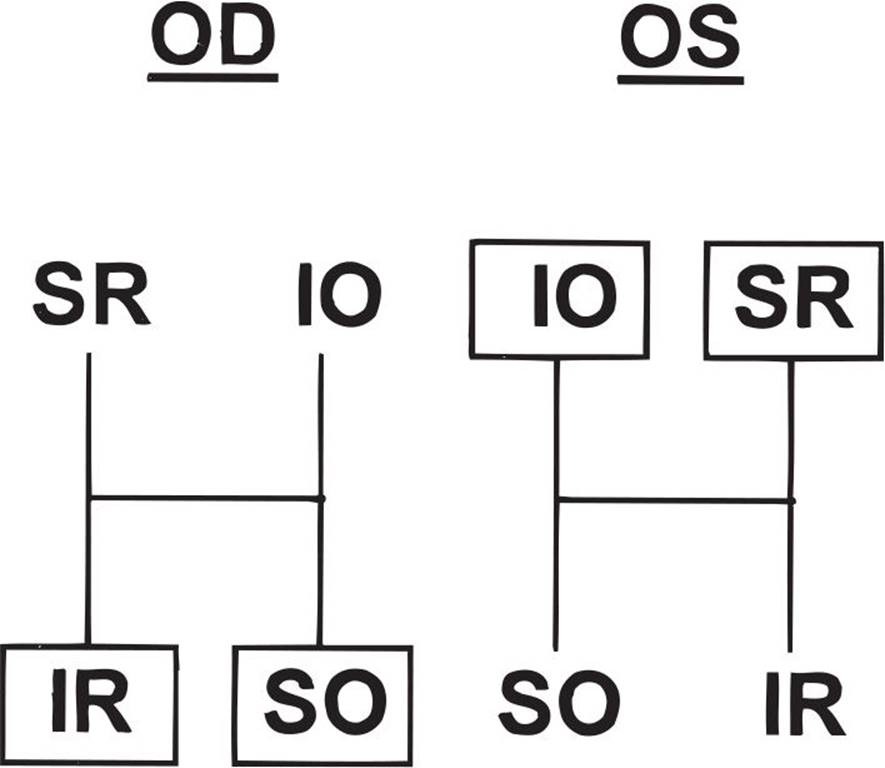
FIGURE 5-43
2. Decide in which gaze the hypertropia is worse. By using the field of action of the four vertically acting muscles, the choices can always be narrowed down to two. In this example, the right hypertropia, which is worse on left gaze, indicates either the right superior oblique (the depressor of the right eye in left gaze) or the left superior rectus (the elevator of the left eye in left gaze) as shown in Figure 5-44.
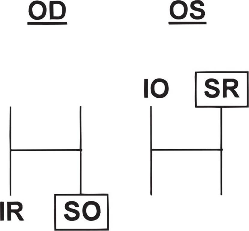
FIGURE 5-44
3. Determine if right or left head tilt worsens the hypertropia. This is the hardest step to understand, but it is really quite simple. When a normal patient tilts his head, one eye intorts and the other eye extorts. The intorters are the superior muscles (superior rectus and superior oblique) and the extortors are the inferior muscles (inferior rectus and inferior oblique). When the head tilts to the right, the right eye intorts and the left eye extorts. This means that both right superior muscles fire to cause the intorsion and both left inferior muscles fire to cause extorsion.
With this in mind, consider the previous example. Remember we have already determined that the palsy is either the right superior oblique or the left superior rectus. If the hyper gets worse with head tilt to the right, then the palsy is either in the right intorter or the left extorter. The right superior oblique is a right intorter, and if it is weakened, the unopposed action of the right superior rectus will cause the right eye to elevate further and worsen the right hyper (Fig. 5-45). In the left eye, the muscle in question is the left superior rectus, which is an intorter and should not be stimulated while extorting with right head tilt.
Two quick and easy patterns to memorize:
RIGHT(hyper) LEFT(gaze) RIGHT(tilt)—right superior oblique palsy
LEFT(hyper) RIGHT(gaze) LEFT(tilt)—left superior oblique palsy
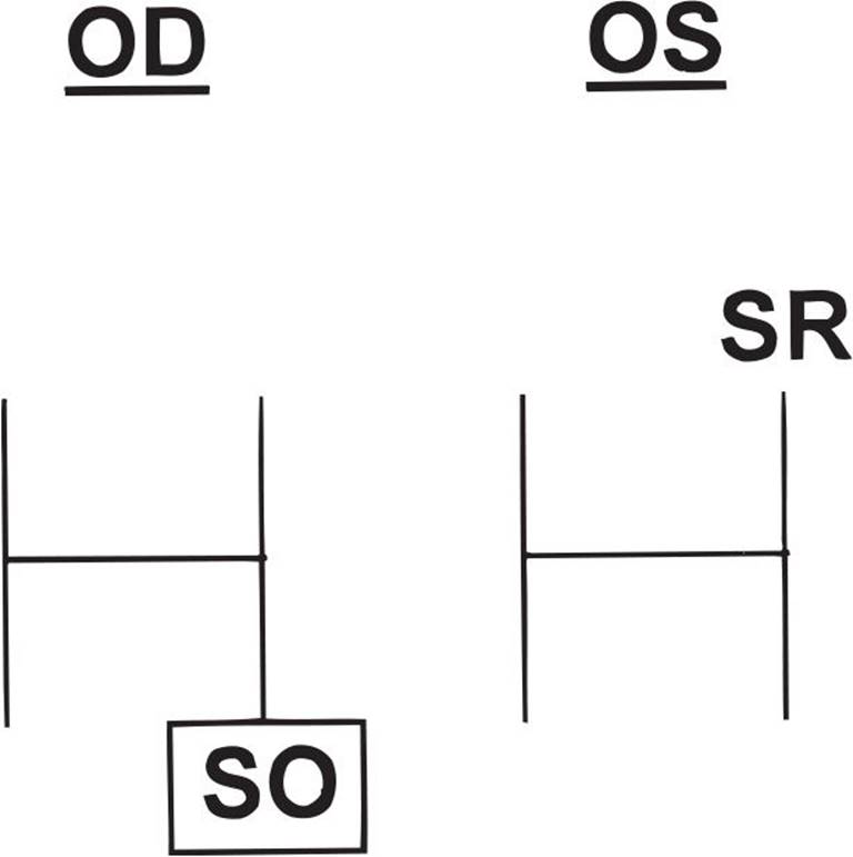
FIGURE 5-45
62 B) V-pattern exotropia
Patients with bilateral superior oblique palsy have excyclotorsion of greater than 10°, V-pattern esotropia, reversal of hypertropia on head tilt, and bilateral IOOA. In contrast to unilateral superior oblique palsies, bilateral cases often do not have a head turn.
63 D) Has its insertion near the macula
The following muscles correspond to each answer:
A = superior oblique
B = superior rectus
C = inferior rectus
D = inferior oblique
64 C) Dense amblyopia
Congenital esotropia, Möbius syndrome, and congenital fibrosis syndrome all can present with both eyes in the adducted position. Because the patient is cross-fixating, dense amblyopia is unlikely.
65 D) Bilateral medial rectus recessions
The patient has infantile esotropia with a large-angle strabismus and cross-fixation. Cycloplegic refraction of +2.00 D would be normal for an infant at this age and unlikely to have a significant effect on the esodeviation. If amblyopia is present, management should be initiated prior to surgery. In this case, the patient is fixating well with each eye and does not demonstrate amblyopia. Alternate patching can be done to improve abduction in patients with tight medial recti, but this patient has full motility. The timing of strabismus surgery is controversial, as some recent data suggests superior stereopsis outcomes with earlier surgery. It is generally accepted that strabismus surgery should be done before 12 months of age, typically at approximately 6 months of age.
Congenital esotropia is a syndrome consisting of the following features:
1. Large-angle esotropia, usually greater than 30 PD
2. Onset usually during the first few months of life, by definition by 6 months of age
3. Cross-fixation may be present
4. Latent nystagmus
5. DVD (in up to 60%–70%)
6. IOOA with V-pattern esotropia (in up to 60%–70%)
7. Mild hyperopia, +1.00 to +2.00 D
The above case is a classic case of congenital ET with IOOA demonstrating both the V-pattern and the right hypertropia on left gaze and left hypertropia on right gaze.
66 C) Fusion on Worth 4-dot testing at 10 feet.
Patients with monofixation syndrome demonstrate a small-angle strabismus. This is frequently esotropia, although exotropia may also be seen. Amblyopia is common. Patients have central suppression with peripheral fusion. They fuse on Worth 4-dot testing at near, but not at distance (10 feet). On the 4Δ base-out prism test, when the prism is placed before the nonfixating eye, no movement is seen because the image is projected onto the central scotoma.
67 A) Always high AC/A ratio
Accommodative esotropia is acquired, developing as the child begins to accommodate around the age of 1 to 2 years, but can occur in infancy. Children with esodeviations usually suppress the deviated eye. Because the deviation is acquired, many patients spontaneously alternate fixation, so less than half will have amblyopia. Patients who constantly suppress one eye develop amblyopia. Patients are usually hyperopic with greater than 3.00 D of hyperopia. Approximately 20% have a high AC/A ratio and will benefit from bifocals.
68 B) Dcc ortho wearing full distance correction of +1.00 D OU
Ncc ET15 wearing full distance correction of +1.00 D OU
The indication for prescribing bifocals to treat esotropia is specific. The patient must be able to fuse in the distance and be esotropic at near while wearing the full hyperopic correction. Giving a bifocal to a patient who has a deviation at distance despite full hyperopic correction will have no effect on the distance deviation. This patient needs surgery.
69 B) Acquired onset from infancy to 4 years of age
Accommodative esotropia occurs from infancy to 4 years of age with hypermetropia ranging from 2.00 to 7.00 D. Most neonates are not yet accommodating, so the esotropia is acquired when the infant starts to accommodate to bring vision in focus. As a result of the hyperopia, additional accommodative effort to bring images into focus is required, and an over-convergence response is seen. Accommodative esotropias are usually hyperopic but rarely more than +7.00 D because if they are very hyperopic they develop bilateral amblyopia and do not even try to accommodate.
70 D) Amblyopia
Suppression occurs when the eyes are deviated or tropic, and there is excellent stereo acuity when the eyes are aligned with intermittent exotropia. During periods of visual concentration, these children often maintain straight eyes with bifoveal fusion and thus develop excellent stereopsis. Because both eyes are stimulated during times of fusion, amblyopia is rare. They do not have monofixation because monofixation is associated with a constant small-angle strabismus, and stereopsis is often worse than 70 seconds of arc.
71 B) Observe the child; tell the parents you are satisfied because this is the desired result postoperative day 1
An immediate overcorrection of 8 to 12 PD is a desirable result. Often, over the first or second postoperative week, the effect of the surgery will lessen and the eyes will straighten. At 8 years old, suppression is unlikely.
72 D) Cannot determine from the information provided
This common pattern of exodeviation requires further evaluation. Exotropia that is farther rather than closer in the distance could represent either pseudo-divergence excess or true divergence excess. One test that could be helpful is the 30-minute patch test. With this dissociating patch on for 30 minutes, the near deviation often increases and approximates the distance deviation. This deviation would then be classified as a pseudo-divergence excess.
A +3.00 D lens may also prove helpful to look for high AC/A ratio patients. This classification is important both for determining how far to recess the recti muscles and for prognosis.
73 D) A deviation of greater than 15 PD
The maintenance or preservation of good binocular function is the goal of surgery for intermittent exotropia. Warning signs that binocular function is becoming threatened are increasing deviation in the tropia phase, increasing ease of dissociation, and poor recovery of fusion. The size of the deviation is of little concern if the patients are able to fuse easily.
74 A) Orthoptic therapy with a base-out prism or pencil push-up exercises
This patient demonstrates convergence insufficiency. This condition is best treated with orthoptic convergence training exercises. Base-out prism therapy and pencil push-up exercises builds up convergence amplitudes. Reading glasses would help accommodative insufficiency but not convergence insufficiency. Very few physicians, if any, advocate surgery for this problem.
75 B) Duane’s syndrome type I
Sherrington’s law states that when one extraocular muscle is stimulated, the ipsilateral antagonist is inhibited. In Duane’s syndrome type I, the lateral rectus muscle is innervated by part of the medial rectus subdivision of the third nerve. This may be a result of a congenital agenesis of the abducens nucleus, which has been demonstrated pathologically. As a result of this aberrant innervation when the medial rectus is stimulated to contract, the lateral rectus also receives stimulatory impulses, thus violating Sherrington’s law. Hering’s law states that when an extraocular muscle receives stimulation, its yoke muscle (the prime mover in the contralateral eye in the same field of gaze) receives equal innervation. An exception to Hering’s law is DVD. In DVD, one eye elevates, extorts, and abducts without any innervation to the contralateral eye.
76 A) Duane’s retraction syndrome (DRS) type I
77 D) Absence of electrical activity in the left lateral rectus muscle on abduction, with paradoxical activity on adduction
78 D) Glaucoma
DRS occurs in approximately 1% of all patients with strabismus. Its clinical features include a unilateral or bilateral abnormality of horizontal gaze, retraction of the globe on attempted adduction, and upshooting or downshooting of the globe on adduction. The left eye is affected more frequently than the right, and females are affected more frequently than males.
DRS may be divided into three types. DRS type I is characterized by marked limitation of abduction with normal or minimal restriction of adduction. Electromyography reveals an absence of electrical activity in the lateral rectus muscle on abduction with paradoxic activity on adduction. DRS type II is characterized by marked limitation of adduction with normal or minimal restriction of abduction. Electromyography reveals electrical activity of the lateral rectus muscle both on adduction and abduction. DRS type III is characterized by marked restriction of abduction as well as adduction. Electromyography reveals electrical activity of both the lateral and medial rectus muscles on both adduction and abduction.
Type I is the most common form of DRS, and type III is the least common. Although the majority of patients with DRS type I and type II will have straight eyes, some type I patients develop an esodeviation in primary position and some type II patients develop an exodeviation. Amblyopia may be present in 10% to 14% of patients with DRS.
Several ocular and systemic anomalies have been associated with DRS: cataracts, iris anomalies, Marcus Gunn jaw winking, microphthalmos, crocodile tears, Goldenhar syndrome, maternal thalidomide use, and Klippel–Feil syndrome.
Brown syndrome is characterized by the inability to actively or passively elevate the eye in the adducted position. It may be acquired or congenital. There is normal or nearly normal elevation of the eye in abduction.
79 B) A Faden procedure may reduce the upshoot of the affected eye on adduction.
The incidence of amblyopia in patients with DRS is only approximately 10%. The lid fissure narrowing is secondary to retraction of the globe with co-contraction of the medial and lateral rectus muscles. The strabismus is generally noncomitant. A Faden procedure (posterior fixation of the horizontal rectus muscles near the equator) may reduce the upshoot of the affected eye on adduction, as it stops vertical slippage of the lateral rectus muscle.
80 A) Esodeviation
Congenital third nerve palsy may result in complete or partial loss of superior, medial, and inferior rectus function, as well as inferior oblique and levator function. The eye is usually deviated down and out. Aberrant reinnervation may manifest as abnormal pupillary constriction with adduction.
81 B) Recession of both lateral recti with supraplacement
This patient exhibits a significant V pattern without significantly overacting inferior obliques. In general, inferior oblique surgery is indicated for overaction of 2+ or more. Without significant oblique overaction, offsetting the horizontal muscles can correct as much as 30 PD of an A or V pattern. Horizontal muscle offset changes the vector of forces. The medial recti are moved toward the apex of the A or V pattern while the lateral recti are moved in the opposite direction. A recess-resect procedure is useful in cases in which surgery must be limited to one eye or when an incomitant deviation exists. Horizontal offset may be performed in conjunction with the recess-resect, but it may not correct large A or V patterns. In this case, the procedure of choice would be to recess the lateral recti for the appropriate deviation in primary position and offset the lateral recti superiorly.
82 C) The deviated eye extorts as it elevates.
DVD is present in 60% to 80% of patients with congenital esotropia. DVD is usually bilateral and asymmetric. The etiology is unknown but appears to be associated with an early disruption of binocular development. Thus, high-grade stereopsis and bifoveal fixation are not seen. During times of visual inattention, the nonfixating eye slowly drifts up, extorts, and abducts without a corresponding hypotropia of the fellow eye on alternate cover testing. This is the hallmark of this disorder—it does not obey Hering’s law. DVD can simulate IOOA in side gaze when the nose acts as an occluder. The hyperdeviation in DVD is of the same amount in adduction, abduction, and primary position. This is in contrast to IOOA, in which the hyperdeviation is greatest in its field of action.
83 B) Occlude one eye for 30 minutes and remeasure the deviation.
This patient exhibits a difference of greater than 10 PD between near and distance. The patch test needs to be performed to differentiate a pseudo-divergence excess from a true divergence excess. This differentiation helps when deciding on the amount of surgery to perform and on the prognosis. The patient is monocularly occluded for 30 to 60 minutes and remeasured without letting him restore binocular fusion. The patch dissociates the eyes to suspend all tonic fusional convergence and to reveal the full latent deviation at near, eliminating the distance/near disparity.
84 B) Remeasure the deviation with a +3.00 D add OU.
This child exhibits true divergence excess. He needs to be measured with a +3.00 D add to determine if he has a high AC/A ratio. If the near deviation increases close to the distance deviation with a +3.00 D add, he has a high AC/A. This factor is important because these patients are prone to overcorrection (75% overcorrection) if surgery for the full distance deviation is performed. Parents should be aware of the poor prognosis before surgery and of the possible need for bifocals to decrease the high AC/A ratio postoperatively.
85 A) Observe and have the patient return in 2 weeks.
A small esotropia of 8 to 15 PD postoperatively is desirable, with 20 PD being the upper limit of normal. Postoperative diplopia associated with an initial overcorrection is normal and usually resolves by 1 to 2 weeks. In younger children (<4 years old), part-time alternate patching helps prevent the development of suppression. Patients with residual exotropia of more than 20 PD in the first postoperative week are unlikely to improve, and many will worsen.
86 C) Prescribe enough base-out prism to alleviate the diplopia but leave a small residual esophoria.
Prescribe just enough prism to alleviate the diplopia but leave a residual esophoria to encourage divergence. If the consecutive esotropia is present only at near, one can consider a bifocal add, miotics, or even base-out prism. If the esotropia persists after 8 weeks, consider reoperation (usually a bilateral medial rectus recession). If the patient demonstrates lateral incomitance or significant limitation of abduction, a slipped muscle is a possibility.
87 B) Lateral rectus recessions plus infraplace the lateral recti
This patient has high-grade stereoacuity and thus bifoveal fusion during his phoric phase. He also has a significant A pattern from bilateral superior oblique overaction. Typically, one would operate on the superior obliques for significant overaction, but in patients with high-grade stereopsis, superior oblique tenotomies could cause consecutive superior oblique paresis with intractable torsional diplopia. In this case, vertical offsets of the horizontal muscles or the Wright superior oblique tendon expander should be done. The Wright silicone expander controls the amount of superior oblique weakening, is reversible, and alleviates the hyper inside gaze. The surgery of choice is lateral rectus recession with infraplacement to alleviate the A pattern.
88 C) Superior oblique overaction with A pattern is common.
The picture exhibits a patient with limited elevation on adduction, which is consistent with Brown syndrome. Brown syndrome can be congenital or acquired, and it represents an inelastic superior oblique muscle tendon complex that leads to a restriction of passive or active elevation in adduction. There is often a downshoot of the eye on adduction and always limited elevation in adduction. This condition can be differentiated from inferior oblique palsy with exaggerated forced duction testing. Associated superior oblique overaction is uncommon.
89 A) Patients with this condition have normal vertical fusion amplitudes.
This patient has congenital fourth nerve palsy. Long-standing fourth nerve palsy results in ocular torticollis with a compensatory head tilt to the side of the palsy that can lead to facial asymmetry of the dependent side. These patients have large vertical fusional amplitudes that help differentiate them from acquired fourth nerve palsies. The superior oblique tendon is rarely long or floppy or absent. When the child has fixation preference for the affected eye, the contralateral superior rectus muscle can appear to underact (inhibitional palsy of the contralateral antagonist) and the contralateral inferior rectus can undergo contracture, leading to a “double elevator palsy.”
90 A) Diplopia
CPEO is an inherited mitochondrial disease. Patients develop early-onset ptosis followed by slowly progressive loss of extraocular motility. Despite the motility deficits, patients typically do not complain of diplopia. CPEO patients may also have concomitant cardiac conduction defects, cerebellar dysfunction, and pigmentary retinopathy.
91 C) Botox has been proven to be effective as the primary treatment of most types of strabismus.
Botox is a purified form of botulinum toxin type A, derived from the Hall strain of Clostridium botulinum. It blocks neuromuscular conduction by binding to receptor sites on motor nerve terminals interfering with the release of ACh into the synaptic cleft. When injected intramuscularly, Botox produces a localized chemical denervation muscle paralysis. The nerve ending atrophies but will resprout over time. Paralysis onset occurs in 2 days, increases in intensity over the next week, and lasts 3 months in extraocular muscles. Botox is indicated for blepharospasm associated with dystonia. The efficacy of Botox in strabismus is low and surgery remains the primary treatment for most types of strabismus. Multiple injections may be necessary but should not exceed 200 units in 1 month to decrease the incidence of antibody production. Reported side effects include ptosis, diplopia, and spatial disorientation. These are, fortunately, temporary. Perforation of the globe has been reported. Systemic effects of Botox are not seen because a dose over 100 times greater than the normal amount is required for toxicity.
92 C) Blepharospasm
Botox is the treatment of choice for most types of blepharospasm associated with dystonia.
93 D) Is often associated with intermittent exotropia
Latent nystagmus increases with monocular occlusion, when the fixing eye is in abduction (null point in adduction) and the fast phase is toward the fixing eye. It is associated with disruption of early binocular visual development (congenital ET, congenital monocular cataracts), not acquired strabismus (intermittent XT or accommodative ET).
94 C) Latent nystagmus with face turn to place the fixing eye in the null point (see answer for Question 92).
95 C) Prescribe full hyperopic correction +3.00 OU
Reduce the latent nystagmus by correcting the residual esotropia and improving binocular fusion.
The best way to correct the small esotropia is by giving the hyperopic correction. Improving binocular fusion will reduce the latent nystagmus, thus reducing the face turn.
When one eye is occluded, the unoccluded eye remains in the adducted position and the patient may turn his and her face to the ipsilateral side to look straight ahead. The degree of esotropia increases when a base-out prism is placed in front of the fixating eye, and the pupil constricts when the eye assumes its adducted position (indicating that this is an accommodative effort). IOOA and DVD occur less frequently in patients with nystagmus blockage syndrome than in patients with essential infantile esotropia.
96 C) Prescribe +3.50 OD and +1.00 OS.
This patient has anisometropic amblyopia. The first line of treatment is to provide optical correction, as a significant number of patients will have vision improvement with glasses alone. It is common practice to reduce the amount of plus prescribed to promote some accommodation. Atropine refraction would be unnecessary in this case, but can be used for incomplete cycloplegia with cyclopentolate. Neuroimaging would not be indicated at this point, as there is a likely etiology of the reduced vision in the right eye.
97 D) Optic atrophy
The classic triad of spasmus nutans includes monocular or dissociated small amplitude nystagmus, head bobbing, and torticollis. The differential diagnosis for this disorder includes patients with chiasmatic gliomas and subacute necrotizing encephalomyopathy. Features of these two disorders may include optic atrophy, irritability, vomiting, and increased intracranial pressure. Spasmus nutans should be considered only after these two disorders are excluded. Spasmus nutans usually disappears within 2 years of onset.
98 A) Oscillopsia is frequently present.
Congenital motor nystagmus is a conjugate, jerk nystagmus that manifests in the perinatal period. It may be associated with a null point and a head turn to minimize the nystagmus. The visual acuity is relatively good—ranging from 20/20 to 20/70—and oscillopsia is typically absent. The nystagmus ceases when the patient is asleep.
99 D) Arnold–Chiari malformation
Congenital sensory nystagmus can result from a number of causes in which visual information does not reach the occipital cortex properly. Media opacities, such as congenital cataracts, foveal or optic nerve hypoplasia, and retinal degenerations, may all lead to sensory nystagmus. An Arnold–Chiari malformation causes a downbeat motor nystagmus.
100 A) Atropine toxicity
In this case, the most likely diagnosis is atropine toxicity. A careful history will confirm the diagnosis. Children, especially young infants are at risk for side effects of atropine. Signs of toxicity include fever, dry mouth, facial flushing, nausea, vomiting, irritability, and tachycardia. Treatment involves immediate discontinuation of the medicine plus supportive measures. In this situation, it would be appropriate to recommend the mother bring her son to the emergency room. A third nerve palsy and trauma could both cause mydriasis, but these etiologies are less likely in this case. Horner syndrome would cause miosis, ptosis, and anhydrosis.