
MARCUS M. MARCET
1 Histologic examination of a corneal button received from penetrating keratoplasty is seen (Fig. 7-1). What is the most likely surgery to cause the finding shown?

FIGURE 7-1
A) Photorefractive keratectomy (PRK)
B) Radial keratotomy (RK)
C) Anterior lamellar keratoplasty (ALK)
D) Laser in situ keratomileusis (LASIK)
2 Which statement about rhabdomyosarcoma of the orbit is incorrect?
A) It is the most common primary malignant orbital tumor in children.
B) The alveolar type has the poorest prognosis.
C) It originates from the extraocular muscles.
D) Patients present usually with very rapid onset of unilateral proptosis that can simulate an orbital inflammation.
3 Gram stain of the purulent material from a dilated, erythematous punctum is shown (Fig. 7-2). Which of the following corresponds to the organisms shown?
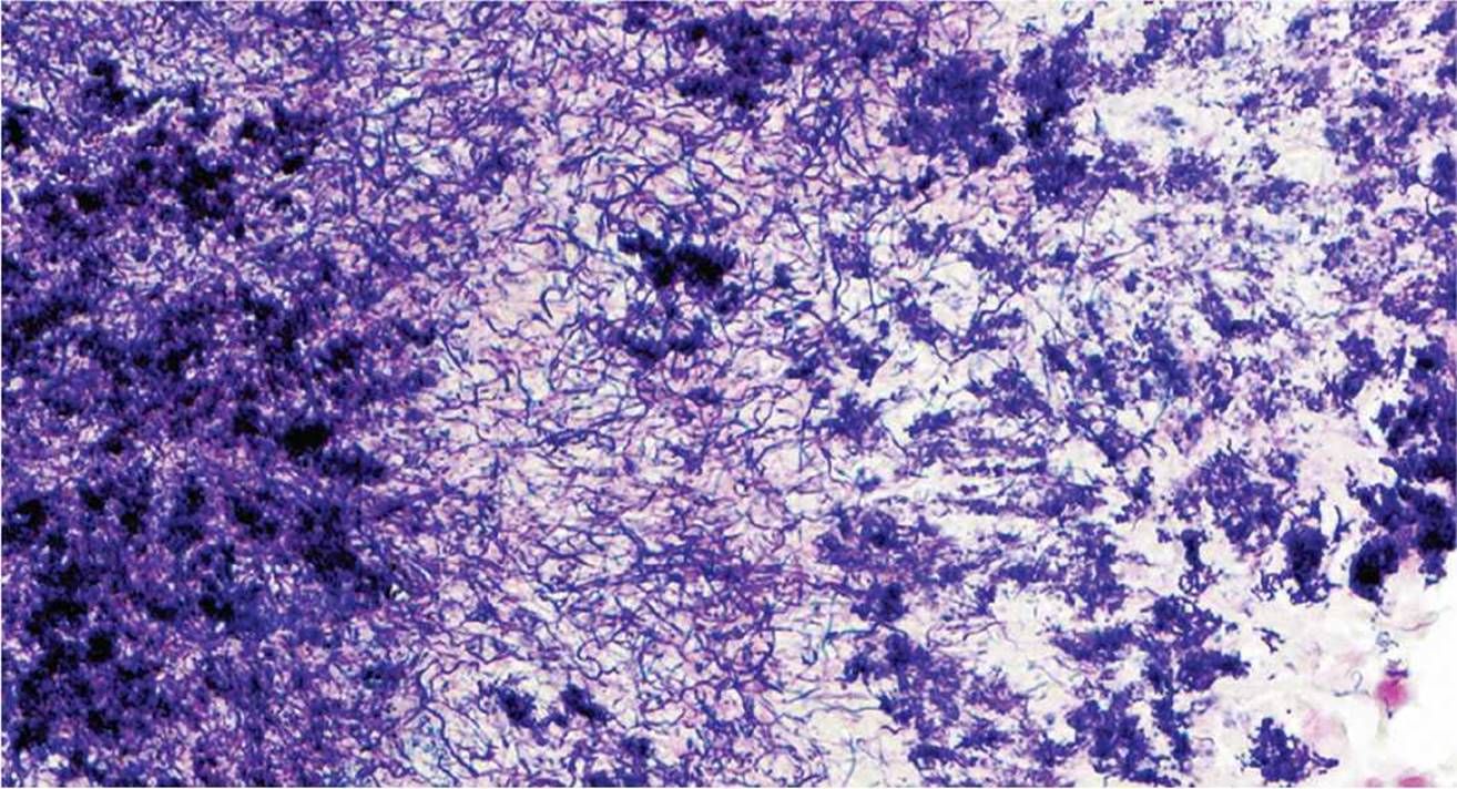
FIGURE 7-2
A) Gram-negative rods
B) Gram-negative filaments
C) Gram-positive cocci
D) Gram-positive hyphae
4 Examination of an eye enucleated for malignant melanoma of the choroid shows a glistening white irregular tumor arising from a ciliary crest measuring 3 mm × 2 mm × 2 mm. A histologic section is shown (Fig. 7-3). Which statement is true?

FIGURE 7-3
A) This is an intraocular metastatic lesion from the uveal melanoma.
B) This is a rare tumor of the nonpigmented epithelium of the ciliary body.
C) This lesion can be found in approximately 25% of older patients.
D) This lesion leads to secondary angle closure if untreated.
QUESTIONS 5 and 6 (FIGS. 7-4 to 7-9) A 42-year-old patient presents with a progressive decrease of visual acuity over 2 months in his right eye. Ophthalmologic examination (Fig. 7-4) shows an orange lesion temporal to the macula with associated retinal detachment in the inferior two quadrants. Fluorescein angiography (Fig. 7-5) displays a hyperfluorescent lesion with a hypofluorescent circular ring around the peripheral border of the lesion. Ultrasonography is shown (Fig. 7-6). The patient was lost to follow-up, but returned 2 months later with a blind, painful eye, and the eye was enucleated. Histologic examination is shown (Fig. 7-7). Higher-magnification images (Figs. 7-8 and 7-9) show the area temporal to the macula, that is, the area of the clinically visible lesion.

FIGURE 7-4

FIGURE 7-5

FIGURE 7-6
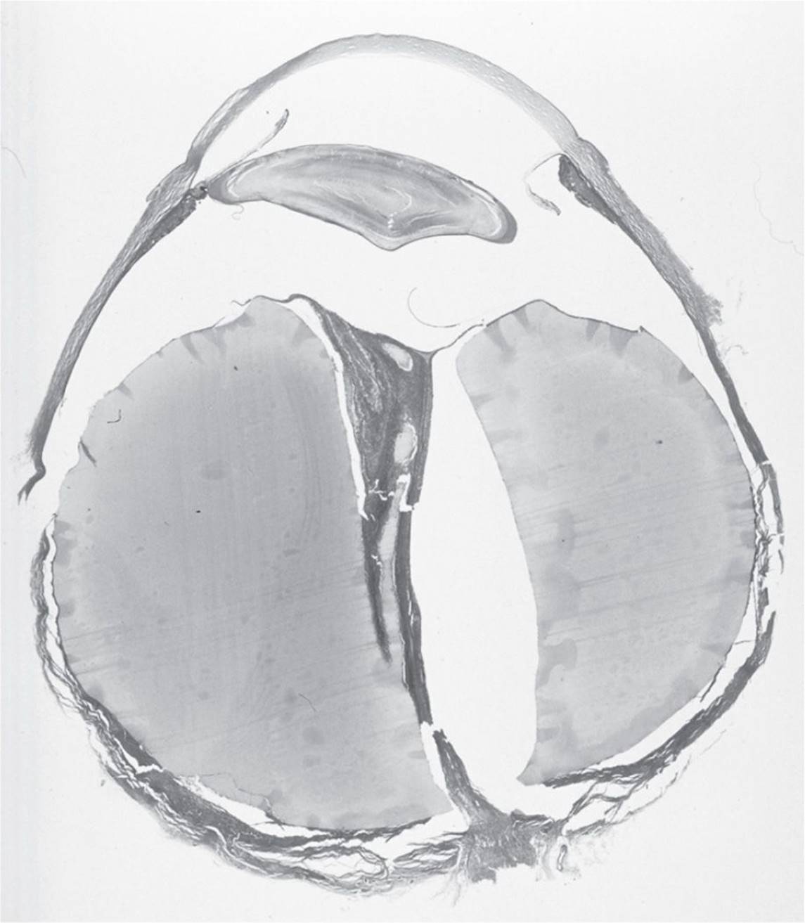
FIGURE 7-7
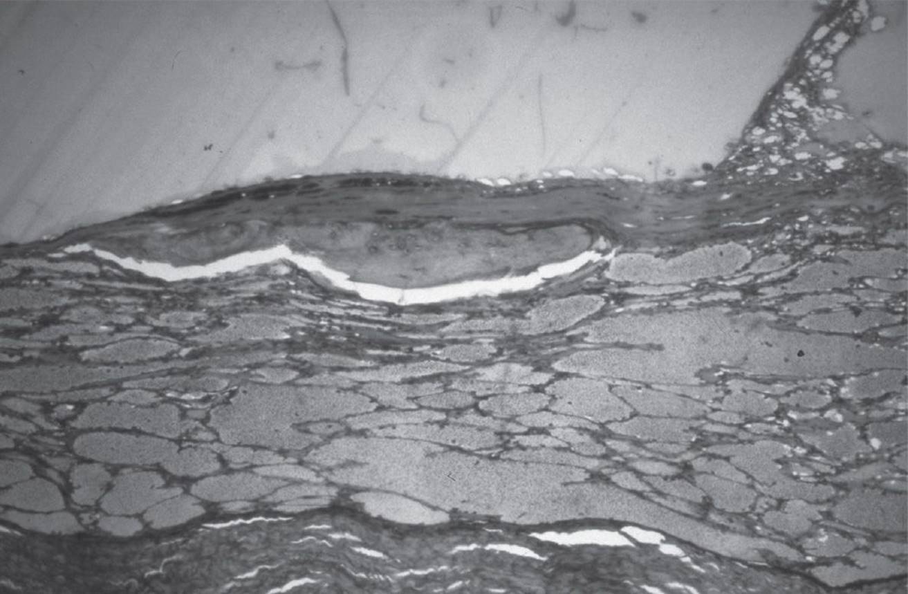
FIGURE 7-8
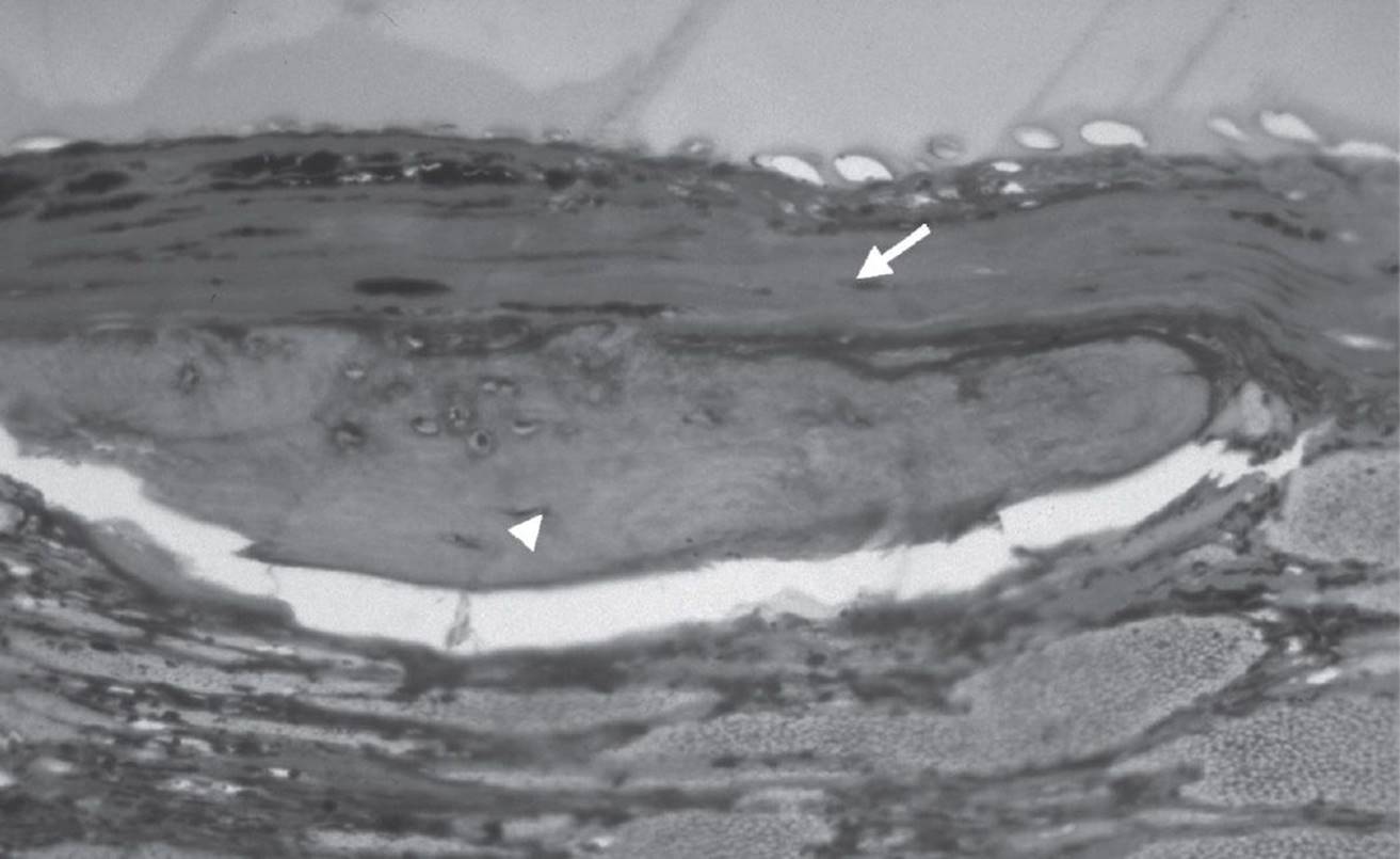
FIGURE 7-9
5 The histologic section of the enucleated eye is shown (Fig. 7-7). What type of retinal detachment is present?
A) Rhegmatogenous
B) Tractional
C) Exudative
D) Artifactitious
6 Figures 7-8 and 7-9 show an area of dense fibrous tissue (arrow) overlying an area of calcified tissue (arrowhead) in the plane of the retinal pigment epithelium and Bruch membrane. Which statement is true?
A) These lesions developed from choroidal melanocytes (melanocytes of neural crest origin).
B) The cells of origin for these lesions are the retinal pigment epithelial cells (cells of neuroectodermal origin).
C) These lesions can undergo malignant transformation.
D) The cell of origin of these lesions is unknown.
QUESTIONS 7 and 8 (FIGS. 7-10 and 7-11) A 5-year-old girl has a facial nevus flammeus and seizures. The lesion is similar to the clinical features of the previous patient (Figs. 7-4 to 7-6). Representative histopathology shows a choroidal lesion (Figs. 7-10 and 7-11).
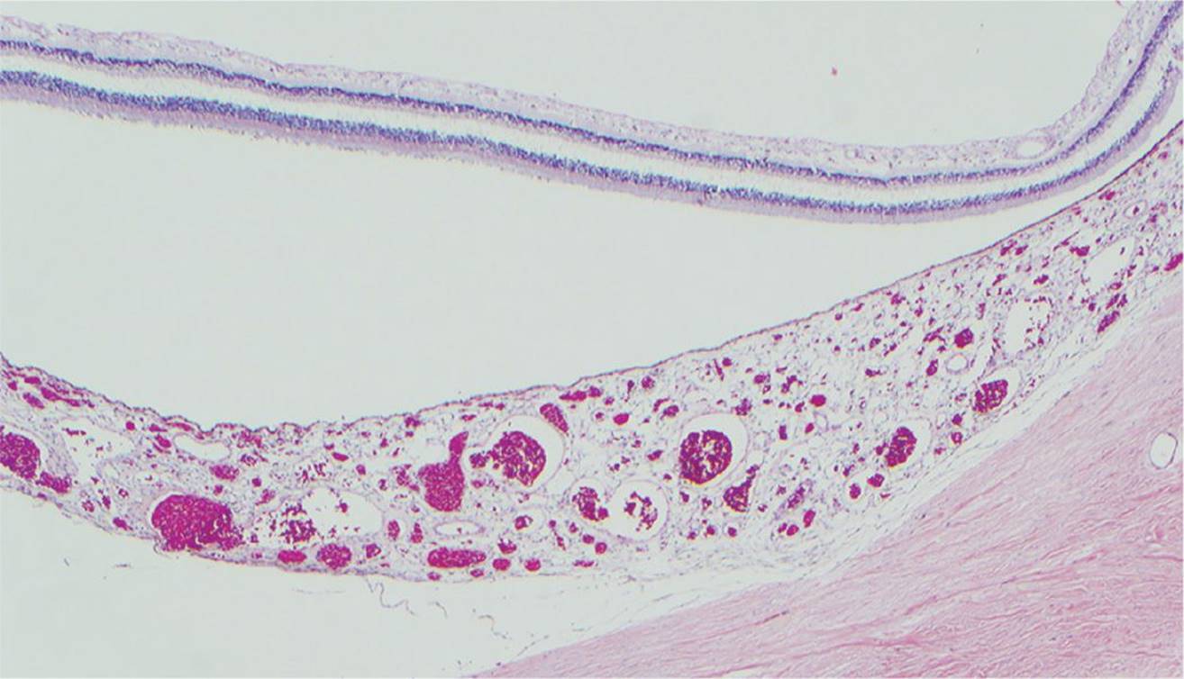
FIGURE 7-10

FIGURE 7-11
7 Which statement is most likely true regarding the girl’s condition?
A) This is a condition with a high probability of spontaneous regression.
B) Glaucoma is not a common feature of this disease.
C) This disease is inherited in an autosomal recessive manner.
D) Ipsilateral hemangiomas of the meninges are common.
8 Which statement is true regarding the histopathology of the lesion?
A) This is a malignant lesion.
B) Lakes of proteinaceous material are separated by fibrovascular septa.
C) This lesion is consistent with a capillary hemangioma.
D) Cavernous spaces filled with erythrocytes are present.
9 The corneal button of a patient is shown (Fig. 7-12). Appropriate steps in the management of the condition may include all of the following, except:
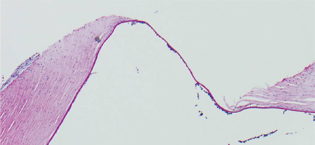
FIGURE 7-12
A) cyanoacrylate adhesive
B) bandage contact lens
C) systemic carbonic anhydrase inhibitors
D) descemet stripping automated endothelial keratoplasty
10 You are asked to read out a temporal artery biopsy specimen (Fig. 7-13) from a patient with headache and jaw claudication. Which of the following is the best answer?

FIGURE 7-13
A) The result is suspicious and contralateral biopsy is warranted.
B) The result is negative and steroids may be tapered off.
C) The result is positive and steroids should be continued.
D) The result is inconclusive and blood laboratory studies are needed.
QUESTIONS 11 and 12 (FIG. 7-14) A 72-year-old female had intracapsular cataract extraction 6 months ago. Recently, she developed increased IOP that was unresponsive to topical treatment. Histologic section through the anterior segment and iris is shown in Figure 7-14.
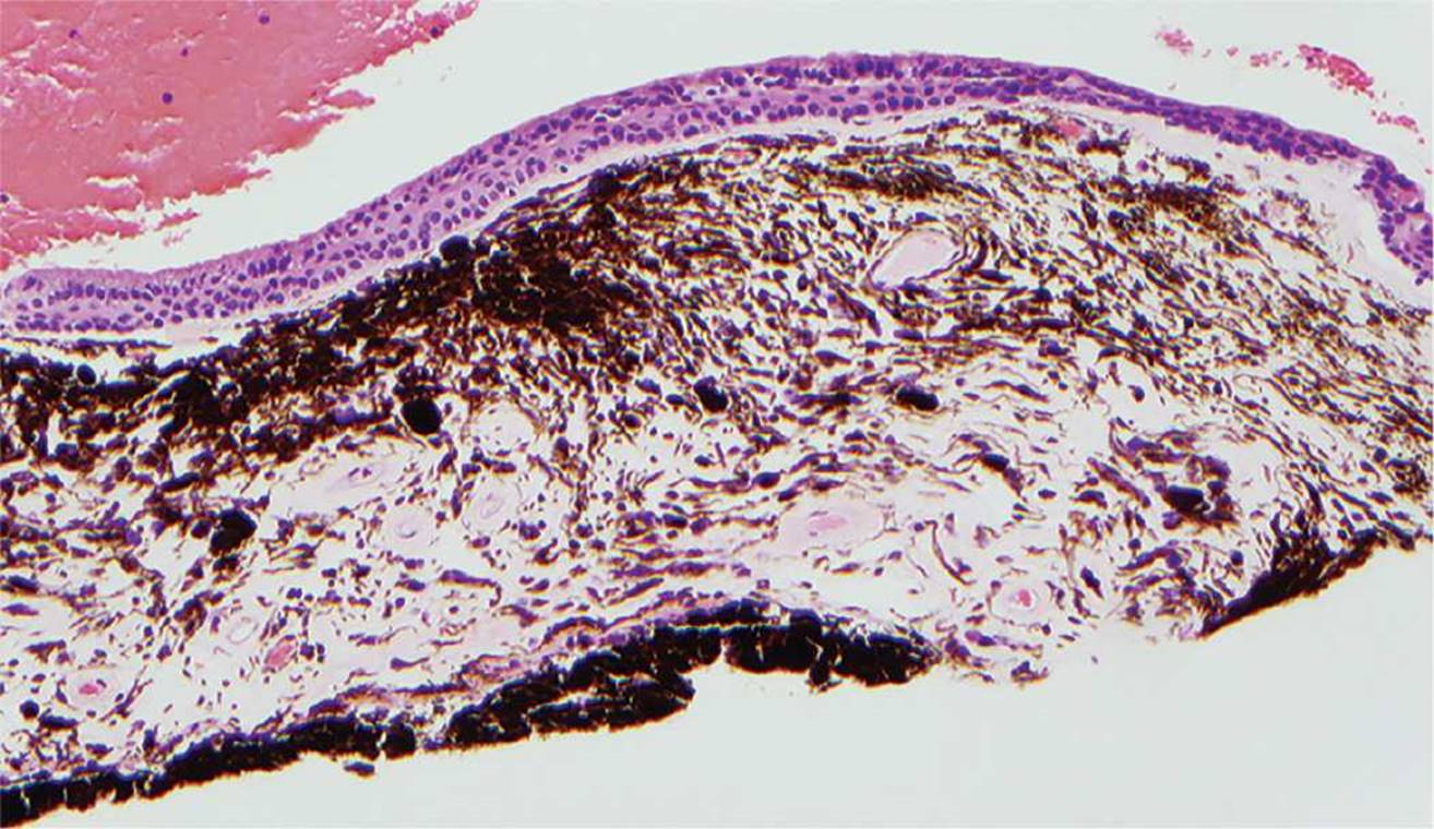
FIGURE 7-14
11 Which statement is correct?
A) This condition is a variant of the iridocorneal endothelial (ICE) syndrome.
B) Surface epithelium is found in the anterior chamber.
C) This is a congenital condition that manifests only in adults.
D) This is a self-limited disorder in most cases.
12 Which stain highlights the characteristic features of the epithelium and allows identification of its origin?
A) Hematoxylin–eosin
B) Oil Red O
C) Periodic acid–Schiff (PAS)
D) Congo red
13 Cells distended by round eosinophilic cytoplasmic inclusions are shown in Figure 7-15 (arrow). Normal versions of the same cell type are seen nearby (arrowheads). What is the cell type of the cells shown in Figure 7-15?
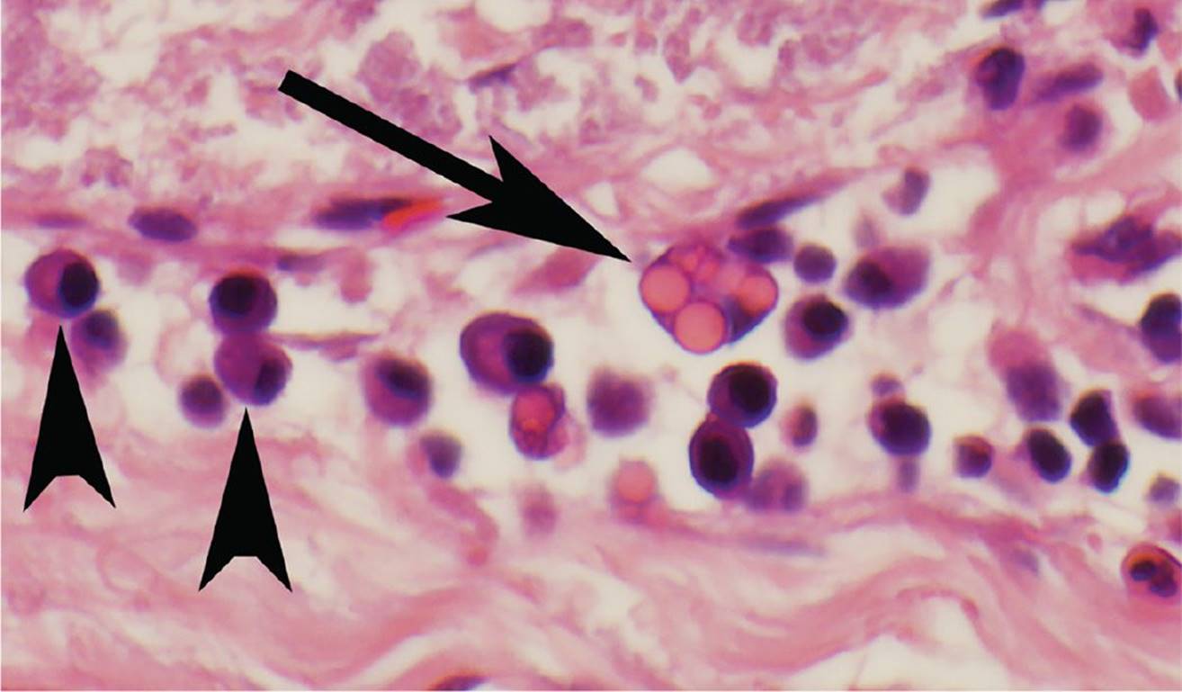
FIGURE 7-15
A) Hemosiderin-laden macrophages
B) Cytomegalovirus-infected retinal cells
C) Plasma cells–containing immunoglobulin
D) Polyhedral melanocytes–containing pigment
QUESTIONS 14 and 15 (FIG. 7-16) A 16-year-old adolescent boy presented with a blind, painful eye. Ophthalmologic examination showed band keratopathy, and the anterior chamber and pupil were covered by a pigmented membrane. Ultrasonographic examination showed a microphthalmic eye with an irregular structure behind the lens extending to the optic disc. The eye was enucleated. A vitreous stalk extended from the optic nerve to the lens and tractional membranes extended to the ciliary body processes (Fig. 7-16).

FIGURE 7-16
14 Which statement is incorrect?
A) A microphthalmic eye is typical for this disease.
B) This is a unilateral disease.
C) Lipomatosis lentis can be a feature of this condition.
D) The diagnosis is confirmed with CT and serologic studies.
15 What is the etiology of this condition?
A) Developmental abnormality
B) Intraocular infection
C) Trauma
D) Neoplasm
16 A 6-month-old child has an iris lesion and recurrent anterior chamber bleeding. A biopsy of this mass was performed. A high-magnification image of the histology is shown in Figure 7-17. Which statement is correct?

FIGURE 7-17
A) The lesion shows pleomorphic cells with a multinucleated tumor cell.
B) This is a benign cutaneous disorder rarely involving the eye.
C) Immunohistochemical stains for macrophages (histiocytes) are negative.
D) Most ocular lesions occur after 1 year of age in this disease entity.
QUESTIONS 17 and 18 (FIGS. 7-18 and 7-19) A 73-year-old woman presents with a lesion in her right eye (Fig. 7-18) that had developed over the course of 2 years. The red, vascular nodule is located in the palpebral conjunctiva in the area of the mucocutaneous junction. Ophthalmologic examination is otherwise normal. Histologic examination is shown in Figure 7-19.
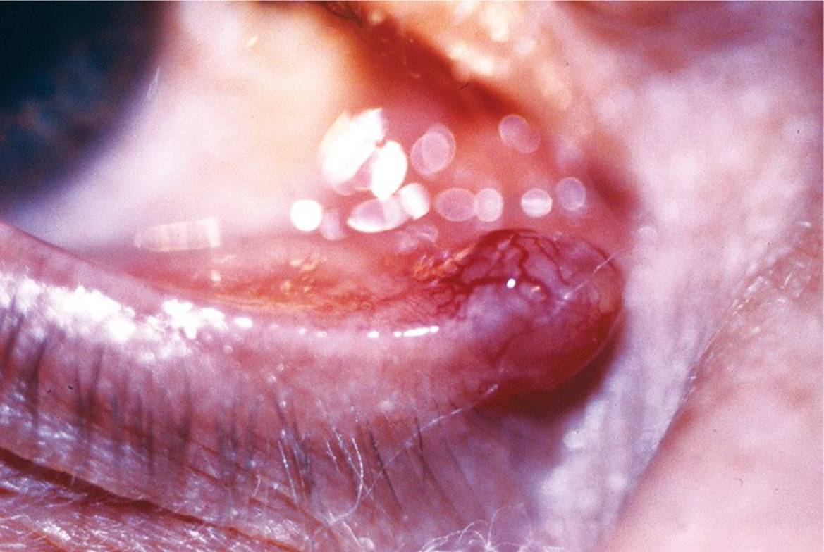
FIGURE 7-18
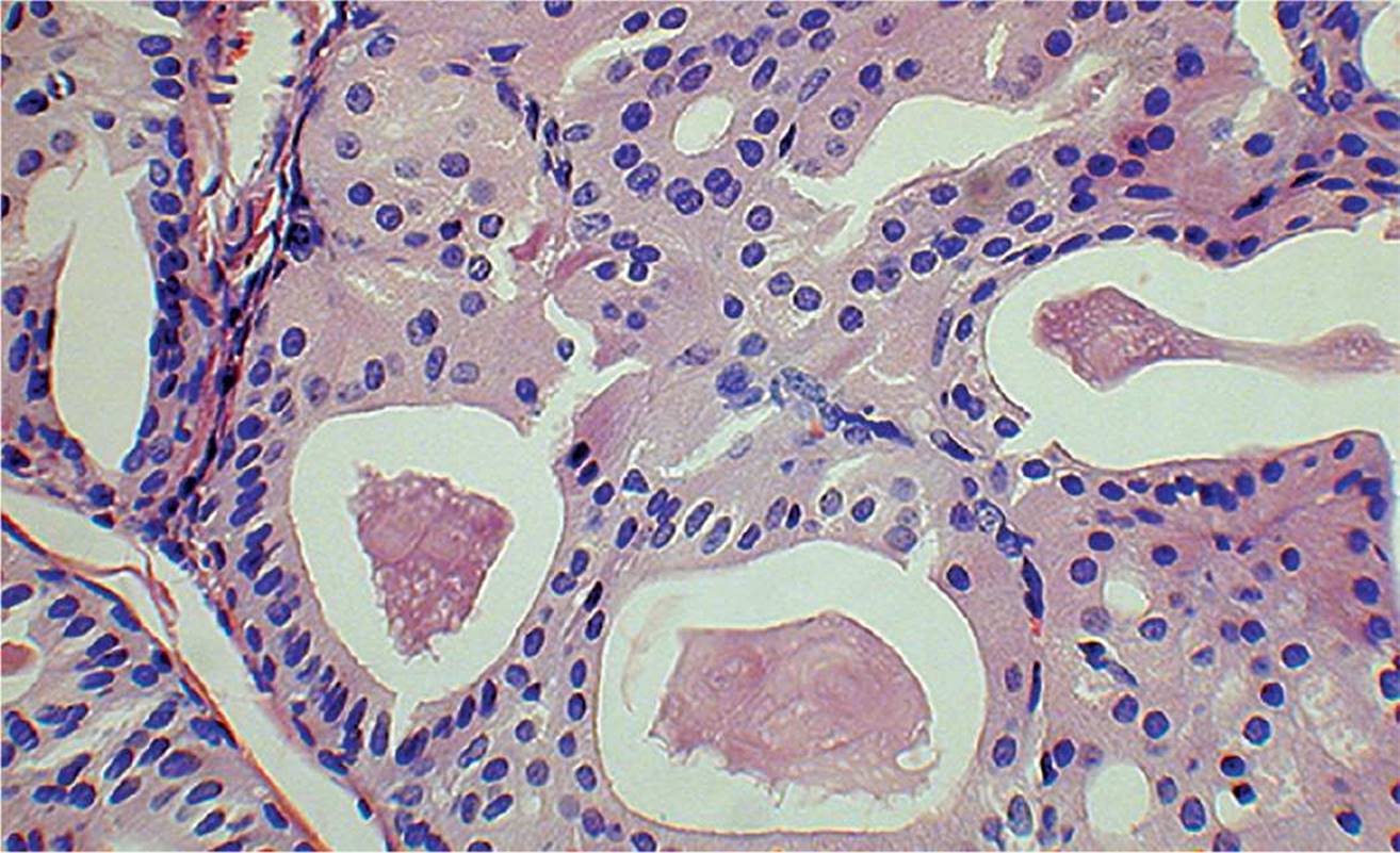
FIGURE 7-19
17 What is the disease process?
A) Adenoid cystic carcinoma
B) Oncocytoma
C) Pyogenic granuloma
D) Papilloma
18 The granular appearance of the cytoplasm of the cells in Figure 7-19 is caused by:
A) rough endoplasmic reticulum
B) multiple partially atypical mitochondria
C) smooth endoplasmic reticulum
D) polyribosomes
QUESTIONS 19 and 20 (FIG. 7-20) A 23-year-old man presents with a hard, painless lump within the left lower eyelid for over 6 weeks. Histologic examination is shown in Figure 7-20.
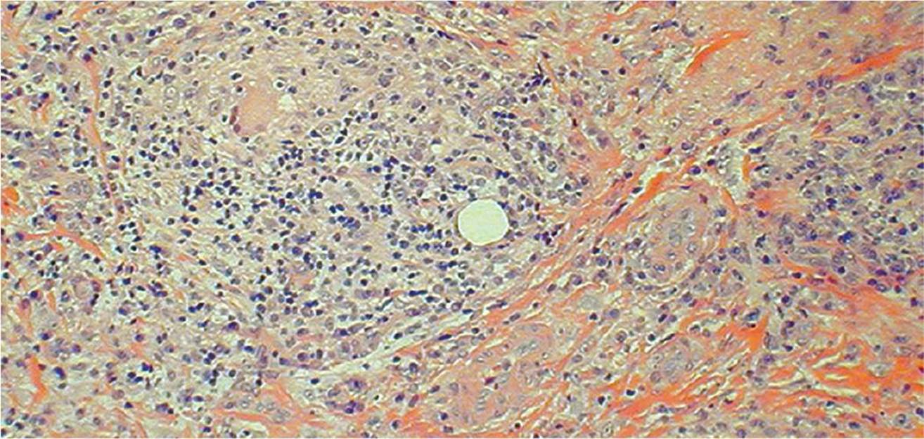
FIGURE 7-20
19 Which statement is correct?
A) This can be a chronic inflammation of the Zeis sebaceous glands.
B) Excision with frozen section control of margins is the treatment of choice.
C) Histologic examination is not necessary because it is always a benign lesion.
D) Moll glands can be the etiologic agent for this inflammation.
20 The clear space in the center of Figure 7-20:
A) is a feature of a xanthoma
B) is also visible on a frozen section
C) is consistent with a zonal lipogranuloma
D) is a vascular channel
21 A 35-year-old woman who has a history of recurrent “corneal inflammation” developed a near full-thickness corneal ulcer. Penetrating keratoplasty was performed (Fig. 7-21). Histologic examination shows a granulomatous reaction to Descemet membrane (Fig. 7-22) and the presence of retrocorneal tissue. What is the most likely etiologic agent for the keratitis?

FIGURE 7-21

FIGURE 7-22
A) Acanthamoeba
B) Herpes virus
C) Pseudomonas
D) Candida albicans
22 A 72-year-old woman has a history of cataract extraction with implantation of an anterior chamber IOL 5 months ago. She reports that the surgeon had “lost some lens material” during surgery. The eye was never “quiet” after surgery. Low- and higher-power photomicrographs around the lens (Figs. 7-23 and 7-24) are shown. Which statement is incorrect?
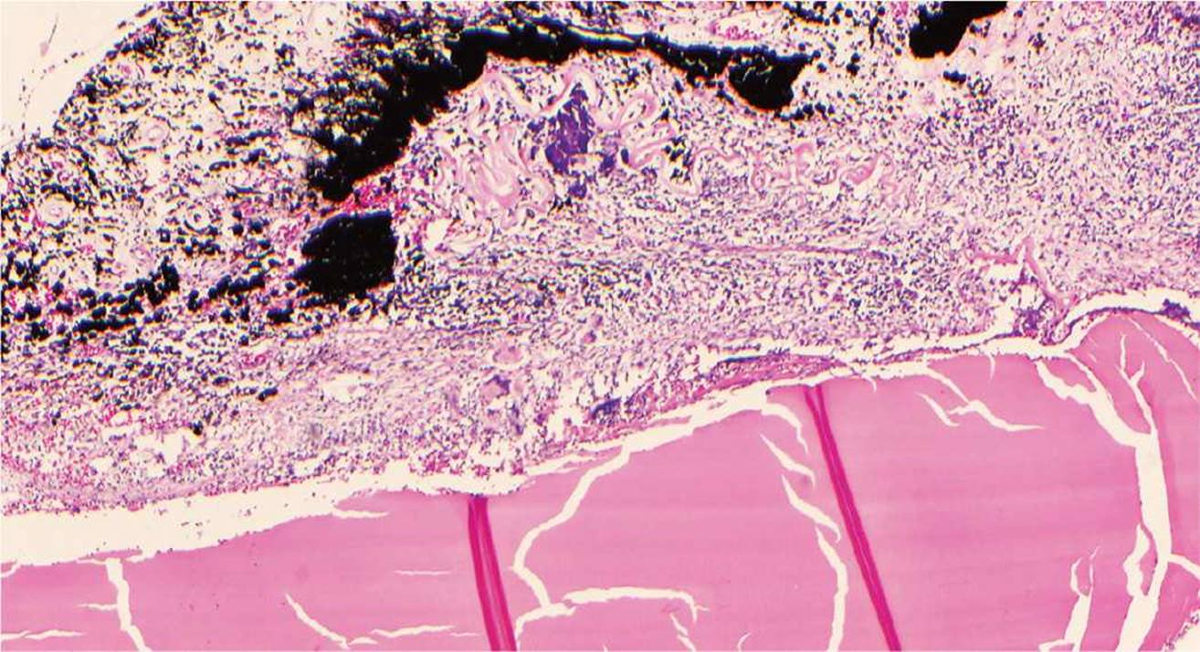
FIGURE 7-23
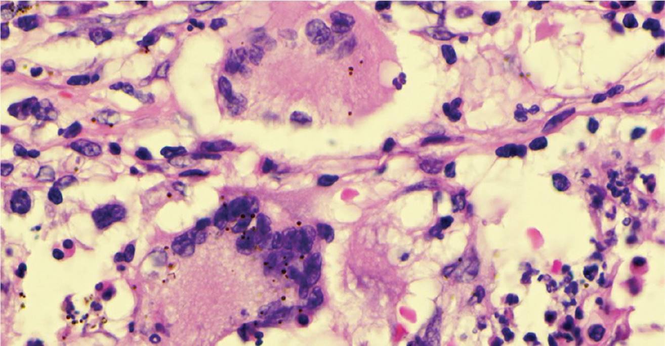
FIGURE 7-24
A) The eye has a chronic choroidal detachment.
B) The lens nucleus was lost into the vitreous during cataract surgery.
C) The eye is hypotonous.
D) The disease is phacolytic glaucoma.
23 An 85-year-old patient is seen preoperatively to have a loose scroll of lens capsule in the anterior chamber. The lens capsule is removed during cataract surgery. High-magnification images of the specimen are shown (Figs. 7-25 and 7-26). Normal lens epithelial cells are seen posterior to the surface of the capsule. The filter paper along the bottom of the figure was used to maintain specimen orientation. Which statement is true regarding the lens capsule?

FIGURE 7-25

FIGURE 7-26
A) This condition can be caused by exposure to infrared radiation.
B) Radial transillumination defects are found in the iris.
C) The incidence of zonular dialysis during cataract surgery is higher in these patients.
D) This condition is much more common in African Americans.
24 What is the abnormal condition in this histologic specimen (Fig. 7-27)?
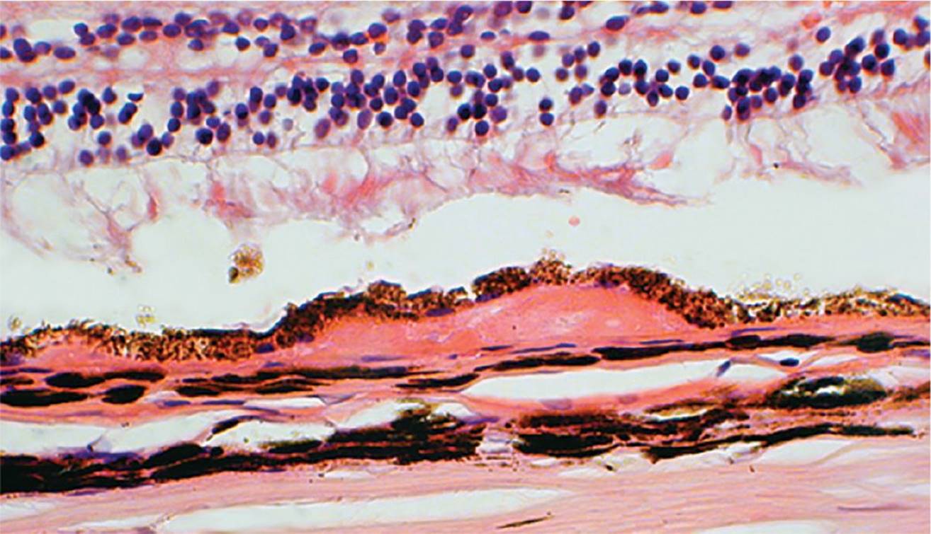
FIGURE 7-27
A) Basal laminar deposits
B) Soft drusen
C) Nodular drusen
D) Basal linear deposits
QUESTIONS 25 and 26 (FIG. 7-28) A 72-year-old woman presents with a lesion on her eyelid. Histologic examination is shown in Figure 7-28.

FIGURE 7-28
25 What is the most accurate diagnosis for this lesion?
A) Basal cell carcinoma, morphea type
B) Basal cell carcinoma, nodular type
C) Basal cell carcinoma, superficial type
D) Basal cell carcinoma, ulcerated type
26 What is the order of probability (from highest to lowest) of the location of this lesion?
A) Upper eyelid—lower eyelid—medial canthus—lateral canthus
B) Lower eyelid—medial canthus—upper eyelid—lateral canthus
C) Medial canthus—upper eyelid—lower eyelid—lateral canthus
D) Lower eyelid—upper eyelid—lateral canthus—medial canthus
27 Histologic examination of an optic nerve head is shown in Figure 7-29. Which statement is correct?
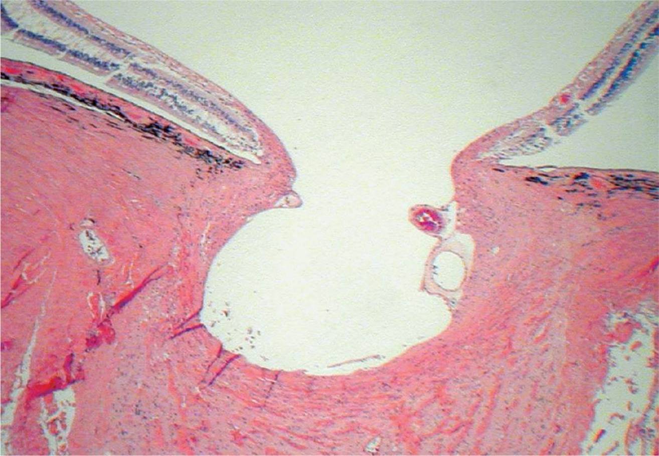
FIGURE 7-29
A) This histologic feature developed quickly over a few days.
B) A special stain for acid mucopolysaccharides (e.g., colloidal iron) can be positive within the optic nerve.
C) A central retinal artery occlusion is a possible cause.
D) In adults, the condition is reversible with appropriate treatment.
28 Histology of an enucleated eye of a patient who sustained trauma is shown (Fig. 7-30). Which statement is the best answer regarding the condition?
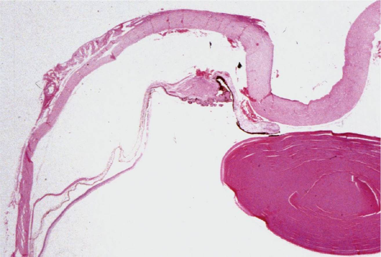
FIGURE 7-30
A) There is a tear in the ciliary body between the circular and longitudinal muscles.
B) Patients with the condition are treated with cycloplegic drops.
C) Clinically, the onset of glaucoma may take years.
D) There is a tear at the iris base.
29 Figure 7-31 shows a histologic section through scleral tissue. Figure 7-32 is a higher magnification of the superior portion of Figure 7-31. What is the most likely scenario in this case?

FIGURE 7-31

FIGURE 7-32
A) Status post repair of a scleral rupture
B) Status post repair of retinal detachment
C) Status post penetrating ocular trauma
D) Scleral staphyloma
30 Homer–Wright rosettes are not found in which of these conditions?
A) Medulloblastoma
B) Retinoblastoma
C) Neuroblastoma
D) Rhabdomyosarcoma
31 Which condition is most likely associated with formation of intraocular cartilage?
A) Phthisis bulbi
B) Posterior PHPV
C) Medulloepithelioma
D) Retinoblastoma
QUESTIONS 32–36 (FIGS. 7-33 to 7-39)
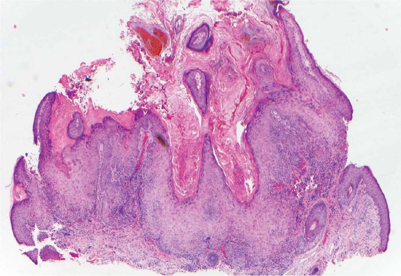
FIGURE 7-33
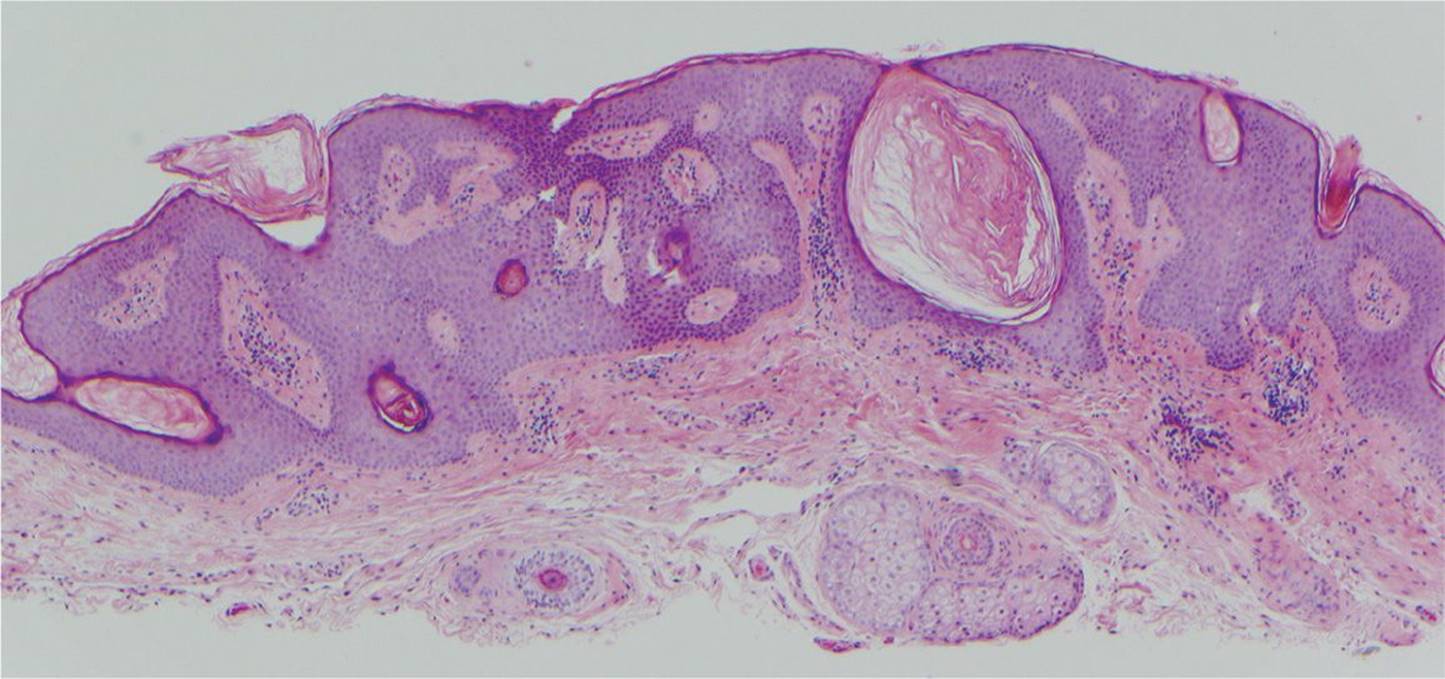
FIGURE 7-34
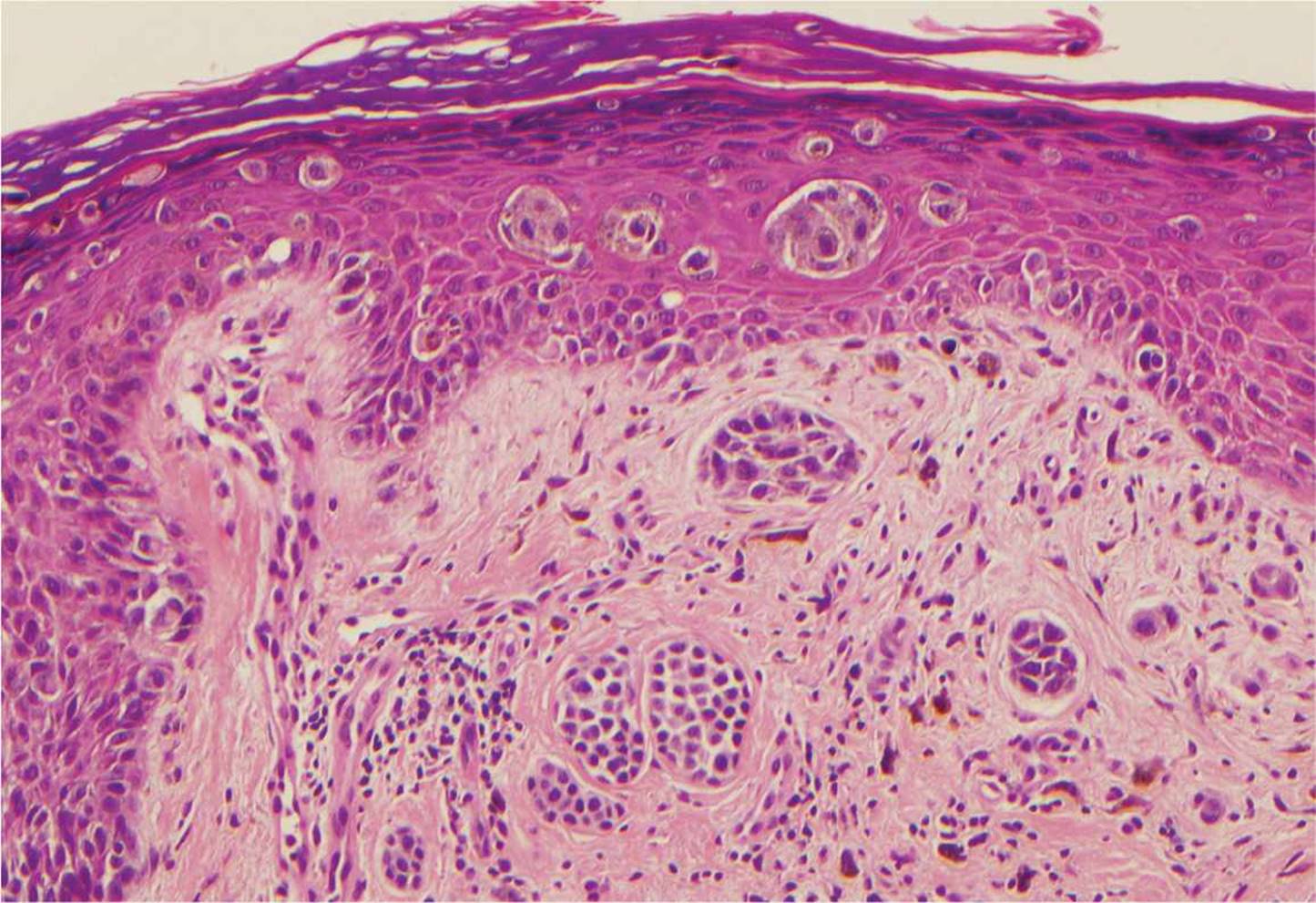
FIGURE 7-35

FIGURE 7-36

FIGURE 7-37

FIGURE 7-38
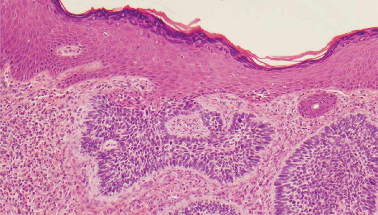
FIGURE 7-39
32 Which lesion may spontaneously disappear?
A) Figure 7-33
B) Figure 7-34
C) Figure 7-35
D) Figure 7-36
33 Which lesion has a stuck-on appearance clinically?
A) Figure 7-33
B) Figure 7-37
C) Figure 7-34
D) Figure 7-36
34 Which lesion is locally invasive with little potential for metastasis?
A) Figure 7-35
B) Figure 7-36
C) Figure 7-38
D) Figure 7-39
35 Which lesion can be found in association with the conjunctival reaction pictured in Figure 7-40?

FIGURE 7-40
A) Figure 7-37
B) Figure 7-38
C) Figure 7-33
D) Figure 7-39
36 Which stain would be helpful in the diagnosis of Figure 7-38?
A) Masson trichrome
B) Gomori methenamine silver
C) Oil Red O
D) Gram stain
QUESTIONS 37–43
37 A 3-year-old patient presents with bilateral leukocoria. What is the least likely diagnosis?
A) Congenital cataracts
B) Retinoblastoma
C) Retinopathy of prematurity
D) Metastasis
38 The right eye was subsequently enucleated. A CT scan was obtained and is shown in Figure 7-41. Which treatment would be least effective for the left eye?

FIGURE 7-41
A) Enucleation
B) Radiation
C) Chemotherapy
D) Cryotherapy
39 The ultrasound of the lesion in the left eye is shown in Figure 7-42. What is the source of the calcification?
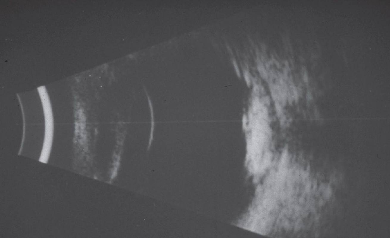
FIGURE 7-42
A) Metaplasia of cells with bone formation
B) Calcium precipitation from the exudative fluid
C) Localized abnormality of calcium metabolism
D) Necrosis of tissue with calcification
40 The histologic specimen is shown in Figures 7-43 and 7-44. Which cell is thought to be the most closely related to these cells?
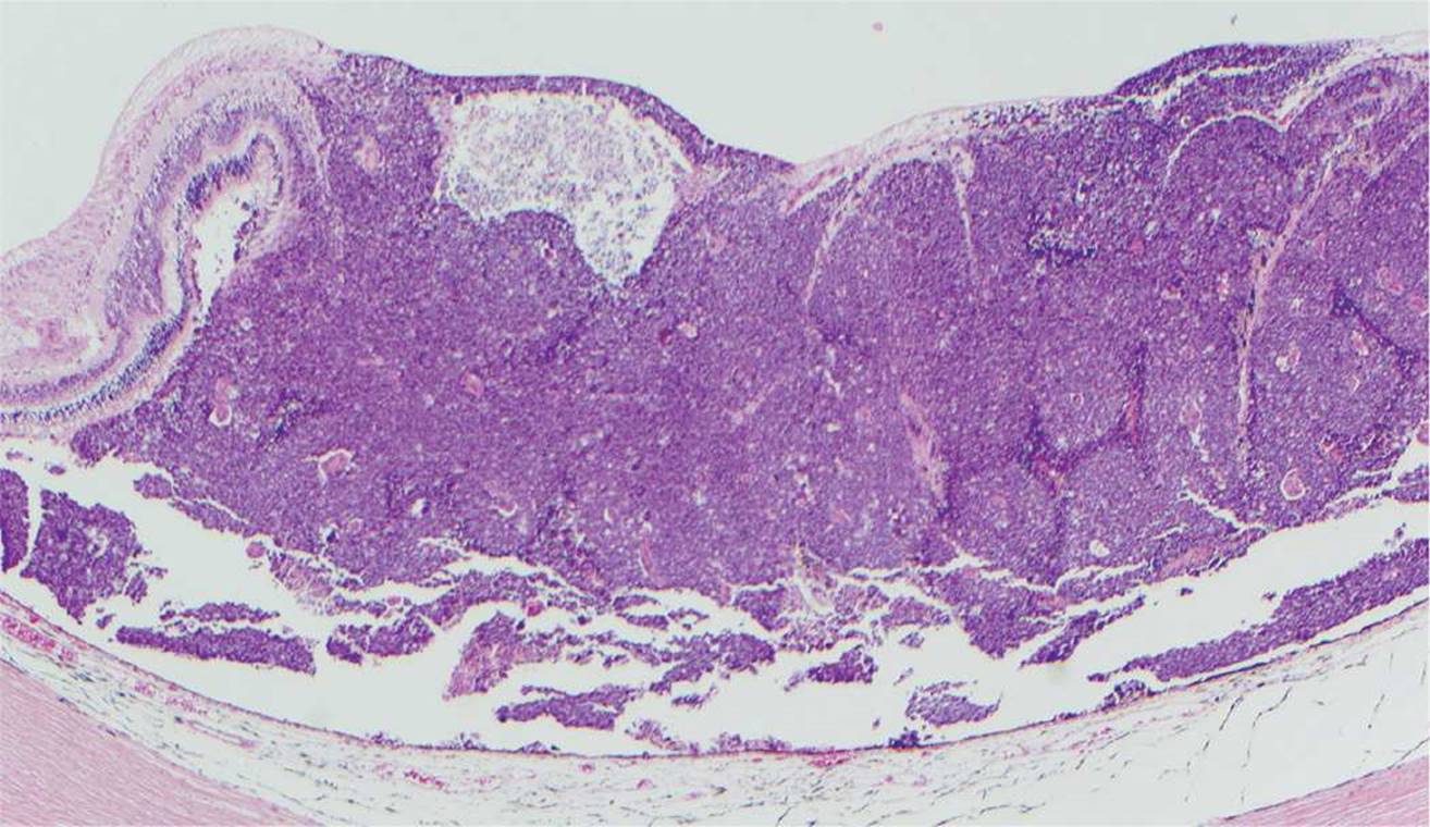
FIGURE 7-43

FIGURE 7-44
A) Ganglion cell
B) Photoreceptor
C) Retinal pigment epithelium
D) Müller cell
41 Which one of the following is not an indicator of a poorer prognosis?
A) Extrascleral extension
B) Tumor cells through the lamina cribrosa of optic nerve
C) Bilateral involvement
D) Tumor diameter greater than 10 DD
42 What type of secondary tumors are these patients at highest risk for?
A) Osteogenic sarcoma
B) Pheochromocytoma
C) Lymphoma
D) Neuroblastoma
43 What is the probability that this child will have an affected child?
A) 40%
B) 6%
C) 1%
D) 80%
44 The central portion of a corneal button (Fig. 7-45) was obtained at time of penetrating keratoplasty. What is the most likely cause for the transplantation?

FIGURE 7-45
A) Pseudophakic bullous keratopathy
B) Keratoconus
C) Fuchs dystrophy
D) Congenital hereditary endothelial dystrophy
45 The nuclei of which cell type is not found in the layer of the retina indicated by the arrow in Figure 7-46?

FIGURE 7-46
A) Bipolar cell
B) Amacrine cell
C) Ganglion cell
D) Horizontal cell
46 A patient is diagnosed with a malignant melanoma localized to the iris. Which statement is false?
A) This melanoma may produce ipsilateral hyperchromia of the iris.
B) Metastases to liver and bone commonly occur.
C) Treatment is by iridectomy.
D) These lesions may be pigmented or amelanotic.
47 Which tumor is found in association with tuberous sclerosis?
A) Choroidal osteoma
B) Melanocytoma
C) Astrocytic hamartoma
D) Capillary hemangioma
48 Which one of the following retinal layers is most severely affected after a central retinal artery occlusion?
A) Inner plexiform layer
B) Retinal pigment epithelium
C) Outer nuclear layer
D) Inner nuclear layer
49 What do ghost cells represent?
A) Hemosiderin-laden macrophages
B) Denatured lens proteins
C) Spherical red blood cells
D) Inflammatory cells
50 Which disease entity does not belong to the group of histiocytosis X?
A) Eosinophilic granuloma of the bone
B) Juvenile xanthogranuloma (JXG)
C) Hand–Schüller–Christian disease
D) Letterer–Siwe disease
1 B) Radial keratotomy (RK)
RK is of historical significance as a refractive surgery, as generally the procedure is no longer performed. There remain special considerations in performing cataract and other types of ocular surgery in these patients when they present to the ophthalmology clinic. The RK technique involved making radial incision into the corneal stroma to produce central corneal flattening. A cross-sectional view of the radial corneal incisions is shown in Figure 7-1. The ALK and LASIK procedures both include a lamellar dissection plane. PRK surgery involves surface ablation.
2 C) It originates from the extraocular muscles.
Children with rhabdomyosarcoma present with rapidly progressive unilateral, painless proptosis. Swelling and ecchymosis may resemble orbital cellulitis. The cell of origin is presumably an undifferentiated, pluripotent cell of the soft tissue. These cells have the capacity to differentiate toward muscle with production of myosin and actin and to show cross-striations. Immunohistochemistry shows positivity for vimentin, myosin, myoglobin, muscle-specific actin, and desmin. The embryonal type is the most common type, followed by the alveolar type. The prognosis is poorest for the alveolar type. The pleomorphic or well-differentiated type occurs in older patients and has the best prognosis.
3 D) Gram-positive hyphae
The Gram stain (Fig. 7-2) is from a patient with canaliculitis. The bacterial organism shown is Actinomyces israelii, which is the most common pathogen associated with canaliculitis. Actinomyces is a filamentous gram-positive rod. Historically, the organism was incorrectly thought to be a fungus, because colonies of the organisms form hyphae.
4 C) This lesion can be found in approximately 25% of older patients.
Figure 7-3 shows a typical Fuchs adenoma (Fuchs reactive hyperplasia, coronal adenoma, Fuchs epithelioma, benign ciliary epithelioma). This lesion is considered to be proliferative rather than neoplastic. The lesion is composed of basement membrane material (type IV collagen and laminin), acid mucopolysaccharides, glycoproteins, and proliferating cells of the nonpigmented ciliary body epithelium. It rarely may cause localized occlusion of the chamber angle. A metastatic lesion from a malignant melanoma would display mostly pleomorphic, pigmented cells with malignant features (high nuclear–cytoplasmic ratio, nucleoli, and mitotic figures).
5 C) Exudative
The histologic section (Fig. 7-7) displays a complete retinal detachment with amorphous eosinophilic material under the retina. This material is not serous as it is in a rhegmatogenous retinal detachment, but contains an abundance of protein. The subretinal fluid in a retinal detachment caused by traction is mostly serous, and the histologic section would show fibrous preretinal membranes in the vitreous. Artifactitious detachments do not have subretinal fluid.
6 B) The cells of origin for these lesions are the retinal pigment epithelial cells (cells of neuroectodermal origin).
The retinal pigment epithelium has the capability for fibrous and osseous metaplasia. This is a nonspecific reaction and is observed most often in phthisical eyes. This metaplasia can lead to intraocular ossification, even with intraocular hematopoiesis. The lesion is benign.
7 D) Ipsilateral hemangiomas of the meninges are common.
The girl has meningocutaneous angiomatosis (Sturge–Weber syndrome, encephalotrigeminal angiomatosis). The syndrome consists of mostly unilateral meningeal calcifications, facial nevus flammeus (port-wine stain) frequently along the distribution of the trigeminal nerve, and congenital glaucoma (30%). Heredity does not seem to be an important factor. Histologically, the syndrome is characterized by cavernous hemangiomas of the skin, lids, choroid (Figs. 7-10 and 7-11), and meninges. The disease has no tendency for spontaneous regression as observed with capillary hemangiomas of the child.
8 D) Cavernous spaces filled with erythrocytes are present.
Histologic section through this choroidal lesion shows a typical cavernous hemangioma, which is characterized by lakes of erythrocytes separated by thin fibrous septa. Choroidal hemangioma is an uncommon hamartoma that has been reported to exhibit two clinical growth patterns: (1) circumscribed tumors without systemic disease and (2) diffuse tumors often associated with the Sturge–Weber syndrome. The lesion is histologically benign; however, the lesion can lead to an exudative retinal detachment if untreated (e.g., as seen in previous case, Figs. 7-4 to 7-7). Therapeutic options are photocoagulation and cryotherapy. The solitary type is histologically characterized by a cavernous hemangioma with sharply demarcated pushing borders, often compressing surrounding melanocytes and choroidal lamellae. This is visible clinically as a ring of hyperpigmentation in the periphery of such lesions (Fig. 7-4) and on fluorescein angiography as a ring of blockage of the underlying choroidal fluorescence (Fig. 7-5). von Hippel–Lindau disease has vascular lesions of the retina and cerebellum; however, these are capillary hemangioblastomas. In contrast, capillary hemangiomas consist of multiple capillaries surrounded by epithelial cells and pericytes.
9 D) Descemet stripping automated endothelial keratoplasty
The corneal button (Fig. 7-12) shows a near full-thickness defect in the corneal with only Descemet membrane intact, which is known as a descemetocele. Descemetoceles and small perforations may initially be managed with cyanoacrylate and a bandage contact lens. A small leak with an intact anterior chamber may also benefit from patching of the eye and an agent to reduce the intraocular pressure, for example, systemic carbonic anhydrase inhibitor. Other treatment options include amniotic membrane transplantation, conjunctival flap, and keratoplasty (anterior lamellar or penetrating). Descemet stripping automated endothelial keratoplasty does not have a role in treating descemetoceles.
10 C) The result is positive and steroids should be continued.
Temporal arteritis is also known as giant cell arteritis (although giant cells are not essential to make the diagnosis). The condition is diagnosed definitively with temporal artery biopsy. Temporal arteritis is characterized by granulomatous inflammation of blood vessels. The disease can result in occlusion of the vessels supplying the optic nerve head (posterior ciliary vessels) and cause arteritic anterior ischemic optic neuropathy. The figure shows extensive granulomatous inflammation. Giant cells are seen along (Fig. 7-13, bottom right). A portion of the narrowed arterial lumen is also visible in the figure.
11 B) Surface epithelium is found in the anterior chamber.
This condition is known as epithelial ingrowth (downgrowth) and occurs when surface epithelium of the eye gains access to the internal structure of the globe. This is generally seen after accidental or surgical penetration of the eye. The multilayered nonkeratinized squamous epithelium grows over any available surface (e.g., along the anterior iris as seen in Fig. 7-14) and can cause an obstruction of the trabecular meshwork, thus causing a secondary glaucoma. In most advanced cases, the eye is lost. The ICE syndrome is characterized by a unilateral disease in young to middle-aged adults. Abnormal corneal endothelium grows over the trabecular meshwork and over the anterior iris surface. Secondary open-angle glaucoma may develop. Three different clinical phenotypes are known: Chandler syndrome, Cogan–Reese (iris nevus), and essential iris atrophy.
12 C) Periodic acid–Schiff (PAS)
The epithelium in the anterior chamber covering the trabecular meshwork and growing over the iris is a nonkeratinized stratified squamous epithelium. To differentiate this epithelium according to conjunctival or corneal origin, the identification of goblet cells may help. Goblet cells are found in the conjunctival epithelium and are highlighted with PAS stain (Fig. 7-14). Hematoxylin–eosin is the standard stain for most tissues. Oil Red O is an excellent stain for lipid; however, this must be done on fresh tissue. Congo red is used to look for amyloid material. With a polarized microscope, amyloid stained with Congo red demonstrates birefringence and dichroism.
13 C) Plasma cells–containing immunoglobulin
Plasma cells, a type of lymphocyte, commonly have a perinuclear halo (Fig. 7-15, arrowheads). Russell bodies appear as rounded eosinophilic cytoplasmic inclusions (Fig. 7-15, arrow). The inclusions represent aggregated unreleased immunoglobulin components within the plasma cells or extracellularly.
14 D) The diagnosis is confirmed with CT and serologic studies.
15 A) Developmental abnormality
The patient has persistent hyperplastic primary vitreous (PHPV). The condition is also known as persistent fetal vasculature, including the presence of a stalk of vitreous. The embryonic primary vitreous and hyaloid vasculature system persist. This condition is mostly unilateral and is characterized clinically by leukocoria (the most common lesion simulating retinoblastoma). However, contrary to retinoblastoma, PHPV is associated with a microphthalmic eye. Histologically, it is characterized by elongated ciliary processes, persistent hyaloid vessel (Fig. 7-16), retrolental fibrovascular tissue (with posterior lens capsule dehiscence) sometimes containing adipose tissue (“lipomatosis lentis”), cartilage, and smooth muscle. The diagnosis is made clinically, and adjunctive tests are unrevealing.
16 B) This is a benign cutaneous disorder rarely involving the eye.
The child has JXG (nevoxanthoendothelioma) of the iris. This is a benign cutaneous disorder of infants with typical raised orange skin lesions occurring singly or in crops and regressing spontaneously. Ocular involvement is rather rare and occurs mostly under 6 months of age. Children may present with spontaneous anterior chamber hemorrhage and secondary glaucoma. Histologically, the lesion is characterized by a diffuse granulomatous inflammatory reaction with many histiocytes and Touton giant cells, as seen in Figure 7-17. The histiocytes are positive for antibodies binding to macrophages (e.g., CD 68).
17 B) Oncocytoma
Ocular oncocytomas (adenolymphomatous tumor, apocrine cystadenoma, oxyphilic cell adenoma) are rare, mostly benign neoplasms. The lesions are most commonly found on the caruncle, especially in elderly women (median age 73). Oncocytoma has also been described in the conjunctiva, mucocutaneous junction of the eyelid (Fig. 7-18), lacrimal sac, and lacrimal gland. The tumor arises from accessory lacrimal glands. Histologic examination reveals solid nests and cords of polyhedral cells exhibiting abundant, finely granular acidophilic cytoplasm and round to oval paracentral nuclei, usually containing a single prominent nucleolus (Fig. 7-19). Cystic cavities are identified within the tumor. Adenoid cystic carcinoma (malignant cylindroma) occurs in the lacrimal gland in young adults. The tumor causes pain (infiltration of perineural lymphatics) and is histologically characterized by a “Swiss cheese” pattern. A pyogenic granuloma is a type of granulation tissue composed of inflammatory cells and budding capillaries that form a radial pattern. A papilloma is characterized by fronds or fingerlike projections of acanthotic epithelium overlying a fibrovascular stroma.
18 B) Multiple partially atypical mitochondria
Electron microscopic examination shows a cytoplasm densely packed with mitochondria. The mitochondria are sometimes atypical. This is characteristic of oncocytomas.
19 A) This can be a chronic inflammation of the Zeis sebaceous glands.
This lesion is a chalazion. A chronic inflammation of the meibomian glands (deep chalazion) or Zeis sebaceous glands (superficial chalazion) results in a hard, painless nodule. Histologically, it is characterized by zonal granulomatous inflammation, including multinucleated giant cells, around clear spaces previously filled with lipid that is dissolved in the processing (Fig. 7-20). It is important to do a biopsy to exclude a sebaceous gland carcinoma in patients with recurrent chalazion. This type of carcinoma can mimic a recurrent chalazion or a chronic blepharitis. Moll glands are apocrine sweat glands in the eyelid and are not associated with chalazion formation.
20 C) Is consistent with a zonal lipogranuloma
The clear spaces in this chalazion were previously filled with lipid that dissolved out during tissue processing. The pathogenic principle of a chalazion is a lipogranulomatous inflammatory process. In contrast to xanthomas, the lipid is located in the extracellular space. In a frozen section without paraffin embedding, the lipid is preserved and visible. A vascular channel should at least show endothelial cells.
21 B) Herpes simplex
A granulomatous reaction to Descemet membrane (including multinucleated giant cells) (Figs. 7-21 and 7-22) is most frequently seen with disciform keratitis with a history of herpes simplex or herpes zoster keratitis. This peculiar reaction to Descemet membrane may be related to an altered antigenicity of the membrane and subsequent development of an autosensitivity reaction. This reaction is very uncommon with other etiologic agents.
22 D) The disease is phacolytic glaucoma.
The patient has a phacoanaphylactic (phacoimmune) endophthalmitis, rather than phacolytic glaucoma. Phacoanaphylactic endophthalmitis (also known as lens-induced granulomatous endophthalmitis) develops after exposure of large amounts of lens antigen to the immune system and abrogation of tolerance to lens protein. The disease is characterized clinically by signs of chronic uveitis, choroidal detachment, and hypotonia bulbi. Normally, lens protein is recognized as “self.” In the case of a breakdown of the T-cell tolerance, antibodies against the lens proteins may be produced, initiating a chronic inflammation. In this case, the lens nucleus was lost during cataract surgery (Fig. 7-23) and a phacoanaphylactic endophthalmitis developed. Histologically, it is characterized by a zonal granuloma with an inner core degenerating lens material surrounded by neutrophils and epithelioid cells and occasionally multinucleated giant cells (Fig. 7-24). There is an outer layer of granulation tissue. In contrast, phacolytic glaucoma is characterized by the leakage of denatured lens proteins through an intact capsule (e.g., hypermature cataract). This initiates a mild foreign body reaction, and macrophages get swollen and engulf denatured lens material. These macrophages can block the trabecular outflow and can cause a secondary open-angle glaucoma (phacolytic glaucoma).
23 A) This condition can be caused by exposure to infrared radiation.
This patient has true exfoliation, a rare condition characterized by a delamination or splitting of the lens capsule (Fig. 7-25). Scrolls of lens capsule may also be seen (Fig. 7-26). Infrared radiation may cause “true exfoliation” of the lens capsule in glass blowers or welders. True exfoliation may also occur secondary to inflammation, metallic intraocular foreign bodies, trauma, and often idiopathically in older patients (as in the case presented). Clinically, on slit lamp examination, this patient showed a loose scroll of lens capsule in the anterior chamber. The only increased risk in cataract surgery is in properly forming the capsulorhexis. There is no association with glaucoma.
This is contrast to pseudoexfoliation syndrome, a systemic condition characterized by deposition of a peculiar white, fluffy material in the eye (e.g., lens capsule, the zonules, the ciliary epithelium, the iris pigment epithelium, and the trabecular meshwork) and in other connective tissues throughout the body. It is more common in Scandinavians and is quite rare in African Americans. The amorphous eosinophilic material lines up perpendicular to the lens (like iron filings lining up on a magnet). The iris pigment epithelium displays a sawtooth posterior configuration. The incidence of zonular dialysis during cataract surgery is higher in these patients. The iris may have peripapillary transillumination defects in this condition in contrast to the radial defects seen with pigment dispersion syndrome.
24 C) Nodular drusen
Nodular (hard) drusen consist of a focal thickening of the basement membrane of the retinal pigment epithelium (Fig. 7-27). Clinically, they appear as small yellow or yellow-white spots measuring ≤63 µm in diameter. Soft drusen are larger (>63 µm) and appear less dense and more fluffy. Basal laminar deposits consist of banded basement membrane material (wide-spaced collagen) located between the basal plasmalemma of the retinal pigment epithelium and its basement membrane. Basal linear deposits refer to material located external to the basement membrane of the retinal pigment epithelium.
25 A) Basal cell carcinoma, morphea type
Basal cell carcinoma is characterized histologically by a proliferation of basophilic cells. It is by far the most common malignant tumor of the eyelid. The tumor can be grouped into three types: nodular, superficial, and morphea form. Ulceration and pigmentation may or may not occur. Typical features of the nodular type are peripheral palisading of the tumor cells. The superficial type shows irregular buds of basaloid cells arising from multiple foci of the epidermal undersurface. The morphea form type is less common and clinically more aggressive. Morphea form lesions are characterized by tumor cells growing in thin, elongated strands or cords within a desmoplastic (fibrotic) dermis (Fig. 7-28).
26 B) Lower eyelid—inner canthus—upper eyelid—lateral canthus
The reason for this order is probably the amount of ultraviolet light exposure during life.
27 B) A special stain for acid mucopolysaccharides (e.g., colloidal iron) can be positive within the optic nerve.
Figure 7-29 shows an excavated, deeply cupped optic disc. The most likely cause is an elevated IOP over the course of many years. In the case of a cavernous optic atrophy (Schnabel’s), cystic spaces generally posterior to the lamina cribrosa are found. The cystic spaces are filled with hyaluronic acid that stains positive with a stain for acid mucopolysaccharides (e.g., colloidal iron). A central retinal occlusion leads to an atrophic optic nerve; however, the disc is not excavated. Reduction and normalization of IOP can result in reversal of optic disc cupping, which is common in children and less frequent in adults.
28 B) Patients with the condition are treated with cycloplegic drops.
A cyclodialysis cleft is shown with characteristic disinsertion of the ciliary body from the scleral spur (Fig. 7-30). The condition can occur as an operative complication, after glaucoma surgery, or after trauma. Typically, the eye is hypotonous; however, there may be an acute pressure elevation with closure of the cleft. Initial management includes medial therapy with cycloplegic agents (e.g., atropine). Larger clefts (>4 clock hours) or those not responding to medical therapy are treated with an argon laser or direct cyclopexy. Answer choices A and C pertain to traumatic angle recession. Choice D refers to iridodialysis.
29 B) Status post repair of retinal detachment
The histologic picture (Figs. 7-31 and 7-32) shows an empty cystic space in the sclera with an overlying polyfilamentous suture associated with a foreign body giant cell reaction. The empty scleral space represents a dissolved encircling band from prior surgery for retinal detachment. The polyfilamentous suture was used to fix the band to the sclera. A ruptured sclera should be associated with full-thickness scleral scars. A scleral staphyloma is ectatic and thinned sclera lined by choroid with or without retina.
30 D) Rhabdomyosarcoma
Homer–Wright rosettes are characterized by cells that line up around an area containing cobweb-like material, but no acid mucopolysaccharides are present. Homer–Wright rosettes do not have central lumen (Fig. 7-47, arrows). These rosettes are not specific for retinoblastoma and are found also in neuroblastoma and medulloepithelioma. Rhabdomyosarcoma displays no rosette formation. In contrast, Flexner–Wintersteiner (F-W) rosettes are the characteristic rosettes of retinoblastoma. F-W rosettes are true rosettes. The ring of cells surrounds a central lumen, as seen prominently in the center of Figure 7-47 and elsewhere in the image. The presence of F-W rosettes makes the diagnosis of a well-differentiated retinoblastoma. However, special stains show the lumen contains hyaluronidase-resistant acid mucopolysaccharides.

FIGURE 7-47
31 C) Medulloepithelioma
The medulloepithelioma (diktyoma) arises from the ciliary epithelium. Nonteratoid and teratoid varieties (presence of heteroplastic elements) are known. Both can be benign or malignant. The teratoid type may contain cartilage and/or rhabdomyoblasts. Phthisis bulbi can result in metaplasia of the retinal pigment epithelium with bone formation. Cartilage formation is virtually unknown. Posterior PHPV is characterized by vitreous membranes extending from the disc usually toward the equatorial zone, posterior radial retinal folds, disturbance of macular function, and retinal detachment. Cartilage formation is not a feature of posterior PHPV, contrasting with the anterior form in which it has been described in rare cases. Retinoblastoma displays calcification secondary to rapid proliferation and necrosis of cells, but no cartilage formation occurs.
32 A) Figure 7-33
33 C) Figure 7-34
34 D) Figure 7-39
35 A) Figure 7-37
36 C) Oil Red O
The lesions pictured are enumerated:
Figure 7-33 = keratoacanthoma
Figure 7-34 = seborrheic keratosis
Figure 7-35 = malignant melanoma
Figure 7-36 = squamous cell carcinoma
Figure 7-37 = molluscum contagiosum
Figure 7-38 = sebaceous carcinoma
Figure 7-39 = basal cell carcinoma, nodular
Sebaceous gland carcinomas may present insidiously as chronic, recurrent chalazia or blepharitis. If sebaceous carcinoma is suspected, fresh tissue should be processed for lipid stains such as Oil Red O. It is worth noting that, during the embedding process in paraffin, different organic solvents (e.g., xylene) are used, which dissolve and leach out the lipids. Therefore, fresh tissue or frozen section has to be used to preserve the lipids and apply a lipid stain such as Oil Red O or Sudan black. Lipid stains are helpful when looking for sebaceous carcinoma.
Sebeceous carcinomas are notorious for skip lesions and require wide excision. Metastasis to regional lymph nodes is possible.
Basal cell carcinoma is the most frequently occurring eyelid lesion, often in sun-exposed areas. These may present as raised lesions with pearly borders, telangiectatic vessels, and an ulcerated crater (rodent ulcer). Histopathologically, there are palisading rests of basophilic cells with scanty cytoplasm and hyperchromatic nuclei. Basal cell carcinoma is locally invasive and rarely metastasizes.
Squamous cell carcinoma (SCC) is also related to sun exposure. Proliferation of eosinophilic epithelium with rests of keratin pearls traverse the basement membrane into the underlying dermis. Surgical excision using the Mohs technique is recommended. Metastasis occurs to regional nodes.
The keratoacanthoma may be confused with the SCC; however, it presents much more rapidly (weeks). It is worth noting that many authors consider keratoacanthoma to be a low-grade variant of SCC. The tumors consist of a dome-shaped lesion with rolled edges and a central crater containing keratin. These may resolve spontaneously over several months; however, complete excision is advised as some may develop into SCC.
Seborrheic keratoses are benign lesions that have a greasy, “stuck-on” appearance. Pathologic examination shows hyperkeratosis and papillomatosis with keratin pseudo-horn cysts.
Molluscum contagiosum is caused by a pox virus and appears as raised, umbilicated lesions on the eyelid, face, and genital regions. Epithelia thickened by intracytoplasmic molluscum bodies are present in the central crater. These may be shed into the tear film from eyelid margin lesions, causing a chronic follicular conjunctivitis. Treatment is by cryotherapy, curettage, or excision.
37 D) Metastasis
Congenital cataracts, retinoblastoma, and retinopathy of prematurity, among others, are in the differential of bilateral leukocoria. Intraocular metastatic disease to the eyes is extremely rare in children.
38 D) Cryotherapy
The CT scan shows a high density lesion consistent with calcification within the tumors (Fig. 7-41) in the left eye. In this case of retinoblastoma, the right eye was enucleated and an spherical orbital implant and prosthesis (curvilinear opacity anteriorly) are visible on the right side. Treatment for the left eye includes enucleation, radiation, and chemotherapy. Cryotherapy may be effective for small lesions. For posterior lesions, as pictured, effective placement of cryotherapy would be difficult to achieve. Moreover, it would be ineffective for multicentric tumors. Chemotherapy or radiation is preferable to try to preserve vision in the left eye.
39 D) Necrosis of tissue with calcification
Retinoblastoma is a rapidly growing tumor that can outgrow its vascular supply. This rapid growth leads to necrosis of tissue and secondary calcification. Whitish flecks of calcification can be seen in the whitish substance of the tumor (Fig. 7-42).
40 B) Photoreceptor
Figure 7-43 shows the tumor at low magnification. The detached retina (left side) is contiguous with the tumor. F-W rosette (Fig. 7-44, center) is characteristic of retinoblastoma. These rosettes represent abnormal photoreceptor differentiation. Homer–Wright rosettes and fleurettes are also along the spectrum of photoreceptor differentiation.
41 D) Tumor diameter greater than 10 DD
Extraocular spread of retinoblastoma indicates a poor prognosis. Patients with bilateral involvement may be at higher risk of trilateral retinoblastoma. The size of the tumor, per se, does not affect prognosis. Figure 7-48 shows tumor cells within the substance of the optic nerve extending to the lamina cribrosa. Numerous Homer–Wright rosettes are seen (especially at the top right, Fig. 7-48).

FIGURE 7-48
42 A) Osteogenic sarcoma
Secondary tumors are more common in these children and include osteogenic sarcoma, malignant melanoma, lymphoma and leukemia, rhabdomyosarcoma, and medulloblastoma. The most common malignancies are sarcomas, particularly osteogenic sarcoma.
43 A) 40%
In this bilateral case, there is a high likelihood that a somatic, heritable mutation exists. Half of this child’s offspring will receive the abnormal chromosome. With 80% penetrance, 40% of his children will be affected.
44 C) Fuchs dystrophy
The cornea has multiple excrescences at the level of Descemet membrane consistent with Fuchs dystrophy (Fig. 7-45). Keratoconus would have breaks in Bowman layer and epithelium along with thinning and scarring of the cornea, but with a normal Descemet membrane. Pseudophakic bullous keratopathy and congenital hereditary endothelial dystrophy have thickening of the corneal stroma and loss of endothelial cells.
45 C) Ganglion cell
The ganglion cell nuclei are found in the innermost layer of nuclei. The inner nuclear layer contains cell bodies of the bipolar, amacrine, horizontal, and Müller cells. The outer nuclear layer has the cell bodies of the photoreceptors (Fig. 7-46).
46 B) Metastases to liver and bone commonly occur.
If isolated to the iris, malignant melanoma behaves in a more benign fashion than does melanoma of the choroid. Involvement of the ciliary body or choroid portends a poorer prognosis. Metastases are rare, and localized iridectomy may remove the tumor entirely. These lesions may be pigmented or amelanotic. Ipsilateral hyperchromia may be present.
47 C) Astrocytic hamartoma
Findings associated with tuberous sclerosis include astrocytic hamartomas, intracerebral calcification, seizures, mental retardation, adenoma sebaceum, and ash leaf spots. Capillary hemangiomas may be found in von Hippel–Lindau disease.
48 A) Inner plexiform layer
The retinal arteries supply oxygen to the superficial layers of the retina, including the nerve fiber layer, ganglion cell layer, inner plexiform layer, and inner third of the inner nuclear layer. The other layers are nourished primarily by the choroid.
49 C) Spherical red blood cells
Ghost cells represent hemolyzed red blood cells. After a long-standing hyphema or vitreous hemorrhage, the red blood cells lose their normal shape and become more spherical. These cells are more rigid and less able to deform to exit through the trabecular meshwork, resulting in a secondary open-angle glaucoma. On slit lamp microscopy, they appear as khaki-colored cells in the anterior chamber.
50 B) Juvenile xanthogranuloma (JXG)
Histiocytosis X (Langerhans cell histiocytosis, Langerhans granulomatosis) is characterized by a proliferation of Langerhans cells in an inflammatory background. Immunohistochemically, the cells stain positively for S-100 and vimentin. Ultrastructurally, the cells contain Birbeck granules (containing a central dense core and a thick outer sheath). Three interrelated clinicopathologic entities are known: (1) eosinophilic granuloma of bone (benign tumor arising often in the outer part of the upper orbital rim), (2) Hand–Schüller–Christian disease (bony lesions in the skull, exophthalmos, and diabetes insipidus), and (3) Letterer–Siwe disease (fatal disease with diffuse histiocytosis). JXG does not belong to the histiocytosis X group. JXG is negative for S-100 and contains no Birbeck granules.