
MARCUS J. KO AND WENDY W. LEE
1 Which statement about the orbital septum is false?
A) During entropion repair, it is very important to recognize the orbital septum of the lower eyelid as being different from the aponeurosis or lower eyelid retractors.
B) The orbital septum arises from a condensation of the periosteum of the orbital rim called the arcus marginalis.
C) The orbital septum inserts on the superior border of the tarsus in the upper eyelid.
D) The orbital septum serves as a barrier to the spread of infection from the superficial eyelids to the orbital tissues.
2 Which statement concerning the medial canthal area is true?
A) All of the attachments anchoring the tarsi to the medial orbital wall lie anterior to the lacrimal sac and attach to the maxillary portion of the frontal bone.
B) The lacrimal sac lies posterior to the orbital septum.
C) The muscle pump of the lacrimal pump mechanism is innervated by the fifth cranial nerve.
D) Lockwood ligament attaches posterior to the lacrimal sac.
3 Which statement regarding fat encountered during eyelid surgery is false?
A) Preaponeurotic fat is orbital fat.
B) Extraconal orbital fat is an important landmark in identifying the levator aponeurosis.
C) The removal of fat from the upper eyelid nasal, central, and lateral fat pads may be done with impunity.
D) In the upper eyelid, the nasal fat pad is small, whereas the lateral fat pad is the small fat pad in the lower eyelid.
4 Which statement regarding Whitnall ligament (superior transverse ligament) is false?
A) Whitnall ligament attaches medially to the trochlea, laterally to the capsule of the lacrimal gland, and to the lateral orbital wall.
B) This ligament is a condensation of the sheath of the levator muscle and serves as a check ligament to prevent excessive elevation of the eyelid.
C) Whitnall ligament acts to change the direction of pull of the levator muscle from horizontal to vertical.
D) This ligament passes anterior to the lacrimal gland.
5 Which statement about eyelid anatomy is false?
A) The gray line is formed by the muscle of Riolan and represents the observable edge of the pretarsal orbicularis at the eyelid margin.
B) The posterior lamella of the eyelid consists of the conjunctiva and tarsus.
C) The mucocutaneous junction occurs where the eyelashes emerge from the eyelid.
D) The peripheral and marginal arterial arcades allow for anastomosis between the internal and external carotid systems.
6 Features of the orbicularis muscle include:
A) closure of the eyelid, depression of the eyebrow, and facilitation of tear drainage.
B) pretarsal orbicularis inserts temporally to become the lateral canthal tendon, contraction narrows the palpebral fissure, and the orbital portion of the muscle inserts medially on the posterior lacrimal crest.
C) the deep head of the medial pretarsal muscle is called Horner tensor tarsi and innervation of the orbicularis muscle by cranial nerve III is divided into three segments (pretarsal, preseptal, and orbital).
D) the zygomaticofacial nerve innervates the upper lid orbicularis, the frontal branch of cranial nerve VII sends motor fibers to the upper lid orbicularis, and the preseptal orbicularis divides to encompass the lacrimal gland.
7 Which one of the following muscle groups is paired incorrectly?
A) Tensor tarsi muscle—deep head of the pretarsal orbicularis
B) Nasalis—preseptal orbicularis
C) Superciliary corrugator muscle—orbital orbicularis
D) Frontalis—procerus muscle
8 Which structure and its bony framework are paired incorrectly?
A) Lacrimal sac fossa—lacrimal and maxillary bones
B) Optic canal—greater and lesser wings of the sphenoid bone
C) Inferior orbital fissure—maxilla, zygomatic bone, palatine bone, and greater wing of the sphenoid bone
D) Anterior and posterior ethmoidal foramen—ethmoid and frontal bones
9 The carbon dioxide laser has all of the following characteristics except:
A) wavelength in the infrared spectrum
B) able to be seen by the human eye
C) utilized for orbital tumor excision
D) operates at 10.6 µm
10 The site of action of botulinum toxin type A (Botox), when used to treat facial movement disorders, is the:
A) motor nerve terminal, inhibiting acetylcholine release
B) motor nerve terminal, promoting cholinesterase release
C) plasma membrane (sarcolemma) of the striated muscle, inhibiting acetylcholine release
D) plasma membrane (sarcolemma) of the striated muscle, promoting cholinesterase release
11 Which one of the following is not a feature of basal cell carcinoma?
A) Pearly elevated margins
B) Likely spread to regional lymph nodes
C) Ulcerated epithelium
D) Telangiectatic vessels
12 The following factors are all associated with cutaneous cancers except:
A) increased sun exposure
B) increased age
C) red hair
D) increased natural skin pigmentation
13 Features most consistent with a malignant eyelid lesion include:
A) tenderness, erythema, alteration in pigment pattern
B) disruption of tarsal architecture, raised pearly margins, pruritus
C) lash loss, central ulceration, rapid growth
D) ipsilateral lymph node metastasis, hyperkeratosis, dark pigmentation
14 A 40-year-old, red-haired, blue-eyed man of Irish descent living in Tucson, Arizona, presents with a raised firm nodule with telangiectasia on his lower eyelid. This lesion most likely represents:
A) nodular basal cell carcinoma
B) morpheaform basal cell carcinoma
C) squamous cell carcinoma
D) sebaceous adenocarcinoma
15 Features of a keratoacanthoma include all of the following except:
A) spontaneous resolution
B) loss of eyelashes
C) ulcerated crater filled with lipids
D) rapid growth
16 The 5-mm raised skin lesion with central ulceration is least likely to be which of the following?
A) Keratoacanthoma
B) Squamous cell carcinoma
C) Basal cell carcinoma
D) Malignant melanoma
17 The CT examination in Figure 6-1 is from a 55-year-old man. Which one of the following is least likely in the differential diagnosis?

FIGURE 6-1
A) Metastatic prostate carcinoma
B) Sphenoid wing meningioma
C) Fibrous dysplasia
D) Metastatic melanoma
QUESTIONS 18 and 19 The defect of this eyelid (Fig. 6-2) of a 65-year-old woman resulted after a Mohs surgical procedure.
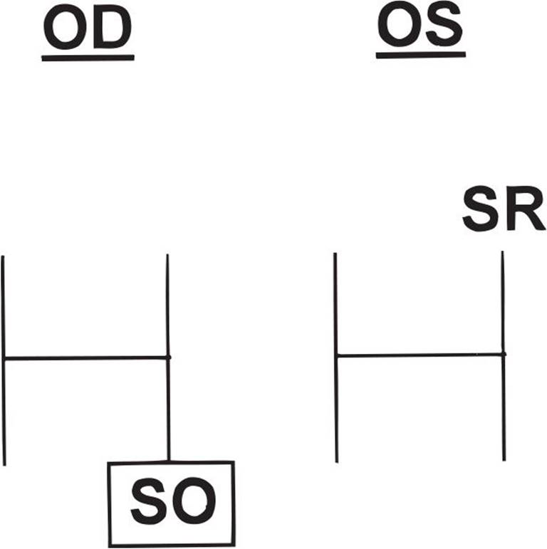
FIGURE 6-2
18 What surgical method would be the most appropriate for reconstruction of the posterior lamella?
A) Cutler–Beard flap
B) Bipedicle myocutaneous flap
C) Full-thickness skin graft
D) Hughes tarsoconjunctival flap
19 What is the least likely cause that led to the eyelid defect pictured in Figure 6-2?
A) Basal cell carcinoma
B) Metastatic cancer
C) Sebaceous cell carcinoma
D) Squamous cell carcinoma
20 All of the following statements concerning lymphatic and venous drainage are true except:
A) Lymphatic vessels of the orbit drain along the lateral portion of the cavernous sinus.
B) Lymphatic vessels serving the medial portion of the upper eyelid drain into submandibular lymph nodes.
C) Lymphatic vessels serving the lateral portions of the upper eyelid drain into preauricular nodes.
D) Pretarsal venous drainage of the medial upper eyelid is into the angular vein and the lateral venous drainage is into the superficial temporal vein system.
21 Xanthelasma eyelid lesions have all the following features except:
A) associated with systemic hyperlipidemic conditions in approximately 25% of patients
B) located in the basal epithelial layer of the skin
C) associated with the Erdheim–Chester disease
D) microscopically contain foamy histiocytes
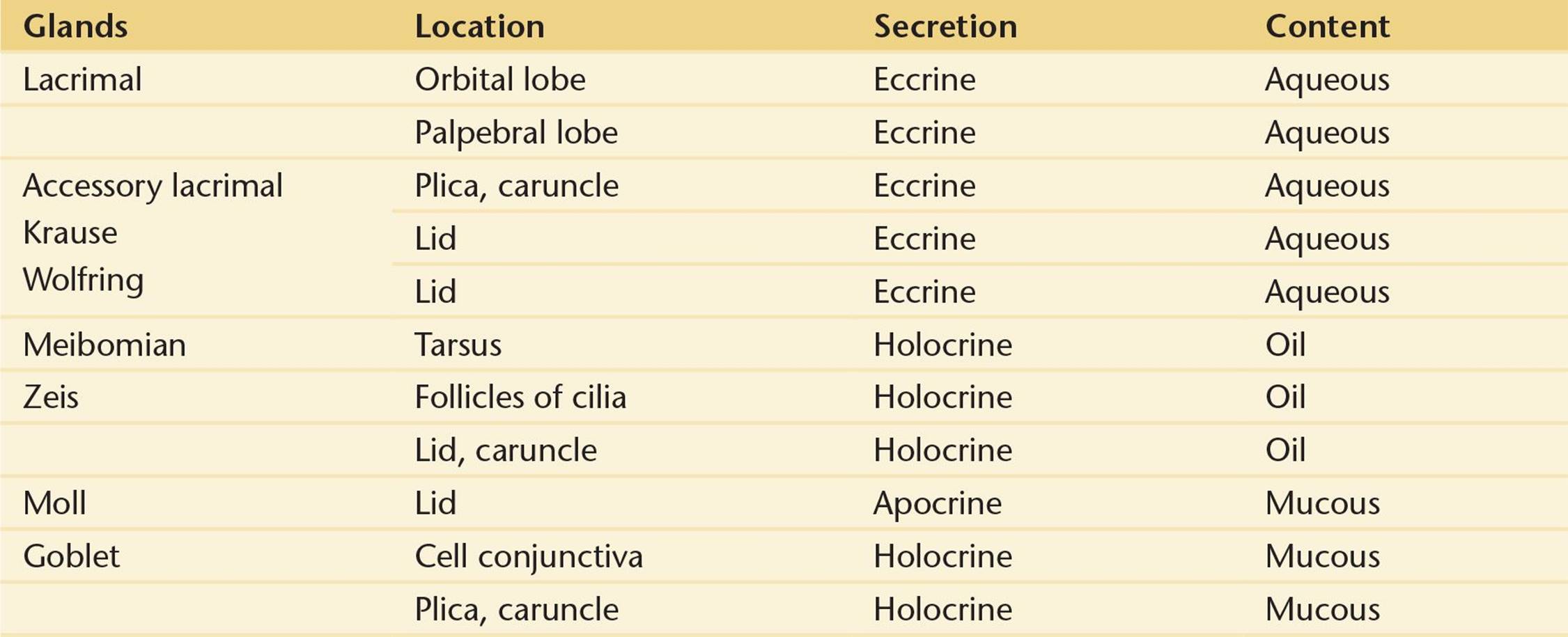
22 Which of the following glands are matched with their correct types of secretions?
A) Moll—apocrine, main lacrimal gland—eccrine, meibomian glands—apocrine
B) Glands of Krause—holocrine, gland of Zeis—apocrine, goblet cells—holocrine
C) Glands of Wolfring—eccrine, gland of Moll—apocrine, goblet cells—holocrine
D) Main lacrimal gland—eccrine, meibomian glands—holocrine, gland of Zeis—apocrine
23 Which gland does not contribute to the aqueous layer of the tear film?
A) Krause
B) Main lacrimal
C) Zeis
D) Wolfring
24 Which statement about eyelid abnormalities is false?
A) Congenital coloboma of the eyelid always involves the lower eyelid and can vary from a small notch to a complete absence of the eyelid.
B) Cryptophthalmos is a rare condition that is caused by a lack of differentiation of eyelid structures and is characterized by absence of a palpebral fissure with uninterrupted skin from the forehead over the eye to the skin of the cheek.
C) Ankyloblepharon filiforme adnatum is a form of ankyloblepharon in which the eyelid margins are connected by thin strands of tissue.
D) Distichiasis is a condition in which an accessory row of eyelashes grows from or are posterior to the meibomian orifices.
25 A patient calls to report pain, sudden swelling, and decreased vision the night after a blepharoplasty procedure. What should be done?
A) Advise the patient to use ice packs to decrease the swelling.
B) Set up an appointment for the patient to see you the next day.
C) Make arrangements to see the patient as soon as possible.
D) Reassure the patient that discomfort, swelling, and blurry vision are normal postoperative findings.
26 Which one of the following is found in the blepharophimosis syndrome?
A) Euryblepharon
B) Ankyloblepharon
C) Epiblepharon
D) Telecanthus
27 A 10-year-old girl has bulging and blepharoptosis of both upper eyelids and repeated episodes of eyelid inflammation and swelling. What is the most likely diagnosis?
A) Blepharochalasis
B) Dermatochalasis
C) Steatoblepharon
D) Blepharospasm
28 The best choice below for surgical repair of a ptotic upper eyelid exhibiting a high eyelid crease, a margin to reflex distance (MRD) of 0 mm, and excellent levator function would be:
A) resection of the superior tarsal muscle
B) unilateral frontalis suspension using autogenous fascia lata
C) reattachment of the dehisced levator aponeurosis
D) plication of the levator muscle (16 mm)
29 The most common form of blepharoptosis is:
A) involutional blepharoptosis (aponeurotic ptosis)
B) neurogenic blepharoptosis
C) myogenic blepharoptosis
D) mechanical blepharoptosis
30 In myogenic congenital ptosis, the levator complex (in the ptotic eye) is:
A) disinserted from the tarsus
B) histologically different from normal levator complex with decreased muscle fibers and fatty infiltrates
C) innervated by cranial nerve VII
D) absent below Whitnall ligament
31 In patients with ptosis, the 2.5% phenylephrine hydrochloride test:
A) may activate the sympathetic receptors in Müller muscle, resulting in elevation of the lid
B) can be used to assess the approximate elevation of the lid with external levator advancement
C) dilates the pupil so that the contralateral eyelid may drop
D) does not affect blood pressure through systemic absorption of the phenylephrine
32 Which one of the following statements regarding blepharoplasty is false?
A) Repair of lower eyelid dermatochalasis and/or steatoblepharon may be followed by lower eyelid retraction.
B) A retroblepharoplasty (transconjunctival blepharoplasty) is a procedure primarily used to perform upper eyelid surgery when trying to avoid an anterior incision.
C) The advantage of eyelid crease fixation in conjunction with blepharoplasty is that it aligns the eyelid crease and the postoperative scar.
D) Damage to the inferior oblique muscle is a potential complication of both anterior and posterior approaches to lower eyelid blepharoplasty.
33 Materials used for frontalis suspension of the eyelid include all of the following except:
A) silicone
B) fascia lata
C) nylon (e.g., Supramid)
D) polyglactin 910 (e.g., Vicryl)
34 All of the following statements are true in describing the lacrimal gland except:
A) The lateral horn of the levator separates the orbital and palpebral lobes.
B) The orbital and palpebral lobes have separate excretory glands that empty into the conjunctival fornix approximately 5 mm above the superior margin of the tarsus.
C) The lacrimal glands are exocrine glands.
D) Blood supply is provided by the lacrimal artery, a branch of the ophthalmic artery.
35 The osteotomy site fashioned at the time of a dacryocystorhinostomy (DCR):
A) is adjacent to the valve of Hasner
B) enlarges the opening of the common duct
C) is adjacent to the superior turbinate
D) is within 10 mm of the cribriform plate
36 What is the most common reason for failure of a DCR?
A) Obstruction at the level of the common canaliculus or bony ostomy site
B) Unsuspected lacrimal sac tumor
C) Recurrent infection of the lacrimal sac
D) Dacryoliths (lacrimal stones)
37 The Jones I test (primary dye test):
A) accurately defines the location of a nasolacrimal system obstruction
B) involves irrigating the lacrimal sac with fluid
C) has a high false-negative rate
D) is a reliable indicator of nasolacrimal duct obstruction
38 What is the most frequently seen primary malignant tumor of the lacrimal sac?
A) Fibrous histiocytoma
B) Hemangiopericytoma
C) Squamous cell carcinoma
D) Lymphoma
39 Which one of the following is an indication for probing of the nasolacrimal system?
A) Acute episode of acquired dacryocystitis
B) Intermittent acquired inflammatory nasolacrimal system obstruction
C) Congenital nasolacrimal duct obstruction unresponsive to massage
D) Workup of all patients with epiphora
40 Adult patients presenting with epiphora with a complete obstruction at the sac–duct junction would be expected to have:
A) negative dye disappearance test/positive Jones III
B) positive dye disappearance test/positive Jones I
C) positive dye disappearance test/negative Jones I
D) negative dye disappearance test/negative Jones II
41 Which one of the following statements regarding dacryocystograms is true?
A) They are a required part of the workup in acquired nasolacrimal system obstruction.
B) They are an excellent test for nasolacrimal function.
C) They demonstrate canaliculi well.
D) They demonstrate the nasolacrimal sac well.
42 All of the following statements regarding tumors of the nasolacrimal sac are true except:
A) They may produce painless irreducible swelling of the lacrimal sac.
B) They may produce bleeding on attempted probing.
C) They do not usually produce secondary dacryocystitis.
D) They may produce epiphora.
43 In regard to canalicular trauma, all of the following are true except:
A) One may wait 24 to 48 hours after injury to allow soft-tissue swelling to decrease.
B) Upper canalicular trauma alone should never be surgically repaired so as not to risk damage to the remaining nasolacrimal system.
C) Silicone stents should be left in place for 3 to 6 months.
D) Surgical microanastomosis of the cut canalicular ends with silicone stent intubation offers the best possibility of successful repair.
44 Regarding the canalicular system, which statement is false?
A) The ampulla has the largest diameter of the canalicular system.
B) A common canaliculus is present in approximately 30% of the population.
C) The canaliculus has a diameter of approximately 1.0 mm.
D) The average distance from the punctum to the nasolacrimal sac is approximately 10 mm.
45 Regarding irrigation of lacrimal outflow system, which statement is false?
A) Syringing saline into the lower canaliculus that irrigates into the nose indicates that no obstruction exists and that the system is functioning normally.
B) Irrigation of the upper punctum with regurgitation through the upper punctum suggests an upper canalicular obstruction.
C) Irrigation of the lower canaliculus into the sac with complete regurgitation through the upper punctum suggests obstruction of the nasolacrimal sac or duct.
D) It may be helpful to recover fluid from the nose to examine for casts.
46 Chronic use of the following medications has been reported to cause canalicular stenosis except:
A) echothiophate
B) idoxuridine
C) epinephrine
D) atropine
47 All of the following are indications for a conjunctivodacryocystorhinostomy or CDCR (Jones tube procedure) except:
A) lacrimal canaliculi have been destroyed.
B) canalicular remnants cannot be anastomosed with the intranasal cavity.
C) common canalicular obstruction combined with nasolacrimal duct obstruction.
D) paralytic or scarred eyelids with absent canalicular pumping mechanism.
48 In acute dacryocystitis:
A) topical antibiotics without systemic antibiotics should be prescribed.
B) cool compresses are applied to the medial canthus.
C) diagnostic probing may be therapeutic in adults.
D) most adults will need a DCR for correction of outflow obstruction.
QUESTIONS 49–52 A 12-year-old boy involved in a motor vehicle accident 3 months ago presents with a complaint of left-sided epiphora since the accident, along with a 1-week history of fever and progressive swelling, redness, and pain in the left medial canthal region with mucopurulent discharge from the medial canthus (Fig. 6-3).

FIGURE 6-3
49 Appropriate initial workup of this patient includes all of the following except:
A) a complete ophthalmic exam
B) CT scan of the orbits and sinuses
C) probing and irrigation of the left nasolacrimal system
D) culture and Gram stain of the medial canthal discharge
50 Appropriate initial therapy of this patient would include all of the following except:
A) DCR
B) IV antibiotics
C) topical antibiotic drops
D) incision and drainage of any pointing abscess
51 In acquired nasolacrimal system obstruction, where is the blockage most frequently located?
A) Canaliculi
B) Nasolacrimal sac
C) Nasolacrimal duct
D) Inferior turbinate
52 What is the most common bacterial etiology in acute dacryocystitis?
A) Actinomyces israelii
B) Pseudomonas aeruginosa
C) Streptococcus pneumoniae
D) Staphylococcal species
QUESTIONS 53–55 A mother brings her 12-month-old child to you because of chronic right-sided epiphora. She has been massaging the right nasolacrimal sac for the past 6 months; however, the epiphora has persisted (Fig. 6-4).
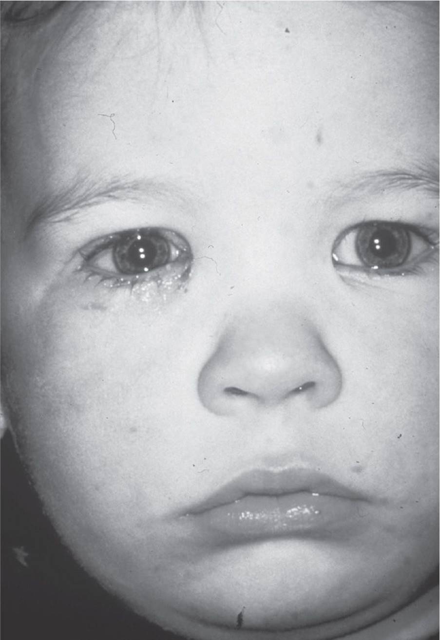
FIGURE 6-4
53 In congenital nasolacrimal system obstruction, where is the level of the obstruction?
A) Common canaliculus
B) Lacrimal sac
C) Valve of Rosenmüller
D) Valve of Hasner
54 The next therapeutic recommendation would include:
A) continuing massage
B) nasolacrimal system probing
C) DCR
D) observation, as most congenital obstructions resolve without therapy
55 Silicone stent intubation (with possible inferior turbinate infracture) is indicated in this patient when:
A) massage therapy has proven unsuccessful.
B) dacryocystography (DCG) shows obstruction at the level of the nasolacrimal duct.
C) nasolacrimal system probing has proven unsuccessful.
D) the patient is older than 12 months.
QUESTIONS 56–58 The 60-year-old woman pictured in Figure 6-5 reports a 3-month history of intermittent tearing and mattering in her right medial canthus. Additionally, she has noted focal swelling and tenderness near her lid margins.
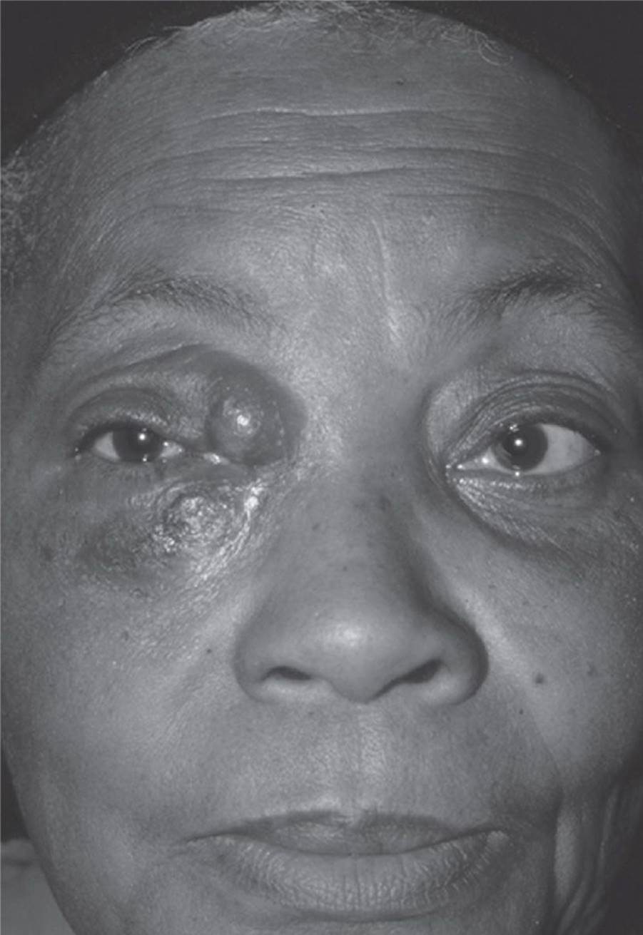
FIGURE 6-5
56 All of the following organisms are associated with canaliculitis except:
A) Actinomyces
B) Candida
C) Acanthamoeba
D) Streptomyces
57 Which one of the following suggests a diagnosis of canaliculitis?
A) Mucopurulent reflux from punctum with compression of the lacrimal sac
B) Gritty sensation on probing with yellow-tinged concretions
C) A palpable subcutaneous mass above the medial canthal tendon
D) Palpable masses in the lacrimal sac
58 Treatment of canaliculitis includes all of the following except:
A) canalicular curettage
B) canalicular incision and debridement
C) canalicular irrigation
D) DCR
59 Trachoma can cause all of the following changes except:
A) distichiasis
B) punctal stenosis
C) conjunctival scarring
D) entropion
60 All of the following pairs match mechanisms of involutional entropion with the surgical repair except:
A) horizontal lower lid laxity—lateral tarsal strip
B) dehiscence of the lower lid retractors—retractor advancement
C) overriding of the pretarsal orbicularis by the preseptal orbicularis—excision of a strip of preseptal orbicularis
D) inward rotation of the lid by steatoblepharon—lower lid blepharoplasty
61 Clinical clues to the disinsertion of the lower lid retractors include all of the following except:
A) white line below the tarsal border caused by the dehisced edge of the disinserted retractors
B) higher than normal lower eyelid position
C) decreased movement of the lower lid on downgaze
D) shrinking of the inferior conjunctival fornix
62 Which one of the following is the least common form of ectropion?
A) Congenital
B) Paralytic
C) Mechanical
D) Cicatricial
63 Repair of lower eyelid involutional entropion would be best accomplished by:
A) suturing the orbicularis to the inferior fornix
B) suturing the retractors to the tarsus
C) suturing the orbital septum to the capsulopalpebral head
D) suturing the Lockwood ligament to the conjunctiva and suspensory ligament of the fornix
64 The most common cause of upper eyelid retraction is:
A) recession of the superior rectus muscle
B) congenital eyelid retraction
C) surgical overcorrection of blepharoptosis
D) thyroid eye disease
QUESTIONS 65–68
65 A 11-year-old girl presents with acute, unilateral, left-sided periocular pain, proptosis, and double vision (Fig. 6-6). Which condition would not be included in the differential diagnosis?
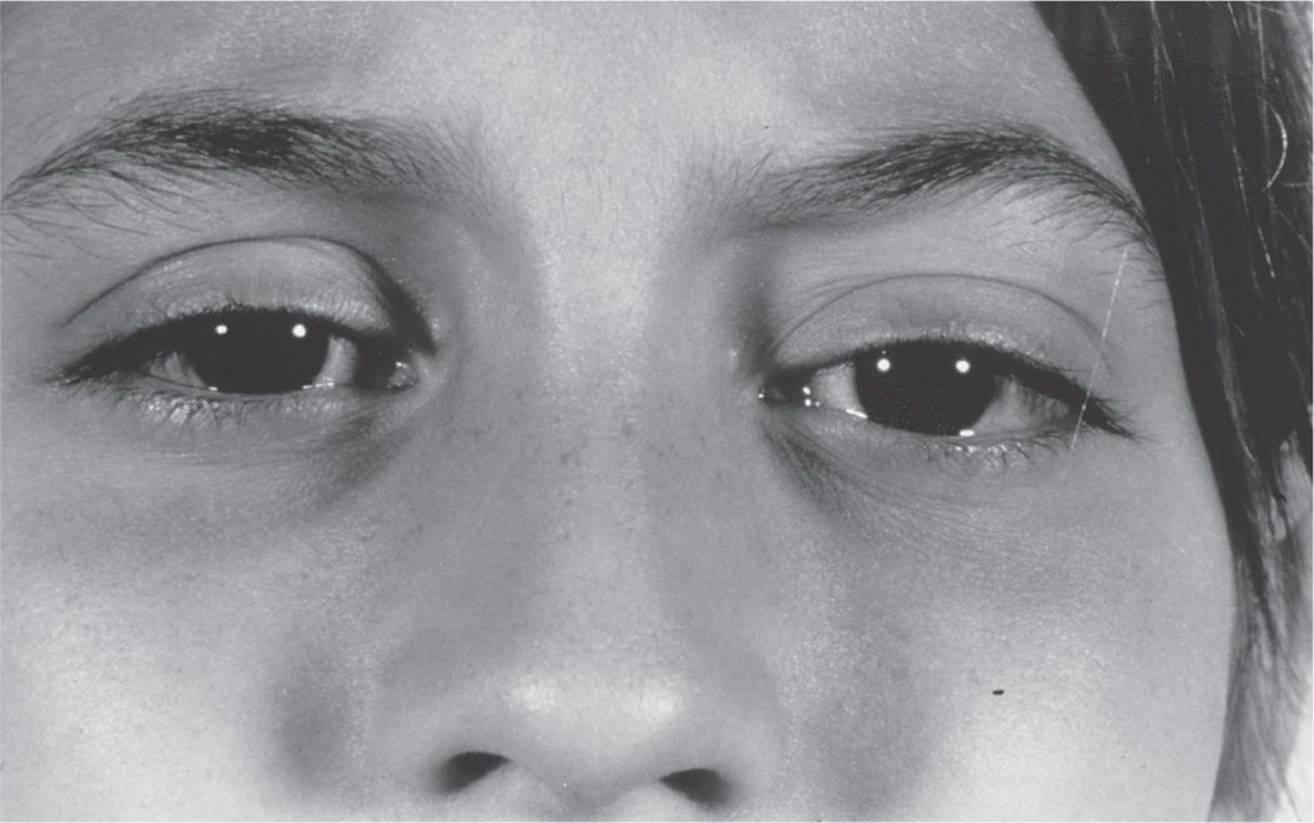
FIGURE 6-6
A) Cavernous hemangioma
B) Sinusitis with orbital abscess
C) Traumatic retrobulbar hemorrhage
D) Orbital lymphangioma
66 Twenty-four hours later (and without any treatment), the pain has resolved. Periocular ecchymosis has developed, and the double vision has stabilized (Fig. 6-7). The MRI results are also available (Fig. 6-8). The most likely diagnosis based on the clinical history and the MRI findings is:

FIGURE 6-7
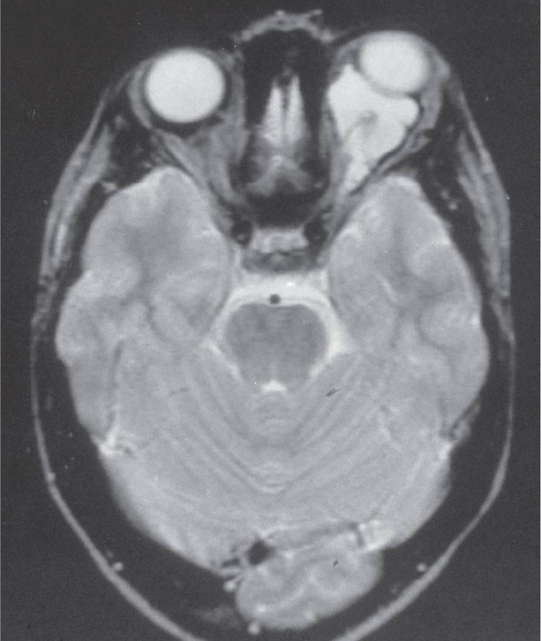
FIGURE 6-8
A) rhabdomyosarcoma
B) capillary hemangioma
C) orbital abscess
D) lymphangioma
67 If the patient was losing vision because of this process, you would consider:
A) open surgery to excise the lesion in its entirety
B) CT-directed drainage of the encysted blood
C) biopsy of the lesion to establish diagnosis and drain
D) B and C
68 This disease process is an example of:
A) the most common cause of proptosis in children
B) the most common primary orbital malignancy in children
C) a tumor that may enlarge with upper respiratory infections
D) an orbital vascular lesion that will involute after intralesional corticosteroids
69 In orbital infectious disease:
A) the presence of a subperiosteal collection of fluid is an indication for surgery.
B) the onset of decreased vision and an afferent pupillary defect in the presence of an orbital abscess is an indication for surgery.
C) proptosis and limitation of motility differentiate an orbital abscess from orbital cellulitis.
D) the maxillary sinus is the most common sinus involved when orbital cellulitis occurs as a result of sinusitis.
70 An 8-year-old boy has a 2-week history of rapidly progressing superonasal mass that does not affect vision. Examination shows proptosis pushing the child’s right eye down and out. The best management includes all of the following except:
A) CT scan
B) anterior orbitotomy with biopsy
C) MRI scan
D) observation
QUESTIONS 71–74
71 The patient in Figures 6-9 and 6-10 has a history of increasing painless proptosis over the past 4 years. She is 48 years old and is otherwise healthy. Based on the history, the CT scan (Fig. 6-11), and the ultrasound (Fig. 6-12), what is the most likely diagnosis?
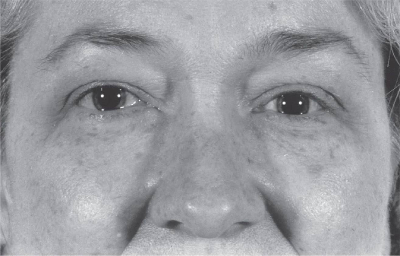
FIGURE 6-9
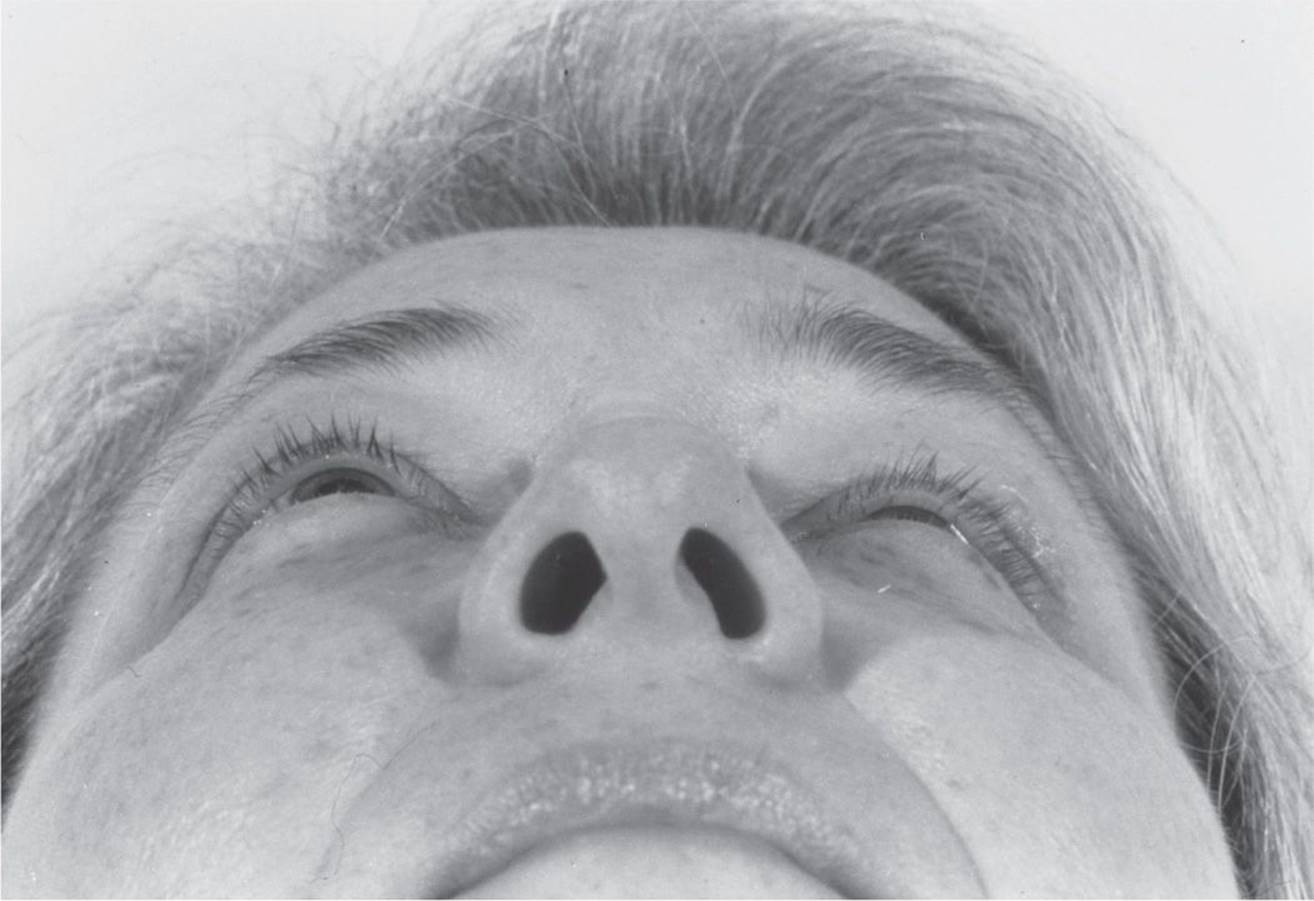
FIGURE 6-10
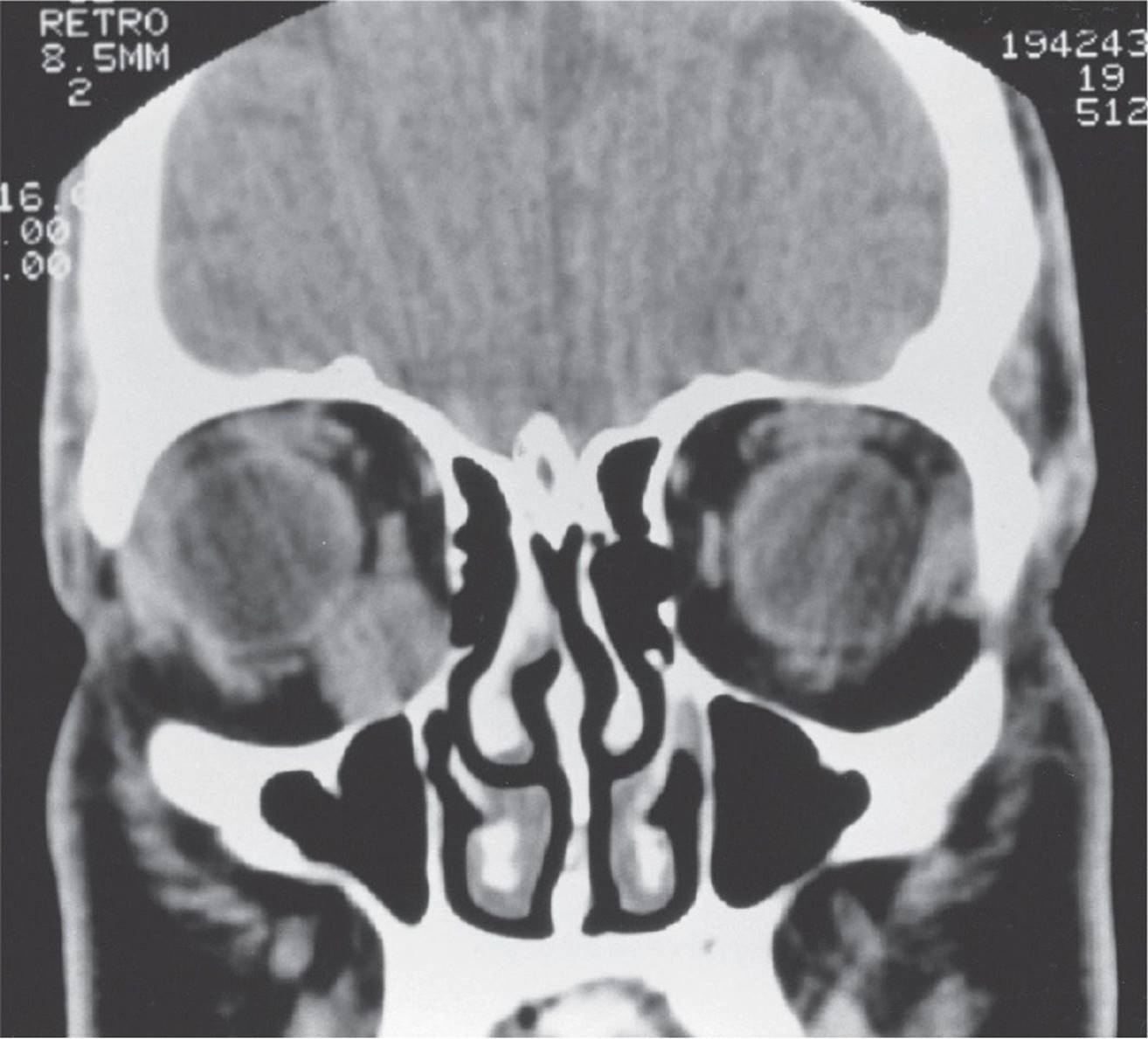
FIGURE 6-11
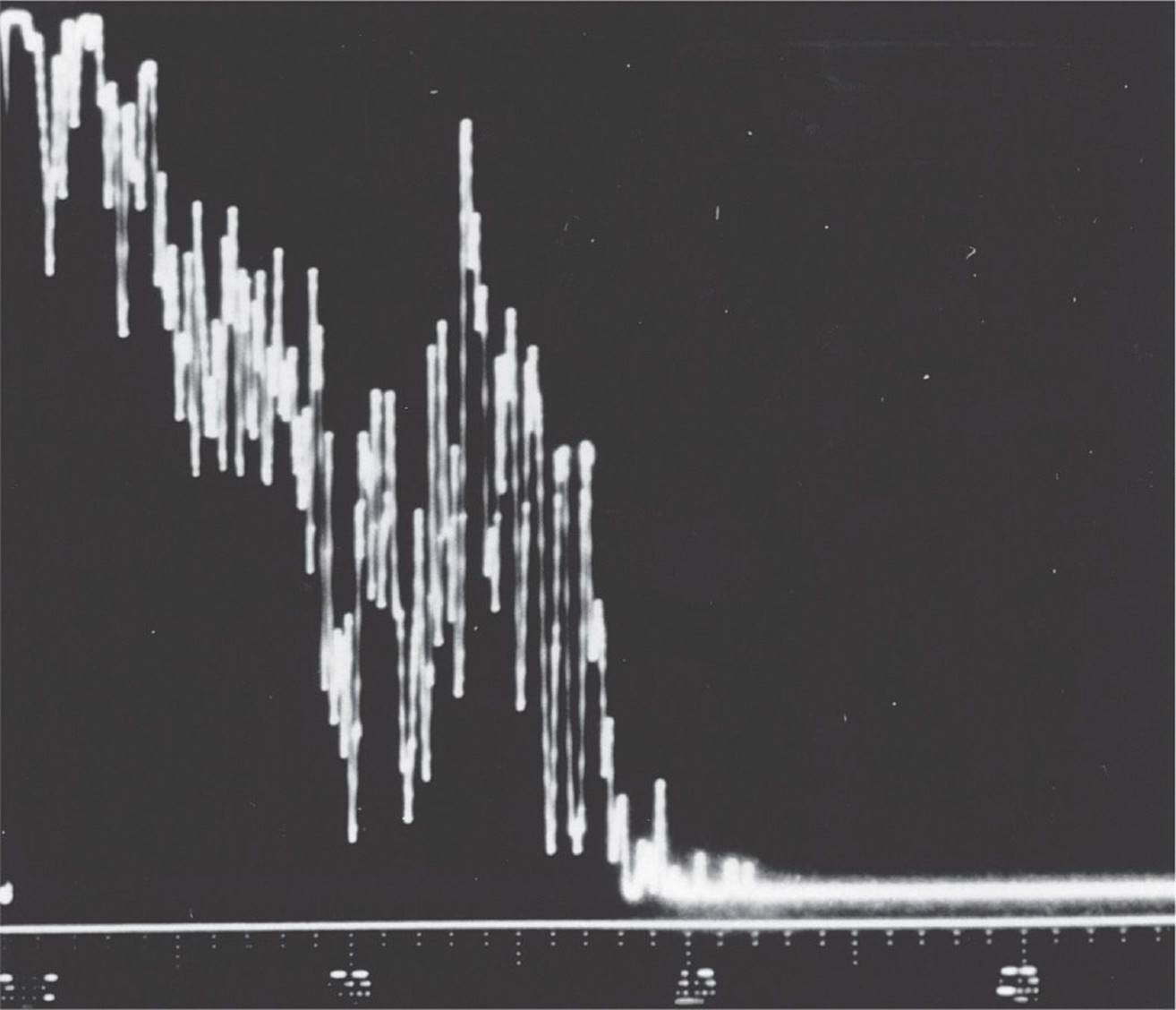
FIGURE 6-12
A) Optic nerve glioma
B) Cavernous hemangioma
C) Metastatic breast cancer
D) Benign mixed cell tumor of the lacrimal gland
72 The orbital ultrasound shows:
A) tissue of homogenous character
B) high internal reflectivity
C) B scan identifying tumor in the anterior inferior orbit
D) low amplitude internal echoes
73 If the lesion in Figure 6-11 is not surgically removed, the patient can expect:
A) slow growth over several years
B) erosion of surrounding bony structure
C) displacement of the globe downward and medially
D) potential for malignant conversion of the presently benign lesion
74 Surgical removal of the lesion in Figure 6-11 would best be approached by:
A) a lateral orbitotomy with en bloc removal of the mass
B) incisional biopsy followed by radiation or chemotherapy
C) an anterior approach through the inferior fornix
D) a medial orbitotomy with reflection of the medial rectus muscle
75 Where do orbital floor fractures most commonly occur?
A) Along the infraorbital canal
B) Within the zygoma medial to the infraorbital canal
C) Within the zygoma medial to the inferior orbital fissure
D) Within the maxilla medial to the infraorbital canal
76 A 7-year-old white boy presents with a sudden onset of rapid evolution of unilateral ptosis for approximately 1 week. Examination shows significant edema in the periorbital area with a palpable mass in the superior nasal quadrant of the eyelid. Biopsy is performed immediately, and the diagnosis of rhabdomyosarcoma is made. How is rhabdomyosarcoma best treated?
A) Radiation and chemotherapy
B) Total surgical excision
C) Exenteration
D) Chemotherapy alone
77 Which one of the following is the most common primary malignancy of the orbit in children?
A) Neuroblastoma
B) Rhabdomyosarcoma
C) Ewing sarcoma
D) None of the above
QUESTIONS 78 and 79 The CT scan in Figure 6-13 is from a 60-year-old woman who presented with painless swelling of the lacrimal gland and anterior orbit for approximately 2 months. There was no significant past or current ophthalmic history. The patient’s medical history was noncontributory.

FIGURE 6-13
78 What is the most likely diagnosis?
A) Primary lacrimal gland lymphoma
B) Pleomorphic adenoma
C) Adenoid cystic carcinoma
D) Malignant pleomorphic adenoma
79 What is the most accurate description of the pathology specimen taken from this lesion?
A) Spindle cells with both ductal epithelium and a mixed stromal pattern
B) “Swiss cheese” pattern—hyperchromatic small cells proliferating around nerves
C) Ductal epithelium in a tubular formation with malignant degeneration
D) Mixture of both B and T cells, with predominance of B cells
80 Enophthalmos in one eye without previous injury is suspicious for:
A) cavernous hemangioma
B) metastatic breast carcinoma in a woman
C) orbital cellulitis
D) all of the above
81 The most common cause of bilateral exophthalmos in adults is:
A) cavernous hemangioma
B) pseudotumor
C) thyroid-related orbitopathy
D) metastatic disease
82 What is the most common cause of unilateral childhood exophthalmos?
A) Capillary hemangioma
B) Thyroid-related orbitopathy
C) Orbital hemorrhage
D) Orbital cellulitis
83 Predisposing conditions for mucormycosis include:
A) diabetes
B) renal disease
C) dehydration
D) all of the above
84 Proper treatment of orbital mucormycosis includes all of the following except:
A) stabilizing the underlying disease process
B) debridement of all devitalized tissue, including exenteration if necessary
C) amphotericin B for 6 weeks
D) radiation to the orbit
QUESTIONS 85–87
85 A 55-year-old white man presents with bilateral proptosis, double vision, and chemosis. Figure 6-14 is an external photograph. What is the most likely diagnosis?

FIGURE 6-14
A) Thyroid-related orbitopathy
B) Orbital cellulitis
C) Lymphangioma
D) Meningioma
86 Concerning thyroid-related orbitopathy, the most common recommended surgical order of therapy is:
A) orbital decompression, strabismus surgery, and eyelid retraction surgical repair
B) eyelid retraction surgery, orbital decompression, and strabismus surgery
C) orbital decompression and eyelid retraction surgery repair
D) eyelid retraction surgery, strabismus surgery, and orbital decompression
87 A CT is performed on this patient with thyroid orbitopathy. Which feature, as demonstrated by CT, helps to clarify that this process is more likely thyroid-related orbitopathy than orbital inflammatory syndrome?
A) Enlarged extraocular muscle
B) Absence of a thickened tendon of the extraocular muscle insertion
C) Enlarged lacrimal glands
D) Periorbital soft-tissue edema of the lids
88 A histopathologic slide of an orbital tumor is shown in Figure 6-15. This tumor is best treated by:
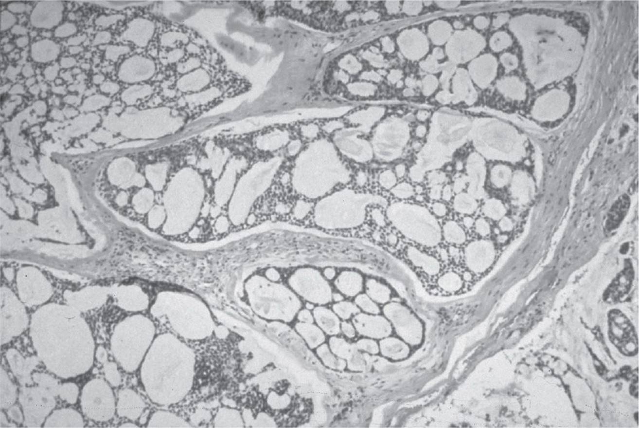
FIGURE 6-15
A) exenteration and removal of involved bone
B) radiation therapy
C) chemotherapy
D) all of the above
QUESTIONS 89–97 (Figs. 6-16–6-22)
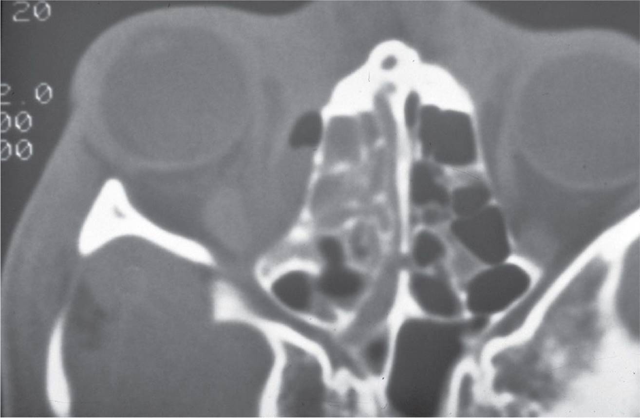
FIGURE 6-16
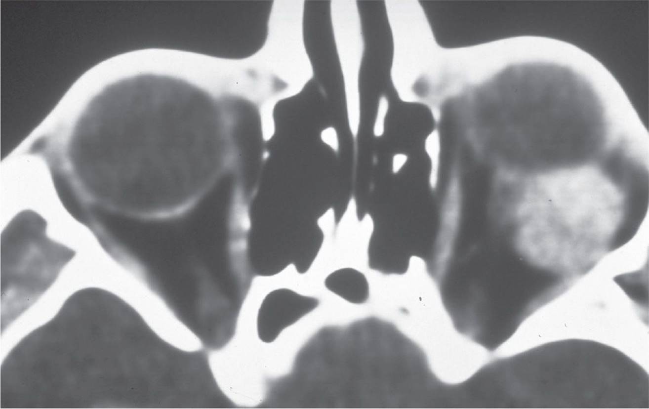
FIGURE 6-17

FIGURE 6-18
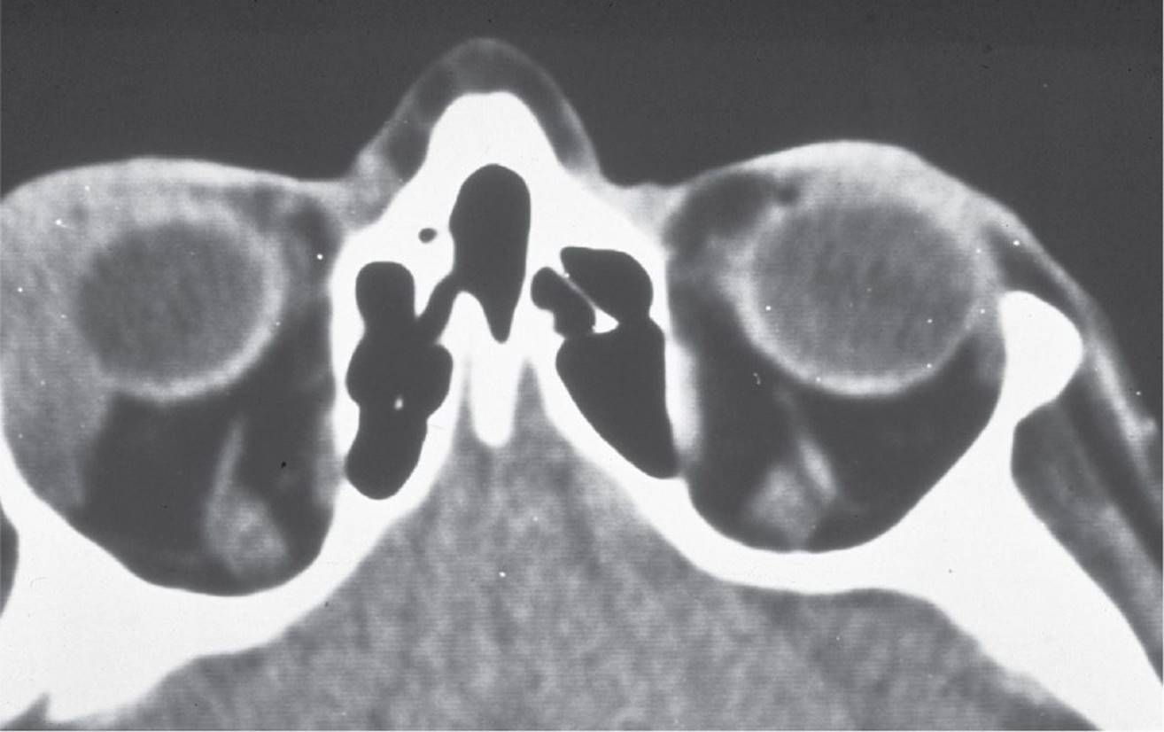
FIGURE 6-19
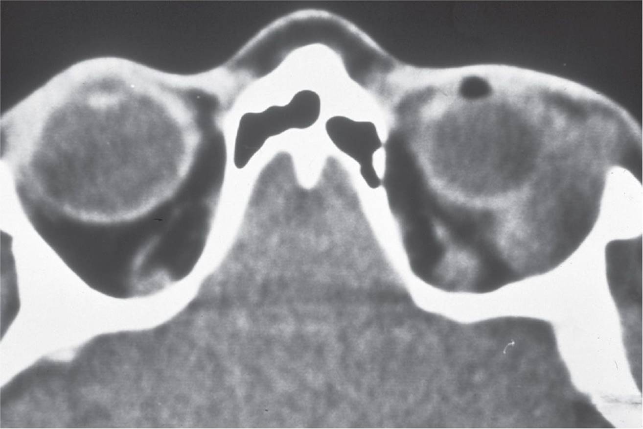
FIGURE 6-20
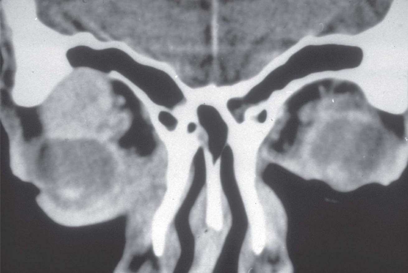
FIGURE 6-21

FIGURE 6-22
89 Staphylococcus aureus is the most common pathogen in patients who have the process as demonstrated in:
A) Figure 6-16
B) Figure 6-18
C) Figure 6-19
D) none of the above
90 Which two CTs best demonstrate orbital processes with a vascular origin?
A) Figures 6-17 and 6-19
B) Figures 6-17 and 6-18
C) Figures 6-16 and 6-17
D) Figure 6-17 alone
91 The lesion shown in Figure 6-17 was found in a 30-year-old woman who noticed mild proptosis that accelerated during her pregnancy. This lesion caused retinal striae and hyperopia. Which etiology is most likely?
A) Metastatic breast carcinoma
B) Cavernous hemangioma
C) Dermoid cyst
D) Hemangiopericytoma
92 Histologic examination of a biopsy specimen from the lesion shows Dutcher body formations that are intranuclear periodic acid–Schiff (PAS)-positive inclusions of immunoglobulin. Which CT demonstrates a characteristic lesion consistent with this finding?
A) Figure 6-18
B) Figure 6-19
C) Figure 6-20
D) Figure 6-21
93 A 45-year-old white man presents with gradual painless proptosis in the right eye that has displaced the globe inferiorly and medially. A firm lobular mass is palpated near the superior lateral orbital rim. Vision is not affected. The CT is obtained and the lesion is demonstrated in Figure 6-20. The next step in therapy should be:
A) a 2-week course of systemic corticosteroids
B) incisional biopsy
C) excisional biopsy
D) metastatic workup
94 A 70-year-old patient presents with severe pain in the left orbit over the past month. Limitation of upgaze and lateral gaze is present. Vision is not affected. The CT is obtained and demonstrated in Figure 6-21. Microscopic pathologic review is most likely to show:
A) tubules—solid nests of cells in a cribriform Swiss cheese pattern with perineural invasion
B) Flexner–Wintersteiner rosettes
C) psammoma bodies
D) Antoni A and Antoni B cells with nuclear palisades
95 Orbital decompression is performed in this condition for extreme proptosis, and development of compressive optic neuropathy occurs as demonstrated on which CT?
A) Figure 6-17
B) Figure 6-20
C) Figure 6-22
D) Figure 6-16
96 The etiology of the process defined on the CT in Figure 6-16 can be from:
A) paranasal sinusitis
B) previous trauma
C) dacryocystitis
D) all of the above
97 Which CT demonstrates a condition in which proptosis increases with the Valsalva maneuver?
A) Figure 6-17
B) Figure 6-18
C) Figure 6-20
D) Figure 6-21
98 All of the following are syndromes involving craniofacial synostosis except:
A) Crouzon syndrome
B) Treacher–Collins syndrome
C) Apert syndrome
D) Pfeiffer syndrome
99 All of the following clinical findings can be associated with Goldenhar syndrome except:
A) eyelid colobomas
B) lipodermoids
C) Duane syndrome
D) proptosis
100 Which systemic condition is incorrectly paired with a skin lesion?
A) Sturge–Weber syndrome (encephalo-trigeminal angiomatosis)—nevus flammeus (port-wine stain)
B) Ataxia-telangiectasia—café-au-lait spots
C) Incontinentia pigmenti—hyperpigmented macules (“splashed paint”)
D) Tuberous sclerosis—facial angiofibromas (adenoma sebaceum)
1 C) The orbital septum inserts on the superior border of the tarsus in the upper eyelid.
The orbital septum is a fibrous sheet that arises from the arcus marginalis, which lies at the junction of the periorbital periosteum (periorbita) and the pericranium. The septum extends toward the eyelid margins to fuse with the levator aponeurosis in the upper eyelid and the lower eyelid retractors (aponeurosis) of the lower eyelid. The septum is immediately deep to the orbicularis oculi muscle and superficial to the preaponeurotic orbital fat. During blepharoptosis surgery and other procedures, such as entropion repair of the lower eyelid, it is important to realize that the aponeurosis and lower eyelid retractors are located deep to the orbital septum and deep to the preaponeurotic fat. Therefore, in an anterior approach during blepharoptosis or entropion repair, the orbital septum must be incised to reach the eyelid retractors. The orbital septum inserts between the superior 2/3 and inferior 1/3 of the tarsal plate.
2 D) Lockwood ligament attaches posterior to the lacrimal sac.
The medial canthal area is complex in its anatomic structures. The medial canthal tendon has anterior and posterior portions. The anterior portion attaches to the frontal process of the maxillary bone and serves as the origin of the superficial head of the pretarsal orbicularis. The posterior portion inserts on the posterior lacrimal crest and fossa. The posterior limb of the medial canthal tendon, the deep head of the pretarsal orbicularis, and the deep head of the preseptal orbicularis muscles are important in maintaining apposition of the eyelids to the globe. The lacrimal sac is positioned anterior to the orbital septum and, therefore, does not lie within the orbit.
During contraction of the orbicularis, the preseptal portion of the orbicularis exerts traction on the fascia lateral to the lacrimal sac (lacrimal diaphragm) as part of the lacrimal pump process. The orbicularis oculi is innervated by cranial nerve VII, not cranial nerve V.
Lockwood ligament (Lockwood suspensory ligament) acts as a suspensory system for the globe. It is the lower eyelid retractor system with contributions from intermuscular septae and Tenon capsule. Posteriorly, it arises from fibrous attachments to the inferior side of the inferior rectus muscle and continues anteriorly as the capsulopalpebral fascia (lower eyelid retractors). Medial and lateral horns extend to attach to the retinacula. The medial retinaculum attaches to the posterior lacrimal crest, and the lateral retinaculum attaches to the lateral orbital tubercle of Whitnall. These tissues form a suspensory hammock for the globe.
3 C) The removal of fat from the upper eyelid nasal, central, and lateral fat pads may be done with impunity.
Orbital fat is located posterior to the orbital septum and is commonly encountered during eyelid surgery. The orbital fat is divided into intraconal fat (central) that is located inside of the muscle cone, and extraconal fat (peripheral) that is located outside of the muscle cone.
The removal of extraconal orbital fat is commonly done during upper and lower eyelid blepharoplasty. Numerous fine connective tissue septae course through the orbital fat and condense in several areas to form compartments. The upper eyelid has two fat pads: a small medial fat pad and a larger, central preaponeurotic fat pad (located between the orbital septum and levator aponeurosis), which primarily is located centrally. The orbital lobe of the lacrimal gland is located laterally and should not be removed. Thus, there is no lateral fat pad in the upper eyelid.
The color of the medial or nasal upper eyelid fat is typically whiter or paler when compared with the yellow color of the preaponeurotic, centrally and laterally located fat. The upper eyelid medial fat pad often moves anteriorly with aging more than the preaponeurotic fat, which results in a bulge that is located inferior to the trochlea of the upper medial orbit.
The lower eyelid has a small lateral fat pad and a larger medial fat pad. The small fat pad is located inferiorly to the lateral canthus and is separated from the large fat pad by fibrous tissue connecting the capsulopalpebral fascia and the orbital septum, which is also known as the lateral expanse of the capsulopalpebral fascia. Posteriorly, the main fat pad of the lower eyelid is divided into two fat pads by the inferior oblique muscle. Therefore, some references will state that the lower eyelid has three fat pads instead of two.
It is important to realize that this is normal fat that occupies the orbit and is bulging from a weakening of the orbital septum. This fat may serve a role in protection of the globe and facilitate its movement. Removal of too much fat may result in restriction of the extraocular muscles or a cicatricial blepharoptosis. In addition, aggressive removal of fat without careful attention to hemostasis can also result in an orbital hemorrhage and blindness.
4 D) This ligament passes anterior to the lacrimal gland.
The superior transverse ligament, or Whitnall ligament, arises from the compaction of the sheath of the anterior portion of the levator muscle. Medially, it arises from the connective tissue of the trochlea. Laterally, it attaches to the capsule of the orbital lobe of the lacrimal gland and to the lateral orbital wall above the lateral orbital tubercle. There are extensions to the medial and lateral retinacula. This structure acts to change the direction of pull of the levator muscle from horizontal to vertical and serves to limit the elevation of the eyelid. Although Whitnall ligament attaches to the fascia on the superior and medial surfaces of the lacrimal gland; it does not pass anteriorly to the lacrimal gland. The lacrimal gland is divided into orbital and palpebral lobes by the lateral horn of the levator aponeurosis.
5 C) The mucocutaneous junction occurs where the eyelashes emerge from the eyelid.
The eyelid is divided by some references into the anterior and posterior lamella. The anterior lamella consists of the skin and the orbicularis oculi (eyelid protractor) muscle. The posterior lamella consists of the conjunctiva, tarsus, and retractors. The eyelid may be subdivided further to include a middle lamella composed of the eyelid retractors. The mucocutaneous junction occurs posterior to the eyelashes near the opening of the meibomian glands. The gray line represents the marginal edge of the pretarsal orbicularis and would be considered part of the anterior lamella.
The ophthalmic (orbital) artery branches from the internal carotid system and contributes to the palpebral arterial arcades. The angular artery and the malar branch of the transverse facial artery also contribute to the arcades. The facial artery branches from the external carotid system.
6 A) Closure of the eyelid, depression of the eyebrow, and facilitation of tear drainage
The orbital portion of the orbicularis muscle runs from the anterior limb of the medial canthal tendon, the orbicularis muscle is innervated by cranial nerve VII (not III), and the origins of the preseptal orbicularis wrap around the lacrimal sac, not the lacrimal gland.
The orbicularis muscle is divided into pretarsal, preseptal, and orbital parts. The orbital portion is involved in forced eyelid closure. The pretarsal portion arises from the posterior lacrimal crest and anterior limb of the medial canthal tendon. The lateral portion of the pretarsal muscle becomes the lateral canthal tendon. The deep head of the pretarsal muscle (tensor tarsi muscle of Horner) encircles the canaliculi to facilitate tear drainage. The corrugator draws the head of the brow to the nose, and the procerus depresses the eyebrow. The preseptal orbicularis originates from the fascia around the lacrimal sac and posterior lacrimal crest. Superficial origins arise from anterior fibers of the medial canthal tendon. Laterally, it forms the lateral palpebral raphe overlying the lateral orbital rim. The orbital portions of the orbicularis originate from the anterior medial canthal tendon and periosteum.
7 B) Nasalis—preseptal orbicularis
The pretarsal portion of the orbicularis lies anterior to the tarsus. Medially, the pretarsal muscle divides into two heads. The superficial head becomes the medial canthal (palpebral) tendon, and the deeper head passes posteriorly to insert on the posterior lacrimal crest and is also referred to as the tensor tarsi muscle of Horner. Laterally, the lateral palpebral tendon arises from the pretarsal orbicularis muscles and attaches to the lateral retinaculum, which attaches to the lateral orbital tubercle of Whitnall.
The superciliary corrugator muscle arises from the periosteum of the frontal bone and inserts laterally into the subcutaneous tissue. The superciliary corrugator muscle is responsible for the vertically oriented glabellar skin creases (rhytids). Its inferior fibers are continuous with the orbicularis muscle. The procerus muscle interdigitates with the inferior edge of the frontalis muscle and is responsible for the horizontal lines seen at the nasion and the dorsum of the nose. The nasalis muscle is located on the lateral aspect of the nose and is a separate muscle from the preseptal orbicularis.
8 B) Optic canal—greater and lesser wings of the sphenoid bone
The lacrimal sac fossa consists of the maxillary bone anteriorly and the lacrimal bone posteriorly. The lacrimal gland fossa is located in the frontal bone in the anterior lateral orbit. The optic canal passes through the lesser wing of the sphenoid, not between the greater and lesser wings of the sphenoid bone.
The inferior orbital fissure is bordered medially by the maxillary bone, anteriorly by the zygomatic bone, and laterally by the greater wing of the sphenoid and is also bounded by the palatine bone. The anterior and posterior ethmoidal foramina are located at the junction of the ethmoid and frontal bones.
9 B) Able to be seen by the human eye
The carbon dioxide laser operates at a wavelength in the infrared portion of the electromagnetic spectrum. At 10.6 µm or 10,600 nm, the carbon dioxide laser is not within the visible spectrum, which means that it is not visible to the human eye. The laser may be visibly guided with a helium–neon (HeNe) laser-aiming beam. The carbon dioxide laser is absorbed by water. The increase in temperature ultimately causes cell destruction. The carbon dioxide laser has a high absorption coefficient with water that is inversely proportional to the absorption length, which is very small. Therefore, carbon dioxide has the ability to remove tissue in small increments. The laser has wide application for orbital tumor removal (Fig. 6-23).

FIGURE 6-23 Modified from Trost D, Zacherl A, Smith MFW. Surgical Laser Properties and Their Tissue Interactions. St. Louis: Mosby, 1992:133.
10 A) Motor nerve terminal, inhibiting acetylcholine release
Botulinum toxin type A inhibits acetylcholine release by binding to receptors of the motor nerve terminals. Specifically, the light chain of the toxin inhibits the release of acetylcholine by cleaving a cytoplasmic protein, SNAP-25, which is responsible for the docking of acetylcholine vesicles to the synaptic cell membrane. The denervation that results usually lasts several months. Some patients develop antibodies to the medication, resulting in a decreased efficacy of the botulinum toxin. Botulinum toxin does not affect the function but may actually increase the level of acetylcholinesterase. In addition, there is no direct effect of botulinum toxin type A on the striated muscle. The introduction of botulinum toxin type A in medicine has a variety of applications. FDA-approved indications for Botox specifically include benign essential blepharospasm, strabismus, axillary hyperhidrosis, chronic migraine, cervical dystonia, urinary incontinence, and upper limb spasticity. Other FDA-approved indications include cosmetic use for facial rhytids in the glabella and, as of late 2013, lateral canthal lines (i.e., crowsfeet). Other commercially available botulinum toxin type A’s include Dysport and Xeomin, both of which have fewer indications. Although botulinum toxin is a potent paralytic agent, the doses used for an adult are well below the amount necessary for systemic toxicity.
11 B) Likely spread to regional lymph nodes
Basal cell carcinoma is the most common primary eyelid malignancy and is located most frequently on the lower eyelid. Nodular basal cell carcinoma has the classic appearance of a pearly raised nodule with centrally ulcerated epithelium. Basal cell carcinoma may recur locally, but basal cell carcinoma is unlikely to metastasize to regional lymph nodes.
12 D) Increased natural skin pigmentation
Sun exposure, fair skin, family history of skin cancer, previous patient history of skin cancer, and red hair are all associated with increased risk of skin cancers. Having more darkly pigmented natural skin pigmentation is not associated with increased risk for cutaneous cancer.
13 C) Lash loss, central ulceration, rapid growth
Erythema, pruritus, and dark pigmentation are not associated with malignancy. Although most cancers have slow, progressive growth, rapid growth does not rule out a malignancy.
14 A) Nodular basal cell carcinoma
Basal cell carcinoma is the most common eyelid malignancy. Basal cell carcinoma accounts for over 90% of eyelid malignancies and is 40 times more common than squamous cell carcinoma. Basal carcinoma occurs most commonly on the lower eyelid (50%–60%), near the medial canthus (25%–30%), the upper eyelid (10%–15%), and in the lateral canthus (5%). Nodular basal cell carcinoma occurs most commonly.
Clinically, telangiectasia and smoothing of the skin texture are early findings. These early changes may be followed by elevation and rolling of the edges of the lesion and central ulceration.
Histopathologically, nodular basal cell carcinoma consists of basal cell nests, and peripheral palisading may be present. Ulceration may be observed both clinically and histologically. Although morpheaform or fibrosing basal cell carcinoma is much more aggressive, it is less common. Histopathologically, morpheaform tumors occur in peripheral radiating cords without peripheral palisading.
Squamous cell carcinoma is less common but is more aggressive than basal cell carcinoma. Squamous cell carcinoma can arise from actinic (solar) keratosis and has been associated with human papilloma virus lesions. Sebaceous adenocarcinoma arises from the tarsal plate of meibomian glands or from other sebaceous glands or periocular tissues. Sebaceous adenocarcinoma is a very aggressive and malignant tumor.
15 C) Ulcerated crater filled with lipids
Keratoacanthoma is a form of pseudoepitheliomatous hyperplasia. The lesion is typically a rapidly growing circular lesion that has a central ulceration filled with keratin. The lesion may cause disruption of the architecture of the lid margin or the cilia. Left untreated, the lesion may resolve spontaneously. Because of the possibility of an underlying malignancy and because the pathologist will need to examine the whole lesion, an excisional biopsy is recommended.
16 D) Malignant melanoma
Many skin lesions develop central ulceration. Some ulcerated lesions, such as the papillomatous lesion with central ulceration seen in molluscum contagiosum, are considered benign. A keratoacanthoma arises quickly and often has central ulceration. Keratoacanthomas typically are less apt to be malignant but can develop into squamous cell carcinoma. Both squamous cell carcinomas and basal cell carcinomas can have central ulcerations. A small melanoma would be least likely to have central ulceration.
17 D) Metastatic melanoma
The CT demonstrates a hyperostotic lesion of the left lateral orbital wall. Choices A, B, and C typically are associated with hyperostosis. In this patient, choices A and B are the most likely possibilities. Fibrous dysplasia can present with hyperostosis, although it normally presents in a somewhat younger patient population. Melanoma, in contrast, will metastasize to the extraocular muscles without hyperostosis.
18 D) Hughes tarsoconjunctival flap
Before selecting an appropriate reconstructive technique, it is necessary to assess the defect. There are numerous methods of reconstructing a defect. In Figure 6-2, there is an approximately 75% full-thickness defect of the lower eyelid, including both the anterior and posterior lamellae. Neither a bipedicle myocutaneous flap nor a full-thickness skin graft adequately replaces the posterior lamella (tarsal plate). The Hughes flap transposes a bridge flap of tarsus and conjunctiva into the defect. In addition, a full-thickness skin graft or advancement flap is used to replace the anterior lamella. A Cutler–Beard flap is a reconstructive technique that replaces full-thickness defects of the upper eyelid with a full-thickness tissue flap from the lower eyelid on the same side. Composite grafting, lateral cantholysis and direct closure, and a tarsoconjunctival flap with a myocutaneous flap (Hughes procedure) are additional reconstructive options.
19 B) Metastatic cancer
Basal cell carcinoma, metastatic cancer, sebaceous cell carcinoma, and squamous cell carcinoma are all malignant. In the malignant category of eyelid lesions, basal cell carcinoma is the most commonly encountered lesion. It is followed in decreasing frequency by squamous cell carcinoma, sebaceous cell carcinoma, and metastatic eyelid lesions.
20 A) Lymphatic vessels of the orbit drain along the lateral portion of the cavernous sinus.
No lymphatic vessels or nodes are typically present within the orbit. The conjunctiva does have lymphatic vessels. The statements regarding lymphatic and venous drainage of the upper eyelid are correct.
21 B) Located in the basal epithelial layer of the skin
Xanthelasma are typically flat, yellow skin lesions, located in the dermis. Histologically, these lesions consist of foamy histiocytes. The majority of patients do not have an associated hyperlipidemic condition. Erdheim–Chester disease is a multisystem disease with lipogranuloma formation in the liver, heart, kidneys, and bones. Histologically, these lipogranulomas contain histiocytes, Touton giant cells, lymphocytes, and plasma cells. Ophthalmic manifestations of this disease can include proptosis and xanthelasma-like skin lesions (Fig. 6-24).

FIGURE 6-24
22 C) Glands of Wolfring—eccrine, gland of Moll—apocrine, goblet cells—holocrine
23 C) Zeis (Table 6-1)
TABLE 6-1 Ocular Adnexal Glands
24 A) Congenital coloboma of the eyelid always involves the lower eyelid and can vary from a small notch to a complete absence of the eyelid.
Colobomas are more common in the upper eyelid, but they can occur in either or both eyelids. When the coloboma is in the medial upper lid, it is usually an isolated finding, whereas in the lower lid, it is often associated with other congenital anomalies such as clefting problems. Not only the surgical repair is indicated, but also the eye must be protected with lubrication before surgery to prevent drying of the cornea and conjunctiva.
Cryptophthalmos is a condition that results from a failure of the eyelids to develop and separate. This condition is usually associated with a malformed eye. Ankyloblepharon is a term used to describe fusion of all or part of the eyelids. Distichiasis refers to an extra row of eyelashes arising posterior to the normal row of eyelashes. This term should not be confused with trichiasis, which is used to describe misdirected eyelashes rather than an extra row.
25 C) Make arrangements to see the patient as soon as possible.
Pain, proptosis, periorbital ecchymosis, and decreased vision are the hallmarks of a postoperative orbital hemorrhage and possible compressive optic neuropathy. This patient requires immediate attention and evaluation. In the postoperative patient with a vision-threatening hemorrhage, the incision may be opened, the hematoma drained, and the tissue evaluated for control of active bleeding.
26 D) Telecanthus
Blepharophimosis means horizontal palpebral fissure narrowing. The blepharophimosis syndrome (congenital eyelid syndrome) is typically congenital and consists of a widened intercanthal distance (telecanthus), blepharoptosis, blepharophimosis, and epicanthus inversus. It has autosomal dominant inheritance with two types: BPES 1 and BPES 2. BPES 1 is associated with premature ovarian failure and infertility in affected women. Lower eyelid ectropion is also commonly seen in blepharophimosis syndrome. Other associated findings include poor development of the orbital rims and nasal bones as well as hypertelorism.
Euryblepharon is a horizontal widening of the palpebral fissure. Ankyloblepharon is a partial or complete fusion of the eyelids. Epiblepharon is commonly seen in Asian children and is caused by altered lower eyelid retractor (aponeurosis) attachments, which allow overriding of the pretarsal orbicularis above the eyelid margin. The result is posterior misdirection of the lower eyelashes and potential keratopathy.
27 A) Blepharochalasis
Blepharochalasis is a rare inherited condition that occurs more commonly in young females than in males and consists of repeated bouts of eyelid inflammation and edema. Recurrent idiopathic eyelid swelling eventually leads to blepharoptosis secondary to dehiscence or attenuation of the levator aponeurosis. Anterior bulging of the lacrimal gland or orbital fat may also occur. There is no proven treatment for the acute bouts of swelling, but some authors suggest that there is benefit with use of oral acetazolamide and topical hydrocortisone.
Dermatochalasis refers to the excess eyelid skin that occurs with aging. Steatoblepharon refers to the bulging of orbital fat that also commonly occurs with aging or attenuation of the orbital septum. Blepharospasm may be associated with blepharoptosis and entropion caused by aponeurotic dehiscence. Dermatochalasis, steatoblepharon, and blepharospasm typically are not associated with repeated bouts of eyelid edema and inflammation.
28 C) Reattachment of the dehisced levator aponeurosis
This patient has the classic signs of a blepharoptosis secondary to an acquired dehiscence of the levator aponeurosis. The patient exhibits a high eyelid crease or high eyelid pull, excellent levator function, and thinning of the eyelid superior to the upper tarsal plate. Therefore, surgery to reattach the dehisced end of the levator to the superior aspect of the tarsus would correct the blepharoptosis in the most anatomically proper fashion. Repair of the dehisced aponeurosis alone does not give as long-lasting a result as suturing the aponeurosis to the edge of the tarsus.
29 A) Involutional blepharoptosis (aponeurotic ptosis)
Ptosis (blepharoptosis) describes drooping of the upper eyelid and can have many different causes. Attempts to classify blepharoptosis can be confusing because there may be overlap of the categories. The term congenital means the blepharoptosis was present at birth, whereas acquired blepharoptosis describes blepharoptosis occurring after birth. Neither of these terms addresses the etiology of the observed blepharoptosis.
Acquired blepharoptosis is the most common ptosis. There are many different types of acquired blepharoptosis, which are often categorized by their etiology. The most common type of acquired blepharoptosis is involutional or aponeurotic blepharoptosis, which is caused by dehiscence of the levator aponeurosis.
Acquired neurogenic blepharoptosis occurs when there is a disruption of the normal innervation to the upper eyelid retractors. A traumatic blepharoptosis caused by a cranial nerve III injury is one example. Another example is acquired Horner syndrome, in which there is interruption of sympathetic innervation to the superior tarsal muscle (i.e., Muller muscle), resulting in miosis, anhydrosis, and a subtle upper eyelid blepharoptosis. Marcus Gunn jaw winking is an example of congenital neurogenic blepharoptosis and is also referred to as synkinetic blepharoptosis. This condition is characterized by an aberrant connection of the levator muscle innervation and the motor supply to the ipsilateral lateral pterygoid muscle.
Aberrant regeneration of cranial nerve III after trauma can result in a ptotic upper eyelid that exhibits synkinetic or abnormal movements. Abnormal regeneration and growth of the motor fibers from the inferior division of cranial nerve III into the superior division of this nerve may result in an eyelid that is ptotic in primary gaze but exhibits retraction in medial or inferior gaze as a result of stimulation of the levator muscle. Myasthenia gravis is caused by a defect at the neuromuscular junction and could be classified as a neurogenic blepharoptosis.
Mechanical blepharoptosis can be acquired or congenital. A tumor or hematoma involving the levator muscle, for example, could be a cause of an acquired or congenital blepharoptosis. Traumatic blepharoptosis can result from injury to the levator muscle, the aponeurosis, or the superior tarsal muscle, or innervation of any of these structures. Acquired myogenic blepharoptosis is rare. Some examples include oculopharyngeal dystrophy, chronic progressive external ophthalmoplegia, and other muscular diseases such as muscular dystrophy.
The most common type of congenital blepharoptosis is myogenic blepharoptosis due to poor development of the levator muscle. These terms are often used interchangeably. Myogenic blepharoptosis is due to dysgenesis of the levator muscle. Striated muscle fibers are diminished, and fibrosis as well as occasional fat may be present in the muscle. This abnormality decreases the ability of the muscle to contract and relax.
30 B) Histologically different from normal levator complex with decreased muscle fibers and fatty infiltrates
Myogenic congenital ptosis is the result of dysgenesis of the levator muscle complex. Both histopathologically and clinically, the complex is atrophic with replacement of muscle fibers by fibrous or adipose tissue. In myogenic ptosis, the levator aponeurosis will insert on the tarsus. Congenital dehiscence of the levator aponeurosis has been described but would not fall into the category of myogenic ptosis. Although the levator complex may be abnormal, the complex is still present below Whitnall ligament. The levator is still innervated by cranial nerve III, but patients may use cranial nerve VII to elevate the brow and thus the lids.
31 A) May activate the sympathetic receptors in Müller muscle, resulting in an elevation of the lid
The 2.5% phenylephrine hydrochloride test can activate the sympathetic fibers of Müller muscle. The resultant elevation of the lid simulates the position of the lid after conjunctivomüllerectomy. The alteration of the lid position also helps the surgeon evaluate the effect of Hering’s law on the contralateral eyelid. Lid position, not pupillary dilation, is responsible for this effect. Phenylephrine can elevate systemic blood pressure. Digital punctal occlusion can be used to help prevent this side effect.
32 B) A retroblepharoplasty (transconjunctival blepharoplasty) is a procedure primarily used to perform upper eyelid surgery when trying to avoid an anterior incision.
Dermatochalasis is a term used to describe redundant eyelid skin of the upper or lower eyelids. Steatoblepharon describes bulging orbital fat that commonly occurs with aging and is often removed during blepharoplasty. This fat tends to bulge forward with the aging process and may be related to a weakening or stretching of the orbital septum.
A retroblepharoplasty (transconjunctival blepharoplasty) or blepharoplasty via the transconjunctival approach is used in lower eyelid blepharoplasty, not upper eyelid blepharoplasty. The advantage of this approach is to avoid a visible scar on the skin of the eyelid. One disadvantage of this approach is that it does not allow for skin removal and is best used in patients with significant lower eyelid steatoblepharon without coexisting significant dermatochalasis.
Blepharoplasty of the upper eyelid is typically done using an eyelid crease incision. When the skin is reapproximated, the suture needle should be passed into the fascia located posterior to the orbicularis. This step accentuates the crease fixation and allows the healed incision to be hidden in the upper eyelid crease.
The inferior oblique muscle originates at the lacrimal spine located on the medial inferior orbital rim. The origin or muscle belly of the inferior oblique can be injured easily during lower eyelid blepharoplasty when the eyelid fat pad is removed.
Lower eyelid retraction is a common complication of lower eyelid blepharoplasty. Although the mechanism is unknown, the retraction may be related to scarring of the lower eyelid retractors and/or excessive removal of skin or muscle proportional to the eyelid’s horizontal laxity.
33 D) Polyglactin 910 (e.g., Vicryl)
Numerous materials have been described for use in frontalis suspension. These materials include donor fascia lata, autogenous fascia lata, temporalis fascia, supramid, silicone, and Gore-Tex. An absorbable suture such as Vicryl would not be effective in suspending the eyelid for the long term.
34 B) The orbital and palpebral lobes have separate excretory glands that empty into the conjunctival fornix approximately 5 mm above the superior margin of the tarsus.
The excretory ducts from the orbital lobe of the lacrimal gland pass into the palpebral lacrimal gland. The ducts continue, and then empty into the conjunctival fornix. This is also why biopsies of the lacrimal gland should only be performed on the orbital lobe, as biopsy of the palpebral lobe can cause injury to these excretory ducts preventing tears produced in the orbital lobe from being expressed onto the fornix.
35 D) Is within 10 mm of the cribriform plate
The osteotomy site created at the time of a DCR or CDCR is at the level of the middle turbinate. This site is most often within 10 mm of the cribriform plate. Typically, the lacrimal sac is divided and secured to the nasal mucosa. This division allows an open outlet from the common opening into the nasal cavity. The valve of Hasner is located at the distal end of the nasolacrimal duct under the inferior turbinate while the bony ostomy of the DCR is placed at the level of the middle turbinate.
36 A) Obstruction at the level of the common canaliculus or bony ostomy site
The two most common causes of DCR failure are obstruction at the common canaliculus and obstruction at the bony ostomy site. Recurrent obstruction can result in recurrent infection and dacryolith formation. Unsuspected tumors may also cause recurrent obstruction but are not the major cause of DCR failure.
37 C) Has a high false-negative rate
The primary dye test (Jones I test) determines whether the tears are passing into the nose under normal physiologic pumping conditions. In this test, 2% fluorescein is instilled in the precorneal tear film. Topical anesthetics and decongestants are sprayed in the ipsilateral inferior turbinate, and a rolled cotton fluff is inserted in the inferior meatus. After 5 minutes, the cotton is removed and examined for fluorescein. If no dye is present, a functional block is possible. However, in 20% to 30% of normal patients, no dye is noted, thus decreasing the reliability of this test when accomplished alone. This test does not indicate where an obstruction exists within the nasolacrimal system. Irrigation of the nasolacrimal system is accomplished on the secondary dye test (Jones II test).
38 C) Squamous cell carcinoma
Although rare, primary malignancies most frequently are of the papillary carcinoma group and are divided into three histologic subgroups: squamous cell, transitional cell, and mixed cell. They are treated by local excision and have a favorable prognosis.
The second most common primary lacrimal sac malignancies are lymphomas. These are treated by radiotherapy after biopsy and systemic evaluation have been completed. Complete excision is not usually necessary.
39 C) Congenital nasolacrimal duct obstruction unresponsive to massage
Probing of the nasolacrimal duct is not indicated in acute dacryocystitis. It is also rarely effective in acquired nasolacrimal duct obstruction. Probing of the duct is not indicated in the routine workup of patients with epiphora.
The majority (80%–90%) of congenital nasolacrimal duct obstructions resolve by 12 months of age. Thereafter, spontaneous resolution becomes less likely and probing is encouraged. In the face of chronic mucopurulent discharge, probing may be considered at 6 to 12 months or sooner, given the clinical situation. After 18 to 24 months of age or after multiple failed probings, consideration may be given to silicone stent intubation of the nasolacrimal system.
40 C) Positive dye disappearance test/negative Jones I
The dye disappearance test, Jones I, Jones II, and canalicular probing and irrigation all provide information that leads to a proper diagnosis in a patient with a tearing problem. The dye disappearance test is performed by placing fluorescein in the cul-de-sac and observing the degree of clearing after 5 minutes. A positive test is interpreted as significant retention of dye after 5 minutes. It does not localize an abnormality within the system but assesses the overall clinical functioning of the lacrimal system. Jones I is positive if dye can be visualized within the nasal cavity. Jones II is a method for determining if dye has entered the lacrimal sac. A positive Jones II indicates dye is in the nasal cavity after lacrimal irrigation. Jones III is useful when assessing the functioning of a surgical osteotomy site. A positive Jones III means dye is found in the nasal cavity of a post DCR or CDCR patient.
Because the location of the complete blockage in this question is at the sac–duct junction, Jones II testing would likely demonstrate reflux of fluid out through the opposite puncta. Jones II testing would thus be negative. In this case, there is no indication of lacrimal surgery, so a Jones III does not apply. Table 6-2 diagrams the various situations and the dye pattern that generally apply.
TABLE 6-2 Functional Lacrimal System Tests

41 D) They demonstrate the nasolacrimal sac well.
DCG involves forcibly injecting radiopaque dye into the lower canaliculus, with radiographs being taken thereafter. Because force is used, it is not a test of lacrimal function. Because the lacrimal cannula is inserted into the canaliculus, a DCG usually does not demonstrate the canalicular system well. A DCG can show the size and filling defects within the sac (diverticula, fistulas, lacrimal tumors, stricture locations). In most situations, diagnosis of a nasolacrimal system obstruction can be made without the use of a DCG.
42 C) They do not usually produce secondary dacryocystitis.
Clinical signs of lacrimal sac tumors include epiphora, irreducible swelling of the lacrimal sac, bleeding on attempted probing and irrigation, and secondary dacryocystitis.
43 B) Upper canalicular trauma alone should never be surgically repaired so as not to risk damage to the remaining nasolacrimal system.
Studies have demonstrated that in up to 50% of patients tested, creation of a monocanalicular state resulted in symptomatic epiphora. Canalicular trauma affects a generally younger patient population. In Figure 6-25, the probe is within the upper canaliculus. Leaving open the possibility of future trauma and disease to the remaining canaliculus, and considering present surgical techniques with a high success rate of repair, many recommend attempted repair of all recent canalicular lacerations. Additionally, acute trauma is generally easier to repair before scarring. One may wait a few days to allow acute soft-tissue swelling to decrease before attempted repair. The longer the silicone stents remain in place, the better the likelihood of patency for the system (after all surrounding scarring has occurred). Many surgeons prefer silicone stent intubation with microanastomosis of the lacerated canalicular system to maximize the success of canalicular patency.

FIGURE 6-25
44 B) A common canaliculus is present in approximately 30% of the population.
The punctum is perpendicular to the lid margin and approximately 2 mm from the ampulla. The canaliculus then turns horizontally and travels approximately 8 mm to the nasolacrimal sac. The ampulla has the largest diameter of the canalicular system (approximately 2 mm). The canaliculus has an average diameter of 1 mm. These relationships are important to keep in mind when probing the canalicular system. A common canaliculus is present in at least 90% of patients.
45 A) Syringing saline into the lower canaliculus that irrigates into the nose indicates that no obstruction exists and that the system is functioning normally.
The Jones II test alone is a nonphysiologic evaluation of absolute nasolacrimal system patency. Therefore, it gives no information on the adequacy of nasolacrimal drainage function. In a canalicular obstruction, irrigation would be expected to regurgitate from the punctum being tested. Reflux of fluid from the opposite punctum when the sac is irrigated indicates obstruction at the level of the sac or duct. Recovery of fluid in the nose after irrigation may be helpful in looking for casts or other debris. Additionally, when accomplished after a negative Jones I testing (no dye recovered from the inferior meatus after instillation of dye in the eye), retrieval of fluorescein-colored irrigant suggests dye-tinged fluid entered the sac, and a partial or functional block may exist.
46 D) Atropine
Canalicular stenosis may follow infections (herpetic, trachoma, infectious mononucleosis), inflammations (Stevens–Johnson syndrome, ocular pemphigoid), trauma (lacerations, chemical or thermal injuries, repeated probings), allergy, irradiation, tumors (rarely), canaliculitis, history of radioactive iodine or chemotherapy, and use of eye drops. Eye drops, including antivirals, strong miotics, and epinephrine-containing compounds, have been implicated most frequently.
47 C) Common canalicular obstruction combined with nasolacrimal duct obstruction
A Jones tube (Fig. 6-26) is indicated in cases in which the canalicular system is disrupted and is inadequate to allow tear flow into the nose. In certain situations, even a patent canalicular system may require a Jones bypass conduit if the lacrimal pump mechanism is disrupted (scarred or paralytic lids). A canaliculodacryocystorhinostomy is indicated for a focal distal canalicular obstruction combined with nasolacrimal duct obstruction.
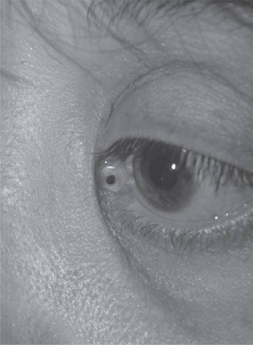
FIGURE 6-26
48 D) Most adults will need a DCR for correction of outflow obstruction.
Acute dacryocystitis is treated with systemic antibiotics and warm compresses. Probing the system does not successfully treat the problem in adults. DCR is usually necessary to re-establish lacrimal outflow in adults.
49 C) Probing and irrigation of the left nasolacrimal system
In acute dacryocystitis, probing of the nasolacrimal system is not indicated. Complete ophthalmic evaluation is necessary to determine whether a surrounding orbital cellulitis is present. Culture and Gram stain may help direct antibiotic therapy. In situations of previous trauma, radiographic imaging will help evaluate any anatomic changes.
50 A) DCR
After a complete evaluation, the patient should be started on systemic (IV or oral) and topical antibiotics, warm compresses, and pain control. Additionally, if a pointing abscess is present, incision and drainage of the abscess may result in more rapid healing and patient comfort. A DCR is not indicated for an acute dacryocystitis.
51 C) Nasolacrimal duct
Although obstruction may be found anywhere in the lacrimal drainage system, acquired nasolacrimal system obstruction is seen most frequently at the level of the mid or lower duct. Obstruction results from chronic low-grade inflammation with ultimate fibrosis of the duct walls.
52 C) Streptococcus pneumoniae
Dacryocystitis results from tear stasis. Pneumococci are the most common organism in dacryocystitis. Other organisms include streptococci, diphtheroids, Klebsiella pneumonia, Haemophilus influenzae, Pseudomonas aeruginosa, and mixed organisms. Actinomyces and fungi, such as Candida, are also frequently seen.
53 D) Valve of Hasner
This is a relatively common cause of epiphora and is seen in 2% to 4% of newborn infants. In 80% to 90% of patients, the membranous valve of Hasner opens spontaneously or with gentle massage of the nasolacrimal sac within the first 6 to 12 months of life. The valve of Rosenmüller is located between the common canaliculus (sinus of Maier) and the lacrimal sac and prevents reflux back into the canalicular system. The valve of Krause (and sinus of Arlt) is located at the junction of the nasolacrimal sac and duct. The spiral valve of Hyrtle and the valve of Taillefer are located within the nasolacrimal duct (Fig. 6-27).

FIGURE 6-27
54 B) Nasolacrimal system probing
55 C) Nasolacrimal system probing has proven unsuccessful.
As mentioned in the previous answer, 80% to 90% of cases of congenital nasolacrimal duct obstruction resolve spontaneously by the first year of life. Thereafter, spontaneous resolution decreases significantly. The next appropriate step would be nasolacrimal system probing under general anesthesia. If the probing is unsuccessful, consideration may be given to silicone stent intubation of the nasolacrimal system (this may be combined with infracture of the inferior turbinate). Only after multiple failed probings and silicone stent intubations would a DCR be considered for a child.
56 C) Acanthamoeba
Although Actinomyces israelii is associated most frequently with canaliculitis, multiple organisms have been shown to cause this disorder. Streptomyces, Arachnia propionica (previously Streptothrix), Nocardia, and fungi, such as Candida albicans and Aspergillus niger, have been documented to cause canaliculitis.
57 B) Gritty sensation on probing with yellow-tinged concretions
Canaliculitis affects the inferior canaliculus most frequently. A diagnosis is suspected when focal swelling of the canaliculus is noted. Probing of the affected canaliculus gives gritty resistance, and frequently, yellow-tinged concretions are noted. Mucopurulent reflux on palpation of the lacrimal sac is seen more frequently in dacryocystitis. In the setting of canaliculitis, one may see mucopurulent discharge on palpation of the canaliculus. Palpable subcutaneous masses above the level of the medial canthal tendon would arouse suspicion of a lacrimal sac tumor, and palpable masses in the lacrimal sac would be more suspicious for dacryoliths.
58 D) DCR
Canaliculitis may be treated by external pressure and curettage of the concretions with irrigation of a penicillin solution. Topical penicillin may also be necessary. Occasionally, incision of the canaliculus may be indicated to debride the tissue. DCR is not a method of treatment.
59 A) Distichiasis
The sequelae of trachoma are a result of the severe inflammatory reaction to the chlamydial antigens in the conjunctiva. Shortening of the posterior lamella resulting in entropion, keratoconjunctivitis sicca, and trichiasis (misdirected lashes) occur, contributing to the high incidence of corneal ulcers and blindness in developing nations. Distichiasis, in contradistinction, is the growth of aberrant lashes in the meibomian gland orifices. These lashes are usually shorter and softer than the usual lid margin cilia. Distichiasis can be congenital or acquired secondary to conditions such as Stevens–Johnson syndrome or ocular cicatricial pemphigoid.
60 D) Inward rotation of the lid by steatoblepharon—lower lid blepharoplasty
Lower lid steatoblepharon is not a cause of lower lid entropion.
61 D) Shrinking of the inferior conjunctival fornix
Retraction of the lower lid retractors may deepen the inferior cul-de-sac rather than shrink it.
62 A) Congenital
Involutional is the most common form of ectropion. It is most commonly attributable to laxity of the eyelid that occurs with aging. Gravity is also a contributing factor. It is very rare to see ectropion at birth (congenital).
Paralytic ectropion occurs when there is interruption of the innervation to the orbicularis oculi as found in cranial nerve VII palsy or Bell palsy. Mechanical ectropion may occur due to a lower eyelid tumor, severe edema, or any other condition that causes the lower eyelid to pull away from the globe.
Cicatricial ectropion arises from a relative shortening of the anterior lamella. Some etiologies that occur are scarring of the skin, chemical or thermal burns, chronic solar damage or inflammatory diseases of the skin, or skin tumors. Treatment of cicatricial ectropion often requires skin grafting.
63 B) Suturing the retractors to the tarsus
The cause of involutional entropion is a dehiscence of the lower eyelid aponeurosis (capsulopalpebral fascia). One way to repair an entropion is to repair the dehisced tissue by suturing the aponeurosis of the lower eyelid retractors to the inferior border of the tarsus. An entropion, therefore, is in many ways analogous to involutional blepharoptosis of the upper eyelid. Repair should be directed at reattaching the lower eyelid retractors and may be combined with other procedures, such as horizontal eyelid tightening procedures. The normal lower eyelid retractors consist of the capsulopalpebral fascia, the suspensory ligaments of the lower fornix, and the inferior tarsal muscle.
The distal portion of the capsulopalpebral fascia, known as the aponeurosis, is analogous to the levator aponeurosis of the upper eyelid. Some investigators think that the aponeurosis attaches to the inferior edge of the tarsus. We believe that the posterior portion of the aponeurosis inserts on the anterior border of the lower eyelid tarsus posteriorly and extends anteriorly into the subcutaneous tissue in a fashion similar to the anatomy of the upper eyelid. Inferior to the tarsus, the orbital septum fuses with the capsulopalpebral fascia just as the septum of the upper eyelid fuses with the levator aponeurosis. Reattachment of the dehisced lower eyelid aponeurosis to the inferior tarsal border does not provide an adequate anchoring of the tarsus for long-term repair of the entropion. However, suturing of the capsulopalpebral fascia (lower eyelid retractors) to the inferior border of the tarsus provides a strong anchor and may be used alone or in conjunction with other techniques for correction of the entropion.
Factors frequently mentioned in literature as causes of involutional entropion include increased horizontal eyelid laxity, the overriding of the preseptal orbicularis, and enophthalmos with fat atrophy. Although we agree that these findings are commonly associated with entropion, we suggest that these findings are incidental to aging (increased laxity and possible fat atrophy) and are not primary causes of the involutional entropic phenomenon. Common surgical procedures for correction of entropion are directed at stabilization of the anatomic abnormalities. Some examples include dehiscence repair with horizontal eyelid shortening, the Quickert four-snip procedure, the Bick procedure, the Wies procedure, the use of Quickert–Rathbun rotational sutures, and the use of a tarsal strip with dehiscence repair.
64 D) Thyroid eye disease
Recession of the superior rectus muscle, congenital eyelid retraction, surgical overcorrection of blepharoptosis, and thyroid eye disease are all causes of upper lid retraction, but the most common cause is thyroid eye disease.
65 A) Cavernous hemangioma
Infection and hemorrhage can both present with rapidly developing proptosis. Orbital lymphangioma may produce acute proptosis when there is a hemorrhage from interstitial capillaries. The cavernous hemangioma is a slow-growing lesion that creates proptosis over years or months, and the lesion is usually not painful.
66 D) Lymphangioma
The MRI scan shows a layered hemorrhage in the lobular and cystic mass behind the eye. The sinuses are clear of infection, making an orbital abscess unlikely. Acute bleeding would not be typical of a capillary hemangioma. The hemorrhage and ecchymosis would be more characteristic of lymphangioma than rhabdomyosarcoma.
67 D) B and C
The hemorrhage from an orbital lymphangioma can cause compressive optic neuropathy. In a patient with visual loss from optic nerve compression, drainage of the cyst with CT guidance or open surgery may be considered. Because the lymphangioma in this patient interdigitates with extraocular muscles and the optic nerve, it would be impractical to attempt total surgical excision of the lesion. Sometimes, a lymphangioma will be more localized and amenable to surgical excision with preservation of normal structures. IV corticosteroids may also be beneficial.
68 C) A tumor that may enlarge with upper respiratory infections
Lymphangiomas may increase in size during viral infection, presumably caused by the lymphocytic components of the tumor. Orbital cellulitis is the most common cause of proptosis in children; lymphangioma is not a malignant tumor; and capillary hemangioma (not lymphangioma) responds to intralesional corticosteroids.
69 B) The onset of decreased vision and an afferent pupillary defect in the presence of an orbital abscess is an indication for surgery.
Orbital infectious disease occurs most commonly as a result of spread from surrounding sinusitis, most commonly the ethmoid sinuses. Whether the presence of a subperiosteal fluid collection on CT (in the presence of stable vision and clinical signs) is an absolute indication for surgery is controversial. Most ophthalmologists would agree that medical management and careful observation may be employed when the patient’s clinical signs are stable and the vision is normal. Proptosis and motility limitation help to differentiate orbital cellulitis from preseptal cellulitis. Indications for surgical intervention in orbital cellulitis include decreasing vision and afferent pupillary defect, failure to respond to IV antibiotics, and progression of clinical signs such as motility changes and proptosis.
70 D) Observation
Rapid evolution of unilateral proptosis in a child should be considered rhabdomyosarcoma until proven otherwise. This finding necessitates a biopsy and then radiation and chemotherapy. Other causes of superonasal masses include myocele, mucopyocele, encephalocoele, and neurofibroma.
71 B) Cavernous hemangioma
72 B) High internal reflectivity
73 A) Slow growth over several years
74 C) An anterior approach through the inferior fornix
The patient is a middle-aged woman with very gradual onset of proptosis and no other symptoms. The CT scan shows a well-circumscribed, rounded mass inferonasal and anterior to the globe that displaces, but does not invade, surrounding tissue. The A-scan ultrasound shows high internal reflectivity consistent with nonhomogeneous tissue. These findings make the most likely diagnosis cavernous hemangioma. Typically, the cavernous hemangioma is a slow-growing mass that becomes symptomatic with proptosis or diplopia as the mass displaces the globe. These lesions are often intraconal. The location of this particular lesion is surgically approached through the inferior conjunctival fornix.
75 D) Within the maxilla medial to the infraorbital canal
With impact to the anterior orbit, forces are transmitted posteriorly. The weakest point along the floor is posterior and medial to the infraorbital canal. The convexity in the posterior orbital floor measures approximately 0.5 mm in thickness.
76 A) Radiation and chemotherapy
Prior to 1965, the standard treatment for orbital rhabdomyosarcoma involved exenteration, and it had a poor survival rate. Since that time, studies such as the Intergroup Rhabdomyosarcoma Study has established radiation and chemotherapy as the mainstay of treatment, replacing radical surgery as primary treatment. The particular chemotherapy and radiation protocol depends on the tumor group at the time of diagnosis. The survival rate using these modalities has improved significantly.
77 B) Rhabdomyosarcoma
The most common primary malignancy in children is rhabdomyosarcoma. The most common metastatic tumor is neuroblastoma, and it may be bilateral. Neuroblastoma typically produces an abrupt ecchymotic proptosis that may be bilateral. Metastasis from neuroblastoma in children most frequently goes to the orbit, whereas metastasis from neuroblastoma in adults usually is found in the uveal tract. In children, the most common benign tumor is capillary hemangioma.
78 A) Primary lacrimal gland lymphoma
Differentiating the variety of lacrimal gland lesions is often very challenging. The short history and the CT examination provided do allow for separating the lesions listed. The choices provided are all considered intrinsic lesions of the lacrimal gland. These lesions have typically been divided into lymphoproliferative lesions and epithelial tumors. Primary lacrimal gland lymphoma, pleomorphic adenoma, and adenoid cystic carcinoma are in the epithelial category. Adenoid cystic carcinoma and malignant pleomorphic adenoma are characteristically painful, with CT findings indicative of bone destruction. Pleomorphic adenoma, also referred to as a benign mixed cell neoplasm, usually has a 9-month or longer history of lacrimal gland enlargement. CT changes of bony erosion and expansion are also commonly seen. Lymphoma characteristically demonstrates “molding or putty-like” qualities on the CT. Lymphomas can present with both a short- and long-standing history of lacrimal gland swelling. In this case, when both history and CT examinations are analyzed, primary lacrimal gland lymphoma is the most appropriate answer.
79 D) Mixture of both B and T cells, with predominance of B cells
Lymphomas have proliferation of lymphocytes, mostly of the B-cell variety. A pleomorphic adenoma or benign mixed cell lacrimal gland neoplasm can be found to have spindle cells with both ductal epithelium and a mixed stromal pattern. Swiss cheese pattern is a classic term for describing an adenoid cystic carcinoma. Ductal epithelium in a tubular formation with malignant degeneration describes a malignant pleomorphic adenoma.
80 B) Metastatic breast carcinoma in a woman
Metastatic breast cancer is by far the most likely to present with enophthalmos, but gastrointestinal and lung are among other tumors, including prostate, which occasionally present with enophthalmos. Cavernous hemangioma and orbital cellulitis are characterized by proptosis, not enophthalmos.
81 C) Thyroid-related orbitopathy
The most common cause of bilateral exophthalmos in adults is most often thyroid-related orbitopathy and less often pseudotumor, Wegener granulomatosis, or neoplasm.
82 D) Orbital cellulitis
The most common cause of unilateral childhood exophthalmos is most often orbital cellulitis, secondary to ethmoid sinusitis or respiratory tract infections.
83 D) All of the above
Of patients with mucormycosis, 70% have diabetes mellitus, 5% have renal disease, 18% have other immunosuppressed states, 3% have leukemia, and only 4% have no systemic illness.
84 D) Radiation to the orbit
It is postulated that patients with diabetes do better than other patients with mucormycosis because they have a disease process that has the potential for control. For example, it is critical to reverse diabetic ketoacidosis in the setting of mucormycosis. It is also necessary to treat concurrent bacterial infections. Debridement of all devitalized tissue until free bleeding tissue is encountered is extremely important; it may include external ethmoidectomy, Caldwell–Luc operations, intranasal ethmoidectomy, sphenoid sinusectomy, palate resection, and exenteration. Exenteration is reserved for potentially lethal disease that is progressing unresponsive to other forms of treatment or as a palliative treatment for severe pain. It has been demonstrated that the more extensive the débridement, the better the prognosis. Since the addition of treatment with amphotericin B, survival rates have jumped from 6% to 73%. Early diagnosis with local excision, systemic stabilization, and IV amphotericin give mucormycosis patients the best chance for survival.
85 A) Thyroid-related orbitopathy
The clinical signs of thyroid-related orbitopathy can be generally grouped into two independent manifestations: type 1 and type 2 orbitopathy. Type 1 orbitopathy comprises symmetric proptosis with symmetric eyelid retraction, minimal orbital inflammation, and minimal extraocular muscle inflammation or restrictive myopathy. Type 2 orbitopathy comprises extraocular muscle myositis, restrictive myopathy, orbital inflammation, and chemosis. Compressive optic neuropathy is more commonly a feature of type 2 orbitopathy.
86 A) Orbital decompression, strabismus surgery, and eyelid retraction surgical repair
This order of therapy allows strabismus surgery to be performed after rather than before orbital decompression because decompression may alter ocular motility alignment. Furthermore, treatment of eyelid retraction must be performed after strabismus because recessing a vertical rectus muscle can also increase eyelid retraction because of the connection of the vertical rectus muscles to the levator or lower lid retractors.
87 B) Absence of a thickened tendon of the extraocular muscle insertion
It is possible to see enlarged extraocular muscles in both orbital inflammatory syndrome and thyroid-related orbitopathy. However, the tendon is almost never involved in thyroid orbitopathy, whereas the extraocular muscle tendon can be thickened in orbital inflammatory syndrome. The most commonly involved muscles in thyroid-related orbitopathy are the inferior rectus and the medial rectus. Lacrimal gland enlargement can occur in both entities.
88 D) All of the above
The histopathologic slide of adenoid cystic carcinoma or cylindroma (Fig. 6-15) shows the most common malignant tumor of the lacrimal gland. These tumor cells grow in tubules, solid nests, or in a cribriform Swiss cheese pattern. Perineural invasion is frequently observed in microscopic sections. Exenteration is almost always performed. Radiation and chemotherapy may be useful in extensive tumors.
89 A) Figure 6-16
Figure 6-16 shows a post–septal orbital cellulitis with a subperiosteal abscess demonstrated on axial CT scans. Staphylococcus aureus is the most common pathogen in children with orbital cellulitis. This infection responds quickly to penicillinase-resistant penicillin such as oxacillin.
90 B) Figures 6-17 and 6-18
Figure 6-17 demonstrates a cavernous hemangioma on an axial CT scan, which is the most common, well-circumscribed orbital lesion in adults. Figure 6-18 shows an orbital varix, which is an irregular mass with a phlebolith. Treatment for orbital varices is usually conservative. Because of the risks of optic nerve or globe damage, surgery is reserved for instances in which the varix is threatening vision. Complete surgical excision is difficult.
91 B) Cavernous hemangioma
Cavernous hemangioma is the most common benign neoplasm in adults. Proptosis is slowly progressive and growth may accelerate during pregnancy. Retinal striae, hyperopia and optic nerve compression, increased IOP, and strabismus may develop. Dermoid cysts occur less frequently but are found in the posterior orbit, usually in the superior and temporal portions. Metastatic breast carcinoma often causes enophthalmos. Hemangiopericytoma are encapsulated; however, they are quite rare.
92 B) Figure 6-19
The CT demonstrates a solid infiltrating tumor that has the characteristic putty-like molding of the tumor to preexisting orbital structures. Bone changes are usually not seen, and these lesions are usually not well-circumscribed. Lymphoplasmacytoid proliferation with Dutcher bodies should bring to mind the possibility of Waldenström macroglobulinemia or a systemic lymphoma with immunoglobulin production.
93 C) Excisional biopsy
Figure 6-20 presents an axial CT showing a lacrimal gland mass with no bony erosion. The patient presents with painless proptosis. This mass is most likely benign mixed pleomorphic adenoma of the lacrimal gland as this is the most common epithelial tumor of the lacrimal gland. It is impossible to tell whether this lacrimal gland tumor is benign or malignant without a biopsy. Excisional biopsy is indicated because complete removal of the tumor within its pseudocapsule is necessary. Performing an incisional biopsy may allow the tumor cells to spill into the orbit. Such cells can lead to a proliferation of an infiltrative recurrent tumor requiring extensive surgery at a later date. Furthermore, incompletely excised benign mixed tumors may later develop into malignant mixed tumors of the lacrimal gland.
94 A) Tubules—solid nests of cells in a cribriform Swiss cheese pattern with perineural invasion
Figure 6-21 shows a mass in the superolateral quadrant that is pressing the globe downward and is most likely of lacrimal gland origin. Bony erosion is present in the superior orbital roof, suggesting a malignant process. A painful orbital mass with rapid progression is most characteristic of adenoid cystic carcinoma, which is the most common malignant tumor of the lacrimal gland. The pain is caused by the perineural invasion and bony destruction. The course is rapid. Treatment consists of exenteration of the orbit with removal of any bone that is involved. Radiation and chemotherapy may be used in the treatment of extensive tumors. Psammoma bodies are composed of monotonous whorls of meningothelial cells interspersed with round calcified material found in meningiomas. Flexner–Wintersteiner rosettes are nearly pathognomonic for retinoblastoma. Antoni A and Antoni B are patterns of proliferation of Schwann cells that occur in Schwannomas.
95 C) Figure 6-22
Figure 6-22 demonstrates enlarged extraocular muscles in thyroid-related orbitopathy on an axial CT scan. Orbital decompression is indicated when there is severe proptosis that causes severe corneal exposure or, more commonly, when compressive optic neuropathy is demonstrated by visual field loss.
96 D) All of the above
Figure 6-16 shows a post–septal cellulitis demonstrated on axial CT scan. Causes of orbital cellulitis include 1) extension of infection from periorbital structures (paranasal sinuses, face and eyelids, dacryocystitis, dental infection, and intracranial infections); 2) exogenous causes, including previous trauma or surgery; 3) endogenous causes, such as bacteremia with septic embolization; and 4) intraorbital causes, such as endophthalmitis and dacryoadenitis.
97 B) Figure 6-18
Figure 6-18 shows an axial CT scan that demonstrates an orbital varix with phleboliths present. Orbital varices occur primarily as dilations of preexisting venous channels. Because dilated veins are present, proptosis can increase after Valsalva or when the head is in a dependent position. Surgery to remove varices is reserved for instances in which the varix is threatening vision because of optic nerve or globe damage. Complete surgical excision is difficult because the varix is often intertwined with other normal orbital structures. Orbital hemorrhages may result from trauma or may occur spontaneously.
98 B) Treacher–Collins syndrome
Crouzon, Apert, and Pfeiffer syndromes all involve craniofacial synostoses. Craniofacial synostosis results from premature closure of one or more cranial sutures. As a result, patients affected have characteristic cranial and facial features. Treacher–Collins syndrome is in the general category of clefting syndromes. These patients typically have hypoplasia of the mid-face, pseudocolobomas of the eyelids, downward angle to the lateral canthi, and dental and ear anomalies.
99 D) Proptosis
Goldenhar syndrome involves abnormalities of the first and second branchial arches. Eyelid colobomas, lipodermoids, and Duane syndrome are all part of a large list of clinical findings associated with Goldenhar’s. Additional findings include microphthalmia, anophthalmia, optic nerve hypoplasia, ocular colobomas, preauricular fistulas and skin tags, and palatal and facial clefts. Proptosis is seen more often with the synostosis, which includes both Crouzon and Apert syndromes.
100 B) Ataxia-telangiectasia—café-au-lait spots
Sturge–Weber syndrome consists of facial cutaneous angioma, which is called nevus flammeus or port-wine stain. Ipsilateral vascular malformations of the leptomeninges are typically present and lead to seizures, calcifications of the brain, and mental deficiency. Sturge–Weber syndrome is not hereditary.
Ataxia–telangiectasia is autosomal recessive and affects the skin, eye, immune system, and brain. The conjunctival and skin lesions consist of telangiectatic changes, usually in sun-exposed areas. These patients typically have involvement of their cerebellum resulting in ataxia as well as difficulty in initiating saccades. Decreased T-cell function and thymus hypoplasia lead to recurrent infections. Café-au-lait spots are found in neurofibromatosis, not ataxia-telangiectasia.
Incontinentia pigmenti is a syndrome displaying X-linked dominant transmission, and occurs almost exclusively in females as it is also X-lethal in males. This condition affects the skin, the eye, and the brain. The skin appears normal at birth, but it gradually develops erythema, bullae, and verrucous lesions. Small, hyperpigmented macules described as “splashed paint” typically occur on the trunk. Associated findings may include seizures; mental deficiency; skull, dental, and palate deformities; microcephaly; hydrocephalus; and dwarfism. The eyes may develop peripheral retinal vascular pathology.
Tuberous sclerosis often occurs as a new mutation, or it may be transmitted as a dominant trait. The “ash leaf” or hypopigmented macule represents an early skin finding in this condition. Angiofibromas of the face (adenoma sebaceum) occur late in childhood and are progressive. Tuberous sclerosis is often associated with calcifications of the basal ganglion and periventricular areas of the brain, mental retardation, seizures, and retinal phakomas.